Infectious Angiogenesis: Bartonella bacilliformis Infection Results in Endothelial Production of Angiopoetin-2 and Epidermal Production of Vascular Endothelial Growth Factor Francesca Cerimele,* Lawrence F. Brown, † Francisco Bravo, ‡ Garret M. Ihler, § Philomene Kouadio, § and Jack L Arbiser* From the Emory Skin Disease Research Core Center,* Department of Dermatology, Emory University School of Medicine, Boston, Massachusetts; the Department of Pathology, † Beth Israel Deaconness Hospital, Boston, Massachusetts; the Department of Medical Biochemistry, § College of Medicine, Texas A&M University System Health Science Center, College Station, Texas; and the Department of Pathology, ‡ Universidad Nacionale Cayetano Heredia, Lima, Peru Pathological angiogenesis , the development of a mi- crovasculature by neoplastic processes , is a critical component of the development of tumors. The role of oncogenes in the induction of angiogenesis has been extensively studied in benign and malignant tumors. However , the role of infection in inducing angiogen- esis is not well understood. Verruga peruana is a clin- ical syndrome caused by the bacterium Bartonella bacilliformis , and is characterized by the develop- ment of hemangioma-like lesions , in which bacteria colonize endothelial cells. To gain insight into how this bacteria induces angiogenesis in vivo , we per- formed in situ hybridization of clinical specimens of verruga peruana for the angiogenesis factors vascular endothelial growth factor (VEGF) , its receptors VEGFR1 and VEGFR2 , and angiopoietin-2. High-level expression of angiopoietin-2 and VEGF receptors was observed in the endothelium of verruga peruana. Sur- prisingly , the major source of VEGF production in verruga peruana is the overlying epidermis. Infection of cultured endothelium with B. bacilliformis also resulted in induction of angiopoetin-2 in vitro. These findings imply a collaboration between infected en- dothelium and overlying epidermis to induce angio- genesis. (Am J Pathol 2003, 163:1321–1327) Angiogenesis, the development of microvessels, is an indispensible component of normal physiological pro- cesses, including pregnancy, menstruation, organ forma- tion, and wound repair. 1,2 These mechanisms have been co-opted by benign and malignant processes, as well as inflammatory processes. Oncogenes have been shown to induce the expression of angiogenic factors including vascular endothelial growth factor (VEGF). 3,4 Infections can also induce angiogenesis, and both viral and bacte- rial infections have been known to result in increased angiogenesis in vivo. The viral infections most commonly associated with increased angiogenesis in vivo include papopaviruses (SV40, human papillomavirus, polyoma), and herpesvirus (Epstein-Barr virus, human herpesvirus 8). 5–9 These viral infections result in neoplasia and in- creased angiogenesis in part through viral-specific on- cogenes, including SV40 large T antigen, polyoma mid- dle T antigen, papillomavirus E6 and E7 genes, latent membrane protein 1 (LMP-1), and HHV8/KSHV-specific G proteins. The mechanisms through which bacterial infections cause angiogenesis is not well understood. 10 Verruga peruana are lesions associated with human infection with B. bacilliformis, an arthropod transmitted disease endemic to the highlands of Peru. 11 Infection with B. bacilliformis results in Oroya fever, a severe immu- nosuppressive infection characterized by high fever and anemia, which has a high mortality rate if untreated. 12 This disorder is also known as Carrion’s disease, after a Peruvian medical student, Daniel Carrion, who infected himself with verruga peruana tissue to prove the etiology of the disease. 13 The convalescent state is associated with the development of hemangioma-like lesions which have been termed verruga peruana. These lesions ultra- structurally demonstrate endothelial colonization with Bartonella organisms. 14 The mechanisms of induction of hemangioma-like lesions by Bartonella infection are not well understood, but has been postulated to be due to elaboration of a proangiogenic factor from infected en- dothelium. We demonstrate that infection of endothelium by Bartonella results in induction of angiopoietin-2 in vitro and in vivo. We found evidence of active angiogenesis in these lesions with high level expression of VEGFR1 and Supported by the American Skin Association and NIAMS grant R03AR44947 (to J. L. A.), Emory Skin Disease Research Core Center grants P30 AR 42687, KO8 AR02030, and RO1 AR 47901 (to J. L. A.). Accepted for publication June 18, 2003. Address reprint requests to Jack L. Arbiser, Department of Dermatol- ogy, Emory University School of Medicine, WMB 5309, Atlanta, GA 30322. E-mail: [email protected]. American Journal of Pathology, Vol. 163, No. 4, October 2003 Copyright © American Society for Investigative Pathology 1321

Welcome message from author
This document is posted to help you gain knowledge. Please leave a comment to let me know what you think about it! Share it to your friends and learn new things together.
Transcript
Infectious Angiogenesis: Bartonella bacilliformisInfection Results in Endothelial Production ofAngiopoetin-2 and Epidermal Production of VascularEndothelial Growth Factor
Francesca Cerimele,* Lawrence F. Brown,†
Francisco Bravo,‡ Garret M. Ihler,§
Philomene Kouadio,§ and Jack L Arbiser*From the Emory Skin Disease Research Core Center,* Department
of Dermatology, Emory University School of Medicine, Boston,
Massachusetts; the Department of Pathology,† Beth Israel
Deaconness Hospital, Boston, Massachusetts; the Department of
Medical Biochemistry,§ College of Medicine, Texas A&M
University System Health Science Center, College Station, Texas;
and the Department of Pathology,‡ Universidad Nacionale
Cayetano Heredia, Lima, Peru
Pathological angiogenesis, the development of a mi-crovasculature by neoplastic processes, is a criticalcomponent of the development of tumors. The role ofoncogenes in the induction of angiogenesis has beenextensively studied in benign and malignant tumors.However, the role of infection in inducing angiogen-esis is not well understood. Verruga peruana is a clin-ical syndrome caused by the bacterium Bartonellabacilliformis , and is characterized by the develop-ment of hemangioma-like lesions, in which bacteriacolonize endothelial cells. To gain insight into howthis bacteria induces angiogenesis in vivo , we per-formed in situ hybridization of clinical specimens ofverruga peruana for the angiogenesis factors vascularendothelial growth factor (VEGF), its receptorsVEGFR1 and VEGFR2, and angiopoietin-2. High-levelexpression of angiopoietin-2 and VEGF receptors wasobserved in the endothelium of verruga peruana. Sur-prisingly, the major source of VEGF production inverruga peruana is the overlying epidermis. Infectionof cultured endothelium with B. bacilliformis alsoresulted in induction of angiopoetin-2 in vitro. Thesefindings imply a collaboration between infected en-dothelium and overlying epidermis to induce angio-genesis. (Am J Pathol 2003, 163:1321–1327)
Angiogenesis, the development of microvessels, is anindispensible component of normal physiological pro-cesses, including pregnancy, menstruation, organ forma-tion, and wound repair.1,2 These mechanisms have beenco-opted by benign and malignant processes, as well as
inflammatory processes. Oncogenes have been shownto induce the expression of angiogenic factors includingvascular endothelial growth factor (VEGF).3,4 Infectionscan also induce angiogenesis, and both viral and bacte-rial infections have been known to result in increasedangiogenesis in vivo. The viral infections most commonlyassociated with increased angiogenesis in vivo includepapopaviruses (SV40, human papillomavirus, polyoma),and herpesvirus (Epstein-Barr virus, human herpesvirus8).5–9 These viral infections result in neoplasia and in-creased angiogenesis in part through viral-specific on-cogenes, including SV40 large T antigen, polyoma mid-dle T antigen, papillomavirus E6 and E7 genes, latentmembrane protein 1 (LMP-1), and HHV8/KSHV-specificG proteins. The mechanisms through which bacterialinfections cause angiogenesis is not well understood.10
Verruga peruana are lesions associated with humaninfection with B. bacilliformis, an arthropod transmitteddisease endemic to the highlands of Peru.11 Infectionwith B. bacilliformis results in Oroya fever, a severe immu-nosuppressive infection characterized by high fever andanemia, which has a high mortality rate if untreated.12
This disorder is also known as Carrion’s disease, after aPeruvian medical student, Daniel Carrion, who infectedhimself with verruga peruana tissue to prove the etiologyof the disease.13 The convalescent state is associatedwith the development of hemangioma-like lesions whichhave been termed verruga peruana. These lesions ultra-structurally demonstrate endothelial colonization withBartonella organisms.14 The mechanisms of induction ofhemangioma-like lesions by Bartonella infection are notwell understood, but has been postulated to be due toelaboration of a proangiogenic factor from infected en-dothelium. We demonstrate that infection of endotheliumby Bartonella results in induction of angiopoietin-2 in vitroand in vivo. We found evidence of active angiogenesis inthese lesions with high level expression of VEGFR1 and
Supported by the American Skin Association and NIAMS grantR03AR44947 (to J. L. A.), Emory Skin Disease Research Core Centergrants P30 AR 42687, KO8 AR02030, and RO1 AR 47901 (to J. L. A.).
Accepted for publication June 18, 2003.
Address reprint requests to Jack L. Arbiser, Department of Dermatol-ogy, Emory University School of Medicine, WMB 5309, Atlanta, GA 30322.E-mail: [email protected].
American Journal of Pathology, Vol. 163, No. 4, October 2003
Copyright © American Society for Investigative Pathology
1321
VEGFR2, which is observed in proliferative but not qui-escent endothelium. However, the major source of theangiogenic factor VEGF is the suprabasal epidermis.These findings imply a collaboration between infectedendothelial cells and overlying epidermis.
Materials and Methods
Infection of Human Umbilical Vein EndotheliumCell (HUVEC) with B. bacilliformis
HUVECs were obtained from Clonetics (San Diego, CA)and were routinely cultured in the manufacturer’s ownmedium (EGM2) supplemented with 2% fetal bovine se-rum. For experimental purposes, HUVECs (passage 2 to4) were trypsinized and seeded on 1 mg/ml gelatin, pre-coated with tissue culture flask in M199 medium (Gibco)containing 20% fetal calf serum (FCS), heparin and bo-vine hypothalamus extract15 the day before infection.Cultures were maintained in a humidified atmosphere 5%CO2 and at 37°C. For infection purposes 2-day-old cul-ture of green fluorescence protein B. bacilliformis wereharvested, washed three times with PBS and resus-pended in plain M199. The cells (about 80% confluent)were washed three times in plain M199. The cells wereincubated with approximately 100 bacteria per cell inM199 medium supplemented with 10% FCS for 26hours.16
RNA Isolation and RT-PCR
Total RNA from Bartonella-infected cells and non-infectedcell were isolated using the TRIzol Reagent Kit (Invitro-gen, Carlsbad, CA) according to the manufacturer’s pro-tocol. Total RNA were dissolved in RNase-free water.Reverse transcription-polymerase chain reaction (RT-PCR) was performed using the Access RT-PCR System(Promega, Madison, WI). For angiopoietin-2 we usedprimers previously described with the following condi-tions: 1 minute at 95°C, 1 minute at 64°C, 45 seconds at72°C for 35 cycles).17,18 For �-actin the following primerswere used: �-actin-F, 5�-AAGATGACCCAGATCATGTTT-GAGAC-3� and �-actin-R, 5�-CTGCTTGCTGATCCA-CATCTGCTGG-3� (1 minute at 95°C, 1 minute at 70°C, 45seconds at 72°C for 35 cycles). RT-PCR was performed
using the Access RT-PCR System (Promega). For angio-poietin-1 the following primers were used: hAng1F, 5�-AGTCCAGAAAACAGTGGGAG-3� and hAng1R, 5�-AG-CAGCTGTATCTCAAGTCG-3� (1 minute at 95°C, 1minute at 59°C, 45 seconds at 72°C for 35 cycles). ForVEGF receptor 1 (VEGFR1) the following primers wereused: VEGFR1F, 5�-GATGTTGAGGAAGAGGAGGATT-3�and VEGFR1R, 5�-AAGCTAGTTTCCTGGGGGTATA-3� (1minute at 95°C, 1 minute at 63°C, 45 seconds at 72°C for35 cycles). For VEGF receptor 2 (VEGFR2) the followingprimers were used VEGFR2F, 5�-GATGTGGTTCT-GAGTCCGTCT-3� and VEGFR2R, 5�-CATGGCTCTGCT-TCTCCTTTG-3� (1 minute at 95°C, 1 minute at 62°C, 45seconds at 72°C for 35 cycles).
In Situ Hybridization (ISH)
ISH was performed on 4-mm-thick sections of formalin-fixed, paraffin-embedded tissue, from four representativespecimens of verruga peruana. Details of ISH have beenreported previously.19,20 Briefly, slides were passagedthrough xylene and graded alcohols; 0.2 mol/L HCl, Tris/EDTA with 3 �g/ml proteinase K/0.2% glycine/4% para-formaldehyde in phosphate-buffered saline (pH 7.4), 0.1mol/L triethanolamine containing 1/200 (v/v) acetic anhy-dride, and 2X SSC. Slides were hybridized overnight at50°C with 35S-labeled riboprobes in the following mixture:0.3 mol/L NaCl, 0.01 mol/L Tris (pH 7.6), 5 mmol/L EDTA0.02% w/v Ficoll, 0.02% w/v polyvinylpyrollidone, 0.02%w/v bovine serum albumin fraction V, 50% formamide,10% dextran sulfate, 0.1 mg/ml yeast tRNA, 0.01 mol/Ldithiothreitol (DTT). Post-hybridization washes included2X SSC, 50% formamide, 10 mmol/L DTT at 50°C; 4XSSC, 10 mmol/L Tris, 1 mmol/L EDTA with 20 �g/mlribonuclease at 37°C; 2X SSC, 50% formamide, 10mmol/L EDTA at 65°C; and 2X SSC. Slides were dehy-drated through graded alcohols containing 0.3 mol/L am-monium acetate, dried, coated with Kodak NTB 2 emul-sion and stored in the dark at 4°C for 2 weeks. Theemulsion was developed with Kodak D19 developer andthe slides were counterstained with hematoxylin. Anti-sense single-stranded 35S-labeled human VPF/VEGFRNA probe and its sense control have been describedpreviously.19 The antisense probe hybridizes specificallywith a region of VPF/VEGF mRNA common to all knownVPF/VEGF splice variants. 35S- labeled single strandedantisense and sense RNA probes for mouse VPF/VEGFmRNA and the mouse VPF/VEGF receptors VEGFR-1 andVEGFR-2 mRNAs were described previously.19
Immunohistochemstry
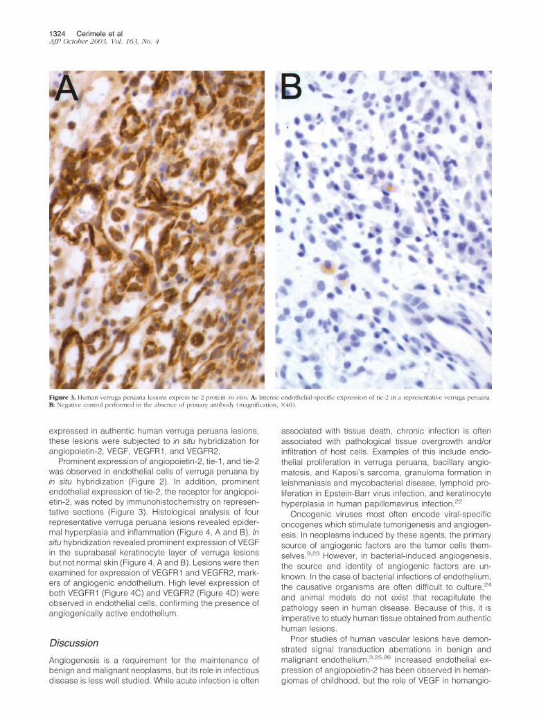
Unstained sections of formalin-fixed, paraffin-embeddedtissue were immunostained with polyclonal antibodiesagainst tie-2 (clone sc-324, 1/40; Santa Cruz Biotechnol-ogies, Santa Cruz, CA) using an avidin-biotin-complextechnique with the pressure-cooker heat-induced antigenretrieval and a DAKO Autostainer (DAKO, Carpinteria,CA). Negative controls had primary antibody substitutedwith buffer.
Figure 1. Infection of HUVEC with B. bacilliformis results in induction ofangiopoietin-2 mRNA. The top bands represent angiopoietin-2 mRNA, whilethe bottom band represents �-actin mRNA used as a loading control. I,MRNA from infected cells, NI, mRNA from non-infected control cells.
1322 Cerimele et alAJP October 2003, Vol. 163, No. 4
Results
Bartonella infection of endothelium has been shown toresult in induction of endothelial proliferation throughstimulation of mediators which have not been fully char-acterized. Given that VEGF production by endotheliumappears to be produced primarily in malignant endothe-lial tumors (angiosarcomas) rather than benign tumors
(hemangiomas),19,21 and that verruga peruana lesionsare biologically benign, we felt it was unlikely that VEGFwas the sole mediator of Bartonella-induced angiogene-sis. Infection of endothelial cells with B. bacilliformis re-sulted in strong induction of angiopoietin-2 mRNA (Figure1). Induction of other angiogenic molecules, such asVEGFR1, VEGFR2, and angiopoietin-1 were not observed(data not shown). To determine whether angiopoietin-2 is
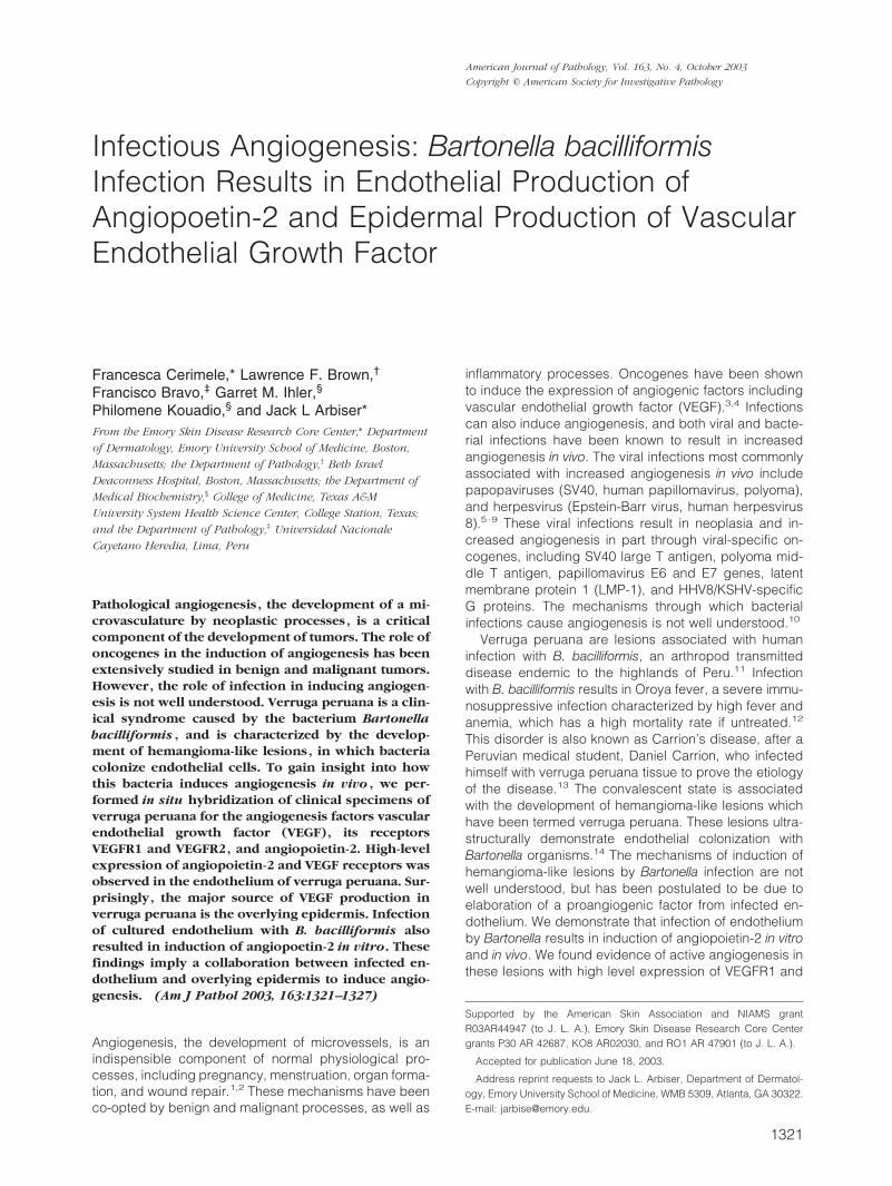
Figure 2. Human verruga peruana lesions express angiopoietin-1 and -2 in vivo. A: A typical hematoxylin and eosin lesion of verruga peruana. B: Strongexpression of angiopoietin-2 in cells surrounding lumens. C and D: Expression of tie-1 and tie-2, respectively.
Bartonella Infection Induces Angiopoeitin-2 1323AJP October 2003, Vol. 163, No. 4
expressed in authentic human verruga peruana lesions,these lesions were subjected to in situ hybridization forangiopoietin-2, VEGF, VEGFR1, and VEGFR2.
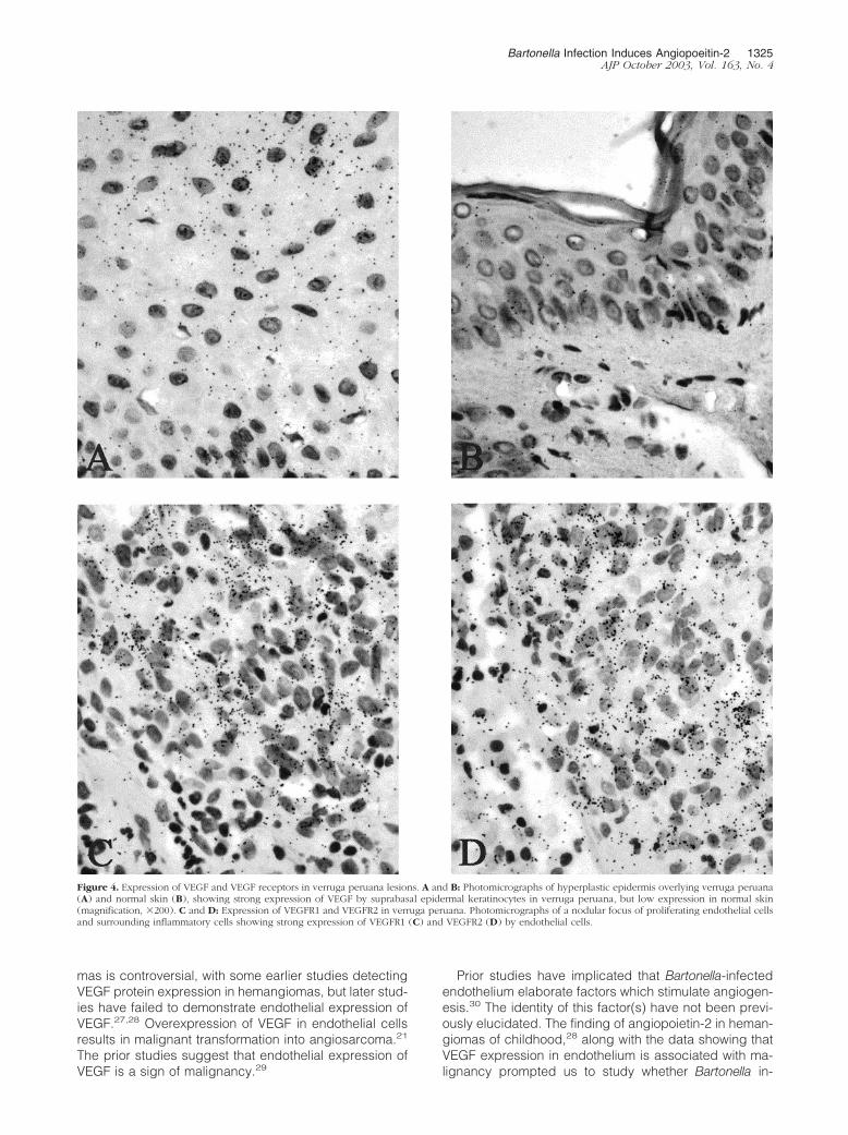
Prominent expression of angiopoietin-2, tie-1, and tie-2was observed in endothelial cells of verruga peruana byin situ hybridization (Figure 2). In addition, prominentendothelial expression of tie-2, the receptor for angiopoi-etin-2, was noted by immunohistochemistry on represen-tative sections (Figure 3). Histological analysis of fourrepresentative verruga peruana lesions revealed epider-mal hyperplasia and inflammation (Figure 4, A and B). Insitu hybridization revealed prominent expression of VEGFin the suprabasal keratinocyte layer of verruga lesionsbut not normal skin (Figure 4, A and B). Lesions were thenexamined for expression of VEGFR1 and VEGFR2, mark-ers of angiogenic endothelium. High level expression ofboth VEGFR1 (Figure 4C) and VEGFR2 (Figure 4D) wereobserved in endothelial cells, confirming the presence ofangiogenically active endothelium.
Discussion
Angiogenesis is a requirement for the maintenance ofbenign and malignant neoplasms, but its role in infectiousdisease is less well studied. While acute infection is often
associated with tissue death, chronic infection is oftenassociated with pathological tissue overgrowth and/orinfiltration of host cells. Examples of this include endo-thelial proliferation in verruga peruana, bacillary angio-matosis, and Kaposi’s sarcoma, granuloma formation inleishmaniasis and mycobacterial disease, lymphoid pro-liferation in Epstein-Barr virus infection, and keratinocytehyperplasia in human papillomavirus infection.22
Oncogenic viruses most often encode viral-specificoncogenes which stimulate tumorigenesis and angiogen-esis. In neoplasms induced by these agents, the primarysource of angiogenic factors are the tumor cells them-selves.9,23 However, in bacterial-induced angiogenesis,the source and identity of angiogenic factors are un-known. In the case of bacterial infections of endothelium,the causative organisms are often difficult to culture,24
and animal models do not exist that recapitulate thepathology seen in human disease. Because of this, it isimperative to study human tissue obtained from authentichuman lesions.
Prior studies of human vascular lesions have demon-strated signal transduction aberrations in benign andmalignant endothelium.3,25,26 Increased endothelial ex-pression of angiopoietin-2 has been observed in heman-giomas of childhood, but the role of VEGF in hemangio-
Figure 3. Human verruga peruana lesions express tie-2 protein in vivo. A: Intense endothelial-specific expression of tie-2 in a representative verruga peruana.B: Negative control performed in the absence of primary antibody (magnification, �40).
1324 Cerimele et alAJP October 2003, Vol. 163, No. 4
mas is controversial, with some earlier studies detectingVEGF protein expression in hemangiomas, but later stud-ies have failed to demonstrate endothelial expression ofVEGF.27,28 Overexpression of VEGF in endothelial cellsresults in malignant transformation into angiosarcoma.21
The prior studies suggest that endothelial expression ofVEGF is a sign of malignancy.29
Prior studies have implicated that Bartonella-infectedendothelium elaborate factors which stimulate angiogen-esis.30 The identity of this factor(s) have not been previ-ously elucidated. The finding of angiopoietin-2 in heman-giomas of childhood,28 along with the data showing thatVEGF expression in endothelium is associated with ma-lignancy prompted us to study whether Bartonella in-
Figure 4. Expression of VEGF and VEGF receptors in verruga peruana lesions. A and B: Photomicrographs of hyperplastic epidermis overlying verruga peruana(A) and normal skin (B), showing strong expression of VEGF by suprabasal epidermal keratinocytes in verruga peruana, but low expression in normal skin(magnification, �200). C and D: Expression of VEGFR1 and VEGFR2 in verruga peruana. Photomicrographs of a nodular focus of proliferating endothelial cellsand surrounding inflammatory cells showing strong expression of VEGFR1 (C) and VEGFR2 (D) by endothelial cells.
Bartonella Infection Induces Angiopoeitin-2 1325AJP October 2003, Vol. 163, No. 4
duces angiopoietin-2. We found that Bartonella infectionresults in induction of angiopoietin-2 in vitro and in vivo,and human verruga peruana lesions express angiopoi-etin-2.
The role of angiopoietin-2 in angiogenesis has notbeen totally elucidated. Initially angiopoietin-2 was foundto have antagonistic behavior to angiopoietin-1 and bindthe same receptor, tie-2.31 These findings led investiga-tors to believe that angiopoietin-2 may be an inhibitor ofangiogenesis. However, later findings have demon-strated that angiopoietin-2 may stimulate tumor growth,and tumor cells overexpressing angiopoietin-2 havedemonstrated highly malignant behavior and leaky ves-sels.32,33 Furthermore, hemangiomas of childhood havebeen demonstrated to express high levels of angiopoi-etin-2.28
Recently, Bartonella infection of endothelial cells in vitrohas shown to lead to decreased apoptosis, which mayaccount for the angiogenic phenotype.34 In addition, Bar-tonella infection has been shown to cause activation ofthe rac GTPase, and rac GTPase activation has beenshown to stimulate angiogenesis and prevent apopto-sis.16,35,36 Consistent with these findings, angiopoietin-2activation of tie receptor signaling has been shown toresult in activation of phosphoinositol-3 kinase/akt signal-ing, which are associated with both protection againstapoptosis and stimulation of endothelial proliferation.37,38
Interestingly, we found that overlying epidermis is theprimary source of VEGF in our lesions, and may serve asa trophic factor to maintain hemangioma-like lesions in-duced by Bartonella infection. Epidermal production ofVEGF appears to be a common response to inflammatorystimuli in the skin, and may be due to elaboration ofcytokines by the inflammatory process39 or through dis-ruption of the epidermis by the verruga peruana lesions.Barrier disruption of the epidermis leads to induction ofVEGF (Cerimele and Arbiser, unpublished data). Wehave observed that verruga peruana specimens expresshigh levels of active mitogen-activated protein kinase(P-MAPK), as do hemangiomas of childhood.40 In con-trast, malignant endothelial lesions (angiosarcoma), ex-press low levels of activated MAP kinase, reflecting adecreased requirement for MAP kinase in tumorigene-sis.40,41 Verruga peruana are highly angiogenic lesions,but are histologically benign. This is a fundamentallydifferent pattern of angiogenesis compared with eithervirally-induced tumors or angiosarcomas (malignant en-dothelial tumors) in which the tumor cells themselves arethe source of angiogenic factors.19,21,42 Our finds point toa novel form of angiogenesis in benign lesions, in whichproliferation of endothelium and/or decreased apoptosismay be due to autocrine loops of angiopoietin-2 andtie-2, with paracrine contributions of VEGF produced out-side the infected endothelium. The paracrine expressionof VEGF may help maintain the lesions.
Bartonella infections are unique among bacteria in in-duction of benign hemangioma-like lesions. Currently,verruga peruana is an uncommon disease, but it histo-logically resembles the benign hemangioma of infancy,the most common neoplasm of children. This report is thefirst demonstration of a potential role of angiopoietin-2 in
the pathogenesis of Bartonella infection, and demon-strates a novel mechanism of bacterial induction of an-giogenesis.
References
1. Klauber N, Rohan RM, Flynn E, D’Amato RJ: Critical components ofthe female reproductive pathway are suppressed by the angiogene-sis inhibitor AGM-1470. Nat Med 1997, 3:443–446
2. Folkman J: Seminars in Medicine of the Beth Israel Hospital, Boston:clinical applications of research on angiogenesis. N Engl J Med 1995,333:1757–1763
3. Arbiser JL, Moses MA, Fernandez CA, Ghiso N, Cao Y, Klauber N,Frank D, Brownlee M, Flynn E, Parangi S, Byers HR, Folkman J:Oncogenic H-ras stimulates tumor angiogenesis by two distinct path-ways. Proc Natl Acad Sci USA 1997, 94:861–866
4. Rak J, Mitsuhashi Y, Sheehan C, Tamir A, Viloria-Petit A, Filmus J,Mansour SJ, Ahn NG, Kerbel RS: Oncogenes and tumorangiogenesis: differential modes of vascular endothelial growth factorup-regulation in ras-transformed epithelial cells and fibroblasts. Can-cer Res 2000, 60:490–498
5. Carbone M, Pass HI, Rizzo P, Marinetti M, Di Muzio M, Mew DJ,Levine AS, Procopio A: Simian virus 40-like DNA sequences in humanpleural mesothelioma. Oncogene 1994, 9:1781–1790
6. Flore O, Rafii S, Ely S, O’Leary JJ, Hyjek EM, Cesarman E: Transfor-mation of primary human endothelial cells by Kaposi’s sarcoma-associated herpesvirus. Nature 1998, 394:588–592
7. Majewski S, Szmurlo A, Marczak M, Jablonska S, Bollag W: Syner-gistic effect of retinoids and interferon-� on tumor-inducedangiogenesis: anti-angiogenic effect on HPV-harboring tumor-celllines. Int J Cancer 1994, 57:81–85
8. Arbeit JM, Olson DC, Hanahan D: Up-regulation of fibroblast growthfactors and their receptors during multi-stage epidermal carcinogen-esis in K14-HPV16 transgenic mice. Oncogene 1996, 13:1847–1857
9. Lopez-Ocejo O, Viloria-Petit A, Bequet-Romero M, Mukhopadhyay D,Rak J, Kerbel RS: Oncogenes and tumor angiogenesis: the HPV-16E6 oncoprotein activates the vascular endothelial growth factor(VEGF) gene promoter in a p53 independent manner. Oncogene2000, 19:4611–4620
10. Arbiser JL: Angiogenesis and the skin: a primer. J Am Acad Dermatol1996, 34:486–497
11. Spach DH, Koehler JE: Bartonella-associated infections. Infect DisClin North Am 1998, 12:137–155
12. Caceres-Rios H, Rodriguez-Tafur J, Bravo-Puccio F, Maguina-VargasC, Diaz CS, Ramos DC, Patarca R: Verruga peruana: an infectiousendemic angiomatosis. Crit Rev Oncog 1995, 6:47–56
13. Sutton J: Daniel Carrion and Oroya fever. Med J Aust 1971, 2:589–590
14. Verma A, Davis GE, Ihler GM: Formation of stress fibres in humanendothelial cells infected with Bartonella bacilliformis is associatedwith altered morphology, impaired migration and defects in cell mor-phogenesis. Cell Microbiol 2001, 3:169–180
15. Friesel RE, Maciag T: Molecular mechanisms of angiogenesis: fibro-blast growth factor signal transduction. EMBO J 1995, 9:919–925
16. Verma A, Davis GE, Ihler GM: Infection of human endothelial cellswith Bartonella bacilliformis is dependent on Rho and results in acti-vation of Rho. Infect Immun 2000, 68:5960–5969
17. Yu Q, Stamenkovic I: Angiopoietin-2 is implicated in the regulation oftumor angiogenesis. Am J Pathol 2001, 158:563–570
18. Pola R, Ling LE, Silver M, Corbley MJ, Kearney M, Blake PR, ShapiroR, Taylor FR, Baker DP, Asahara T, Isner JM: The morphogen Sonichedgehog is an indirect angiogenic agent upregulating two familiesof angiogenic growth factors. Nat Med 2001, 7:706–711
19. McLaughlin ER, Brown LF, Weiss SW, Mulliken JB, Perez-Atayde A,Arbiser JL: VEGF and its receptors are expressed in a pediatricangiosarcoma in a patient with Aicardi’s syndrome. J Invest Dermatol2000, 114:1209–1210
20. Claffey KP, Brown LF, del Aguila LF, Tognazzi K, Yeo KT, ManseauEJ, Dvorak HF: Expression of vascular permeability factor/vascularendothelial growth factor by melanoma cells increases tumor growth,angiogenesis, and experimental metastasis. Cancer Res 1996, 56:172–181
1326 Cerimele et alAJP October 2003, Vol. 163, No. 4
21. Arbiser JL, Larsson H, Claesson-Welsh L, Bai X, LaMontagne K,Weiss SW, Soker S, Flynn E, Brown LF: Overexpression of VEGF 121in immortalized endothelial cells causes conversion to slowly growingangiosarcoma and high-level expression of the VEGF receptorsVEGFR-1 and VEGFR-2 in vivo. Am J Pathol 2000, 156:1469–1476
22. Koehler JE, Quinn FD, Berger TG, LeBoit PE, Tappero JW: Isolation ofRochalimaea species from cutaneous and osseous lesions of bacil-lary angiomatosis. N Engl J Med 1992, 327:1625–1631
23. Weninger W, Uthman A, Pammer J, Pichler A, Ballaun C, Lang IM,Plettenberg A, Bankl HC, Sturzl M, Tschachler E: Vascular endothelialgrowth factor production in normal epidermis and in benign andmalignant epithelial skin tumors. Lab Invest 1996, 75:647–657
24. Garcia FU, Wojta J, Broadley KN, Davidson JM, Hoover RL: Bar-tonella bacilliformis stimulates endothelial cells in vitro and is angio-genic in vivo. Am J Pathol 1990, 136:1125–1135
25. Bielenberg DR, Bucana CD, Sanchez R, Mulliken JB, Folkman J,Fidler IJ: Progressive growth of infantile cutaneous hemangiomas isdirectly correlated with hyperplasia and angiogenesis of adjacentepidermis and inversely correlated with expression of the endoge-nous angiogenesis inhibitor, IFN-�. Int J Oncol 1999, 14:401–408
26. North PE, Waner M, Mizeracki A, Mihm MC Jr: GLUT1: a newlydiscovered immunohistochemical marker for juvenile hemangiomas.Hum Pathol 2000, 31:11–22
27. Takahashi K, Mulliken JB, Kozakewich HP, Rogers RA, Folkman J,Ezekowitz RA: Cellular markers that distinguish the phases of hem-angioma during infancy and childhood. J Clin Invest 1994, 93:2357–2364
28. Boye E, Yu Y, Paranya G, Mulliken JB, Olsen BR, Bischoff J: Clonalityand altered behavior of endothelial cells from hemangiomas. J ClinInvest 2001, 107:745–752
29. Brown LF, Dezube BJ, Tognazzi K, Dvorak HF, Yancopoulos GD:Expression of Tie1, Tie2, and angiopoietins 1, 2, and 4 in Kaposi’ssarcoma and cutaneous angiosarcoma. Am J Pathol 2000, 156:2179–2183
30. Garcia FU, Wojta J, Hoover RL: Interactions between live Bartonellabacilliformis and endothelial cells. J Infect Dis 1992, 165:1138–1141
31. Maisonpierre PC, Suri C, Jones PF, Bartunkova S, Wiegand SJ,Radziejewski C, Compton D, McClain J, Aldrich TH, Papadopoulos N,Daly TJ, Davis S, Sato TN, Yancopoulos GD: Angiopoietin-2, a naturalantagonist for Tie2 that disrupts in vivo angiogenesis. Science 1997,277:55–60
32. Tanaka S, Mori M, Sakamoto Y, Makuuchi M, Sugimachi K, Wands JR:
Biologic significance of angiopoietin-2 expression in human hepato-cellular carcinoma. J Clin Invest 1999, 103:341–345
33. Zagzag D, Hooper A, Friedlander DR, Chan W, Holash J, WiegandSJ, Yancopoulos GD, Grumet M: In situ expression of angiopoietins inastrocytomas identifies angiopoietin-2 as an early marker of tumorangiogenesis. Exp Neurol 1999, 159:391–400
34. Kirby JE, Nekorchuk DM: Bartonella-associated endothelial prolifer-ation depends on inhibition of apoptosis. Proc Natl Acad Sci USA2002, 99:4656–4661
35. Eriksson A, Cao R, Roy J, Tritsaris K, Wahlestedt C, Dissing S,Thyberg J, Cao Y: Small GTP-binding protein Rac is an essentialmediator of vascular endothelial growth factor-induced endothelialfenestrations and vascular permeability. Circulation 2003, 107:1532–1538
36. Jiang K, Zhong B, Ritchey C, Gilvary DL, Hong-Geller E, Wei S, DjeuJY: Regulation of Akt-dependent cell survival by Syk and Rac. Blood2003, 101:236–244
37. Kim I, Kim JH, Moon SO, Kwak HJ, Kim NG, Koh GY: Angiopoietin-2at high concentration can enhance endothelial cell survival throughthe phosphatidylinositol 3�-kinase/Akt signal transduction pathway.Oncogene 2000, 19:4549–4552
38. Zagzag D, Amirnovin R, Greco MA, Yee H, Holash J, Wiegand SJ,Zabski S, Yancopoulos GD, Grumet M: Vascular apoptosis and invo-lution in gliomas precede neovascularization: a novel concept forglioma growth and angiogenesis. Lab Invest 2000, 80:837–849
39. Schon MP, Detmar M, Parker CM: Murine psoriasis-like disorderinduced by naive CD4� T cells. Nat Med 1997, 3:183–188
40. Arbiser JL, Weiss SW, Arbiser ZK, Bravo F, Govindajaran B, Caceres-Rios H, Cotsonis G, Recavarren S, Swerlick RA, Cohen C: Differentialexpression of active mitogen-activated protein kinase in cutaneousendothelial neoplasms: implications for biologic behavior and re-sponse to therapy. J Am Acad Dermatol 2001, 44:1–5
41. LaMontagne Jr KR Jr., Moses MA, Wiederschain D, Mahajan S,Holden J, Ghazizadeh H, Frank DA, Arbiser JL: Inhibition of MAPkinase causes morphological reversion and dissociation between softagar growth and in vivo tumorigenesis in angiosarcoma cells. Am JPathol 2000, 157:1937–1945
42. Brown LF, Tognazzi K, Dvorak HF, Harrist TJ: Strong expression ofkinase insert domain-containing receptor, a vascular permeabilityfactor/vascular endothelial growth factor receptor in AIDS-associatedKaposi’s sarcoma and cutaneous angiosarcoma. Am J Pathol 1996,148:1065–1074
Bartonella Infection Induces Angiopoeitin-2 1327AJP October 2003, Vol. 163, No. 4
Related Documents