APPLIED AND ENVIRONMENTAL MICROBIOLOGY, Apr. 1992, p. 1175-1182 0099-2240/92/041175-08$02.00/0 Copyright © 1992, American Society for Microbiology Corrosion and Electrochemical Oxidation of a Pyrite by Thiobacillus ferrooxidans C. MUSTIN,l* J. BERTHELIN,1 P. MARION,2 AND P. DE DONATO2 Centre de Pedologie Biologique du C.N.R. S., UPR 6831 Associe a l 'Universite de Nancy I, 17 Rue Notre-Dame-des-Pauvres, B.P. 5,1 and Ecole Nationale Supenieure de Geologie, Laboratoire "Environnement et Mineralurgie, " U.R.A. 235 du C.N.R.S., B.P. 40, 2 54501 Vandeuvre-les-Nancy Cedex, France Received 23 July 1991/Accepted 31 January 1992 The oxidation of a pure pyrite by Thiobacilusfferrooxidans is not really a constant phenomenon; it must be considered to be more like a succession of different steps which need characterization. Electrochemical studies using a combination of a platinum electrode and a specific pyrite electrode (packed-ground-pyrite electrode) revealed four steps in the bioleaching process. Each step can be identified by the electrochemical behavior (redox potentials) of pyrite, which in turn can be related to chemical (leachate content), bacterial (growth), and physical (corrosion patterns) parameters of the leaching process. A comparison of the oxidation rates of iron and sulfur indicated the nonstoichiometric bacterial oxidation of a pure pyrite in which superficial phenomena, aqueous oxidation, and deep crystal dissolution are successively involved. Since its discovery (5, 6), the acidophilic iron-oxidizing bacterium Thiobacillus ferrooxidans has been used for the recovery of copper and uranium from low-grade ores in industrial heap, dump, and in situ leaching processes (26) and more recently in a pilot plant for gold extraction (11, 13). Knowledge of the physiology, biochemistry, and genetic characteristics of the bacteria and of the leaching processes is growing (3, 10, 11). Despite this interest and the important results presented in many articles, the mechanisms and processes involved in bioleaching are poorly understood. To increase knowledge on the subject, different fundamental studies of acidophilic iron- and sulfur-oxidizing bacteria, the nature of the bacterium-mineral (sulfide) interface, and the reactions occurring at the interface are required. Pyrite oxidation in the presence of T. ferrooxidans involves not only direct action by the bacterium but also chemical oxida- tion by ferric ions produced by the bacteria and released in the leachate (16, 23). The strictly chemical oxidation and electrochemical dis- solution of pyrite have been discussed (18-20), but only a few works have been done on observing a relationship between electrochemical properties and physical-chemical evolution during the bacterial leaching process (15). Mea- surements of the redox potential of a ground-pyrite suspen- sion during oxidation by T. ferrooxidans have been deter- mined and provided information only on a global redox condition of the pulp (2). The redox potential of pyrite was used to measure the chemical activity of bacteria and the oxidation rate of iron (17, 22) but only during very short incubation periods that do not correspond to complete leaching or weathering processes. Moreover, the measurement of pyrite redox potential in those experiments was performed with a pyrite slab elec- trode that does not represent the diversity of pyrite grains and which consequently promoted the influence of only one major crystalline orientation. Crystalline orientation can strongly modify the resistivity and semiconductivity of py- rite (18) and, as a consequence, the activation energy of * Corresponding author. oxidation. Thus, it seems important to take into account the diversity of a suspension of pyrite particles to establish a relationship between the electrochemical behavior and the chemical or physical transformation that occurs during the different steps of their oxidation by T. ferrooxidans. The aim of this article is to describe more precisely the mechanisms of the bacterial oxidation of pyrite in relation to the content of the solution, bacterial growth, crystal corro- sion, and measured redox potentials. For this purpose, experiments were performed in a pulp reactor equipped with a three-electrode open circuit, including an original pyrite- packed electrode (21) used to take into account the cathodic and anodic reactions occurring at or near the pyrite surface. MATERUILS AND METHODS Experimental device. Studies of the relationship between the electrochemical behavior of pyrite and the different steps of its chemical and physical evolution during bacterial oxi- dation were performed by using a reactor equipped with a platinum electrode and a special pyrite electrode. Reactor. The reactor consisted of a Wheaton Pyrex- jacketed device, in which the temperature was maintained at 30°C by circulating water (Fig. 1). The vessel was open to the atmosphere. A special, original Teflon turbine was used to stir and oxygenate the medium efficiently. Electrodes. The open-circuit potentials of the platinum electrode and pyrite bed electrode were measured versus a saturated calomel electrode (Fig. 1). The platinum electrode (Ingold) had a special design to accept an external reference. The pyrite electrode (Fig. 1) was made of a polyamide woven bag (pore size, 20 ,um) which contained 1 g of pyrite grains (ca. 106 grains of the 53- to 80-,um fraction) packed around a platinum wire (Tacussel Ptl). The bag was closed by heat sealing. The reference electrode was a saturated calomel electrode (SCE Ingold reference), which was placed into a water- jacketed compartment, connected to the platinum electrode by a plastic tube (Fig. 1). The filling solution for the reference electrode compartment and the connector was a 1175 Vol. 58, No. 4 on June 13, 2018 by guest http://aem.asm.org/ Downloaded from

Welcome message from author
This document is posted to help you gain knowledge. Please leave a comment to let me know what you think about it! Share it to your friends and learn new things together.
Transcript

APPLIED AND ENVIRONMENTAL MICROBIOLOGY, Apr. 1992, p. 1175-11820099-2240/92/041175-08$02.00/0Copyright © 1992, American Society for Microbiology
Corrosion and Electrochemical Oxidation of a Pyriteby Thiobacillus ferrooxidans
C. MUSTIN,l* J. BERTHELIN,1 P. MARION,2 AND P. DE DONATO2
Centre de Pedologie Biologique du C.N.R. S., UPR 6831 Associe a l'Universite de Nancy I,17 Rue Notre-Dame-des-Pauvres, B.P. 5,1 and Ecole Nationale Supenieure de Geologie,
Laboratoire "Environnement et Mineralurgie, " U.R.A. 235 du C.N.R.S.,B.P. 40, 2 54501 Vandeuvre-les-Nancy Cedex, France
Received 23 July 1991/Accepted 31 January 1992
The oxidation of a pure pyrite by Thiobacilusfferrooxidans is not really a constant phenomenon; it must beconsidered to be more like a succession of different steps which need characterization. Electrochemical studiesusing a combination of a platinum electrode and a specific pyrite electrode (packed-ground-pyrite electrode)revealed four steps in the bioleaching process. Each step can be identified by the electrochemical behavior(redox potentials) of pyrite, which in turn can be related to chemical (leachate content), bacterial (growth), andphysical (corrosion patterns) parameters of the leaching process. A comparison of the oxidation rates of ironand sulfur indicated the nonstoichiometric bacterial oxidation of a pure pyrite in which superficial phenomena,aqueous oxidation, and deep crystal dissolution are successively involved.
Since its discovery (5, 6), the acidophilic iron-oxidizingbacterium Thiobacillus ferrooxidans has been used for therecovery of copper and uranium from low-grade ores inindustrial heap, dump, and in situ leaching processes (26)and more recently in a pilot plant for gold extraction (11, 13).Knowledge of the physiology, biochemistry, and geneticcharacteristics of the bacteria and of the leaching processesis growing (3, 10, 11). Despite this interest and the importantresults presented in many articles, the mechanisms andprocesses involved in bioleaching are poorly understood. Toincrease knowledge on the subject, different fundamentalstudies of acidophilic iron- and sulfur-oxidizing bacteria, thenature of the bacterium-mineral (sulfide) interface, and thereactions occurring at the interface are required. Pyriteoxidation in the presence of T. ferrooxidans involves notonly direct action by the bacterium but also chemical oxida-tion by ferric ions produced by the bacteria and released inthe leachate (16, 23).The strictly chemical oxidation and electrochemical dis-
solution of pyrite have been discussed (18-20), but only afew works have been done on observing a relationshipbetween electrochemical properties and physical-chemicalevolution during the bacterial leaching process (15). Mea-surements of the redox potential of a ground-pyrite suspen-sion during oxidation by T. ferrooxidans have been deter-mined and provided information only on a global redoxcondition of the pulp (2). The redox potential of pyrite wasused to measure the chemical activity of bacteria and theoxidation rate of iron (17, 22) but only during very shortincubation periods that do not correspond to completeleaching or weathering processes.Moreover, the measurement of pyrite redox potential in
those experiments was performed with a pyrite slab elec-trode that does not represent the diversity of pyrite grainsand which consequently promoted the influence of only one
major crystalline orientation. Crystalline orientation can
strongly modify the resistivity and semiconductivity of py-rite (18) and, as a consequence, the activation energy of
* Corresponding author.
oxidation. Thus, it seems important to take into account thediversity of a suspension of pyrite particles to establish a
relationship between the electrochemical behavior and thechemical or physical transformation that occurs during thedifferent steps of their oxidation by T. ferrooxidans.The aim of this article is to describe more precisely the
mechanisms of the bacterial oxidation of pyrite in relation tothe content of the solution, bacterial growth, crystal corro-
sion, and measured redox potentials. For this purpose,experiments were performed in a pulp reactor equipped witha three-electrode open circuit, including an original pyrite-packed electrode (21) used to take into account the cathodicand anodic reactions occurring at or near the pyrite surface.
MATERUILS AND METHODS
Experimental device. Studies of the relationship betweenthe electrochemical behavior of pyrite and the different stepsof its chemical and physical evolution during bacterial oxi-dation were performed by using a reactor equipped with a
platinum electrode and a special pyrite electrode.Reactor. The reactor consisted of a Wheaton Pyrex-
jacketed device, in which the temperature was maintained at30°C by circulating water (Fig. 1). The vessel was open to theatmosphere. A special, original Teflon turbine was used tostir and oxygenate the medium efficiently.
Electrodes. The open-circuit potentials of the platinumelectrode and pyrite bed electrode were measured versus a
saturated calomel electrode (Fig. 1).The platinum electrode (Ingold) had a special design to
accept an external reference.The pyrite electrode (Fig. 1) was made of a polyamide
woven bag (pore size, 20 ,um) which contained 1 g of pyritegrains (ca. 106 grains of the 53- to 80-,um fraction) packedaround a platinum wire (Tacussel Ptl). The bag was closedby heat sealing.The reference electrode was a saturated calomel electrode
(SCE Ingold reference), which was placed into a water-jacketed compartment, connected to the platinum electrodeby a plastic tube (Fig. 1). The filling solution for thereference electrode compartment and the connector was a
1175
Vol. 58, No. 4
on June 13, 2018 by guesthttp://aem
.asm.org/
Dow
nloaded from
APPL. ENVIRON. MICROBIOL.
Dual penrecorder - ' [ I_
Current density (PA/m2)
)V ~--
250 350 450 550 650 750 850
Potential (mV/SHE)
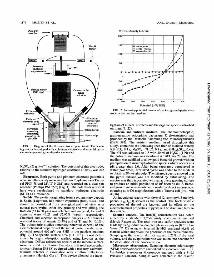
FIG. 1. Diagram of the three-electrode open circuit. The leach-ing reactor is equipped with a platinum electrode and a special pyriteelectrode (packed ground-pyrite electrode).
K2SO4 (25 g liter-1) solution. The potential of this electrode,relative to the standard hydrogen electrode at 30°C, was 242mV.
Electronics. Both pyrite and platinum electrode potentialswere simultaneously measured by two Eh-pH meters (Tacus-sel PHN 78 and MVIT-NUM) and recorded on a dual-penrecorder (Phillips PM 8222) (Fig. 1). The potentials reportedhere were recalculated to standard hydrogen electrode(SHE) as a reference.
Sulfide. The pyrite, originating from a sedimentary depositin Spain (Logrofio), had minor impurities (total, 0.4%) andshould be considered from geological point of view as anatural pure pyrite. After dry grinding and wet sifting, thefraction (53 to 80 ,um) was selected and analyzed; Fe and Scontents were 46.23 and 52.97% (wt/wt), respectively.Chemical and electron microprobe analysis (SX Cameca)revealed traces of arsenic (0.3%) and of Ti and Ni (0.1%).The voltametric studies (intensity-potential curves) of theelectrochemical properties of the initial pyrite revealed a restpotential around 660 mV per SHE in the nutrient medium(Fig. 2). The specific surface area (1.11 m2 g-1) was deter-mined by a gas adsorption method using krypton as theadsorbate. Diffuse reflectance spectra of the mineral surfacewere recorded on a Fourier Transform Infrared Spectropho-tometer (Bruker IFS 88) equipped with a mercury-cadmium-telluride detector in association with a diffuse reflectanceattachment (Harrick Corp.). This device allowed the inves-
FIG. 2. Intensity-potential curves of packed ground-pyrite elec-trode in the nutrient medium.
tigation of mineral surfaces and the organic species adsorbedon them (9, 25).
Bacteria and nutrient medium. The chemolithotrophic,gram-negative acidophilic bacterium T. ferrooxidans wasprovided by the Deutsche Sammlung von Mikroorganismen(DSM 583). The nutrient medium, used throughout thisstudy, contained the following (per liter of distilled water):KH2PO4, 0.4 g; MgSO4- 7H20, 0.4 g; and (NH4)2SO4, 0.4 g.The pH was adjusted to 1.8 with 30 ml of H2SO4 (1 N) andthe nutrient medium was sterilized at 120°C for 20 min. Themedium was acidified to allow good bacterial growth withoutprecipitation of iron oxyhydroxide species which occurs at apH greater than 2.0. After being separately autoclaved inwater (two times), sterilized pyrite was added to the mediumto obtain a 2% weight pulp. The infrared spectra showed thatthe pyrite surface was not modified by autoclaving. Thereactor was then inoculated with an actively growinf cultureto produce an initial population of 107 bacteria ml- . Bacte-rial growth measurements were made by direct microscopiccounting at x400 magnification with a Thoma cell (0.01-mmdepth).An inoculated reactor with thymol (2-isopropyl, 5-methyl,
phenol CloH140) served as the control. The bacteriostaticproperties of thymol are known, and its effect on theelectrochemical properties of pyrite will be discussed later inthis article.
Solution analysis. The iron(II) concentration was deter-mined by a standard 2,2'-bipyridyl colorimetric method(Merck Reagents). The total iron and sulfur analyses weremade by using inductive coupled plasma analysis (ICP JobinYvon JY 32) using an internal Sr-HCl standard (0.6% ofmatrix) which improved the precision of the measurements.Sampling in the reactor did not exceed 0.25% of the totalvolume, and the evaporation rate was taken into account forthe calculation of the concentration.Microscopy observations. Scanning electron microscopy
(SEM) observations were carried out on a conventional F.B.Cambridge Stereoscan Microscope equipped with a Si-LiPrinceton detector. Samples were collected in the reactor
1176 MUSTIN ET AL.
on June 13, 2018 by guesthttp://aem
.asm.org/
Dow
nloaded from

OXIDATION OF A PYRITE BY T. FERROOXIDANS 1177
and dried under an 02-free atmosphere to preserve thesurface and the corrosion pits. Optical microscopy observa-tions were made with a Nachet microscope (NS 400),equipped with a Nomarsky interferential contrast device.The high optical resolution permitted the simultaneous ob-servation of the bacteria and corroded pyrite grains.Comparison of the iron and sulfur oxidation rates. A
derivative ratio of the oxidation rates of sulfur and iron wasused to compare these data more accurately. This ratio (R.,)is written in the following form: R0X(t) = d(SO4)/dt x[d(Fe)Idtf-', where (SO4) and (Fe) represent the amounts inmoles per liter of sulfate and iron solubilized in the solution,respectively. It can be used to show the incongruent disso-lution of pyrite. The R., value for a congruent dissolution ofa pure pyrite must be constant versus time and equal to 2.The R0x ratio was obtained after a polynomial regression(order 3) of experimental data. The polynomial equations ofthe regression curves corresponding to the iron and sulfateconcentrations (in moles per liter), up to 30 days, weredetermined as follows:
(Fe) = 2.80 x 10⁻⁴ - 1.30 x 10⁻⁴t + 1.81 x 10⁻⁵t2+ 1.13 x 10⁻⁶t3 (r2 = 0.999)
(SO4) = 1.94 x 10⁻² - 6.09 x 10⁻⁴t + 8.76 x 10⁻⁵t2+ 7.97 x 10⁻⁷t3 (r2 = 0.995)
where t is the elapsed time and r is the regression coefficient.The zero-order terms in each equation were in a good
agreement with the initial concentrations of iron and sulfurspecies in the solution after pyrite addition and inoculation.After 30 days, the appearance of precipitates disturbed thecalculation.
RESULTSThe changes in the redox potentials of the platinum and
pyrite electrodes, the dissolution of iron and sulfur, and theSEM observation of corrosion patterns allowed the descrip-tion of four main steps found during a bioleaching cycle.
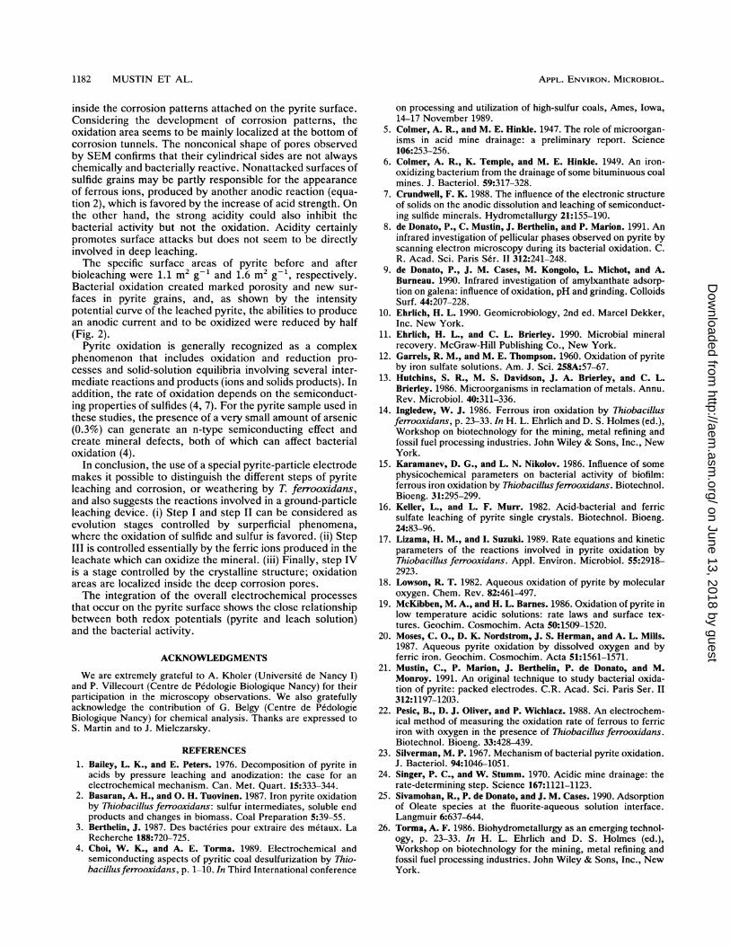
Step I. The lag phase. Initially, ferric ions introduced withthe inoculum were reduced (Fig. 3). The open-circuit poten-tial of the platinum electrode converged around the pyritepotential (Fig. 4a). The open-circuit potential of pyritedecreased slightly and had a lower value than the previouslydefined rest potential in nutrient medium (Fig. 2). At the endof this lag phase (5 days in the present experimental condi-tions), platinum and pyrite electrodes had the same potential(640 mV per SHE). Iron and sulfur solubilizations were veryweak (Fig. 5); therefore, the calculation of the Ro. ratio wasnot performed (Fig. 6).The number of bacteria present in the liquid medium
decreased (Fig. 7). Adsorption onto pyrite, which involved90% of inoculated bacteria, began immediately after theinoculation, and the pH increased slightly (Fig. 7). Nocorrosion was observed by SEM examination (Fig. 8a). Inthe control treatment (with addition of thymol), except foriron(III) reduction (Fig. 3), no detectable evolution wasnoted.
Step II. The start of the dissolution. At the beginning of thesecond step, the ferrous ions produced in step I were quicklyand completely oxidized (Fig. 3). After a small decrease ofpyrite potential (1 day), both platinum and pyrite potentialsincreased simultaneously and exceeded the rest potential ofpyrite (Fig. 4a). The pyrite potential, which was slightlyhigher than that of platinum during this increasing step,reached a stable value, about 80 mV lower than the platinumelectrode potential.
Fe(II) (mg/i)
Time (days)FIG. 3. Ferrous iron contents in solution versus time during the
four steps (I, II, III, IV) of bioleaching.
The bioleaching process began, and the solubilization ofiron and sulfur became significant (Fig. 5). The R., value of2.5 indicated a preferential solubilization of sulfur (Fig. 6).The number of bacteria in the nutrient medium increasedwith the beginning of the exponential growth phase, and thepH began to decrease (Fig. 7). Corrosion patterns, as in theprevious step, were not detectable by SEM even at highmagnification (x6,000) (Fig. 8b).
In the control treatment, during this step and the followingones, only very little ferrous iron solubilization resulted fromthe chemical solubilization of pyrite in the nutrient medium(Fig. 3). The potentials remained low (Fig. 4c) and had atendency to decrease slowly. The acidity was not greatlymodified (results not reported), but the number of bacteriadecreased (Fig. 7).
Step III. The superficial attack. The third step was domi-nated by the high content of ferric ions in solution (Fig. 5).The iron(II) concentration was very low (Fig. 3). An equi-librium between both potentials was reached around 80 mV(Fig. 4b).
Iron and sulfur solubilizations increased strongly (Fig. 5).The Ro. ratio indicated that sulfur was always preferentiallysolubilized over iron (Fig. 6); however, the R., ratio de-creased and became stoichiometric after 18 days (Rox =2.01).The increase in bacterial population in the nutrient me-
dium corresponded to the last part of its exponential growthphase. The pH decreased constantly (Fig. 7). The firstpatterns of corrosion (Fig. 8c) were evident from the appear-ance of some cracks (0.6 ,um wide).
In the control experiment, only a very small amount offerrous iron was observed. In this treatment, both potentialsshowed similar decreases (Fig. 4c). The bacteria were notdetectable by the direct counting method.
Step IV. The deep attack. Progressively after 25 days,ferrous ions appeared again but always in a very lowconcentration in comparison to the ferric ions (Fig. 3). Thepyrite potential increased, and the platinum potential de-
VOL. 58, 1992
on June 13, 2018 by guesthttp://aem
.asm.org/
Dow
nloaded from

1178 MUSTIN ET AL.
ISA,
Step I ' Step Hn 000
0 0750' z <Ds
00 Platinum potential ,* Pyrite potentUal
0@9..
700 .
Inoculation
I~~~~~~~~~60 0 0 0
6000 25 50 75 100 125 150 175 200
Time (hours) (a)850- ;
I i II, III , IV*
0
I
800 *.- -0-.--01520 00 0 0o:00000 0 0
0~~~~~~~~750-~~~~
700 :
o 0 Platinum potential10 Pyrite potential
650 ~j
6000 5 10 15 20 25 30 35 40 45
Time (days) (b)
850-
1II III IV
800-
'
_750- __ ...
6 700
I700li| a °0 Platinum Potential
650 Cl s | ' * Pyrite Potential
600.,''.,.,.,'....0 5 10 15 20 25 30
Time (days)35 40 45
(c)FIG. 4. Evolution of both pyrite and platinum potentials. (a)
Details of the first two steps; (b) record of the four steps; (c)potentials in the control reactor without bacterial activity in thepresence of thymol.
DISCUSSIONThe experimental setup allowed us to measure simulta-
neously the redox potential of a pyrite electrode and aplatinum electrode over the entire bioleaching cycle, whichrevealed more accurately the different steps of the biooxida-tion process in relation to the evolution of pyrite solution andmineral parameters.
In this work, the properties of the population of T.ferrooxidans could be modified during leaching despite inoc-ulation of a pure strain and may have influenced the redoxpotential of the solution and that of the mineral. Neverthe-less, it appears, in particular with voltametric studies, thatelectrochemical properties of pyrite are modified by bacte-rial oxidation. In addition, this modification can certainly be
creased. After 30 to 40 days of bioleaching, the differencesbetween the platinum and pyrite potentials were no morethan 40 mV (Fig. 4b).
Iron and sulfur were always strongly solubilized as ferricsulfate. The preferential iron dissolution was supported bythe low Ro. value of less than 2 (Fig. 6).The bacterial population reached the stationary phase at
the beginning of this step. The pH decreased strongly,reaching 1.3 after 45 days.The SEM photographs showed hexagonal or square
etched pits (internal diameter, about 2 ,um) which penetratedthe mineral to varying depths (Fig. 8d). The shape and thedirection of the corrosion patterns seemed to be related tothe cubic lattice of pyrite (16).At the end of the experiment, it was observed that
corrosion patterns were the same for the mineral grainssampled in pulp and those in the electrode bag.The bioleaching experiment was repeated three times, and
very similar relationships between the measured parameterswere observed. Only a small delay in the lag phase (less than1 day) and a small variation in the amount of solubilizedspecies (less than 10%) were observed. By using a pure,carefully ground pyrite, a pure strain, and a 400-ml reactor,the reproducibility of the experimental results was im-proved. The previously described delineation of the bi-oleaching cycle was always observed.
.-(g/l)
17.5
15,0
12,5
10.0
0 10 20 30 40 50
Time (days)
FIG. 5. Iron and sulfur contents in the leachate.
CO
6
i)
la
:P
(/2
6
a)00)
APPL. ENVIRON. MICROBIOL.
0
5
on June 13, 2018 by guesthttp://aem
.asm.org/
Dow
nloaded from

OXIDATION OF A PYRITE BY T. FERROOXIDANS 1179
Rox2.6- I'I' III IV
2.5-
2,4
2,3
2,2
2,1-Stoichiometric ratio
2.0-
1,9-
1.8-
1,7
1,60 5 10 15 20 25 30 35
Time (days)FIG. 6. Comparison of sulfur and iron oxidation rates (R., ratio
versus time).
related to the presence of compounds on the pyrite surfacewhich disturb the electrochemical behavior of pyrite, bacte-rial activity, and congruent dissolution of pyrite. Preliminaryresults (8) have shown that the surface layer can be stronglymodified during bacterial leaching.The packed-ground-pyrite electrode that contains 106 py_
rite grains represents the great diversity of pyrite grains and,in particular, the different crystalline orientations and sur-face properties. This type of electrode shows a highersensitivity towards the phenomena which occur at the min-eral interface. By the formation inside the electrode of amicroenvironment accessible to the bacteria, diverse bacte-rial and chemical reactions at the interface can take placeand be more extensively integrated. The pyrite potential isalso related to the presence of superficial pellicular phases
pH bact/rn, 10
I; 100
20 30Time (days)
10
.1
FIG. 7. Evolution of pH and bacterial growth.
(8) that are better randomly distributed in a ground-pyriteelectrode than in a polished-pyrite electrode.The platinum and pyrite electrode potentials are related to
each other and to the parameters measured in solution (pH,bacterial growth, and sulfate and ferric ions in solution,etc.). The difference between both potentials corresponds todifferent electrochemical behaviors of the electrodes. Whilethe platinum electrode is inert and reflects the redox poten-tial of the pulp suspension, the pyrite electrode can reactwith ferric ions, dissolved oxygen, protons (acid), and bac-teria present in the solution and the measured potential is atypical mixed potential. As mentioned earlier (24), the over-all process of the dissolution of a pyrite is the sum ofcathodic and anodic reactions occurring at the pyrite sur-face. The anodic process is a complex collection of oxidationreactions in which the pyrite reacts mainly with water toproduce ferric ions, sulfates, and protons (equation 1) orferrous ions and elementary sulfur (equation 2) when theacid strength increases (1):
FeS2 + 8H20- Fe3+ + 2SO42- + 16H+ + 15e- (1)
FeS2-*Fe2+ + 2S" + 2e- (2)
This second reaction shows a possible pathway for theformation of a sulfur-passive film.The anodic process has been discussed (18) in terms of a
two-layer system, where one layer is associated with theoxidation of iron(II) to iron(III) and the other involves theoxidation of sulfide (S22-). The sulfide is oxidized throughintermediates such as thiosulfate, elemental sulfur, or sul-fate. The electrons are transferred to a cathodic site (at themineral surface or on the bacteria) where the principalreaction is an oxygen reduction process:
02 + 4H+ + 4e-*2H20 (3)Concurrently the ferric ion may be reduced to ferrous ion
on the pyrite surface. Assuming that this process is con-trolled by the Fe(II)-Fe(III) couple, the stoichiometric equa-tion has been postulated (12):
FeS2 + 8H20 + 14Fe3+ -* 15Fe2+ + 2SO42- + 16H+ (4)
The presence of an active T. ferrooxidans culture provokesand catalyzes the completion of a cyclic process by regen-erating the ferric ions through the oxidation of ferrous ions.The bacterial growth and oxidation of sulfide are inhibited
in the control treatment by the presence of thymol. Thebacteriostatic effect of thymol is well understood; however,thymol can also modify the electrochemical behavior ofmineral particles. Figure 2 exhibits the electrochemicalinhibition effect of this molecule: a pyrite treated withthymol is not electrochemically active. The diffuse reflec-tance spectra (Fig. 9) confirm the presence of an adsorbedspecies on a mineral surface. The infrared spectrum showedin Fig. 9, spectrum a, indicates the methyl-stretching vibra-tions of the adsorbed thymol molecule that can be, in a firstapproximation, decomposed as follows: asymmetric 2,961and symmetric 2,871 cm-' stretching vibration of the twomethyl groups of the isopropyl radical; 2,927 and 2,871 cm-1in phase symmetric CH3 stretch of the methyl group in a ofthe hydroxyl function in Fermi resonance with a CH3deformation overtone. The surface coverage can be suffi-cient to saturate all of the reactive sites of pyrite grains and,thus, to inhibit bacterial nutrition.
Finally, the study of chemical, electrochemical, and bac-terial processes involved in pyrite bioleaching has proventhat the redox potential is controlled by the ferrous-ferric
VOL. 58, 1992
on June 13, 2018 by guesthttp://aem
.asm.org/
Dow
nloaded from

APPL. ENVIRON. MICROBIOL.
FIG. 8. SEM and Nomarsky optical microscopy photographsshowing evolution of the surface of pyrite grains during the bioleach-ing process. (a) At 2 days, clean surfaces and no corrosion patternare detectable; (b) at 17 days, cracks (0.6-,um wide) appear; (c) at 24days, small pits appear on the cracked surface; (d) at 43 days, deepand oriented corrosion pores which pass through the pyrite particlesare evident; (e) at 43 days, bacterial groups inside corrosion pores oradsorbed on the pyrite surface are evident.
redox couple (10, 20). Pyrite and platinum redox potentialsare expressed by the following Nernst's equation: E = Eo +(RT/nF) x [In (Fe3+/Fe2+)], where the standard potential Eoand the (RT/nF) value are well defined under standardconditions for a platinum electrode. In the case of a pyriteelectrode (22), differences were observed between the theo-retical and the measured potentials, which can be explainedby the existence of mixed potential, the influence of semi-conducting properties, or the appearance and dissolution ofsome superficial species that were previously neglected (8);
pyrite oxidation occurs through formation of several inter-mediate ions and surface products.At this stage of study, it is not necessary to know the exact
numerical expression of pyrite potential versus content ofsolubilized species. A qualitative observation of the varia-tion of both redox potentials, assuming that they are con-trolled by the ferrous-ferric redox couple, is sufficient todescribe the bioleaching process summarized in Table 1.
In step I, the pyrite electrode potential, lower than the restpotential of pyrite, exhibits the reducing behavior of thesulfide. The convergence and the stability of both potentialsprove that a the balance of ferrous and ferric species existsat the mineral-solution interface. On the pyrite surface,ferric ions and molecular oxygen are reduced. The anodicreaction may involve the sulfur species.
In the second step, both potentials increase rapidly bybacterial oxidation of soluble ferrous iron which is mainlyresponsible for the potential wave. During a short timeperiod of bioleaching, the pyrite potential, slightly greaterthan the platinum potential, shows that the mineral surface isthe main oxidation area. The ferric ions produced are
1180 MUSTIN ET AL.
on June 13, 2018 by guesthttp://aem
.asm.org/
Dow
nloaded from

OXIDATION OF A PYRITE BY T. FERROOXIDANS 1181
0.16 - l l
('4
0.093200 3100 3000 2900 2800 2700
WAVENUMBER (cm )
FIG. 9. Diffuse reflectance infrared spectra of pyrite treated withthymol (adsorbed on the surface) (a) and pyrite inoculated withbacteria (b).
diffused through the solution and are involved in the increaseof redox solution potential. This step corresponds to apreferential oxidative solubilization of sulfur (Fig. 6) andincreases in acidity and bacterial population (Fig. 7). Itshould be noted that the potential of pyrite becomes greaterthan the redox potential of the rusticyanine (680 mV perSHE) (14), which is considered to be the first electronacceptor of the T. ferrooxidans respiratory chain during ironoxidation. Yet, the pyrite becomes an electron donor and itsbacterial oxidation is made easier. The nature of superficialspecies (like ferrous sulfate) produced during the bacterialoxidation of pyrite (8) could explain the incongruent oxida-tion of the pyrite (Fig. 6) and a certain degree of indepen-dence of iron and sulfur oxidations. Iron binding by thebacteria is too low to interfere with the measurement of ironcontent in the medium.
In the third step, the ferric ions present in a significantamount in solution can electrochemically oxidize the mineral(equation 4). The pyrite is attacked and its redox potential(Fig. 4) is lower than the platinum potential, which provesthat the reductive behavior of the sulfide surface dependsupon the interfacial iron(II)-iron(III) balance. Because ofthis aspect, the concentration of ferrous iron can be consid-ered to be more important near the surface particles than inthe solution where the iron oxidation rate increases. Thus,the difference between both potentials can be an indicationof the intensity of the attack of pyrite and the ability ofbacteria to regenerate ferric ions through the oxidation offerrous ions.
In the last step, a progressive decrease of the differencebetween potentials of the pyrite and platinum electrodes isobserved. The decrease of the platinum potential resultsfrom the formation, in the solution, of a detectable quantityof ferrous ions, which are not completely reoxidized bybacteria (Fig. 3). On the other hand, the increase of thepyrite potential reveals the higher concentration of ferricions close to the sulfide surface, which certainly results froma bacterial activity inside the corrosion pores (Fig. 8d).Optical microscopic observation (Fig. 8e) shows bacteria
^ 0
cm 0
°Xg0
_.0,__00
_.-D
CD
Dc
0
CD
CD
0
CDCD.i
CD
++
0P00
3D
CD
_.'
sD
(DCD
'TI CACDN+TI
CD
0i+
++ or+n tjC +
+CA
++4. CD
(A0
+'Ti
CD
'TICD+
+
+
'TICDcn
'ICDw
+
+
0
+w
'TICD
I
'TICD
+
0
'I
CD
'-ICD~
'TI 'TIED CD CD
+ + +
'I
CD CD CD
+ + +
'TICD CD CD
CD
.-
000q VI
I --
110
I-,
s~~~:: -04 -Z-O.
, - -
+ ++
-~~~
I D~~~~.IV + V CD
CD
11
+
0 -) C C
.i'CD
CDO C_ CD CDla 3 X p
X, CD CD
CD Q ,WCD U -CD D
P0_l. _
:r0lD
M.
CD
'A0r_
CD
c
CD
0CD
IV0
0
CD
0CD
_.
3
CD "VCL _3. 3
CD CD
'OU)
~o
0>
0
rA0.laC0
ID
0)
VOL. 58, 1992
r
0.0CD
_.
CD
CD
0
0
0
CD
-0.
CD
rCD'0
ci'
m
-1.
CD
10CL0
CD
CD_.
CDI
ro
CD
U,ci
11I-,
on June 13, 2018 by guesthttp://aem
.asm.org/
Dow
nloaded from
APPL. ENVIRON. MICROBIOL.
inside the corrosion patterns attached on the pyrite surface.Considering the development of corrosion patterns, theoxidation area seems to be mainly localized at the bottom ofcorrosion tunnels. The nonconical shape of pores observedby SEM confirms that their cylindrical sides are not alwayschemically and bacterially reactive. Nonattacked surfaces ofsulfide grains may be partly responsible for the appearanceof ferrous ions, produced by another anodic reaction (equa-tion 2), which is favored by the increase of acid strength. Onthe other hand, the strong acidity could also inhibit thebacterial activity but not the oxidation. Acidity certainlypromotes surface attacks but does not seem to be directlyinvolved in deep leaching.The specific surface areas of pyrite before and after
bioleaching were 1.1 m2 g-1 and 1.6 m2 g-1, respectively.Bacterial oxidation created marked porosity and new sur-faces in pyrite grains, and, as shown by the intensitypotential curve of the leached pyrite, the abilities to producean anodic current and to be oxidized were reduced by half(Fig. 2).
Pyrite oxidation is generally recognized as a complexphenomenon that includes oxidation and reduction pro-cesses and solid-solution equilibria involving several inter-mediate reactions and products (ions and solids products). Inaddition, the rate of oxidation depends on the semiconduct-ing properties of sulfides (4, 7). For the pyrite sample used inthese studies, the presence of a very small amount of arsenic(0.3%) can generate an n-type semiconducting effect andcreate mineral defects, both of which can affect bacterialoxidation (4).
In conclusion, the use of a special pyrite-particle electrodemakes it possible to distinguish the different steps of pyriteleaching and corrosion, or weathering by T. ferrooxidans,and also suggests the reactions involved in a ground-particleleaching device. (i) Step I and step II can be considered asevolution stages controlled by surperficial phenomena,where the oxidation of sulfide and sulfur is favored. (ii) StepIII is controlled essentially by the ferric ions produced in theleachate which can oxidize the mineral. (iii) Finally, step IVis a stage controlled by the crystalline structure; oxidationareas are localized inside the deep corrosion pores.The integration of the overall electrochemical processes
that occur on the pyrite surface shows the close relationshipbetween both redox potentials (pyrite and leach solution)and the bacterial activity.
ACKNOWLEDGMENTS
We are extremely grateful to A. Kholer (Universite de Nancy I)and P. Villecourt (Centre de P6dologie Biologique Nancy) for theirparticipation in the microscopy observations. We also gratefullyacknowledge the contribution of G. Belgy (Centre de PedologieBiologique Nancy) for chemical analysis. Thanks are expressed toS. Martin and to J. Mielczarsky.
REFERENCES1. Bailey, L. K., and E. Peters. 1976. Decomposition of pyrite in
acids by pressure leaching and anodization: the case for anelectrochemical mechanism. Can. Met. Quart. 15:333-344.
2. Basaran, A. H., and 0. H. Tuovinen. 1987. Iron pyrite oxidationby Thiobacillus ferrooxidans: sulfur intermediates, soluble endproducts and changes in biomass. Coal Preparation 5:39-55.
3. Berthelin, J. 1987. Des bacteries pour extraire des metaux. LaRecherche 188:720-725.
4. Choi, W. K., and A. E. Torma. 1989. Electrochemical andsemiconducting aspects of pyritic coal desulfurization by Thio-bacillusferrooxidans, p. 1-10. In Third International conference
on processing and utilization of high-sulfur coals, Ames, Iowa,14-17 November 1989.
5. Colmer, A. R., and M. E. Hinkle. 1947. The role of microorgan-isms in acid mine drainage: a preliminary report. Science106:253-256.
6. Colmer, A. R., K. Temple, and M. E. Hinkle. 1949. An iron-oxidizing bacterium from the drainage of some bituminuous coalmines. J. Bacteriol. 59:317-328.
7. Crundwell, F. K. 1988. The influence of the electronic structureof solids on the anodic dissolution and leaching of semiconduct-ing sulfide minerals. Hydrometallurgy 21:155-190.
8. de Donato, P., C. Mustin, J. Berthelin, and P. Marion. 1991. Aninfrared investigation of pellicular phases observed on pyrite byscanning electron microscopy during its bacterial oxidation. C.R. Acad. Sci. Paris Ser. II 312:241-248.
9. de Donato, P., J. M. Cases, M. Kongolo, L. Michot, and A.Burneau. 1990. Infrared investigation of amylxanthate adsorp-tion on galena: influence of oxidation, pH and grinding. ColloidsSurf. 44:207-228.
10. Ehrlich, H. L. 1990. Geomicrobiology, 2nd ed. Marcel Dekker,Inc. New York.
11. Ehrlich, H. L., and C. L. Brierley. 1990. Microbial mineralrecovery. McGraw-Hill Publishing Co., New York.
12. Garrels, R. M., and M. E. Thompson. 1960. Oxidation of pyriteby iron sulfate solutions. Am. J. Sci. 258A:57-67.
13. Hutchins, S. R., M. S. Davidson, J. A. Brierley, and C. L.Brierley. 1986. Microorganisms in reclamation of metals. Annu.Rev. Microbiol. 40:311-336.
14. Ingledew, W. J. 1986. Ferrous iron oxidation by Thiobacillusferrooxidans, p. 23-33. In H. L. Ehrlich and D. S. Holmes (ed.),Workshop on biotechnology for the mining, metal refining andfossil fuel processing industries. John Wiley & Sons, Inc., NewYork.
15. Karamanev, D. G., and L. N. Nikolov. 1986. Influence of somephysicochemical parameters on bacterial activity of biofilm:ferrous iron oxidation by Thiobacillusferrooxidans. Biotechnol.Bioeng. 31:295-299.
16. Keller, L., and L. F. Murr. 1982. Acid-bacterial and ferricsulfate leaching of pyrite single crystals. Biotechnol. Bioeng.24:83-96.
17. Lizama, H. M., and I. Suzuki. 1989. Rate equations and kineticparameters of the reactions involved in pyrite oxidation byThiobacillus ferrooxidans. Appl. Environ. Microbiol. 55:2918-2923.
18. Lowson, R. T. 1982. Aqueous oxidation of pyrite by molecularoxygen. Chem. Rev. 82:461-497.
19. McKibben, M. A., and H. L. Barnes. 1986. Oxidation of pyrite inlow temperature acidic solutions: rate laws and surface tex-tures. Geochim. Cosmochim. Acta 50:1509-1520.
20. Moses, C. O., D. K. Nordstrom, J. S. Herman, and A. L. Mills.1987. Aqueous pyrite oxidation by dissolved oxygen and byferric iron. Geochim. Cosmochim. Acta 51:1561-1571.
21. Mustin, C., P. Marion, J. Berthelin, P. de Donato, and M.Monroy. 1991. An original technique to study bacterial oxida-tion of pyrite: packed electrodes. C.R. Acad. Sci. Paris Ser. II312:1197-1203.
22. Pesic, B., D. J. Oliver, and P. Wichlacz. 1988. An electrochem-ical method of measuring the oxidation rate of ferrous to ferriciron with oxygen in the presence of Thiobacillus ferrooxidans.Biotechnol. Bioeng. 33:428-439.
23. Silverman, M. P. 1967. Mechanism of bacterial pyrite oxidation.J. Bacteriol. 94:1046-1051.
24. Singer, P. C., and W. Stumm. 1970. Acidic mine drainage: therate-determining step. Science 167:1121-1123.
25. Sivamohan, R., P. de Donato, and J. M. Cases. 1990. Adsorptionof Oleate species at the fluorite-aqueous solution interface.Langmuir 6:637-644.
26. Torma, A. F. 1986. Biohydrometallurgy as an emerging technol-ogy, p. 23-33. In H. L. Ehrlich and D. S. Holmes (ed.),Workshop on biotechnology for the mining, metal refining andfossil fuel processing industries. John Wiley & Sons, Inc., NewYork.
1182 MUSTIN ET AL.
on June 13, 2018 by guesthttp://aem
.asm.org/
Dow
nloaded from
Related Documents