Correlation between Hemichrome Stability and the Root Effect in Tetrameric Hemoglobins Alessandro Vergara, †‡ Marisa Franzese, † Antonello Merlino, †‡ Giovanna Bonomi, † Cinzia Verde, § Daniela Giordano, § Guido di Prisco, § H. Caroline Lee, { Jack Peisach, { and Lelio Mazzarella †‡ * † Department of Chemistry, University of Naples ‘‘Federico II’’, Complesso Universitario Monte S. Angelo, Naples, Italy; ‡ Istituto di Biostrutture e Bioimmagini, and § Institute of Protein Biochemistry, Consiglio Nazionale delle Ricerche, Naples, Italy; and { Department of Physiology and Biophysics, Albert Einstein College of Medicine, Yeshiva University, New York, New York ABSTRACT Oxidation of Hbs leads to the formation of different forms of Fe(III) that are relevant to a range of biochemical and physiological functions. Here we report a combined EPR/x-ray crystallography study performed at acidic pH on six ferric tetrameric Hbs. Five of the Hbs were isolated from the high-Antarctic notothenioid fishes Trematomus bernacchii, Trematomus newnesi, and Gymnodraco acuticeps, and one was isolated from the sub-Antarctic notothenioid Cottoperca gobio. Our EPR analysis reveals that 1), in all of these Hbs, at acidic pH the aquomet form and two hemichromes coexist; and 2), only in the three Hbs that exhibit the Root effect is a significant amount of the pentacoordinate (5C) high-spin Fe(III) form found. The crystal struc- ture at acidic pH of the ferric form of the Root-effect Hb from T. bernacchii is also reported at 1.7 A ˚ resolution. This structure reveals a 5C state of the heme iron for both the a- and b-chains within a T quaternary structure. Altogether, the spectroscopic and crystallographic results indicate that the Root effect and hemichrome stability at acidic pH are correlated in tetrameric Hbs. Furthermore, Antarctic fish Hbs exhibit higher peroxidase activity than mammalian and temperate fish Hbs, suggesting that a partial hemichrome state in tetrameric Hbs, unlike in monomeric Hbs, does not remove the need for protection from peroxide attack, in contrast to previous results from monomeric Hbs. INTRODUCTION Hbs are proteins that are devoted to oxygen transport in blood. They carry out their function when the iron atom, which binds the oxygen molecule, is in the reduced Fe(II) state. However, it is well known that Hbs may undergo spon- taneous oxidation even under physiological conditions. Although the ferric forms are physiologically inert to further oxygenation, several subsequent side reactions in the Hb autoxidation may interfere with or merge into other biochem- ical pathways. Oxidized Hbs are involved in a range of biomedical and physiological functions. For example, autox- idation is a serious problem because it limits the storage time of acellular Hb-based blood substitutes (1). In addition to the commonly observed aquomet and hydroxylmet species, oxidation of Hbs can lead to the formation of pentacoordi- nate and endogenous hexacoordinate species, including bis-His adducts (hemichromes) (2). In the past, hemichromes were considered to be precursors of Hb denaturation because their formation is accelerated by denaturing agents (3). More recently, it was shown that hemichromes can be obtained under nondenaturing as well as physiological conditions (4). However, the physiological role of hemichromes is still disputed. It has been suggested that bis-His adducts can be involved in nitric oxide (NO) detoxification by acting as a NO scavenger (5), in the in vivo reduction of met-Hb, in Heinz-body formation (3), and in ligand binding (6,7). Hemichrome detection may also represent a valuable tool for tumor diagnosis (8). More recently, hemichromes were suggested to be involved in protecting Hbs from peroxide attack (9), given that the hemichrome derivative of human a-subunit complexed with AHSP does not exhibit peroxidase activity. Under physiological conditions, mammalian Hbs contain a low level of hemichrome. In contrast, Antarctic fish Hbs at room temperature are easily oxidized to a partial hemi- chrome state in which only the iron of the b-chain is bonded to the distal histidine (10–13). Cold adaptation (e.g., the biosynthesis of antifreeze glycoproteins) and isolation due to the Antarctic polar front are the major peculiarities of the Antarctic Notothenioidei (the dominant suborder of tele- osts in the Southern ocean), but other features, such as the high mitochondrial content of slow muscle fibers (14), can also be cold-adaptive. Despite the high level of sequence homology among the different members, Antarctic fish Hbs show marked differences in their functional properties, and therefore provide an intriguing system in which to explore the structural determinants and functional role of Submitted November 24, 2008, and accepted for publication April 28, 2009. *Correspondence: [email protected] Abbreviations used: Hbs, hemoglobins; AHSP, a-hemoglobin stabilizing protein; bis-His, bis-histidyl; CT, charge transfer; CW-EPR, continuous- wave electron paramagnetic resonance; deo-HbTb, the deoxy form of HbTb at pH 6.0; Hb, hemoglobin; HbA, adult human hemoglobin; Hb1Cg, Hb1 of Cottoperca gobio; HbGa, Hb of Gymnodraco acuticeps; Hb1Tn, major Hb of Trematomus newnesi; Hb2Tn, minor Hb of Tremato- mus newnesi; HbCTn, cathodic Hb of Trematomus newnesi; HbTb, Hb of Trematomus bernacchii; hemi-HbTb, the b-bis-histidyl ferric form of HbTb at pH 7.6; pH6-HbTb, the ferric form of HbTb at pH 6.0; PDB, Protein Data Bank; TrI, Hb I of trout; RMSD, root mean-square deviation; TrIV, Hb IV of trout; T state, tense state. Editor: Marilyn Gunner. Ó 2009 by the Biophysical Society 0006-3495/09/08/0866/9 $2.00 doi: 10.1016/j.bpj.2009.04.056 866 Biophysical Journal Volume 97 August 2009 866–874

Welcome message from author
This document is posted to help you gain knowledge. Please leave a comment to let me know what you think about it! Share it to your friends and learn new things together.
Transcript
866 Biophysical Journal Volume 97 August 2009 866–874
Correlation between Hemichrome Stability and the Root Effectin Tetrameric Hemoglobins
Alessandro Vergara,†‡ Marisa Franzese,† Antonello Merlino,†‡ Giovanna Bonomi,† Cinzia Verde,§
Daniela Giordano,§ Guido di Prisco,§ H. Caroline Lee,{ Jack Peisach,{ and Lelio Mazzarella†‡*†Department of Chemistry, University of Naples ‘‘Federico II’’, Complesso Universitario Monte S. Angelo, Naples, Italy; ‡Istituto di Biostrutture eBioimmagini, and §Institute of Protein Biochemistry, Consiglio Nazionale delle Ricerche, Naples, Italy; and {Department of Physiology andBiophysics, Albert Einstein College of Medicine, Yeshiva University, New York, New York
ABSTRACT Oxidation of Hbs leads to the formation of different forms of Fe(III) that are relevant to a range of biochemicaland physiological functions. Here we report a combined EPR/x-ray crystallography study performed at acidic pH on six ferrictetrameric Hbs. Five of the Hbs were isolated from the high-Antarctic notothenioid fishes Trematomus bernacchii, Trematomusnewnesi, and Gymnodraco acuticeps, and one was isolated from the sub-Antarctic notothenioid Cottoperca gobio. Our EPRanalysis reveals that 1), in all of these Hbs, at acidic pH the aquomet form and two hemichromes coexist; and 2), only in the threeHbs that exhibit the Root effect is a significant amount of the pentacoordinate (5C) high-spin Fe(III) form found. The crystal struc-ture at acidic pH of the ferric form of the Root-effect Hb from T. bernacchii is also reported at 1.7 A resolution. This structurereveals a 5C state of the heme iron for both the a- and b-chains within a T quaternary structure. Altogether, the spectroscopicand crystallographic results indicate that the Root effect and hemichrome stability at acidic pH are correlated in tetramericHbs. Furthermore, Antarctic fish Hbs exhibit higher peroxidase activity than mammalian and temperate fish Hbs, suggestingthat a partial hemichrome state in tetrameric Hbs, unlike in monomeric Hbs, does not remove the need for protection fromperoxide attack, in contrast to previous results from monomeric Hbs.
doi: 10.1016/j.bpj.2009.04.056
INTRODUCTION
Hbs are proteins that are devoted to oxygen transport in
blood. They carry out their function when the iron atom,
which binds the oxygen molecule, is in the reduced Fe(II)
state. However, it is well known that Hbs may undergo spon-
taneous oxidation even under physiological conditions.
Although the ferric forms are physiologically inert to further
oxygenation, several subsequent side reactions in the Hb
autoxidation may interfere with or merge into other biochem-
ical pathways. Oxidized Hbs are involved in a range of
biomedical and physiological functions. For example, autox-
idation is a serious problem because it limits the storage time
of acellular Hb-based blood substitutes (1). In addition to the
commonly observed aquomet and hydroxylmet species,
oxidation of Hbs can lead to the formation of pentacoordi-
nate and endogenous hexacoordinate species, including
bis-His adducts (hemichromes) (2).
Submitted November 24, 2008, and accepted for publication April 28, 2009.
*Correspondence: [email protected]
Abbreviations used: Hbs, hemoglobins; AHSP, a-hemoglobin stabilizing
protein; bis-His, bis-histidyl; CT, charge transfer; CW-EPR, continuous-
wave electron paramagnetic resonance; deo-HbTb, the deoxy form of
HbTb at pH 6.0; Hb, hemoglobin; HbA, adult human hemoglobin;
Hb1Cg, Hb1 of Cottoperca gobio; HbGa, Hb of Gymnodraco acuticeps;
Hb1Tn, major Hb of Trematomus newnesi; Hb2Tn, minor Hb of Tremato-
mus newnesi; HbCTn, cathodic Hb of Trematomus newnesi; HbTb, Hb of
Trematomus bernacchii; hemi-HbTb, the b-bis-histidyl ferric form of
HbTb at pH 7.6; pH6-HbTb, the ferric form of HbTb at pH 6.0; PDB, Protein
Data Bank; TrI, Hb I of trout; RMSD, root mean-square deviation; TrIV,
Hb IV of trout; T state, tense state.
Editor: Marilyn Gunner.
� 2009 by the Biophysical Society
0006-3495/09/08/0866/9 $2.00
In the past, hemichromes were considered to be precursors
of Hb denaturation because their formation is accelerated
by denaturing agents (3). More recently, it was shown that
hemichromes can be obtained under nondenaturing as well
as physiological conditions (4). However, the physiological
role of hemichromes is still disputed. It has been suggested
that bis-His adducts can be involved in nitric oxide (NO)
detoxification by acting as a NO scavenger (5), in the
in vivo reduction of met-Hb, in Heinz-body formation (3),
and in ligand binding (6,7). Hemichrome detection may
also represent a valuable tool for tumor diagnosis (8).
More recently, hemichromes were suggested to be involved
in protecting Hbs from peroxide attack (9), given that the
hemichrome derivative of human a-subunit complexed
with AHSP does not exhibit peroxidase activity.
Under physiological conditions, mammalian Hbs contain
a low level of hemichrome. In contrast, Antarctic fish Hbs
at room temperature are easily oxidized to a partial hemi-
chrome state in which only the iron of the b-chain is bonded
to the distal histidine (10–13). Cold adaptation (e.g., the
biosynthesis of antifreeze glycoproteins) and isolation due
to the Antarctic polar front are the major peculiarities of
the Antarctic Notothenioidei (the dominant suborder of tele-
osts in the Southern ocean), but other features, such as the
high mitochondrial content of slow muscle fibers (14), can
also be cold-adaptive. Despite the high level of sequence
homology among the different members, Antarctic fish
Hbs show marked differences in their functional properties,
and therefore provide an intriguing system in which to
explore the structural determinants and functional role of
Hemichrome Stability in Root-Effect Hemoglobins 867
hemichrome formation (15). Crystallographic studies of the
major Hbs from the Antarctic fish Trematomus bernacchiiand Trematomus newnesi at pH 7.6 have revealed that the
hemichrome derivative at physiological pH is associated
with a quaternary assembly that is intermediate between
the R and T states (10,11). In this form (hereafter denoted
the H state), several features of the tertiary organization are
also intermediate.
In addition to hemichrome formation, some Antarctic fish
Hbs also display a drastic reduction of the oxygen affinity
and binding rate coupled with a loss of cooperativity at the
lower values of the physiological pH range (16,17). This
property, known as the Root effect (17), is also common to
several Hbs of temperate fish. Many unsuccessful attempts
to interpret the Root effect on structural grounds have been
conducted in the past and include sequence comparison
(18), site-directed mutagenesis (19), and x-ray structural
comparison of Root-effect and non-Root-effect Hbs. Based
on crystallographic analysis, the most current hypothesis
attributes the Root effect to overstabilization of the T quater-
nary structure at physiological pH (20,21), although tertiary-
structure features may modulate the strength of the effect
(21,22).
We recently characterized the oxidized states of five Hbs
isolated from the Antarctic fish species T. bernacchii(HbTb), T. newnesi (Hb1Tn, Hb2Tn, and HbCTn), and Gym-nodraco acuticeps (HbGa) by EPR at physiological pH (11).
That investigation revealed the existence of a variety of ferric
forms, ranging from aquomet/hydroxymet to two distinct
hemichromes, including the presence of a pentacoordinate
(5C) high-spin Fe(III) form. Of interest, some Hbs of Arctic
fish can also adopt a 5C high-spin Fe(III) form, and in solu-
tion show only a low content of hemichrome species (11,23).
In the investigation presented here, we explored the inter-
connection between hemichrome stability and Root-effect
occurrence by using a combined EPR/x-ray crystallography
method to study the oxidation of six notothenioid Hbs at
acidic pH. Our data reveal that the two hemichromes
observed at physiological pH (11) persist at acidic pH in
the five Hbs from the Antarctic species T. bernacchii, T. new-nesi, G. acuticeps, as well as in the Hb of the sub-Antarctic
species Cottoperca gobio (Hb1Cg). The choice of these six
Hbs was motivated by the fact that, although in the ferric
state they form the a(aquomet)/b(bis-His) derivative, only
three of them exhibit the Root effect (HbTb, HbCTn, and
Hb1Cg), whereas the remaining three (HbGa, Hb1Tn, and
Hb2Tn) are scarcely sensitive to pH. The results indicate
that the three Hbs endowed with the Root effect have a larger
amount of the 5C Fe(III) form at acidic pH in comparison
with the other three. Moreover, x-ray analysis of ferric
HbTb crystallized at pH 6.0 shows that the latter is in the
5C-coordination state and adopts a T quaternary structure.
The observation that the 5C forms are present in Antarctic
fish Hbs in a suite of pH conditions prompted us to investi-
gate whether these proteins are endowed with a peroxidase
function. Indeed, all of these Antarctic fish Hbs (both with
and without the Root effect) exhibit a much higher peroxi-
dase activity than human and temperate fish Hbs, suggesting
that a partial hemichrome state in tetrameric Hbs does not
remove the need for protection from peroxide action, in
contrast to previous results from monomeric Hbs (9).
MATERIALS AND METHODS
Hb purification
Purification and storage of HbTb (24); Hb1Tn, Hb2Tn, and HbCTn (25);
HbGa (26); and the major Hb from C. gobio (Hb1Cg) (27) were performed
as previously described. Hbs were oxidized with K3Fe(CN)6, and the excess
was removed by gel filtration on a Sephadex G-25 column.
EPR
Ferric Hbs were studied by means of CW-EPR at 12 K using a Bruker ESP
300 spectrometer equipped with an Oxford Instrument ESR 10 continuous-
flow cryostat and model 3120 temperature controller. Data were exported to
Bruker WinEPR, version 2.11, for processing in a manner similar to that
previously described (11,23). All EPR samples were at 0.5 mM tetramer
concentration. The buffers (50 mM citrate pH 4.5, MES pH 6.0, HEPES
pH 7.6, and BICINE pH 9.0 and 10.4) were chosen for their low protonation
enthalpy (28). Spectra were recorded at 9.29-GHz microwave frequency,
10-mW microwave power, 100-kHz modulation frequency, and 5-G modu-
lation amplitude.
Optical spectroscopy
Optical pH titration of HbTb was performed in a JASCO 530V spectropho-
tometer. The buffers were the same as those used for the EPR analysis.
X-ray crystallography
In a first attempt, crystals grown at pH 7.6 (11) were equilibrated in a step-
wise fashion in mother liquor with a final pH value of 6.0. In all trials, the
crystals readily developed cracks on their surface and became disordered.
Direct crystallization of oxidized HbTb (pH6-HbTb) was performed in
a capillary at pH 6.0 and room temperature by means of the liquid-diffusion
technique (final conditions 6.0 mg/mL HbTb and 8% w/v MPEG 5K), which
provided well-diffracting crystals.
Diffraction data on pH6-HbTb were collected at high resolution (1.7 A)
with the XRD1 beam line of the Elettra Synchrotron (Trieste, Italy). A
data set was collected at 100 K using glycerol as the cryoprotectant and pro-
cessed with the program suite HKL (29). A summary of the data-processing
statistics is provided in Table 1. Crystals of pH6-HbTb were isomorphous to
those of deoxy HbTb (21,30). A statistical analysis of the intensities indi-
cates that the diffraction data are affected by pseudo-merohedral twinning
similar to that previously described in detail for deoxy HbTb crystals (30).
The twin fraction for the crystal used in the data collection is 0.47, as deter-
mined by the algorithm implemented in the program SHELX (31). Despite
twinning, the high resolution of the diffraction pattern still allows a highly
significant refinement of the model. The coordinates of the high-resolution
(1.3 A) deoxy structure of HbTb (PDB code 2H8F) (21) were used as a start-
ing model, which was then refined using the program SHELX-L (31). The
refinement runs were followed by manual intervention using the molecular
graphic program O (32) to correct minor errors in side-chain positions.
Water molecules were identified by evaluating the shape of the electron
density and the distance of potential hydrogen-bond donors and/or accep-
tors. At convergence, the R-factor value was 0.156 (Rfree ¼ 0.204). A
summary of the refinement statistics is given in Table 1. The coordinates
of the structure have been deposited in the PDB with entry code 3GQG.
Biophysical Journal 97(3) 866–874
868 Vergara et al.
Heme geometry
To compare the heme coordination of pH6-HbTb with those of other hemo-
proteins, we performed a statistical analysis of the structure of the heme
regions in all of the globin structures in the PDB. In particular, we found
998 iron-containing hemes in 317 crystal structures of Hbs, myoglobins,
or leghemoglobins. All data analyses were performed using programs devel-
oped in-house.
Peroxidase assays
Peroxidase activity was measured in HbTb, HbGa, and T. newnesi hemolysate
at 20�C according to a published procedure (33), using dopamine and guaia-
col as substrates. The data are the average of three independent kinetic traces.
The peroxidase activity of HbA was measured as a control to reproduce the
literature data (34). In the cases of HbTb and HbA, the experiments were
also repeated at 4�C at the highest Hb concentration used for the 20�C data.
RESULTS
EPR and optical spectra
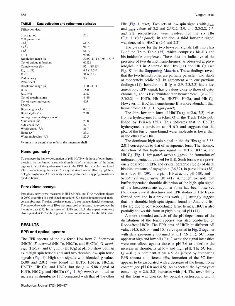
The EPR spectra of the six ferric Hbs from T. bernacchi(HbTb), T. newnesi (Hb1Tn, Hb2Tn, and HbCTn), G. acuti-ceps (HbGa), and C. gobio (Hb1Cg) at pH 6.0 show both an
axial high-spin ferric signal and two rhombic low-spin ferric
signals (Fig. 1). High-spin signals with identical g-values
(5.88 and 2.01) were found in HbTb, Hb1Tn, Hb2Tn,
HbCTn, Hb1Cg, and HbGa, but the g ¼ 5.88 signal of
HbTb, Hb1Cg, and HbCTn (Fig. 1, left panel) exhibited an
increase in rhombicity (11) compared with that of the other
TABLE 1 Data collection and refinement statistics
Diffraction data
Space group P21
Cell parameters
a (A) 61.72
b (A) 94.78
c (A) 61.72
b (�) 90.09
Resolution range (A) 30.00–1.71 (1.76–1.71)*
No. of unique reflections 69821
Completeness (%) 95.1 (88.1)*
Rmerge (%) 4.3 (17.5)*
I/s(I) 31.6 (5.1)
Redundancy 3.7
Refinement
Resolution range (A) 30.00–1.71
R (%) 15.0
Rfree (%) 20.8
No. of protein atoms 4449
No. of water molecules 405
RMSD
Bond lengths (A) 0.010
Bond angles (�) 2.28
Average atomic displacement
Main chain (A2) 20.8
Side chain (A2) 22.7
Whole chain (A2) 21.7
Heme (A2) 23.3
Water molecules (A2) 27.5
*Numbers in parentheses refer to the outermost shell.
Biophysical Journal 97(3) 866–874
Hbs (Fig. 1, inset). Two sets of low-spin signals with gmax
and gmid values of 3.2 and 2.3/2.2, 2.9, and 2.3/2.2, 2.6,
and 2.2, respectively, were resolved for the six Hbs
(Fig. 1, right panel). In addition, a third low-spin signal
was detected in HbCTn (2.6 and 2.2).
The g-values for the two low-spin signals fall into class
B of the Truth Table (35), which comprises bis-His and
bis-imidazole complexes. These data are indicative of the
presence of two distinct hemichromes, as observed at phys-
iological pH in Antarctic fish Hbs (11) and Hb1Cg (see
Fig. S1 in the Supporting Material). These findings reveal
that the two hemichromes are partially persistent and stable
at moderately acidic pH. In agreement with our previous
findings (11), hemichrome II (g ¼ 2.9, 2.3/2.2) has a less
anisotropic EPR signal, has g-values close to those of cyto-
chrome b5, and is less abundant than hemichrome I (g ¼ 3.2,
2.3/2.2) in HbTb, Hb1Tn, Hb2Tn, HbGa, and Hb1Cg.
However, in HbCTn, hemichrome II is more abundant than
hemichrome I (Fig. 1, right panel).The third low-spin form of HbCTn (g ¼ 2.6, 2.2) arises
from a hydroxymet form (class O of the Truth Table pub-
lished by Peisach (35)). This indicates that in HbCTn
hydroxymet is persistent at pH 6.0, and suggests that the
pKa of the ferric heme-bound water molecule is lower than
in the other five Hbs.
The dominant high-spin signal in the six Hbs (g ¼ 5.88,
2.01) corresponds to that of an aquomet form. The rhombic
distortion of this high-spin signal in HbTb, HbCTn, and
Hb1Cg (Fig. 1, left panel, inset) suggests the formation of
unligated, pentacoordinated Fe (III). Such forms were previ-
ously observed in EPR and crystallographic studies of distal
histidine mutants of myoglobin (36,37), in peroxidases (38),
in a flavo Hb (39), in a giant Hb at acidic pH (40), and in
Scapharca inequivalvis Hb (41). Although we note that
buffer-dependent rhombic distortion of the high-spin signal
of the hexacoordinate aquomet form has been observed
(36), x-ray crystal structures and EPR studies of HbTb per-
formed here and in a previous work (11) strongly suggest
that the rhombic high-spin signals found in Antarctic fish
Hbs are due to pentacooordinate ferric hemes. HbCTn also
partially shows this form at physiological pH (11).
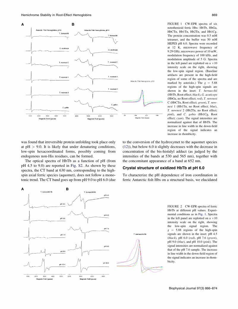
A more extended analysis of the pH dependence of the
distribution of the ferric species was also conducted on
Root-effect HbTb. The EPR data of HbTb at different pH
values (4.5, 6.0, 9.0, and 10.4) are reported in Fig. 2 together
with data previously obtained at pH 7.6 (11). 5C forms
appear at high and low pH (Fig. 2, inset; the signal intensities
were normalized against those at pH 7.6 to underline the
increase in rhombicity at low and high pH). The 5C form
(g ¼ 6.1) is dominant at pH 4.5. As judged by comparing
EPR spectra at different pHs, formation of the 5C form
appears to be associated with a decrease of the hemichrome
content (see pH 6.0 and 4.5). As expected, the hydroxymet
content (g ¼ 2.6, 2.2) increases with pH. The reversibility
of the form was checked by optical spectroscopy, and it
Hemichrome Stability in Root-Effect Hemoglobins 869
FIGURE 1 CW-EPR spectra of six
notothenioid ferric Hbs: HbTb, HbGa,
HbCTn, Hb1Tn, Hb2Tn, and Hb1Cg.
The protein concentration was 0.5 mM
tetramer, and the buffer was 50 mM
HEPES pH 6.0. Spectra were recorded
at 12 K, microwave frequency of
9.29 GHz, microwave power of 10 mW,
modulation frequency of 100 kHz, and
modulation amplitude of 5 G. Spectra
in the left panel are replotted on a �10
intensity scale on the right, showing
the low-spin signal region. (Baseline
artifacts are present in the high-field
region of some of the spectra and are
marked by asterisks.) The g ¼ 5.88
regions of the high-spin signals are
shown in the inset: T. bernacchii
(HbTb, Root effect; black), G. acuticeps(HbGa, no Root effect; red), T. newnesi
C (HbCTn, Root effect; green), T. new-
nesi 1 (Hb1Tn, no Root effect; blue),
T. newnesi 2 (Hb2Tn, no Root effect;
pink), and C. gobio (Hb1Cg, Root
effect; cyan). The signal intensities are
normalized against that of HbTb. The
increase in line width in the down-field
region of the signal indicates an
increase in rhombicity.
was found that irreversible protein unfolding took place only
at pH > 9.0. It is likely that under denaturing conditions,
low-spin hexacoordinated forms, possibly coming from
endogenous non-His residues, can be formed.
The optical spectra of HbTb as a function of pH (from
pH 4.5 to 9.0) are reported in Fig. S2. As shown by these
spectra, the CT band at 630 nm, corresponding to the high-
spin axial ferric species (aquomet), does not follow a mono-
tonic trend. The CT band goes up from pH 9.0 to pH 6.0 (due
to the conversion of the hydroxymet to the aquomet species
(12)), but below 6.0 it slightly decreases with the decrease in
concentration of the bis-histidyl adduct (as judged by the
intensities of the bands at 530 and 565 nm), together with
the concomitant appearance of a band at 652 nm.
Crystal structure of oxidized HbTb at pH 6.0
To characterize the pH dependence of iron coordination in
ferric Antarctic fish Hbs on a structural basis, we elucidated
FIGURE 2 CW-EPR spectra of ferric
HbTb at different pH values. Experi-
mental conditions as in Fig. 1. Spectra
in the left panel are replotted on a �10
intensity scale on the right, showing
the low-spin signal region. The
g ¼ 5.88 regions of the high-spin
signals are shown in the inset: pH 4.5
(black), pH 6.0 (red), pH 7.6 (green),
pH 9.0 (blue), and pH 10.0 (pink). The
signal intensities are normalized against
that of the pH 7.6 sample. The increase
in line width in the down-field region of
the signal indicates an increase in rhom-
bicity.
Biophysical Journal 97(3) 866–874
870 Vergara et al.
the novel structure of the oxidized form of HbTb at pH 6.0
(pH6-HbTb) using synchrotron data at 1.7 A resolution
(Table 1) and compared it with that solved at pH 7.6 (11).
Although crystals are affected by pseudo-merohedral twin-
ning, the resolution of the diffraction pattern ensures suffi-
cient data to produce a well-refined molecular structure, as
evidenced by the good quality of the omit electron density
maps for most of the residues (Figs. 3–5), the distribution
of the thermal displacement parameters (Table 1), and the
final refinement statistics (crystallographic R-factor ¼0.156, Rfree ¼ 0.204) The RMSD of the Ca atoms between
the two halves a1b1 and a2b2 of the tetramer, related by
a noncrystallographic twofold axis, is only 0.26 A.
The crystallographic model demonstrates that at acidic pH
the iron-to-histidine bond on the distal side of the hemi-HbTb
(PDB code 2PEG) b-chain (11) is broken, and the molecule
modifies the quaternary structure to adopt the typical quater-
nary T structure of the deoxy ferrous state. Indeed, the
RMSD between the Ca atoms of pH6-HbTb and deo-HbTb
(PDB code 2H8F) (21) is only 0.19 A, which has to be
compared with the value of 1.39 A for the superposition of
the structure presented here with that obtained at pH 7.6.
With respect to the latter, the stacking interaction between
the imidazole groups of the two C-terminal histidines
observed at the b1b2 interface at physiological pH is broken
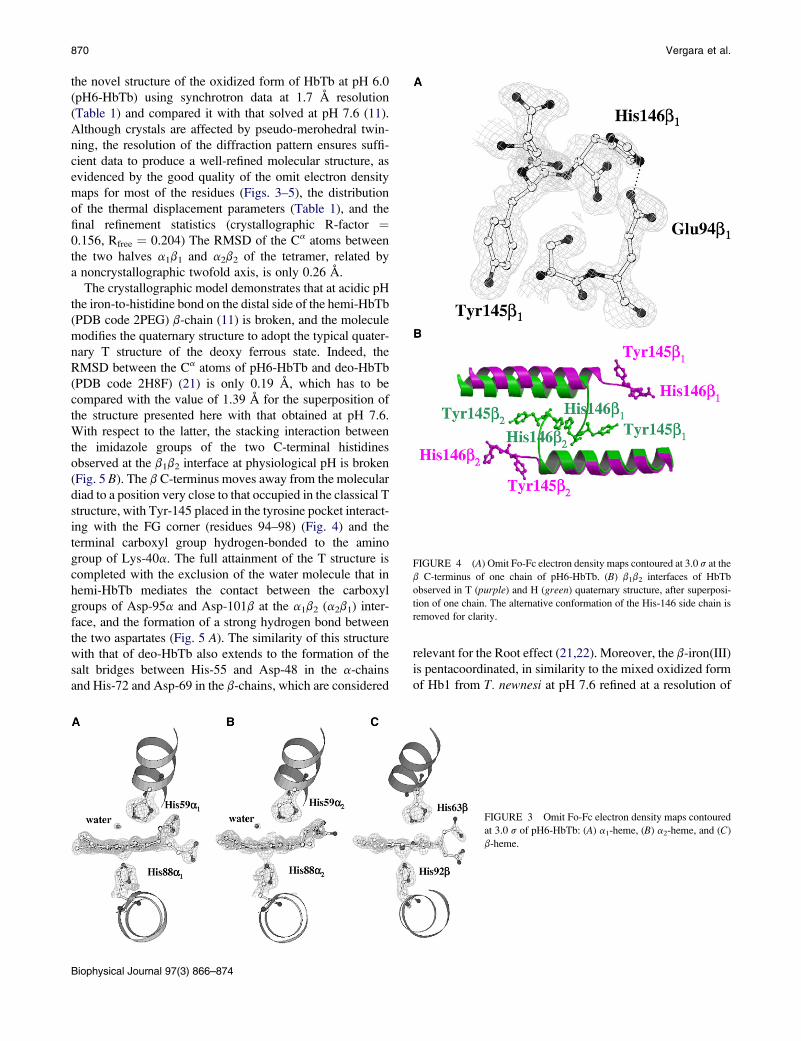
(Fig. 5 B). The b C-terminus moves away from the molecular
diad to a position very close to that occupied in the classical T
structure, with Tyr-145 placed in the tyrosine pocket interact-
ing with the FG corner (residues 94–98) (Fig. 4) and the
terminal carboxyl group hydrogen-bonded to the amino
group of Lys-40a. The full attainment of the T structure is
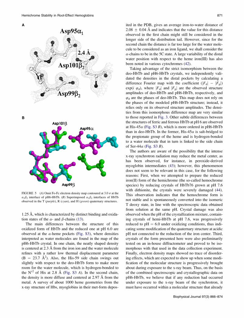
completed with the exclusion of the water molecule that in
hemi-HbTb mediates the contact between the carboxyl
groups of Asp-95a and Asp-101b at the a1b2 (a2b1) inter-
face, and the formation of a strong hydrogen bond between
the two aspartates (Fig. 5 A). The similarity of this structure
with that of deo-HbTb also extends to the formation of the
salt bridges between His-55 and Asp-48 in the a-chains
and His-72 and Asp-69 in the b-chains, which are considered
Biophysical Journal 97(3) 866–874
relevant for the Root effect (21,22). Moreover, the b-iron(III)
is pentacoordinated, in similarity to the mixed oxidized form
of Hb1 from T. newnesi at pH 7.6 refined at a resolution of
FIGURE 4 (A) Omit Fo-Fc electron density maps contoured at 3.0 s at the
b C-terminus of one chain of pH6-HbTb. (B) b1b2 interfaces of HbTb
observed in T (purple) and H (green) quaternary structure, after superposi-
tion of one chain. The alternative conformation of the His-146 side chain is
removed for clarity.
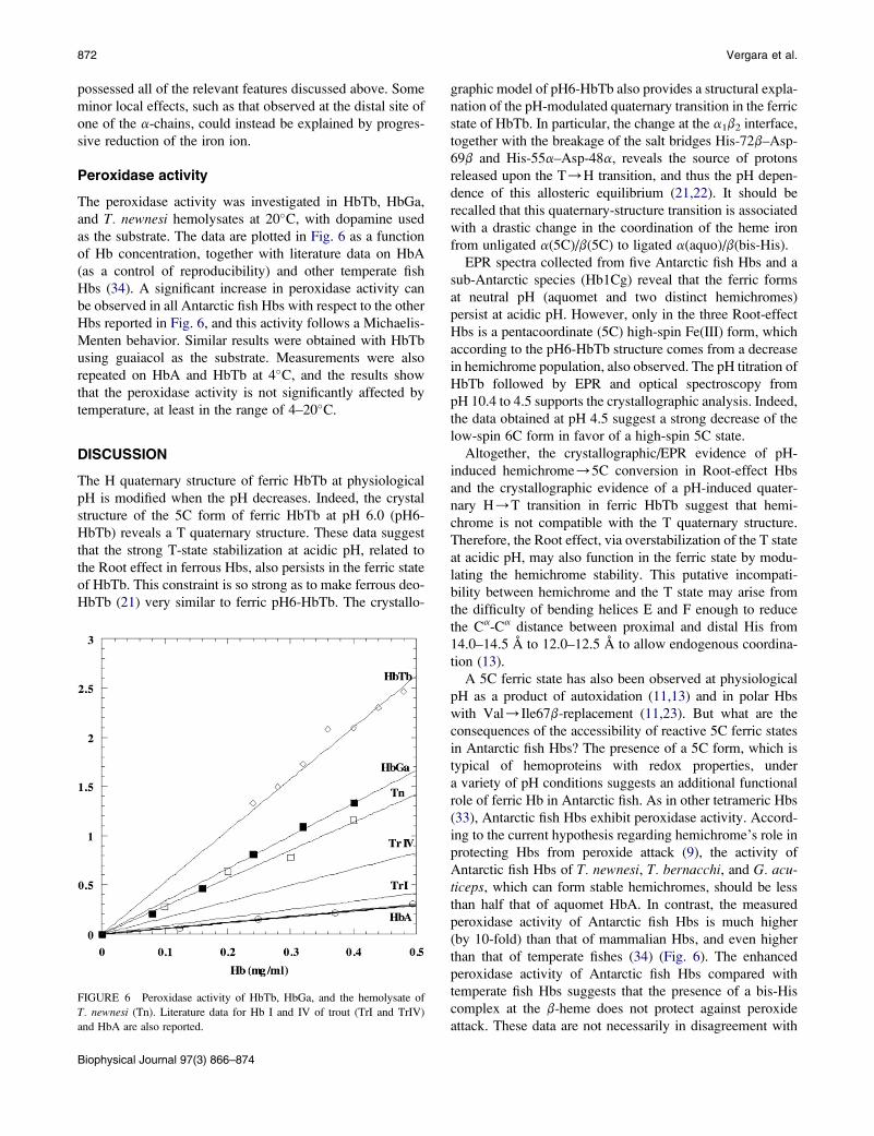
FIGURE 3 Omit Fo-Fc electron density maps contoured
at 3.0 s of pH6-HbTb: (A) a1-heme, (B) a2-heme, and (C)
b-heme.
Hemichrome Stability in Root-Effect Hemoglobins 871
1.25 A, which is characterized by distinct binding and oxida-
tion states of the a- and b-chains (13).
The main differences between the structure of this
oxidized form of HbTb and the reduced one at pH 6.0 are
observed at the a-heme pockets (Fig. S3), where densities
interpreted as water molecules are found in the map of the
pH6-HbTb crystal. In one chain, the neatly shaped density
is centered at 2.3 A from the iron ion and the water molecule
refines with a rather low thermal displacement parameter
(B ¼ 23.7 A2). Also, the His-59 side chain swings out
slightly with respect to the deo-HbTb form to make more
room for the water molecule, which is hydrogen-bonded to
the N32 of His at 2.8 A (Fig. S3 A). In the second chain,
the density is more diffuse and centered at 2.97 A from the
metal. A survey of about 1000 heme geometries from the
x-ray structure of Hbs, myoglobins in their met-form depos-
FIGURE 5 (A) Omit Fo-Fc electron density map contoured at 3.0 s at the
a1b2 interface of pH6-HbTb. (B) Superimposed a1b2 interfaces of HbTb
observed in the T (purple), R (cyan), and H (green) quaternary structures.
ited in the PDB, gives an average iron-to-water distance of
2.08 5 0.04 A and indicates that the value for this distance
observed in the first chain might still be considered in the
longer side of the distribution tail. However, since for the
second chain the distance is far too large for the water mole-
cule to be considered as an iron ligand, we shall consider the
a-chains to be in the 5C state. A large variability of the distal
water position with respect to the heme iron(III) has also
been noted in various cytochromes (42).
Taking advantage of the strict isomorphism between the
deo-HbTb and pH6-HbTb crystals, we independently vali-
dated the densities in the distal pockets by calculating a
difference Fourier map with the coefficient (jFdj � jFpj)exp(i 4d), where jFdj and jFpj are the observed structure
amplitudes of deo-HbTb and pH6-HbTb, respectively, and
4d are the phases of deo-HbTb. This map does not rely on
the phases of the modeled pH6-HbTb structure; instead, it
relies only on its observed structure amplitudes. The densi-
ties from this isomorphous difference map are very similar
to those reported in Fig. 3. Other subtle differences between
the structures of ferric and ferrous HbTb at pH 6 are observed
at His-45a (Fig. S3 B), which is more ordered in pH6-HbTb
than in deo-HbTb. In the former, His-45a is salt-bridged to
the propionate group of the heme and is hydrogen-bonded
to a water molecule that in turn is linked to the side chain
of Ser-44a (Fig. S3 B).
The authors are aware of the possibility that the intense
x-ray synchrotron radiation may reduce the metal center, as
has been observed, for instance, in peroxide-derived
myoglobin intermediates (43); however, this phenomenon
does not seem to be relevant in this case, for the following
reasons: First, when we attempted to prepare the reduced
iron(II) form of the hemichrome (the so-called hemochrome
species) by reducing crystals of HbTb76 grown at pH 7.6
with dithionite, the crystals were severely damaged (44).
This observation indicates that the hemochrome form is
not stable and is spontaneously converted into the isomeric
T deoxy state, in line with the spectroscopic data obtained
from solution at the same pH. Crystal damage was also
observed when the pH of the crystallization mixture, contain-
ing crystals of hemi-HbTb at pH 7.6, was progressively
reduced to pH ¼ 6.0 under oxidizing conditions, thus indi-
cating some modification of the quaternary structure at acidic
pH not connected to the reduction of the iron center. Third,
crystals of the form presented here were also preliminarily
tested on an in-house diffractometer and proved to be iso-
morphous with that used in the data collection experiment.
Finally, electron density maps showed no trace of disorder-
ing effects, which are expected to show up when some modi-
fication of the molecular structure is progressively brought
about during exposure to the x-ray beam. Thus, on the basis
of the combined spectroscopic and crystallographic data on
pH6-HbTb, we believe that if any reduction had occurred
under exposure to the x-ray beam of the synchrotron, it
must have occurred within a molecular structure that already
Biophysical Journal 97(3) 866–874
872 Vergara et al.
possessed all of the relevant features discussed above. Some
minor local effects, such as that observed at the distal site of
one of the a-chains, could instead be explained by progres-
sive reduction of the iron ion.
Peroxidase activity
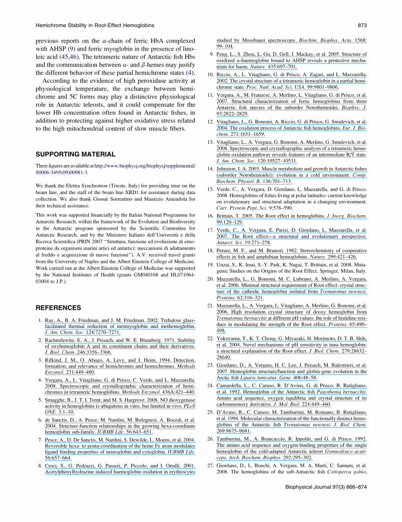
The peroxidase activity was investigated in HbTb, HbGa,
and T. newnesi hemolysates at 20�C, with dopamine used
as the substrate. The data are plotted in Fig. 6 as a function
of Hb concentration, together with literature data on HbA
(as a control of reproducibility) and other temperate fish
Hbs (34). A significant increase in peroxidase activity can
be observed in all Antarctic fish Hbs with respect to the other
Hbs reported in Fig. 6, and this activity follows a Michaelis-
Menten behavior. Similar results were obtained with HbTb
using guaiacol as the substrate. Measurements were also
repeated on HbA and HbTb at 4�C, and the results show
that the peroxidase activity is not significantly affected by
temperature, at least in the range of 4–20�C.
DISCUSSION
The H quaternary structure of ferric HbTb at physiological
pH is modified when the pH decreases. Indeed, the crystal
structure of the 5C form of ferric HbTb at pH 6.0 (pH6-
HbTb) reveals a T quaternary structure. These data suggest
that the strong T-state stabilization at acidic pH, related to
the Root effect in ferrous Hbs, also persists in the ferric state
of HbTb. This constraint is so strong as to make ferrous deo-
HbTb (21) very similar to ferric pH6-HbTb. The crystallo-
FIGURE 6 Peroxidase activity of HbTb, HbGa, and the hemolysate of
T. newnesi (Tn). Literature data for Hb I and IV of trout (TrI and TrIV)
and HbA are also reported.
Biophysical Journal 97(3) 866–874
graphic model of pH6-HbTb also provides a structural expla-
nation of the pH-modulated quaternary transition in the ferric
state of HbTb. In particular, the change at the a1b2 interface,
together with the breakage of the salt bridges His-72b–Asp-
69b and His-55a–Asp-48a, reveals the source of protons
released upon the T/H transition, and thus the pH depen-
dence of this allosteric equilibrium (21,22). It should be
recalled that this quaternary-structure transition is associated
with a drastic change in the coordination of the heme iron
from unligated a(5C)/b(5C) to ligated a(aquo)/b(bis-His).
EPR spectra collected from five Antarctic fish Hbs and a
sub-Antarctic species (Hb1Cg) reveal that the ferric forms
at neutral pH (aquomet and two distinct hemichromes)
persist at acidic pH. However, only in the three Root-effect
Hbs is a pentacoordinate (5C) high-spin Fe(III) form, which
according to the pH6-HbTb structure comes from a decrease
in hemichrome population, also observed. The pH titration of
HbTb followed by EPR and optical spectroscopy from
pH 10.4 to 4.5 supports the crystallographic analysis. Indeed,
the data obtained at pH 4.5 suggest a strong decrease of the
low-spin 6C form in favor of a high-spin 5C state.
Altogether, the crystallographic/EPR evidence of pH-
induced hemichrome/5C conversion in Root-effect Hbs
and the crystallographic evidence of a pH-induced quater-
nary H/T transition in ferric HbTb suggest that hemi-
chrome is not compatible with the T quaternary structure.
Therefore, the Root effect, via overstabilization of the T state
at acidic pH, may also function in the ferric state by modu-
lating the hemichrome stability. This putative incompati-
bility between hemichrome and the T state may arise from
the difficulty of bending helices E and F enough to reduce
the Ca-Ca distance between proximal and distal His from
14.0–14.5 A to 12.0–12.5 A to allow endogenous coordina-
tion (13).
A 5C ferric state has also been observed at physiological
pH as a product of autoxidation (11,13) and in polar Hbs
with Val/Ile67b-replacement (11,23). But what are the
consequences of the accessibility of reactive 5C ferric states
in Antarctic fish Hbs? The presence of a 5C form, which is
typical of hemoproteins with redox properties, under
a variety of pH conditions suggests an additional functional
role of ferric Hb in Antarctic fish. As in other tetrameric Hbs
(33), Antarctic fish Hbs exhibit peroxidase activity. Accord-
ing to the current hypothesis regarding hemichrome’s role in
protecting Hbs from peroxide attack (9), the activity of
Antarctic fish Hbs of T. newnesi, T. bernacchi, and G. acu-ticeps, which can form stable hemichromes, should be less
than half that of aquomet HbA. In contrast, the measured
peroxidase activity of Antarctic fish Hbs is much higher
(by 10-fold) than that of mammalian Hbs, and even higher
than that of temperate fishes (34) (Fig. 6). The enhanced
peroxidase activity of Antarctic fish Hbs compared with
temperate fish Hbs suggests that the presence of a bis-His
complex at the b-heme does not protect against peroxide
attack. These data are not necessarily in disagreement with
Hemichrome Stability in Root-Effect Hemoglobins 873
previous reports on the a-chain of ferric HbA complexed
with AHSP (9) and ferric myoglobin in the presence of lino-
leic acid (45,46). The tetrameric nature of Antarctic fish Hbs
and the communication between a- and b-hemes may justify
the different behavior of these partial hemichrome states (4).
According to the evidence of high peroxidase activity at
physiological temperature, the exchange between hemi-
chrome and 5C forms may play a distinctive physiological
role in Antarctic teleosts, and it could compensate for the
lower Hb concentration often found in Antarctic fishes, in
addition to protecting against higher oxidative stress related
to the high mitochondrial content of slow muscle fibers.
SUPPORTING MATERIAL
Three figures are available at http://www.biophysj.org/biophysj/supplemental/
S0006-3495(09)00981-3.
We thank the Elettra Synchrotron (Trieste, Italy) for providing time on the
beam line, and the staff of the beam line XRD1 for assistance during data
collection. We also thank Giosue Sorrentino and Maurizio Amendola for
their technical assistance.
This work was supported financially by the Italian National Programme for
Antarctic Research, within the framework of the Evolution and Biodiversity
in the Antarctic program sponsored by the Scientific Committee for
Antarctic Research, and by the Ministero Italiano dell’Universita e della
Ricerca Scientifica (PRIN 2007 ‘‘Struttura, funzione ed evoluzione di emo-
proteine da organismi marini artici ed antartici: meccanismi di adattamento
al freddo e acquisizione di nuove funzioni’’). A.V. received travel grants
from the University of Naples and the Albert Einstein College of Medicine.
Work carried out at the Albert Einstein College of Medicine was supported
by the National Institutes of Health (grants GM040168 and HL071064-
03004 to J.P.).
REFERENCES
1. Ray, A., B. A. Friedman, and J. M. Friedman. 2002. Trehalose glass-facilitated thermal reduction of metmyoglobin and methemoglobin.J. Am. Chem. Soc. 124:7270–7271.
2. Rachmilewitz, E. A., J. Peisach, and W. E. Blumberg. 1971. Stabilityof oxyhemoglobin A and its constituent chains and their derivatives.J. Biol. Chem. 246:3356–3366.
3. Rifkind, J. M., O. Abugo, A. Levy, and J. Heim. 1994. Detection,formation, and relevance of hemichromes and hemochromes. MethodsEnzymol. 231:449–480.
4. Vergara, A., L. Vitagliano, G. di Prisco, C. Verde, and L. Mazzarella.2008. Spectroscopic and crystallographic characterization of hemi-chromes in tetrameric hemoglobins. Methods Enzymol. 436A:421–440.
5. Smagghe, B., J. T. I. Trent, and M. S. Hargrove. 2008. NO dioxygenaseactivity in hemoglobins is ubiquitous in vitro, but limited in vivo. PLoSONE. 3:1–10.
6. de Sanctis, D., A. Pesce, M. Nardini, M. Bolognesi, A. Bocedi, et al.2004. Structure-function relationships in the growing hexa-coordinatehemoglobin sub-family. IUBMB Life. 56:643–651.
7. Pesce, A., D. De Sanctis, M. Nardini, S. Dewilde, L. Moens, et al. 2004.Reversible hexa- to penta-coordination of the heme Fe atom modulatesligand binding properties of neuroglobin and cytoglobin. IUBMB Life.56:657–664.
8. Croci, S., G. Pedrazzi, G. Passeri, P. Piccolo, and I. Ortalli. 2001.Acetylphenylhydrazine induced haemoglobin oxidation in erythrocytes
studied by Mossbauer spectroscopy. Biochim. Biophys. Acta. 1568:99–104.
9. Feng, L., S. Zhou, L. Gu, D. Gell, J. Mackay, et al. 2005. Structure of
oxidized a-haemoglobin bound to AHSP reveals a protective mecha-
nism for haem. Nature. 435:697–701.
10. Riccio, A., L. Vitagliano, G. di Prisco, A. Zagari, and L. Mazzarella.
2002. The crystal structure of a tetrameric hemoglobin in a partial hemi-
chrome state. Proc. Natl. Acad. Sci. USA. 99:9801–9806.
11. Vergara, A., M. Franzese, A. Merlino, L. Vitagliano, G. di Prisco, et al.
2007. Structural characterization of ferric hemoglobins from threeAntarctic fish species of the suborder Notothenioidei. Biophys. J.93:2822–2829.
12. Vitagliano, L., G. Bonomi, A. Riccio, G. di Prisco, G. Smulevich, et al.
2004. The oxidation process of Antarctic fish hemoglobins. Eur. J. Bio-chem. 271:1651–1659.
13. Vitagliano, L., A. Vergara, G. Bonomi, A. Merlino, G. Smulevich, et al.
2008. Spectroscopic and crystallographic analysis of a tetrameric hemo-
globin oxidation pathway reveals features of an intermediate R/T state.
J. Am. Chem. Soc. 120:10527–10531.
14. Johnston, I. A. 2003. Muscle metabolism and growth in Antarctic fishes
(suborder Notothenioidei): evolution in a cold environment. Comp.Biochem. Physiol. B. 136:701–713.
15. Verde, C., A. Vergara, D. Giordano, L. Mazzarella, and G. di Prisco.
2008. Hemoglobins of fishes living at polar latitudes: current knowledgeon evolutionary and structural adaptation in a changing environment.
Curr. Protein Pept. Sci. 9:578–590.
16. Brittain, T. 2005. The Root effect in hemoglobins. J. Inorg. Biochem.99:120–129.
17. Verde, C., A. Vergara, E. Parisi, D. Giordano, L. Mazzarella, et al.2007. The Root effect—a structural and evolutionary perspective.
Antarct. Sci. 19:271–278.
18. Perutz, M. F., and M. Brunori. 1982. Stereochemistry of cooperative
effects in fish and amphibian hemoglobins. Nature. 299:421–426.
19. Unzai, S., K. Imai, S.-Y. Park, K. Nagai, T. Brittain, et al. 2008. Muta-genic Studies on the Origins of the Root Effect. Springer, Milan, Italy.
20. Mazzarella, L., G. Bonomi, M. C. Lubrano, A. Merlino, A. Vergara,
et al. 2006. Minimal structural requirement of Root effect: crystal struc-
ture of the cathodic hemoglobin isolated from Trematomus newnesi.Proteins. 62:316–321.
21. Mazzarella, L., A. Vergara, L. Vitagliano, A. Merlino, G. Bonomi, et al.
2006. High resolution crystal structure of deoxy hemoglobin from
Trematomus bernacchii at different pH values: the role of histidine resi-
dues in modulating the strength of the Root effect. Proteins. 65:490–498.
22. Yokoyama, T., K. T. Chong, G. Miyazaki, H. Morimoto, D. T. B. Shih,
et al. 2004. Novel mechanisms of pH sensitivity in tuna hemoglobin:
a structural explanation of the Root effect. J. Biol. Chem. 279:28632–
28640.
23. Giordano, D., A. Vergara, H. C. Lee, J. Peisach, M. Balestrieri, et al.
2007. Hemoglobin structure/function and globin-gene evolution in the
Arctic fish Liparis tunicatus. Gene. 406:48–58.
24. Camardella, L., C. Caruso, R. D’Avino, G. di Prisco, B. Rutigliano,
et al. 1992. Hemoglobin of the Antarctic fish Pagothenia bernacchii.Amino acid sequence, oxygen equilibria and crystal structure of its
carbonmonoxy derivative. J. Mol. Biol. 224:449–460.
25. D’Avino, R., C. Caruso, M. Tamburrini, M. Romano, B. Rutigliano,
et al. 1994. Molecular characterization of the functionally distinct hemo-globins of the Antarctic fish Trematomus newnesi. J. Biol. Chem.269:9675–9681.
26. Tamburrini, M., A. Brancaccio, R. Ippoliti, and G. di Prisco. 1992.
The amino acid sequence and oxygen-binding properties of the single
hemoglobin of the cold-adapted Antarctic teleost Gymnodraco acuti-ceps. Arch. Biochem. Biophys. 292:295–302.
27. Giordano, D., L. Boechi, A. Vergara, M. A. Martı, U. Samuni, et al.
2008. The hemoglobins of the sub-Antarctic fish Cottoperca gobio,
Biophysical Journal 97(3) 866–874
874 Vergara et al.
a phyletically basal species. Oxygen-binding equilibria, kinetics andmolecular dynamics. FEBS J. 276:2266–2277.
28. Fukada, H., and K. Takahashi. 1998. Enthalpy and heat capacitychanges for the proton dissociation of various buffer components in0.1 M potassium chloride. Proteins. 33:159–166.
29. Otwinowski, Z., and W. Minor. 1997. Processing of X-ray diffractiondata collected in oscillation mode. Methods Enzymol. 276:307–326.
30. Ito, N., N. H. Komiyama, and G. Fermi. 1995. Structure of deoxyhemo-globin of the Antarctic fish Pagothenia bernacchii with an analysis ofthe structural basis of the Root effect by comparison of the ligandedand unliganded hemoglobin structures. J. Mol. Biol. 250:648–658.
31. Sheldrick, G., and T. Schneider. 1997. SHELXL: high-resolution refine-ment. Methods Enzymol. 277:319–343.
32. Jones, T. A., J. Y. Zou, S. W. Cowan, and M. Kjedgaard. 1991.Improved methods for binding protein models in electron densitymaps and the location of errors in these models. Acta Crystallogr.D Biol. Crystallogr. 56:714–721.
33. Everse, J., M. Johnson, and M. Marini. 1994. Peroxidative activitiesof hemoglobin and hemoglobin derivatives. Methods Enzymol.231:547–559.
34. Gabbianelli, R., G. Zolese, E. Bertoli, and G. Falcioni. 2004. Correla-tion between functional and structural changes of reduced and oxidizedtrout hemoglobins I and IV at different pHs. A circular dichroism study.Eur. J. Biochem. 271:1971–1979.
35. Peisach, J. 1998. EPR of metalloproteins: truth tables revisited. In Foun-dations of Modern EPR. S. S. Eaton, G. R. Eaton, and K. Salikhov,editors. World Scientific Press, Singapore. 346–360.
36. Ikeda-Saito, M., H. Hori, L. A. Andersson, R. C. Prince, I. J. Pickering,et al. 1992. Coordination structure of the ferric heme iron in engineereddistal histidine myoglobin mutants. J. Biol. Chem. 267:22843–22852.
37. Quillin, M., R. Arduini, J. Olson, and G. J. Phillips. 1993. High-resolu-tion crystal structures of distal histidine mutants of sperm whalemyoglobin. J. Mol. Biol. 234:140–155.
Biophysical Journal 97(3) 866–874
38. Smulevich, G., A. Feis, and B. D. Howes. 2005. Fifteen years of Raman
spectroscopy of engineered heme containing peroxidases: what have we
learned? Acc. Chem. Res. 38:433–440.
39. Ilari, A., A. Bonamore, A. Farina, K. Johnson, and A. Boffi. 2002. The
X-ray structure of ferric Escherichia coli flavohemoglobin reveals an
unexpected geometry of the distal heme pocket. J. Biol. Chem.26:23725–23732.
40. Marmo Moreira, L., A. Lima Poli, A. J. Costa-Filho, and H. Imasato.
2006. Pentacoordinate and hexacoordinate ferric hemes in acid medium:
EPR, UV-Vis and CD studies of the giant extracellular hemoglobin of
Glossoscolex paulistus. Biophys. Chem. 124:62–72.
41. Boffi, A., S. Takahashi, C. Spagnuolo, D. L. Rousseau, and E. Chian-
cone. 1994. Structural characterization of oxidized dimeric Scapharcainaequivalvis hemoglobin by resonance Raman spectroscopy. J. Biol.Chem. 269:20437–20440.
42. Smulevich, G. 1998. Understanding heme cavity structure of peroxi-
dases: comparison of electronic absorption and resonance Raman
spectra with crystallographic results. Biospectroscopy. 4:S3–S17.
43. Hersleth, H. P., Y. W. Hsiao, U. Ryde, C. H. Gorbitz, and K. K. Ander-
sson. 2008. The influence of X-rays on the structural studies of peroxide-
derived myoglobin intermediates. Chem. Biodivers. 5:2067–2089.
44. Merlino, A., C. Verde, G. di Prisco, L. Mazzarella, and A. Vergara.
2008. Reduction of ferric hemoglobin from Trematomus bernacchii in
a partial bis-histidyl state produces a deoxy coordination even when
encapsulated into the crystal phase. Spectroscopy. 22:143–152.
45. Baron, C. P., L. H. Skibsted, and H. J. Andersen. 2000. Peroxidation of
linoleate at physiological pH: hemichrome formation by substrate
binding protects against metmyoglobin activation by hydrogen
peroxide. Free Radic. Biol. Med. 28:549–558.
46. Baron, C. P., L. H. Skibsted, and H. J. Andersen. 2002. Concentration
effects in myoglobin-catalyzed peroxidation of linoleate. J. Agric. FoodChem. 50:883–888.
Related Documents