ORIGINAL RESEARCH ARTICLE published: 31 December 2013 doi: 10.3389/fnsys.2013.00125 Corpus callosal microstructure influences intermanual transfer in chimpanzees Kimberley A. Phillips 1,2 *, Jennifer A. Schaeffer 3 and William D. Hopkins 3,4 1 Department of Psychology, Trinity University, San Antonio, TX, USA 2 Southwest National Primate Research Center, Texas Biomedical Research Institute, San Antonio, TX, USA 3 Division of Cognitive and Developmental Neuroscience, Yerkes National Primate Research Center, Atlanta, GA, USA 4 Neuroscience Institute and Language Research Center, Georgia State University, Atlanta, GA, USA Edited by: Sven Bestmann, University College London, UK Reviewed by: Rogier B. Mars, University of Oxford, UK Fahad Sultan, University Tübingen, Germany *Correspondence: Kimberley A. Phillips, Department of Psychology, Trinity University, 1 Trinity Place, San Antonio, TX 78212, USA e-mail: [email protected] Learning a new motor skill with one hand typically results in performance improvements in the alternate hand. The neural substrates involved with this skill acquisition are poorly understood. We combined behavioral testing and non-invasive brain imaging to study how the organization of the corpus callosum was related to intermanual transfer performance in chimpanzees. Fifty-three chimpanzees were tested for intermanual transfer of learning using a bent-wire task. Magnetic resonance and diffusion tensor images were collected from 39 of these subjects. The dominant hand showed greater performance benefits than the nondominant hand. Further, performance was associated with structural integrity of the motor and sensory regions of the CC. Subjects with better intermanual transfer of learning had lower fractional anisotropy values. The results are consistent with the callosal access model of motor programming. Keywords: intermanual transfer, manual performance, fractional anisotropy, chimpanzees INTRODUCTION Learning a new motor skill with one hand typically results in performance improvements in the alternate hand (Parlow and Kinsbourne, 1989; Grafton et al., 2002; Japikse et al., 2003). A typical paradigm is to have one hand learn and practice a spe- cific motor task, and then to test whether the opposite (untrained) hand shows performance improvements. Inconsistent results have been reported as to whether greater performance improvements are shown from the dominant hand (DOM) to the nondom- inant hand (NDOM) (Milisen and Riper, 1939; Laszlo et al., 1970; Parlow and Kinsbourne, 1989; Halsband, 1992), or from the NDOM to the DOM (Hicks, 1974; Taylor and Heilman, 1980). The callosal access model postulates that motor programs are stored in the dominant hemisphere (in humans, typically the left), irrespective of the hand used during training. Thus, the DOM (in humans, typically the right) has direct access to these programs, whereas the nondominant (left) hand has indirect access via the corpus callosum (CC) (Taylor and Heilman, 1980). According to this model greater transfer of learning would be seen in DOM from NDOM training. The CC, the major white matter tract connecting the two cere- bral hemispheres, is crucial for interhemispheric transfer of infor- mation (Wahl and Ziemann, 2008). Two subdivisions of the CC appear to be most associated with intermanual transfer: that con- taining transcallosal fibers of the primary motor cortex (M1), and that containing transcallosal fibers of the supplementary motor area (SMA). Bonzano et al. (2011) reported a significant positive correlation between the structural integrity of the region contain- ing M1 transcallosal fibers and an intermanual transfer task of reaction-time. In another study, reduced structural integrity of the anterior CC was associated with impairment in a coordinated bimanual task (Bonzano et al., 2008). The SMA is involved in the intermanual transfer of a newly acquired motor skill (Perez et al., 2007). In a test of learned sequential finger movements, greater activity was observed in the SMA when a skill transferred well than when the skill transferred poorly. Furthermore, blocking activity of the SMA through transcranial magnetic stimulation resulted in a blocking of intermanual transfer. Thus, the SMA is centrally involved in the interhemisphic transfer of motor skill learning, and we could reasonably expect the CC region where these transcallosal fibers cross to be associated with intermanual transfer performance. Chimpanzees (Pan troglodytes) have evolved several motor characteristics in common with humans; including complex manipulation, use of feeding tools in the wild, corticospinal ter- minals in the ventral horn of the spinal cord, and the use of precision grips (Padberg et al., 2007). Thus, they are good mod- els for understanding patterns of intermanual transfer and its association with the organization of the CC. Determining these relationships in chimpanzees will thus provide information about the fundamental aspects of neurobiological organization that underlie skilled motor actions and interhemispheric transfer. The first aim of this study was to investigate intermanual trans- fer of learning in chimpanzees. Chimpanzees were presented with a skilled motor task that required them to use a specific hand to guide a metal washer off a curved rod. To determine which would result in greater performance improvements, some indi- viduals trained on the DOM, others trained on the NDOM. Following the callosal access model, we hypothesized that those individuals trained on NDOM would see greater performance improvement. The second aim was to relate intermanual trans- fer to the structural integrity of the CC, specifically the regions Frontiers in Systems Neuroscience www.frontiersin.org December 2013 | Volume 7 | Article 125 | 1 SYSTEMS NEUROSCIENCE

Welcome message from author
This document is posted to help you gain knowledge. Please leave a comment to let me know what you think about it! Share it to your friends and learn new things together.
Transcript
ORIGINAL RESEARCH ARTICLEpublished: 31 December 2013doi: 10.3389/fnsys.2013.00125
Corpus callosal microstructure influences intermanualtransfer in chimpanzeesKimberley A. Phillips1,2*, Jennifer A. Schaeffer3 and William D. Hopkins3,4
1 Department of Psychology, Trinity University, San Antonio, TX, USA2 Southwest National Primate Research Center, Texas Biomedical Research Institute, San Antonio, TX, USA3 Division of Cognitive and Developmental Neuroscience, Yerkes National Primate Research Center, Atlanta, GA, USA4 Neuroscience Institute and Language Research Center, Georgia State University, Atlanta, GA, USA
Edited by:
Sven Bestmann, University CollegeLondon, UK
Reviewed by:
Rogier B. Mars, University ofOxford, UKFahad Sultan, University Tübingen,Germany
*Correspondence:
Kimberley A. Phillips, Department ofPsychology, Trinity University,1 Trinity Place, San Antonio,TX 78212, USAe-mail: [email protected]
Learning a new motor skill with one hand typically results in performance improvementsin the alternate hand. The neural substrates involved with this skill acquisition are poorlyunderstood. We combined behavioral testing and non-invasive brain imaging to study howthe organization of the corpus callosum was related to intermanual transfer performancein chimpanzees. Fifty-three chimpanzees were tested for intermanual transfer of learningusing a bent-wire task. Magnetic resonance and diffusion tensor images were collectedfrom 39 of these subjects. The dominant hand showed greater performance benefits thanthe nondominant hand. Further, performance was associated with structural integrity ofthe motor and sensory regions of the CC. Subjects with better intermanual transfer oflearning had lower fractional anisotropy values. The results are consistent with the callosalaccess model of motor programming.
Keywords: intermanual transfer, manual performance, fractional anisotropy, chimpanzees
INTRODUCTIONLearning a new motor skill with one hand typically results inperformance improvements in the alternate hand (Parlow andKinsbourne, 1989; Grafton et al., 2002; Japikse et al., 2003). Atypical paradigm is to have one hand learn and practice a spe-cific motor task, and then to test whether the opposite (untrained)hand shows performance improvements. Inconsistent results havebeen reported as to whether greater performance improvementsare shown from the dominant hand (DOM) to the nondom-inant hand (NDOM) (Milisen and Riper, 1939; Laszlo et al.,1970; Parlow and Kinsbourne, 1989; Halsband, 1992), or from theNDOM to the DOM (Hicks, 1974; Taylor and Heilman, 1980).The callosal access model postulates that motor programs arestored in the dominant hemisphere (in humans, typically the left),irrespective of the hand used during training. Thus, the DOM (inhumans, typically the right) has direct access to these programs,whereas the nondominant (left) hand has indirect access via thecorpus callosum (CC) (Taylor and Heilman, 1980). According tothis model greater transfer of learning would be seen in DOMfrom NDOM training.
The CC, the major white matter tract connecting the two cere-bral hemispheres, is crucial for interhemispheric transfer of infor-mation (Wahl and Ziemann, 2008). Two subdivisions of the CCappear to be most associated with intermanual transfer: that con-taining transcallosal fibers of the primary motor cortex (M1), andthat containing transcallosal fibers of the supplementary motorarea (SMA). Bonzano et al. (2011) reported a significant positivecorrelation between the structural integrity of the region contain-ing M1 transcallosal fibers and an intermanual transfer task ofreaction-time. In another study, reduced structural integrity ofthe anterior CC was associated with impairment in a coordinated
bimanual task (Bonzano et al., 2008). The SMA is involved in theintermanual transfer of a newly acquired motor skill (Perez et al.,2007). In a test of learned sequential finger movements, greateractivity was observed in the SMA when a skill transferred wellthan when the skill transferred poorly. Furthermore, blockingactivity of the SMA through transcranial magnetic stimulationresulted in a blocking of intermanual transfer. Thus, the SMA iscentrally involved in the interhemisphic transfer of motor skilllearning, and we could reasonably expect the CC region wherethese transcallosal fibers cross to be associated with intermanualtransfer performance.
Chimpanzees (Pan troglodytes) have evolved several motorcharacteristics in common with humans; including complexmanipulation, use of feeding tools in the wild, corticospinal ter-minals in the ventral horn of the spinal cord, and the use ofprecision grips (Padberg et al., 2007). Thus, they are good mod-els for understanding patterns of intermanual transfer and itsassociation with the organization of the CC. Determining theserelationships in chimpanzees will thus provide information aboutthe fundamental aspects of neurobiological organization thatunderlie skilled motor actions and interhemispheric transfer.
The first aim of this study was to investigate intermanual trans-fer of learning in chimpanzees. Chimpanzees were presented witha skilled motor task that required them to use a specific handto guide a metal washer off a curved rod. To determine whichwould result in greater performance improvements, some indi-viduals trained on the DOM, others trained on the NDOM.Following the callosal access model, we hypothesized that thoseindividuals trained on NDOM would see greater performanceimprovement. The second aim was to relate intermanual trans-fer to the structural integrity of the CC, specifically the regions
Frontiers in Systems Neuroscience www.frontiersin.org December 2013 | Volume 7 | Article 125 | 1
SYSTEMS NEUROSCIENCE

Phillips et al. Intermanual transfer in chimpanzees
containing transcallosal connections of M1 and SMA. We hypoth-esized that greater structural integrity of these regions would beassociated with greater intermanual transfer.
METHODSSUBJECTSFifty-three chimpanzees (Pan troglodytes) were tested on thebehavioral component of the study (male = 18; female = 35),ranging in age from 9 to 47 years. Of these, 31 subjects trainedon the DOM (male = 12; female = 19) and 22 subjects trainedon NDOM (male = 6, female = 16). All of the chimpanzeesresided at the Yerkes National Primate Research Center (Atlanta,Georgia). All aspects of this study were conducted in accordancewith ethical guidelines associated with the care and use of non-human primates and with the approval of the Emory UniversityInstitutional Animal Care and Use Committee.
MATERIALSThe device used for intermanual transfer testing was made usinga 1 cm thick metal wire that has been curved into a pattern similarto an “S” shape but with an additional loop. The wire was weldedto a metal plate measuring 11.35 by 11.35 cm which was designedto prevent the device from being pulled into the enclosure, and ahandle was welded on the opposite side of the plate. The openingof each curve measured approximately 10 cm, and the distancefrom the wire end to the plate was 39 cm (see Figure 1).
PROCEDUREDetermination of DOM and NDOM handIndividual’s DOM and NDOM hands were determined based onhand preferences on a simple reaching paradigm (Hopkins et al.,2002). Briefly, hand preferences for simple reaching were mea-sured by throwing small food items into the subject’s outdoorenclosure and recording which hand was used to grasp the item.Hand use was recorded for 50 responses. Between each reachingresponse, the subject had to reposition or locomote at least 3 mbefore reaching for another food item. Based on the frequencyin right and left hand use, a handedness index was computedfollowing the formula [HI = (#R - #L)/(#R + #L)] where #R
FIGURE 1 | Curved metal rod used to test intermanual transfer of
learning.
and #L represent the number of left and right hand responses.To simplify hand preference assignment in this paper, we classi-fied subjects with positive HI scores as right-handed and subjectswith negative HI scores as left-handed.
Intermanual transfer proceduresAll subjects were initially trained and subsequently tested on theS wire (see Figure 2) and reliably used only the designated handbefore beginning testing on the intermanual transfer task. Eachsubject received 12 trials per test session and received 1 testingsession per day.
At the start of each test session, the experimenter placed astationing stimulus (a 9 cm PVC pipe extending into the sub-jects cage) to the left or right of the chimpanzee, depending onwhich hand they were going to use on the motor task duringthe session. During each trial, the chimpanzee had to grasp thestationing stimulus with the hand not being tested so as to pre-vent them from attempting to switch hands during the trial andthereby assure that they would use the designated hand during asession. A nut (2.5 cm in diameter) was then placed at the end ofthe bent wire and inserted into to the subject’s home cage. Thesubject was then allowed to remove the nut from the wire withthe target hand. If they were successful in removing the nut fromthe bent wire, they were rewarded with a small piece of food orsquirt of juice.
If at any point during the trial the subject tried to use thenon-target hand or mouth to remove the nut, the experimenterretracted the wire. If this occurred, the subject was not rein-forced for this trial but the trial was repeated. During each trial,the experimenter recorded the time needed to remove the nutfrom the wire, and whether the subject was successful in remov-ing the nut. Time was recorded from the moment the subjectgrasped the nut until it was successfully removed at the end ofthe wire. If the subject turned their attention away from the taskor stopped working on the task, that attempt was not counted
FIGURE 2 | Subject removing the nut from the curved metal rod with
the target hand. Note that the non-target hand is grasping the stationingstimulus.
Frontiers in Systems Neuroscience www.frontiersin.org December 2013 | Volume 7 | Article 125 | 2
Phillips et al. Intermanual transfer in chimpanzees
and the trial was repeated or if possible, the experimenter stoppedthe time until the subject re-engaged with the task. Each subjectreceived 12 trials during each test session with the target hand.Test sessions were repeated until subject reached the performancecriterion. Performance criterion was defined as each subject com-pleting at least 10 of 12 trials within a single testing session inunder 5 s. Once criterion was met, the subject was tested with theopposite hand following the methods and procedure describedabove.
Image acquisition and processingWe acquired non-invasive MRI and DTI scans from 39 chim-panzees who completed the intermanual testing (16 males, 23females; average age = 20.19 years ± 7.34; 11 left- and 28 right-handers). As the animals needed to be anesthetized for this proce-dure, the collection of the brain images was coordinated with eachsubject’s annual physical exam. Anesthesia was used only for thepurpose of restraint and to keep the subject immobilized duringtheir physical exam and collection of the brain images. Subjectswere initially immobilized using ketamine (10 mg/kg) and subse-quently anesthetized with propofol [40–60 mg (kg/h)] followingstandard procedures at the YNPRC. Subjects were under anesthe-sia for transport to and from the imaging facility, and remainedanesthetized throughout the imaging procedure. Respiration rate,heart rate, and oxygen consumption were continually monitoredby a veterinarian.
As described previously (Phillips and Hopkins, 2012), subjectswere scanned on a Siemens 3.0 T Trio at the YNPRC. T1-weightedimages were acquired using a 3D gradient echo sequence (pulserepetition = 2300 ms, echo time = 4.4 ms, number of signalsaveraged = 3, matrix size = 320 × 320, with 0.6 mm isotropic res-olution). We acquired two sets of whole brain diffusion-weighteddata with a single-shot EPI sequence with a b-value of 1000 s/mm2
with 64 diffusion directions; plus one image without diffusionweighting (b-value of 0 s/mm2). DTI data were acquired transax-ially (FOV = 243 × 243) using 42 contiguous slices with nogap that covered the entire brain with resolution of 1.9 × 1.9 ×1.9 mm. Averages of two sets of diffusion-weighted data werecollected per subject with phase-encoding directions of oppo-site polarity (left–right) to correct for susceptibility distortion.Acquisition time for both the MRI and DTI scans was approx-imately 1 h. After completing the DTI and MRI procedures thesubjects were temporarily housed in a single cage for 6–12 h, toallow for the effects of anesthesia to wear off, after which theywere returned to their home cage and social group.
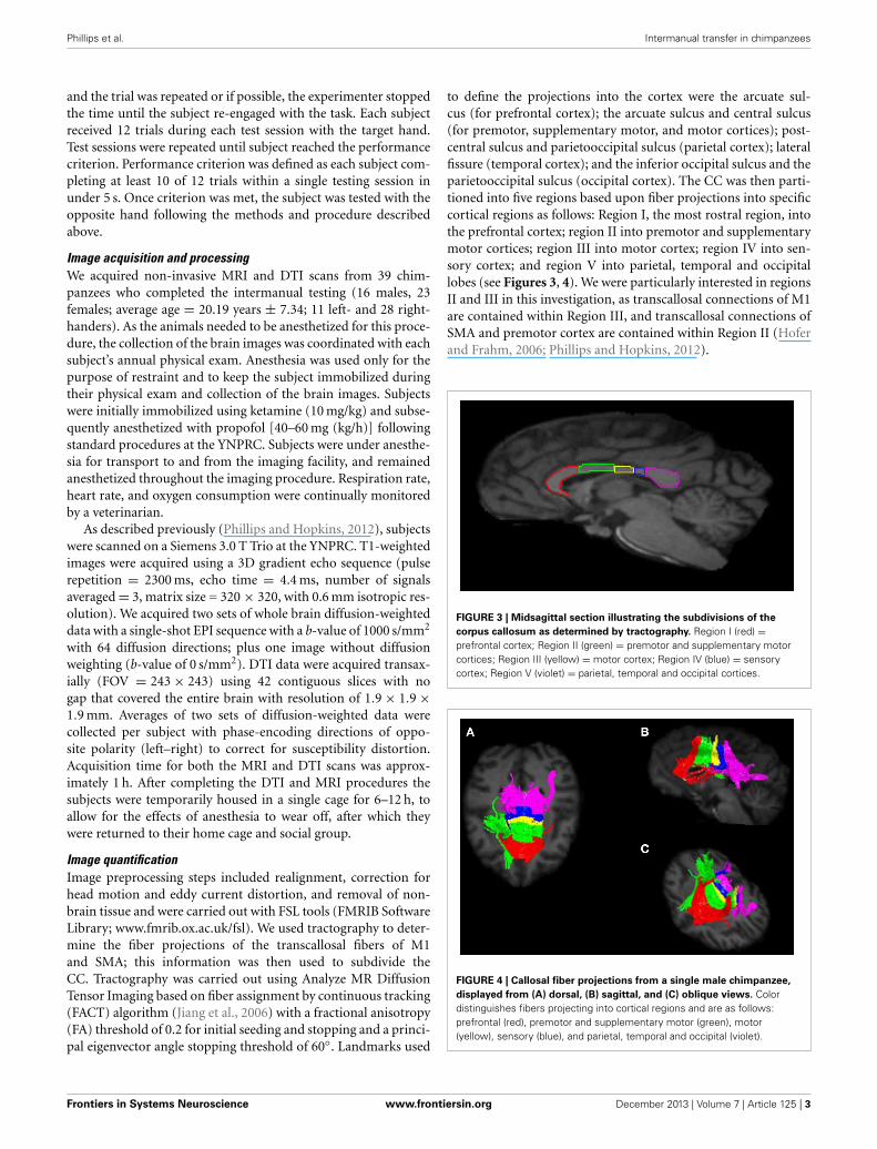
Image quantificationImage preprocessing steps included realignment, correction forhead motion and eddy current distortion, and removal of non-brain tissue and were carried out with FSL tools (FMRIB SoftwareLibrary; www.fmrib.ox.ac.uk/fsl). We used tractography to deter-mine the fiber projections of the transcallosal fibers of M1and SMA; this information was then used to subdivide theCC. Tractography was carried out using Analyze MR DiffusionTensor Imaging based on fiber assignment by continuous tracking(FACT) algorithm (Jiang et al., 2006) with a fractional anisotropy(FA) threshold of 0.2 for initial seeding and stopping and a princi-pal eigenvector angle stopping threshold of 60◦. Landmarks used
to define the projections into the cortex were the arcuate sul-cus (for prefrontal cortex); the arcuate sulcus and central sulcus(for premotor, supplementary motor, and motor cortices); post-central sulcus and parietooccipital sulcus (parietal cortex); lateralfissure (temporal cortex); and the inferior occipital sulcus and theparietooccipital sulcus (occipital cortex). The CC was then parti-tioned into five regions based upon fiber projections into specificcortical regions as follows: Region I, the most rostral region, intothe prefrontal cortex; region II into premotor and supplementarymotor cortices; region III into motor cortex; region IV into sen-sory cortex; and region V into parietal, temporal and occipitallobes (see Figures 3, 4). We were particularly interested in regionsII and III in this investigation, as transcallosal connections of M1are contained within Region III, and transcallosal connections ofSMA and premotor cortex are contained within Region II (Hoferand Frahm, 2006; Phillips and Hopkins, 2012).
FIGURE 3 | Midsagittal section illustrating the subdivisions of the
corpus callosum as determined by tractography. Region I (red) =prefrontal cortex; Region II (green) = premotor and supplementary motorcortices; Region III (yellow) = motor cortex; Region IV (blue) = sensorycortex; Region V (violet) = parietal, temporal and occipital cortices.
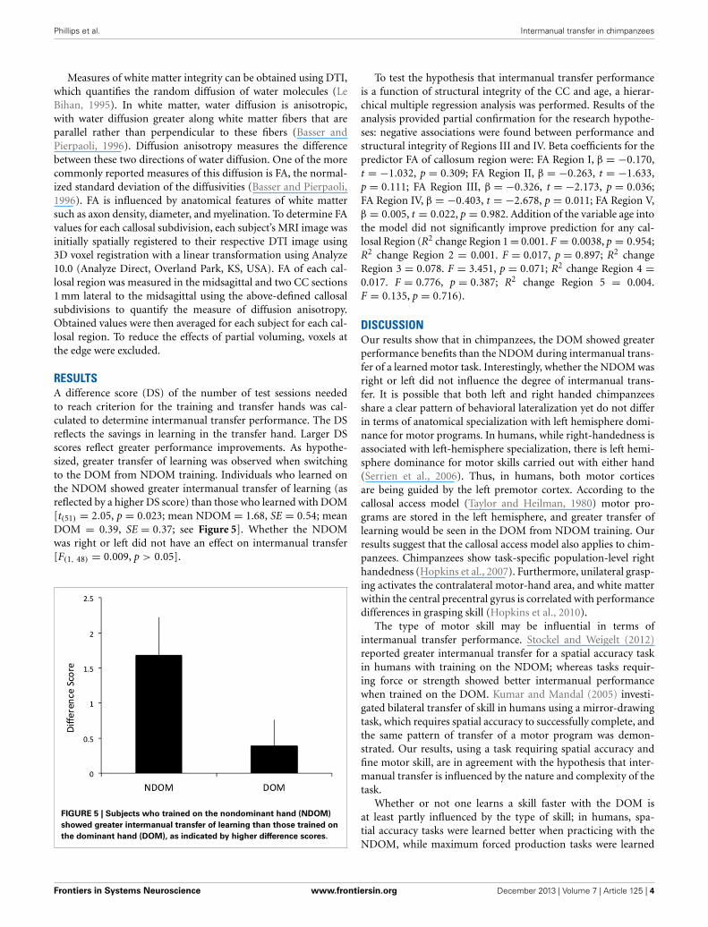
FIGURE 4 | Callosal fiber projections from a single male chimpanzee,
displayed from (A) dorsal, (B) sagittal, and (C) oblique views. Colordistinguishes fibers projecting into cortical regions and are as follows:prefrontal (red), premotor and supplementary motor (green), motor(yellow), sensory (blue), and parietal, temporal and occipital (violet).
Frontiers in Systems Neuroscience www.frontiersin.org December 2013 | Volume 7 | Article 125 | 3
Phillips et al. Intermanual transfer in chimpanzees
Measures of white matter integrity can be obtained using DTI,which quantifies the random diffusion of water molecules (LeBihan, 1995). In white matter, water diffusion is anisotropic,with water diffusion greater along white matter fibers that areparallel rather than perpendicular to these fibers (Basser andPierpaoli, 1996). Diffusion anisotropy measures the differencebetween these two directions of water diffusion. One of the morecommonly reported measures of this diffusion is FA, the normal-ized standard deviation of the diffusivities (Basser and Pierpaoli,1996). FA is influenced by anatomical features of white mattersuch as axon density, diameter, and myelination. To determine FAvalues for each callosal subdivision, each subject’s MRI image wasinitially spatially registered to their respective DTI image using3D voxel registration with a linear transformation using Analyze10.0 (Analyze Direct, Overland Park, KS, USA). FA of each cal-losal region was measured in the midsagittal and two CC sections1 mm lateral to the midsagittal using the above-defined callosalsubdivisions to quantify the measure of diffusion anisotropy.Obtained values were then averaged for each subject for each cal-losal region. To reduce the effects of partial voluming, voxels atthe edge were excluded.
RESULTSA difference score (DS) of the number of test sessions neededto reach criterion for the training and transfer hands was cal-culated to determine intermanual transfer performance. The DSreflects the savings in learning in the transfer hand. Larger DSscores reflect greater performance improvements. As hypothe-sized, greater transfer of learning was observed when switchingto the DOM from NDOM training. Individuals who learned onthe NDOM showed greater intermanual transfer of learning (asreflected by a higher DS score) than those who learned with DOM[t(51) = 2.05, p = 0.023; mean NDOM = 1.68, SE = 0.54; meanDOM = 0.39, SE = 0.37; see Figure 5]. Whether the NDOMwas right or left did not have an effect on intermanual transfer[F(1, 48) = 0.009, p > 0.05].
FIGURE 5 | Subjects who trained on the nondominant hand (NDOM)
showed greater intermanual transfer of learning than those trained on
the dominant hand (DOM), as indicated by higher difference scores.
To test the hypothesis that intermanual transfer performanceis a function of structural integrity of the CC and age, a hierar-chical multiple regression analysis was performed. Results of theanalysis provided partial confirmation for the research hypothe-ses: negative associations were found between performance andstructural integrity of Regions III and IV. Beta coefficients for thepredictor FA of callosum region were: FA Region I, β = −0.170,t = −1.032, p = 0.309; FA Region II, β = −0.263, t = −1.633,p = 0.111; FA Region III, β = −0.326, t = −2.173, p = 0.036;FA Region IV, β = −0.403, t = −2.678, p = 0.011; FA Region V,β = 0.005, t = 0.022, p = 0.982. Addition of the variable age intothe model did not significantly improve prediction for any cal-losal Region (R2 change Region 1 = 0.001. F = 0.0038, p = 0.954;R2 change Region 2 = 0.001. F = 0.017, p = 0.897; R2 changeRegion 3 = 0.078. F = 3.451, p = 0.071; R2 change Region 4 =0.017. F = 0.776, p = 0.387; R2 change Region 5 = 0.004.F = 0.135, p = 0.716).
DISCUSSIONOur results show that in chimpanzees, the DOM showed greaterperformance benefits than the NDOM during intermanual trans-fer of a learned motor task. Interestingly, whether the NDOM wasright or left did not influence the degree of intermanual trans-fer. It is possible that both left and right handed chimpanzeesshare a clear pattern of behavioral lateralization yet do not differin terms of anatomical specialization with left hemisphere domi-nance for motor programs. In humans, while right-handedness isassociated with left-hemisphere specialization, there is left hemi-sphere dominance for motor skills carried out with either hand(Serrien et al., 2006). Thus, in humans, both motor corticesare being guided by the left premotor cortex. According to thecallosal access model (Taylor and Heilman, 1980) motor pro-grams are stored in the left hemisphere, and greater transfer oflearning would be seen in the DOM from NDOM training. Ourresults suggest that the callosal access model also applies to chim-panzees. Chimpanzees show task-specific population-level righthandedness (Hopkins et al., 2007). Furthermore, unilateral grasp-ing activates the contralateral motor-hand area, and white matterwithin the central precentral gyrus is correlated with performancedifferences in grasping skill (Hopkins et al., 2010).
The type of motor skill may be influential in terms ofintermanual transfer performance. Stockel and Weigelt (2012)reported greater intermanual transfer for a spatial accuracy taskin humans with training on the NDOM; whereas tasks requir-ing force or strength showed better intermanual performancewhen trained on the DOM. Kumar and Mandal (2005) investi-gated bilateral transfer of skill in humans using a mirror-drawingtask, which requires spatial accuracy to successfully complete, andthe same pattern of transfer of a motor program was demon-strated. Our results, using a task requiring spatial accuracy andfine motor skill, are in agreement with the hypothesis that inter-manual transfer is influenced by the nature and complexity of thetask.
Whether or not one learns a skill faster with the DOM isat least partly influenced by the type of skill; in humans, spa-tial accuracy tasks were learned better when practicing with theNDOM, while maximum forced production tasks were learned
Frontiers in Systems Neuroscience www.frontiersin.org December 2013 | Volume 7 | Article 125 | 4
Phillips et al. Intermanual transfer in chimpanzees
better when practicing with the DOM Stockel and Weigelt (2012).As we did not test both types of switches or different types of tasksin the present study we cannot exclude this possibility.
The second aim of this study was to relate microstructuralintegrity of the CC to intermanual transfer. We hypothesized thatgreater intermanual transfer would be associated with increasedFA of the callosal regions containing transcallosal connectionsfor cortical areas previously identified as involved in interman-ual transfer, M1 and SMA. Instead, negative relationships betweenintermanual transfer performance and FA of callosal Regions IIIand IV were identified. Subjects who showed greater interman-ual transfer of a learned motor task had lower FA of the callosalregion containing fibers connecting motor and sensory regions.This would seem to indicate reduced structural integrity of theCC in those subjects who showed greater performance benefits.While no other relationships were significant, all showed a neg-ative association between FA and DS score. While this may seemcounterintuitive, studies relating callosal area, FA and functionin humans are inconsistent. Anisotropy measures of the poste-rior callosal region correlated positively with pronounced left-hemisphere language lateralization (Westerhausen et al., 2006).Westerhausen et al. suggested this indicated individuals showingextreme left lateralization of language had a greater number ofaxons and/or thicker myelin sheaths in posterior callosal regionsthan individuals who showed less language lateralization. In indi-viduals with autism, FA correlated positively with performanceIQ in the total CC and subregions (Alexander et al., 2007). Sistiet al. (2012), using a similar parcellation of the CC in humansbased on tractography, reported FA in callosal regions connectingprefrontal cortices predicted performance on the acquisition of anovel bimanual coordination task.
In a study most similar to the present study in terms ofmethodologies (Bonzano et al., 2008), a positive correlation wasfound between FA of the anterior CC and performance of abimanual task.
Another consideration concerns age differences in CC macro-and micro-structure that could influence intermanual transfer.Structural MRI has demonstrated callosal size changes duringdevelopment in chimpanzees (Hopkins and Phillips, 2010) andhumans (Allen et al., 1991; Pujol et al., 1993; Hasan et al., 2008).FA changes are related to these developmental patterns of callosalsize in humans (Lebel et al., 2012; Yap et al., 2013). These struc-tural changes of the CC across development influence behavior.For example, less lateralized task processing occurs in cognitiveand motor tasks, and is associated with reduced callosal area inolder adults (Muller-Oehring et al., 2007; Langan et al., 2010)While age was not significant to intermanual performance in thepresent study, other variables should be explored in lieu of orcombination with FA to improve the predictability of intermanualtransfer.
The microstructure of the transcallosal fibers of primarymotor cortices reflects the capacity for interhemispheric inhibi-tion (Wahl et al., 2007; Koerte et al., 2009). Wahl et al. reported apositive relationship between microstructural integrity (FA) andstrength of inhibition in adults. Koerte et al. found a similar rela-tionship across development (7–26 years), but Fling et al. (2013)suggest these data were largely driven by the child group. Further
analysis indicated that the observed relationship between FA andstrength of inhibition may actually show an opposite pattern inadults. Thus, adults with higher microstructural integrity mayhave reduced interhemispheric inhibition during volitional cor-tical activity. In support of this hypothesis, Fling et al. reporthigher CC microstructural integrity was associated with poorerperformance on bimanual tasks requiring a large degree of inter-hemispheric inhibition.
FA is undoubtedly influenced by multiple characteristics ofwater diffusion, including myelination, axon diameter and axondensity. Hopkins et al. (2012) examined axon density acrossregions of the CC in chimpanzees. The highest fiber densities(for both small and large myelinated axons) were reported in thegenu and splenium (the rostrum was not analyzed in this study),and there was no difference between genu and splenium in axondensity. No differences were found across the CC for density oflarge axonal fibers. Considering small axonal fibers, the highestdensity was in the genu. If FA primarily reflects axonal densityrather than axon diameter or myelination, as has been posited bysome [e.g., Wahl et al. (2007)], then FA of these callosal regionsin chimpanzees should show a similar pattern to the axon densitypattern reported by Hopkins et al. However, it does not. Phillipsand Hopkins (2012), in an examination of FA in chimpanzee cal-losal subdivisions, reported the posterior region of the CC (whichwould include the splenium as discussed above) had the highestFA value, and was significantly higher than the genu. Thus, FAand fiber density data do not precisely match.
In sum, chimpanzees showed greater performance benefitswhen switching to the DOM from NDOM during intermanualtransfer of a learned motor task, and intermanual transfer per-formance was associated with variation in white matter integrityof regions of the body of the CC that contain motor and sen-sory projections. We suggest that in chimpanzees, lower structuralintegrity in these regions of the CC indicates less interhemi-spheric inhibition, which leads to greater intermanual transfer oflearning.
AUTHOR CONTRIBUTIONSKimberley A. Phillips and William D. Hopkins designed the study.William D. Hopkins and Jennifer A. Schaeffer collected the data;Kimberley A. Phillips analyzed the data. Kimberley A. Phillips andWilliam D. Hopkins wrote the manuscript.
ACKNOWLEDGMENTSThis work was supported by the National Institute of NeurologicalDisorders and Stroke, grants NS070717 (to Kimberley A.Phillips), NS42867 and NS73134 (to William D. Hopkins). Thecontent is solely the responsibility of the authors and does notnecessarily represent the official views of the National Institute ofNeurological Disorders And Stroke or the National Institutes ofHealth.
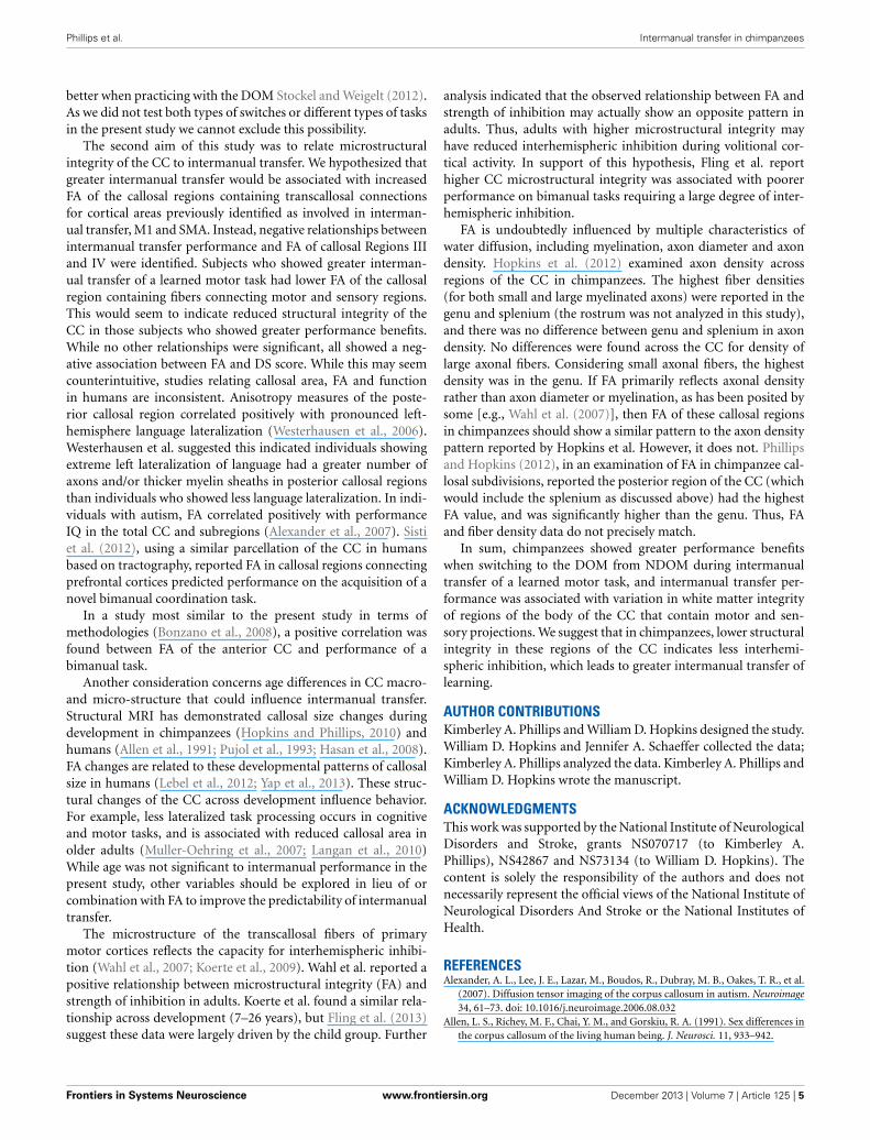
REFERENCESAlexander, A. L., Lee, J. E., Lazar, M., Boudos, R., Dubray, M. B., Oakes, T. R., et al.
(2007). Diffusion tensor imaging of the corpus callosum in autism. Neuroimage34, 61–73. doi: 10.1016/j.neuroimage.2006.08.032
Allen, L. S., Richey, M. F., Chai, Y. M., and Gorskiu, R. A. (1991). Sex differences inthe corpus callosum of the living human being. J. Neurosci. 11, 933–942.
Frontiers in Systems Neuroscience www.frontiersin.org December 2013 | Volume 7 | Article 125 | 5
Phillips et al. Intermanual transfer in chimpanzees
Basser, P. J., and Pierpaoli, C. (1996). Microstructural and physiological featuresof tissues elucidated by quantitative-diffusion-tensor MRI. J. Magn. Reson. 111,209–219. doi: 10.1006/jmrb.1996.0086
Bonzano, L., Tacchino, A., Roccatagliata, L., Abbruzzese, G., Mancardi, G. L.,and Bove, M. (2008). Callosal contributions to simultaneous bimanual fin-ger movements. J. Neurosci. 28, 3227–3233. doi: 10.1523/JNEUROSCI.4076-07.2008
Bonzano, L., Tacchino, A., Roccatagliata, L., Mancardi, G. L., Abbruzzese, G., andBove, M. (2011). Structural integrity of callosal midbody influences interman-ual transfer in a motor reaction-time task. Hum. Brain Mapp. 32, 218–228. doi:10.1002/hbm.21011
Fling, B. W., Benson, B. L., and Seidler, R. D. (2013). Transcallosal sensorimotorfiber tract structure-function relationships. Hum. Brain Mapp. 34, 384–395. doi:10.1002/hbm.21437
Grafton, S. T., Hazeltine, E., and Ivry, R. B. (2002). Motor sequence learning withthe nondominant left hand. A PET functional imaging study. Exp. Res. 146,369–378. doi: 10.1007/s00221-002-1181-y
Halsband, U. (1992). Left hemisphere preponderance in trajectorial learning.Neuroreport 3, 397–400. doi: 10.1097/00001756-199205000-00005
Hasan, K. M., Kamali, A., Kramer, L. A., Papnicolaou, A. C., Fletcher, J. M., andEwing-Cobbs, L. (2008). Diffusion tensor quantification of the human mid-sagittal corpus callosum subdivisions across the lifespan. Brain Res. 1227, 52–67.doi: 10.1016/j.brainres.2008.06.030
Hicks, R. E. (1974). Asymmetry of bilateral transfer. Am. J. psychol. 87, 667–674.doi: 10.2307/1421973
Hofer, S., and Frahm, J. (2006). Topography of the human corpus cal-losum revisited comprehensive fiber tractography using diffusiontensor magnetic resonance imaging. Neuroimage 32, 989–994. doi:10.1016/j.neuroimage.2006.05.044
Hopkins, W. D., Cantalupo, C., Wesley, M. J., Hostetter, A., and Pilcher, D. (2002).Grip morphology and hand use in chimpanzees (Pan troglodytes): evidence of aleft hemisphere specialization in motor skill. J. Exp. Psychol. Gen. 131, 412–423.doi: 10.1037/0096-3445.131.3.412
Hopkins, W. D., and Phillips, K. A. (2010). Cross-sectional analysis of the associ-ation between age and corpus callosum size in chimpanzees (Pan troglodytes).Dev. Psychobiol. 52, 133–141. doi: 10.1002/dev.20421
Hopkins, W. D., Pilger, J. F., Storz, R., Ambrose, A., Hof, P. R., and Sherwood, C.C. (2012). Planum temporale asymmetries correlate with corpus callosum axonfiber density in chimpanzees (Pan troglodytes). Behav. Brain Res. 234, 248–254.doi: 10.1016/j.bbr.2012.06.030
Hopkins, W. D., Russell, J., Lambeth, S., and Schapiro, S. (2007). “Handedness andneuroanatomical asymmetries in captive chimpanzees: a summary of 15 yearsof research,” in Evolution of Hemispheric Specialization in Primates, Vol. 5, ed W.D. Hopkins (Oxford: Elsevier), 146–181. doi: 10.1016/S1936-8526(07)05006-3
Hopkins, W. D., Taglialatela, J. P., Russell, J. L., Nir, T. M., and Schaeffer, J. (2010).Cortical representation of lateralized grasping in chimpanzees (Pan troglodytes):a combined MRI and PET study. PLoS ONE 5:e13383. doi: 10.1371/jour-nal.pone/0013383
Japikse, K. C., Negash, S., Howard, J. H. Jr., and Howard, D. V. (2003).Intermanual transfer of procedural learning after extended practice of proba-bilistic sequences. Exp. Brain Res. 148, 38–49. doi: 10.1007/s00221-002-1264-9
Jiang, H., van Zijl, P. C., Kim, J., Pearlson, G. D., and Mori, S. (2006).DtiStudio: resource program for diffusion tensor computation and fiberbundle tracking. Comput. Methods Programs Biomed. 81, 106–116. doi:10.1016/j.cmpb.2005.08.004
Koerte, I., Heinen, F., Fuchs, T., Laubender, R. P., Pomscar, A., Stahl, R., et al. (2009).Anisotropy of callosal motor fibers in combination with transcranial magneticstimulation in the course of motor development. Invest. Radiol. 44, 279–284.doi: 10.1097/RLI.0b013e31819e9362
Kumar, S., and Mandal, M. K. (2005). Bilateral transfer of skill in left- and right-handers. Laterality 10, 337–344. doi: 10.1080/13576500442000120
Langan, J., Peltier, S. J., Bo, J., Fling, B. W., Welsh, R. C., and Seidler, R. D. (2010).Functional implications of age differences in motor system connectivity. Front.Syst. Neurosci. 4:17. doi: 10.3389/fnsys.2010.00017
Laszlo, J. L., Baguley, R. A., and Bairstow, P. J. (1970). Bilateral transfer in tap-ping skill in the absence of peripheral information. J. Motor Behav. 2, 271. doi:10.1080/00222895.1970.10734884
Lebel, C., Gee, M., Camicioli, R., Wieler, M., Martin, W., and Beaulieu,C. (2012). Diffusion tensor imaging of white matter tract evolution
over the lifespan. Neuroimage 60, 340–352. doi: 10.1016/j.neuroimage.2011.11.094
Le Bihan, D. (1995). Diffusion and Perfusion MRI. New York, NY: Raven.Milisen, R., and Riper, C. V. (1939). Differential transfer of training in a rotary
activity. J. Exp. Psychol. 24, 640–646. doi: 10.1037/h0063064Muller-Oehring, E. M., Schulte, T., Raassi, C., Pfefferbaum, A., and Sullivan, E.
V. (2007). Local-global interference is modulated by age, sex and anteriorcorpus callosum size. Brain Res. 1142, 189–205. doi: 10.1016/j.brainres.2007.01.062
Padberg, J., Franca, J. G., DCooke, D. F., Soares, J. G. M., Rosa, M. G. P.,Fiorani, M. Jr., et al. (2007). Parallel evolution of cortical areas involved inskilled hand use. J. Neurosci. 27, 10106–10115. doi: 10.1523/JNEUROSCI.2632-07.2007
Parlow, S. E., and Kinsbourne, M. (1989). Asymmetric transfer of train-ing between hands: implications for interhemispheric communication inthe normal brain. Brain Cogn. 11, 98–113. doi: 10.1016/0278-2626(89)90008-0
Perez, M. A., Tanaka, S., Wise, S. P., Sadato, N., Tanabe, H. C., Willingham, D.T., et al. (2007). Neural substrates of intermanual transfer of a newly acquiredmotor skill. Curr. Biol. 17, 1896–1902. doi: 10.1016/j.cub.2007.09.058
Phillips, K. A., and Hopkins, W. D. (2012). Topography of the chimpanzee corpuscallosum. PLoS ONE 7:e31941. doi: 10.1371/journal.pone.0031941
Pujol, J., Vendrell, P., Junque, C., Marti-Vilalta, J. L., and Capdevila, A. (1993).When does human brain development end? Evidence of corpus callosumgrowth up to adulthood. Ann. Neurol. 34, 71–75. doi: 10.1002/ana.410340113
Serrien, D. J., Ivry, R. B., and Swinnen, S. P. (2006). Dynamics of hemispheric spe-cialization and integration in the context of motor control. Nat. Rev. Neurosci.7, 160–167. doi: 10.1038/nrn1849
Sisti, H. M., Geurts, M., Gooijers, J., Heitger, M. H., Caeyenberghs, K., Beets, I. A.M., et al. (2012). Microstructural organization of corpus callosum projections toprefrontal cortex predicts bimanual motor learning. Learn. Mem. 19, 351–357.doi: 10.1101/lm.026534.112
Stockel, T., and Weigelt, M. (2012). Brain lateralisation and motor learn-ing: selective effects of dominant and non-dominant hand practiceon the early acquisition of throwing skills. Laterality 17, 18–37. doi:10.1080/1357650X.2010.524222
Taylor, H. G., and Heilman, K. M. (1980). Left-hemisphere motor dominance inrighthanders. Cortex 16, 587–603. doi: 10.1016/S0010-9452(80)80006-2
Wahl, M., Lauterbach-Soon, B., Hattingen, E., Jung, P., Singer, O., Volz, S.,et al. (2007). Human motor corpus callosum: topography, somatography, andlink between microstructure and function. J. Neurosci. 27, 12132–12138. doi:10.1523/JNEUROSCI.2320-07.2007
Wahl, M., and Ziemann, U. (2008). The human motor corpus callosum. Rev.Neurosci. 19, 451–466. doi: 10.1515/REVNEURO.2008.19.6.451
Westerhausen, R., Kreuder, F., Sequeria, S. D. S., Walter, C., Woerner, W., Wittling,R. A., et al. (2006). The association of macro- and microstructure of the corpuscallosum and language. Brain Lang. 97, 80–90. doi: 10.1016/j.bandl.2005.07.133
Yap, Q. J., Teh, I., Fusar-Poli, P., Sum, M. Y., Kuswanto, C., andSim, K. (2013). Tracking cerebral white matter changes acrossthe lifespan: insights from diffusion tensor imaging studies.J. Neural Transm. 120, 1369–1395. doi: 10.1007/s00702-013-0971-7
Conflict of Interest Statement: The authors declare that the research was con-ducted in the absence of any commercial or financial relationships that could beconstrued as a potential conflict of interest.
Received: 19 August 2013; accepted: 15 December 2013; published online: 31 December2013.Citation: Phillips KA, Schaeffer JA and Hopkins WD (2013) Corpus callosalmicrostructure influences intermanual transfer in chimpanzees. Front. Syst. Neurosci.7:125. doi: 10.3389/fnsys.2013.00125This article was submitted to the journal Frontiers in Systems Neuroscience.Copyright © 2013 Phillips, Schaeffer and Hopkins. This is an open-access articledistributed under the terms of the Creative Commons Attribution License (CC BY).The use, distribution or reproduction in other forums is permitted, provided theoriginal author(s) or licensor are credited and that the original publication in thisjournal is cited, in accordance with accepted academic practice. No use, distribution orreproduction is permitted which does not comply with these terms.
Frontiers in Systems Neuroscience www.frontiersin.org December 2013 | Volume 7 | Article 125 | 6
Related Documents