Cooking enhances curcumin anti-cancerogenic activity through pyrolytic formation of ‘‘deketene curcumin’’ Indra N. Dahmke a,e , Stefan P. Boettcher b , Matthias Groh c , Ulrich Mahlknecht d,e,⇑ a Institute of Clinical and Experimental Surgery, Saarland University, Homburg/Saar, Germany b Institute of Pharmaceutical and Medicinal Chemistry, Saarland University, Saarbrucken, Germany c Helmholtz-Institute of Pharmaceutical Research Saarland, Department of Drug Design and Optimization, Saarland University, Saarbrucken, Germany d Department of Internal Medicine – Haematology/Oncology, St. Lukas Clinic, D-42697 Solingen, Germany e Department of Internal Medicine, Division of Immunotherapy and Gene Therapy, Saarland University Medical Center, Homburg/Saar, Germany article info Article history: Received 29 July 2013 Received in revised form 9 October 2013 Accepted 19 November 2013 Available online 27 November 2013 Keywords: Curcumin Pyrolysis Bioavailability HPLC Anti-cancerogenic G2-arrest abstract Curcumin is widely used in traditional Asian kitchen as a cooking ingredient. Despite its low bioavailabil- ity, epidemiological data, on low cancer incidence in Asia, suggest beneficial health effects of this compound. Therefore, the question arose whether cooking modifies the anti-cancerogenic effects of curcumin. To evaluate this, we pyrolysed curcumin with and without coconut fat or olive oil, and ana- lysed the products by high-performance liquid chromatography (HPLC). A number of more hydrophilic curcumin isoforms and decomposition products, including a compound later identified by nuclear mag- netic resonance spectroscopy (NMR) as ‘‘deketene curcumin’’ (1,5-bis(4-hydroxy-3-methoxyphenyl)-1,4- pentadiene-3-one), formerly described as a synthetic curcumin derivative, were detected. Additionally, we proved that deketene curcumin, compared to curcumin, exhibits higher toxicity on B78H1 melanoma cells resulting in G2 arrest. In conclusion, deketene curcumin is formed as a consequence of pyrolysis during common household cooking, showing stronger anti-cancer effects than curcumin. Moreover, we propose a chemical reaction-pathway for this process. Ó 2013 Elsevier Ltd. All rights reserved. 1. Introduction Curcumin (1,7-bis(4-hydroxy-3-methoxyphenyl)-1,6-heptadi- ene-3,5-dione), which is the main phenolic component found in turmeric prepared from the rhizome of Curcuma longa, is long known for its anti-inflammatory, anti-oxidant and anti-cancero- genic properties (Shehzad, Lee, & Lee, 2013). Based on epidemio- logical data, from the WHO, curcumin, as part of the traditional Indian diet, is associated with low incidence rates on colorectal, prostate and lung cancers in India (Mohandas, 2011; Sinha, Ander- son, McDonald, & Greenwald, 2003). The low bioavailability of or- ally administered curcumin, which is described by numerous investigators, as well as the conversion into less active metabolites, is contradictory to this assumption (Cheng et al., 2001; Garcea et al., 2005; Sharma et al., 2001). Moreover, curcumin shows low buffer and plasma stability (Griesser et al., 2011; Tonnesen & Karl- sen, 1985; Wang et al., 1997). Hence, we questioned whether the pre-consumptional processing, as performed in typical Indian cooking, changes the quality or bioavailability of curcumin. To address this question, we pyrolysed curcumin, with and without coconut fat, which is traditionally used on the Indian subcontinent, or olive oil, as in modern style western cooking, and analysed the products in reverse phase high-performance liquid chromatogra- phy (HPLC). Although we found the major part of curcumin un- changed, a number of more hydrophilic products were observed, including isoforms of curcumin derivatives, known degradation products, as well as an unknown product. This product was isolated by preparative HPLC and identified by nuclear magnetic resonance (NMR) spectroscopy as 1,5-bis(4-hydroxy-3-methoxy- phenyl)-1,4-pentadiene-3-one), which has already been described as a potent synthetic curcumin derivative (Liang et al., 2009; Quincoces Suarez et al., 2010). We then evaluated the activity of this derivative, named ‘‘deketene curcumin’’ (DKC) and compared it to curcumin and pyrolysed curcumin, in different in vitro exper- iments. In conclusion, DKC showed significantly stronger cytotoxic activity on murine B78H1 melanoma cells when compared to the reactant. Therefore, we argue that traditional pre-consumptional processing of curcumin strongly enhances its bioactivity thus potentiating its beneficial effects. Furthermore, we propose a pref- erential reaction-pathway for the pyrolytic formation of DKC based on the identification of certain intermediates. 0308-8146/$ - see front matter Ó 2013 Elsevier Ltd. All rights reserved. http://dx.doi.org/10.1016/j.foodchem.2013.11.102 ⇑ Corresponding author at: Department of Internal Medicine – Haematology/ Oncology, St. Lukas Clinic, D-42697 Solingen, Germany. Tel.: +49 212 705 2150; fax: +49 212 705 2152. E-mail address: [email protected] (U. Mahlknecht). Food Chemistry 151 (2014) 514–519 Contents lists available at ScienceDirect Food Chemistry journal homepage: www.elsevier.com/locate/foodchem

Welcome message from author
This document is posted to help you gain knowledge. Please leave a comment to let me know what you think about it! Share it to your friends and learn new things together.
Transcript
Food Chemistry 151 (2014) 514–519
Contents lists available at ScienceDirect
Food Chemistry
journal homepage: www.elsevier .com/locate / foodchem
Cooking enhances curcumin anti-cancerogenic activity through pyrolyticformation of ‘‘deketene curcumin’’
0308-8146/$ - see front matter � 2013 Elsevier Ltd. All rights reserved.http://dx.doi.org/10.1016/j.foodchem.2013.11.102
⇑ Corresponding author at: Department of Internal Medicine – Haematology/Oncology, St. Lukas Clinic, D-42697 Solingen, Germany. Tel.: +49 212 705 2150; fax:+49 212 705 2152.
E-mail address: [email protected] (U. Mahlknecht).
Indra N. Dahmke a,e, Stefan P. Boettcher b, Matthias Groh c, Ulrich Mahlknecht d,e,⇑a Institute of Clinical and Experimental Surgery, Saarland University, Homburg/Saar, Germanyb Institute of Pharmaceutical and Medicinal Chemistry, Saarland University, Saarbrucken, Germanyc Helmholtz-Institute of Pharmaceutical Research Saarland, Department of Drug Design and Optimization, Saarland University, Saarbrucken, Germanyd Department of Internal Medicine – Haematology/Oncology, St. Lukas Clinic, D-42697 Solingen, Germanye Department of Internal Medicine, Division of Immunotherapy and Gene Therapy, Saarland University Medical Center, Homburg/Saar, Germany
a r t i c l e i n f o
Article history:Received 29 July 2013Received in revised form 9 October 2013Accepted 19 November 2013Available online 27 November 2013
Keywords:CurcuminPyrolysisBioavailabilityHPLCAnti-cancerogenicG2-arrest
a b s t r a c t
Curcumin is widely used in traditional Asian kitchen as a cooking ingredient. Despite its low bioavailabil-ity, epidemiological data, on low cancer incidence in Asia, suggest beneficial health effects of thiscompound. Therefore, the question arose whether cooking modifies the anti-cancerogenic effects ofcurcumin. To evaluate this, we pyrolysed curcumin with and without coconut fat or olive oil, and ana-lysed the products by high-performance liquid chromatography (HPLC). A number of more hydrophiliccurcumin isoforms and decomposition products, including a compound later identified by nuclear mag-netic resonance spectroscopy (NMR) as ‘‘deketene curcumin’’ (1,5-bis(4-hydroxy-3-methoxyphenyl)-1,4-pentadiene-3-one), formerly described as a synthetic curcumin derivative, were detected. Additionally,we proved that deketene curcumin, compared to curcumin, exhibits higher toxicity on B78H1 melanomacells resulting in G2 arrest. In conclusion, deketene curcumin is formed as a consequence of pyrolysisduring common household cooking, showing stronger anti-cancer effects than curcumin. Moreover, wepropose a chemical reaction-pathway for this process.
� 2013 Elsevier Ltd. All rights reserved.
1. Introduction
Curcumin (1,7-bis(4-hydroxy-3-methoxyphenyl)-1,6-heptadi-ene-3,5-dione), which is the main phenolic component found inturmeric prepared from the rhizome of Curcuma longa, is longknown for its anti-inflammatory, anti-oxidant and anti-cancero-genic properties (Shehzad, Lee, & Lee, 2013). Based on epidemio-logical data, from the WHO, curcumin, as part of the traditionalIndian diet, is associated with low incidence rates on colorectal,prostate and lung cancers in India (Mohandas, 2011; Sinha, Ander-son, McDonald, & Greenwald, 2003). The low bioavailability of or-ally administered curcumin, which is described by numerousinvestigators, as well as the conversion into less active metabolites,is contradictory to this assumption (Cheng et al., 2001; Garceaet al., 2005; Sharma et al., 2001). Moreover, curcumin shows lowbuffer and plasma stability (Griesser et al., 2011; Tonnesen & Karl-sen, 1985; Wang et al., 1997). Hence, we questioned whether thepre-consumptional processing, as performed in typical Indian
cooking, changes the quality or bioavailability of curcumin. Toaddress this question, we pyrolysed curcumin, with and withoutcoconut fat, which is traditionally used on the Indian subcontinent,or olive oil, as in modern style western cooking, and analysed theproducts in reverse phase high-performance liquid chromatogra-phy (HPLC). Although we found the major part of curcumin un-changed, a number of more hydrophilic products were observed,including isoforms of curcumin derivatives, known degradationproducts, as well as an unknown product. This product wasisolated by preparative HPLC and identified by nuclear magneticresonance (NMR) spectroscopy as 1,5-bis(4-hydroxy-3-methoxy-phenyl)-1,4-pentadiene-3-one), which has already been describedas a potent synthetic curcumin derivative (Liang et al., 2009;Quincoces Suarez et al., 2010). We then evaluated the activity ofthis derivative, named ‘‘deketene curcumin’’ (DKC) and comparedit to curcumin and pyrolysed curcumin, in different in vitro exper-iments. In conclusion, DKC showed significantly stronger cytotoxicactivity on murine B78H1 melanoma cells when compared to thereactant. Therefore, we argue that traditional pre-consumptionalprocessing of curcumin strongly enhances its bioactivity thuspotentiating its beneficial effects. Furthermore, we propose a pref-erential reaction-pathway for the pyrolytic formation of DKC basedon the identification of certain intermediates.
I.N. Dahmke et al. / Food Chemistry 151 (2014) 514–519 515
2. Materials and methods
2.1. Chemicals
Curcumin that was used for pyrolysis was purchased from Sig-ma Aldrich (Deisenhofen, Germany). Curcumin employed for thepreparative HPLC was obtained from Sabinsa Cooperation (EastWindsor, USA). Both samples contained about 80% curcumin(CUR), 17% demethoxycurcumin (DMC) and 3% bisdemethoxycur-cumin (BDMC). All chemicals were purchased from Sigma Aldrich(Deisenhofen) if not indicated otherwise.
2.2. Pyrolysis procedure
5.0 mg of curcumin was filled into a glass tube (£ 20 mm,height 40 mm) for each single sample. For the fat treated samples,50.0 mg of the particular fat was added. The samples were subse-quently heated to 250 �C for 20 min, using a hot plate. For theisolation of DKC 100 mg curcumin were heated to the same tem-perature for 120 min in a crystallizing dish.
2.3. Instruments and reagents
The quantitation of the pyrolysis products was carried out usinga ThermoFisher SpectraSystem HPLC–UV–MS (Thermo Fisher Scien-tific, Waltham), equipped with a degasser, a quaternary pump, anautosampler, a MWD (254 and 278 nm) and a MSQ ESI mass spec-trometer in positive mode (source temperature 350 �C, capillaryvoltage 3.9 kV, nitrogen sheath gas pressure 4.0 � 105 Pa, sheathgas flow: 58 ml/min according to descriptions of the manufacturer,auxiliary gas flow: 29 ml/min). Xcalibur software was deployed fordata acquisition and plotting. The isolation of DKC, from thepyrolysis mixture, was performed using a Waters AutopurificationHPLC–DAD–MS. For the chromatographic methods bidistilled water(Elix� water, pure, Millipore Corporation, Billerica) was used asaquatic phase. All solvents used for HPLC were of chromatographicgrade.
NMR spectra were recorded on a Bruker Fourier 300 (BrukerCorporation, Billerica) (1H, 300 MHz; 13C, 75 MHz) spectrometerat 300 K. Chemical shifts are recorded as d values in ppm unitsby reference to the hydrogenated residues of deuterated solventas internal standard (DMSO-d6: d = 2.50, 39.99). Splitting patternsdescribe apparent multiplicities and are designated as s (singlet),br s (broad singlet), d (doublet), dd (doublet of doublet). Couplingconstants (J) are given in Hertz (Hz).
2.3.1. LC–UV–MSThe pyrolysis residues were diluted in 1 ml acetone. All samples
were injected by an autosampler (Surveyor�, Finnigan™, ThermoFisher Scientific) with an injection volume of 25 ll. A RP C18NUCLEODUR� 100–5 (125 � 3 mm) column (Macherey–NagelGmbH, Düren) was used as the stationary phase. The solvent sys-tem consisted of water (A) and acetonitrile (B), each containing0.1% trifluoroacetic acid (TVA) (v/v).
The flow rate was set to 800 ll/min. The percentage of B startedat an initial of 5%, was increased to 100% during 16 min, kept at100% for 2 min and flushed back to the 5% in 2 min.
2.3.2. Preparative HPLC–MSIn 30 single runs each time 1 ml of methanol, containing
3–4 mg of pyrolysis residue was injected and separated on a C-18 Waters X-Bridge OBD 19 � 150 mm, 5 lM column. The solventsystem consisted of water (A) and methanol (B), each containing0.1% TFA (v/v).
The flow rate was set to 20 ml/min. The percentage of B startedat an initial of 10%, was increased up to 95% during 7 min, kept atthat percentage for 1 min and flushed back to 10% in 1 min. Thefraction collector was triggered by the mass spectrometer in SIMmode, collecting the m/z 327.2 with a width of 1.0 m/z.
2.4. Cell culture
Murine amelanotic B78H1 melanoma cells, employed for thein vitro experiments, were a kind gift from the Laboratory of Immu-nology and Biology of Metastasis of the Department of Experimen-tal Pathology at the University of Bologna (Lollini et al., 1987). TheB78H1 cells were cultured in Dulbecco’s modified Eagle’s medium(DMEM; PAA, Cölbe) supplemented with 10% fetal calf serum (FCS),100 U/ml penicillin and 0.1 mg/ml streptomycin (PAA) at 37 �C in ahumidified atmosphere containing 5% CO2. They were grown to80–90% confluence and subjected to no more than five cell pas-sages after cryostorage.
2.5. Fluorescence microscopy of cells
For qualitative analysis of the incorporation of curcumin deriv-atives by B78H1 cells 1 � 105 cells were seeded per well into a24-well plate. After 24 h, cells were incubated either with 20 lMof curcumin derivatives or DMSO at 37 �C and 5% CO2 and micro-scopic pictures were taken after 1 and 24 h with the BIOREVOBZ-8000 (Keyence, Osaka). The auto-fluorescent curcumin deriva-tives incorporated by the cells were detectable in the GFP fluores-cence channel. All substances were tested in triplicate.
2.6. Water-soluble tetrazolium (WST)-1 assay
To assess the effect of curcumin derivatives on the viability ofB78H1 cells, a WST-1 assay (Roche Diagnostics, Mannheim) wasperformed according to the manufacturer’s instructions. Briefly,5 � 103 cells were seeded into 96-well plates, and were treatedeither with vehicle (DMSO) or serial dilutions of curcumin deriva-tives (Santa Cruz Biotechnology, Heidelberg). All derivatives weretested in quadruplicate. After 24 h, 10 ll of WST-1 reagent per100 ll medium was added to each well. After 30 min incubationat 37 �C, the absorbance of each well was measured at a wave-length of 450 nm (reference at 620 nm) and corrected againstblanks (medium ± curcumin derivatives without cells).
2.7. Analysis of apoptotic cells by flow cytometry
The number of apoptotic versus necrotic cells was assessed byflow cytometry. In short, B78H1 cells (3 � 106 cells per well) wereseeded into a 12-well plate and allowed to adhere overnight. Cellswere incubated with either serial dilutions of curcumin derivativesor vehicle for 24 h at 37 �C, 5% CO2. They were cropped, washedtwice with PBS, transferred to 5 ml round bottom polystyrenetubes (BD Falcon, REF. 352054) and incubated for 15 min at roomtemperature in the dark with APC-Annexin V (1:24 in AnnexinBinding Buffer, Immunotools, Friesoythe). Immediately prior toanalysis on a FACSCanto™ (BD Biosciences, San Jose) 2 ll propidi-um iodide [1 mg/ml] was added to each tube. Three independentexperiments were performed; each sample was analysed induplicate.
2.8. Cell cycle analysis
B78H1 were plated as described above and incubated in thepresence of 20 lM curcumin derivatives or DMSO for 24 h at37 �C and 5% CO2. Subsequently, cells were cropped, washed twicewith PBS and resuspended in 200 ll NaCl (0.9%). With the help of a
516 I.N. Dahmke et al. / Food Chemistry 151 (2014) 514–519
1 ml syringe and a 25 gauge needle the resuspended cells weresquirted into 1800 ll 100% methanol and stored at �20 �Covernight. Cells were shortly thawed, centrifuged and washed withPBS. Supernatant was carefully removed, cells were resuspended inPBS with RNAse A (1:400, 10 mg/ml stock solution; Macherey–Na-gel GmbH, Düren), transferred to 5 ml round bottom polystyrenetubes and incubated for 1 h at room temperature. Afterwards, anequal volume of PBS containing propidium iodide (1:100, 1 mg/ml stock solution) was added to the suspension and incubatedfor 1 h, at room temperature in the dark. Samples were analysedon a FACSCanto™. Three independent experiments wereperformed; each sample was analysed in duplicate.
2.9. Statistical analyses
All values are shown as mean ± standard error of the mean(SEM). Data was first analysed for normal distribution and equalvariance. Differences between the two experimental groups werecalculated by unpaired Student’s t-test (SigmaStat, Jandel Corpora-tion, San Rafael). A value of P < 0.05 was considered statisticallysignificant.
3. Results and discussion
3.1. Pyrolysis with a fatty matrix changes composition of products
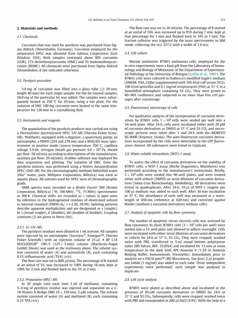
After mixing curcumin with fatty acids a reduction of spontane-ously formed hydrophilic isoforms was detected (Fig. 1). Afterpyrolysis of curcumin, we measured an increase of more hydro-philic CUR-, BMC- and BDMC-isoforms, both with and without afatty matrix. By performing curcumin pyrolysis, without coconutfat or olive oil, the formation of curcumin- and dehydrocurcu-min-dimers with a higher retention time (RT) compared to curcu-min were observed. Furthermore, we discovered a yet unidentifiedproduct at RT 9.6 min with a molecular weight of 326 g mol�1 (m/z: 327). Since a high deviation among the same preparations wasnoticed, probably due to unequal heating processes, this analysiswas primarily of qualitative nature.
In 1986 Nagabhushan and Bhide demonstrated a dose-depen-dent reduction of chilli induced mutagenicity by pyrolysed curcu-min (Nagabhushan & Bhide, 1986). It was also shown that heating
Fig. 1. Pyrolysis of curcumin leads to formation of hydrophilic products. Products of pyrreverse phase HPLC. The content of products is displayed in single ion monitoring areapyrolysis. Compared to unpyrolysed controls the content of isoforms of curcumin derivatcompared to curcumin was considerably higher in pyrolysed samples. In the pyrolysed cnew product deketene curcumin (DKC) at RT 9.6 min with a molecular weight of 326 g mthe molecular weight (g mol�1) as well as the RT (min) is included. Curcumin (Cur), bisd
curcumin for 15 min in water increased its solubility and alsopharmacological activity for example on toxic lipid peroxidationproducts or human auto-antibodies in vitro (Kurien, D’Souza, &Scofield, 2010; Kurien, Singh, Matsumoto, & Scofield, 2007). Inour HPLC analysis unmodified CUR showed a retention time of11.7 min. Roughly, about 30% of CUR was converted by thepyrolytic process into more hydrophilic products with a shorterretention time compared to the educts.
The formation of CUR dimers under slightly more radical condi-tions (1 h heating at 70 �C with 2,20-azobis(isobutyronitrile)) wasdescribed by Masuda et al. (1999), and the authors suggest thedimers importance for the antioxidant activity of CUR (Masudaet al., 1999).
3.2. Identification of the pyrolysis product with RT 9.6 and m/z 327 byNMR spectroscopy
The pyrolysis product of interest was isolated in sufficientquantity by the use of preparative HPLC as described above andyielded 4.2 mg. Based on the molecular structure and atomiccomposition mass of curcumin, the substance could be identifiedby NMR spectroscopy as 1,5-bis(4-hydroxy-3-methoxyphenyl)-1,4-pentadiene-3-one), named ‘‘deketene curcumin’’ (DKC). TheNMR data showed good accordance to a formerly published refer-ence spectrum (Masuda, Jitoe, Isobe, Nakatani, & Yonemori,1993).
1H NMR (300 MHz, DMSO-d6): d = 9.63 (br s, 2 H), 7.65 (d,J = 15.8 Hz, 2 H), 7.37 (d, J = 1.7 Hz, 2 H), 7.20 (dd, J = 8.1, 1.7 Hz,2 H), 7.15 (d, J = 15.8 Hz, 2 H), 6.84 (d, J = 8.1 Hz, 2 H), 3.85 (s, 6H) ppm. 13C NMR (75 MHz, DMSO-d6): d = 188.0, 149.4, 148.0,142.8, 126.4, 123.3, 123.0, 115.7, 111.4, 55.7 ppm.
3.3. Reaction-pathway for the pyrolytic formation of deketenecurcumin (DKC)
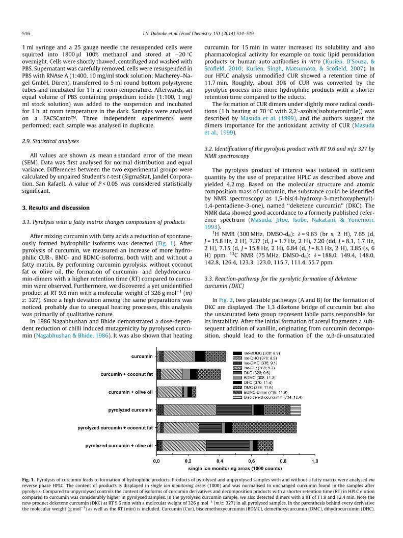
In Fig. 2, two plausible pathways (A and B) for the formation ofDKC are displayed. The 1,3 diketone bridge of curcumin but alsothe unsaturated keto group represent labile parts responsible forits instability. After the initial formation of acetyl fragments a sub-sequent addition of vanillin, originating from curcumin decompo-sition, should lead to the formation of the a,b-di-unsaturated
olysed and unpyrolysed samples with and without a fatty matrix were analysed vias (1000) and was normalised to unchanged curcumin found in the samples afterives and decomposition products with a shorter retention time (RT) in HPLC elutionurcumin sample, we also detected dimers with a RT of 11.9 and 12.4 min. Note theol�1 (m/z: 327) in all pyrolysed samples. In the parenthesis behind every derivativeemethoxycurcumin (BDMC), demethoxycurcumin (DMC), dihydrocurcumin (DHC).
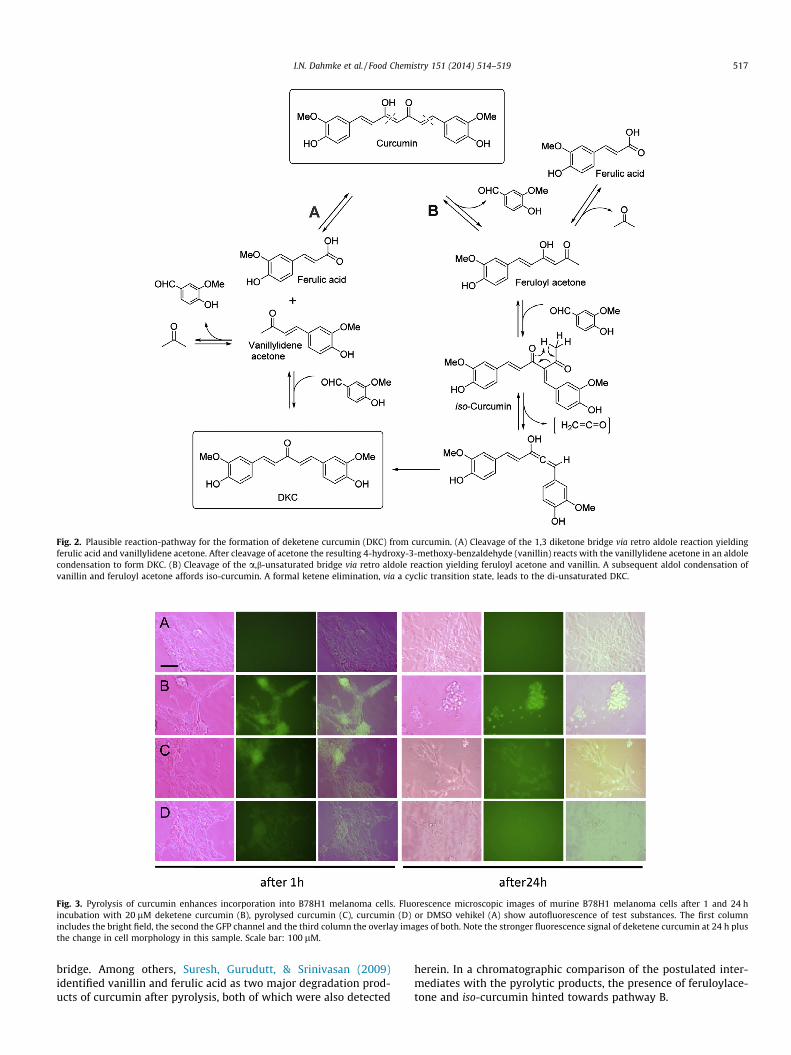
Fig. 2. Plausible reaction-pathway for the formation of deketene curcumin (DKC) from curcumin. (A) Cleavage of the 1,3 diketone bridge via retro aldole reaction yieldingferulic acid and vanillylidene acetone. After cleavage of acetone the resulting 4-hydroxy-3-methoxy-benzaldehyde (vanillin) reacts with the vanillylidene acetone in an aldolecondensation to form DKC. (B) Cleavage of the a,b-unsaturated bridge via retro aldole reaction yielding feruloyl acetone and vanillin. A subsequent aldol condensation ofvanillin and feruloyl acetone affords iso-curcumin. A formal ketene elimination, via a cyclic transition state, leads to the di-unsaturated DKC.
Fig. 3. Pyrolysis of curcumin enhances incorporation into B78H1 melanoma cells. Fluorescence microscopic images of murine B78H1 melanoma cells after 1 and 24 hincubation with 20 lM deketene curcumin (B), pyrolysed curcumin (C), curcumin (D) or DMSO vehikel (A) show autofluorescence of test substances. The first columnincludes the bright field, the second the GFP channel and the third column the overlay images of both. Note the stronger fluorescence signal of deketene curcumin at 24 h plusthe change in cell morphology in this sample. Scale bar: 100 lM.
I.N. Dahmke et al. / Food Chemistry 151 (2014) 514–519 517
bridge. Among others, Suresh, Gurudutt, & Srinivasan (2009)identified vanillin and ferulic acid as two major degradation prod-ucts of curcumin after pyrolysis, both of which were also detected
herein. In a chromatographic comparison of the postulated inter-mediates with the pyrolytic products, the presence of feruloylace-tone and iso-curcumin hinted towards pathway B.
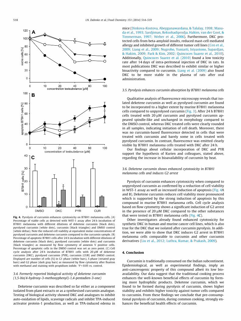
Fig. 4. Pyrolysis of curcumin enhances cytotoxicity on B78H1 melanoma cells. (A)Percentage of viable cells as detected with WST-1 assay after 24 h incubation ofB78H1 melanoma with different dilutions of deketene curcumin (black dots),pyrolysed curcumin (white dots), curcumin (black triangles) and DMSO control(white deltas). Note the reduced cell viability at equivalent molar concentrations ofpyrolysed curcumin and deketene curcumin compared to the curcumin sample. (B)Percentage of apoptotic B78H1 cells after 24 h incubation with different dilutions ofdeketene curcumin (black dots), pyrolysed curcumin (white dots) and curcumin(black triangles) as measured by flow cytometry of annexin V positive cells.Percentage of apoptotic cells in the DMSO control was set as zero point. (C) Cellcycle analysis after 24 h incubation of B78H1 cells with 20 lM of deketenecurcumin (DKC), pyrolysed curcumin (PYR), curcumin (CUR) and DMSO control.Displayed are number of cells [%] in G1 phase (white bars), S phase (striated graybars) and G2 phase (dark gray bars) as measured by flow cytometry after fixationwith methanol and staining with propidium iodide. ⁄P < 0.05 vs. control.
518 I.N. Dahmke et al. / Food Chemistry 151 (2014) 514–519
3.4. Formerly reported biological activity of deketene curcumin(1,5-bis(4-hydroxy-3-methoxyphenyl)-1,4-pentadien-3-one)
Deketene curcumin was described so far either as a componentisolated from plant extracts or as a synthesised curcumin analogue.Testing of biological activity showed DKC and analogues to inhibitauto-oxidation of lipids, scavenge radicals and inhibit TPA-inducedactivator protein-1 production, as well as TPA-induced edema in
mice (Dinkova-Kostova, Abeygunawardana, & Talalay, 1998; Masu-da et al., 1993; Sardjiman, Reksohadiprodja, Hakim, van der Goot, &Timmerman, 1997; Weber et al., 2006). Furthermore, DKC pro-tected cells from beta-amyloid insults, reduced mast-cell mediatedallergy and inhibited growth of different tumor cell lines (Cen et al.,2009; Liang et al., 2009; Nugroho, Yuniarti, Istyastono, Supardjan,& Hakim, 2009; Park & Kim, 2002; Quincoces Suarez et al., 2010).Additionally, Quincoces Suarez et al. (2010) found a low toxicityrate after 14 days of intra-peritoneal injection of DKC in rats. Inmost publications DKC was described to exhibit similar or higherbioactivity compared to curcumin. Liang et al. (2009) also foundDKC to be more stable in the plasma of rats after oraladministration.
3.5. Pyrolysis enhances curcumin absorption by B78H1 melanoma cells
Qualitative analysis of fluorescence microscopy reveals that iso-lated deketene curcumin as well as pyrolysed curcumin are foundto be incorporated to a higher extent by murine B78H1 melanomacells compared to unpyrolysed curcumin (Fig. 3). After 24 h B78H1cells treated with 20 lM curcumin and pyrolysed curcumin ap-peared spindle-like and unchanged in morphology compared tothe DMSO control, whereas DKC treated cells were clearly roundedin all samples, indicating initiation of cell death. Moreover, therewas no curcumin-based fluorescence detected in cells that weretreated with curcumin and barely some in cells treated withpyrolysed curcumin. In contrast, fluorescence was emitted clearlyvisible by B78H1 melanoma cells treated with DKC after 24 h.
Our findings about cellular incorporation of DKC and PYRsupport the hypothesis of Kurien and colleagues, stated above,regarding the increase in bioavailability of curcumin by heat.
3.6. Deketene curcumin shows enhanced cytotoxicity in B78H1melanoma cells and induces G2 arrest
Pyrolysis of curcumin enhances cytotoxicity when compared tounpyrolysed curcumin as confirmed by a reduction of cell viabilityin WST-1 assay as well as increased induction of apoptosis (Fig. 4Aand B). Deketene curcumin reduces cell viability more pronouncedwhich is supported by the strong induction of apoptosis by thiscompound in murine B78H1 melanoma cells. Cell cycle analysisbased on flow cytometry shows a significant induction of G2 arrestin the presence of 20 lM DKC compared to the other substancesthat were tested in B78H1 melanoma cells (Fig. 4C).
Other investigators already found enhanced cytotoxicity forsynthetic DKC in human and murine cancer cell lines, which is alsotrue for the DKC that we isolated after curcumin pyrolysis. In addi-tion, we were able to show that DKC induces G2 arrest in B78H1melanoma cells comparable to curcumin and other curcuminderivatives (Liu et al., 2012; Luthra, Kumar, & Prakash, 2009).
4. Conclusion
Curcumin is traditionally consumed on the Indian subcontinent.Epidemiological, as well as experimental findings, imply ananti-cancerogenic property of this compound albeit its low bio-availability. Our data suggest that the traditional cooking processenhances the well-known beneficial effects of curcumin by form-ing more hydrophilic products. Deketene curcumin, which wefound to be formed during pyrolysis of curcumin, shows higherstability and exhibits higher toxicity against tumor cells comparedto curcumin. From these findings we conclude that pre-consump-tional pyrolysis of curcumin, during common cooking, strongly en-hances the beneficial health effects of curcumin.
I.N. Dahmke et al. / Food Chemistry 151 (2014) 514–519 519
Acknowledgements
We thank Julia Parakenings, Dr. Claudia Scheuer and Dr. Dag-mar Keil for excellent technical assistance. We also want to thankProf. Andreas Speicher for fruitful discussions, and Dr. Josef Zappfor excellent assistance with the NMR.
References
Cen, L., Hutzen, B., Ball, S., DeAngelis, S., Chen, C. L., Fuchs, J. R., et al. (2009). Newstructural analogues of curcumin exhibit potent growth suppressive activity inhuman colorectal carcinoma cells. BMC Cancer, 9, 99.
Cheng, A. L., Hsu, C. H., Lin, J. K., Hsu, M. M., Ho, Y. F., Shen, T. S., et al. (2001). Phase Iclinical trial of curcumin, a chemopreventive agent, in patients with high-risk orpre-malignant lesions. Anticancer Research, 21(4B), 2895–2900.
Dinkova-Kostova, A. T., Abeygunawardana, C., & Talalay, P. (1998). Chemoprotectiveproperties of phenylpropanoids, bis(benzylidene)cycloalkanones, and relatedMichael reaction acceptors: Correlation of potencies as phase 2 enzymeinducers and radical scavengers. Journal of Medicinal Chemistry, 41(26),5287–5296.
Garcea, G., Berry, D. P., Jones, D. J., Singh, R., Dennison, A. R., Farmer, P. B., et al.(2005). Consumption of the putative chemopreventive agent curcumin bycancer patients: Assessment of curcumin levels in the colorectum and theirpharmacodynamic consequences. Cancer Epidemiology, Biomarkers andPrevention, 14(1), 120–125.
Griesser, M., Pistis, V., Suzuki, T., Tejera, N., Pratt, D. A., & Schneider, C. (2011).Autoxidative and cyclooxygenase-2 catalyzed transformation of the dietarychemopreventive agent curcumin. Journal of Biological Chemistry, 286(2),1114–1124.
Kurien, B. T., D’Souza, A., & Scofield, R. H. (2010). Heat-solubilized curry spicecurcumin inhibits antibody–antigen interaction in in vitro studies: A possibletherapy to alleviate autoimmune disorders. Molecular Nutrition and FoodResearch, 54(8), 1202–1209.
Kurien, B. T., Singh, A., Matsumoto, H., & Scofield, R. H. (2007). Improving thesolubility and pharmacological efficacy of curcumin by heat treatment. Assayand Drug Development Technology, 5(4), 567–576.
Liang, G., Shao, L., Wang, Y., Zhao, C., Chu, Y., Xiao, J., et al. (2009). Exploration andsynthesis of curcumin analogues with improved structural stability bothin vitro and in vivo as cytotoxic agents. Bioorganic Medicinal Chemistry, 17(6),2623–2631.
Liu, H., Liang, Y., Wang, L., Tian, L., Song, R., Han, T., et al. (2012). In vivo and in vitrosuppression of hepatocellular carcinoma by EF24, a curcumin analog. PLoS ONE,7(10), e48075.
Lollini, P. L., De Giovanni, C., Del Re, B., Nicoletti, G., Prodi, G., & Nanni, P. (1987).Interferon-mediated enhancement of metastasis. Are MHC antigens involved?Clinical and Experimental Metastasis, 5(4), 277–287.
Luthra, P. M., Kumar, R., & Prakash, A. (2009). Demethoxycurcumin induces Bcl-2mediated G2/M arrest and apoptosis in human glioma U87 cells. Biochemicaland Biophysical Research Communications, 384(4), 420–425.
Masuda, T., Hidaka, K., Shinohara, A., Maekawa, T., Takeda, Y., & Yamaguchi, H.(1999). Chemical studies on antioxidant mechanism of curcuminoid: Analysisof radical reaction products from curcumin. Journal of Agricultural and FoodChemistry, 47(1), 71–77.
Masuda, T., Jitoe, A., Isobe, J., Nakatani, N., & Yonemori, S. (1993). Anti-oxidative andanti-inflammatory curcumin-related phenolics from rhizomes of Curcumadomestica. Phytochemistry, 32(6), 1557–1560.
Mohandas, K. M. (2011). Colorectal cancer in India: Controversies, enigmas andprimary prevention. Indian Journal of Gastroenterology, 30(1), 3–6.
Nagabhushan, M., & Bhide, S. V. (1986). Nonmutagenicity of curcumin and itsantimutagenic action versus chili and capsaicin. Nutrition and Cancer, 8(3),201–210.
Nugroho, A. E., Yuniarti, N., Istyastono, E. P., Supardjan, M. K., & Hakim, L. (2009).Anti-allergic effects of 1,5-bis(40hydroxy-30-methoxyphenyl)-1,4-pentdiene-3-one on mast cell-mediated allergy model. Malaysian Journal of PharmaceuticalSciences, 7(1), 51–71.
Park, S. Y., & Kim, D. S. (2002). Discovery of natural products from Curcuma longathat protect cells from beta-amyloid insult: A drug discovery effort againstAlzheimer’s disease. Journal of Natural Products, 65(9), 1227–1231.
Quincoces Suarez, J. A., Rando, D. G., Santos, R. P., Goncalves, C. P., Ferreira, E., deCarvalho, J. E., et al. (2010). New antitumoral agents I: In vitro anticanceractivity and in vivo acute toxicity of synthetic 1,5-bis(4-hydroxy-3-methoxyphenyl)-1,4-pentadien-3-one and derivatives. Bioorganic andMedicinal Chemistry, 18(17), 6275–6281.
Sardjiman, S. S., Reksohadiprodja, M. S., Hakim, L., van der Goot, H., & Timmerman,H. (1997). 1,5-Diphenyl-1,4-pentadiene-3-ones and cyclic analogues asantioxidative agents. Synthesis and structure-activity relationship. EuropeanJournal of Medicinal Chemistry, 32(7), 625–630.
Sharma, R. A., McLelland, H. R., Hill, K. A., Ireson, C. R., Euden, S. A., Manson, M. M.,et al. (2001). Pharmacodynamic and pharmacokinetic study of oral curcumaextract in patients with colorectal cancer. Clinical Cancer Research, 7(7),1894–1900.
Shehzad, A., Lee, J., & Lee, Y. S. (2013). Curcumin in various cancers. Biofactors, 39(1),56–68.
Sinha, R., Anderson, D. E., McDonald, S. S., & Greenwald, P. (2003). Cancer risk anddiet in India. Journal of Postgraduate Medicine, 49(3), 222–228.
Suresh, D., Gurudutt, K. N., & Srinivasan, K. (2009). Degradation of bioactive spicecompound: Curcumin during domestic cooking. European Food Research andTechnology, 228, 807–812.
Tonnesen, H. H., & Karlsen, J. (1985). Studies on curcumin and curcuminoids. VI.Kinetics of curcumin degradation in aqueous solution. Z Lebensm Unters Forsch,180(5), 402–404.
Wang, Y. J., Pan, M. H., Cheng, A. L., Lin, L. I., Ho, Y. S., Hsieh, C. Y., et al. (1997).Stability of curcumin in buffer solutions and characterization of its degradationproducts. Journal of Pharmaceutical Biomedical Analysis, 15(12), 1867–1876.
Weber, W. M., Hunsaker, L. A., Gonzales, A. M., Heynekamp, J. J., Orlando, R. A., Deck,L. M., et al. (2006). TPA-induced up-regulation of activator protein-1 can beinhibited or enhanced by analogs of the natural product curcumin. BiochemicalPharmacology, 72(8), 928–940.
Related Documents