INTRODUCTION W ith advances in diagnostic imaging techniques and their widespread application, the ability to diagnose pancreatic cancer has improved significantly. Among these techniques, external ultrasonography, a simple and non-invasive method of examination, plays an important role in the diagnosis of pancreatic carci- noma. Since Levovist (Schering AG, Berlin, Germany), an ultrasonic contrast agent that can be used intrave- nously, became available in September 1999, it has been possible to evaluate in detail the hemodynamics of lesions non-invasively by using images [1,2]. Using Levovist with the coded harmonic angio mode developed by General Electric Health Care (Milwaukee, Wisconsin, USA) provides high sensitiv- ity that enables detailed examination of the hemody- Kurume Medical Journal, 60, 71-78, 2013 Summary: Comparative study of contrast-enhanced ultrasonography (CE-US) and histopathology of surgi- cally resected specimens in 13 patients with pancreatic carcinoma. A time intensity curve was used to determine the percentage brightness increase in cancerous and normal regions and the patients were divided into two groups, hyperperfusion, with a percentage brightness increase over 80% (n=6) and hypoperfusion, with an increase of less than 80% (n=7) on CE-US. The hyperperfusion group included well-differentiated tubular adenocarcinoma, adenosquamous cell carcinoma and acinar cell carcinoma, while all 7 patients in the hypoperfusion group had moderately differentiated tubular adenocarcinoma. Immunological staining (α-SMA and anti-CD34) of the resected specimens showed significantly higher microartery count (MAC) in the hyperperfusion group (p<0.005) than in the hypoperfusion group or normal pancreas. In the normal pancreas, the mean vessel diame- ter was significantly higher (over 100 μm) than in the hyperperfusion group (30 μm; p<0.005). It was concluded that a muscular arterial vessel density of less than 30 μm is an important factor in determining staining degree and carcinoma progression by CE-US in pancreatic carcinoma. Key words pancreatic carcinoma, Contrast-Enhanced Ultrasonography, microartery count, muscular arterial vessels, Levovist Contrast-Enhanced Ultrasonograpic Studies on Pancreatic Carcinoma with Special Reference to Staining and Muscular Arterial Vessels HIDEYA SUGA* , §§ , YOSHINOBU OKABE*, OSAMU TSURUTA*, YOSHIKI NAITO**, HISAFUMI KINOSHITA † , ATSUSHI TOYONAGA ‡ , NAOFUMI ONO § , KAZUHIKO OHO §§ , MASAMICHI KOJIRO §§ AND MICHIO SATA* *Division of Gastroenterology Department of Medicine, **Department of Pathology and † Department of Surgery, Kurume University School of Medicine, Kurume, 830-0011, ‡ Yasumoto Hospital, Fukuoka 830-0112, § Eguchi Hospital, Saga 830-0032, §§ Yanagawa Hospital, Fukuoka 832-0077, Japan Received 13 September 2013, accepted 3 December 2013 J-STAGE advance publication 17 February 2014 Edited by MINORU YAGI Correspondence to: Hideya Suga, MD. Division of Gastroenterology Department of Medicine,Kurume University School of Medicne Address; 67 Asahi- machi, Kurume, 830-0011, Japan. Tel: +81-942-31-7561 Fax: +81-942-34-9413 E-mail: [email protected] Abbreviation: CE-US, contrast enhanced ultrasonography; MAC, microartery count; MVD, micro vessel density; TIC, time intensity curve; TI, tumor inten- sity; VEGF, vascular endothelial growth factor; PI, pancreas intensity. Original Contribution

Welcome message from author
This document is posted to help you gain knowledge. Please leave a comment to let me know what you think about it! Share it to your friends and learn new things together.
Transcript

INTRODUCTION
With advances in diagnostic imaging techniques and their widespread application, the ability to diagnose pancreatic cancer has improved significantly. Among these techniques, external ultrasonography, a simple and non-invasive method of examination, plays an important role in the diagnosis of pancreatic carci-noma.
Since Levovist (Schering AG, Berlin, Germany), an ultrasonic contrast agent that can be used intrave-nously, became available in September 1999, it has been possible to evaluate in detail the hemodynamics of lesions non-invasively by using images [1,2].
Using Levovist with the coded harmonic angio mode developed by General Electric Health Care (Milwaukee, Wisconsin, USA) provides high sensitiv-ity that enables detailed examination of the hemody-
Kurume Medical Journal, 60, 71-78, 2013
Summary: Comparative study of contrast-enhanced ultrasonography (CE-US) and histopathology of surgi-cally resected specimens in 13 patients with pancreatic carcinoma. A time intensity curve was used to determine the percentage brightness increase in cancerous and normal regions and the patients were divided into two groups, hyperperfusion, with a percentage brightness increase over 80% (n=6) and hypoperfusion, with an increase of less than 80% (n=7) on CE-US. The hyperperfusion group included well-differentiated tubular adenocarcinoma, adenosquamous cell carcinoma and acinar cell carcinoma, while all 7 patients in the hypoperfusion group had moderately differentiated tubular adenocarcinoma. Immunological staining (α-SMA and anti-CD34) of the resected specimens showed significantly higher microartery count (MAC) in the hyperperfusion group (p<0.005) than in the hypoperfusion group or normal pancreas. In the normal pancreas, the mean vessel diame-ter was significantly higher (over 100 μm) than in the hyperperfusion group (30 μm; p<0.005).It was concluded that a muscular arterial vessel density of less than 30 μm is an important factor in determining staining degree and carcinoma progression by CE-US in pancreatic carcinoma.
Key words pancreatic carcinoma, Contrast-Enhanced Ultrasonography, microartery count, muscular arterial vessels, Levovist
Contrast-Enhanced Ultrasonograpic Studies on Pancreatic Carcinoma with Special Reference to Staining and Muscular Arterial Vessels
HIDEYA SUGA*, §§, YOSHINOBU OKABE*, OSAMU TSURUTA*, YOSHIKI NAITO**, HISAFUMI KINOSHITA†, ATSUSHI TOYONAGA‡, NAOFUMI ONO§,
KAZUHIKO OHO§§, MASAMICHI KOJIRO§§ AND MICHIO SATA*
*Division of Gastroenterology Department of Medicine, **Department of Pathology and †Department of Surgery, Kurume University School of Medicine, Kurume, 830-0011,
‡Yasumoto Hospital, Fukuoka 830-0112, §Eguchi Hospital, Saga 830-0032, §§Yanagawa Hospital, Fukuoka 832-0077, Japan
Received 13 September 2013, accepted 3 December 2013 J-STAGE advance publication 17 February 2014
Edited by MINORU YAGI
Correspondence to: Hideya Suga, MD. Division of Gastroenterology Department of Medicine,Kurume University School of Medicne Address; 67 Asahi-machi, Kurume, 830-0011, Japan. Tel: +81-942-31-7561 Fax: +81-942-34-9413 E-mail: [email protected]
Abbreviation: CE-US, contrast enhanced ultrasonography; MAC, microartery count; MVD, micro vessel density; TIC, time intensity curve; TI, tumor inten-sity; VEGF, vascular endothelial growth factor; PI, pancreas intensity.
Original Contribution
72 SUGA ET AL.
Kurume Medical Journal Vol. 60, No. 3, 4 2013
namics of the pancreas as well as precise evaluation of the staining degree and the extent of pancreatic carci-noma.
It has been reported that since tumor vascularity is an important factor for determining cancer progres-sion [3], detailed evaluation of blood flow with contrast enhanced ultrasonography (CE-US) can have an impact on chemotherapy for pancreatic carcinoma and can be a prognostic predictor [4].
While other diagnostic imaging techniques have shown poor blood flow in pancreatic carcinoma [5], few studies have examined intratumoral microvessels and contrast enhanced staining. There have been no comparative studies of US and histology to examine the effect of histological factors on staining enhance-ment of CE-US.
The aim of the study was to clarify the relationship between intratumoral muscular arterial vessels and staining degree in CE-US.
SUBJECTS AND METHODSSubjects
CE-US was performed on 84 consecutive patients with pancreatic carcinoma from September 2002 through June 2006 in Kurume University hospital. Surgical treatment was indicated and successfully per-formed in 13 cases, in which normal and cancerous regions of the pancreas were simultaneously deline-ated by CE-US and surgical enucleation was possible. They were 8 males and 5 females with a mean age of 64.8±7.5 years. These 13 patients with surgical speci-
mens were enrolled in the study.The tumor was located in the pancreas head, body
or tail of the pancreas in 5, 7 and 1 patient, respec-tively. Tumor size averaged 24.4±12.5 mm in diam-eter. There were 2 well-differentiated adenocarcinomas, 8 moderately differentiated adenocarcinomas, 2 ade-nosquamous cell carcinomas, and 1 acinar cell carci-noma (Table 1). The study was performed according to the guidelines of the Helsinki Declaration. Informed consent was obtained from all patients.
CE-US examinationThe coded harmonic angio mode of a GE LOGIQ
7 (General Electric Health Care, Milwaukee, Wiscon-sin, USA) ultrasonic scanner was used, transmitting at 2.2 MHz and receiving at 4.4 MHz. Acoustic power was at the default setting with a mechanical index of 0.6-1.2. The focus point was just below the bottom of the lesion. The US contrast agent Levovist, which is composed of 99.9% galactose and 0.1% palamic acid, was used in all patients. Levovist at 300 mg/ml (2.5 g) was administered intravenously at 1 ml/second, and after 10 to 15 seconds the patient was made to hold his or her breath during observation of the lesional area with a continuous transmission (7Hz). When the le-sion and peripheral blood vessels were delineated, the transmission frequency was changed to 2 Hz and after observation of blood vessels intermittent transmission was performed for 2 to 4 seconds to obtain a perfusion image.
US Image AnalysisFollowing the completion of examination, using
low data stored hard disk of the ultrasound observa-tion device, regions of interest were established in can-cerous and normal regions of the pancreas and time-
72
TABLE 1.Characteristics of patients with pancreatic carcinoma (n=13)
Sex (Male/ Female) 8/5Age (years) 64.8±7.5
Tumor location head 5 body 7 tail 1 Tumor size (mm) 24.4±12.5
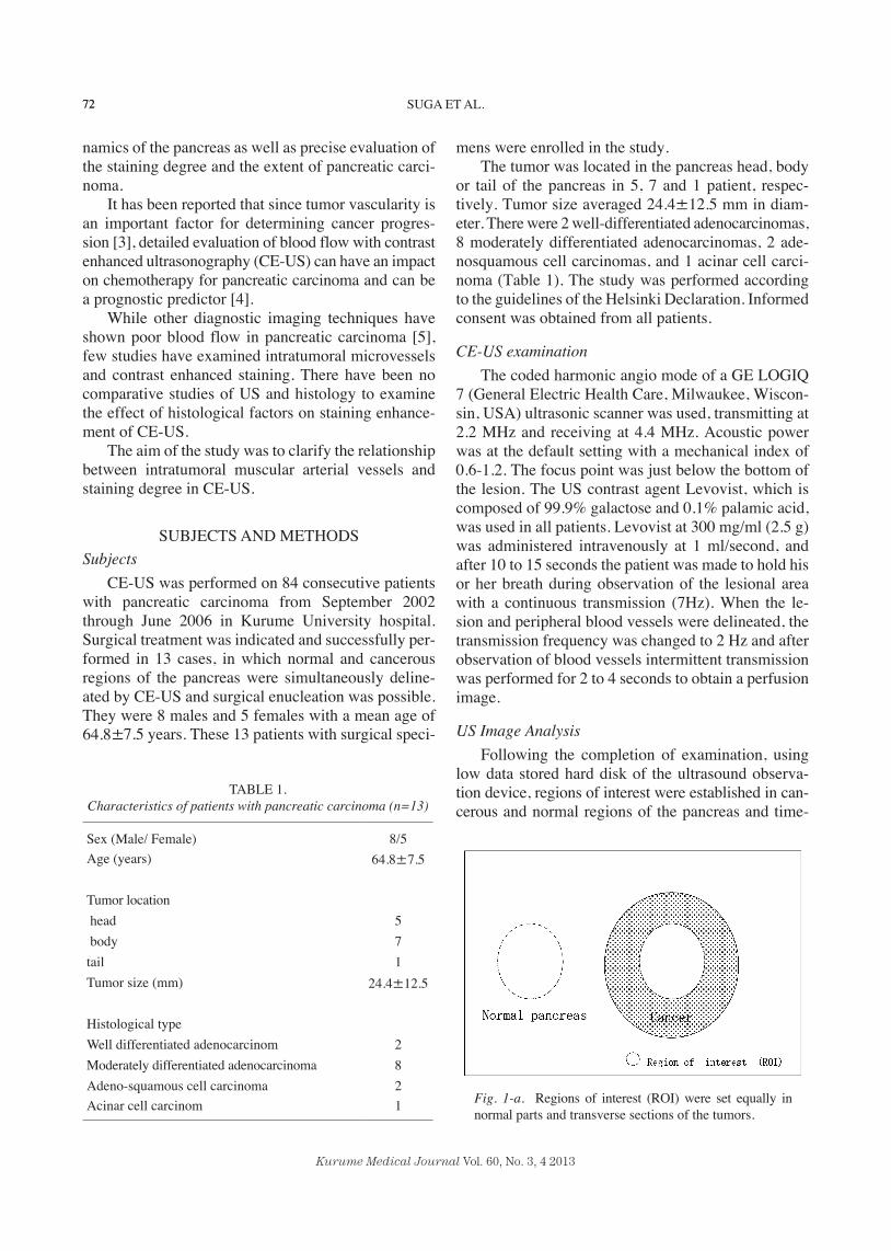
Histological type Well differentiated adenocarcinom 2 Moderately differentiated adenocarcinoma 8Adeno-squamous cell carcinoma 2Acinar cell carcinom 1 Fig. 1-a. Regions of interest (ROI) were set equally in
normal parts and transverse sections of the tumors.
73
Kurume Medical Journal Vol. 60, No. 3, 4 2013
CONTRAST-ENHANCED ULTRASONOGRAPHY IN PANCREATIC CANCER
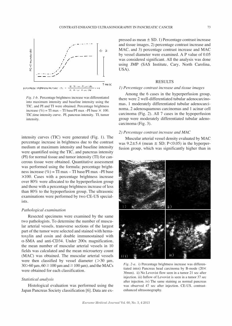
intensity curves (TIC) were generated (Fig. 1). The percentage increase in brightness due to the contrast medium at maximum intensity and baseline intensity were quantified using the TIC, and pancreas intensity (PI) for normal tissue and tumor intensity (TI) for can-cerous tissue were obtained. Quantitative assessment was performed using the formula; percentage bright-ness increase (%) = TI max – TI base/PI max –PI base ×100. Cases with a percentage brightness increase over 80% were allocated to the hyperperfusion group and those with a percentage brightness increase of less than 80% to the hypoperfusion group. The ultrasonic examinations were performed by two CE-US special-ists.
Pathological examinationResected specimens were examined by the same
two pathologists. To determine the number of muscu-lar arterial vessels, transverse sections of the largest part of the tumor were selected and stained with hema-toxylin and eosin and double immunostained with α-SMA and anti-CD34. Under 200x magnification, the mean number of muscular arterial vessels in 10 fields was calculated and the mean microartery count (MAC) was obtained. The muscular arterial vessels were then classified by vessel diameter (>30 μm, 30>60 μm, 60 >_ 100 μm and >_ 100 μm), and the MACs were obtained for each classification.
Statistical analysisHistological evaluation was performed using the
Japan Pancreas Society classification [6]. Data are ex-
pressed as mean ± SD. 1) Percentage contrast increase and tissue images, 2) percentage contrast increase and MAC, and 3) percentage contrast increase and MAC by vessel diameter were examined. A P value of 0.05 was considered significant. All the analysis was done using JMP (SAS Institute, Cary, North Carolina, USA).
RESULTS1) Percentage contrast increase and tissue images
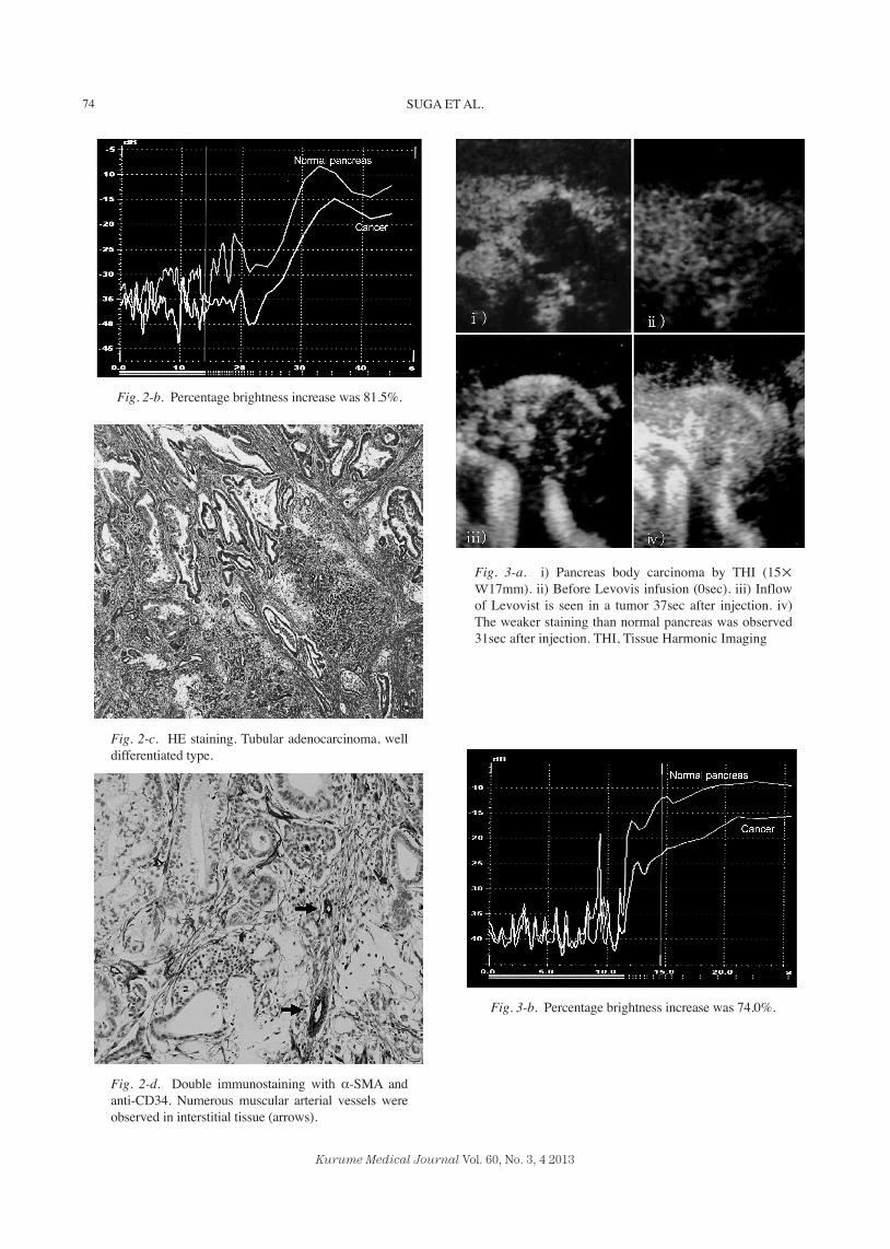
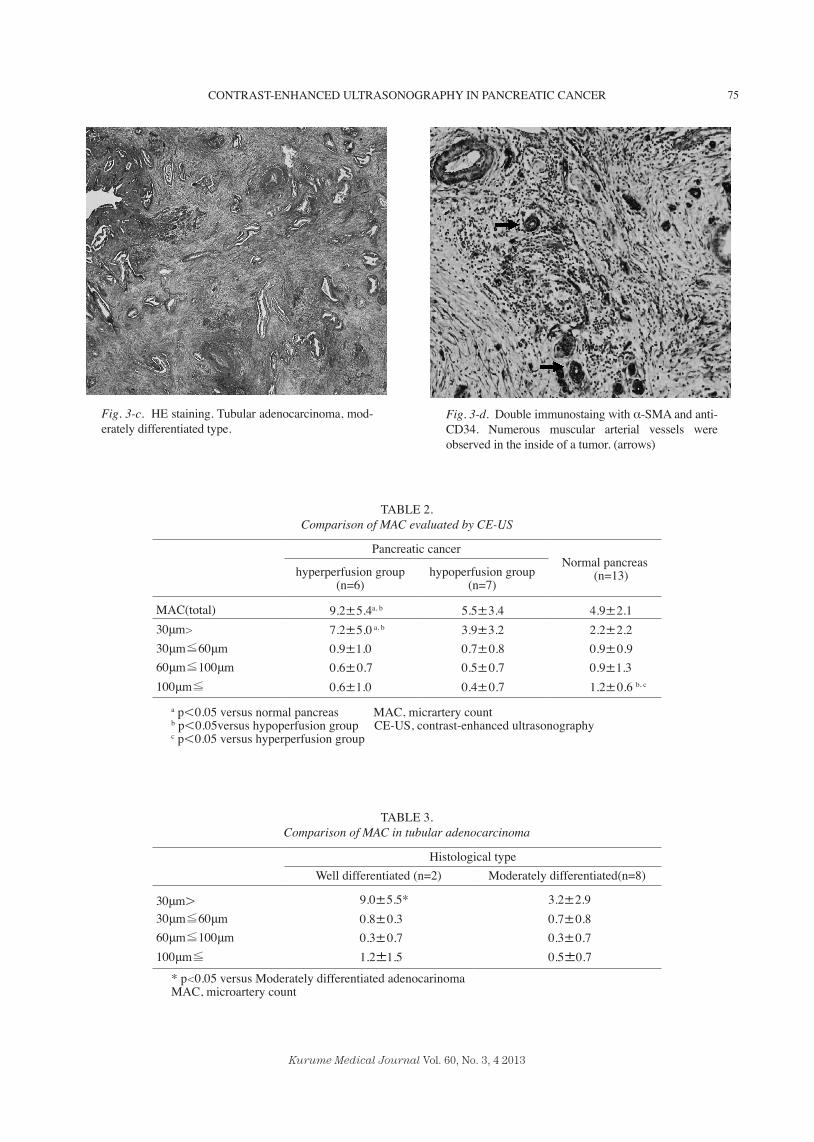
Among the 6 cases in the hyperperfusion group, there were 2 well-differentiated tubular adenocarcino-mas, 1 moderately differentiated tubular adenocarci-noma, 2 adenosquamous carcinomas and 1 acinar cell carcinoma (Fig. 2). All 7 cases in the hypoperfusion group were moderately differentiated tubular adeno-carcinoma (Fig. 3).
2) Percentage contrast increase and MACMuscular arterial vessel density evaluated by MAC
was 9.2±5.4 (mean ± SD; P<0.05) in the hyperper-fusion group, which was significantly higher than in
Fig. 1-b. Percentage brightness increase was differentiated into maximum intensity and baseline intensity using the TIC, and PI and TI were obtained. Percentage brightness increase (%) = TI max – TI base/PI max –PI base × 100. TIC,time intensity curve. PI, pancreas intensity. TI, tumor intensity.
Fig. 2-a. i) Percentage brightness increase was differen-tiated intoi) Pancreas head carcinoma by B-mode (20× 30mm). ii) No Levovist flow seen in a tumor 21 sec after injection. iii) Inflow of Levovist is seen in a tumor 37 sec after injection. iv) The same staining as normal pancreas was observed 47 sec after injection. CE-US, contrast-enhanced ultrasonography.
74 SUGA ET AL.
Kurume Medical Journal Vol. 60, No. 3, 4 2013
Fig. 2-b. Percentage brightness increase was 81.5%.
Fig. 3-b. Percentage brightness increase was 74.0%.
Fig. 2-c. HE staining. Tubular adenocarcinoma, well differentiated type.
Fig. 2-d. Double immunostaining with α-SMA and anti-CD34. Numerous muscular arterial vessels were observed in interstitial tissue (arrows).
Fig. 3-a. i) Pancreas body carcinoma by THI (15× w17mm). ii) Before Levovis infusion (0sec). iii) Inflow of Levovist is seen in a tumor 37sec after injection. iv) The weaker staining than normal pancreas was observed 31sec after injection. THI, Tissue Harmonic Imaging
75
Kurume Medical Journal Vol. 60, No. 3, 4 2013
CONTRAST-ENHANCED ULTRASONOGRAPHY IN PANCREATIC CANCER
Fig. 3-c. HE staining. Tubular adenocarcinoma, mod-erately differentiated type.
Fig. 3-d. Double immunostaing with α-SMA and anti-CD34. Numerous muscular arterial vessels were observed in the inside of a tumor. (arrows)
TABLE 2.Comparison of MAC evaluated by CE-US
Pancreatic cancerNormal pancreas
(n=13)hyperperfusion group (n=6)
hypoperfusion group (n=7)
MAC(total) 9.2±5.4a, b 5.5±3.4 4.9±2.130μm> 7.2±5.0 a, b 3.9±3.2 2.2±2.2 30μm≦60μm 0.9±1.0 0.7±0.8 0.9±0.9 60μm≦100μm 0.6±0.7 0.5±0.7 0.9±1.3 100μm≦ 0.6±1.0 0.4±0.7 1.2±0.6 b, c
a p<0.05 versus normal pancreas MAC, micrartery countb p<0.05versus hypoperfusion group CE-US, contrast-enhanced ultrasonographyc p<0.05 versus hyperperfusion group
TABLE 3.Comparison of MAC in tubular adenocarcinoma
Histological typeWell differentiated (n=2) Moderately differentiated(n=8)
30μm> 9.0±5.5* 3.2±2.930μm≦60μm 0.8±0.3 0.7±0.8 60μm≦100μm 0.3±0.7 0.3±0.7 100μm≦ 1.2±1.5 0.5±0.7
* p<0.05 versus Moderately differentiated adenocarinomaMAC, microartery count

76 SUGA ET AL.
Kurume Medical Journal Vol. 60, No. 3, 4 2013
the hypoperfusion group (5.3±3.4) or normal tissue (4.9±2.1). There was no significant difference in MAC between the hypoperfusion group and the nor-mal pancreas.
3) Percentage contrast increase and MAC by vessel diameter
MAC with a diameter of less than 30 μm was 7.2± 5.0 in the hyperperfusion group (P<0.05), which was significantly higher than in the hypoperfusion group (2.2±2.2) and normal pancreas (4.9±2.1). There was no significant difference between the MAC values in the 3 groups for vessels with a diameter of 30 to 60 μm and 60 to 100 μm.
For vessels with a diameter greater than 100 μm, MAC was 1.2±0.6 (P<0.05) in normal pancreas, which was significantly higher than the 0.6±1.0 in the hy-perperfusion group and 0.4±0.7 in the hypoperfusion group (Table 2).
In tubular adenocarcinoma the MAC for vessels with a diameter less than 30 μm was 9.5±5.5 (P<0.05) in well-differentiated adenocarcinoma, which was sig-nificantly higher than the 3.2±2.9 found in moderately differentiated adenocarcinoma (Table 3).
No significant difference was observed in the amount of interstitial tissue in cancerous lesions be-tween the hyperperfusion and hypoperfusion groups (Table 4).
DISCUSSION
Imaging diagnostic methods show hypovascular tu-mors in pancreatic carcinoma and until now, the blood flow in tumors could not be delineated. Folkman pro-posed the idea that in solid tumors, the supply of oxy-gen and nutrients by diffusion limits the size of the tu-mor to approximately 250 to 300 μm [7]. His assumption was that unless angiogenesis occurred, a tumor could become no larger than 1 to 2 mm, and that vasculariza-tion was essential for tumor metastasis and enlarge-
ment. Following publication of these proposals, stud-ies were undertaken to examine microvessel density (MVD), vascular endothelial growth factor (VEGF), thymidine phosphorylase (TP) and other angiogenesis factors, and there have also been reports of how their expression and mechanisms are related to angiogen-esis [8-10]. MVD in particular is an indicator of the degree of angiogenesis and its use as a prognostic fac-tor is anticipated [11].
Many studies on the different prognoses of hyper vascular tumors by means of MVD were reported con-cerning breast cancer [12], colon cancer [13] and gas-tric cancer[14]. Benckert et al. examined the correla-tion between MVD and VEGF and prognosis in pancreatic cancer, and reported that multivariable anal-ysis showed high MVD to be an independent prognos-tic factor [15]. D’Onofrio et al. examined the relation between CE-US and angiogenesis and found a corre-lation between the contrast effect of CE-US and CD34-positive blood vessels pathologically examined in 42 pancreatic tumor patients [16]. There have, however, been only a few reports on the correlation between CE-US and MVD in pancreatic carcinoma, and a re-view of Pub Med and Japan Medical Abstracts Society found no reports on the direct correlation between CE-US and pathological findings. Until the advent of Lev-ovist, US-angiography (US-A) was performed using carbon dioxide microbubbles injected into the hepatic artery as a contrast medium.
In pancreatic tumors, Koito et al. reported that US-A was superior to contrast CT and angiography for differentiating between mass forming pancreatitis and pancreatic ductal carcinoma [17]. However, angi-ographic examination is essential but highly invasive. On the other hand, Levovist enables intravenous eval-uation of blood flow. Compared to other contrast agents, Levovist has not been reported to cause allergic reac-tions or renal disorders and seems to be an extremely safe contrast agent [18]. Furthermore, unlike the con-trast medium used by CT, it is able to show a pure
TABLE 4.Comparison of cancer histology with CE-US findings
CE-US findings hyperperfusion group (n=6) hypoperfusion group (n=7)
HistologyMedullary type 1 0Intermediate type 3 4
Scirrhous type 2 3
CE-US, contrast-enhanced ultrasonography.
77
Kurume Medical Journal Vol. 60, No. 3, 4 2013
CONTRAST-ENHANCED ULTRASONOGRAPHY IN PANCREATIC CANCER
vessel image. Although pancreatic carcinoma is thought to be a
hypovascular tumor, it should have some blood flow for growth. With US-A, the cancerous part of the pan-creas is delineated as a poorly stained region [17]. Therefore, it was judged as hypovascular tumor. With CE-US, pancreatic tumors are shown distinctly and both sensitivity and specificity are high [19,20], be-cause blood-flow evaluation is attained. There have also been reports of the usefulness of the staining pat-terns for tumor differentiation and for determining the surgical indication [1,21]. In previous reports, how-ever, the presence or absence of blood flow was deter-mined visually and staining patterns were analyzed on that basis rather than by evaluating the degree of stain-ing by intensity. We analyzed the TIC obtained by CE-US and compared the percentage brightness increase of the cancerous and normal parts of the pancreas. The results suggested that percentage brightness increase might be correlated with histological type in pancre-atic carcinoma.
Differences in staining in US-A and CE-US are believed to be due to differences in bubble size (CO2 > 30 μm, Levovist approximately 1.3 μm). This study showed that since there were many muscular arterial vessels with a diameter of less than 30 μm in cancerous parts, CO2 bubbles could not pass through these blood vessels and they would therefore have been delineated as poorly stained areas. With Levovist, however, the vessels were delineated as stained areas. Therefore, the percentage of muscular arterial vessels less than 30 μm is the factor that determines the staining intensity in pancreatic cancer. As a result, with CE-US, quanti-tative evaluation of staining becomes an indirect eval-uation of microvessel density, which could be a useful prognostic predictor for pancreatic carcinoma.
In conclusion, the CE-US density of muscular ar-terial vessels less than 30 μm is an important factor in determining staining degree and carcinoma progres-sion in pancreatic carcinoma.
REFERENCES1. Kitano M, Kudo M, Maekawa K, Suetomi Y, Sakamoto H
et al. Dynamic imaging of pancreatic disease by contrast enhanced coded phase inversion harmonic ultrasonography. Gut 2004; 53:854-859.
2. Sofuni A, Iijima H, Moriyasu F, Nakayama M, Nakamura K et al. Differential diagnosis of pancreatic tumor using ultrasound contrast imaging. J Gastroenterol 2005; 40:518-525.
3. Klagusbrum M, Folkman J: Angiogenesis. Inpeptide growth factor and their receptors II. Handbook of Experimental Pharmacology. Springer-Veriag, Berlin 1990; 95:549-665.
4. Masaki T, Ohkawa S, Amano A, Ueno M, Miyakawa K et al. Noninvasive assessment of tumor vasucularity by con-trast-enhanced ultrasonography and the progressive of patients with nonresectable pancreatic carcinoma. Am Cancer Society 2005; 103:1026-1035.
5. Ichikawa T, Haradome H, Hachiya J, Nitatori T, Ohyomo K et al. Pancreatic ductal adenocarcinoma: preoperative assessment with helical CT versus dynamic MR imageing. Radiology 1997; 202:655-662.
6. Japan Pancreas Society. Classification of Pancreatic Cancer 2nd English edition. Tokyo: Kanehara & CO., Ltd.,2003.
7. Folkman J: Tumor angiogenesis: therapeutic implications. N Engl J Med 1971; 285:1182-1186.
8. Poon RT, Fan ST, Wong J: Clinical significance of angio-genesis in gastrointestinal cancers: a target for novel prog-nostic and therapeutic approaches. Ann Surg 2003; 238:9-28.
9. Kutami R, Nakashima Y, Nakashima O, Siota K, and Kojiro M. Pathomorphologic study on the mechanism of fatty change in small hepatoellular carcinoma of humans. J Hepatol 2000; 33:282-289.
10. Vizo B, Navarino A, Giacobino A, Cristiano C, Parti A et al. Pilot study to relate outcome in pancreatic carcinoma and angiogenic plasma factors /circulating mature / pro-genitor endothelial cells: Preliminay result. Cancer Sci 2010; 101:2448-2454.
11. Fujimoto K, Hosotani R, Wada M, Lee JU, Koshiba T et al. Expresson of two angiogenic factors, vasuccular endothelial growth factor in human pancreatic cancer, and its relation-ships to angiogenesis. Eur J cancer 1998; 34:1439-1447.
12. Kim SW, Park SS, Ahn SJ, Chung KW, Moon WK et al. Identification of angiogenesis in primary breast carcinoma according to the image analysis. Breast Cancer Res Treat 2002; 74:121-129.
13. Takahashi Y, Kitadai Y, Bucana CD, Cleary KR, and Ellis LM. Expression of endothelial growth factor and its recep-tor, KDR, correlates with vascularity, metastasis, and prolif-eration of human colon cancer. Cancer Res 1995; 55:3964-3968.
14. Tanigawa N, Amaya H, Matsumura M, Shimomatsuya T, and Horiuchi T. Extent of tumor vascularization correlation with prognosis and hematogenous metastasis in gastric car-cinoma. Cancer Res 1996; 56:2671-2679.
15. Benckert C, Thelen A, Cramer T, Weichert W, Gaebelein G et al. Impact of microvessel density on lymph node metas-tasis and survival after curative resection on pancreatic can-cer. Surg Today 2012; 42:169-176.
16. D’Onofiro M, Mallago R, Zamboni G, Vasori S, Falconi M et al. Contrast-enhanced ultrasonography better indentifies pancreatic tumor vascularization than helical CT. Pancreatology 2005; 5:398-402.
17. Koito K, Namieno T, Nagakawa T, and Morita K. Inflammatory pancreatic mass: differentitation from ductal carcinoma with ontrast-enhanced sonography using carbon dioxide microbubbles. AJR 1997; 169:1263-1267.
18. Otis S, Rush M, Boyajian R. Contrast-enhanced transcrani-al imaging: result of an American two phase study. Stroke 1995; 26: 203-209.
19. Rickes S, Unkrodt K, Neye H, Ocran KW, and Wermke W. Differentiation of Pancreatic Tumor by Conventional
78 SUGA ET AL.
Kurume Medical Journal Vol. 60, No. 3, 4 2013
Ultrasound, Unenhanced and Echo-enhanced Power Doppler Sonography. Scand J Gastoroenterol 2002; 37:1313-1320.
20. Oshikawa O, Tanaka S, Ioka T, Nakaizumi A, Hamada Y et al. Dynamic Sonography of Pancreatic Tumors: Comparison with Dynamic CT. AJR 2002; 178:1133-1137.
21. Faccioli N, D’Onofrio M, Malogo R, Zamboni G, Falconi M, et al. Resectable pancreatic carcinoma; depiction of tumoral margin at contrast enhanced ultrasonography. Pancreas 2008; 37:265-268.
Related Documents


