Acute complicated pyelonephritis: contrast-enhanced ultrasound Teresa Fontanilla, Javier Minaya, Cristina Corte ´s, Concepcio ´n Gonza ´lez Hernando, Rafael Pe ´rez Arangu ¨ena, Jesu ´s Arriaga, Maria Soledad Carmona, Ana Alcolado Servicio de Radiologı´a, Hospital Universitario Puerta de Hierro Majadahonda, Madrid, Spain Abstract Imaging is required if complication is suspected in acute pyelonephritis to assess the nature and extent of the lesions, and to detect underlying causes. The current imaging modality of choice in clinical practice is com- puted tomography. Because of associated radiation and potential nephrotoxicity, CEUS is an alternative that has been proven to be equally accurate in the detection of acute pyelonephritis renal lesions. The aims of this study of 48 patients are to describe in detail the CEUS findings in acute pyelonephritis, and to determine if abscess and focal pyelonephritis may be distinguished. Very charac- teristic morphologic and temporal patterns of enhance- ment are described. These allow differentiation of focal pyelonephritis from renal abscess, and detection of tiny suppurative foci within focal pyelonephritis. The detec- tion of abscesses is important because follow-up in 25 patients revealed a longer clinical course. Typical pyelonephritis CEUS features permit distinction from other renal lesions. As a whole, CEUS is an excellent tool in the work-up of complicated acute pyelonephritis, so it may be considered as the imaging technique of choice in the evaluation and follow-up of these patients who fre- quently are very young, so as to minimise radiation exposure. Key words: Acute pyelonephritis—CEUS—Contrast- enhanced ultrasound—Renal abscess—Complicated pyelonephritis Acute pyelonephritis (APN) is a common disease which affects mostly young women. Diagnosis is usually made on the basis of clinical and laboratory findings [1], and imaging is not required unless complication is suspected or in high-risk patients (immunocompromised patients, diabetics and the elderly) [2]. Complication is suspected when there is no clinical response after 72 h of intrave- nous antibiotic treatment or when there is obstruction of the urinary tract. Grey-scale ultrasound may show findings in complicated pyelonephritis [3], but its sensi- tivity is lower than that of computed tomography (CT). A 2007 study demonstrated a very similar diagnostic performance of CT and contrast-enhanced ultrasound (CEUS) in the diagnosis of acute pyelonephritis [4]. But to date, a detailed description of the imaging findings of complicated pyelonephritis in CEUS has not been pub- lished. The aims of this study are to describe the different findings of complicated acute pyelonephritis in CEUS, and to determine if CEUS is able to distinguish focal pyelonephritis from abscess. Materials and methods Patients and study Carried out over a 3-year period in a single centre, this is a retrospective study which comprises 48 patients (43 women, 5 men) with a mean age of 33 years (range 14–85), who underwent CEUS for the evaluation of complicated acute pyelonephritis. Most of the patients were previously healthy, except three renal transplant recipients, a liver transplant-recipient, a patient with only one kidney secondary to nephrectomy due to calculi, a patient with double excretory system and previous sur- gery, and a drug abuser. Informed consent of patients was waived. The presence of APN was correlated with clinical and laboratory findings. Four of the patients had urinary sepsis. The clinical improvement after antibiotic therapy, the disappearance of the lesions, the develop- ment of scars at CEUS follow-up; or concordant con- trast-enhanced CT findings (15 patients) were considered as a confirmation of the pyelonephritic nature of the lesions. 25 patients participated in CEUS follow-up. The imaging follow-up schedule was performance of basal and CEUS 1 week and 1 month after diagnosis. In ten Correspondence to: Teresa Fontanilla; email: [email protected] ª Springer Science+Business Media, LLC 2011 Abdominal Imaging Abdom Imaging (2011) DOI: 10.1007/s00261-011-9781-2

Welcome message from author
This document is posted to help you gain knowledge. Please leave a comment to let me know what you think about it! Share it to your friends and learn new things together.
Transcript
Acute complicated pyelonephritis:contrast-enhanced ultrasound
Teresa Fontanilla, Javier Minaya, Cristina Cortes, Concepcion Gonzalez Hernando,
Rafael Perez Aranguena, Jesus Arriaga, Maria Soledad Carmona, Ana Alcolado
Servicio de Radiologıa, Hospital Universitario Puerta de Hierro Majadahonda, Madrid, Spain
Abstract
Imaging is required if complication is suspected in acutepyelonephritis to assess the nature and extent of thelesions, and to detect underlying causes. The currentimaging modality of choice in clinical practice is com-puted tomography. Because of associated radiation andpotential nephrotoxicity, CEUS is an alternative that hasbeen proven to be equally accurate in the detection ofacute pyelonephritis renal lesions. The aims of this studyof 48 patients are to describe in detail the CEUS findingsin acute pyelonephritis, and to determine if abscess andfocal pyelonephritis may be distinguished. Very charac-teristic morphologic and temporal patterns of enhance-ment are described. These allow differentiation of focalpyelonephritis from renal abscess, and detection of tinysuppurative foci within focal pyelonephritis. The detec-tion of abscesses is important because follow-up in 25patients revealed a longer clinical course. Typicalpyelonephritis CEUS features permit distinction fromother renal lesions. As a whole, CEUS is an excellent toolin the work-up of complicated acute pyelonephritis, so itmay be considered as the imaging technique of choice inthe evaluation and follow-up of these patients who fre-quently are very young, so as to minimise radiationexposure.
Key words: Acute pyelonephritis—CEUS—Contrast-enhanced ultrasound—Renal abscess—Complicatedpyelonephritis
Acute pyelonephritis (APN) is a common disease whichaffects mostly young women. Diagnosis is usually madeon the basis of clinical and laboratory findings [1], andimaging is not required unless complication is suspectedor in high-risk patients (immunocompromised patients,
diabetics and the elderly) [2]. Complication is suspectedwhen there is no clinical response after 72 h of intrave-nous antibiotic treatment or when there is obstructionof the urinary tract. Grey-scale ultrasound may showfindings in complicated pyelonephritis [3], but its sensi-tivity is lower than that of computed tomography (CT).A 2007 study demonstrated a very similar diagnosticperformance of CT and contrast-enhanced ultrasound(CEUS) in the diagnosis of acute pyelonephritis [4]. Butto date, a detailed description of the imaging findings ofcomplicated pyelonephritis in CEUS has not been pub-lished. The aims of this study are to describe the differentfindings of complicated acute pyelonephritis in CEUS,and to determine if CEUS is able to distinguish focalpyelonephritis from abscess.
Materials and methods
Patients and study
Carried out over a 3-year period in a single centre, thisis a retrospective study which comprises 48 patients(43 women, 5 men) with a mean age of 33 years (range14–85), who underwent CEUS for the evaluation ofcomplicated acute pyelonephritis. Most of the patientswere previously healthy, except three renal transplantrecipients, a liver transplant-recipient, a patient with onlyone kidney secondary to nephrectomy due to calculi, apatient with double excretory system and previous sur-gery, and a drug abuser. Informed consent of patientswas waived. The presence of APN was correlated withclinical and laboratory findings. Four of the patients hadurinary sepsis. The clinical improvement after antibiotictherapy, the disappearance of the lesions, the develop-ment of scars at CEUS follow-up; or concordant con-trast-enhanced CT findings (15 patients) were consideredas a confirmation of the pyelonephritic nature of thelesions. 25 patients participated in CEUS follow-up. Theimaging follow-up schedule was performance of basaland CEUS 1 week and 1 month after diagnosis. In tenCorrespondence to: Teresa Fontanilla; email: [email protected]
ª Springer Science+Business Media, LLC 2011
AbdominalImaging
Abdom Imaging (2011)
DOI: 10.1007/s00261-011-9781-2
patients with persistence of lesions, CEUS was per-formed 3 months later.
Ultrasound technique
The machines used were Sequoia and S 2000 (Siemens-Acuson). Convex multifrequency probes and a specificCEUS technology (Cadence Contrast Pulse Sequencing,CPS) with low mechanical indexes (0.07–0.18) wereemployed. A sulphur hexafluoride-filled microbubblecontrast agent (SonoVue, Bracco, Milan) was used. Allof the patients underwent grey-scale basal sonographyand power Doppler of both kidneys. Presence or absenceof renal and perirenal lesions and urinary tract dilatationor obstruction were noted. Afterwards, CEUS of thesuspect kidney in all of the phases, and CEUS of thecontralateral kidney in the parenchymal phase wereperformed. Each patient received a 2.4 mL intravenousbolus of contrast in a peripheral vein through a 20 gaugecannula, flushed with 10 mL of saline. In six cases, asecond dose was needed to study the contralateral kid-ney. The CEUS exploration lasted until most of thebubbles had disappeared from the kidney (mean 3 min).
CEUS image analysis
Two main enhancement phases were considered: a cor-tical phase, in which enhancement of the cortex was seen(15–30 s after contrast injection), and a parenchymalphase, in which enhancement of both cortex andmedullae was seen (25 s—4m after contrast injection).For practical reasons, the parenchymal phase was sepa-rated into early parenchymal phase (25 s—1m) and lateparenchymal phase (after 1m).
The explorations were performed by two radiologistswith 4 year experience in CEUS and were stored (dividedin 40 s clips) in the hard disc and DVD or PACS. Thecomplete explorations were reviewed afterwards by bothof the radiologists in consensus.
None of the studied lesions met the criteria of asimple cyst on baseline US. The CEUS diagnostic criteriafor abscess were defined as the presence of areas of non-enhancement throughout the whole exploration, with orwithout an enhancing rim or septa. The CEUS imagingcriteria to diagnose focal pyelonephritis were the pres-ence of a cortical or corticomedullary focal wedge-shaped or round lesion, hypoechoic (less enhancing) ascompared to the surrounding parenchyma. In all of thepatients, the presence, number, location and size of thelesions as well as unilateral or bilateral involvement wereevaluated. Also, temporal patterns of enhancement(enhancement and washout during the different phases)were analysed. In follow-up explorations, the previousones were reviewed, so as to assess the change in size,appearance and enhancement pattern of the lesions.
Results
According to CEUS findings, the lesions were classifiedinto five groups (Table 1). All of the findings were uni-lateral. In those patients in whom different types oflesions existed, the patient was considered as belongingto the group with the most severe lesions.
Abscess
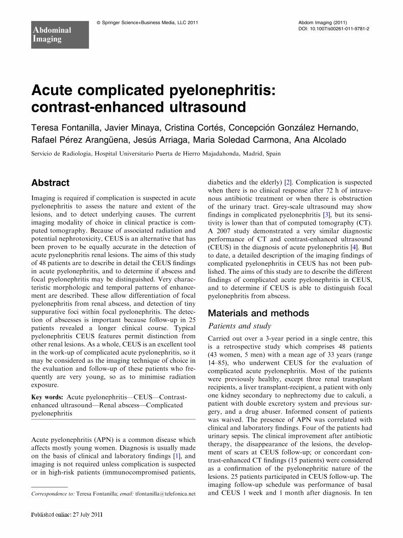
In 10 patients, 16 medium (2.5–4 cm) or large (4–6 cm)renal abscesses were detected. Their shape was roundedor geographical. In all of these lesions, there was absenceof enhancement throughout the whole exploration, withrim enhancement in two lesions and enhancing thicksepta in 3 lesions (Figs. 1, 2). CEUS detected five morelesions. CT was performed in seven patients with goodcorrelation.
Focal pyelonephritis
In 14 patients, 22 areas suggestive of focal pyelonephritiswere identified. These were wedge-shaped or roundedhypoechoic areas, located in the cortex or cortex andmedullae with size ranging from 1.5 to 4 cm (Fig. 3). Asfor the enhancement temporal patterns, all of the lesionswere best depicted during the late parenchymal phase,whereas the findings were variable in the rest of theexploration (Fig. 1). Most of them were initially hypo-echoic (cortical or very early parenchymal phase), thenisoechoic, to turn hypoechoic again in the late paren-chymal phase. Some of the lesions were hypoechoicthroughout the exploration, and some were only detectedon the late parenchymal phase. Five new lesions weredetected after CEUS.
Focal pyelonephritis with small abscess
In 14 patients, a combination of the two previouslydescribed enhancing patterns was seen. These lesionsappeared as areas of focal pyelonephritis (with any of theaforementioned temporal patterns), but with areas ofnon-enhancement inside (n = 24), corresponding tosmall abscesses, most of them around 1 cm in size (range0.8–3 cm) (Fig. 4). CEUS detected ten more lesions.
Table 1. CEUS types of renal parenchymal lesions
CEUS diagnosis Patients Number oflesions ongrey-scale
Number oflesions onCEUS
Focal PN 14 17 22Focal PN small abscess 14 14 24Abscess 10 11 16Normal 9 0 0Scar 1 1 1Total (n = 48) 48 43 62
T. Fontanilla et al.: Acute complicated pyelonephritis
Correlation with CT (eight cases) was good, but CTdetected additional lesions in two patients. On the otherhand, CEUS depicted better than CT very smallabscesses (size around 1 cm).
Scar
In one patient, a scar related to previous pyelonephritiswas the only finding. In another patient with focalpyelonephritis, there were scars related to previous APN.These were seen as focal thinning of the cortex, hypo-echoic on CEUS.
Normal
In nine patients, CEUS showed normal enhancement,with no renal lesions. In all of these cases, no furtherimaging was performed (unless obstruction was present)and clinical resolution ensued.
Grey-scale ultrasound and power Doppler
Findings on basal grey-scale ultrasound and powerDoppler and their correspondence to the CEUS diag-nosis are shown in Table 2. The most common finding incomplicated APN was a focal hyperechoic lesion, almostalways with bulging of the cortex. This correspondedmost frequently to focal pyelonephritis without abscess,but corresponded also to pyelonephritis with smallabscess, and even to a medium size abscess (one case).Most medium or large abscesses were seen as heteroge-neous or hypoechoic focal lesions with cortical bulging.
All focal lesions appeared as hypovascular or avascularareas on power Doppler, but power Doppler was normalor non-evaluable in eight cases with focal pyelonephritisor focal pyelonephritis with abscess.
Other findings
Among those patients with parenchymal lesions onCEUS, pyonephrosis due to obstructive ureteral lithiasiswas seen in two cases, one of them with a perirenal ab-scess. In five other cases, obstructive lithiasis was found.
In nine cases, parenchymal CEUS was normal. Thisexcluded complicated pyelonephritis, but it did notexclude uncomplicated pyelonephritis. In two of thesecases, the diagnosis was pyonephrosis; in another caseacute diverticulitis was found.
Other findings included very frequently perirenal fluidand urothelial thickening and enhancement; and lesscommonly biliary bladder wall edema, pleural effusionand ascites.
Follow-up
Follow-up was performed in 25 patients. Resolution wasvery fast in focal pyelonephritis without abscess (days),without residual lesions. In all of the cases with mediumsize or large abscesses, the evolution was slow (morethan a month), even though the patients were doingwell clinically. The lesion gradually turned smaller andsmaller, the rim enhancement disappeared, and ulti-mately ended in complete resolution or focal atrophiccortical lesions (scars) (three cases).The cases with smallabscesses inside focal pyelonephritis had an intermediateevolution, with residual scarring in two cases.
Differential diagnosis
From an imaging standpoint, differential diagnosesincluded infarction, in two cases, and residual postpye-lonephritic lesion (scar), in one case. One patient whohad an abscess had previously had a lower pole infarc-tion due to a critical stenosis in a lower pole segmentalartery in the early posttransplantation period (Fig. 5).This was seen as non-enhancing areas within a large areaof retarded and very diminished enhancement in thelower pole, with preservation of a cortical rim. Inanother patient who had multiple coalescent abscessesthe possibility of multiple infarctions or septic emboliwas considered because he was a drug abuser.
Discussion
Imaging is necessary in complicated APN both to eval-uate the presence and extent of renal and perirenal le-sions, and to detect potentially treatable causes. Up tillnow, CT has been the preferred imaging technique to
Fig. 1. Focal pyelonephritis and abscess are shown in thedifferent phases of CEUS. m, medullae; s sinus. Pyelone-phritis areas are always most conspicuous during theparenchymal late phase, as round or wedge-shaped corticalor corticomedullar hypoechoic areas (shown in the drawing asgrey areas). These areas may be seen also during the corticalphase, and are less conspicuous during parenchymal earlyphase. Abscesses are always seen as anechoic areas(shown in the drawing as black areas), with or without rim orseptal enhancement, alone or within areas of pyelonephritis.
T. Fontanilla et al.: Acute complicated pyelonephritis
diagnose complications of acute pyelonephritis. CT issuperior to grey-scale ultrasound and power Doppler [3]in detecting and characterizing APN lesions and has anexcellent performance in identifying obstructive causessuch as lithiasis or anatomic congenital alterations [2, 3].But CT has associated an amount of radiation which isfar from negligible (especially in this group of youngpatients, mostly women in bearing age), as well aspotential contrast media nephrotoxicity. Mitterbergeret al. [4] demonstrated CEUS and contrast-enhanced CTto be almost equally sensitive and specific for detectingrenal parenchymal changes in APN. To date, there isvery little written about CEUS in the evaluation ofcomplicated pyelonephritis [5–8] and the EFSUMB 2008guide does not include complicated APN as an indicationfor CEUS [9].To our knowledge, up till now the CEUSfindings of APN have not been described in detail. Thisstudy describes typical CEUS enhancement features ofthe different parenchymal lesions, which make it possibleto distinguish abscess from focal pyelonephritis, and to
detect even very small abscesses within pyelonephriticareas.
Abscess enhancement findings in our study are con-cordant with CT findings (performed in eight patientswith abscess) and coincident with those reported for CT,as expected [3]. An abscess is a necrotic cavity whichcontains pus and debris, so no enhancement is seen insidethroughout all phases, or else enhancement of septa isseen if the abscess is partially liquefied. Peripheral rimenhancement in cortical phase may be seen. In our series,both rim enhancement and septal enhancement dimin-ished during the parenchymal phase. These CEUS find-ings are similar to those described for hepatic abscesses[10], in which rim or septal enhancement diminishesduring portal or late phase. In all of our cases CEUSclearly improved the performance of grey-scale sonog-raphy, since basal sonography of large abscesses wasnonspecific in most of the cases. Just two of the casesshowed lesions which were very suggestive of abscesson grey-scale US (hypoechoic with posterior sonic
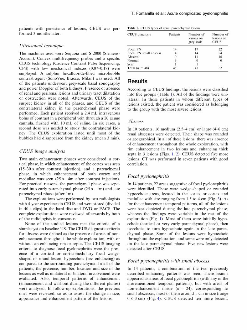
Fig. 2. Renal abscess. 22 year old female transplant-recipient. A Grey-scale sonography of upper pole of trans-planted kidney shows a heterogeneous round area of corticalthickening (black arrows) m, medullae; s sinus. It is impossi-ble to distinguish focal pyelonephritis from abscess, and even
from solid mass with this image. B Power Doppler sagittalview shows that this is a hypovascular area (arrowheads).C CEUS at 10 s shows abscess as an anechoic area with rimenhancement (black arrows). D CEUS follow-up 1 month latershows shrinkage of the lesion, without rim enhancement.
T. Fontanilla et al.: Acute complicated pyelonephritis
enhancement) and most of them were seen as heteroge-neous lesions. Thus, basal sonography was not able todistinguish them from focal pyelonephritis or from otherentities in most of the cases (Fig. 2).CEUS permittedassessment of the size, internal structure and extent of theabscesses, and detected five abscesses not detected bybasal ultrasound.
Focal pyelonephritis appeared most frequently ongrey-scale ultrasound as a hyperechoic focal lesion withcortical bulging. This represents the presence of numer-ous interfaces mostly due to the presence of abundantinflammatory cells in the interstitium and tubules [3, 11].Besides the inflammatory infiltrate and edema, severecapillary damage has been demonstrated, with presenceof intraluminal leukocyte and fibrin plugs [11].Thisexplains the CEUS hypoechoic appearance of theselesions in comparison to the surrounding cortex, due todiminished enhancement. CEUS allows real-time assess-ment of renal perfusion, obtaining valuable temporal
information (Fig. 1). Focal pyelonephritis areas are mostconspicuous during the late parenchymal phase, becausethese areas (wedge or round-shaped) appear more hypo-echoic during this phase. Even tiny lesions (smaller than1 cm, may be detected). But care must be taken and all ofthe phases must be carefully analysed because during thelate phase, if the pyelonephritis area is very hypoechoic,small abscesses may not be distinguished from the sur-rounding pyelonephritic area, or else the size of the ab-scesses may be overestimated (Fig. 4) Although it was notan objective of our study, we found a good correlationwith CT findings (performed in seven cases with focalpyelonephritis); except in one case in which three lesionswere missed by CEUS. This may be because some CTfindings cannot be extrapolated to CEUS; such as thestriated nephrogram or the delayed and persistentenhancement [12, 13]. Both of these signs occur because ofunderlying tubular obstruction; CEUS cannot detectthem because US contrast is purely intravascular.
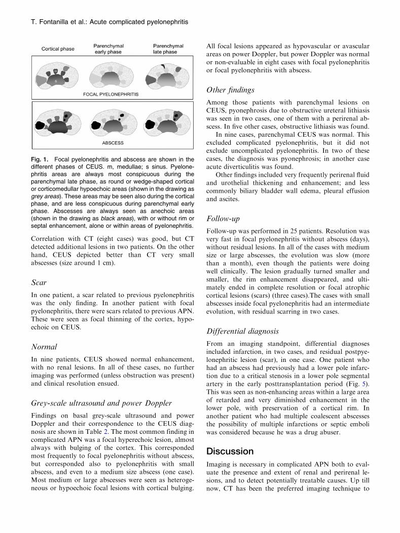
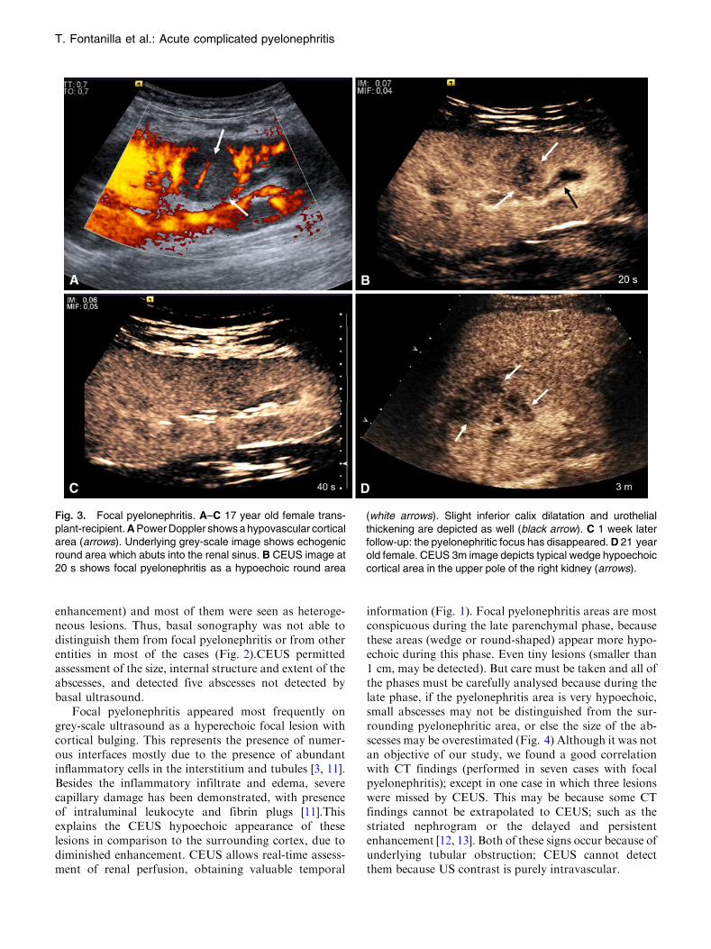
Fig. 3. Focal pyelonephritis. A–C 17 year old female trans-plant-recipient. A Power Doppler shows a hypovascular corticalarea (arrows). Underlying grey-scale image shows echogenicround area which abuts into the renal sinus. B CEUS image at20 s shows focal pyelonephritis as a hypoechoic round area
(white arrows). Slight inferior calix dilatation and urothelialthickening are depicted as well (black arrow). C 1 week laterfollow-up: the pyelonephritic focus has disappeared. D 21 yearold female. CEUS 3m image depicts typical wedge hypoechoiccortical area in the upper pole of the right kidney (arrows).
T. Fontanilla et al.: Acute complicated pyelonephritis
The CEUS temporal and morphologic featuresdescribed above in complicatedAPNare quite typical, andusually differential diagnosis is not under consideration.
Infarctions may be distinguished from focal APN becausepyelonephritis has a typical temporal pattern of enhance-ment and always shows some degree of enhancement,
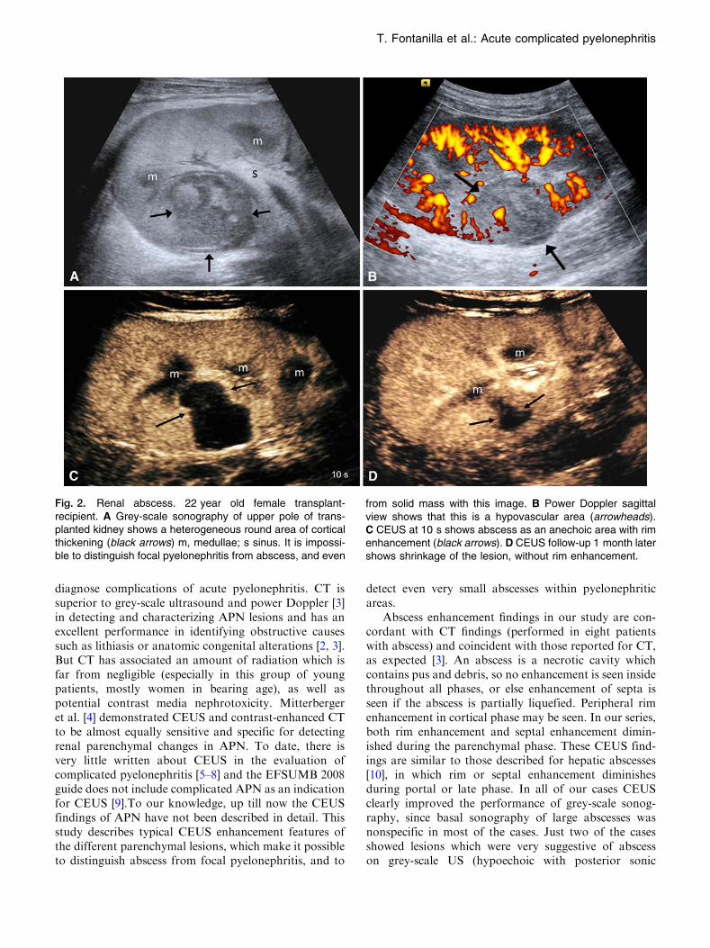
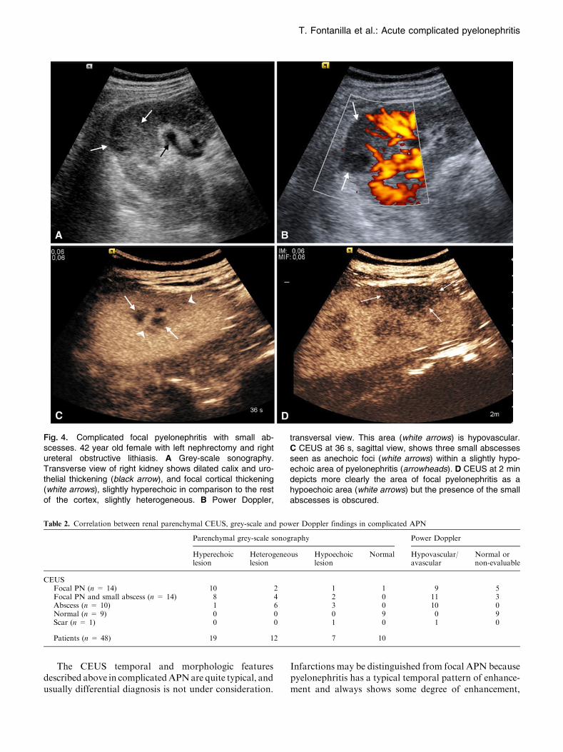
Fig. 4. Complicated focal pyelonephritis with small ab-scesses. 42 year old female with left nephrectomy and rightureteral obstructive lithiasis. A Grey-scale sonography.Transverse view of right kidney shows dilated calix and uro-thelial thickening (black arrow), and focal cortical thickening(white arrows), slightly hyperechoic in comparison to the restof the cortex, slightly heterogeneous. B Power Doppler,
transversal view. This area (white arrows) is hypovascular.C CEUS at 36 s, sagittal view, shows three small abscessesseen as anechoic foci (white arrows) within a slightly hypo-echoic area of pyelonephritis (arrowheads). D CEUS at 2 mindepicts more clearly the area of focal pyelonephritis as ahypoechoic area (white arrows) but the presence of the smallabscesses is obscured.
Table 2. Correlation between renal parenchymal CEUS, grey-scale and power Doppler findings in complicated APN
Parenchymal grey-scale sonography Power Doppler
Hyperechoiclesion
Heterogeneouslesion
Hypoechoiclesion
Normal Hypovascular/avascular
Normal ornon-evaluable
CEUSFocal PN (n = 14) 10 2 1 1 9 5Focal PN and small abscess (n = 14) 8 4 2 0 11 3Abscess (n = 10) 1 6 3 0 10 0Normal (n = 9) 0 0 0 9 0 9Scar (n = 1) 0 0 1 0 1 0
Patients (n = 48) 19 12 7 10
T. Fontanilla et al.: Acute complicated pyelonephritis
whereas focal infarctions are seen as non-enhancing areas(although rarely there may be peripheral partially en-hanced areas) (Fig. 5).Infarctions are usually wedge-shaped, whereas abscesses are round, geographical orcoalescent. Also, infarctions usually do not produce cor-tical bulging, whereas large ormedium size pyelonephritis,with or without abscess, usually does. Another distinctivefeature is that infarctions may show a characteristic en-hanced external border due to the presence of corticalcollaterals [14]. In the long run, infarctions may producescars indistinguishable from pyelonephritic scars. Pyelo-nephritis may repeat itself, especially in those cases withurologic anatomic alterations which cause reflux orobstruction, so pyelonephritic lesions may coexist withscars, as happened in one of our patients. Although scarsare hypoechoic on CEUS, they are not difficult to distin-guish from complicated APN, because they lack the
characteristic temporal pattern and there is focal atrophyof the cortex. Other hypovascular lesions that can poten-tially mimic APN lesions in CT are renal lymphoma,lesions in autoimmune pancreatitis or Wegener granulo-matosis, but there are scarce or no reports on CEUSfindings in these entities [15–18].
Although it was not an aim of this study, CEUSproved to be very useful in the follow-up of thesepatients. During follow-up the radiological and theclinical course was longer in those patients withabscesses. Even though the course of small abscesses isusually benign, the clinical improvement is slower thanwhen there is no abscess. The larger the abscess is, thelonger the course of the disease, as well as the risk forkidney scarring. If parenchymal abscesses exist, a longercourse of antibiotics or sequential therapy may be neces-sary [19]. Follow-upwas done in all of the abscess patients,
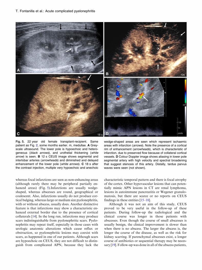
Fig. 5. 22 year old female transplant-recipient. Samepatient as Fig. 2, some months earlier. m, medullae. A Grey-scale ultrasound. The lower pole is hypoechoic and hetero-geneous (black arrows), and urothelial thickening (whitearrow) is seen. B 12 s CEUS image shows segmental andinterlobar arteries (arrowheads) and diminished and delayedenhancement of the lower pole (white arrows). C 18 s afterthe contrast injection, multiple very hypoechoic and anechoic
wedge-shaped areas are seen which represent ischaemicareas with infarction (arrows). Note the presence of a corticalrim of enhancement (arrowheads), which is characteristic ofinfarction, due to preserved flow because of collateral corticalvessels. D Colour Doppler image shows aliasing in lower polesegmental artery with high velocity and spectral broadeningthat suggest stenosis of this artery. Distally, tardus parvuswaves were seen (not shown).
T. Fontanilla et al.: Acute complicated pyelonephritis
but not in all of the focal pyelonephritis patients, so it ispossible that some scarring may have been missed.
An important factor to bear in mind is that UScontrast media are not nephrotoxic and can be employedsafely, even in patients with marginal renal function.Detection of complicated APN is of outmost importancein renal transplant recipients because it may be relatedwith deterioration of graft function [8], but it is also veryimportant to minimise the risk of nephrotoxicity. Inthese patients CEUS is very accurate due to the excellentvisualization of the kidney.
Taking everything into consideration, it seems rea-sonable to choose CEUS first in complicated APNpatients, and to performCT in selected cases, so as to avoidradiation. In our institution, renal CEUS is now per-formed in the first place, with follow-up at 1 week and1 month. Depending on the initial lesion (if there wasabscess or not) and the evolution of the lesion, follow-up at3 months and a year is done, so as to ascertain the com-plete resolution or the development of scars.CTorMRareperformed if large renal abscesses or extrarenal collectionsare seen, because they perform better in the evaluation ofperirenal or pararenal involvement; also if anatomicalterations are suspected. But even in these cases, follow-up is usually done with CEUS. Emphysematous pyelone-phritis must always be evaluated and followed with CT[20]. DMSA scintigraphy remains the imaging method ofchoice in children [21], because US contrast agents are notapproved for intravascular use in children.
Our study had some limitations. First of all, it is aretrospective study, so the analysis of clinical andimaging records had some inhomogeneity, and the fol-low-up schedule was not established from the beginning.In our study, the exploration throughout all phases wasdone on the symptomatic side and the contralateralkidney was examined for a short time during the earlyparenchymal phase. This makes it possible that smalllesions (not abscesses) may have been unnoticed on thecontralateral kidney, but we feel that the detection ofthese lesions would not have altered the patient man-agement and evolution. Non-optimal kidney visualiza-tion due to patient body habitus or bowel gas is apotential CEUS limitation that was not frequent in thisstudy, probably because of the young age and thin bodyhabitus of most of the patients. Another limitation of thisstudy is the absence of confirmation of the findings withCT in all patients. CT imaging is more panoramic andmore sensitive than US in detecting calculi and under-lying urinary tract anatomic abnormalities, so it is pos-sible that some of these may have been unnoticed inthose cases in which just CEUS was done.
Conclusions
Contrast-enhanced ultrasound is able to diagnose focalpyelonephritis and to distinguish focal pyelonephritis
from abscess, with very typical findings. Because ofabsence of associated radiation and nephrotoxicity,CEUS should be considered the first imaging method forevaluating those patients suspected of complication inacute pyelonephritis. CT should be done when largeabscess or extrarenal collections are seen, or when ana-tomic obstructive alterations or emphysematous pyelo-nephritis are suspected. The authors recommend CEUSas the method of choice for complicated APN follow-up.
References
1. Demertzis J, Menias CO (2007) State of the art: imaging of renalinfections. Emerg Radiol 14:13–22
2. Stunell H, Buckley O, Feeney J, et al. (2007) Imaging of acutepyelonephritis in the adult. Eur Radiol 17:1820–1828
3. Craig W, Wagner B, Travis M (2008) Pyelonephritis: radiologic-pathologic review. RadioGraphics 28:255–276
4. Mitterberger M, Pinggera GM, Colleselli D, et al. (2007) Acutepyelonephritis: comparison of diagnosis with computed tomogra-phy and contrast enhanced ultrasonography. BJU Int 101:341–344
5. Quaia E (2007) Microbubble ultrasound contrast agents: an up-date. Eur Radiol 17:1995–2008
6. Setola SV, Catalano O, Sandomenico F, Siani A (2007) Contrast-enhanced sonography of the kidney. Abdom Imaging 32:21–28
7. Kim B, Lim HK, Choi MH, et al. (2001) Detection of parenchymalabnormalities in acute pyelonephritis by pulse inversion harmonicimaging with or without microbubble ultrasonographic contrastagent: correlation with computed tomography. J Ultrasound Med20(1):5–14
8. Granata A, Andrulli S, Fiorini F, et al. (2011) Diagnosis of acutepyelonephritis by contrast-enhanced ultrasonography in kidneytransplant patients. Nephrol Dial Transplant 26:715–720
9. Claudon M, Cosgrove D, Albrecht T, et al. (2008) Guidelines andgood clinical practice recommendations for contrast enhancedultrasound (CEUS)—update 2008. Ultraschall Med 29:28–44
10. Fontanilla T, Mendo M, Canas T, et al. (2009) Diagnosis anddifferential diagnosis of liver abscesses using contrast-enhanced(SonoVue) ultrasonography. Radiologıa 51(4):403–410
11. Ivanyi B, Thoenes W (1987) Microvascular injury and repair inacute human bacterial pyelonephritis. Virchows Arch A 411(3):257–265
12. Kawashima A, Le Roy AJ (2003) Radiologic evaluation of patientswith renal infections. Infect Dis Clin North Am 17:433–456
13. Kawashima A, Sandler CM, Goldman SM, Raval BK, FishmanEK (1997) CT of renal inflammatory disease. RadioGraphics17:851–866
14. Bertolotto M, Martegani A, Aiani L, et al. (2008) Value of con-trast-enhanced ultrasonography for detecting renal infarcts provenby contrast enhanced CT—a feasibility study. Eur Radiol 18(2):376–383
15. Takahashi N, Kawashima A, Fletcher JG, Chari ST (2007) Renalinvolvement in patients with autoimmune pancreatitis: CT and MRimaging findings. Radiology 242(3):791–801
16. Triantopoulou C, Malachias G, Maniatis P, et al. (2010) Renallesions associated with autoimmune pancreatitis: CT findings. ActaRadiol 51:702–707
17. Ruiz E, Medina A, Lopez G, et al. (2001) Multiple renal masses asinitial manifestation of Wegener’s granulomatosis. AJR Am JRoentgenol 176:116–118
18. Urban BA, Fishman EK (2000) Renal lymphoma: CT patterns withemphasis on helical CT. RadioGraphics 20:197–212
19. Ramakrishnan K, Scheid D (2005) Diagnosis and management ofacute pyelonephritis in adults. Am Fam Phys 71:5
20. Wan YL, Lee TY, Bullard MJ, Tsai CC (1996) Acute gas-pro-ducing bacterial renal infection: correlation between imaging find-ings and clinical outcome. Radiology 198:433–438
21. Boubakery A, Priory JO, Meuwly J-Y, Bischof-Delaloye A (2006)Radionuclide investigations of the urinary tract in the era of mul-timodality imaging. J Nucl Med 47:1819–1836
T. Fontanilla et al.: Acute complicated pyelonephritis
Related Documents