ISSN:1748 0132 © Elsevier Ltd 2007 Constructing novel materials with DNA DNA, apart from being a natural biological information carrier, has also been recognized as a useful building material in the field of nanotechnology. Its miniature scale, geometric properties, and molecular recognition capacity make DNA an appealing candidate for the construction of novel nanomaterials. Here we summarize the latest developments and describe the challenges and emerging applications of this field. Thom H. LaBean 1 * and Hanying Li 2 1 *Departments of Computer Science and Chemistry, Duke University, Durham, NC 27708, USA 2 *Department of Pathology, Duke University Medical Center, Durham, NC 27710, USA E-mail: [email protected] Although the detailed structure of DNA was revealed by Watson and Crick 1,2 back in 1953, even today we continue to discover stunning and useful new structural modes for this versatile macromolecule. Taking lessons from its in vivo role and aided by technological advances, nanoengineers have begun to explore novel and creative uses for DNA including: molecular detection 3 , therapeutic regimens 4 , complex nanodevices 5 , nanomechanical actuators and motors 6-8 , directed organic synthesis 9,10 , and molecular computation 11,12 . Excellent reviews of many of these aspects of DNA can be found in this issue of Nano Today and elsewhere 10,12,13 . In this review, we will focus on the ‘materials’ side of DNA by examining major architectural strategies (linear, branching, and multibranched complexes) and application strategies (directed organization of nanomaterials including biomolecules, templating of inorganics, and approaches combining preformed and templated materials) in which DNA nanotechnology plays a starring role. Architectural strategies Linear DNA for conducting nanowires The use of DNA to form building blocks for nanoelectronic constructs is quite promising, although for many years the conductivity of bare DNA remained controversial. Electron transfer in neat DNA has been observed in many cases 14-17 but there are other experiments suggesting that DNA might display semiconducting 18,19 , insulating 20 , or even superconducting behavior 21 . Although coherent electron tunneling and diffusive thermal hopping – the two most fundamental processes for charge transfer – have been clearly demonstrated in DNA 22,23 , the electron transfer behavior of bare DNA is typically insufficient for nanoelectronic engineering purposes. However, DNA has been used as a template upon which to organize more highly conductive materials such as metals for electronic applications. Lee et al. 24 described a new form of DNA, M-DNA, in which the imino proton of the DNA base- pairs is replaced by a Zn 2+ , Ni 2+ , or Co 2+ ion (Fig. 1a). It has been shown that M-DNA behaves like a molecular wire and has potential for the development of future molecular electronics 25-27 . APRIL 2007 | VOLUME 2 | NUMBER 2 26

Welcome message from author
This document is posted to help you gain knowledge. Please leave a comment to let me know what you think about it! Share it to your friends and learn new things together.
Transcript
ISSN:1748 0132 © Elsevier Ltd 2007
Constructing novel materials with DNADNA, apart from being a natural biological information carrier, has also been recognized as a useful building material in the field of nanotechnology. Its miniature scale, geometric properties, and molecular recognition capacity make DNA an appealing candidate for the construction of novel nanomaterials. Here we summarize the latest developments and describe the challenges and emerging applications of this field.
Thom H. LaBean1* and Hanying Li2
1*Departments of Computer Science and Chemistry, Duke University, Durham, NC 27708, USA
2*Department of Pathology, Duke University Medical Center, Durham, NC 27710, USA
E-mail: [email protected]
Although the detailed structure of DNA was revealed by Watson
and Crick1,2 back in 1953, even today we continue to discover
stunning and useful new structural modes for this versatile
macromolecule. Taking lessons from its in vivo role and aided by
technological advances, nanoengineers have begun to explore
novel and creative uses for DNA including: molecular detection3,
therapeutic regimens4, complex nanodevices5, nanomechanical
actuators and motors6-8, directed organic synthesis9,10, and
molecular computation11,12 . Excellent reviews of many of these
aspects of DNA can be found in this issue of Nano Today and
elsewhere10,12,13.
In this review, we will focus on the ‘materials’ side of DNA by
examining major architectural strategies (linear, branching, and
multibranched complexes) and application strategies (directed
organization of nanomaterials including biomolecules, templating
of inorganics, and approaches combining preformed and
templated materials) in which DNA nanotechnology plays a
starring role.
Architectural strategiesLinear DNA for conducting nanowiresThe use of DNA to form building blocks for nanoelectronic constructs
is quite promising, although for many years the conductivity of
bare DNA remained controversial. Electron transfer in neat DNA has
been observed in many cases14-17 but there are other experiments
suggesting that DNA might display semiconducting18,19, insulating20, or
even superconducting behavior21. Although coherent electron tunneling
and diffusive thermal hopping – the two most fundamental processes
for charge transfer – have been clearly demonstrated in DNA22,23,
the electron transfer behavior of bare DNA is typically insufficient for
nanoelectronic engineering purposes. However, DNA has been used as
a template upon which to organize more highly conductive materials
such as metals for electronic applications. Lee et al.24 described a new
form of DNA, M-DNA, in which the imino proton of the DNA base-
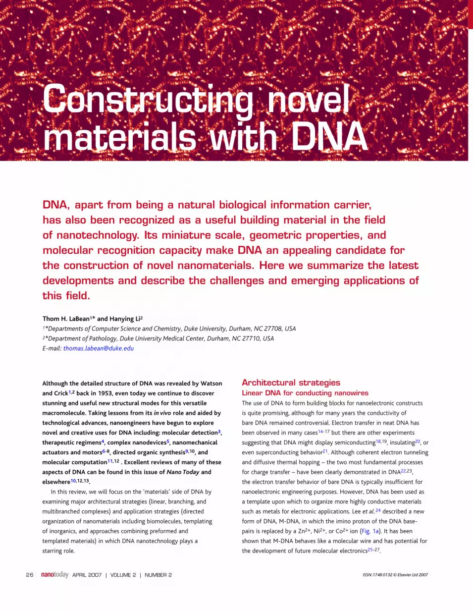
pairs is replaced by a Zn2+, Ni2+, or Co2+ ion (Fig. 1a). It has been
shown that M-DNA behaves like a molecular wire and has potential for
the development of future molecular electronics25-27.
APRIL 2007 | VOLUME 2 | NUMBER 226
NT202p26_35.indd 26NT202p26_35.indd 26 23/02/2007 08:38:2623/02/2007 08:38:26
Novel materials from DNA REVIEW
APRIL 2007 | VOLUME 2 | NUMBER 2 27
Other methods to enhance the conductivity of linear DNA have also
been explored. Braun et al.20 successfully constructed DNA templated
Ag nanowires by electroless deposition, producing nanowires ~100 nm
thick and 15 µm long. They attached two short oligonucleotides
to electrodes and introduced λ-phage DNA as a bridge (with ends
complementary to electrode-linked DNAs). Ag ions were then loaded
onto the DNA and reduced to form Ag nanoparticles (AgNPs) and
fine nanowires. The deposition of Pd28, Au29, and Pt30 on DNA have
also been investigated as a potential approach for creating conductive
nanowires. Monson et al.31 described the construction of DNA-
templated Cu nanowires. These wires were about 3 nm tall and may
prove useful in construction of single electron devices. Keren et al.32
recently reported protecting specific regions of DNA molecules from
metal deposition by associating proteins along selected sections of
the DNA. The ability to control metallization spatially provides an
important technological advantage for the assembly of functional
nanocircuits. Recently, Ag nanowires with widths down to 15 nm and
several microns in length (Fig. 1b) have been templated on various
DNA nanostructures and characterized electrically at room temperature
and low temperature33-36.
Linear DNA as smart glueSince the binding strength of DNA double-helices can be easily
controlled (by tuning helix length and base composition) and a huge
lexicon of unique sequences exist, linear DNA is extremely useful
as a structural linker for controlled aggregation of nanomaterials.
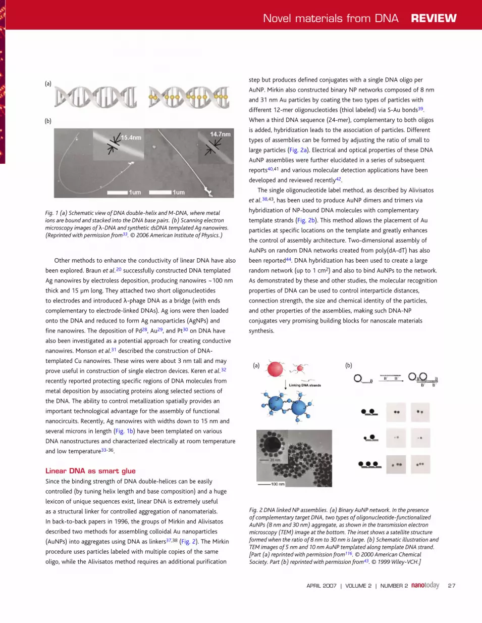
In back-to-back papers in 1996, the groups of Mirkin and Alivisatos
described two methods for assembling colloidal Au nanoparticles
(AuNPs) into aggregates using DNA as linkers37,38 (Fig. 2). The Mirkin
procedure uses particles labeled with multiple copies of the same
oligo, while the Alivisatos method requires an additional purification
step but produces defined conjugates with a single DNA oligo per
AuNP. Mirkin also constructed binary NP networks composed of 8 nm
and 31 nm Au particles by coating the two types of particles with
different 12-mer oligonucleotides (thiol labeled) via S-Au bonds39.
When a third DNA sequence (24-mer), complementary to both oligos
is added, hybridization leads to the association of particles. Different
types of assemblies can be formed by adjusting the ratio of small to
large particles (Fig. 2a). Electrical and optical properties of these DNA
AuNP assemblies were further elucidated in a series of subsequent
reports40,41 and various molecular detection applications have been
developed and reviewed recently42.
The single oligonucleotide label method, as described by Alivisatos
et al.38,43, has been used to produce AuNP dimers and trimers via
hybridization of NP-bound DNA molecules with complementary
template strands (Fig. 2b). This method allows the placement of Au
particles at specific locations on the template and greatly enhances
the control of assembly architecture. Two-dimensional assembly of
AuNPs on random DNA networks created from poly(dA-dT) has also
been reported44. DNA hybridization has been used to create a large
random network (up to 1 cm2) and also to bind AuNPs to the network.
As demonstrated by these and other studies, the molecular recognition
properties of DNA can be used to control interparticle distances,
connection strength, the size and chemical identity of the particles,
and other properties of the assemblies, making such DNA-NP
conjugates very promising building blocks for nanoscale materials
synthesis.
Fig. 1 (a) Schematic view of DNA double-helix and M-DNA, where metal
ions are bound and stacked into the DNA base pairs. (b) Scanning electron
microscopy images of λ-DNA and synthetic dsDNA templated Ag nanowires.
(Reprinted with permission from33. © 2006 American Institute of Physics.)
Fig. 2 DNA linked NP assemblies. (a) Binary AuNP network. In the presence
of complementary target DNA, two types of oligonucleotide-functionalized
AuNPs (8 nm and 30 nm) aggregate, as shown in the transmission electron
microscopy (TEM) image at the bottom. The inset shows a satellite structure
formed when the ratio of 8 nm to 30 nm is large. (b) Schematic illustration and
TEM images of 5 nm and 10 nm AuNP templated along template DNA strand.
[Part (a) reprinted with permission from116. © 2000 American Chemical
Society. Part (b) reprinted with permission from43. © 1999 Wiley-VCH.]
(b)
(a)
(b)(a)
NT202p26_35.indd 27NT202p26_35.indd 27 23/02/2007 08:38:3823/02/2007 08:38:38

REVIEW Novel materials from DNA
APRIL 2007 | VOLUME 2 | NUMBER 228
Branching DNA motifsLinear DNA can be used to assemble a range of different structures,
however, in order to make more diverse constructs, branching DNA
units are highly useful. Substantial progress has been made recently
in designing branching structures from DNA. In 1997, Shchepinov
et al.45 reported the synthesis of DNA dendrimers using synthon, a
novel phosphoramidite. By chemically connecting DNA to different
synthon molecules, dendrimers with a branching backbone and
varying numbers of DNA arms were synthesized46. The development
of DNA block copolymers was also described47,48, in which DNA was
covalently attached to an organic polymer backbone and provided
bridges for assembly and subsequent chemical modification. Recently,
Luo and coworkers49 demonstrated well-defined dendrimer-like
structures assembled via sticky end cohesion from DNA units with
branched secondary structure (Fig. 3a). This study demonstrates
that nearly-monodisperse dendrimeric DNA nanostructures can be
synthesized in a highly controlled fashion with relatively high yield
and purity. More recently, the construction of three-dimensional
hydrogels made entirely from flexible branched DNA building blocks
has been reported50. X-, Y-, and T-shaped DNA units were hybridized
and crosslinked with each other via ligase catalyzed assembly. The size
and shape of these large, three-dimensional hydrogels can be easily
controlled by using different molds (Fig. 3b). Novel soft materials
created from DNA hydrogels may find future applications in cell and
tissue culture, drug delivery, and cell-free protein synthesis.
Complex DNA motifs for structural building blocksApart from playing the role of smart glue to facilitate the assembly
of other molecules, DNA itself can be used to form rigid building
blocks for the construction of complex nanostructures. Seeman has
referred to this strategy as ‘bricks plus mortar’ because DNA itself
makes up the building blocks and the cement holding them together.
The obvious advantage is that it is easier to define solution conditions
in which DNA is soluble and well-behaved than conditions under
which both DNA and metallic NPs (for example) are soluble and
stable. Seeman and coworkers were the first to exploit DNA’s self-
complementarity for construction of novel nanostructures51. Since
the simple double-helix lacks the complexity needed for forming
tightly controlled two- and three-dimensional structures, they sought
to design more complex building blocks. They succeeded in making
branched junction motifs with four double-helical arms, which resemble
Holliday junctions, a natural conformation of DNA found in biological
homologous recombination complexes. In theory, these branched
junction units should assemble into a quadrilateral lattice by sticky end
cohesion52-54, yet in solution, the junctions (illustrated in Fig. 4) do
not assemble into a two-dimensional lattice because the structure is
Fig. 3 DNA dendrimer and hydrogel. (a) Dendrimer-like DNA (DL-DNA) formed by the ligation of Y-shaped DNA. The scheme in the middle shows higher generation
DL-DNA, which corresponds to the AFM image on the right. Scale bar corresponds to 100 nm. (b) Left and middle: schematic view of Y-shaped DNA monomer and
three-dimensional DNA hydrogels. (Right) DNA hydrogels built from X-shaped monomers patterned into different shapes. Scale bar corresponds to 1 cm. [Part (a)
reprinted with permission from49. © 2004 Nature Publishing Group. Part (b) reprinted with permission from50,117. © 2006 Nature Publishing Group.]
(b)
(a)
NT202p26_35.indd 28NT202p26_35.indd 28 23/02/2007 08:38:4023/02/2007 08:38:40

Novel materials from DNA REVIEW
APRIL 2007 | VOLUME 2 | NUMBER 2 29
not stiff enough to hold the helical domains in the same plane and
instead they twist by ~60° (Fig. 4a). Seeman’s group had previously
reported the construction of a closed DNA cube55 and a truncated
octahedron56,57 (Fig. 4b) albeit at very low yields. Interestingly, the
four-arm branch motif can be stiffened when combined in pairs
and larger constructs. Mao et al.58 have fused four junctions into
a rhombus-like building block and successfully demonstrated the
further assembly into two-dimensional lattices (Fig. 5e). Turberfield
recently reported the use of RuvA, a Holliday junction binding protein,
to control the conformation and facilitate the assembly of square
lattices59.
Based on the idea of employing immobile DNA junctions, a large
number of distinct DNA building blocks (or tiles) have been designed
and experimentally implemented in the past two decades (Fig. 5).
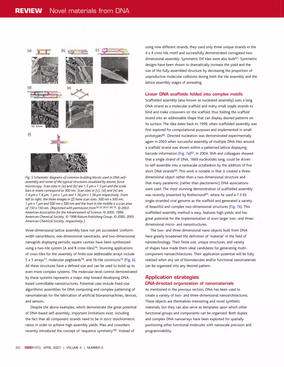
Li et al.60 have reported the construction of double-crossover (DX)
complexes, which consist of two juxtaposed Holliday-junction-like
crossover motifs joined together by two double-helical domains.
Properly designed sticky ends further facilitate the assembly into
periodic one- and two-dimensional lattices (Fig. 5a,b)61,62. A related
motif combining a stem-loop hairpin with one of the duplex arms of
DX (known as DX+J) has also been reported61. The extra hairpin is
used as a topographical marker, visible by atomic force microscopy
(AFM)61,63. Seeman and coworkers also describe the generation of
paranemic crossovers (PX)64,65, which arise from fusion of two close,
parallel double-helices by reciprocal exchange at every possible contact
point. By controlling the interconversion between a PX junction and its
topoisomeric JX state, a robust DNA nanomechanical device has been
built and studied66,67.
A more complex planar building block was reported by LaBean
et al.68 in 2000. This triple crossover complex (TX) contained three
helices and four crossovers, and as in DX tiles, two adjacent helices
were connected by two four-arm junctions. Compared to DX tiles, TXs
provide larger space for gaps in two-dimensional arrays and further
extend the prototyping of useful branching building blocks (Fig. 5c).
Another DNA motif, the 4 x 4 cross-tile, consisting of four four-arm
junctions was reported in 2003 (Fig. 5e)34. Since the cross-tile has a
square aspect ratio and helix stacking in all four directions in the plane,
they can assemble into very large two-dimensional lattices. Structures
with triangular building blocks have also been developed. In 1998, DX
tiles were successfully ligated with DNA triangles, to create a unique
zigzag pattern69. Two triangle tile types have been prototyped, which
feature the formation of triangular and hexagonal patterns70,71.
These basic building blocks and their variants have been used
in the construction of self-assembled lattices. DX and TX lattices,
ribbons, and tubes have been observed. More complicated designs
including double-double crossover72, 4-, 8-, and 12-helix DNA tile
complexes have also been realized recently73. These tiles have also
been used for the assembly of planar and tubular structures. DNA tiles
that hold their helices in nonplanar domains have been prototyped
by several groups35,74,75, although attempts at using these tiles for
Fig. 4 (a) Schematic drawings of four-arm junctions and the proposed assembly of a two-dimensional lattice via sticky ends cohesion. The twist between the two
helical domains is illustrated in the bottom row. (b) DNA molecule with the connectivity of a cube and an octahedron. Each of the edges is composed of double-
helical DNA. [Part (a) adapted from54. Part (b) reprinted with permission from54. © 1998 Institute of Physics.]
(b)
(a)
NT202p26_35.indd 29NT202p26_35.indd 29 23/02/2007 08:38:4123/02/2007 08:38:41
REVIEW Novel materials from DNA
APRIL 2007 | VOLUME 2 | NUMBER 230
three-dimensional lattice assembly have not yet succeeded. Uniform-
width nanoribbons, one-dimensional nanotracks, and two-dimensional
nanogrids displaying periodic square cavities have been synthesized
using a two-tile system (A and B cross-tiles)76. Stunning applications
of cross-tiles for the assembly of finite-size addressable arrays include
5 x 5 arrays77, molecular pegboards78, and 16-tile constructs79 (Fig. 6).
All these structures have a defined size and can be used to build up to
even more complex systems. The molecular-level control demonstrated
by these systems represents a major step toward developing DNA-
based controllable nanostructures. Potential uses include fixed-size
algorithmic assemblies for DNA computing and complex patterning of
nanomaterials for the fabrication of artificial bionanomachines, devices,
and sensors.
Despite the above examples, which demonstrate the great potential
of DNA-based self-assembly, important limitations exist, including
the fact that all component strands need to be in strict stoichiometric
ratios in order to achieve high assembly yields. Mao and coworkers
recently introduced the concept of ‘sequence symmetry’80. Instead of
using nine different strands, they used only three unique strands in the
4 x 4 cross-tile motif and successfully demonstrated corrugated two-
dimensional assembly. Symmetric DX tiles were also built81. Symmetric
designs have been shown to dramatically increase the yield and the
size of the fully-assembled structure by decreasing the proportion of
unproductive molecular collisions during both the tile assembly and the
lattice assembly stages of annealing.
Linear DNA scaffolds folded into complex motifs Scaffolded assembly (also known as nucleated assembly) uses a long
DNA strand as a molecular scaffold and many small staple strands to
bind and make crossovers on the scaffold, thus folding the scaffold
strand into an addressable shape that can display desired patterns on
its surface. The idea dates back to 1999, when scaffolded assembly was
first explored for computational purposes and implemented in small
prototypes82. Directed nucleation was demonstrated experimentally
again in 2003 when successful assembly of multiple DNA tiles around
a scaffold strand was shown within a patterned lattice displaying
barcode information (Fig. 7a)83. In 2004, Shih and colleagues showed
that a single strand of DNA, 1669 nucleotides long, could be driven
to self-assemble into a nanoscale octahedron by the addition of five
short DNA strands84. This work is notable in that it created a three-
dimensional object rather than a two-dimensional structure and
that many paranemic (rather than plectonemic) DNA associations
were used. The most stunning demonstration of scaffolded assembly
was recently presented by Rothemund85, where he used a 7.3 Kb
single-stranded viral genome as the scaffold and generated a variety
of beautiful and complex two-dimensional structures (Fig. 7b). This
scaffolded assembly method is easy, features high yields, and has
great potential for the implementation of even larger two- and three-
dimensional micro- and nanostructures.
The two- and three-dimensional nano-objects built from DNA
have greatly broadened the definition of ‘material’ in the field of
nanotechnology. Their finite size, unique structures, and variety
of shapes have made them ideal candidates for generating multi-
component nanoarchitectures. Their application potential will be fully
realized when any set of biomolecules and/or functional nanomaterials
can be organized into any desired pattern.
Application strategiesDNA-directed organization of nanomaterialsAs mentioned in the previous section, DNA has been used to
create a variety of two- and three-dimensional nanoarchitectures.
These objects are themselves interesting and novel synthetic
materials, but they can also serve as templates upon which other
functional groups and components can be organized. Both duplex
and complex DNA nanoarrays have been exploited for spatially
positioning other functional molecules with nanoscale precision and
programmability.
Fig. 5 Schematic diagrams of common building blocks used in DNA self-
assembly and some of the typical structures visualized by atomic force
microscopy. Scan sizes in (a) and (b) are 1.5 µm x 1.5 µm and the scale
bars in insets correspond to 300 nm. Scan sizes in (c), (d) and (e) are
1.4 µm x 1.4 µm, 1 µm x 1 µm and 1.56 µm x 1.56 µm respectively. From
left to right, the three images in (f) have scan sizes: 500 nm x 500 nm,
1 µm x 1 µm and 500 nm x 500 nm and the inset in the middle is a scan area
of 150 x 150 nm. (Reprinted with permission from34,35,58,61,68,76. © 2003
American Association for the Advancement of Science, © 2005, 1999,
American Chemical Society, © 1998 Nature Publishing Group, © 2000, 2005
American Chemical Society, respectively.)
(b)(a) (c)
(d) (e)
(f)
NT202p26_35.indd 30NT202p26_35.indd 30 23/02/2007 08:38:4223/02/2007 08:38:42

Novel materials from DNA REVIEW
APRIL 2007 | VOLUME 2 | NUMBER 2 31
Fig. 6 Finite-sized and addressable DNA patterns. (a) DNA nanoarrays with increased complexity and defined sizes based on the cross-tile or 8-helix bundle. From
left to right: molecular pegboard, 4 x 4, symmetric 5 x 5, and 8-helix bundle based 5 x 5 arrays. (b) Full addressability of the ten tile system with an additional index
tile added to the pegboard design. (c) The letters ‘D’, ‘N’, and ‘A’ displayed on self-assembled 4 x 4 cross-tile arrays. [Part (a) reprinted with permission from77-79.
© 2005 American Chemical Society, © 2006 Wiley-VCH. Part (b) reprinted with permission from78. © 2005 American Chemical Society. Part (c) reprinted with
permission from79. © 2006 Wiley-VCH.]
Fig. 7 Nucleated DNA self-assembly and scaffolded origami. (a) Self-assembly of 01101 barcode lattice around scaffold DNA strand and corresponding atomic
force microscopy (AFM) visualization. (b) The left schematic shows an arbitrary shape formed when the scaffold strand (black) is folded by hybridization with staple
strands (colors). The inset in the left panel shows a staple strand with a protruding stem-loop, used to produce the raised (lighter) pixels in the next panels. The
middle and right panels show a schematic drawing and an AFM image, respectively, of structures formed by folding six copies of the 7.3 Kb M13 virus genomic DNA
into triangles then hexagons. [Part (a) reprinted with permission from83. © 2003 National Academy of Sciences. Part (b) reprinted with permission from85. © 2006
Nature Publishing Group.]
(b)
(a)
(c)
(b)
(a)
NT202p26_35.indd 31NT202p26_35.indd 31 23/02/2007 08:38:4323/02/2007 08:38:43
REVIEW Novel materials from DNA
APRIL 2007 | VOLUME 2 | NUMBER 232
In the past, many strategies have been exploited for DNA-directed
NP assemblies and a wide variety of patterns have been built. First
of all, metal or semiconductor ions can directly adsorb to DNA
templates via electrostatic interactions, groove binding, or intercalation.
Reduction of these ions will facilitate the formation of NPs along the
DNA templates. Ag20, Au32, Pd28,86, Pt30, and semiconductor NPs87,88
have been successfully templated on DNA using this approach. These
DNA-NP arrays can serve as precursors for nanowires or nanoelectronic
devices, as previously discussed. However, it is hard to achieve
consistent interparticle spacing at long range by using this technique
and only coarse and irregular metallized structures can be built.
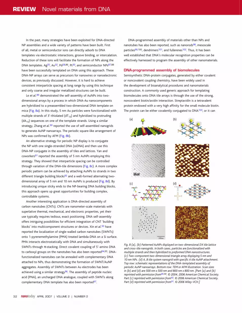
Le et al.89 demonstrated the self-assembly of AuNPs into two-
dimensional arrays by a process in which DNA-Au nanocomponents
are hybridized to a preassembled two-dimensional DNA template on
mica (Fig. 8a). In this study, 5 nm Au particles were functionalized with
multiple strands of 3’-thiolated (dT15) and hybridized to protruding
(dA15) sequences on one of the template strands. Using a similar
strategy, Zhang et al.90 reported the use of self-assembled nanogrids
to generate AuNP nanoarrays. The periodic square-like arrangement of
NPs was confirmed by AFM (Fig. 8b).
An alternative strategy for periodic NP display is to conjugate
the NP with one single-stranded DNA (ssDNA) and then use this
DNA-NP conjugate in the assembly of tiles and lattices. Yan and
coworkers91 reported the assembly of 5 nm AuNPs employing this
strategy. They showed that interparticle spacing can be controlled
through variation of the DNA-tile dimensions (Fig. 8c). A more complex
periodic pattern can be achieved by attaching AuNPs to strands in two
different triangle building blocks92 and a well-formed alternating two-
dimensional array of 5 nm and 10 nm AuNPs is produced (Fig. 8d). By
introducing unique sticky ends to the NP-bearing DNA building blocks,
this approach opens up great opportunities for building complex,
controllable systems.
Another interesting application is DNA-directed assembly of
carbon nanotubes (CNTs). CNTs are nanometer-scale materials with
superlative thermal, mechanical, and electronic properties, yet their
use typically requires tedious, exact positioning. DNA self-assembly
offers intriguing possibilities for efficient integration of CNT ‘building
blocks’ into multicomponent structures or devices. Xin et al.93 have
reported the localization of single-walled carbon nanotubes (SWNTs)
onto 1-pyrenemethylamine (PMA) treated lambda-DNA on a Si surface.
PMA interacts electrostatically with DNA and simultaneously with
SWNTs through π-stacking. Direct covalent coupling of 5’-amino DNA
to carboxyl groups on the nanotubes has also been reported94,95. DNA-
functionalized nanotubes can be annealed with complementary DNA
attached to NPs, thus demonstrating the formation of SWNT/AuNP
aggregates. Assembly of SWNTs between Au electrodes has been
achieved using a similar strategy96. The assembly of peptide nucleic
acid (PNA), an uncharged DNA analogue, coupled with SWNTs along
complementary DNA template has also been reported97.
DNA-programmed assembly of materials other than NPs and
nanotubes has also been reported, such as nanorods98, mesoscale
particles99,100, dendrimers101, and fullerenes102. Thus, it has been
well established that DNA’s molecular recognition properties can be
effectively harnessed to program the assembly of other nanomaterials.
DNA-programmed assembly of biomolecules Semisynthetic DNA-protein conjugates, generated by either covalent
or noncovalent coupling chemistry, have been widely used in
the development of bioanalytical procedures and nanomaterials
construction. A commonly used generic approach for templating
biomolecules onto DNA tile arrays is through the use of the strong,
noncovalent biotin/avidin interaction. Streptavidin is a tetravalent
protein endowed with a very high affinity for the small molecule biotin.
The protein can be either covalently conjugated to DNA103, or it can
Fig. 8 (a), (b) Patterned AuNPs displayed on two-dimensional DX tile lattice
and cross-tile nanogrids. In both cases, particles are functionalized with
multiple strands and then hybridized to preformed DNA nanostructures.
(c) Two-component two-dimensional triangle array displaying 5 nm and
10 nm NPs. (d) A, B tile system nanogrid with specific A tile AuNP attachment.
Top row: schematic representations of the DNA-templated assembly of
periodic AuNP nanoarrays. Bottom row: TEM or AFM illustration. Scan sizes
in (b) and (d) are 500 nm x 500 nm and 800 nm x 800 nm. [Part (a) and (b)
reprinted with permission from89,90. © 2004, 2006 American Chemical Society.
Part (c) reprinted with permission from92. © 2006 American Chemical Society.
Part (d) reprinted with permission from91. © 2006 Wiley-VCH.]
(b)(a)
(c) (d)
NT202p26_35.indd 32NT202p26_35.indd 32 23/02/2007 08:38:4523/02/2007 08:38:45
Novel materials from DNA REVIEW
APRIL 2007 | VOLUME 2 | NUMBER 2 33
interact with biotinylated DNA104. The conjugates can be utilized as
biomolecular adapters for positioning biotinylated components along
nucleic acid backbones. Any type of biotinylated compound can be
arranged, such as peptides, antibodies, enzymes, and low molecular
weight components104,105. Because of the tetravalent nature of
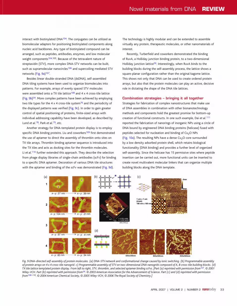
streptavidin (STV), more complex DNA-STV networks can be built,
such as supramolecular nanocircles106 and supercoiling mediated STV
networks (Fig. 9a)107.
Besides linear double-stranded DNA (dsDNA), self-assembled
DNA tiling systems have been used to organize biomolecules into
patterns. For example, arrays of evenly spaced STV molecules
were assembled onto a TX tile lattice108 and 4 x 4 cross-tile lattice
(Fig. 9b)34. More complex patterns have been achieved by employing
two tile types for the 4 x 4 cross-tile system76 and the periodicity of
the displayed patterns was verified (Fig. 9c). In order to gain greater
control of spatial positioning of proteins, finite-sized arrays with
individual addressing capability have been developed, as described by
Lund et al.78, Park et al.79, etc.
Another strategy for DNA-templated protein display is to employ
specific DNA binding proteins. Liu and coworkers109 first demonstrated
the use of aptamer to direct the assembly of thrombin onto sites on
TX tile arrays. Thrombin binding aptamer sequence is introduced into
the TX tiles and acts as docking sites for the thrombin molecules.
Li et al.110 further extended this approach. They describe the selection
from phage display libraries of single-chain antibodies (scFv) for binding
to a specific DNA aptamer. Decoration of various DNA tile structures
with the aptamer and binding of the scFv was demonstrated (Fig. 9d).
The technology is highly modular and can be extended to assemble
virtually any protein, therapeutic molecules, or other nanomaterials of
interest.
Recently, Turberfield and coworkers demonstrated the binding
of RuvA, a Holliday junction binding protein, to a two-dimensional
Holliday junction lattice59. Interestingly, when RuvA binds to the
building blocks during the self-assembly process, the lattice shows a
square-planar configuration rather than the original kagome lattice.
This shows not only that DNA can be used to create ordered protein
arrays, but also that the protein molecules can play an active, decisive
role in dictating the shape of the DNA tile lattices.
Combination strategies – bringing it all togetherStrategies for fabrication of complex nanostructures that make use
of DNA assemblies in combination with other bionanotechnology
methods and components hold the greatest promise for bottom-up
creation of functional constructs. In one such example, Dai et al.111
reported the fabrication of nanorings of inorganic NPs using a circle of
DNA bound by engineered DNA binding proteins (helicase) fused with
peptides selected for nucleation and binding of Cu2O NPs
(Fig. 10a). The resulting NPs have a dense Cu2O core surrounded
by a low density adsorbed protein shell, which retains biological
functionality (DNA binding) and provides a further level of organized
self-assembly. Since the helicase has 15 permissive sites where peptide
insertion can be carried out, more functional units can be inserted to
create novel multivalent molecular linkers that can organize multiple
building blocks along the DNA template.
Fig. 9 DNA-directed self-assembly of protein molecules. (a) DNA-STV network and conformational change caused by ionic switching. (b) Programmable assembly
of protein arrays on 4 x 4 cross-tile nanogrid. c) Programmable assembly of STV on two-dimensional DNA nanogrids composed of A, B cross-tile building blocks. (d)
TX tile lattice templated protein display. From left to right, STV, thrombin, and selected aptamer binding scFvs. [Part (a) reprinted with permission from107. © 2001
Wiley-VCH. Part (b) reprinted with permission from34. © 2003 American Association for the Advancement of Science. Part (c) and (d) reprinted with permission
from108-110. © 2004 American Chemical Society, © 2005 Wiley-VCH, © 2006 The Royal Society of Chemistry.]
(b)(a) (c)
(d)
NT202p26_35.indd 33NT202p26_35.indd 33 23/02/2007 08:38:4723/02/2007 08:38:47

REVIEW Novel materials from DNA
APRIL 2007 | VOLUME 2 | NUMBER 234
Keren et al.32,112 recently reported another combination strategy
involving DNA, a DNA binding protein, and inorganic nanomaterials.
RecA, a homologous DNA sequence binding protein, was polymerized
onto ssDNA and used to localize a SWNT at a desired position along
the dsDNA template. The RecA also serves to protect the covered
DNA segment against metallization thereby creating an insulating
gap where the SWNT could sit with its ends contacted by the
conductive metal nanowire, thus creating a field-effect transistor
(Fig. 10b). Mixed biomolecular structures comprised of DNA and
other materials including lipids have also been used for templating
inorganics. For example, a multilamellar structure composed of anionic
DNA and cationic lipid membranes has been used to achieve Cd2+ ion
condensation and growth of CdS nanorods113,114 (Fig. 10c). Lieberman
and coworkers115 recently reported the deposition of monolayer DNA
rafts onto (3-amino-propyl) triethoxysilane stripes on a specially
treated Si surface, which shows the possibility of attaching complex
DNA nanostructures at specific sites and demonstrates a promising
approach for combining bottom-up DNA self-assembly with the top-
down nanolithography technique.
Summary and outlookIn summary, DNA has many unique properties that make it a
promising material for development of self-assembly systems in
nanotechnology. The wide range of DNA tile building blocks
already available makes possible the construction of complex
nanostructures. Combined with the wide variety of other available
nanomaterials, a powerful method for nanofabrication by precise
spatial positioning of functional units is quickly developing. Multiple
functions of DNA can be utilized simultaneously and numerous
techniques can be employed. As the understanding of DNA and
the set of tools available in the molecular toolbox continue to
expand, our ability to engineer novel nanomaterials will continue to
advance.
The outlook for DNA assembly in nanotechnology is very promising
with application areas reaching both down toward the atomic level
and up toward the micron level. The studies summarized here have
helped to flesh out many of the detailed design and construction rules
necessary for successful application of artificial biomolecular structures
to future nanotechnologies.
Fig. 10 Combination strategies for DNA-nanomaterial constructs. (a) TEM image of Cu2O-NPs assembled onto circular DNA. (b) SWNT localization in the middle
of a scaffold λ-DNA molecule mediated by RecA. From top to bottom: RecA nucleoprotein filament (black arrow) bound to template DNA; SWNT bound to RecA
filament; scanning conductance image showing the contrast between the conductive nanotube and the DNA template. Scale bars are 200 nm, 300 nm, and 300 nm,
respectively. (c) DNA-membrane templates for organizing the growth of CdS NPs. TEM images of CdS grown in free solution compared with that templated by
DNA-membrane complexes. [Part (a) reprinted with permission from111. © 2005 American Chemical Society. Part (b) reprinted with permission from112. © 2003
American Association for the Advancement of Science. Part (c) reprinted with permission from113,114. © 2003, 2004 American Chemical Society.]
(b)(a)
(c)
REFERENCES
1. Watson, J. D., and Crick, F. H., Nature (1953) 171, 737
2. Watson, J. D., and Crick, F. H., Cold Spring Harb. Symp. Quant. Biol. (1953) 18,
123
3. Bunka, D. H., and Stockley, P. G., Nat. Rev. Microbiol. (2006) 4, 588
4. Nimjee, S. M., et al., Annu. Rev. Med. (2005) 56, 555
5. Beyer, S., and Simmel, F. C., Nucleic Acids Res. (2006) 34, 1581
6. Mao, C., et al., Nature (1999) 397, 144
7. Yurke, B., et al., Nature (2000) 406, 605
8. Turberfield, A. J., et al., Phys. Rev. Lett. (2003) 90, 118102
9. Liu, D. R., PLoS Biol. (2004) 2, E223
10. Gothelf, K. V., and LaBean, T. H., Org. Biomol. Chem. (2005) 3, 4023
11. Amos, M., Theoretical and Experimental DNA Computation, Springer-Verlag, Berlin
(2005)
12. Stojanovic, M. N., et al., Computing with Nucleic Acids. In Bioelectronics: From
Theory to Applications, Willner, I., and Katz, E. (eds.), Wiley-VCH, Weinheim,
Germany, (2005)
NT202p26_35.indd 34NT202p26_35.indd 34 23/02/2007 08:38:4923/02/2007 08:38:49

Novel materials from DNA REVIEW
APRIL 2007 | VOLUME 2 | NUMBER 2 35
13. Xu, J., et al., DNA Structures and their Applications in Nanotechnology, In
Supramolecular Polymers, 2nd edition, Ciferri, A., (ed.) CRC Press, Boca Raton,
Florida, (2005)
14. Murphy, C. J., et al., Science (1993) 262, 1025
15. Arkin, M. R., et al., Science (1996) 273, 475
16. Beratan, D. N., et al., Chem. Biol. (1997) 4, 3
17. Taubes, G., Science (1997) 275, 1420
18. Fink, H.-W., and Schonenberger, C., Nature (1999) 398, 407
19. Porath, D., et al., Nature (2000) 403, 635
20. Braun, E., et al., Nature (1998) 391, 775
21. Kasumov, A. Y., et al., Science (2001) 291, 280
22. Berlin, Y. A., et al., J. Am. Chem. Soc. (2001) 123, 260
23. Berlin, Y., et al., Top. Curr. Chem. (2004) 237, 1
24. Lee, J., et al., Biochem. Cell Biol. (1993) 71, 162
25. Aich, P., et al., J. Mol. Biol. (1999) 294, 477
26. Rakitin, A., et al., Phys. Rev. Lett. (2001) 86, 3670
27. Wettig, S. D., et al., Anal. Sci. (2003) 19, 23
28. Richter, J., et al., Adv. Mater. (2000) 12, 507
29. Patolsky, F., et al., Angew. Chem., Int. Ed. (2002) 41, 2323
30. Ford, W. E., et al., Adv. Mater. (2001) 13, 1793
31. Monson, C. F., and Woolley, A. T., Nano Lett. (2003) 3, 359
32. Keren, K., et al., Science (2002) 297, 72
33. Park, S. H., et al., Appl. Phys. Lett. (2006) 89, 033901
34. Yan, H., et al., Science (2003) 301, 1882
35. Park, S. H., et al., Nano Lett. (2005) 5, 693
36. Park, S. H., et al., Nanotechnology (2004) 15, S525
37. Mirkin, C. A., et al., Nature (1996) 382, 607
38. Alivisatos, A. P., et al., Nature (1996) 382, 609
39. Mucic, R. C., et al., J. Am. Chem. Soc. (1998) 120, 12674
40. Storhoff, J. J., et al., J. Am. Chem. Soc. (2000) 122, 4640
41. Lazarides, A. A., and Schatz, G. C., J. Phys. Chem. B (2000) 104, 460
42. Rosi, N. L., and Mirkin, C. A., Chem. Rev. (2005) 105, 1547
43. Loweth, C. J., et al., Angew. Chem., Int. Ed. (1999) 38, 1808
44. Maeda, Y., et al., Appl. Phys. Lett. (2001) 79, 1181
45. Shchepinov, M. S., et al., Nucleic Acids Res. (1997) 25, 4447
46. Shchepinov, M. S., et al., Nucleic Acids Res. (1999) 27, 3035
47. Watson, K. J., et al., J. Am. Chem. Soc. (2001) 123, 5592
48. Li, Z., et al., Nano Lett. (2004) 4, 1055
49. Li, Y., et al., Nat. Mater. (2004) 3, 38
50. Um, S. H., et al., Nat. Mater. (2006) 5, 797
51. Seeman, N. C., J. Theor. Biol. (1982) 99, 237
52. Kallenbach, N. R., et al., Nature (1983) 305, 829
53. Seeman, N. C., and Kallenbach, N. R., Biophys. J. (1983) 44, 201
54. Seeman, N. C., et al., Nanotechnology (1998) 9, 257
55. Chen, J., and Seeman, N. C., Nature (1991) 350, 631
56. Zhang, Y., and Seeman, N. C., Biophys. J. (1994) 66, A24
57. Zhang, Y., and Seeman, N. C., J. Am. Chem. Soc. (1994) 116, 1661
58. Mao, C., et al., J. Am. Chem. Soc. (1999) 121, 5437
59. Malo, J., et al., Angew. Chem., Int. Ed. (2005) 44, 3057
60. Li, X., et al., J. Am. Chem. Soc. (1996) 118, 6131
61. Winfree, E., et al., Nature (1998) 394, 539
62. Sha, R., et al., Chem. Biol. (2000) 7, 743
63. Liu, F., et al., J. Am. Chem. Soc. (1999) 121, 917
64. Seeman, N. C., Nano. Lett. (2001) 1, 22
65. Zhang, X., et al., J. Am. Chem. Soc. (2002) 124, 12940
66. Yan, H., et al., Nature (2002) 415, 62
67. Liao, S., and Seeman, N. C., Science (2004) 306, 2072
68. LaBean, T. H., et al., J. Am. Chem. Soc. (2000) 122, 1848
69. Yang, X., et al., J. Am. Chem. Soc. (1998) 120, 9779
70. Ding, B., et al., J. Am. Chem. Soc. (2004) 126, 10230
71. Liu, D., et al., J. Am. Chem. Soc. (2004) 126, 2324
72. Reishus, D., et al., J. Am. Chem. Soc. (2005) 127, 17590
73. Ke, Y., et al., J. Am. Chem. Soc. (2006) 128, 4414
74. Wei, B., and Mi, Y., Biomacromolecules (2005) 6, 2528
75. Mathieu, F., et al., Nano Lett. (2005) 5, 661
76. Park, S. H., et al., Nano Lett. (2005) 5, 729
77. Liu, Y., et al., J. Am. Chem. Soc. (2005) 127, 17140
78. Lund, K., et al., J. Am. Chem. Soc. (2005) 127, 17606
79. Park, S. H., et al., Angew. Chem., Int. Ed. (2006) 45, 735
80. He, Y., et al., Angew. Chem. Int., Ed. (2005) 44, 6694
81. Liu, H., et al., Biomacromolecules (2005) 6, 2943
82. LaBean, T. H., et al., Experimental Progress in Computation by Self-Assembly
of DNA Tilings. In DIMACS Series in Discrete Mathematics and Theoretical
Computer Science, Winfree, E., and Gifford, D. K., (eds.) Proceedings of the 5th
DIMACS Workshop on DNA Based Computers, MIT, Cambridge, (1999) 54, 123
83. Yan, H., et al., Proc. Natl. Acad. Sci. USA (2003) 100, 8103
84. Shih, W. M., et al., Nature (2004) 427, 618
85. Rothemund, P. W., Nature (2006) 440, 297
86. Richter, J., et al., Appl. Phys. Lett. (2001) 78, 536
87. Coffer, J. L., et al., Appl. Phys. Lett. (1996) 69, 3851
88. Torimoto, T., J. Phys. Chem. B (1999) 103, 8799
89. Le, J., et al., Nano Lett. (2004) 4, 2343
90. Zhang, J., et al., Nano Lett. (2006) 6, 248
91. Sharma, J., et al., Angew. Chem., Int. Ed. (2006) 45, 730
92. Zheng J., et al., Nano Lett. (2006) 6, 1502
93. Xin, H. J., and Woolley, A. T., J. Am. Chem. Soc. (2003) 125, 8710
94. Dwyer C., et al., Nanotechnology (2002) 13, 601
95. Li, S., et al., J. Am. Chem. Soc. (2005) 127, 14
96. Hazani, M., et al., Chem. Phys. Lett. (2004) 391, 389
97. Williams, K. A., et al., Nature (2002) 420, 761
98. Dujardin, E., et al., Chem. Commun. (2001), 1264
99. Soto, C. M., et al., J. Am. Chem. Soc. (2002) 124, 8508
100. Milam, V. T., et al., Langmuir (2003) 19, 10317
101. Choi, Y. S., et al., Nano Lett. (2004) 4, 391
102. Cassell, A. M., et al., Angew. Chem., Int. Ed. (1998) 37, 1528
103. Niemeyer, C. M., et al., Angew. Chem., Int. Ed. (1998) 37, 2265
104. Niemeyer, C. M., Trends Biotechnol. (2002) 20, 395
105. Niemeyer, C. M., Angew. Chem., Int. Ed. (2001) 40, 4128
106. Niemeyer, C. M., et al., Angew. Chem., Int. Ed. (2000) 39, 3055
107. Niemeyer, C. M., et al., ChemBioChem (2001) 2, 260
108. Li, H., et al., J. Am. Chem. Soc. (2004) 126, 418
109. Liu, Y., et al., Angew. Chem., Int. Ed. (2005) 44, 4333
110. Li, H., et al., Org. Biomol. Chem. (2006) 4, 3420
111. Dai, H., et al., J. Am. Chem. Soc. (2005) 127, 15637
112. Keren, K, et al., Science (2003) 302, 1380
113. Liang, H., et al., J. Am. Chem. Soc. (2003) 125, 11786
114. Liang, H., et al., J. Am. Chem. Soc. (2004) 126, 14157
115. Sarveswaran, K., et al., Langmuir (2006) 22, 11279
116. Mirkin, C. A., Inorg. Chem. (2000) 39, 2258
117. LaBean, T. H., Nat. Mater. (2006) 5, 767
NT202p26_35.indd 35NT202p26_35.indd 35 23/02/2007 08:38:5123/02/2007 08:38:51
Related Documents