Communication Vol. 263, No. 8, Issue of Msrch 15, pp. 3554-3557, 1988 THE JOURNAL OF BIOLOGICAL CHEMISTRY Printed in U.S.A. Bending of the Bacteriophage X Attachment Site by Escherichia coli Integration Host Factor* (Received for publication, October 30, 1987) Carol A. Robertson and Howard A. Nash From the Laboratory of Molecular Bwbgy, National Institute of Mental Health, National Institutes of Health, Bethesda, Marylund 20892 Escherichia coli integration host factor (IHF) is a small basic protein that is required for efficient inte- grative recombination of bacteriophage X. IHF binds specifically to sequences within attP, the site in bac- teriophage X that undergoes recombination.It has been suggested that the binding of IHF creates bends in DNA SO as to help attP condense into a compact structure that is activated for recombination. In this work we show that IHF binding to either of two sites found within attP does indeed produce bending of DNA. In contrast, the other recombination protein needed for integrative recombination, Int, does not appreciably bend the DNA to which it is bound. Inagreement with the proposal that IHF bending is important for creating a condensed attP, bending by IHF persists in the pres- ence of boundInt. Our conclusions about protein-directed bends in DNA are based on the study of the electrophoretic mobility of a set of permuted DNA fragments in the presence or absence of IHF and/or Int. To facilitate this study, we have constructed a novel vector that simplifies the generation of permuted fragments. This vector should be useful in studying the bending of other DNA sequences by specific binding proteins. When bacteriophage X infects Escherichia coli, it can follow one of two alternative life cycles (1). On the one hand, the virus can replicate its chromosome, make large amounts of viral protein, and ultimately lyse thehost cell, releasing hundreds of viral progeny. On the other hand, the virus can enter a quiescent statewhere it coexists stably with the host. In this lysogenic pathway, replication of the virus is assured by the integration of the viral genome into the host genome. Integration is a site-specific event, taking place at unique loci, called attachment sites, on the viral and host chromosomes. In recent years, a large body of evidence from a variety of biochemical studies has made it clear that the viral attach- ment site, attP, is the dominant partner. attP encompasses about 250 bp’ of essential DNA sequence, making it 10 times larger than bacterial attachment site, attB (2). Recombination proteins bind to multiple sites within attP and, under the influence of supercoiling, organize this DNA into a compact, ordered structure, the intasome (3,4). Recent evidence’ shows * The costs of publication of this article were defrayed in part by the payment of page charges. This article must therefore be hereby marked “aduertisement” in accordance with 18 U.S.C. Section 1734 solely to indicate this fact. The abbreviations used are: bp, base pair; IHF, integration host factor. * E. Richet, P. Abcarian, and H. Nash, manuscript in preparation. that recombination depends on the capacity of this nucleo- protein assembly to interact with sequences in attB and thereby juxtapose thetwo attachment sites. In this synaptic step, attB behaves as apassive target while the intasome provides all thenecessary proteins and structural features. It is therefore clear that in order to understand the mechanism of integrative recombination, one must understand the orga- nization of the intasome. Much is known about the binding of recombination proteins to attP. Int protein, a recombinase encoded by the virus binds to sequences located at the boundary of the core region of attP, the region common to attP and attB that contains the crossover point (5). When bound to thesesequences, Int can direct the breakage and reunion of attachment site DNA (6). Int also binds to different sequences that lie far from the core, in the so-called arms of the attachment site (7). Binding of Int to both arm and core sites is necessary but not sufficient for recombination. To form an intasome, attP must also bind IHF (4), a small (M, = 20,000) basic protein encoded by the E. coli host (8). There are three binding sites for IHF scattered throughout the arms of attP (9) and all three are essential for efficient recombination (10). The IHF binding sites are inter- mingled with Int binding sites and IHF can help Int bind to attP (9). However, even under conditions in which all Int binding sites are saturated with protein, IHF is still required to form a functional intasome (4). What critical role(s) does IHF play in generating this structure? We have speculated that IHF might be needed simply to help bend attachment site DNA into a suitably compact form (4, 9). In this paper, we test the most straightforward prediction of this hypothesis, i.e. whether IHF bends the segments of attP to which it binds. To detect bending of DNA we have used the strategy introduced by Crothers and colleagues (11). In this method, gel electrophoresis is performed on a series of fragments of identical length and composition. Each member of the series differs only by a permutation in the position of its ends. If the permuted segment of DNA containsa bend, the effect on electrophoresis will be greatest for that member of the set in which the bend is near the middle of the fragment. This is thoughtto reflect thetendency of DNAto move end-on through the pores of the gel by a snake-like motion (12); a bend near the middle creates the maximum drag for such “reptation.” Thus, a permutation-dependent change in mo- bility is diagnostic of a bend in DNA, and the permutation that causes the greatest retardation identifies the position of the bend. Several strategies can be used to generate permuted sets of fragments. One can circularize a fragment containing a site of interest and then cleave it with a set of enzymes, each of which cuts the circle only once. Alternatively, one can clone a tandem dimer of the desired fragment and liberate permuted monomers from the surrounding DNA by restriction, again using enzymes that cut only once in thedesired fragment (11, 13). Shuey and Parker (14) have introduced a variant of this technique in which the dimer is composed of directly repeated vector sequences thatflank a single copy of a fragment containing the site of interest. Here, the permuted fragments are generated by restricting with enzymes that cut once within the repeated vector but not at all within the target site. To generate material for their permutation analysis, Shuey and 3554

Welcome message from author
This document is posted to help you gain knowledge. Please leave a comment to let me know what you think about it! Share it to your friends and learn new things together.
Transcript

Communication Vol. 263, No. 8, Issue of Msrch 15, pp. 3554-3557, 1988 THE JOURNAL OF BIOLOGICAL CHEMISTRY
Printed in U.S.A.
Bending of the Bacteriophage X Attachment Site by Escherichia coli Integration Host Factor*
(Received for publication, October 30, 1987)
Carol A. Robertson and Howard A. Nash From the Laboratory of Molecular Bwbgy, National Institute of Mental Health, National Institutes of Health, Bethesda, Marylund 20892
Escherichia coli integration host factor (IHF) is a small basic protein that is required for efficient inte- grative recombination of bacteriophage X. IHF binds specifically to sequences within attP, the site in bac- teriophage X that undergoes recombination. It has been suggested that the binding of IHF creates bends in DNA SO as to help attP condense into a compact structure that is activated for recombination. In this work we show that IHF binding to either of two sites found within attP does indeed produce bending of DNA. In contrast, the other recombination protein needed for integrative recombination, Int, does not appreciably bend the DNA to which it is bound. In agreement with the proposal that IHF bending is important for creating a condensed attP, bending by IHF persists in the pres- ence of bound Int.
Our conclusions about protein-directed bends in DNA are based on the study of the electrophoretic mobility of a set of permuted DNA fragments in the presence or absence of IHF and/or Int. To facilitate this study, we have constructed a novel vector that simplifies the generation of permuted fragments. This vector should be useful in studying the bending of other DNA sequences by specific binding proteins.
When bacteriophage X infects Escherichia coli, it can follow one of two alternative life cycles (1). On the one hand, the virus can replicate its chromosome, make large amounts of viral protein, and ultimately lyse the host cell, releasing hundreds of viral progeny. On the other hand, the virus can enter a quiescent state where it coexists stably with the host. In this lysogenic pathway, replication of the virus is assured by the integration of the viral genome into the host genome. Integration is a site-specific event, taking place at unique loci, called attachment sites, on the viral and host chromosomes. In recent years, a large body of evidence from a variety of biochemical studies has made it clear that the viral attach- ment site, attP, is the dominant partner. attP encompasses about 250 bp’ of essential DNA sequence, making it 10 times larger than bacterial attachment site, attB (2). Recombination proteins bind to multiple sites within attP and, under the influence of supercoiling, organize this DNA into a compact, ordered structure, the intasome (3,4). Recent evidence’ shows
* The costs of publication of this article were defrayed in part by the payment of page charges. This article must therefore be hereby marked “aduertisement” in accordance with 18 U.S.C. Section 1734 solely to indicate this fact.
The abbreviations used are: bp, base pair; IHF, integration host factor.
* E. Richet, P. Abcarian, and H. Nash, manuscript in preparation.
that recombination depends on the capacity of this nucleo- protein assembly to interact with sequences in attB and thereby juxtapose the two attachment sites. In this synaptic step, attB behaves as a passive target while the intasome provides all the necessary proteins and structural features. It is therefore clear that in order to understand the mechanism of integrative recombination, one must understand the orga- nization of the intasome.
Much is known about the binding of recombination proteins to attP. Int protein, a recombinase encoded by the virus binds to sequences located at the boundary of the core region of attP, the region common to attP and attB that contains the crossover point (5). When bound to these sequences, Int can direct the breakage and reunion of attachment site DNA (6). Int also binds to different sequences that lie far from the core, in the so-called arms of the attachment site ( 7 ) . Binding of Int to both arm and core sites is necessary but not sufficient for recombination. To form an intasome, attP must also bind IHF (4), a small ( M , = 20,000) basic protein encoded by the E. coli host (8). There are three binding sites for IHF scattered throughout the arms of attP (9) and all three are essential for efficient recombination (10). The IHF binding sites are inter- mingled with Int binding sites and IHF can help Int bind to attP (9). However, even under conditions in which all Int binding sites are saturated with protein, IHF is still required to form a functional intasome (4). What critical role(s) does IHF play in generating this structure? We have speculated that IHF might be needed simply to help bend attachment site DNA into a suitably compact form (4, 9). In this paper, we test the most straightforward prediction of this hypothesis, i.e. whether IHF bends the segments of attP to which it binds.
To detect bending of DNA we have used the strategy introduced by Crothers and colleagues (11). In this method, gel electrophoresis is performed on a series of fragments of identical length and composition. Each member of the series differs only by a permutation in the position of its ends. If the permuted segment of DNA contains a bend, the effect on electrophoresis will be greatest for that member of the set in which the bend is near the middle of the fragment. This is thought to reflect the tendency of DNA to move end-on through the pores of the gel by a snake-like motion (12); a bend near the middle creates the maximum drag for such “reptation.” Thus, a permutation-dependent change in mo- bility is diagnostic of a bend in DNA, and the permutation that causes the greatest retardation identifies the position of the bend.
Several strategies can be used to generate permuted sets of fragments. One can circularize a fragment containing a site of interest and then cleave it with a set of enzymes, each of which cuts the circle only once. Alternatively, one can clone a tandem dimer of the desired fragment and liberate permuted monomers from the surrounding DNA by restriction, again using enzymes that cut only once in the desired fragment (11, 13). Shuey and Parker (14) have introduced a variant of this technique in which the dimer is composed of directly repeated vector sequences that flank a single copy of a fragment containing the site of interest. Here, the permuted fragments are generated by restricting with enzymes that cut once within the repeated vector but not at all within the target site. To generate material for their permutation analysis, Shuey and
3554
Bending of DNA by IHF Protein 3555
Parker (14) purified each component segment, assembled them by ligation, purified the vector-target-vector cassette, and joined it to a carrier plasmid. We have simplified their procedure by constructing a plasmid that contains a repeated segment of vector DNA, with a unique restriction site placed at the novel joint of the repeat. With this construct one can easily clone a fragment of interest between the two copies of the repeat. Fig. 1 shows the structure of our "bending vector" plasmid and indicates how it was derived; a related set of vectors has also been constructed by Prentki and co-workers (26).
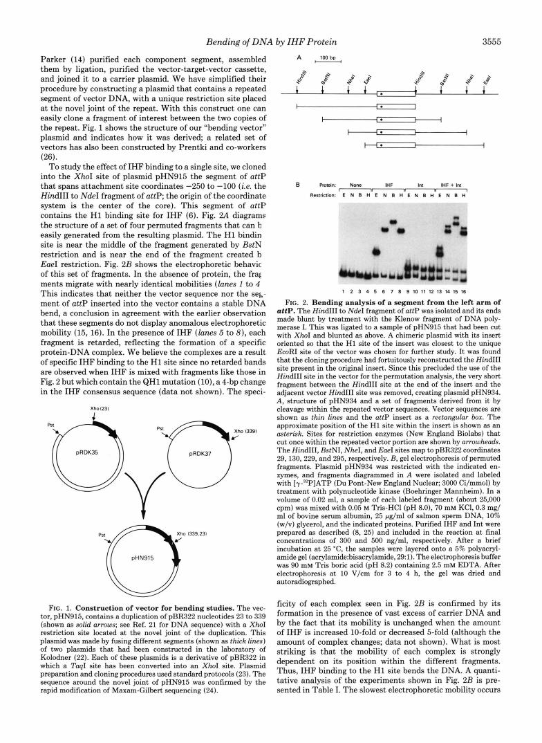
To study the effect of IHF binding to a single site, we cloned into the XhoI site of plasmid pHN915 the segment of attP that spans attachment site coordinates -250 to -100 (i.e. the HindIII to NdeI fragment of attP; the origin of the coordinate system is the center of the core). This segment of attP contains the H1 binding site for IHF (6). Fig. 2A diagrams the structure of a set of four permuted fragments that can b easily generated from the resulting plasmid. The H1 bindin site is near the middle of the fragment generated by BstN restriction and is near the end of the fragment created b EueI restriction. Fig. 2B shows the electrophoretic behavic of this set of fragments. In the absence of protein, the frag ments migrate with nearly identical mobilities (lanes 1 to 4 This indicates that neither the vector sequence nor the seg- ment of attP inserted into the vector contains a stable DNA bend, a conclusion in agreement with the earlier observation that these segments do not display anomalous electrophoretic mobility (15, 16). In the presence of IHF (lanes 5 to 8) , each fragment is retarded, reflecting the formation of a specific protein-DNA complex. We believe the complexes are a result of specific IHF binding to the H1 site since no retarded bands are observed when IHF is mixed with fragments like those in Fig. 2 but which contain the QH1 mutation (lo), a 4-bp change in the IHF consensus sequence (data not shown). The speci-
Xho1231
[ pRDK37
FIG. 1. Construction of vector for bending studies. The vec- tor, pHN915, contains a duplication of pBR322 nucleotides 23 to 339 (shown as solid arrows; see Ref. 21 for DNA sequence) with a XhoI restriction site located at the novel joint of the duplication. This plasmid was made by fusing different segments (shown as thick lines) of two plasmids that had been constructed in the laboratory of Kolodner (22). Each of these plasmids is a derivative of pBR322 in which a TaqI site has been converted into an XhoI site. Plasmid preparation and cloning procedures used standard protocols (23). The sequence around the novel joint of pHN915 was confirmed by the rapid modification of Maxam-Gilbert sequencing (24).
A , 100bp ,
I
I I * I I
H* 1 I
B Rotein: Nme IHF Inl IHF + Inl I I
Restriction: E N B H E N B H E N B H E N B H
1 2 3 4 5 6 7 8 9 1 0 1 1 1 2 1 3 1 4 1 5 1 6
FIG. 2. Bending analysis of a segment from the left arm of attP. The HindIII to NdeI fragment of attP was isolated and its ends made blunt by treatment with the Klenow fragment of DNA poly- merase I. This was ligated to a sample of pHN915 that had been cut with XhoI and blunted as above. A chimeric plasmid with its insert oriented so that the H1 site of the insert was closest to the unique EcoRI site of the vector was chosen for further study. It was found that the cloning procedure had fortuitously reconstructed the HindIII site present in the original insert. Since this precluded the use of the HindIII site in the vector for the permutation analysis, the very short fragment between the HindIII site at the end of the insert and the adjacent vector HindIII site was removed, creating plasmid pHN934. A, structure of pHN934 and a set of fragments derived from it by cleavage within the repeated vector sequences. Vector sequences are shown as thin lines and the attP insert as a rectangulur box. The approximate position of the HI site within the insert is shown as an asterisk. Sites for restriction enzymes (New England Biolabs) that cut once within the repeated vector portion are shown by arrowheads. The HindIII, BstNI, NheI, and EaeI sites map to pBR322 coordinates 29,130, 229, and 295, respectively. B, gel electrophoresis of permuted fragments. Plasmid pHN934 was restricted with the indicated en- zymes, and fragments diagrammed in A were isolated and labeled with [y"P]ATP (Du Pont-New England Nuclear; 3000 Ci/mmol) by treatment with polynucleotide kinase (Boehringer Mannheim). In a volume of 0.02 ml, a sample of each labeled fragment (about 25,000 cpm) was mixed with 0.05 M Tris-HCI (pH 8.0), 70 mM KCI, 0.3 mg/ ml of bovine serum albumin, 25 pg/ml of salmon sperm DNA, 10% (w/v) glycerol, and the indicated proteins. Purified IHF and Int were prepared as described (8, 25) and included in the reaction at final concentrations of 300 and 500 ng/ml, respectively. After a brief incubation at 25 "C, the samples were layered onto a 5% polyacryl- amide gel (acrylamide:bisacrylamide, 291). The electrophoresis buffer was 90 mM Tris boric acid (pH 8.2) containing 2.5 mM EDTA. After electrophoresis at 10 V/cm for 3 to 4 h, the gel was dried and autoradiographed.
ficity of each complex seen in Fig. 2B is confirmed by its formation in the presence of vast excess of carrier DNA and by the fact that its mobility is unchanged when the amount of IHF is increased 10-fold or decreased 5-fold (although the amount of complex changes; data not shown). What is most striking is that the mobility of each complex is strongly dependent on its position within the different fragments. Thus, IHF binding to the H1 site bends the DNA. A quanti- tative analysis of the experiments shown in Fig. 2B is pre- sented in Table I. The slowest electrophoretic mobility occurs

3556 Bending of DNA by IHF Protein TABLE I
Electrophoretic mobility of IHF. DNA complexes
Plasmid" Enzyme RF=
pHN934 EaeI 0.16, 0.84 0.70 pHN934 NheI 0.30, 0.70 0.41 pHN934 BstNI 0.51, 0.49 0.31 pHN934 HindIII 0.72, 0.28 0.42
pHN919 Em1 0.23, 0.77 0.55 pHN919 NheI 0.41, 0.59 0.31 pHN919 BstNI 0.67, 0.33 0.34 pHN919 HindIII 0.93, 0.07 0.90
"Plasmid pHN934 is described in Fig. 2. Plasmid pHN919 was constructed by isolating the 45-bp AatII to BarnHI fragment of plasnlid pWR221 (5), blunting the ends with T4 DNA polymerase, and ligating to a sample pHN915 that had been cut with Xhol endonuclease and blunted with the Klenow fragment of DNA polym- erase I. The insert in plasmid pHN919 is oriented so that its IHF site is furthest from the unique EcoRI site of the vector.
The position of each IHF site is taken to be at the center of the consensus sequence for the binding of IHF (6); this is expressed as a fractional length to either end of the fragment.
Ratio of mobility of the protein-DNA complex to the mobility of the free DNA.
Position of IHF Siteb
when restriction places the H1 binding site in the middle of the DNA fragment (ie. BstNI digestion). It is also noteworthy that two different permutations in which this IHF binding site is off-center to a similar degree (HindIII and NheI frag- ments) have nearly identical (and faster) mobilities. Taken together, our data confirm the expectation that the bend created by IHF is located at the H1 binding site.
We have also probed for bending of DNA by the other component of the intasome, Int. The segment of attP cloned in pHN934 contains two Int arm-type binding sites, called P1 and P2, that flank the IHF H1 binding site. In addition, the pBR322 vector sequences, contained in the fragments shown in Fig. 2 A , are known to contain a fortuitous Int arm-type binding site (7). Fig. 2B, lanes 9 to 12, shows that Int makes specific complexes with fragments containing these binding sites. We do not know which combination of the P1, P2, and vector arm-type binding sites are occupied under these con- ditions. Nevertheless, the important point is that, in contrast to the complexes of DNA with IHF, there is no dependence of electrophoretic mobility on the permutation of the frag- ment. This implies that, unless a bend at one binding site compensates for a bend at another site, Int does not bend DNA when it binds to an arm-type site.
Since Int and IHF are present together in the intasome, it is important to ask about the behavior of DNA loaded with both proteins. This is presented in Fig. 2B, lanes 13 to 16. Because limiting amounts of protein were used in this exper- iment, one can readily observe bands corresponding to frag- ments with only IHF bound or only Int bound. However, one also observes a new species that corresponds to DNA bearing both IHF and Int. Note that the mobility of this species is strongly position-dependent and that the BstNI fragment again has the slowest mobility. We conclude that the bend introduced by IHF persists in the presence of Int protein.
We have also examined the effect of IHF binding to another of the sites found in attP, the H' site. A fragment (attachment site coordinates +4 to +46) containing this site was cloned into pHN915 and a permuted set of fragments was isolated from the resulting plasmid, pHN919, by restriction with the same set of enzymes used to analyze the H1 site. We found that, in the absence of protein, the mobility of the DNA fragments was insensitive to permutation (data not shown) showing that this region of attP is not stably bent. However,
the mobility of IHF .DNA complexes was strongly dependent on the location of the IHF site (Table I). The placement of the IHF site within these fragments does not permit as precise an assignment of the position of the bend as could be made with the H1 site, but the data are consistent with the bend being located at or near the H' binding site. Int binding to fragments of plasmid pHN919 was also studied. The only strong Int binding site in these constructs should be the arm- type site present in the pBR322 portion of each fragment. (The attP insert does contain a core-type binding site but these are known to be very weak and do not survive challenge with excess carrier DNA.2) We find that specific complexes made by Int with each fragment have virtually identical mobilities ( RF values range from 0.84 to 0.87; data not shown). In agreement with our earlier suggestion, this indicates that Int binding to arm-type sites causes little or no bending. However, it should be noted that, using different gel electro- phoresis conditions, Thompson and Landy have observed a slightly larger range of mobility shifts upon Int binding to the arm sites of ~ t t P . ~
Our data establish that IHF bends the segments of the viral attachment site to which it binds. The same conclusion has been independently reached by A. Landy and colleagues3 who examined H2, the third binding site for IHF within attP as well as the H1 and H' sites. These findings support, but do not prove, the hypothesis (4) that the major role for IHF in h integration is to assist the formation of an intasome by bending attP into the appropriate configuration. Two addi- tional features of attachment sites should help in the forma- tion of a compact structure. First, as evidenced by anomalous gel mobility of fragments, one segment of attP (coordinates 0 to -100) is stably bent in the absence of protein (15). Second, attP is normally present on a supercoiled circle (17). We believe that the bends introduced by IHF combine with these two features to assure folding of attP into the correct config- uration. If this is so, one expects that the placement of the bends made by IHF binding to the H1, H2, and H' sites would be critical. This is because the length of intervening DNA between two bends determines their relative spatial orienta- tion, e.g. bends separated by the integral number of helical turns will produce a very different structure than identical bends separated by a half-integral number of turns (27). The possibility that phasing of the IHF bends is important for attP function is amenable to test. A more direct test of the role of IHF-promoted bending would seek variants of the protein or its binding sites that permit binding but show altered degrees of bending. Ongoing studies in our laboratory of the detailed contacts between IHF and DNA4 may provide sufficient insight into the nature of protein-DNA interaction so as to permit rational engineering of such changes. AS a extreme possibility, one might replace an IHF site with a stably bent piece of DNA, such as found in kinetoplasts (ll), and determine whether IHF is rendered dispensable.
The bending of DNA may be a universal feature of specific complexes with IHF. While this work was in preparation, reports appeared showing bending of DNA as a result of IHF binding to sites in the origin region of pSClOl (18) and the ends of insertion element IS1 (19). As in the case of the intasome, in both of these cases, IHF sites are intermingled with other protein binding sites. It may be that the bending of DNA is the fundamental activity of IHF in all systems where it is used, an idea that has appealed to other workers as well (18-20).
J. Thompson and A. Landy, personal communication. C.-C. Yang, unpublished experiments.

Bending of DNA by IHF Protein 3557
Acknowledgments-We are grateful to Paul Kitts for suggesting the strategy used to construct the bending vector. We thank Steven Goodman and David Galas for their comments on the manuscript and Mamta Vasudeva for preparing it.
REFERENCES
1. Arber, W. (1983) in Lambda ZZ (Hendrix, R. W., Roberts, J. W., Stahl, F. W., and Weisberg, R. A., eds) pp. 381-394, Cold Spring Harbor Laboratory, Cold Spring Harbor, NY
2. Weisberg, R. A., and Landy, A. (1983) in Lambda ZZ (Hendrix, R. W., Roberts, J. W., Stahl, F. W., and Weisberg, R. A., eds) pp.
NY 211-250, Cold Spring Harbor Laboratory, Cold Spring Harbor,
3. Echols, H. (1986) Science 233, 1050-1056 4. Richet, E., Abcarian, P., and Nash, H. A. (1986) Cell 46, 1011-
5. Ross, W., and Landy, A. (1983) Cell 33, 261-272 6. Craig, N. L., and Nash, H. A. (1983) Cell 35, 795-803 7. Ross, W., and Landy, A. (1982) Proc. Natl. Acad. Sci. U. S. A.
8. Nash, H. A., and Robertson, C. A. (1981) J. Biol. Chem. 266,
9. Craig, N. L., and Nash, H. A. (1984) Cell 39, 707-716
1021
79 , 7724-7728
9246-9253
10. Gardner, J. F., and Nash, H. A. (1986) J. Mol. Biol. 191, 181-
11. Wu, H.-M., and Crothers, D. M. (1984) Nature 308, 509-513 189
12. Lumpkin, 0. J., and Zimm, B. H. (1982) Biopolymers 21,2315-
13. Zahn, K., and Blattner, F. R. (1985) Nature 317,451-453 14. Shuey, D. J., and Parker, C. S. (1986) Nature 323,459-461 15. Ross, W., Shulman, M., and Landy, A. (1982) J. Mol. BWZ. 166,
16. Stellwagen, N. C. (1983) Biochemistry 22, 6186-6193 17. Mizuuchi, K., Gellert, M., and Nash, H. A. (1978) J. Mol. Biol.
18. Stenzel, T. T., Patel, P., and Bastia, D. (1987) Cell 49, 709-717 19. Prentki, P., Chandler, M., and Galas, D. J. (1987) EMBO J. 6 ,
20. Drlica, K., and Rouviere-Yaniv, J. (1987) Microbiol. Rev. 51,
21. Sutcliffe, J. G. (1979) Cold Spring Harbor Symp. Quant. Biol. 43,
2316
505-529
121,375-392
2479-2487
301-319
77-90 22.
23.
24.
25. 26.
27.
Doherty, M. J., Morrison, P. T., and Kolodner, R. (1983) J. Mol.
Maniatis, T., Fritsch, E. F., and Sambrook, J. (1982) Molecular
Cold Spring Harbor, NY Cloning, A Laboratory Manual, Cold Spring Harbor Laboratory,
Bencini, D. A., O’Donovan, G. A., and Wild, J. R. (1984) Bio-
Nash, H. A. (1983) Methods Enzymol. 100,210-216 Techniques 2,4-5
Prentki, P., Pham, M.-H., and Galas, D. J. (1987) Nucleic Acid
Zinkel, S. S., and Crothers, D. M. (1987) Nature 328, 178-181
Bid. 167,539-560
Res. 16, 10060
Related Documents