Conformational Changes in the Active Site Loops of Dihydrofolate Reductase during the Catalytic Cycle ² Rani P. Venkitakrishnan, ‡ Eduardo Zaborowski, ‡ Dan McElheny, ‡ Stephen J. Benkovic, § H. Jane Dyson, ‡ and Peter E. Wright* ,‡ Department of Molecular Biology and Skaggs Institute for Chemical Biology, The Scripps Research Institute, La Jolla, California 92037, and Department of Chemistry, The PennsylVania State UniVersity, UniVersity Park, PennsylVania 16802 ReceiVed August 31, 2004; ReVised Manuscript ReceiVed October 7, 2004 ABSTRACT: Escherichia coli dihydrofolate reductase (DHFR) has several flexible loops surrounding the active site that play a functional role in substrate and cofactor binding and in catalysis. We have used heteronuclear NMR methods to probe the loop conformations in solution in complexes of DHFR formed during the catalytic cycle. To facilitate the NMR analysis, the enzyme was labeled selectively with [ 15 N]- alanine. The 13 alanine resonances provide a fingerprint of the protein structure and report on the active site loop conformations and binding of substrate, product, and cofactor. Spectra were recorded for binary and ternary complexes of wild-type DHFR bound to the substrate dihydrofolate (DHF), the product tetrahydrofolate (THF), the pseudosubstrate folate, reduced and oxidized NADPH cofactor, and the inactive cofactor analogue 5,6-dihydroNADPH. The data show that DHFR exists in solution in two dominant conformational states, with the active site loops adopting conformations that closely approximate the occluded or closed conformations identified in earlier X-ray crystallographic analyses. A minor population of a third conformer of unknown structure was observed for the apoenzyme and for the disordered binary complex with 5,6-dihydroNADPH. The reactive Michaelis complex, with both DHF and NADPH bound to the enzyme, could not be studied directly but was modeled by the ternary folate:NADP + and dihydrofolate:NADP + complexes. From the NMR data, we are able to characterize the active site loop conformation and the occupancy of the substrate and cofactor binding sites in all intermediates formed in the extended catalytic cycle. In the dominant kinetic pathway under steady-state conditions, only the holoenzyme (the binary NADPH complex) and the Michaelis complex adopt the closed loop conformation, and all product complexes are occluded. The catalytic cycle thus involves obligatory conformational transitions between the closed and occluded states. Parallel studies on the catalytically impaired G121V mutant DHFR show that formation of the closed state, in which the nicotinamide ring of the cofactor is inserted into the active site, is energetically disfavored. The G121V mutation, at a position distant from the active site, interferes with coupled loop movements and appears to impair catalysis by destabilizing the closed Michaelis complex and introducing an extra step into the kinetic pathway. Dihydrofolate reductase (DHFR) 1 (5,6,7,8-tetrahydrofolate: NADP + oxidoreductase, EC 1.5.1.3) utilizes NADPH to reduce 7,8-dihydrofolate (DHF) to form the product 5,6,7,8- tetrahydrofolate (THF). DHFR plays a central role in maintenance of the cellular pools of THF and its derivatives, which are essential for purine and thymidylate biosynthesis and hence for cell growth, and is a target for several anticancer and antibacterial drugs (1, 2). E. coli DHFR has been the subject of intensive structural and mechanistic studies (see ref 3 for a recent review). The kinetics of the wild-type enzyme have been thoroughly investigated, result- ing in a complete description of the overall kinetic pathway (4). The kinetic intermediates in the catalytic cycle under cellular conditions of substrate and cofactor concentration are shown in Figure 1a, and the chemical structures of the various substrates, products, and cofactors are shown in Figure 1b. Binding of DHF substrate to the holoenzyme (the binary complex with reduced NADPH cofactor, henceforth denoted E:NADPH) leads to formation of the ternary Michaelis complex (E:DHF:NADPH) which, following hy- dride transfer, forms the ternary product complex (E:THF: NADP + ). Dissociation of the oxidized cofactor leads to formation of the product binary complex E:THF, which then rebinds NADPH to form the product release complex (E: THF:NADPH). The fundamental chemical event, hydride transfer from NADPH to dihydrofolate, occurs at a rate of ∼10 3 s -1 at pH 6-7 and is not rate-limiting under these ² This work was supported by Grant GM56879 from the National Institutes of Health. E.Z. was the recipient of a Rothschild Foundation postdoctoral fellowship. * Corresponding author. Address: Department of Molecular Biology, The Scripps Research Institute, 10550 North Torrey Pines Rd, La Jolla CA 92037. Phone: 858 784 9721. Fax: 858 784 9822. E-mail: [email protected]. ‡ The Scripps Research Institute. § The Pennsylvania State University. 1 Abbreviations: DHFR, dihydrofolate reductase; DHF, 7,8-dihy- drofolate; THF, 5,6,7,8-tetrahydrofolate; NADP + , nicotinamide adenine dinucleotide phosphate; NADPH, reduced nicotinamide adenine di- nucleotide phosphate; DHNADPH, 5,6-dihydroNADPH; nuclear mag- netic resonance, NMR; HSQC, heteronuclear single quantum correla- tion. 16046 Biochemistry 2004, 43, 16046-16055 10.1021/bi048119y CCC: $27.50 © 2004 American Chemical Society Published on Web 11/25/2004

Welcome message from author
This document is posted to help you gain knowledge. Please leave a comment to let me know what you think about it! Share it to your friends and learn new things together.
Transcript
Conformational Changes in the Active Site Loops of Dihydrofolate Reductaseduring the Catalytic Cycle†
Rani P. Venkitakrishnan,‡ Eduardo Zaborowski,‡ Dan McElheny,‡ Stephen J. Benkovic,§ H. Jane Dyson,‡ andPeter E. Wright*,‡
Department of Molecular Biology and Skaggs Institute for Chemical Biology, The Scripps Research Institute,La Jolla, California 92037, and Department of Chemistry, The PennsylVania State UniVersity,
UniVersity Park, PennsylVania 16802
ReceiVed August 31, 2004; ReVised Manuscript ReceiVed October 7, 2004
ABSTRACT: Escherichia colidihydrofolate reductase (DHFR) has several flexible loops surrounding theactive site that play a functional role in substrate and cofactor binding and in catalysis. We have usedheteronuclear NMR methods to probe the loop conformations in solution in complexes of DHFR formedduring the catalytic cycle. To facilitate the NMR analysis, the enzyme was labeled selectively with [15N]-alanine. The 13 alanine resonances provide a fingerprint of the protein structure and report on the activesite loop conformations and binding of substrate, product, and cofactor. Spectra were recorded for binaryand ternary complexes of wild-type DHFR bound to the substrate dihydrofolate (DHF), the producttetrahydrofolate (THF), the pseudosubstrate folate, reduced and oxidized NADPH cofactor, and the inactivecofactor analogue 5,6-dihydroNADPH. The data show that DHFR exists in solution in two dominantconformational states, with the active site loops adopting conformations that closely approximate theoccluded or closed conformations identified in earlier X-ray crystallographic analyses. A minor populationof a third conformer of unknown structure was observed for the apoenzyme and for the disordered binarycomplex with 5,6-dihydroNADPH. The reactive Michaelis complex, with both DHF and NADPH boundto the enzyme, could not be studied directly but was modeled by the ternary folate:NADP+ anddihydrofolate:NADP+ complexes. From the NMR data, we are able to characterize the active site loopconformation and the occupancy of the substrate and cofactor binding sites in all intermediates formed inthe extended catalytic cycle. In the dominant kinetic pathway under steady-state conditions, only theholoenzyme (the binary NADPH complex) and the Michaelis complex adopt the closed loop conformation,and all product complexes are occluded. The catalytic cycle thus involves obligatory conformationaltransitions between the closed and occluded states. Parallel studies on the catalytically impaired G121Vmutant DHFR show that formation of the closed state, in which the nicotinamide ring of the cofactor isinserted into the active site, is energetically disfavored. The G121V mutation, at a position distant fromthe active site, interferes with coupled loop movements and appears to impair catalysis by destabilizingthe closed Michaelis complex and introducing an extra step into the kinetic pathway.
Dihydrofolate reductase (DHFR)1 (5,6,7,8-tetrahydrofolate:NADP+ oxidoreductase, EC 1.5.1.3) utilizes NADPH toreduce 7,8-dihydrofolate (DHF) to form the product 5,6,7,8-tetrahydrofolate (THF). DHFR plays a central role inmaintenance of the cellular pools of THF and its derivatives,which are essential for purine and thymidylate biosynthesisand hence for cell growth, and is a target for several
anticancer and antibacterial drugs (1, 2). E. coli DHFR hasbeen the subject of intensive structural and mechanisticstudies (see ref3 for a recent review). The kinetics of thewild-type enzyme have been thoroughly investigated, result-ing in a complete description of the overall kinetic pathway(4). The kinetic intermediates in the catalytic cycle undercellular conditions of substrate and cofactor concentrationare shown in Figure 1a, and the chemical structures of thevarious substrates, products, and cofactors are shown inFigure 1b. Binding of DHF substrate to the holoenzyme (thebinary complex with reduced NADPH cofactor, henceforthdenoted E:NADPH) leads to formation of the ternaryMichaelis complex (E:DHF:NADPH) which, following hy-dride transfer, forms the ternary product complex (E:THF:NADP+). Dissociation of the oxidized cofactor leads toformation of the product binary complex E:THF, which thenrebinds NADPH to form the product release complex (E:THF:NADPH). The fundamental chemical event, hydridetransfer from NADPH to dihydrofolate, occurs at a rate of∼103 s-1 at pH 6-7 and is not rate-limiting under these
† This work was supported by Grant GM56879 from the NationalInstitutes of Health. E.Z. was the recipient of a Rothschild Foundationpostdoctoral fellowship.
* Corresponding author. Address: Department of Molecular Biology,The Scripps Research Institute, 10550 North Torrey Pines Rd, La JollaCA 92037. Phone: 858 784 9721. Fax: 858 784 9822. E-mail:[email protected].
‡ The Scripps Research Institute.§ The Pennsylvania State University.1 Abbreviations: DHFR, dihydrofolate reductase; DHF, 7,8-dihy-
drofolate; THF, 5,6,7,8-tetrahydrofolate; NADP+, nicotinamide adeninedinucleotide phosphate; NADPH, reduced nicotinamide adenine di-nucleotide phosphate; DHNADPH, 5,6-dihydroNADPH; nuclear mag-netic resonance, NMR; HSQC, heteronuclear single quantum correla-tion.
16046 Biochemistry2004,43, 16046-16055
10.1021/bi048119y CCC: $27.50 © 2004 American Chemical SocietyPublished on Web 11/25/2004
conditions (4). Rather, the rate-limiting step is a physicalprocess, tetrahydrofolate release, which occurs preferentiallyafter NADPH replaces NADP+ in the ternary productcomplex. Identical or highly similar kinetic schemes havebeen found for theLactobacillus caseiand mouse DHFRs(5, 6).
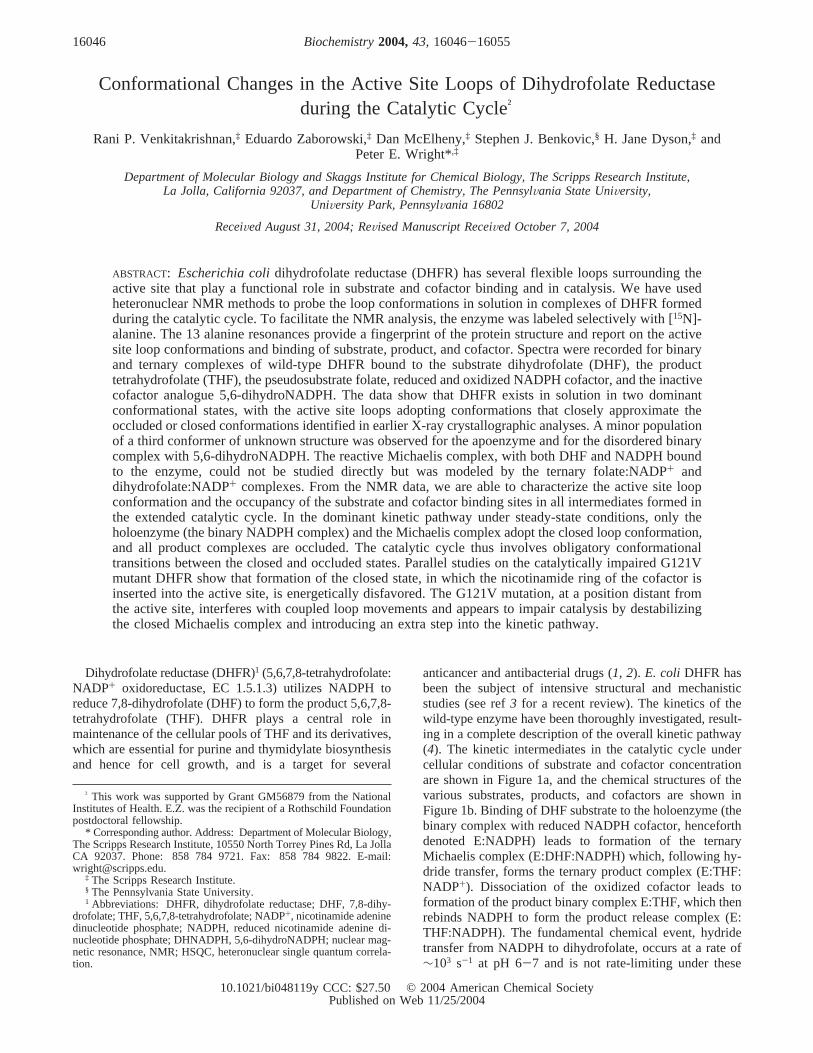
X-ray structures have been determined for more than 50complexes ofE. coli DHFR with various ligands. The proteinfold (Figure 2) consists of an eight-strandedâ-sheet formedof â-strandsâA to âH, four R-helices (designatedRB, RC,RE, andRF), and five loops (7). The substrate and cofactorbind in a 15 Å deep hydrophobic cleft that contains only asingle hydrophilic residue, Asp27. The hydride donor atom,C4 of the NADPH cofactor, and the C6 hydride acceptor atomon DHF are in van der Waals contact (8). The active sitecleft divides the protein into two structural domains, with
K38 and V88 functioning as a hinge (9). The major domainis formed by residues 1-37 and 89-159 and provides manyresidues critical for substrate binding. The smaller domain(residues 38-88) binds the adenosine moiety of the cofactorand is termed the adenosine-binding domain. Hinge motionscause the adenosine binding domain to move relative to themajor domain upon binding of various ligands, resulting inclosure of the cleft that binds the (p-aminobenzoyl)-L-glutamate tail of the substrate (9).
Conformational changes are also observed in three loopsin the major domain of the protein upon binding of ligandsto apo-DHFR (9). These are the Ala9-Asn23 loop connect-ing âA andRB (termed the Met20 loop by Sawaya and Kraut(10)), and the loops connectingâF and âG (residuesAla117-Asp131) andâG andâH (residues Asp142-His149)(designated the FG and GH loop, respectively). The Met20loop serves as a “gate or lid”, which closes over the pteridineand nicotinamide rings of the bound folate and cofactor.From their detailed analysis of the numerous X-ray structuresof E. coli DHFR, Sawaya and Kraut (10) have identifiedthree conformations of the Met20 loop, termed open, closed,and occluded. The open conformation is ligand-independentand is observed in space groups where the Met20 loopparticipates in lattice contacts. In space groups where latticecontacts involving the Met20 loop are weak or absent, theenzyme adopts the closed or occluded conformation, depend-ing on the nature of the bound ligands. The occludedconformation occurs only when the binding pocket for thenicotinamide-ribose moiety of the cofactor is empty; in thisconformation, residues 14-16 of the Met20 loop protrudeinto the pocket and prevent binding of the nicotinamide ring.In the closed conformation, the Met20 loop packs tightlyagainst the nicotinamide-ribose and seals the active site. Theloop conformational transitions are accompanied by changesin hydrogen bonding to the FG and GH loops (10). Theclosed and occluded conformations have also been observedin solution by NMR (11).
On the basis of numerous X-ray structures ofE. coliDHFRcomplexes, Sawaya and Kraut (10) have proposed a structuralmodel for the conformational changes that occur during thecatalytic cycle. Due to the instability of the substrate, product,and cofactor, stable analogues were used to model many ofthe intermediate complexes in the reaction cycle. Thestructures suggest that the Met20 loop adopts the closedconformation, with the nicotinamide ring positioned in theactive site, in the holoenzyme (E:NADPH), the Michaeliscomplex, and the transition state. On the basis of thestructures of complexes with product analogues, Sawaya andKraut propose that, following hydride transfer, the nico-tinamide ring is excluded from the binding pocket due tosteric clash with the puckered pterin ring of THF, and theysuggest that the remaining complexes in the reaction cycleare therefore in the occluded conformation (10).
In the course of NMR studies of the structure anddynamics of complexes ofE. coli DHFR with the substrateanalogue folate, with NADPH, and with folate plus cofactoror a cofactor analogue (11-13), we have identified a numberof chemical shift markers for the closed and occluded Met20loop conformations and for the presence of the pterin andnicotinamide rings within the binding pocket. In the presentwork, we use NMR to probe directly the active site loopconformation for all intermediates in the catalytic cycle, with
FIGURE 1: (a) Simplified catalytic cycle for DHFR, derived fromref 4. (b) Chemical structures of the substrates, cofactors, andanalogues used in the present studies.
Solution Conformations of Mutant and Wild-Type DHFR Biochemistry, Vol. 43, No. 51, 200416047
the sole exception of the E:DHF:NADPH Michaelis complex,which reacts too rapidly to permit NMR measurements. Tofacilitate these measurements and circumvent the need formaking complete backbone resonance assignments for eachcomplex, DHFR was labeled selectively with [15N]alanineor [13C,15N]alanine to yield highly simplified subspectracontaining only alanine backbone NH resonances. Alaninewas chosen for labeling because of its high abundance inthe E. coli DHFR sequence, because the Ala amide reso-nances are well-resolved, and because several Ala resonancesare sensitive reporters of the active site loop conformationsand the presence of cofactor and substrate in their bindingpockets. Our NMR experiments confirm the Sawaya andKraut model (10) for the conformational changes occurringduring the catalytic cycle, provide new insights into theinteractions of DHFR with substrate and ligands, and suggesta mechanism by which mutations at Gly121 in the FG loopimpair the catalytic activity of the enzyme.
MATERIALS AND METHODS
Sample Preparation. E. coliBL21 (DE3) cells weretransformed with the expression plasmid containing theDHFR and carbenicillin resistance gene. Both wild-typeenzyme and the G121V mutant were expressed by growingthe cells on M9 medium containing glucose (2 g/L), [15N]-Alanine (75 mg/L), and a mixture of all other 19 unlabeledamino acids in proportion to their average abundance in cells(total 1.925 g/L). The expressed DHFR contains selectively15N-labeled alanine at positions 6, 7, 9, 19, 26, 29, 81, 83,84, 107, 117, 143, and 145. Wild-type DHFR was alsoexpressed as above using13C,15N-labeled alanine. Both wild-type and mutant enzyme were purified by previouslydescribed methods (14, 15). The amount of protein wasestimated spectrophotometrically using an extinction coef-ficient of 31 100 M-1 cm-1 at 280 nm.
Due to the oxygen and light sensitivity of the cofactorand substrate used in the investigations, NMR sample buffer[70 mM potassium phosphate (pH 7.6), 25 mM KCl in 93%H2O/7% D2O, containing 0.02% sodium azide and 1 mMDTT] was thoroughly degassed in a vacuum by severalfreeze-thaw cycles. The protein samples were exchanged
into the NMR buffer using an Amicon NAP5 column in anargon-equilibrated glovebox. The protein concentration was1 mM for all NMR experiments, and a 4-40-fold excess ofsubstrate/product or cofactor and their analogues was used.NMR samples were subjected to an additional freeze-thawcycle and stored in argon atmosphere. Amberized NMR tubesequipped with Teflon valves for connecting to the vacuummanifold were used wherever necessary.
NMR Spectroscopy.NMR spectra were acquired at 282K on Bruker DRX600 and AMX600 spectrometers equippedwith 5 mm triple resonance probes with single axis or tripleaxis gradients. The probe temperature was calibrated usingneat methanol. Spectra of the DHNADPH complexes wereacquired at 287 K on a Bruker Avance 500 spectrometerequipped with a cryoprobe.
2D 1H-15N HSQC spectra were recorded on the variousenzyme complexes with 2048 complex data points int2 anda variable number of data points (160-256) in the t1dimension and with 128 transients per experiment. Spectrawere processed using NMRPIPE (16) and analyzed usingNMRVIEW (17). Data sets were zero-filled to 4096 pointsin ω2 and 1024 points in theω1 dimension and apodizedwith sine bell or shifted sine bell window functions prior toprocessing. The1H chemical shifts were referenced tointernal 2,2-dimethyl-2-silapentane-5-sulfonate (DSS). The15N dimension was referenced indirectly (18). HNCO spectra(19) were acquired at 282 K on samples of wild-type DHFRlabeled with [13C,15N]alanine. 1D HNCO spectra wererecorded with 4096 transients, and 2D spectra were typicallyacquired with 2048 complex data points int2, 32 data pointsin the t1 dimension, and 1024 transients per experiment.
RESULTS
1H-15N HSQC spectra were recorded for complexes ofwild-type and the G121V mutant DHFR to probe theconformation of the intermediates formed in the catalyticcycle. To simplify the HSQC spectra and facilitate assign-ment of resonances, the protein was labeled specifically with[15N]alanine. The 13 alanine residues are distributed through-out the protein structure, with several (Ala6, Ala7, Ala9,Ala19, Ala26, Ala29, and Ala117) located in the active site
FIGURE 2: Stereoview of the superposition of the backbones of X-ray crystal structures of DHFR (10) in complex with folate (PDB 1rx7,green) and with NADP+ and folate (PDB 1rx2, pink). Folate and NADP+ in the 1rx2 structure are shown in blue and yellow, respectively.The backbone N atoms of the alanine residues are shown as red spheres. The Met20 loop is represented by thicker lines to emphasize theconformational change between the occluded and closed states.
16048 Biochemistry, Vol. 43, No. 51, 2004 Venkitakrishnan et al.
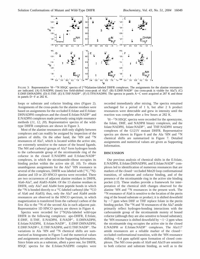
loops or substrate and cofactor binding sites (Figure 2).Assignments of the cross-peaks for the alanine residues werebased on assignments for the occluded E:folate and E:folate:DHNADPH complexes and the closed E:folate:NADP+ andE:NADPH complexes made previously using triple resonancemethods (11, 12, 20). Representative spectra of the wild-type DHFR complexes are shown in Figure 3.
Most of the alanine resonances shift only slightly betweencomplexes and can readily be assigned by inspection of thepattern of shifts. On the other hand, the1HN and 15Nresonances of Ala7, which is located within the active site,are extremely sensitive to the nature of the bound ligands.The NH and carbonyl groups of Ala7 form hydrogen bondsto the carboxamide group of the nicotinamide ring of thecofactor in the closed E:NADPH and E:folate:NADP+
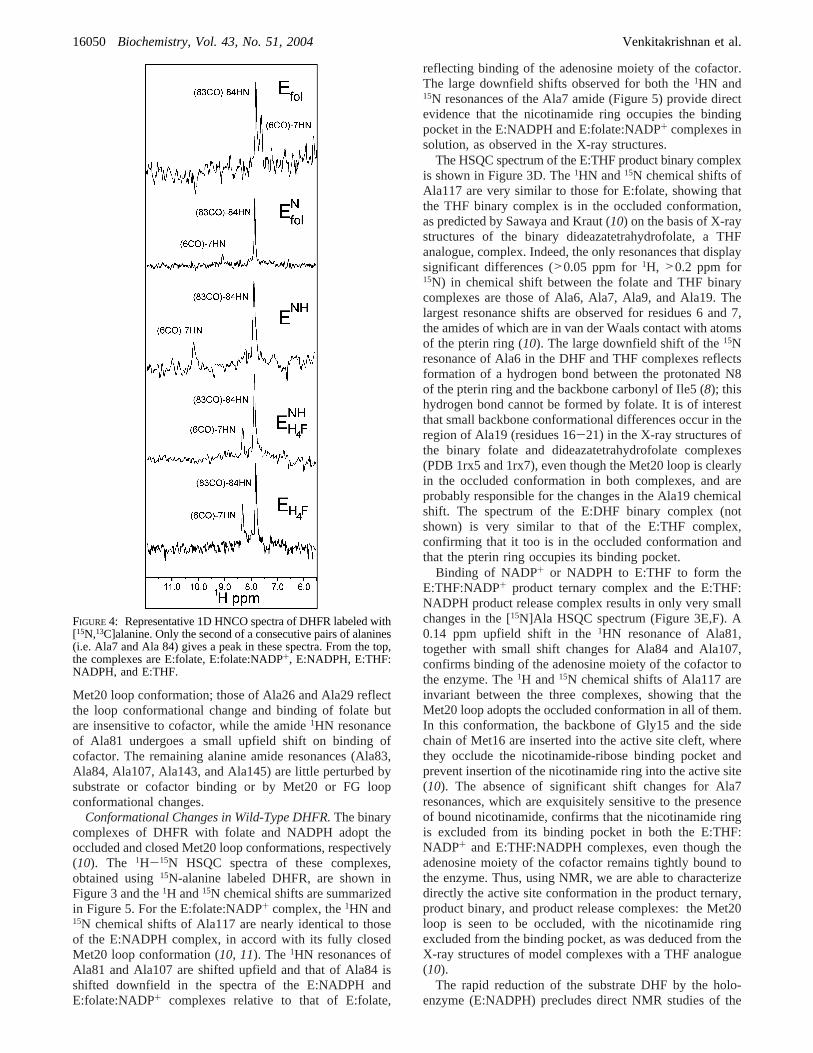
complexes, in which the nicotinamide-ribose occupies itsbinding pocket within the active site (8, 10). To obtainunambiguous assignments for the Ala71HN resonance inseveral of the complexes, DHFR was labeled with [13C,15N]-alanine and 1D or 2D HNCO spectra were recorded. Thereare two occurrences of adjacent alanine residues in DHFR,Ala6-Ala7, and Ala83-Ala84. Of the 13 alanine residues inDHFR, only Ala7 and Ala84 form peptide bonds in whichthe15N is bonded directly to a13C-labeled carbonyl (the13COof Ala6 and Ala83); thus, only the Ala7 and Ala84 amideresonances are observed in the HNCO spectrum, in whichmagnetization is transferred from the carbonyl carbon of thefirst Ala to the15N of the second Ala in each adjacent pair.Representative 1D HNCO spectra are shown in Figure 4.
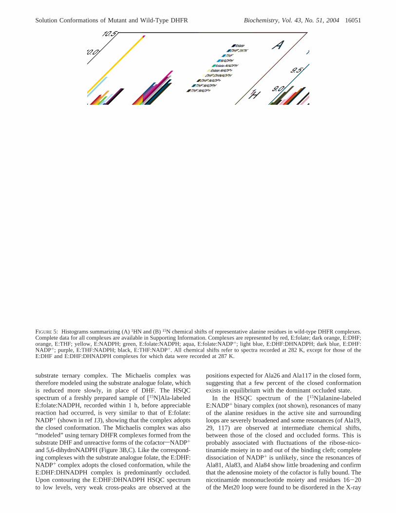
Spectra were recorded for [15N]Ala-labeled wild-typeDHFR in the following complexes: apo-DHFR, E:folate,E:DHF, E:THF, E:NADPH, E:NADP+, E:DHNADPH,E:folate:NADPH, E:folate:NADP+, E:DHF:DHNADPH,E:DHF:NADP+, E:THF:NADPH, and E:THF:NADP+. Thevariations in Ala 1HN and 15N chemical shifts are sum-marized as histograms in Figure 5 and the numerical valuesand assignments are tabulated in the Supporting Information.Since folate acts as a substrate, albeit a poor one, for DHFR,HSQC spectra for the E:folate:NADPH complex were
recorded immediately after mixing. The spectra remainedunchanged for a period of 1 h, but after 2 h productresonances were detectable and grew in intensity until thereaction was complete after a few hours at 282 K.
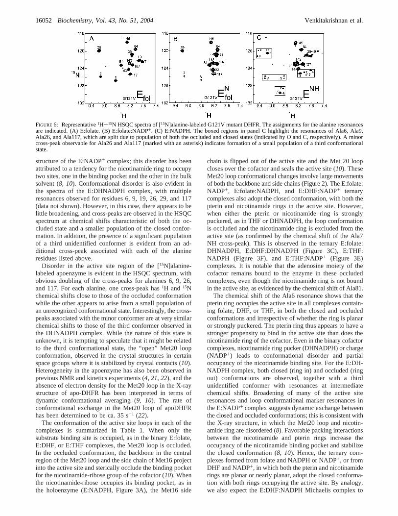
1H-15N HSQC spectra were recorded for the apoenzyme,the folate, DHF, and NADPH binary complexes, and thefolate:NADPH, folate:NADP+, and THF:NADPH ternarycomplexes of the G121V mutant DHFR. Representativespectra are shown in Figure 6 and the Ala1HN and 15Nchemical shifts are summarized in Figure 7. Detailedassignments and numerical values are given as SupportingInformation.
DISCUSSION
Our previous analysis of chemical shifts in the E:folate,E:NADPH, E:folate:DHNADPH, and E:folate:NADP+ com-plexes led to identification of numerous resonances that aremarkers of the closed-occluded Met20 loop conformationaltransition, of substrate and cofactor binding, and of thepresence of the nicotinamide ring in the active site bindingpocket (13). These studies provide a framework for inter-pretation of the chemical shift changes observed for thealanine1HN and 15N resonances in the present work. The15N resonance of Ala6 is sensitive to the location of the pterinring of the bound substrate or product; it is shifted downfieldby ∼7 ppm when DHF or THF replace folate in the pterinbinding pocket. The15N and1H resonances of the Ala7 amideprimarily reflect hydrogen-bonding interactions with thecarboxamide group of the nicotinamide moiety of boundcofactor (although they are also sensitive to bound substrate);the1HN resonance is shifted downfield by∼1-2 ppm whenthe nicotinamide ring occupies the active site in the closedE:NADPH or E:folate:NADP+ complexes. The Ala117amide resonances are a reliable marker of the closed-occluded conformational transition, with the1HN resonanceshifting ∼0.4 ppm upfield upon formation of closed com-plexes. The NH cross-peaks of Ala9 and Ala19 are sensitiveto both cofactor and substrate binding, as well as to the
FIGURE 3: Representative1H-15N HSQC spectra of [15N]alanine-labeled DHFR complexes. The assignments for the alanine resonancesare indicated. (A) E:NADPH; (inset) low field-shifted cross-peak of Ala7. (B) E:DHF:NADP+ (no cross-peak is visible for Ala7). (C)E:DHF:DHNADPH. (D) E:THF. (E) E:THF:NADP+. (F) E:TFH:NADPH. The spectra in panels A-C were acquired at 287 K and thosein panels D-F at 282 K.
Solution Conformations of Mutant and Wild-Type DHFR Biochemistry, Vol. 43, No. 51, 200416049
Met20 loop conformation; those of Ala26 and Ala29 reflectthe loop conformational change and binding of folate butare insensitive to cofactor, while the amide1HN resonanceof Ala81 undergoes a small upfield shift on binding ofcofactor. The remaining alanine amide resonances (Ala83,Ala84, Ala107, Ala143, and Ala145) are little perturbed bysubstrate or cofactor binding or by Met20 or FG loopconformational changes.
Conformational Changes in Wild-Type DHFR.The binarycomplexes of DHFR with folate and NADPH adopt theoccluded and closed Met20 loop conformations, respectively(10). The 1H-15N HSQC spectra of these complexes,obtained using15N-alanine labeled DHFR, are shown inFigure 3 and the1H and15N chemical shifts are summarizedin Figure 5. For the E:folate:NADP+ complex, the1HN and15N chemical shifts of Ala117 are nearly identical to thoseof the E:NADPH complex, in accord with its fully closedMet20 loop conformation (10, 11). The 1HN resonances ofAla81 and Ala107 are shifted upfield and that of Ala84 isshifted downfield in the spectra of the E:NADPH andE:folate:NADP+ complexes relative to that of E:folate,
reflecting binding of the adenosine moiety of the cofactor.The large downfield shifts observed for both the1HN and15N resonances of the Ala7 amide (Figure 5) provide directevidence that the nicotinamide ring occupies the bindingpocket in the E:NADPH and E:folate:NADP+ complexes insolution, as observed in the X-ray structures.
The HSQC spectrum of the E:THF product binary complexis shown in Figure 3D. The1HN and15N chemical shifts ofAla117 are very similar to those for E:folate, showing thatthe THF binary complex is in the occluded conformation,as predicted by Sawaya and Kraut (10) on the basis of X-raystructures of the binary dideazatetrahydrofolate, a THFanalogue, complex. Indeed, the only resonances that displaysignificant differences (>0.05 ppm for1H, >0.2 ppm for15N) in chemical shift between the folate and THF binarycomplexes are those of Ala6, Ala7, Ala9, and Ala19. Thelargest resonance shifts are observed for residues 6 and 7,the amides of which are in van der Waals contact with atomsof the pterin ring (10). The large downfield shift of the15Nresonance of Ala6 in the DHF and THF complexes reflectsformation of a hydrogen bond between the protonated N8of the pterin ring and the backbone carbonyl of Ile5 (8); thishydrogen bond cannot be formed by folate. It is of interestthat small backbone conformational differences occur in theregion of Ala19 (residues 16-21) in the X-ray structures ofthe binary folate and dideazatetrahydrofolate complexes(PDB 1rx5 and 1rx7), even though the Met20 loop is clearlyin the occluded conformation in both complexes, and areprobably responsible for the changes in the Ala19 chemicalshift. The spectrum of the E:DHF binary complex (notshown) is very similar to that of the E:THF complex,confirming that it too is in the occluded conformation andthat the pterin ring occupies its binding pocket.
Binding of NADP+ or NADPH to E:THF to form theE:THF:NADP+ product ternary complex and the E:THF:NADPH product release complex results in only very smallchanges in the [15N]Ala HSQC spectrum (Figure 3E,F). A0.14 ppm upfield shift in the1HN resonance of Ala81,together with small shift changes for Ala84 and Ala107,confirms binding of the adenosine moiety of the cofactor tothe enzyme. The1H and15N chemical shifts of Ala117 areinvariant between the three complexes, showing that theMet20 loop adopts the occluded conformation in all of them.In this conformation, the backbone of Gly15 and the sidechain of Met16 are inserted into the active site cleft, wherethey occlude the nicotinamide-ribose binding pocket andprevent insertion of the nicotinamide ring into the active site(10). The absence of significant shift changes for Ala7resonances, which are exquisitely sensitive to the presenceof bound nicotinamide, confirms that the nicotinamide ringis excluded from its binding pocket in both the E:THF:NADP+ and E:THF:NADPH complexes, even though theadenosine moiety of the cofactor remains tightly bound tothe enzyme. Thus, using NMR, we are able to characterizedirectly the active site conformation in the product ternary,product binary, and product release complexes: the Met20loop is seen to be occluded, with the nicotinamide ringexcluded from the binding pocket, as was deduced from theX-ray structures of model complexes with a THF analogue(10).
The rapid reduction of the substrate DHF by the holo-enzyme (E:NADPH) precludes direct NMR studies of the
FIGURE 4: Representative 1D HNCO spectra of DHFR labeled with[15N,13C]alanine. Only the second of a consecutive pairs of alanines(i.e. Ala7 and Ala 84) gives a peak in these spectra. From the top,the complexes are E:folate, E:folate:NADP+, E:NADPH, E:THF:NADPH, and E:THF.
16050 Biochemistry, Vol. 43, No. 51, 2004 Venkitakrishnan et al.
substrate ternary complex. The Michaelis complex wastherefore modeled using the substrate analogue folate, whichis reduced more slowly, in place of DHF. The HSQCspectrum of a freshly prepared sample of [15N]Ala-labeledE:folate:NADPH, recorded within 1 h, before appreciablereaction had occurred, is very similar to that of E:folate:NADP+ (shown in ref13), showing that the complex adoptsthe closed conformation. The Michaelis complex was also“modeled” using ternary DHFR complexes formed from thesubstrate DHF and unreactive forms of the cofactorsNADP+
and 5,6-dihydroNADPH (Figure 3B,C). Like the correspond-ing complexes with the substrate analogue folate, the E:DHF:NADP+ complex adopts the closed conformation, while theE:DHF:DHNADPH complex is predominantly occluded.Upon contouring the E:DHF:DHNADPH HSQC spectrumto low levels, very weak cross-peaks are observed at the
positions expected for Ala26 and Ala117 in the closed form,suggesting that a few percent of the closed conformationexists in equilibrium with the dominant occluded state.
In the HSQC spectrum of the [15N]alanine-labeledE:NADP+ binary complex (not shown), resonances of manyof the alanine residues in the active site and surroundingloops are severely broadened and some resonances (of Ala19,29, 117) are observed at intermediate chemical shifts,between those of the closed and occluded forms. This isprobably associated with fluctuations of the ribose-nico-tinamide moiety in to and out of the binding cleft; completedissociation of NADP+ is unlikely, since the resonances ofAla81, Ala83, and Ala84 show little broadening and confirmthat the adenosine moiety of the cofactor is fully bound. Thenicotinamide mononucleotide moiety and residues 16-20of the Met20 loop were found to be disordered in the X-ray
FIGURE 5: Histograms summarizing (A)1HN and (B)15N chemical shifts of representative alanine residues in wild-type DHFR complexes.Complete data for all complexes are available in Supporting Information. Complexes are represented by red, E:folate; dark orange, E:DHF;orange, E:THF; yellow, E:NADPH; green, E:folate:NADPH; aqua, E:folate:NADP+; light blue, E:DHF:DHNADPH; dark blue, E:DHF:NADP+; purple, E:THF:NADPH; black, E:THF:NADP+. All chemical shifts refer to spectra recorded at 282 K, except for those of theE:DHF and E:DHF:DHNADPH complexes for which data were recorded at 287 K.
Solution Conformations of Mutant and Wild-Type DHFR Biochemistry, Vol. 43, No. 51, 200416051
structure of the E:NADP+ complex; this disorder has beenattributed to a tendency for the nicotinamide ring to occupytwo sites, one in the binding pocket and the other in the bulksolvent (8, 10). Conformational disorder is also evident inthe spectra of the E:DHNADPH complex, with multipleresonances observed for residues 6, 9, 19, 26, 29, and 117(data not shown). However, in this case, there appears to belittle broadening, and cross-peaks are observed in the HSQCspectrum at chemical shifts characteristic of both the oc-cluded state and a smaller population of the closed confor-mation. In addition, the presence of a significant populationof a third unidentified conformer is evident from an ad-ditional cross-peak associated with each of the alanineresidues listed above.
Disorder in the active site region of the [15N]alanine-labeled apoenzyme is evident in the HSQC spectrum, withobvious doubling of the cross-peaks for alanines 6, 9, 26,and 117. For each alanine, one cross-peak has1H and 15Nchemical shifts close to those of the occluded conformationwhile the other appears to arise from a small population ofan unrecognized conformational state. Interestingly, the cross-peaks associated with the minor conformer are at very similarchemical shifts to those of the third conformer observed inthe DHNADPH complex. While the nature of this state isunknown, it is tempting to speculate that it might be relatedto the third conformational state, the “open” Met20 loopconformation, observed in the crystal structures in certainspace groups where it is stabilized by crystal contacts (10).Heterogeneity in the apoenzyme has also been observed inprevious NMR and kinetics experiments (4, 21, 22), and theabsence of electron density for the Met20 loop in the X-raystructure of apo-DHFR has been interpreted in terms ofdynamic conformational averaging (9, 10). The rate ofconformational exchange in the Met20 loop of apoDHFRhas been determined to be ca. 35 s-1 (22).
The conformation of the active site loops in each of thecomplexes is summarized in Table 1. When only thesubstrate binding site is occupied, as in the binary E:folate,E:DHF, or E:THF complexes, the Met20 loop is occluded.In the occluded conformation, the backbone in the centralregion of the Met20 loop and the side chain of Met16 projectinto the active site and sterically occlude the binding pocketfor the nicotinamide-ribose group of the cofactor (10). Whenthe nicotinamide-ribose occupies its binding pocket, as inthe holoenzyme (E:NADPH, Figure 3A), the Met16 side
chain is flipped out of the active site and the Met 20 loopcloses over the cofactor and seals the active site (10). TheseMet20 loop conformational changes involve large movementsof both the backbone and side chains (Figure 2). The E:folate:NADP+, E:folate:NADPH, and E:DHF:NADP+ ternarycomplexes also adopt the closed conformation, with both thepterin and nicotinamide rings in the active site. However,when either the pterin or nicotinamide ring is stronglypuckered, as in THF or DHNADPH, the loop conformationis occluded and the nicotinamide ring is excluded from theactive site (as confirmed by the chemical shift of the Ala7NH cross-peak). This is observed in the ternary E:folate:DHNADPH, E:DHF:DHNADPH (Figure 3C), E:THF:NADPH (Figure 3F), and E:THF:NADP+ (Figure 3E)complexes. It is notable that the adenosine moiety of thecofactor remains bound to the enzyme in these occludedcomplexes, even though the nicotinamide ring is not boundin the active site, as evidenced by the chemical shift of Ala81.
The chemical shift of the Ala6 resonance shows that thepterin ring occupies the active site in all complexes contain-ing folate, DHF, or THF, in both the closed and occludedconformations and irrespective of whether the ring is planaror strongly puckered. The pterin ring thus appears to have astronger propensity to bind in the active site than does thenicotinamide ring of the cofactor. Even in the binary cofactorcomplexes, nicotinamide ring pucker (DHNADPH) or charge(NADP+) leads to conformational disorder and partialoccupancy of the nicotinamide binding site. For the E:DH-NADPH complex, both closed (ring in) and occluded (ringout) conformations are observed, together with a thirdunidentified conformer with resonances at intermediatechemical shifts. Broadening of many of the active siteresonances and loop conformational marker resonances inthe E:NADP+ complex suggests dynamic exchange betweenthe closed and occluded conformations; this is consistent withthe X-ray structure, in which the Met20 loop and nicotin-amide ring are disordered (8). Favorable packing interactionsbetween the nicotinamide and pterin rings increase theoccupancy of the nicotinamide binding pocket and stabilizethe closed conformation (8, 10). Hence, the ternary com-plexes formed from folate and NADPH or NADP+, or fromDHF and NADP+, in which both the pterin and nicotinamiderings are planar or nearly planar, adopt the closed conforma-tion with both rings occupying the active site. By analogy,we also expect the E:DHF:NADPH Michaelis complex to
FIGURE 6: Representative1H-15N HSQC spectra of [15N]alanine-labeled G121V mutant DHFR. The assignments for the alanine resonancesare indicated. (A) E:folate. (B) E:folate:NADP+. (C) E:NADPH. The boxed regions in panel C highlight the resonances of Ala6, Ala9,Ala26, and Ala117, which are split due to population of both the occluded and closed states (indicated by O and C, respectively). A minorcross-peak observable for Ala26 and Ala117 (marked with an asterisk) indicates formation of a small population of a third conformationalstate.
16052 Biochemistry, Vol. 43, No. 51, 2004 Venkitakrishnan et al.
be closed. However, when either of the rings is stronglypuckered, as in the ternary complexes containing DHNADPHor THF, the nicotinamide ring is excluded from the bindingpocket and the active site loops adopt the occluded confor-mation. The driving force for this transition appears to besteric clash between the pterin and nicotinamide rings (10).
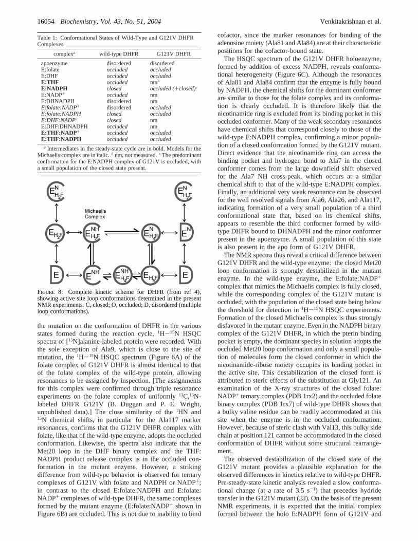
Loop Conformational Changes during the Catalytic Cycle.The NMR experiments reported here provide direct insightsinto the active site occupancy and loop conformations of allof the intermediates that participate in the catalytic cycle ofE. coli DHFR. The results are summarized on the completecatalytic scheme in Figure 8 (4); the heavy arrows indicatethe kinetic pathway (shown in Figure 1) for steady-stateturnover under saturating substrate concentrations. The mostnotable feature is that the large amplitude conformationaltransitions between the closed and occluded conformations,with movements of backbone and side chain atoms by as
much as 12 Å, are an integral part of the reaction cycle.While both the holoenzyme (E:NADPH) and the Michaeliscomplex (E:DHF:NADPH) adopt the closed conformation,all of the THF product complexes (E:THF:NADP+, E:THF,and E:THF:NADPH) are occluded. Thus, the enzyme mustcycle between the closed and the occluded states at two stepsin the reaction cycle, immediately following hydride transferand again after product release.
Loop Conformations in the G121V Mutant Enzyme.Substitution of Gly121 by valine and other bulky side chainshas a dramatic effect on the kinetics of DHFR, decreasingthe hydride transfer rate by up to 1000-fold and resulting inan additional kinetic step in the reaction mechanism (23). Arecent molecular dynamics, CD, and fluorescence study ofapoDHFR suggested that the G121V mutation may desta-bilize the contacts between the Met20 and FG loops and leadto some structural perturbation (24). To probe the effect of
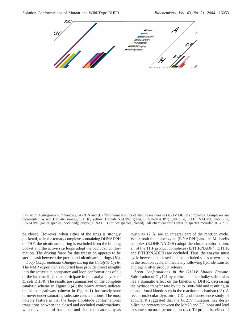
FIGURE 7: Histograms summarizing (A)1HN and (B)15N chemical shifts of alanine residues in G121V DHFR complexes. Complexes arerepresented by red, E:folate; orange, E:DHF; yellow, E:folate:NADPH; green, E:folate:NADP+; light blue, E:THF:NADPH; dark blue,E:NADPH (major species, occluded); purple, E:NADPH (minor species, closed). All chemical shifts refer to spectra recorded at 282 K.
Solution Conformations of Mutant and Wild-Type DHFR Biochemistry, Vol. 43, No. 51, 200416053
the mutation on the conformation of DHFR in the variousstates formed during the reaction cycle,1H-15N HSQCspectra of [15N]alanine-labeled protein were recorded. Withthe sole exception of Ala9, which is close to the site ofmutation, the1H-15N HSQC spectrum (Figure 6A) of thefolate complex of G121V DHFR is almost identical to thatof the folate complex of the wild-type protein, allowingresonances to be assigned by inspection. [The assignmentsfor this complex were confirmed through triple resonanceexperiments on the folate complex of uniformly13C,15N-labeled DHFR G121V (B. Duggan and P. E. Wright,unpublished data).] The close similarity of the1HN and15N chemical shifts, in particular for the Ala117 markerresonances, confirms that the G121V DHFR complex withfolate, like that of the wild-type enzyme, adopts the occludedconformation. Likewise, the spectra also indicate that theMet20 loop in the DHF binary complex and the THF:NADPH product release complex is in the occluded con-formation in the mutant enzyme. However, a strikingdifference from wild-type behavior is observed for ternarycomplexes of G121V with folate and NADPH or NADP+;in contrast to the closed E:folate:NADPH and E:folate:NADP+ complexes of wild-type DHFR, the same complexesformed by the mutant enzyme (E:folate:NADP+ shown inFigure 6B) are occluded. This is not due to inability to bind
cofactor, since the marker resonances for binding of theadenosine moiety (Ala81 and Ala84) are at their characteristicpositions for the cofactor-bound state.
The HSQC spectrum of the G121V DHFR holoenzyme,formed by addition of excess NADPH, reveals conforma-tional heterogeneity (Figure 6C). Although the resonancesof Ala81 and Ala84 confirm that the enzyme is fully boundby NADPH, the chemical shifts for the dominant conformerare similar to those for the folate complex and its conforma-tion is clearly occluded. It is therefore likely that thenicotinamide ring is excluded from its binding pocket in thisoccluded conformer. Many of the weak secondary resonanceshave chemical shifts that correspond closely to those of thewild-type E:NADPH complex, confirming a minor popula-tion of a closed conformation formed by the G121V mutant.Direct evidence that the nicotinamide ring can access thebinding pocket and hydrogen bond to Ala7 in the closedconformer comes from the large downfield shift observedfor the Ala7 NH cross-peak, which occurs at a similarchemical shift to that of the wild-type E:NADPH complex.Finally, an additional very weak resonance can be observedfor the well resolved signals from Ala6, Ala26, and Ala117,indicating formation of a very small population of a thirdconformational state that, based on its chemical shifts,appears to resemble the third conformer formed by wild-type DHFR bound to DHNADPH and the minor conformerpresent in the apoenzyme. A small population of this stateis also present in the apo form of G121V DHFR.
The NMR spectra thus reveal a critical difference betweenG121V DHFR and the wild-type enzyme: the closed Met20loop conformation is strongly destabilized in the mutantenzyme. In the wild-type enzyme, the E:folate:NADP+
complex that mimics the Michaelis complex is fully closed,while the corresponding complex of the G121V mutant isoccluded, with the population of the closed state being belowthe threshold for detection in1H-15N HSQC experiments.Formation of the closed Michaelis complex is thus stronglydisfavored in the mutant enzyme. Even in the NADPH binarycomplex of the G121V DHFR, in which the pterin bindingpocket is empty, the dominant species in solution adopts theoccluded Met20 loop conformation and only a small popula-tion of molecules form the closed conformer in which thenicotinamide-ribose moiety occupies its binding pocket inthe active site. This destabilization of the closed form isattributed to steric effects of the substitution at Gly121. Anexamination of the X-ray structures of the closed folate:NADP+ ternary complex (PDB 1rx2) and the occluded folatebinary complex (PDB 1rx7) of wild-type DHFR shows thata bulky valine residue can be readily accommodated at thissite when the enzyme is in the occluded conformation.However, because of steric clash with Val13, this bulky sidechain at position 121 cannot be accommodated in the closedconformation of DHFR without some structural rearrange-ment.
The observed destabilization of the closed state of theG121V mutant provides a plausible explanation for theobserved differences in kinetics relative to wild-type DHFR.Pre-steady-state kinetic analysis revealed a slow conforma-tional change (at a rate of 3.5 s-1) that precedes hydridetransfer in the G121V mutant (23). On the basis of the presentNMR experiments, it is expected that the initial complexformed between the holo E:NADPH form of G121V and
Table 1: Conformational States of Wild-Type and G121V DHFRComplexes
complexa wild-type DHFR G121V DHFR
apoenzyme disordered disorderedE:folate occluded occludedE:DHF occluded occludedE:THF occluded nmb
E:NADPH closed occluded (+closed)c
E:NADP+ occluded nmE:DHNADPH disordered nmE:folate:NADP+ disordered occludedE:folate:NADPH closed occludedE:DHF:NADP+ closed nmE:DHF:DHNADPH occluded nmE:THF:NADP + occluded occludedE:THF:NADPH occluded occluded
a Intermediates in the steady-state cycle are in bold. Models for theMichaelis complex are in italic.b nm, not measured.c The predominantconformation for the E:NADPH complex of G121V is occluded, witha small population of the closed state present.
FIGURE 8: Complete kinetic scheme for DHFR (from ref4),showing active site loop conformations determined in the presentNMR experiments. C, closed; O, occluded; D, disordered (multipleloop conformations).
16054 Biochemistry, Vol. 43, No. 51, 2004 Venkitakrishnan et al.
the DHF substrate would be in the occluded conformation,with the nicotinamide ring excluded from the active site. Theslow conformational rearrangement observed in the kineticsprobably reflects formation of an unstable “closed” state, inwhich the nicotinamide ring is inserted into its binding pocketadjacent to the pterin ring of the substrate. While our presentexperiments provide no direct insights into the origins ofthe substantial decrease in hydride transfer rate observed forthe mutant protein (23), it is likely that the steric destabiliza-tion of the closed state might result in changes in active sitegeometry that would be manifest in the kinetics. Indeed, wenote that recent quantum mechanical and molecular mechan-ical simulations suggest that the G121V mutation leads toan altered transition state structure, with a concomitantincrease in the activation free energy barrier (25, 26), andalso perturbs the critical hydrogen-bonding interactionsbetween the Met20 and FG loops (27).
ACKNOWLEDGMENT
We thank Drs. Jason Schnell and Jim Huntley for valuablediscussions and Linda Tennant for technical assistance.
SUPPORTING INFORMATION AVAILABLE1H and 15N chemical shifts for wild-type DHFR (Table
S1) and G121V DHFR (Table S2). This material is availablefree of charge via the Internet at http://pubs.acs.org.
REFERENCES
1. Blakley, R. L. (1969)The Biochemistry of Folic Acid and RelatedPteridines, Elsevier/North-Holland, Amsterdam.
2. Hitchings, G. H., Jr. (1989) Nobel lecture in physiology ormedicine--1988. Selective inhibitors of dihydrofolate reductase,In Vitro Cell DeV. Biol. 25, 303-310.
3. Schnell, J. R., Dyson, H. J., and Wright, P. E. (2004) Structure,dynamics and catalytic function of dihydrofolate reductase,Annu.ReV. Biophys. Biomol. Struct. 33, 140.
4. Fierke, C. A., Johnson, K. A., and Benkovic, S. J. (1987)Construction and evaluation of the kinetic scheme associated withdihydrofolate reductase fromEscherichia coli, Biochemistry 26,4085-4092.
5. Andrews, J., Fierke, C. A., Birdsall, B., Ostler, G., Feeney, J.,Roberts, G. C. K., and Benkovic, S. J. (1989) A kinetic study ofwild-type and mutant dihydrofolate reductases fromLactobacilluscasei, Biochemistry 28, 5743-5750.
6. Thillet, J., Adams, J. A., and Benkovic, S. J. (1990) The kineticmechanism of wild-type and mutant mouse dihydrofolate reduc-tases,Biochemistry 29, 5195-5202.
7. Matthews, D. A., Alden, R. A., Bolin, J. T., Freer, S. T., Hamlin,R., Xuong, N., Kraut, J., Poe, M., Williams, M., and Hoogsteen,K. (1977) Dihydrofolate reductase: X-ray structure of the binarycomplex with methotrexate,Science 197, 452-455.
8. Bystroff, C., Oatley, S. J., and Kraut, J. (1990) Crystal structuresof Escherichia colidihydrofolate reductase: The NADP+ ho-loenzyme and the folate. NADP+ ternary complex. Substratebinding and a model for the transition state,Biochemistry 29,3263-3277.
9. Bystroff, C., and Kraut, J. (1991) Crystal structure of unligandedEscherichia colidihydrofolate reductase. Ligand-induced confor-mational changes and cooperativity in binding,Biochemistry 30,2227-2239.
10. Sawaya, M. R., and Kraut, J. (1997) Loop and subdomainmovements in the mechanism ofEscherichia colidihydrofolatereductase: Crystallographic evidence,Biochemistry 36, 586-603.
11. Osborne, M. J., Schnell, J., Benkovic, S. J., Dyson, H. J., andWright, P. E. (2001) Backbone dynamics in dihydrofolate reduc-tase complexes: Role of loop flexibility in the catalytic mecha-nism,Biochemistry 40, 9846-9859.
12. Zaborowski, E., Chung, J., Kroon, G. J. A., Dyson, H. J., andWright, P. E. (2000) Backbone HN, N, CR, C′ and Câ assignmentsof the 19 kDa DHFR/NADPH complex at 9°C and pH 7.6,J.Biomol. NMR 16, 349-350.
13. Osborne, M. J., Venkitakrishnan, R. P., Dyson, H. J., and Wright,P. E. (2003) Diagnostic chemical shift markers for loop conforma-tion and cofactor binding in dihydrofolate reductase complexes,Protein Sci. 12, 2230-2238.
14. Miller, G. P., and Benkovic, S. J. (1998) Deletion of a highlymotional residue affects formation of the Michaelis complex forEscherichia colidihydrofolate reductase,Biochemistry 37, 6327-6335.
15. Osborne, M. J., and Wright, P. E. (2001) Anisotropic rotationaldiffusion in model-free analysis for a ternary-DHFR complex,J.Biomol. NMR 19, 209-230.
16. Delaglio, F., Grzesiek, S., Vuister, G. W., Guang, Z., Pfeifer, J.,and Bax, A. (1995) NMRPipe: A multidimensional spectralprocessing system based on UNIX pipes,J. Biomol. NMR 6, 277-293.
17. Johnson, B. A., and Blevins, R. A. (1994) NMRView: A computerprogram for the visualization and analysis of NMR data,J. Biomol.NMR 4, 604-613.
18. Wishart, D. S., Bigam, C. G., Yao, J., Abildgaard, F., Dyson, H.J., Oldfield, E., Markley, J. L., and Sykes, B. D. (1995)1H, 13Cand 15N chemical shift referencing in biomolecular NMR,J.Biomol. NMR 6, 135-140.
19. Muhandiram, D. R., and Kay, L. E. (1994) Gradient-enhancedtriple-resonance three-dimensional NMR experiments with im-proved sensitivity,J. Magn. Reson. Series B 103, 203-216.
20. Falzone, C. J., Cavanagh, J., Cowart, M., Palmer, A. G., Matthews,C. R., Benkovic, S. J., and Wright, P. E. (1994)1H, 15N and13Cresonance assignments, secondary structure, and the conformationof substrate in the binary folate complex ofEscherichia colidihydrofolate reductase,J. Biomol. NMR 4, 349-366.
21. Li, L., Falzone, C. J., Wright, P. E., and Benkovic, S. J. (1992)Functional role of a mobile loop ofEscherichia colidihydrofolatereductase in transition-state stabilization,Biochemistry 31, 7826-7833.
22. Falzone, C. J., Wright, P. E., and Benkovic, S. J. (1994) Dynamicsof a flexible loop in dihydrofolate reductase fromEscherichia coliand its implication for catalysis,Biochemistry 33, 439-442.
23. Cameron, C. E., and Benkovic, S. J. (1997) Evidence for afunctional role of the dynamics of glycine-121 ofEscherichia colidihydrofolate reductase obtained from kinetic analysis of a site-directed mutant,Biochemistry 36, 15792-15800.
24. Swanwick, R. S., Shrimpton, P. J., and Allemann, R. K. (2004)Pivotal Role of Gly 121 in Dihydrofolate Reductase fromEscherichia coli: The Altered Structure of a Mutant Enzyme MayForm the Basis of Its Diminished Catalytic Performance,Bio-chemistry 43, 4119-4127.
25. Watney, J. B., Agarwal, P. K., and Hammes-Schiffer, S. (2003)Effect of mutation on enzyme motion in dihydrofolate reductase,J. Am. Chem. Soc. 125, 3745-3750.
26. Thorpe, I. F., and Brooks, C. L., III (2003) Barriers to hydridetransfer in wild type and mutant dihydrofolate reductase fromE-coli, J. Phys. Chem. B 107, 14042-14051.
27. Rod, T. H., Radkiewicz, J. L., and Brooks, C. L., III (2003)Correlated motion and the effect of distal mutations in dihydro-folate reductase,Proc. Natl. Acad. Sci. U.S.A. 100, 6980-6985.
BI048119Y
Solution Conformations of Mutant and Wild-Type DHFR Biochemistry, Vol. 43, No. 51, 200416055
Related Documents