ORIGINAL ARTICLE Comparison of titanium and resorbable copolymer fixation after Le Fort I maxillary impaction Walinder S. Dhol, a Johan P. Reyneke, b Bryan Tompson, c and George K. B. Sándor d Johannesburg, South Africa, and Toronto, Ontario, Canada Introduction: Advances in skeletal stabilization techniques have led to the use of titanium devices for rigid fixation. Their advantages include strength and skeletal stability, but they also have disadvantages. The purpose of this study was to investigate the stability of a resorbable copolymer as a potential alternative to titanium for fixation of Le Fort I maxillary impaction. Methods: Fifty consecutive patients underwent maxillary impaction with nonsegmental monopiece Le Fort I osteotomy. Twenty-five patients were treated with titanium fixation; 25 patients were treated with resorbable copolymer fixation (82% poly-L-lactic acid: 18% polyglycolic acid). Lateral cephalograms were obtained 1 week preoperatively, 1 week postoperatively, and a minimum of 8 months postoperatively. Linear and angular measurements were recorded digitally to evaluate 2-dimensional skeletal changes. Results: Statistical analysis showed no significant radiographic differences (P 0.05) in long-term stability in or between the 2 groups. No clinical or radiographic evidence of wound healing problems was noted. Conclusions: These results support the use of resorbable copolymer fixation for Le Fort I impaction as a viable alternative to titanium fixation. (Am J Orthod Dentofacial Orthop 2008;134:67-73) A dvances in skeletal stabilization techniques have led to the use of titanium devices for rigid fixation. 1 Although these devices have proven to be reliable for providing strength and skeletal stability, 1,2 they also have potential disadvantages. The concerns expressed in the literature related to metal devices include bone atrophy, palpability, loos- ening, temperature sensitivity, infections, and interfer- ence with radiation therapy and imaging. 3-8 Migration of metal particles to adjacent tissues and regional lymph nodes has also been shown. 3,9-11 Any of these factors might ultimately require reoperation for re- moval of metal plates and screws. A reliable resorbable fixation system would avoid all these potential complications. If resorbable materi- als could provide sufficiently rigid fixation to allow for satisfactory bone healing, with the added advantage of elimination by the body, this would be a significant development. Although research about resorbable fixation dates back over 30 years, recent advances in techniques and material composition have resulted in renewed interest in their use. 12,13 Lactosorb (Walter Lorenz, Jackson- ville, Fla) is a resorbable copolymer composed of 82% poly-L-lactic acid (PLLA) and 18% polyglycolic acid (PGA), and has been in clinical use (approved by the Food and Drug Administration) since 1996. 14 Since that time, various studies have examined this material with regard to clinical application, bone healing, and device degradation. 15-18 A number of resorbable poly- mers have been developed with varying patterns of resorption and strengths. 16-18 A major requirement of any fixation system in orthognathic surgery is that it must ensure long-term skeletal stability. If a resorbable copolymer is to be considered a viable alternative to the current standard of care for orthognathic surgery (titanium fixation), then long-term, postsurgical stability of the resorbable a Former orthodontic resident, University of Toronto, Toronto, Ontario, Canada; private practice, Calgary, Alberta, Canada. b Honorary professor, Department of Maxillofacial and Oral Surgery, Univer- sity of the Witwatersrand, Johannesburg, South Africa; clinical professor, Department of Oral and Maxillofacial Surgery, University of Oklahoma, Oklahoma City, Okla; private practice, Johannesburg, South Africa. c Associate professor and head, Discipline of Orthodontics, Faculty of Den- tistry, University of Toronto; head, Division of Orthodontics, Hospital for Sick Children, Toronto, Ontario, Canada. d Director, Graduate Program in Oral and Maxillofacial Surgery and Anesthesia, Department of Dentistry, Mount Sinai Hospital, New York, New York; Coordinator, Pediatric Oral and Maxillofacial Surgery, The Hospital for Sick Children and Bloorview Kids Rehab, Toronto, Ontario, Canada; Professor of Oral and Maxillofacial Surgery, University of Toronto, Toronto, Ontario, Canada; Professor, Regea Institute for Regenerative Medicine, University of Tampere, Tampere, Finland; Docent in Oral and Maxillofacial Surgery, University of Oulu, Oulu, Finland. Reprint requests to: Bryan Tompson, Director of Orthodontics, Hospital for Sick Children, S-526, 555 University Ave, Toronto, Ontario, Canada M5G 1X8; e-mail, [email protected]. Submitted, October 2005; revised and accepted, April 2006. 0889-5406/$34.00 Copyright © 2008 by the American Association of Orthodontists. doi:10.1016/j.ajodo.2006.04.049 67

Welcome message from author
This document is posted to help you gain knowledge. Please leave a comment to let me know what you think about it! Share it to your friends and learn new things together.
Transcript

ORIGINAL ARTICLE
Comparison of titanium and resorbablecopolymer fixation after Le Fort Imaxillary impactionWalinder S. Dhol,a Johan P. Reyneke,b Bryan Tompson,c and George K. B. Sándord
Johannesburg, South Africa, and Toronto, Ontario, Canada
Introduction: Advances in skeletal stabilization techniques have led to the use of titanium devices for rigidfixation. Their advantages include strength and skeletal stability, but they also have disadvantages. Thepurpose of this study was to investigate the stability of a resorbable copolymer as a potential alternative totitanium for fixation of Le Fort I maxillary impaction. Methods: Fifty consecutive patients underwent maxillaryimpaction with nonsegmental monopiece Le Fort I osteotomy. Twenty-five patients were treated withtitanium fixation; 25 patients were treated with resorbable copolymer fixation (82% poly-L-lactic acid: 18%polyglycolic acid). Lateral cephalograms were obtained 1 week preoperatively, 1 week postoperatively, anda minimum of 8 months postoperatively. Linear and angular measurements were recorded digitally toevaluate 2-dimensional skeletal changes. Results: Statistical analysis showed no significant radiographicdifferences (P �0.05) in long-term stability in or between the 2 groups. No clinical or radiographic evidenceof wound healing problems was noted. Conclusions: These results support the use of resorbable copolymerfixation for Le Fort I impaction as a viable alternative to titanium fixation. (Am J Orthod Dentofacial Orthop
2008;134:67-73)Advances in skeletal stabilization techniques haveled to the use of titanium devices for rigidfixation.1 Although these devices have proven to
be reliable for providing strength and skeletal stability,1,2
they also have potential disadvantages.The concerns expressed in the literature related to
metal devices include bone atrophy, palpability, loos-ening, temperature sensitivity, infections, and interfer-ence with radiation therapy and imaging.3-8 Migrationof metal particles to adjacent tissues and regional
aFormer orthodontic resident, University of Toronto, Toronto, Ontario, Canada;private practice, Calgary, Alberta, Canada.bHonorary professor, Department of Maxillofacial and Oral Surgery, Univer-sity of the Witwatersrand, Johannesburg, South Africa; clinical professor,Department of Oral and Maxillofacial Surgery, University of Oklahoma,Oklahoma City, Okla; private practice, Johannesburg, South Africa.cAssociate professor and head, Discipline of Orthodontics, Faculty of Den-tistry, University of Toronto; head, Division of Orthodontics, Hospital for SickChildren, Toronto, Ontario, Canada.dDirector, Graduate Program in Oral and Maxillofacial Surgery and Anesthesia,Department of Dentistry, Mount Sinai Hospital, New York, New York;Coordinator, Pediatric Oral and Maxillofacial Surgery, The Hospital for SickChildren and Bloorview Kids Rehab, Toronto, Ontario, Canada; Professor ofOral and Maxillofacial Surgery, University of Toronto, Toronto, Ontario,Canada; Professor, Regea Institute for Regenerative Medicine, University ofTampere, Tampere, Finland; Docent in Oral and Maxillofacial Surgery,University of Oulu, Oulu, Finland.Reprint requests to: Bryan Tompson, Director of Orthodontics, Hospital forSick Children, S-526, 555 University Ave, Toronto, Ontario, Canada M5G1X8; e-mail, [email protected], October 2005; revised and accepted, April 2006.0889-5406/$34.00Copyright © 2008 by the American Association of Orthodontists.
doi:10.1016/j.ajodo.2006.04.049lymph nodes has also been shown.3,9-11 Any of thesefactors might ultimately require reoperation for re-moval of metal plates and screws.
A reliable resorbable fixation system would avoidall these potential complications. If resorbable materi-als could provide sufficiently rigid fixation to allow forsatisfactory bone healing, with the added advantage ofelimination by the body, this would be a significantdevelopment.
Although research about resorbable fixation datesback over 30 years, recent advances in techniques andmaterial composition have resulted in renewed interestin their use.12,13 Lactosorb (Walter Lorenz, Jackson-ville, Fla) is a resorbable copolymer composed of 82%poly-L-lactic acid (PLLA) and 18% polyglycolic acid(PGA), and has been in clinical use (approved by theFood and Drug Administration) since 1996.14 Sincethat time, various studies have examined this materialwith regard to clinical application, bone healing, anddevice degradation.15-18 A number of resorbable poly-mers have been developed with varying patterns ofresorption and strengths.16-18
A major requirement of any fixation system inorthognathic surgery is that it must ensure long-termskeletal stability. If a resorbable copolymer is to beconsidered a viable alternative to the current standardof care for orthognathic surgery (titanium fixation),
then long-term, postsurgical stability of the resorbable67
American Journal of Orthodontics and Dentofacial OrthopedicsJuly 2008
68 Dhol et al
copolymer must be demonstrated for the various or-thognathic procedures when the device is used.19-21
Most studies about the long-term stability of orthog-nathic procedures with resorbable copolymer devicesconcentrated on mandibular advancement surgery,22
with little information about maxillary procedures.23
MATERIAL AND METHODS
The study comprised 50 consecutively treated pa-tients who satisfied the following inclusion criteria. Allpatients underwent Le Fort I maxillary impaction pro-cedures with or without a mandibular procedure. Eachpatient received maxillary impaction of at least 2 mm.All patients required orthognathic surgery for condi-tions not associated with syndromes. All patients weretreated by the same surgeon (J.P.R.) using the same LeFort I down-fracture technique. Each patient’s cepha-lograms were taken on the same cephalostat understandardized conditions, at the appropriate time inter-vals. The 50 patients were divided into 2 equal groups.Twenty-five patients were treated with titanium fixationdevices only; the others received fixation with Lac-tosorb, a resorbable copolymer of 82% PLLA and 18%PGA. Each patient received either 2 titanium or 2resorbable plates to the antero-lateral aspect of themaxilla lateral to the piriform apertures, 1 on the rightand 1 on the left. Each plate had at least 2 screws oneither side of each osteotomy. If the plates wereresorbable, so were the screws; if the plates weretitanium, the screws were also titanium. In addition,each patient had an interosseous stainless steel wireplaced at the right and left buttress areas.
Patient assignment to treatment groups was notrandomized. The patients selected the fixation materialthrough an informed consent process. The first 25patients who chose titanium fixation and met theinclusion criteria comprised the titanium group. Thefirst 25 patients who chose resorbable fixation and metall inclusion criteria formed the resorbable group. Theresearch protocol was approved by the Committee forResearch on Human Subjects of the University of theWitwatersrand, Johannesburg, South Africa, where thestudy was conducted.
The 2 groups of patients were similar with regard tosex, age, and surgical movements (Tables I and II). Anindependent samples t test showed no significant dif-ference in patient age between the groups (P � 0.903),and repeated measures ANOVA showed no significantdifferences in surgical movements.
The follow-up protocol consisted of postoperativeappointments at 1 and 6 weeks, and at 3, 6, and 12months. At each appointment, patients had a clinical
examination of the surgical sites to identify swelling,discharge, pain, or discoloration of the mucosa andskin. Panoramic radiographic examination was per-formed at 1 week, 6 months, and 12 months to identifyany adverse effects of the titanium devices or degrada-tion of the copolymer on the surrounding bone.
Lateral cephalometric radiographs were obtained incentric relation, 1 week preoperatively (T1), 1 weekpostoperatively (T2), and a minimum of 8 monthspostoperatively (T3). To evaluate 2-dimensional skele-tal changes in the 2 groups of patients, these radio-graphs were first scanned (Expression 1680, EpsonCanada, Toronto, Ontario, Canada) into digital format(JPEG). The digital cephalograms were then traceddirectly on the computer screen by using QuickCeph2000 software (San Diego, Calif).
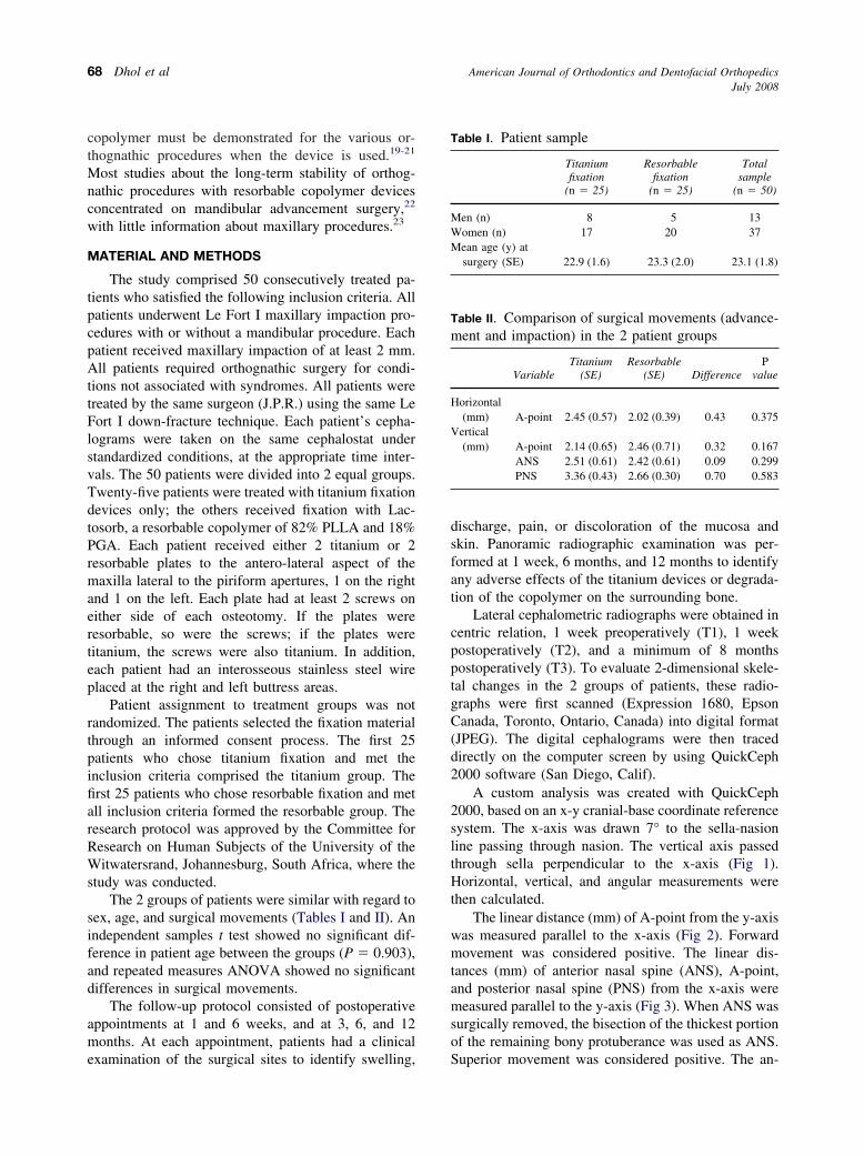
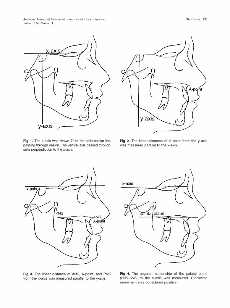
A custom analysis was created with QuickCeph2000, based on an x-y cranial-base coordinate referencesystem. The x-axis was drawn 7° to the sella-nasionline passing through nasion. The vertical axis passedthrough sella perpendicular to the x-axis (Fig 1).Horizontal, vertical, and angular measurements werethen calculated.
The linear distance (mm) of A-point from the y-axiswas measured parallel to the x-axis (Fig 2). Forwardmovement was considered positive. The linear dis-tances (mm) of anterior nasal spine (ANS), A-point,and posterior nasal spine (PNS) from the x-axis weremeasured parallel to the y-axis (Fig 3). When ANS wassurgically removed, the bisection of the thickest portionof the remaining bony protuberance was used as ANS.
Table I. Patient sample
Titaniumfixation
(n � 25)
Resorbablefixation
(n � 25)
Totalsample
(n � 50)
Men (n) 8 5 13Women (n) 17 20 37Mean age (y) at
surgery (SE) 22.9 (1.6) 23.3 (2.0) 23.1 (1.8)
Table II. Comparison of surgical movements (advance-ment and impaction) in the 2 patient groups
VariableTitanium
(SE)Resorbable
(SE) DifferenceP
value
Horizontal(mm) A-point 2.45 (0.57) 2.02 (0.39) 0.43 0.375
Vertical(mm) A-point 2.14 (0.65) 2.46 (0.71) 0.32 0.167
ANS 2.51 (0.61) 2.42 (0.61) 0.09 0.299PNS 3.36 (0.43) 2.66 (0.30) 0.70 0.583
Superior movement was considered positive. The an-
was measured parallel to the x-axis.
American Journal of Orthodontics and Dentofacial OrthopedicsVolume 134, Number 1
Dhol et al 69
Fig 1. The x-axis was drawn 7° to the sella-nasion linepassing through nasion. The vertical axis passed through
sella perpendicular to the x-axis.from the x-axis was measured parallel to the y-axis.
Fig 2. The linear distance of A-point from the y-axis
Fig 3. The linear distance of ANS, A-point, and PNS
Fig 4. The angular relationship of the palatal plane(PNS-ANS) to the x-axis was measured. Clockwise movement was considered positive.
American Journal of Orthodontics and Dentofacial OrthopedicsJuly 2008
70 Dhol et al
gular relationship of the palatal plane (PNS-ANS) tothe x-axis was measured (Fig 4). Clockwise movementwas considered positive.
All measurements were made for each patient at T1,T2, and T3. The differences from T1 to T2 showed thesurgical movements. The differences from T2 to T3measured change (relapse) during the postoperativeobservation period.
Fifteen radiographs were chosen at random andtraced by the same investigator (W.S.D.) on two sepa-rate occasions, one month apart. Reliability coefficientswere calculated for each measurement used in the studyto determine intra-examiner reliability.
Statistical analysis
Descriptive statistics consisting of means and stan-dard errors were calculated for the various measure-ments in each group. Repeated measures ANOVA wasused to assess significant changes in the cephalometricmeasurements in the groups (within-group differences)from T2 to T3 and any significant differences betweenthe groups (between-group differences). Statistical testswere 2-tailed at the 5% significance level (P �0.05). Inaddition, clinically significant thresholds of 1 mm forlinear measurements and 1° for angular measurementswere used to test for clinical significance of postsurgi-cal changes. Results were considered significant only if
Table III. Intra-examiner reliability
Variable Intraclass correlation
Horizontal (mm) A-point 0.9949Vertical (mm) A-point 0.9822
ANS 0.9651PNS 0.9782
Angular (°) Palatal plane 0.9835
Table IV. Surgical and postsurgical maxillary move-ment in titanium fixation group
T2-T1 T3-T2
Variable Mean SE Mean SEP
value
Horizontal(mm) A-point 2.45 0.57 �0.80 0.43 0.063
Vertical(mm) A-point 2.14 0.65 �0.64 0.57 0.264
ANS 2.51 0.61 �0.32 0.50 0.520PNS 3.36 0.43 �0.28 0.35 0.429
Angular (°) Palatal plane 2.36 0.74 0.00 0.69 1.000
they were both statistically and clinically significant.
RESULTS
The results of the precision study show a high levelof intra-examiner reliability (Table III).
For the titanium fixation group, the surgical move-ments (T2-T1) and results (T3-T2) are summarized inTable IV.
Horizontal changes included a mean surgical ad-vancement at A-point of 2.45 � 0.57 mm, and a meanrelapse occurred posteriorly but had a clinically andstatistically insignificant value of 0.80 � 0.43 mm.
Vertical changes included a mean surgical impac-tion at ANS of 2.51 � 0.61 mm; mean relapse was inan inferior direction, clinically and statistically insig-nificant at 0.32 � 0.50 mm. The mean surgical impac-tion at PNS was 3.36 � 0.43 mm, and mean relapsewas in an inferior direction, with a clinically andstatistically insignificant value of 0.28 � 0.35 mm.
Angular changes included a mean sagittal rotationof the palatal plane of 2.36° � 0.74° in a clockwisemanner (posterior impacted more than anterior), andmean relapse of the palatal plane angle was essentiallyzero.
In the resorbable fixation group, the surgical move-ments (T2-T1) and results (T3-T2) are summarized inTable V.
Horizontal changes included a mean surgical ad-vancement at A-point of 2.02 � 0.39 mm. Mean
Table V. Surgical and postsurgical maxillary movementin resorbable fixation group
T2-T1 T3-T2
Variable Mean SE Mean SEP
value
Horizontal(mm) A-point 2.02 0.39 �0.20 0.43 0.639
Vertical(mm) A-point 2.46 0.71 �0.12 0.57 0.834
ANS 2.42 0.61 �0.04 0.50 0.936PNS 2.66 0.30 �0.24 0.35 0.498
Angular (°) Palatal plane 0.99 0.84 �0.04 0.69 0.954
Table VI. Difference in postsurgical maxillary move-ment between titanium and resorbable groups
VariableMean (T3-T2)
difference SE P value
Horizontal (mm) A-point 0.60 0.60 0.321Vertical (mm) A-point 0.52 0.80 0.520
ANS 0.28 0.70 0.690PNS 0.04 0.50 0.936
Angular (°) Palatal plane 0.04 0.97 0.967
relapse occurred in a posterior direction but had a
American Journal of Orthodontics and Dentofacial OrthopedicsVolume 134, Number 1
Dhol et al 71
clinically and statistically insignificant value of 0.20 �0.43 mm.
Vertical changes included a mean surgical impac-tion at ANS of 2.42 � 0.61 mm. Mean relapse was inan inferior direction, with a clinically and statisticallyinsignificant value of 0.04 � 0.50 mm. The meansurgical impaction at PNS was 2.66 � 0.30 mm. Meanrelapse was in an inferior direction, with a clinicallyand statistically insignificant value of 0.24 � 0.35 mm.
Angular changes included a mean sagittal rotationof the palatal plane of 0.99° � 0.84° in a clockwisemanner (posterior impacted more than anterior). Meanrelapse of the palatal plane angle was 0.04° � 0.69° ina counterclockwise direction; this was clinically andstatistically insignificant.
Table V summarizes the differences of relapsevalues (T3-T2 changes) between the 2 groups for eachmeasurement. The difference in relapse between thegroups was insignificant for each measurement.
DISCUSSION
The use of resorbable fixation devices for osteosyn-thesis was first investigated over 3 decades ago.12,13
Renewed interest in their use has been spawned bypotential concerns about metal devices and has led toadvances in techniques and material composition.14,16,18
A resorbable copolymer of 18% PGA and 82%PLLA (Lactosorb) has shown promise as an alternativeto metal for fixation of maxillofacial osteotomies.19-21
These resorbable devices are being used increasinglyfor orthognathic surgery, with reports of successfulapplication in Le Fort I osteotomy19,23,24 and bilateralsagittal split osteotomy.25,26 Various human trials havedemonstrated, qualitatively, the ease of application,strength, and the stability of surgical results withresorbable copolymer devices.15,19,20,25,26 To date,there have been no reports of healing complicationsrelated to this material.
Until recently, no data quantified the skeletal sta-bility of orthognathic procedures when resorbable co-polymer was used. Ferretti and Reyneke22 were the firstto carry out such a study. Their prospective trialassessed the skeletal stability of bilateral sagittal splitosteotomy advancement and compared 20 patients withtitanium screws with 20 patients in whom resorbablecopolymer screws were used. Their results showed that,after 1 year, bilateral sagittal split osteotomy fixed withresorbable copolymer screws relapsed 0.83 mm com-pared with 0.25 mm for titanium fixation. Thesechanges were both statistically and clinically insignifi-cant.
The purpose of our study was to similarly examine
Le Fort I maxillary impaction procedures. A previousstudy examined the stability of maxillary proceduresfixated with a resorbable copolymer.23 Norholt et al23
reported a statistically significant difference in thevertical position of the maxilla after 6 weeks in Le FortI osteotomies fixated with a resorbable copolymer, asthe position became slightly more superior than theimmediate postoperative position with an average of0.6 mm. No such changes were noted in the titaniumgroup, and the changes in maxillary position were notclinically noticeable in either treatment group.23
Our study accurately and reliably measured param-eters from lateral cephalometric radiographs of eachpatient at T1, T2, and T3.
Horizontal, vertical, and angular measurementsshowed no significant statistical or radiographic evi-dence of relapse in either the titanium or the resorbablegroup. Differences between the groups were similarlyinsignificant (Tables IV-VI).
Compared with previous stability studies27-29 of LeFort I procedures, this study was unique, and its designhad numerous advantages. All operations were per-formed by 1 surgeon, using the same technique for allpatients. The sample was gathered prospectively over arelatively short period (18 months) and included con-secutively treated patients who met specific inclusioncriteria. The indications for surgery were kept narrowby restricting the study groups to patients who requirednonsegmental maxillary impaction procedures withminimal anteroposterior changes. All cephalometricradiographs were obtained with the same cephalostatunder standardized conditions. Also, the cephalometricanalysis focused on pertinent skeletal landmarks toexamine maxillary position and ignored extraneouslandmarks to improve the statistical power of the study.
Our primary goal was to determine whether resorb-able copolymer plates and screws offer sufficient sta-bility for fixation of Le Fort I impaction procedures.Radiographically, the results indicate sufficient strengthand stability from resorbable copolymer plates andscrews to resist relapse forces such as occlusal ormuscular forces.
Clinical examinations of the surgical sites werecarried out postoperatively at 1 and 6 weeks, and at 3,6, and 12 months. Signs of swelling, discharge, pain, ordiscoloration of the mucosa and skin were monitored asindicators of complications during healing. There wasno evidence of wound healing complications at thesefollow-up visits for any patient. Panoramic radio-graphic examinations were performed at 1 week, 6months, and 12 months, again to identify any adverseeffects of the fixation devices on the surrounding bone.No evidence of healing complications were noted.
However, Norholt et al23 found 2 patients with infec-
American Journal of Orthodontics and Dentofacial OrthopedicsJuly 2008
72 Dhol et al
tion and wound dehiscence in their resorbable copoly-mer group of 30 patients, whereas titanium osteosyn-thesis was more often palpable after 6 to 12 months andrequired removal in 3 of the 30 patients.
Our results support the use of resorbable copolymerdevices as a viable alternative to titanium for fixation inLe Fort I maxillary impaction.
Future studies should examine the stability ofother orthognathic procedures, particularly those thatare inherently more susceptible to relapse such asinferior repositioning of the maxilla, maxillary seg-mental surgery, and mandibular setback procedures.We assessed only the stability of single piece orsingle fragment nonsegmental Le Fort I level osteot-omies. These results might not apply to segmental LeFort I osteotomies or Le Fort I osteotomies withmajor anteroposterior repositioning. In addition, dif-ferent copolymer material compositions, the use ofself reinforcement, the incorporation of bone-stimu-lating factors into the polymers, and the use ofcyanoacrylate or rivets to replace screws for platefixation need further investigation.18,30-33
REFERENCES
1. Van Sickels JE, Larsen AJ, Thrash WJ. A retrospective study ofrelapse in rigidly fixated sagittal split osteotomies: contributingfactors. Am J Orthod Dentofacial Orthop 1988;93:413-8.
2. Proffit WR, Turvey TA, Phillips C. Orthognathic surgery: ahierarchy of stability. Int J Adult Orthod Orthognath Surg1996;11:191-204.
3. Alpert B, Seligson D. Removal of asymptomatic bone platesused for orthognathic surgery and facial fractures. J Oral Max-illofac Surg 1996;54:618-21.
4. Viljanen J, Kinnunen J, Bondestam S, Majola A, Rokkanen P,Törmälä P. Bone changes after experimental osteotomies fixedwith absorbable self-reinforced poly-L-lactide screws or metallicscrews studied by plain radiographs, quantitative computedtomography and magnetic resonance imaging. Biomaterials1995;16:1353-8.
5. Postlethwaite KR, Philips JG, Booth S, Shaw J, Slater A. Theeffects of small plate osteosynthesis on postoperative radiother-apy. Br J Oral Maxillofac Surg 1989;27:375-8.
6. Scher N, Poe D, Kucnmir F, Reft C, Weichselbaum R, PanjeWR. Radiotherapy of the resected mandible following stainlesssteel plate fixation. Laryngoscope 1988;98:561-3.
7. Kennady MC, Tucker MR, Lester GE, Buckley MJ. Histomor-phometric evaluation of stress shielding in mandibular continuitydefects treated with rigid fixation plates and bone grafts. IntJ Oral Maxillofac Surg 1989;18:170-4.
8. Kennady MC, Tucker MR, Lester GE, Buckley MJ. Stressshielding effect of rigid internal fixation plates on mandibularbone grafts. A photon absorption densitometry and quantitativecomputerized tomographic evaluation. Int J Oral MaxillofacSurg 1989;18:307-10.
9. Jorgenson DS, Mayer MH, Ellenbogen RG, Centeno JA,Johnson FB, Mullick FG, et al. Detection of titanium inhuman tissues after craniofacial surgery. Plast Reconstr Surg
1997;99:976-9.10. Rosenberg A, Gratz KW, Sailer HF. Should titanium miniplatesbe removed after bone healing is complete? Int J Oral MaxillofacSurg 1993;22:185-8.
11. Schliephake H, Lehmann H, Kunz U, Schmelzeisen R. Ultra-structural findings in soft tissues adjacent to titanium plates usedin jaw fracture treatment. Int J Oral Maxillofac Surg 1993;22:20-5.
12. Cutright DE, Hunsuck EE, Beasley JD. Fracture reduction usinga biodegradable material, polylactic acid. J Oral Surg 1971;29:393-7.
13. Cutright DE, Perez B, Beasley JD, Larson WJ, Posey WR.Degradation rates of polymers and copolymers of polylactic andpolyglycolic acids. Oral Surg Oral Med Oral Pathol 1974;37:142-52.
14. Eppley BL, Reilly M. Degradation characteristics of PLLA-PGAbone fixation devices. J Craniofac Surg 1997;8:116-20.
15. Eppley BL, Sadove AM, Havlik RJ. Resorbable plate fixationin pediatric craniofacial surgery. Plast Reconstr Surg 1997;100:1-7.
16. Suuronen R. Comparison of absorbable self-reinforced poly-L-lactide screws and metallic screws in the fixation of mandibularcondyle osteotomies: an experimental study in sheep. J OralMaxillofac Surg 1991;49:989-95.
17. Suurronen R, Pohjonen T, Hietanen J, Lindqvist C. A 5-year invitro and in vivo study of the biodegradation of polylactideplates. J Oral Maxillofac Surg 1998;56:604-14.
18. Ylikontiola LP, Sundquist K, Sándor GKB, Törmälä P,Ashammakhi N. Self-reinforced bioresorbable poly-L/DL-lactide [SR-P(L/D)LA] 70/30 miniplates and miniscrews arereliable for fixation of anterior mandibular fractures: a pilotstudy. Oral Surg Oral Med Oral Pathol Oral Radiol Endod2004;97:312-7.
19. Edwards RC, Kiely KD, Eppley BL. The fate of resorbablepoly-L-lactic/polyglycolic acid (Lactosorb) bone fixation de-vices in orthognathic surgery. J Oral Maxillofac Surg 2001;59:19-25.
20. Edwards RC, Kiely KD, Eppley BL. Resorbable fixation tech-niques for genioplasty. J Oral Maxillofac Surg 2000;58:269-72.
21. Edwards RC, Kiely KD, Eppley BL. Resorbable PLLA-PGAscrew fixation of mandibular sagittal split osteotomies. J Cranio-fac Surg 1999;10:230-6.
22. Ferretti C, Reyneke JP. Mandibular, sagittal split osteotomiesfixed with biodegradable or titanium screws: a prospective,comparative study of postoperative stability. Oral Surg Oral MedOral Pathol Oral Radiol Endod 2002;93:534-7.
23. Norholt SE, Pedersen TK, Jensen J. Le Fort I miniplate osteo-synthesis: a randomized, prospective study comparing resorbablePLLA/PGA with titanium. Int J Oral Maxillofac Surg 2004;33:245-52.
24. Edwards RC, Kiely KD, Eppley BL. Fixation of bimaxillaryosteotomies with resorbable plates and screws: experience in 20consecutive cases. J Oral Maxillofac Surg 2001;59:271-6.
25. Westermark A. Lactosorb resorbable osteosynthesis after sagittalsplit osteotomy of the mandible: a 2-year follow-up. J CraniofacSurg 1999;10:519-22.
26. Long-term stability and predictability of soft tissue change afterLe Fort I surgery. Am J Orthod Dentofacial Orthop 1993;104:544-55.
27. Bishara SE, Chu GW, Jakobson JR. Stability of the Le Fort Ione-piece maxillary osteotomy. Am J Orthod Dentofacial Orthop
1988;94:184-200.
American Journal of Orthodontics and Dentofacial OrthopedicsVolume 134, Number 1
Dhol et al 73
28. Bishara SE, Chu GW. Comparison of postsurgical stability of theLe Fort I impaction and maxillary advancement. Am J OrthodDentofacial Orthop 1992;102:335-41.
29. Turvey TA, Phillips C, Zaytoun HS, Proffit WR. Simultane-ous superior respositioning of the maxilla and mandibularadvancement. Am J Orthod Dentofacial Orthop 1988;94:372-83.
30. Illi OE, Feldmann CP. Stimulation of fracture healing by localapplication of humoral factors integrated in biodegradable im-
plants. Eur J Pediatr Surg 1998;8:251-5.31. Tielinen L, Manninen M, Puolakkinen P, Pihlajamaki H, Po-hjonen T, Rautaruori J, et al. Polylactide pin with transforminggrowth factor beta 1 in delayed osteotomy fixation. Clin OrthopRelat Res 1998;355:312-22.
32. Gosain AK, Song LS, Corrao MA, Pintar FA. Biomechanicalevaluation of titanium, biodegradable plate and screw, andcyanoacrylate glue fixation systems in craniofacial surgery. PlastReconstr Surg 1998;101:582-91.
33. Iera D, Haddad AJ, Sándor GK, Ashmmakhi N. Bioabsorbable
fixation devices. Ann Chir Plast Esthet 2005;50:723-32.
Related Documents