Comparison of the Developmental and Reproductive Toxicity of Diethylstilbestrol Administered to Rats in Utero, Lactationally, Preweaning, or Postweaning J. Odum,* P. A. Lefevre,* H. Tinwell,* J. P. Van Miller,² R. L. Joiner,‡ R. E. Chapin,§ N. T. Wallis,* and J. Ashby* ,1 *Syngenta Central Toxicology Laboratory, Alderley Park, Macclesfield, Cheshire, SK10 4TJ, United Kingdom; ²Toxicology/Regulatory Services, Inc., Charlottesville, Virginia 22911; ‡General Electric Company, Pittsfield, Massachusetts 01201; and §Bristol-Myers Squibb, Drug Safety Evaluation, 1090 Elkton Rd., Newark, Delaware 19714 Received November 29, 2001; accepted February 5, 2002 The objective of the study was to determine which period of exposure produces the most marked effects on the reproductive capacity and sexual development of the rat, with particular em- phasis on the relative sensitivity of in utero and postnatal expo- sures. The endocrine active chemical, diethylstilbestrol (DES) was used as an agent known to affect many of the endpoints examined. Hitherto, such comparisons have been made between studies, rather than within a study. Our data will be helpful in the inter- pretation of future multigenerational assay data. In preliminary studies, DES was shown to be active in the immature rat utero- trophic assay with a lowest detected dose of 0.05 mg DES/kg body weight by sc injection and 10 mg DES/l (1.6 mg DES/kg body weight) by administration in drinking water. A dose of 60 mg DES/l drinking water (; 6.5mg DES/kg body weight/day) was selected for the main study since this represented the midpoint of the drinking water uterotrophic dose response and produced no overt maternal toxicity. The study used 10 groups of concomi- tantly pregnant animals, including 2 control groups. The first comparison was between the effects of exposure to DES in utero, and exposure from conception to weaning. Another group of animals was exposed to DES in utero and cross-fostered to un- treated pregnant females to prevent lactational transfer of DES to pups. Two groups were exposed to DES neonatally, either from birth to postnatal day (PND) 10 (pups thus having only lactational exposure), or from birth until weaning (PND 21; pups thus having both lactational exposure and self-exposure via drinking water). In addition, a dose response study to DES was conducted on animals exposed from weaning to PND 100, when the first phase of the study was terminated. Pups exposed to DES in utero and pups exposed from weaning to PND 100 were bred to assess fertility of the F1 animals and the sexual development of F2 offspring. This last comparison was to determine the extent to which weanling rats could be used in endocrine toxicity studies to assess their potential to show activity in utero. The most sensitive period of exposure for inducing developmental effects in F1 animals was from weaning onwards. The neonatal to weaning period (PND 1–21) was the next most sensitive. Essentially no effects were induced in F1 animals exposed in utero. No effects of any kind were observed in animals only exposed over the early neonatal period of PND 1–10. The mean day of vaginal opening, testes descent, and prepuce separation was only altered in groups where postnatal exposure to DES continued beyond PND 10, or was started at weaning. No changes were observed in anogenital dis- tance or caudal sperm counts. Some changes in organ weights were observed, but the interpretation of these was often confused by concomitant changes in body weight. In general, histopatho- logical examination of tissues yielded no additional information. In breeding studies with animals exposed to DES in utero, or from weaning, reduced litter sizes and marginal advances in the day of vaginal opening were observed in the offspring, together with changes in organ weights. However, no unique sensitivity was noted for exposure in utero. Evaluation of the several exposure periods and the many markers monitored in this study may have individual strengths in individual cases, but when rigorously com- pared using the reference estrogen DES, many preconceptions regarding their absolute or relative value were not upheld. Fur- ther, each of these markers is subject to natural variability, as demonstrated by comparisons made among the 5 separate control groups available in parts of the present study. This variability increases the chance that small changes observed in endocrine toxicity studies employing small group sizes and a single control group, or no concomitant control group, may be artifactual. The most marked effects observed in this study were on the develop- mental landmarks in the F1 animals induced by exposures after PND 10. Some effects on developmental landmarks and organ weights were observed in F2 animals following exposure either in utero or postweaning. This study therefore does not establish a unique role for exposures in utero or during the early neonatal period. Key Words: diethylstilbestrol; reproductive toxicity; in utero; neonatal period; lactational exposure; rat. One of the main uncertainties identified by the United States Endocrine Disrupter Screening and Testing Advisory Commit- tee (EDSTAC), and others, was whether it is necessary to evaluate specifically the endocrine toxicity of chemicals in utero and in the early neonatal period, or whether exposures in 1 To whom correspondence should be addressed. Fax: (44) (0) 1625 590249. E-mail: [email protected]. TOXICOLOGICAL SCIENCES 68, 147–163 (2002) Copyright © 2002 by the Society of Toxicology 147 by guest on February 3, 2014 http://toxsci.oxfordjournals.org/ Downloaded from

Welcome message from author
This document is posted to help you gain knowledge. Please leave a comment to let me know what you think about it! Share it to your friends and learn new things together.
Transcript
Comparison of the Developmental and Reproductive Toxicityof Diethylstilbestrol Administered to Rats in Utero,
Lactationally, Preweaning, or Postweaning
J. Odum,* P. A. Lefevre,* H. Tinwell,* J. P. Van Miller,† R. L. Joiner,‡ R. E. Chapin,§ N. T. Wallis,* and J. Ashby*,1
*Syngenta Central Toxicology Laboratory, Alderley Park, Macclesfield, Cheshire, SK10 4TJ, United Kingdom;†Toxicology/Regulatory Services, Inc.,Charlottesville, Virginia 22911;‡General Electric Company, Pittsfield, Massachusetts 01201; and§Bristol-Myers Squibb,
Drug Safety Evaluation, 1090 Elkton Rd., Newark, Delaware 19714
Received November 29, 2001; accepted February 5, 2002
The objective of the study was to determine which period ofexposure produces the most marked effects on the reproductivecapacity and sexual development of the rat, with particular em-phasis on the relative sensitivity of in utero and postnatal expo-sures. The endocrine active chemical, diethylstilbestrol (DES) wasused as an agent known to affect many of the endpoints examined.Hitherto, such comparisons have been made between studies,rather than within a study. Our data will be helpful in the inter-pretation of future multigenerational assay data. In preliminarystudies, DES was shown to be active in the immature rat utero-trophic assay with a lowest detected dose of 0.05 mg DES/kg bodyweight by sc injection and 10 mg DES/l (1.6 mg DES/kg bodyweight) by administration in drinking water. A dose of 60 mgDES/l drinking water (; 6.5mg DES/kg body weight/day) wasselected for the main study since this represented the midpoint ofthe drinking water uterotrophic dose response and produced noovert maternal toxicity. The study used 10 groups of concomi-tantly pregnant animals, including 2 control groups. The firstcomparison was between the effects of exposure to DES in utero,and exposure from conception to weaning. Another group ofanimals was exposed to DES in utero and cross-fostered to un-treated pregnant females to prevent lactational transfer of DES topups. Two groups were exposed to DES neonatally, either frombirth to postnatal day (PND) 10 (pups thus having only lactationalexposure), or from birth until weaning (PND 21; pups thus havingboth lactational exposure and self-exposure via drinking water). Inaddition, a dose response study to DES was conducted on animalsexposed from weaning to PND 100, when the first phase of thestudy was terminated. Pups exposed to DES in utero and pupsexposed from weaning to PND 100 were bred to assess fertility ofthe F1 animals and the sexual development of F2 offspring. Thislast comparison was to determine the extent to which weanlingrats could be used in endocrine toxicity studies to assess theirpotential to show activity in utero. The most sensitive period ofexposure for inducing developmental effects in F1 animals wasfrom weaning onwards. The neonatal to weaning period (PND1–21) was the next most sensitive. Essentially no effects wereinduced in F1 animals exposed in utero. No effects of any kind
were observed in animals only exposed over the early neonatalperiod of PND 1–10. The mean day of vaginal opening, testesdescent, and prepuce separation was only altered in groups wherepostnatal exposure to DES continued beyond PND 10, or wasstarted at weaning. No changes were observed in anogenital dis-tance or caudal sperm counts. Some changes in organ weightswere observed, but the interpretation of these was often confusedby concomitant changes in body weight. In general, histopatho-logical examination of tissues yielded no additional information.In breeding studies with animals exposed to DES in utero, or fromweaning, reduced litter sizes and marginal advances in the day ofvaginal opening were observed in the offspring, together withchanges in organ weights. However, no unique sensitivity wasnoted for exposure in utero. Evaluation of the several exposureperiods and the many markers monitored in this study may haveindividual strengths in individual cases, but when rigorously com-pared using the reference estrogen DES, many preconceptionsregarding their absolute or relative value were not upheld. Fur-ther, each of these markers is subject to natural variability, asdemonstrated by comparisons made among the 5 separate controlgroups available in parts of the present study. This variabilityincreases the chance that small changes observed in endocrinetoxicity studies employing small group sizes and a single controlgroup, or no concomitant control group, may be artifactual. Themost marked effects observed in this study were on the develop-mental landmarks in the F1 animals induced by exposures afterPND 10. Some effects on developmental landmarks and organweights were observed in F2 animals following exposure either inutero or postweaning. This study therefore does not establish aunique role for exposures in utero or during the early neonatalperiod.
Key Words: diethylstilbestrol; reproductive toxicity; in utero;neonatal period; lactational exposure; rat.
One of the main uncertainties identified by the United StatesEndocrine Disrupter Screening and Testing Advisory Commit-tee (EDSTAC), and others, was whether it is necessary toevaluate specifically the endocrine toxicity of chemicalsinuteroand in the early neonatal period, or whether exposures in
1 To whom correspondence should be addressed. Fax: (44) (0) 1625 590249.E-mail: [email protected].
TOXICOLOGICAL SCIENCES68, 147–163 (2002)Copyright © 2002 by the Society of Toxicology
147
by guest on February 3, 2014http://toxsci.oxfordjournals.org/
Dow
nloaded from
other life stages will suffice to reveal endocrine toxicity (Gold-manet al.,2000; NTP, 2001; Pryoret al.,2000; Schmidt, 2001;Sharpe, 1994; Stokeret al.,2000; Williamset al.,2001). Thereare few, if any, comparative data available to answer thisquestion. This question is particularly pertinent to interpreta-tion of data derived from the rodent multigenerational assay(OECD, 1983), appropriately classed as an apical endocrineperturbation assay by EDSTAC. This assay requires constantadministration of the test chemical through 2 generations,including exposures occurringin utero, during lactation, andduring sexual maturation. As a consequence of the constantexposure, it is impossible to distinguish effects induced duringgestation from those occurring postnatally, a distinction ofrelevance to human risk assessments. The multigenerationalassay protocol was recently updated to include, among otherthings, observation of developmental landmarks such as themean day of vaginal opening and prepuce separation (OECD,2001a), landmarks not accessible in the prenatal developmentaltoxicity assay (OECD, 2001b) because of the culling of somenewly born pups. These additions exacerbate the above prob-lem because these newly added landmarks are modulated byexposure during the peripubertal period. In an attempt to ad-dress these uncertainties we have evaluated the comparativeendocrine toxicity of diethylstilbestrol (DES) in different lifestages of the rat.
DES is one of the most widely studied of endocrine toxi-cants. Endocrine toxicity has been observed for DES in allspecies studied and for all endpoints evaluated. Further, activ-ity has been reported over a wide dose range, extending fromunconfirmed reports of effects on the mouse ventral prostategland following exposurein utero to 0.02 mg DES/kg bodyweight, to carcinogenic effects on the mouse uterus followingneonatal exposure to 1000mg DES/kg (representative data areshown in Table 1). Despite the extent of this database there hasbeen no systematic comparative evaluation of the relativesensitivity of the many different windows of exposure. Theobjective of the present study was to determine which exposureperiod produced the most marked effects of DES on the repro-ductive capacity and sexual development of the rat, with par-ticular emphasis on the relative sensitivity ofin utero andpostnatal exposures. Hitherto, such comparisons have beenmade between studies, rather than within a study.
Use of drinking water as the route of exposure to DES waschosen for the study to reduce stress to the treated animals andto be consistent with the general use of the po route of exposurein multigenerational studies. Likewise the rat was selected forstudy since this is the primary test species used in the multi-generational assay. Since most of the earlier studies on DEShad used sc injection as the route of exposure, the relationshipbetween these 2 routes for DES was first investigated using theimmature rat uterotrophic assay. The uterotrophic activity ofDES to the immature mouse is well established (Ashby, 2000;Shelbyet al.,1996), but its activity in the immature rat utero-trophic assay has not been reported. The dose of 60mg DES/l
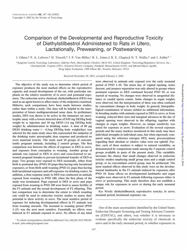
drinking water (; 6.5 mg DES/kg body weight/day) wasselected for the main part of the study as this was found torepresent both the midpoint of the rat drinking water uterotro-phic dose response curve and the midpoint of all previousendocrine toxicity studies on DES (see Fig. 1). This dose wasalso shown in preliminary experiments not to be overtly toxicto pregnant rats. The transplacental transfer of DES is known(Table 1), and some degree of lactational exposure of pups ofdams treated with DES may be assumed from the results ofVorherr et al. (1979).
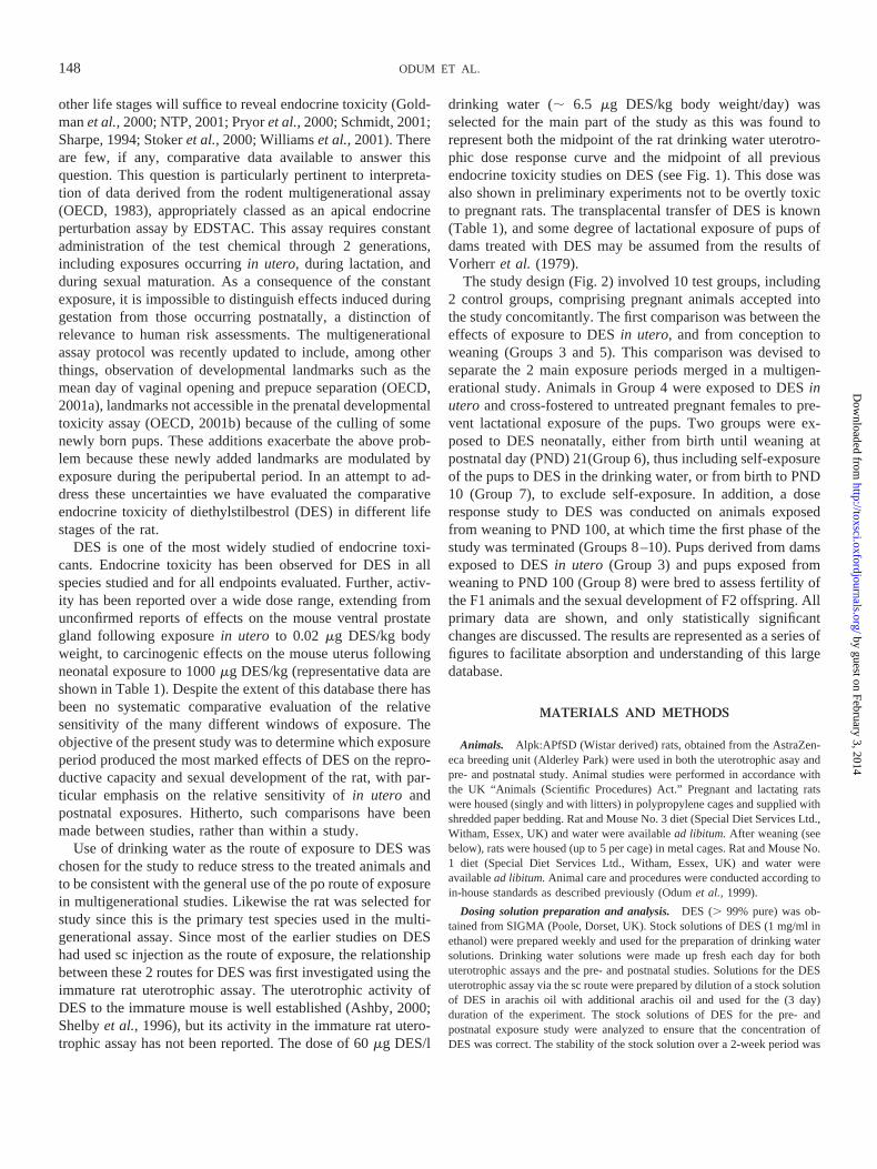
The study design (Fig. 2) involved 10 test groups, including2 control groups, comprising pregnant animals accepted intothe study concomitantly. The first comparison was between theeffects of exposure to DESin utero, and from conception toweaning (Groups 3 and 5). This comparison was devised toseparate the 2 main exposure periods merged in a multigen-erational study. Animals in Group 4 were exposed to DESinutero and cross-fostered to untreated pregnant females to pre-vent lactational exposure of the pups. Two groups were ex-posed to DES neonatally, either from birth until weaning atpostnatal day (PND) 21(Group 6), thus including self-exposureof the pups to DES in the drinking water, or from birth to PND10 (Group 7), to exclude self-exposure. In addition, a doseresponse study to DES was conducted on animals exposedfrom weaning to PND 100, at which time the first phase of thestudy was terminated (Groups 8–10). Pups derived from damsexposed to DESin utero (Group 3) and pups exposed fromweaning to PND 100 (Group 8) were bred to assess fertility ofthe F1 animals and the sexual development of F2 offspring. Allprimary data are shown, and only statistically significantchanges are discussed. The results are represented as a series offigures to facilitate absorption and understanding of this largedatabase.
MATERIALS AND METHODS
Animals. Alpk:APfSD (Wistar derived) rats, obtained from the AstraZen-eca breeding unit (Alderley Park) were used in both the uterotrophic asay andpre- and postnatal study. Animal studies were performed in accordance withthe UK “Animals (Scientific Procedures) Act.” Pregnant and lactating ratswere housed (singly and with litters) in polypropylene cages and supplied withshredded paper bedding. Rat and Mouse No. 3 diet (Special Diet Services Ltd.,Witham, Essex, UK) and water were availablead libitum.After weaning (seebelow), rats were housed (up to 5 per cage) in metal cages. Rat and Mouse No.1 diet (Special Diet Services Ltd., Witham, Essex, UK) and water wereavailablead libitum.Animal care and procedures were conducted according toin-house standards as described previously (Odumet al., 1999).
Dosing solution preparation and analysis.DES (. 99% pure) was ob-tained from SIGMA (Poole, Dorset, UK). Stock solutions of DES (1 mg/ml inethanol) were prepared weekly and used for the preparation of drinking watersolutions. Drinking water solutions were made up fresh each day for bothuterotrophic assays and the pre- and postnatal studies. Solutions for the DESuterotrophic assay via the sc route were prepared by dilution of a stock solutionof DES in arachis oil with additional arachis oil and used for the (3 day)duration of the experiment. The stock solutions of DES for the pre- andpostnatal exposure study were analyzed to ensure that the concentration ofDES was correct. The stability of the stock solution over a 2-week period was
148 ODUM ET AL.
by guest on February 3, 2014http://toxsci.oxfordjournals.org/
Dow
nloaded from
also established. Analysis of DES was by HPLC using an Ultracarb 7 mm ODS30 column (ID 25 cm3 4.6 mm; Phenomenex, Macclesfield, Cheshire, UK),and a mobile phase of methanol (55%), acetonitrile (25%), and water (20%)(v/v) with a flow rate of 1.0 ml/min. A UV detector (210 nm) was used.
Uterotrophic assays. Immature rat uterotrophic assays were conductedusing weanling rats (either 19 to 20 days of age, or 20 to 21 days of age onarrival) for sc and drinking water studies, respectively) as described previously(Odumet al., 1997). DES was administered sc in arachis oil (5 ml/kg), daily
TABLE 1Literature Data for DES in the Mouse and Rat
Rodent/strainDosingwindow
Doseroute
Min 1vedose Reference Summary of effects seen at all doses detected as1ve
Mouse/CD1 GD 9–16 sc 5 McLachlanet al., 1980 Females only. Reproductive tract lesions at 18 months.Genital tract tumors at 100mg/kg
Mouse/CD1 GD 9–16 sc 100 Newbold and McLachlan, 1982 Females only studied. Reproductive tract abnormalities at 1and 18 months.
Mouse/CD1 GD 9–16 sc 2.5 Newboldet al., 1998 Females only studied. Fertility reduced in F1. F2 fertilityunaffected. Reproductive tract tumors 19–24 months.
Mouse/CD1 GD 18 sc 1000 Newboldet al., 1998 Females only studied. Fertility reduced in F1. F2 fertilityunaffected. Reproductive tract tumors at 19–24 months.
Mouse/CD1 GD 9–16 sc 2.5 Newboldet al., 2000 Males only studied. F1 and F2 fertility unaffected.Reproductive tract tumors at 19–24 months.
Mouse/CD1 GD 18 sc 1000 Newboldet al., 2000 Males only studied. F1 and F2 fertility unaffected.Reproductive tract tumors at 19–24 months.
Mouse/CF1 GD 11–17 po 0.02 Vom Saalet al., 1997 Males only studied. Increased ventral prostate weight at 0.02.0.2, 2mg/kg. Decreased ventral prostate weight at 200mg/kg (repeat studies listed below).
Mouse/CF1 GD 11–17 po — Cagenet al., 1999 No effects at 90 daysMouse/CF1 GD 11–17 po — Ashbyet al., 1999 No effects at 6 months.Mouse/CD1 PND 1–5 sc 1000a Newbold and McLachlan, 1982 Females only studied. Adenosis and hypotrophic uterus at 35
days.Mouse/CD1 PND 1–5 sc 1000a Newboldet al., 1990 Females only studied. Reproductive tract lesions at 8 months
onwards. Uterine adenocarcinoma at 18 months.Mouse/CD1 PND 1–5 sc 1b Newboldet al., 1998 Females only studied. Fertility reduced in F1. F2 fertility
unaffected. Reproductive tract tumors at 19–24 months.Mouse/CD1 PND 1–5 sc 1b Newboldet al., 2000 Males only studied. F1 and F2 fertility unaffected.
Reproductive tract tumors at 19–24 months.Mouse/CD1 PND 1–5 sc 1 Newboldet al., 2001 Females only studied. Uterine adenocarcinoma at 18 months.Rat (strain not
specified)PND 6–20 sc 400 Vorherret al., 1979 Reproductive tract abnormalities (males and females),
reproductive tract carcinomas (females).Rat/ACI GD 15 and 18 sc 4.8 Rothschildet al., 1988 Females only studied. Decreased live births, decreased age at
vaginal opening, no changes in cyclicity, atypical uterinepithelia, cystically dilated uterine glands, thickened vaginalepithelium.
Rat/CD GD 16–20 sc 17b Levy et al., 1995 Anogenital distance reduced in males, no effect on time ofvaginal opening.
Rat/Wistar PND 2, 4, 6, 8,10, 12
sc 0.37 Atanassovaet al., 2000 Males only studied; testis wt reduced. Spermatogeneisreduced (PND 18) in all groups except 0.37mg/kg where itwas increased. Testis wt. and mating reduced in adults at370 mg/kg and 37mg/kg.
Rat/Wistar PND 2, 4, 6, 8,10, 12
sc 0.37 Williamset al., 2001 Males only studied. Description of –ve effects at high dosesand1ve effects at low doses on male reproductivesystems.
Rat/SD GD 11–PND 20 po 15 Kwonet al., 2000 Litter size and survival unaffected. Increase in area of thesexually dimorphic nucleus of the preoptic area in females.No effect on age of vaginal opening or first estrus.Irregular cycles at 4 months.
Rat/Alpk PND 21–23 sc 0.05 Data presented herein Females only studied. Increase in uterine weight at PND 24Rat/Alpk PND 21–23 dw 1.6 Data presented herein Females only studied. Increase in uterine weight at PND 24
Note.A representative selection of pre- and postnatal DES studies are shown. The only available rat uterotrophic data for DES are those included in this article.Min 1ve dose, minimum dose detected as positive (mg/kg).
aDose estimated by us.bDose estimate included in reference.
149TOXICITY OF DIETHYLSTILBESTROL
by guest on February 3, 2014http://toxsci.oxfordjournals.org/
Dow
nloaded from
for 3 days at 0.01–2.5mg DES/kg body weight/day to 20 to 21-day-old rats.Control animals received vehicle only. DES was administered at 5–50mgDES/l in the drinking water,ad libitum for 3 days to 21 to 22-day-old animals.Control animals received water only. Water consumption was measured dailyand the intake of DES was calculated. In all cases, on the fourth day, animalswere killed by an overdose of halothane (AstraZeneca, PLC) followed bycervical dislocation. Uteri were removed, trimmed free of fat, blotted andweighed, as described earlier (Odumet al., 1997).
DES Pre- and Postnatal Exposure Study
Administration of DES to F0 and F1 generations. The experimentaldesign is shown in Figure 2. Pregnant female rats (F0; 10–12 weeks old) wereassigned to 10 groups on gestational day (GD) 0 (day of sperm positive smear).Two extra groups of untreated rats (with the same specification) were used tosupply foster mothers for rearing of pups in Groups 2 and 4 (Fig. 2). Theanimals were administered DES in the drinking water (ad libitum) at differentperiods throughout pregnancy, lactation, or postweaning.
Group 1 (control) received water. Group 2 (cross-fostered control) receivedwater. Group 3 received DES (60mg DES/l) from GD 0 to birth. Group 4received DES (60mg DES/l) from GD 0 to birth but were cross-fostered ontountreated mothers. Group 5 received DES (60mg DES/l) from GD 0 toweaning (PND 21). Group 6 received DES (60mg DES/l) from birth toweaning (PND 21). Group 7 received DES (60mg DES/l) from birth to PND10 (this group represented exposure during lactation only, since after this timethe pups would start to consume the drinking water). Group 8 received DES(60 mg DES/l) from weaning to PND 100. Group 9 received DES (30mgDES/l) from weaning to PND 100. Group 10 received DES (10mg DES/l)from weaning to PND 100.
Freshly prepared drinking water solutions were supplied daily. The maxi-mum concentration of DES (60mg DES/l in drinking water) was selectedbased on the results of preliminary studies that indicated that this concentrationgave body weight reductions of no greater than 20% for treated pregnant ratscompared to the concurrent control animals.
Birth occurred naturally, and pups in Groups 2 and 4 were cross-fosteredonto untreated mothers immediately after birth. The pups (F1) were culled to8 per litter (4 of each sex if possible) on PND 4 (day of birth5 D 0). Atweaning (PND 21), the sexes were separated and housed with littermates. Thedams were killed. All animals were weighed at 3- or 4-day intervals from GD0 until termination of the study. Consumption of drinking water was deter-mined daily throughout the study and the intake of DES calculated. Foodconsumption per cage was recorded weekly throughout the study.
The following developmental landmarks were monitored: anogenital dis-tance (AGD; within 24 h of birth, using the method described in Ashby[1997]), testis descent (TD, from PND 21), onset of vaginal opening (VO, fromPND 21), and prepuce separation (PPS, from PND 35). Animals were alsoweighed at the time of the observation of these specific landmarks. Vaginalsmears were taken between PND 52 and 69 to determine the percentage ofdays spent in estrus and cyclicity.
At PND 100, postweaning administration of DES to Groups 8–10 ceased.Animals were selected for the F1 breeding phase, and the remaining animalswere terminated between PND 107 and 111. Sex organ weights were deter-mined for up to 3 pups per sex per litter. Ovaries, uterus, cervix, vagina, ventralprostate, and seminal vesicles were fixed in formal/saline. Testes and epidid-ymides were fixed overnight in Bouins. The tissues were then dehydrated andprocessed by standard histopathological procedures to haemotoxylin/eosinstained paraffin-mounted sections (5 microns). The tissues from groups 1, 3, 5,and 8 were processed for standard histopathological analysis. The right caudaepididymis was taken for sperm analysis.
Breeding of the F1 generation. Twenty-six animals of each sex wereselected from Groups 1, 5, and 8 for breeding within the groups (Figure 2).
FIG. 1. Uterotrophic activity of DES to the rat when administered for 3days either by sc injection or in drinking water (dw; based on Table 2). Thelowest and highest doses for which endocrine activity has been reported forDES in the literature are also shown (based on mouse data, see Table 1). Thedose of DES in the pre- and postnatal exposure study (Groups 3–8) wasestimated from Table 3 (;6.5 mg/kg/day); **p , 0.01 different from con-comitant control values.
FIG. 2. Study design for the DESpre- and postnatal exposure study (1 and2). Open boxes indicate untreated drink-ing water; filled boxes indicate adminis-tration of DES in drinking water at theconcentrations (mg/l) shown. The cross-fostering (XF) period is also indicated(when the pups were fostered onto un-treated dams). *Groups that were se-lected for breeding; gd, gestational day;pnd, postnatal day; F, female; M, male.
150 ODUM ET AL.
by guest on February 3, 2014http://toxsci.oxfordjournals.org/
Dow
nloaded from
These groups were representative of control, gestational, and postweaningexposure to 60mg DES/l. Animals were randomly selected taking siblings intoaccount. Exposure to DES had ceased in “Administration of DES to F0 and F1generations” above and was not resumed. The animals (114–128 days old)were mated in pairs for up to 2 weeks. Females were examined daily and thepresence of sperm in the vaginal smear was taken as evidence of mating. Whenthis occurred the animals were separated, males killed, and the females singlyhoused. Culling of pups was conducted as described above. TD, VO, and PPSwere determined as described above. Females were terminated on PND; 50(after VO was complete for all animals) and males on PND; 85 (after PPSwas complete for all animals). Sex organ weights were determined for allanimals, as in the F1 generation, and tissues were processed for histopatho-logical analysis as above.
Sperm analysis. The right cauda epididymis was scissor minced in 10 mlof 199 medium containing 25mM HEPES (Invitrogen, Groningen, The Neth-erlands). Sperm were isolated and counted as described in Ashby (1997).Analyses were carried out on all F1 groups using 2 pups per litter.
Statistical Methods
Uterotrophic assays. Uterine weights were analyzed by covariance withthe terminal body weights. Terminal body weights were adjusted for covari-ance with initial body weights. Differences from control values were assessedstatistically using a two-sided Student’st-test based on the error mean squarefrom the analysis of covariance. The individual was considered to be thestatistical unit.
DES pre- and postnatal exposure.Initial body weights were analyzed byvariance and subsequent body weights by covariance with the initial bodyweight (taken on the first day of pregnancy, at birth, or at weaning, asappropriate). Water and food consumption were analyzed by variance. Per-centage pup survival and pup sex were analyzed by variance following thedouble arcsine transformation of Freeman and Tukey (1950). Litter size andpup weights at birth were analyzed by variance. AGD was analyzed byvariance and by covariance with birth weight. The proportions of animalsrecorded each day with developmental landmarks were analyzed by Fisher’sExact test and the observed days for the developmental landmarks wereanalyzed by variance. Body weights recorded at the time of observation of thelandmark were also analyzed by variance. Estrus cycle data and sperm num-
bers were analyzed by variance. Organ weights were analyzed by variance andby covariance with the terminal body weights (Shirley, 1996) because thisanalysis provides a better means of allowing for differences in terminal body
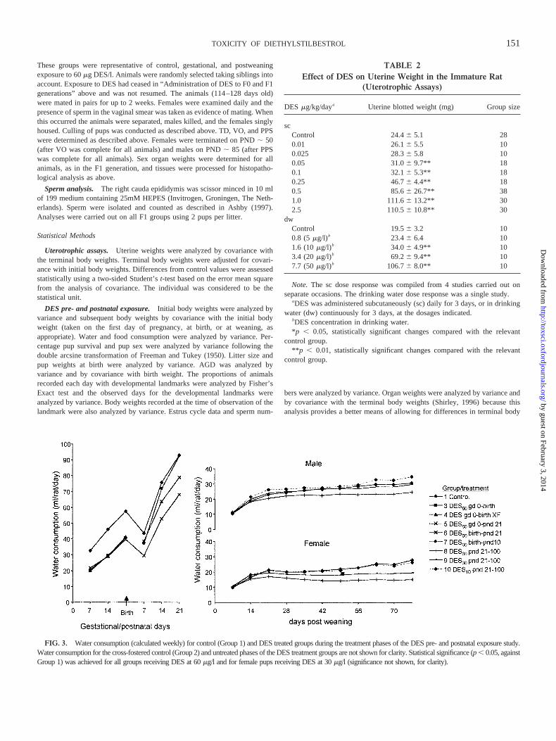
FIG. 3. Water consumption (calculated weekly) for control (Group 1) and DES treated groups during the treatment phases of the DES pre- and postnatal exposure study.Water consumption for the cross-fostered control (Group 2) and untreated phases of the DES treatment groups are not shown for clarity. Statistical significance (p, 0.05, againstGroup 1) was achieved for all groups receiving DES at 60mg/l and for female pups receiving DES at 30mg/l (significance not shown, for clarity).
TABLE 2Effect of DES on Uterine Weight in the Immature Rat
(Uterotrophic Assays)
DES mg/kg/daya Uterine blotted weight (mg) Group size
scControl 24.46 5.1 280.01 26.16 5.5 100.025 28.36 5.8 100.05 31.06 9.7** 180.1 32.16 5.3** 180.25 46.76 4.4** 180.5 85.66 26.7** 381.0 111.66 13.2** 302.5 110.56 10.8** 30
dwControl 19.56 3.2 100.8 (5 mg/l)b 23.46 6.4 101.6 (10mg/l)b 34.06 4.9** 103.4 (20mg/l)b 69.26 9.4** 107.7 (50mg/l)b 106.76 8.0** 10
Note. The sc dose response was compiled from 4 studies carried out onseparate occasions. The drinking water dose response was a single study.
aDES was administered subcutaneously (sc) daily for 3 days, or in drinkingwater (dw) continuously for 3 days, at the dosages indicated.
bDES concentration in drinking water.*p , 0.05, statistically significant changes compared with the relevant
control group.**p , 0.01, statistically significant changes compared with the relevant
control group.
151TOXICITY OF DIETHYLSTILBESTROL
by guest on February 3, 2014http://toxsci.oxfordjournals.org/
Dow
nloaded from
weights than organ to body weight ratios. Differences from control Group 1values were assessed for Groups 3 and 5–10, and differences from controlGroup 2 values were assessed for Group 4; using a two-sided Student’st-testbased on the error mean square from ANOVA or covariance. In all cases thelitter was considered to be the statistical unit. Analyses were carried out asdescribed in SAS (1996).
RESULTS
Uterotrophic Assays
DES was active in the immature rat uterotrophic assay whenadministered for 3 days by sc injection, giving a lowest statis-tically significant dose level of 0.05mg DES/kg body weight/day and reaching a plateau at 1mg DES/kg/day (Table 2, Fig.1). Administration of DES in drinking water gave a similarshaped dose response curve with a lowest statistically signifi-
cant effect dose of 1.6mg DES/kg/day (10mg DES/l drinkingwater; Table 2, Fig. 1).
DES Pre- and Postnatal Exposure Study
Dosing solution analysis. The mean concentration for allbatches of dosing preparations of DES analyzed were within7% of the nominal concentration. The stability of the stocksolutions used to prepare dosing preparations was 100% over 2weeks.
Administration of DES to F0 and F1 generations.Thechanges described below are those that are statistically signif-icant. Changes that do not attain statistical significance are notdescribed unless they are considered to be of importance inunderstanding a trend, or lack thereof, in which case the lack ofstatistical significance is clearly stated.
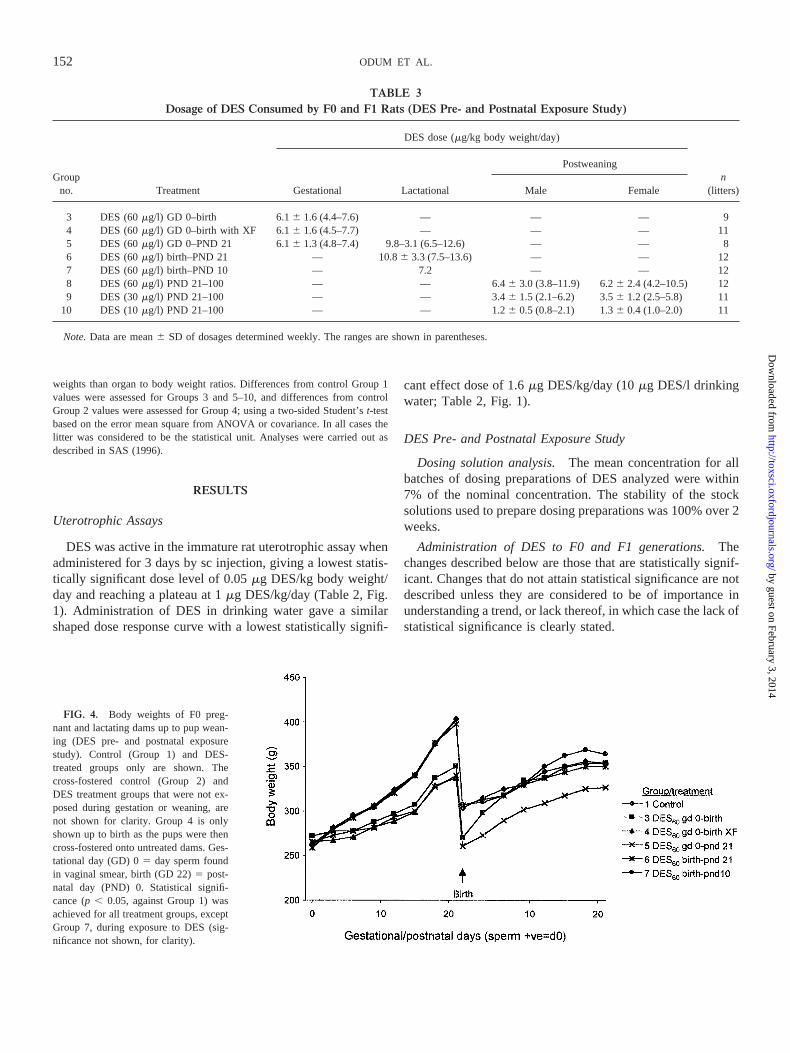
FIG. 4. Body weights of F0 preg-nant and lactating dams up to pup wean-ing (DES pre- and postnatal exposurestudy). Control (Group 1) and DES-treated groups only are shown. Thecross-fostered control (Group 2) andDES treatment groups that were not ex-posed during gestation or weaning, arenot shown for clarity. Group 4 is onlyshown up to birth as the pups were thencross-fostered onto untreated dams. Ges-tational day (GD) 05 day sperm foundin vaginal smear, birth (GD 22)5 post-natal day (PND) 0. Statistical signifi-cance (p, 0.05, against Group 1) wasachieved for all treatment groups, exceptGroup 7, during exposure to DES (sig-nificance not shown, for clarity).
TABLE 3Dosage of DES Consumed by F0 and F1 Rats (DES Pre- and Postnatal Exposure Study)
Groupno. Treatment
DES dose (mg/kg body weight/day)
n(litters)Gestational Lactational
Postweaning
Male Female
3 DES (60mg/l) GD 0–birth 6.16 1.6 (4.4–7.6) — — — 94 DES (60mg/l) GD 0–birth with XF 6.16 1.6 (4.5–7.7) — — — 115 DES (60mg/l) GD 0–PND 21 6.16 1.3 (4.8–7.4) 9.8–3.1 (6.5–12.6) — — 86 DES (60mg/l) birth–PND 21 — 10.86 3.3 (7.5–13.6) — — 127 DES (60mg/l) birth–PND 10 — 7.2 — — 128 DES (60mg/l) PND 21–100 — — 6.46 3.0 (3.8–11.9) 6.26 2.4 (4.2–10.5) 129 DES (30mg/l) PND 21–100 — — 3.46 1.5 (2.1–6.2) 3.56 1.2 (2.5–5.8) 11
10 DES (10mg/l) PND 21–100 — — 1.26 0.5 (0.8–2.1) 1.36 0.4 (1.0–2.0) 11
Note.Data are mean6 SD of dosages determined weekly. The ranges are shown in parentheses.
152 ODUM ET AL.
by guest on February 3, 2014http://toxsci.oxfordjournals.org/
Dow
nloaded from
Consumption of drinking water containing DES by F0 andF1 animals during the study is shown in Figure 3. Groupsreceiving DES at 60mg DES/l consumed less water thancontrol groups (up to 60% of concurrent control values). At 10mg DES/l there was no effect on water consumption although30 mg DES/l reduced water consumption in females, but notmales. The dosage of DES received (asmg DES/kg bodyweight/day) is shown in Table 3. Gestational and postweaning
exposure to a concentration of 60mg DES/l resulted in; 6 mgDES/kg/day while the dosage during lactation rose to; 10 mgDES/kg/day. These doses of DES produced no adverse clinicalsigns in any animals. Body weights of treated F0 dams werereduced in the groups exposed to DES but were no less than80% of concurrent control values throughout pregnancy (Fig.4). Dams exposed only during pregnancy (Group 3) regainedthis weight, once exposure had ceased. The body weights of
TABLE 4Numbers of Births and Litter Survival (F1 pups, DES Pre- and Postnatal Exposure Study)
Groupno. Treatment
Litters born/no. pregnant
Litter sizeat birtha
% Pupsborn livea
% Survivalto PND 4(pre-cull)a
Pup weight at birth (g)a
Male Female
1 Control (water only) 12/12 13.16 2.1 99.46 2.2 90.66 10.4 (11) 5.96 0.5 (12) 5.46 0.5 (11)2 Control with XF 12/12 12.06 3.9 93.56 2.5 94.16 8.8 (10) 5.86 0.7 (12) 5.56 0.7 (12)3 DES (60mg/l) GD 0–birth 11/11 11.66 3.6 99.16 3.0 81.46 16.8 (11) 5.66 0.7 (11) 5.36 0.7 (11)4 DES (60mg/l) GD 0–birth with XFb 9/9 11.46 3.3 98.56 4.4 82.26 21.9 (8)**b 5.56 0.6 (9) 5.26 0.6 (9)5 DES (60mg/l) GD 0–PND 21 8/8 10.76 4.0 96.46 5.5 84.56 21.5 (8) 5.56 0.6 (8) 5.26 0.7 (8)6 DES (60mg/l) birth–PND 21 12/12 11.86 3.0 99.46 2.1 93.76 6.8 (7) 5.96 0.6 (12) 5.56 0.6 (12)7 DES (60mg/l) birth-PND 10 12/12 11.16 2.6 98.86 2.9 98.46 3.1 (9)* 6.16 0.9 (12) 5.66 0.5 (11)8 DES (60mg/l) PND 21–100 12/12 12.46 2.2 1006 0.0 96.16 5.2 (12) 6.06 0.5 (12) 5.76 0.5 (12)9 DES (30mg/l) PND 21–100 11/11 11.76 2.5 99.26 2.2 96.26 5.4 (11) 6.46 0.6 (11) 6.06 0.6 (11)*
10 DES (10mg/l) PND 21–100 11/11 12.56 1.7 97.86 5.0 94.76 9.6 (10) 6.06 0.3 (11) 5.76 0.4 (11)
aData are mean6 SD wheren (in parentheses)5 no. of litters.*p , 0.05; statistically significant changes compared with Group 1 as control group orbGroup 2 as control group.**p , 0.01; statistically significant changes compared with Group 1 as control group orbGroup 2 as control group.
TABLE 5Body Weights of F1 Generation (DES Pre- and Postnatal Exposure Study)
Groupno. Treatment
Male pups (PND) Female pups (PND)
4a 9 21 61 97 n 4a 9 21 61 97 n
1 Control (wateronly)
8.76 1.0 17.46 1.6 48.96 3.5 3116 14.2 4566 16.3 11 8.26 0.9 16.66 1.8 45.96 3.1 1996 8.9 2486 11.6 11
2 Control with XF 8.86 1.2 17.56 2.2 48.96 4.8 3146 25.6 4586 38.1 10 8.06 1.8 17.06 2.3 47.46 4.5 2016 14.5 2506 17.8 103 DES (60mg/l)
GD 0–birth8.06 1.7 16.16 2.5 47.66 4.9 3106 18.8 4516 32.0 11 7.96 1.7 16.26 2.5 47.66 4.1 2046 9.0 2536 11.3 11
4 DES (60mg/l)GD 0–birthwith XFb
8.36 2.1 16.56 3.5 48.26 6.8 3086 40.6 4446 52.6 8 7.66 1.8 14.76 3.5 44.56 7.0 1986 18.7 2466 18.0 8
5 DES (60mg/l)GD 0–PND 21
7.36 1.8 13.26 4.2** 35.46 5.8** 266 6 34.1 3876 41.0 7 7.26 2.2 13.16 4.4** 34.76 5.6** 181 6 22.7 2306 29.2 7
6 DES (60mg/l)birth–PND 21
8.46 1.1 15.56 1.6* 37.66 5.4** 279 6 23.8 4086 29.7 11 8.06 1.2 15.06 2.1* 36.76 5.9** 180 6 20.1 2296 18.9 11
7 DES (60mg/l)birth–PND 10
8.56 1.2 15.36 1.8** 42.46 3.9** 300 6 14.9 4316 24.2 9 8.06 1.3 14.56 2.1** 40.96 4.1** 193 6 11.9 2456 15.5 9
8 DES (60mg/l)PND 21–100
9.26 1.6 18.46 2.4 50.86 5.6 2746 22.7** 3946 30.3** 12 9.06 1.4 18.16 2.0 49.66 4.9 1746 9.4** 215 6 8.0** 12
9 DES (30mg/l)PND 21–100
9.06 0.8 18.16 1.4 49.76 3.1 3076 15.5 4356 25.0* 11 8.66 0.9 17.56 1.6 48.36 3.5 1936 10.8* 2376 13.1** 10
10 DES (10mg/l)PND 21–100
9.26 0.8 18.86 2.3 50.76 4.1 3236 22.5 4576 30.9 11 8.76 0.7 17.76 2.0 48.46 4.2 2016 11.9 2466 12.9 11
aData are post-cull on PND 4 and are unadjusted;n 5 no. of litters.*p , 0.05, statistically significant changes are shown after adjusting for covariance with initial body weights, at birth (PND, 22) or weaning (PND. 22)
and compared with Group 1 orbGroup 2 as control group.**p , 0.01, statistically significant changes are shown after adjusting for covariance with initial body weights, at birth (PND, 22) or weaning (PND. 22)
and compared with Group 1 orbGroup 2 as control group.
153TOXICITY OF DIETHYLSTILBESTROL
by guest on February 3, 2014http://toxsci.oxfordjournals.org/
Dow
nloaded from
dams administered DES postpartum only (Groups 6 and 7)were unaffected. Dams exposed through pregnancy and lacta-tion (Group 5) were consistently lighter than those in all theother groups (Fig. 4).
The numbers of litters born and litter sizes at birth weresimilar for all groups. Pup survival to PND 4 (when numberswere culled to 8 per litter) among the groups which were notcross-fostered was similar among treated and untreated groups(Table 4). Pup survival was significantly reduced in Group 4(exposed through gestation, with cross-fostering) when com-pared with the cross-fostered controls (Group 2), but this wasdue to the higher survival rate of pups in Group 2 comparedwith the noncross-fostered control group (Group1) againstwhich the other groups were compared. Body weights of maleand female pups up to weaning were reduced in pups exposedto DES after birth (Groups 5–7), but not in pups exposedgestationally (Table 5, Figs. 5 and 6). In the postweaningperiod, body weights were reduced in pups exposed to 30 and60 mg DES/l after weaning (Groups 8 and 9), no other groupswere affected (Table 5, Figs. 5 and 6).
Food consumption by the F0 dams was reduced to approx-imately 80% of control values during the DES exposure period,but returned to that of the control animals once exposure hadceased. Food consumption was similarly reduced in F1 animalsexposed to 60mg DES/l during the postweaning phase. F1females exposed to 30mg DES/l showed intermittent reduc-tions (approximately 90% of control values) in food consump-tion (data not shown).
AGD was unchanged in males and females in all groups(Tables 6 and 7). Since body weights at birth were similar in allgroups, AGD adjusted for body weight was also unchanged(data not shown).
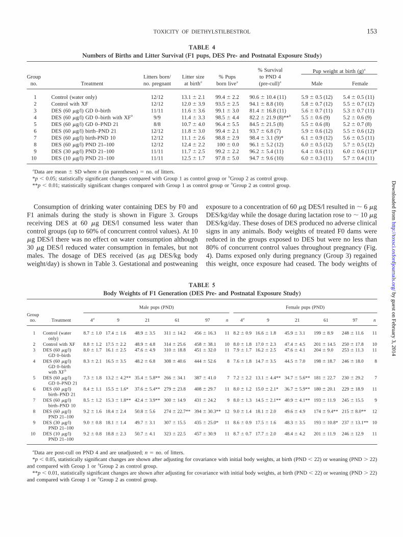
TD and PPS are sexual developmental landmarks in malerats. The age and weight at which these occur were analyzedindependently (i.e., they were unadjusted for body weightchanges). There were no changes observed in these parametersin F1 pups exposed to DES during the gestational period only(Groups 3 and 4). Animals exposed to DES postnatally wereolder, and sometimes heavier, at TD and PPS (Table 6, Fig. 5).Exposure to 60mg DES/l from GD 1 to PND 21 (Group 5) orexposure from birth to PND 21 (Group 6) gave a delay of 2–3days for TD. These effects on these 2 groups were, however,considered to be of uncertain biological significance (Fig. 5)since they were not accompanied by increased body weight atTD. Group 5 animals were lighter but not statistically differentfrom controls (Table 6). A delay in TD was also observed inanimals exposed to 60mg DES/l from PND 21 onwards (Group8) and was accompanied by a body weight increase at the timeof TD. An increase in body weight only at TD was observed inanimals exposed to 30mg DES/l from PND 21 onwards (Group9), giving some indication of a dose response (Table 6, Fig. 5).PPS was also delayed in animals exposed to 60mg DES/l fromGD 1 to PND 21 (Group 5). However this group, and Group 6,also had reduced body weights at PPS. As with TD, thedecreased body weights led us to consider the effects in Groups5 and 6 to be of uncertain biological significance (Fig. 5). PPS
FIG. 5. Summary of results for F0 dams and F1 male pups (DES pre- and postnatal exposure study). Exposure period, open boxes, water only; filled boxes,DES mg/l in drinking water. XF, cross-fostered; gd, gestational day; pnd, postnatal day. Arrows indicate statistically significant increases or decreases atp ,0.05, open arrows are considered to be of uncertain biological relevance (see Results). The light gray shaded boxes (discussed in the text) indicate the strongestresponses observed.aBody weight decreases during the study, occurring at timepoints within the period shown (d, effect on dam; p, effect on pup).bOnly Groups1, 3, 5, and 8 were examined histopathologically. Developmental landmarks: AG dist, anogenital distance; TD, testis descent, PPS; preputial separation. Animalsnot selected for breeding were terminated at PND 107–111, organ weight results are after adjusting for covariance with terminal body weight.
154 ODUM ET AL.
by guest on February 3, 2014http://toxsci.oxfordjournals.org/
Dow
nloaded from
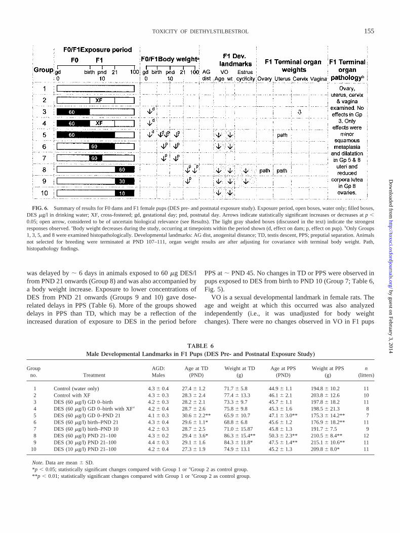
was delayed by; 6 days in animals exposed to 60mg DES/lfrom PND 21 onwards (Group 8) and was also accompanied bya body weight increase. Exposure to lower concentrations ofDES from PND 21 onwards (Groups 9 and 10) gave dose-related delays in PPS (Table 6). More of the groups showeddelays in PPS than TD, which may be a reflection of theincreased duration of exposure to DES in the period before
PPS at; PND 45. No changes in TD or PPS were observed inpups exposed to DES from birth to PND 10 (Group 7; Table 6,Fig. 5).
VO is a sexual developmental landmark in female rats. Theage and weight at which this occurred was also analyzedindependently (i.e., it was unadjusted for body weightchanges). There were no changes observed in VO in F1 pups
FIG. 6. Summary of results for F0 dams and F1 female pups (DES pre- and postnatal exposure study). Exposure period, open boxes, water only; filled boxes,DES mg/l in drinking water; XF, cross-fostered; gd, gestational day; pnd, postnatal day. Arrows indicate statistically significant increases or decreases atp ,0.05; open arrow, considered to be of uncertain biological relevance (see Results). The light gray shaded boxes (discussed in the text) indicate the strongestresponses observed.aBody weight decreases during the study, occurring at timepoints within the period shown (d, effect on dam; p, effect on pup).bOnly Groups1, 3, 5, and 8 were examined histopathologically. Developmental landmarks: AG dist, anogenital distance; TD, testis descent, PPS; preputial separation. Animalsnot selected for breeding were terminated at PND 107–111, organ weight results are after adjusting for covariance with terminal body weight. Path,histopathology findings.
TABLE 6Male Developmental Landmarks in F1 Pups (DES Pre- and Postnatal Exposure Study)
Groupno. Treatment
AGD:Males
Age at TD(PND)
Weight at TD(g)
Age at PPS(PND)
Weight at PPS(g)
n(litters)
1 Control (water only) 4.36 0.4 27.46 1.2 71.76 5.8 44.96 1.1 194.86 10.2 112 Control with XF 4.36 0.3 28.36 2.4 77.46 13.3 46.16 2.1 203.86 12.6 103 DES (60mg/l) GD 0–birth 4.26 0.3 28.26 2.1 73.36 9.7 45.76 1.1 197.86 18.2 114 DES (60mg/l) GD 0–birth with XFa 4.26 0.4 28.76 2.6 75.86 9.8 45.36 1.6 198.56 21.3 85 DES (60mg/l) GD 0–PND 21 4.16 0.3 30.66 2.2** 65.96 10.7 47.16 3.0** 175.36 14.2** 76 DES (60mg/l) birth–PND 21 4.36 0.4 29.66 1.1* 68.86 6.8 45.66 1.2 176.96 18.2** 117 DES (60mg/l) birth–PND 10 4.26 0.3 28.76 2.5 71.06 15.87 45.86 1.3 191.76 7.5 98 DES (60mg/l) PND 21–100 4.36 0.2 29.46 3.6* 86.36 15.4** 50.36 2.3** 210.56 8.4** 129 DES (30mg/l) PND 21–100 4.46 0.3 29.16 1.6 84.36 11.8* 47.56 1.4** 215.16 10.6** 11
10 DES (10mg/l) PND 21–100 4.26 0.4 27.36 1.9 74.96 13.1 45.26 1.3 209.86 8.0* 11
Note.Data are mean6 SD.*p , 0.05; statistically significant changes compared with Group 1 oraGroup 2 as control group.**p , 0.01; statistically significant changes compared with Group 1 oraGroup 2 as control group.
155TOXICITY OF DIETHYLSTILBESTROL
by guest on February 3, 2014http://toxsci.oxfordjournals.org/
Dow
nloaded from
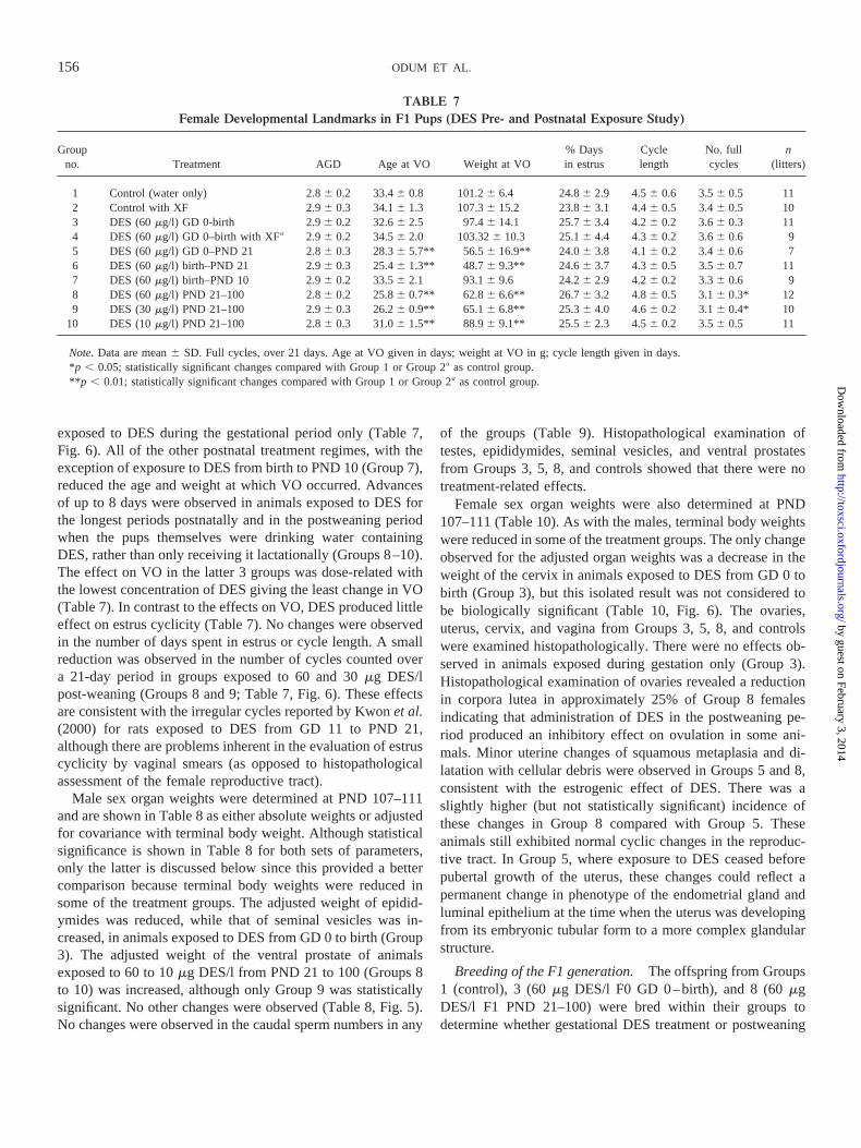
exposed to DES during the gestational period only (Table 7,Fig. 6). All of the other postnatal treatment regimes, with theexception of exposure to DES from birth to PND 10 (Group 7),reduced the age and weight at which VO occurred. Advancesof up to 8 days were observed in animals exposed to DES forthe longest periods postnatally and in the postweaning periodwhen the pups themselves were drinking water containingDES, rather than only receiving it lactationally (Groups 8–10).The effect on VO in the latter 3 groups was dose-related withthe lowest concentration of DES giving the least change in VO(Table 7). In contrast to the effects on VO, DES produced littleeffect on estrus cyclicity (Table 7). No changes were observedin the number of days spent in estrus or cycle length. A smallreduction was observed in the number of cycles counted overa 21-day period in groups exposed to 60 and 30mg DES/lpost-weaning (Groups 8 and 9; Table 7, Fig. 6). These effectsare consistent with the irregular cycles reported by Kwonet al.(2000) for rats exposed to DES from GD 11 to PND 21,although there are problems inherent in the evaluation of estruscyclicity by vaginal smears (as opposed to histopathologicalassessment of the female reproductive tract).
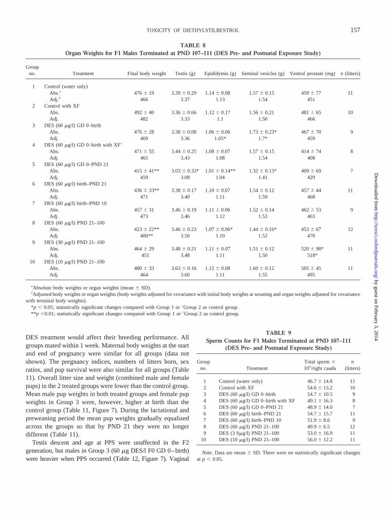
Male sex organ weights were determined at PND 107–111and are shown in Table 8 as either absolute weights or adjustedfor covariance with terminal body weight. Although statisticalsignificance is shown in Table 8 for both sets of parameters,only the latter is discussed below since this provided a bettercomparison because terminal body weights were reduced insome of the treatment groups. The adjusted weight of epidid-ymides was reduced, while that of seminal vesicles was in-creased, in animals exposed to DES from GD 0 to birth (Group3). The adjusted weight of the ventral prostate of animalsexposed to 60 to 10mg DES/l from PND 21 to 100 (Groups 8to 10) was increased, although only Group 9 was statisticallysignificant. No other changes were observed (Table 8, Fig. 5).No changes were observed in the caudal sperm numbers in any
of the groups (Table 9). Histopathological examination oftestes, epididymides, seminal vesicles, and ventral prostatesfrom Groups 3, 5, 8, and controls showed that there were notreatment-related effects.
Female sex organ weights were also determined at PND107–111 (Table 10). As with the males, terminal body weightswere reduced in some of the treatment groups. The only changeobserved for the adjusted organ weights was a decrease in theweight of the cervix in animals exposed to DES from GD 0 tobirth (Group 3), but this isolated result was not considered tobe biologically significant (Table 10, Fig. 6). The ovaries,uterus, cervix, and vagina from Groups 3, 5, 8, and controlswere examined histopathologically. There were no effects ob-served in animals exposed during gestation only (Group 3).Histopathological examination of ovaries revealed a reductionin corpora lutea in approximately 25% of Group 8 femalesindicating that administration of DES in the postweaning pe-riod produced an inhibitory effect on ovulation in some ani-mals. Minor uterine changes of squamous metaplasia and di-latation with cellular debris were observed in Groups 5 and 8,consistent with the estrogenic effect of DES. There was aslightly higher (but not statistically significant) incidence ofthese changes in Group 8 compared with Group 5. Theseanimals still exhibited normal cyclic changes in the reproduc-tive tract. In Group 5, where exposure to DES ceased beforepubertal growth of the uterus, these changes could reflect apermanent change in phenotype of the endometrial gland andluminal epithelium at the time when the uterus was developingfrom its embryonic tubular form to a more complex glandularstructure.
Breeding of the F1 generation.The offspring from Groups1 (control), 3 (60mg DES/l F0 GD 0–birth), and 8 (60mgDES/l F1 PND 21–100) were bred within their groups todetermine whether gestational DES treatment or postweaning
TABLE 7Female Developmental Landmarks in F1 Pups (DES Pre- and Postnatal Exposure Study)
Groupno. Treatment AGD Age at VO Weight at VO
% Daysin estrus
Cyclelength
No. fullcycles
n(litters)
1 Control (water only) 2.86 0.2 33.46 0.8 101.26 6.4 24.86 2.9 4.56 0.6 3.56 0.5 112 Control with XF 2.96 0.3 34.16 1.3 107.36 15.2 23.86 3.1 4.46 0.5 3.46 0.5 103 DES (60mg/l) GD 0-birth 2.96 0.2 32.66 2.5 97.46 14.1 25.76 3.4 4.26 0.2 3.66 0.3 114 DES (60mg/l) GD 0–birth with XFa 2.96 0.2 34.56 2.0 103.326 10.3 25.16 4.4 4.36 0.2 3.66 0.6 95 DES (60mg/l) GD 0–PND 21 2.86 0.3 28.36 5.7** 56.56 16.9** 24.06 3.8 4.16 0.2 3.46 0.6 76 DES (60mg/l) birth–PND 21 2.96 0.3 25.46 1.3** 48.76 9.3** 24.66 3.7 4.36 0.5 3.56 0.7 117 DES (60mg/l) birth–PND 10 2.96 0.2 33.56 2.1 93.16 9.6 24.26 2.9 4.26 0.2 3.36 0.6 98 DES (60mg/l) PND 21–100 2.86 0.2 25.86 0.7** 62.86 6.6** 26.76 3.2 4.86 0.5 3.16 0.3* 129 DES (30mg/l) PND 21–100 2.96 0.3 26.26 0.9** 65.16 6.8** 25.36 4.0 4.66 0.2 3.16 0.4* 10
10 DES (10mg/l) PND 21–100 2.86 0.3 31.06 1.5** 88.96 9.1** 25.56 2.3 4.56 0.2 3.56 0.5 11
Note.Data are mean6 SD. Full cycles, over 21 days. Age at VO given in days; weight at VO in g; cycle length given in days.*p , 0.05; statistically significant changes compared with Group 1 or Group 2a as control group.**p , 0.01; statistically significant changes compared with Group 1 or Group 2a as control group.
156 ODUM ET AL.
by guest on February 3, 2014http://toxsci.oxfordjournals.org/
Dow
nloaded from
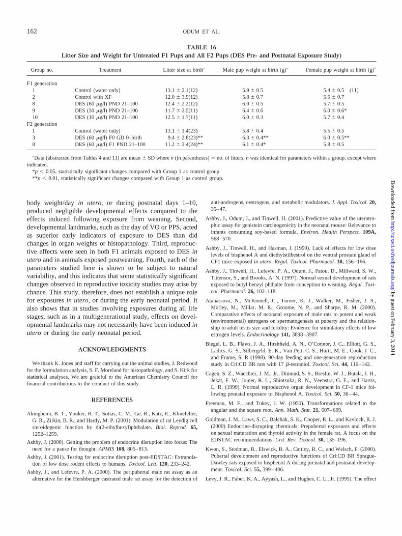
DES treatment would affect their breeding performance. Allgroups mated within 1 week. Maternal body weights at the startand end of pregnancy were similar for all groups (data notshown). The pregnancy indices, numbers of litters born, sexratios, and pup survival were also similar for all groups (Table11). Overall litter size and weight (combined male and femalepups) in the 2 treated groups were lower than the control group.Mean male pup weights in both treated groups and female pupweights in Group 3 were, however, higher at birth than thecontrol group (Table 11, Figure 7). During the lactational andpreweaning period the mean pup weights gradually equalizedacross the groups so that by PND 21 they were no longerdifferent (Table 11).
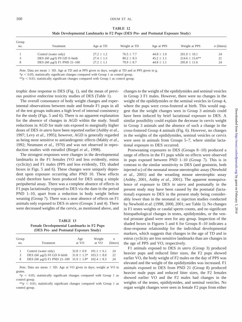
Testis descent and age at PPS were unaffected in the F2generation, but males in Group 3 (60mg DES/l F0 GD 0–birth)were heavier when PPS occurred (Table 12, Figure 7). Vaginal
TABLE 8Organ Weights for F1 Males Terminated at PND 107–111 (DES Pre- and Postnatal Exposure Study)
Groupno. Treatment Final body weight Testis (g) Epididymis (g) Seminal vesicles (g) Ventral prostate (mg)n (litters)
1 Control (water only)Abs.a 4766 19 3.396 0.29 1.146 0.08 1.576 0.15 4596 77 11Adj.b 466 3.37 1.13 1.54 451
2 Control with XFAbs. 4926 40 3.366 0.66 1.126 0.17 1.566 0.21 4816 65 10Adj. 482 3.33 1.1 1.50 466
3 DES (60mg/l) GD 0–birthAbs. 4766 28 3.386 0.08 1.066 0.06 1.736 0.23* 4676 70 9Adj. 469 3.36 1.05* 1.7* 459
4 DES (60mg/l) GD 0–birth with XFc
Abs. 4716 55 3.446 0.25 1.086 0.07 1.576 0.15 4146 74 8Adj. 465 3.43 1.08 1.54 408
5 DES (60mg/l) GD 0–PND 21Abs. 4156 41** 3.03 6 0.33* 1.016 0.14** 1.326 0.13* 4096 69 7Adj. 459 3.08 1.04 1.41 429
6 DES (60mg/l) birth–PND 21Abs. 4366 33** 3.38 6 0.17 1.106 0.07 1.546 0.12 4576 44 11Adj. 471 3.40 1.11 1.59 468
7 DES (60mg/l) birth–PND 10Abs. 4576 31 3.466 0.19 1.116 0.06 1.526 0.14 4626 53 9Adj. 473 3.46 1.12 1.53 463
8 DES (60mg/l) PND 21–100Abs. 4236 22** 3.46 6 0.23 1.076 0.06* 1.446 0.16* 4536 67 12Adj. 406** 3.50 1.10 1.52 470
9 DES (30mg/l) PND 21–100Abs. 4646 29 3.486 0.21 1.116 0.07 1.516 0.12 5206 98* 11Adj. 451 3.48 1.11 1.50 518*
10 DES (10mg/l) PND 21–100Abs. 4806 33 3.636 0.16 1.126 0.08 1.606 0.12 5056 45 11Adj. 464 3.60 1.11 1.55 495
aAbsolute body weights or organ weights (mean6 SD).bAdjusted body weights or organ weights (body weights adjusted for covariance with initial body weights at weaning and organ weights adjusted for covariance
with terminal body weights).*p , 0.05; statistically significant changes compared with Group 1 orcGroup 2 as control group.**p ,0.01; statistically significant changes compared with Group 1 orcGroup 2 as control group.
TABLE 9Sperm Counts for F1 Males Terminated at PND 107–111
(DES Pre- and Postnatal Exposure Study)
Groupno. Treatment
Total sperm3106/right cauda
n(litters)
1 Control (water only) 46.76 14.8 112 Control with XF 54.66 13.2 103 DES (60mg/l) GD 0–birth 54.76 10.5 94 DES (60mg/l) GD 0–birth with XF 49.16 16.3 85 DES (60mg/l) GD 0–PND 21 48.96 14.0 76 DES (60mg/l) birth–PND 21 54.76 15.7 117 DES (60mg/l) birth–PND 10 51.96 8.6 98 DES (60mg/l) PND 21–100 49.96 6.5 129 DES (3 0mg/l) PND 21–100 53.06 16.9 11
10 DES (10mg/l) PND 21–100 56.06 12.2 11
Note.Data are mean6 SD. There were no statistically significant changesat p , 0.05.
157TOXICITY OF DIETHYLSTILBESTROL
by guest on February 3, 2014http://toxsci.oxfordjournals.org/
Dow
nloaded from
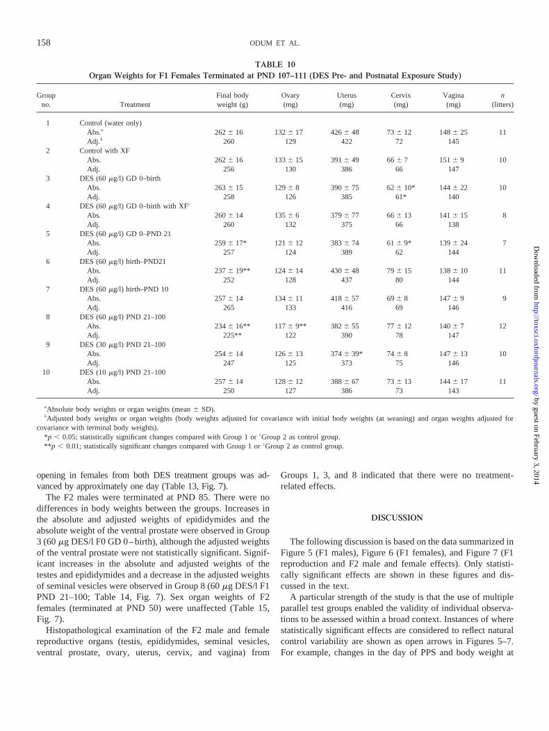
opening in females from both DES treatment groups was ad-vanced by approximately one day (Table 13, Fig. 7).
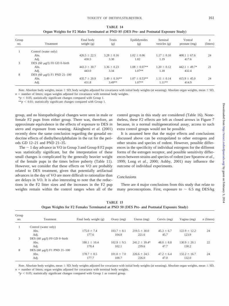
The F2 males were terminated at PND 85. There were nodifferences in body weights between the groups. Increases inthe absolute and adjusted weights of epididymides and theabsolute weight of the ventral prostate were observed in Group3 (60mg DES/l F0 GD 0–birth), although the adjusted weightsof the ventral prostate were not statistically significant. Signif-icant increases in the absolute and adjusted weights of thetestes and epididymides and a decrease in the adjusted weightsof seminal vesicles were observed in Group 8 (60mg DES/l F1PND 21–100; Table 14, Fig. 7). Sex organ weights of F2females (terminated at PND 50) were unaffected (Table 15,Fig. 7).
Histopathological examination of the F2 male and femalereproductive organs (testis, epididymides, seminal vesicles,ventral prostate, ovary, uterus, cervix, and vagina) from
Groups 1, 3, and 8 indicated that there were no treatment-related effects.
DISCUSSION
The following discussion is based on the data summarized inFigure 5 (F1 males), Figure 6 (F1 females), and Figure 7 (F1reproduction and F2 male and female effects). Only statisti-cally significant effects are shown in these figures and dis-cussed in the text.
A particular strength of the study is that the use of multipleparallel test groups enabled the validity of individual observa-tions to be assessed within a broad context. Instances of wherestatistically significant effects are considered to reflect naturalcontrol variability are shown as open arrows in Figures 5–7.For example, changes in the day of PPS and body weight at
TABLE 10Organ Weights for F1 Females Terminated at PND 107–111 (DES Pre- and Postnatal Exposure Study)
Groupno. Treatment
Final bodyweight (g)
Ovary(mg)
Uterus(mg)
Cervix(mg)
Vagina(mg)
n(litters)
1 Control (water only)Abs.a 2626 16 1326 17 4266 48 736 12 1486 25 11Adj.b 260 129 422 72 145
2 Control with XFAbs. 2626 16 1336 15 3916 49 666 7 1516 9 10Adj. 256 130 386 66 147
3 DES (60mg/l) GD 0–birthAbs. 2636 15 1296 8 3906 75 626 10* 1446 22 10Adj. 258 126 385 61* 140
4 DES (60mg/l) GD 0–birth with XFc
Abs. 2606 14 1356 6 3796 77 666 13 1416 15 8Adj. 260 132 375 66 138
5 DES (60mg/l) GD 0–PND 21Abs. 2596 17* 1216 12 3836 74 616 9* 1396 24 7Adj. 257 124 389 62 144
6 DES (60mg/l) birth–PND21Abs. 2376 19** 124 6 14 4306 48 796 15 1386 10 11Adj. 252 128 437 80 144
7 DES (60mg/l) birth–PND 10Abs. 2576 14 1346 11 4186 57 696 8 1476 9 9Adj. 265 133 416 69 146
8 DES (60mg/l) PND 21–100Abs. 2346 16** 117 6 9** 382 6 55 776 12 1406 7 12Adj. 225** 122 390 78 147
9 DES (30mg/l) PND 21–100Abs. 2546 14 1266 13 3746 39* 746 8 1476 13 10Adj. 247 125 373 75 146
10 DES (10mg/l) PND 21–100Abs. 2576 14 1286 12 3886 67 736 13 1446 17 11Adj. 250 127 386 73 143
aAbsolute body weights or organ weights (mean6 SD).bAdjusted body weights or organ weights (body weights adjusted for covariance with initial body weights (at weaning) and organ weights adjusted for
covariance with terminal body weights).*p , 0.05; statistically significant changes compared with Group 1 orcGroup 2 as control group.**p , 0.01; statistically significant changes compared with Group 1 orcGroup 2 as control group.
158 ODUM ET AL.
by guest on February 3, 2014http://toxsci.oxfordjournals.org/
Dow
nloaded from
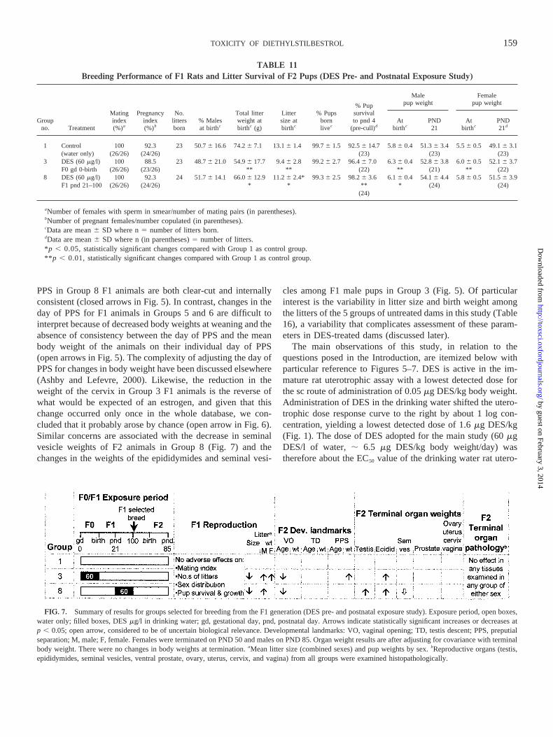
PPS in Group 8 F1 animals are both clear-cut and internallyconsistent (closed arrows in Fig. 5). In contrast, changes in theday of PPS for F1 animals in Groups 5 and 6 are difficult tointerpret because of decreased body weights at weaning and theabsence of consistency between the day of PPS and the meanbody weight of the animals on their individual day of PPS(open arrows in Fig. 5). The complexity of adjusting the day ofPPS for changes in body weight have been discussed elsewhere(Ashby and Lefevre, 2000). Likewise, the reduction in theweight of the cervix in Group 3 F1 animals is the reverse ofwhat would be expected of an estrogen, and given that thischange occurred only once in the whole database, we con-cluded that it probably arose by chance (open arrow in Fig. 6).Similar concerns are associated with the decrease in seminalvesicle weights of F2 animals in Group 8 (Fig. 7) and thechanges in the weights of the epididymides and seminal vesi-
cles among F1 male pups in Group 3 (Fig. 5). Of particularinterest is the variability in litter size and birth weight amongthe litters of the 5 groups of untreated dams in this study (Table16), a variability that complicates assessment of these param-eters in DES-treated dams (discussed later).
The main observations of this study, in relation to thequestions posed in the Introduction, are itemized below withparticular reference to Figures 5–7. DES is active in the im-mature rat uterotrophic assay with a lowest detected dose forthe sc route of administration of 0.05mg DES/kg body weight.Administration of DES in the drinking water shifted the utero-trophic dose response curve to the right by about 1 log con-centration, yielding a lowest detected dose of 1.6mg DES/kg(Fig. 1). The dose of DES adopted for the main study (60mgDES/l of water, ; 6.5 mg DES/kg body weight/day) wastherefore about the EC50 value of the drinking water rat utero-
TABLE 11Breeding Performance of F1 Rats and Litter Survival of F2 Pups (DES Pre- and Postnatal Exposure Study)
Groupno. Treatment
Matingindex(%)a
Pregnancyindex(%)b
No.littersborn
% Malesat birthc
Total litterweight atbirthc (g)
Littersize atbirthc
% Pupsbornlivec
% Pupsurvivalto pnd 4
(pre-cull)d
Malepup weight
Femalepup weight
Atbirthc
PND21
Atbirthc
PND21d
1 Control 100 92.3 23 50.76 16.6 74.26 7.1 13.16 1.4 99.76 1.5 92.56 14.7 5.86 0.4 51.36 3.4 5.56 0.5 49.16 3.1(water only) (26/26) (24/26) (23) (23) (23)
3 DES (60mg/l) 100 88.5 23 48.76 21.0 54.96 17.7 9.46 2.8 99.26 2.7 96.46 7.0 6.36 0.4 52.86 3.8 6.06 0.5 52.16 3.7F0 gd 0-birth (26/26) (23/26) ** ** (22) ** (21) ** (22)
8 DES (60mg/l) 100 92.3 24 51.76 14.1 66.06 12.9 11.26 2.4* 99.36 2.5 98.26 3.6 6.16 0.4 54.16 4.4 5.86 0.5 51.56 3.9F1 pnd 21–100 (26/26) (24/26) * * ** * (24) (24)
(24)
aNumber of females with sperm in smear/number of mating pairs (in parentheses).bNumber of pregnant females/number copulated (in parentheses).cData are mean6 SD where n5 number of litters born.dData are mean6 SD where n (in parentheses)5 number of litters.* p , 0.05, statistically significant changes compared with Group 1 as control group.**p , 0.01, statistically significant changes compared with Group 1 as control group.
FIG. 7. Summary of results for groups selected for breeding from the F1 generation (DES pre- and postnatal exposure study). Exposure period, open boxes,water only; filled boxes, DESmg/l in drinking water; gd, gestational day, pnd, postnatal day. Arrows indicate statistically significant increases or decreases atp , 0.05; open arrow, considered to be of uncertain biological relevance. Developmental landmarks: VO, vaginal opening; TD, testis descent; PPS, preputialseparation; M, male; F, female. Females were terminated on PND 50 and males on PND 85. Organ weight results are after adjusting for covariance with terminalbody weight. There were no changes in body weights at termination.aMean litter size (combined sexes) and pup weights by sex.bReproductive organs (testis,epididymides, seminal vesicles, ventral prostate, ovary, uterus, cervix, and vagina) from all groups were examined histopathologically.
159TOXICITY OF DIETHYLSTILBESTROL
by guest on February 3, 2014http://toxsci.oxfordjournals.org/
Dow
nloaded from
trophic dose response to DES (Fig. 1), and the mean of previ-ous positive endocrine toxicity studies of DES (Table 1).
The overall consonance of body weight changes and exper-imental observations between male and female F1 pups in allof the test groups indicates a high level of internal consistencyfor the study (Figs. 5 and 6). There is no apparent explanationfor the absence of changes in AGD within the study. Smallreductions in AGD for male rats exposed to marginally higherdoses of DESin uterohave been reported earlier (Ashbyet al.,1997; Levyet al.,1995); however, AGD is generally regardedas being most sensitive to antiandrogenic effects (Mablyet al.,1992; Neumannet al., 1970) and was not observed in repro-duction studies with estradiol (Biegelet al., 1998).
The strongest responses were changes in the developmentallandmarks in the F1 females (VO and less evidently, estruscyclicity) and F1 males (PPS and less evidently, TD; shadedboxes in Figs. 5 and 6). These changes were uniquely depen-dent upon exposure occurring after PND 10. These effectscould therefore have been produced for DES using a simpleperipubertal assay. There was a complete absence of effects inF1 pups lactationally exposed to DES via the dam in the periodPND 1–10, apart from a reduction in body weight beforeweaning (Group 7). There was a near absence of effects on F1animals only exposed to DESin utero(Groups 3 and 4). Therewere decreased weights of the cervix, as mentioned above, and
changes to the weight of the epididymides and seminal vesiclesin Group 3 F1 males. However, there were no changes in theweight of the epididymides or the seminal vesicles in Group 4,where the pups were cross-fostered at birth. This would sug-gest that the weight changes seen in Group 3 animals couldhave been induced by brief lactational exposure to DES. Asimilar possibility could explain the decrease in cervix weightin Group 3 animals and the absence of such a change in thecross-fostered Group 4 animals (Fig. 6). However, no changesin the weights of the epididymides, seminal vesicles or cervixwere seen in animals from Groups 5–7, where similar lacta-tional exposure to DES occurred.
Postweaning exposures to DES (Groups 8–10) produced arange of effects in the F1 pups while no effects were observedin pups exposed between PND 1–10 (Group 7). This is incontrast to the similar sensitivity to DES (and genistein, bothinjected sc) of the neonatal mouse uterotrophic assay (Newboldet al., 2001) and the weanling mouse uterotrophic assay(Ashby, 2001, Ashbyet al., 2001). The apparent nonequiva-lence of exposure to DESin utero and postnatally in thepresent study may have been caused by the postnatal (lacta-tional) exposure to DES in the present study being consider-ably lower than in the neonatal sc injection studies conductedby Newboldet al.(1998, 2000, 2001; see Table 1). No changesin F1 testes weights or caudal sperm counts, and no significanthistopathological changes in testes, epididymides, or the ven-tral prostate gland were seen for any group. Inspection of theshaded boxes in Figures 5 and 6 for Groups 8–10 indicates adose-response relationship for the individual developmentalmarkers, which suggests that changes in the age of TD and inestrus cyclicity are less sensitive landmarks than are changes inthe age of PPS and VO, respectively.
F1 animals exposed to DESin utero (Group 3) producedheavier pups and reduced litter sizes, the F2 pups showedearlier VO, the body weight of F2 males on the day of PPS waselevated and the weight of the epididymides was increased. F1animals exposed to DES from PND 21 (Group 8) producedheavier male pups and reduced litter sizes, the F2 femalesshowed earlier VO and the F2 males had changes in theweights of the testes, epididymides, and seminal vesicles. Noorgan weight changes were seen in female F2 pups from either
TABLE 12Male Developmental Landmarks in F2 Pups (DES Pre- and Postnatal Exposure Study)
Groupno. Treatment Age at TD Weight at TD Age at PPS Weight at PPS n (litters)
1 Control (water only) 27.26 1.2 76.56 7.7 44.86 1.0 201.06 10.1 243 DES (60mg/l) F0 GD 0–birth 27.46 1.3 80.26 8.3 45.26 1.1 214.66 15.6** 218 DES (60mg/l) F1 PND 21–100 27.26 1.1 79.96 8.7 44.86 1.1 205.86 11.6 24
Note.Data are mean6 SD. Age at TD and at PPS given in days, weight at TD and at PPS given in g.*p , 0.05; statistically significant changes compared with Group 1 as control group.**p , 0.01; statistically significant changes compared with Group 1 as control group.
TABLE 13Female Developmental Landmarks in F2 Pups
(DES Pre- and Postnatal Exposure Study)
Groupno. Treatment
Ageat VO
Weightat VO
n(litters)
1 Control (water only) 32.86 0.9 101.16 6.1 243 DES (60mg/l) F0 GD 0–birth 31.86 1.3* 103.36 8.0 228 DES (60mg/l) F1 PND 21–100 31.96 1.8* 102.46 8.3 24
Note. Data are mean6 SD. Age at VO given in days; weight at VO ingrams.
*p , 0.05; statistically significant changes compared with Group 1 ascontrol group.
**p , 0.01; statistically significant changes compared with Group 1 ascontrol group.
160 ODUM ET AL.
by guest on February 3, 2014http://toxsci.oxfordjournals.org/
Dow
nloaded from
group, and no histopathological changes were seen in male orfemale F2 pups from either group. There was, therefore, anapproximate equivalence in the effects of exposure to DESinutero and exposure from weaning. Akingbemiet al. (2001)recently drew the same conclusion regarding the gonadal en-docrine effects of diethylhexylphthalate in the rat for the peri-ods GD 12–21 and PND 21–35.
The; 1 day advance in VO in Group 3 and Group 8 F2 pupswas statistically significant, but the interpretation of thesesmall changes is complicated by the generally heavier weightof the female pups in the times before puberty (Table 11).However, we consider that these effects on VO are probablyrelated to DES treatment, given that potentially artifactualadvances in the day of VO are more difficult to rationalize thanare delays in VO. It is also interesting to note that the reduc-tions in the F2 litter sizes and the increases in the F2 pupweights remain within the control ranges when all of the
control groups in this study are considered (Table 16). None-theless, these F2 effects are left as closed arrows in Figure 7because, in a normal multigenerational assay, access to suchextra control groups would not be possible.
It is assumed here that the major effects and conclusionsdiscussed above can be extrapolated to other estrogens andother strains and species of rodent. However, possible differ-ences in the specificity of individual estrogens for the differentforms of the estrogen receptor, and possible sensitivity differ-ences between strains and species of rodent (see Spearowet al.,1999; Long et al., 2000; Ashby, 2001) may influence theoutcome of individual experiments.
Conclusions
There are 4 major conclusions from this study that relate tomany preconceptions. First, exposure to; 6.5 mg DES/kg
TABLE 14Organ Weights for F2 Males Terminated at PND 85 (DES Pre- and Postnatal Exposure Study)
Groupno. Treatment
Final bodyweight (g)
Testis(g)
Epididymides(g)
Seminalvesicles (g)
Ventralprostate (mg)
n(litters)
1 Control (water only)Abs. 426.56 22.5 3.286 0.16 1.026 0.06 1.176 0.10 408.16 67.6 24Adj. 430.5 3.30 1.02 1.19 417.6
3 DES (60mg/l) F0 GD 0–birthAbs. 443.36 30.7 3.366 0.23 1.086 0.07** 1.206 0.12 442.16 49.7* 21Adj. 443.0 3.34 1.07** 1.18 432.4
8 DES (60mg/l) F1 PND 21–100Abs. 435.76 20.9 3.496 0.16** 1.076 0.53** 1.116 0.14 415.96 45.0 24Adj. 431.8 3.49** 1.07** 1.11** 414.9
Note.Absolute body weights, mean6 SD; body weights adjusted for covariance with initial body weights (at weaning). Absolute organ weights, mean6 SD,n 5 number of litters; organ weights adjusted for covariance with terminal body weights.
*p , 0.05; statistically significant changes compared with Group 1.**p , 0.01; statistically significant changes compared with Group 1.
TABLE 15Organ Weights for F2 Females Terminated at PND 50 (DES Pre- and Postnatal Exposure Study)
Groupno. Treatment Final body weight (g) Ovary (mg) Uterus (mg) Cervix (mg) Vagina (mg)n (litters)
1 Control (water only)Abs. 175.06 7.4 103.76 8.1 219.56 30.0 45.36 6.7 122.96 12.2 24Adj. 177.6 104.8 221.6 45.7 123.9
3 DES (60mg/l) F0 GD 0–birthAbs. 180.16 10.4 102.96 9.5 241.26 39.4* 48.06 8.8 130.96 20.1 22Adj. 178.4 102.1 239.6 47.7 130.2
8 DES (60mg/l) F1 PND 21–100Abs. 178.76 8.5 101.06 7.0 226.66 24.5 47.26 6.4 132.26 16.7 24Adj. 177.7 100.7 226.0 47.0 132.0
Note.Absolute body weights, mean6 SD; body weights adjusted for covariance with initial body weights (at weaning). Absolute organ weights, mean6 SD,n 5 number of litters; organ weights adjusted for covariance with terminal body weights.
*p , 0.05; statistically significant changes compared with Group 1 as control group.
161TOXICITY OF DIETHYLSTILBESTROL
by guest on February 3, 2014http://toxsci.oxfordjournals.org/
Dow
nloaded from
body weight/dayin utero, or during postnatal days 1–10,produced negligible developmental effects compared to theeffects induced following exposure from weaning. Second,developmental landmarks, such as the day of VO or PPS, actedas superior early indicators of exposure to DES than didchanges in organ weights or histopathology. Third, reproduc-tive effects were seen in both F1 animals exposed to DESinuteroand in animals exposed postweaning. Fourth, each of theparameters studied here is shown to be subject to naturalvariability, and this indicates that some statistically significantchanges observed in reproductive toxicity studies may arise bychance. This study, therefore, does not establish a unique rolefor exposuresin utero, or during the early neonatal period. Italso shows that in studies involving exposures during all lifestages, such as in a multigenerational study, effects on devel-opmental landmarks may not necessarily have been inducedinutero or during the early neonatal period.
ACKNOWLEDGMENTS
We thank K. Jones and staff for carrying out the animal studies, J. Redwoodfor the formulation analysis, S. F. Moreland for histopathology, and S. Kirk forstatistical analyses. We are grateful to the American Chemistry Council forfinancial contributions to the conduct of this study.
REFERENCES
Akingbemi, B. T., Youker, R. T., Sottas, C. M., Ge, R., Katz, E., Klinefelter,G. R., Zirkin, B. R., and Hardy, M. P. (2001). Modulation of rat Leydig cellsteroidogenic function by di(2-ethylhexyl)phthalate.Biol. Reprod. 65,1252–1259.
Ashby, J. (2000). Getting the problem of endocrine disruption into focus: Theneed for a pause for thought.APMIS108,805–813.
Ashby, J. (2001). Testing for endocrine disruption post-EDSTAC: Extrapola-tion of low dose rodent effects to humans.Toxicol. Lett.120,233–242.
Ashby, J., and Lefevre, P. A. (2000). The peripubertal male rat assay as analternative for the Hershberger castrated male rat assay for the detection of
anti-androgens, oestrogens, and metabolic modulators.J. Appl. Toxicol.20,35–47.
Ashby, J., Odum, J., and Tinwell, H. (2001). Predictive value of the uterotro-phic assay for genistein carcinogenicity in the neonatal mouse: Relevance toinfants consuming soy-based formula.Environ. Health Perspect.109A,568–570.
Ashby, J., Tinwell, H., and Hasman, J. (1999). Lack of effects for low doselevels of bisphenol A and diethylstilbestrol on the ventral prostate gland ofCF1 mice exposedin utero.Regul. Toxicol. Pharmacol.30, 156–166.
Ashby, J., Tinwell, H., Lefevre, P. A., Odum, J., Patou, D., Millward, S. W.,Tittensor, S., and Brooks, A. N. (1997). Normal sexual development of ratsexposed to butyl benzyl phthalte from conception to weaning.Regul. Toxi-col. Pharmacol.26, 102–118.
Atanassova, N., McKinnell, C., Turner, K. J., Walker, M., Fisher, J. S.,Morley, M., Millar, M. R., Groome, N. P., and Sharpe, R. M. (2000).Comparative effects of neonatal exposure of male rats to potent and weak(environmental) estrogens on spermatogenesis at puberty and the relation-ship to adult testis size and fertility: Evidence for stimulatory effects of lowestrogen levels.Endocrinology141,3898–3907.
Biegel, L. B., Flaws, J. A., Hirshfield, A. N., O’Connor, J. C., Elliott, G. S.,Ladics, G. S., Silbergeld, E. K., Van Pelt, C. S., Hurtt, M. E., Cook, J. C.,and Frame, S. R (1998). 90-day feeding and one-generation reproductionstudy in Crl:CD BR rats with 17b-estradiol.Toxicol. Sci.44, 116–142.
Cagen, S. Z., Waechter, J. M., Jr., Dimond, S. S., Breslin, W. J., Butala, J. H.,Jekat, F. W., Joiner, R. L., Shiotsuka, R. N., Veenstra, G. E., and Harris,L. R. (1999). Normal reproductive organ development in CF-1 mice fol-lowing prenatal exposure to Bisphenol A.Toxicol. Sci.50, 36–44.
Freeman, M. F., and Tukey, J. W. (1950). Transformations related to theangular and the square root.Ann. Math. Stat.21, 607–609.
Goldman, J. M., Laws, S. C., Balchak, S. K., Cooper, R. L., and Kavlock, R. J.(2000) Endocrine-disrupting chemicals: Prepubertal exposures and effectson sexual maturation and thyroid activity in the female rat. A focus on theEDSTAC recommendations.Crit. Rev. Toxicol.30, 135–196.
Kwon, S., Stedman, B., Elswick, B. A., Cattley, R. C., and Welsch, F. (2000).Pubertal development and reproductive functions of Crl:CD BR Sprague-Dawley rats exposed to bisphenol A during prenatal and postnatal develop-ment.Toxicol. Sci.55, 399–406.
Levy, J. R., Faber, K. A., Ayyash, L., and Hughes, C. L., Jr. (1995). The effect
TABLE 16Litter Size and Weight for Untreated F1 Pups and All F2 Pups (DES Pre- and Postnatal Exposure Study)
Group no. Treatment Litter size at birtha Male pup weight at birth (g)a Female pup weight at birth (g)a
F1 generation1 Control (water only) 13.16 2.1(12) 5.96 0.5 5.46 0.5 (11)2 Control with XF 12.06 3.9(12) 5.86 0.7 5.56 0.78 DES (60mg/l) PND 21–100 12.46 2.2(12) 6.06 0.5 5.76 0.59 DES (30mg/l) PND 21–100 11.76 2.5(11) 6.46 0.6 6.06 0.6*10 DES (10mg/l) PND 21–100 12.56 1.7(11) 6.06 0.3 5.76 0.4
F2 generation1 Control (water only) 13.16 1.4(23) 5.86 0.4 5.56 0.53 DES (60mg/l) F0 GD 0–birth 9.46 2.8(23)** 6.36 0.4** 6.0 6 0.5**8 DES (60mg/l) F1 PND 21–100 11.26 2.4(24)** 6.16 0.4* 5.86 0.5
aData (abstracted from Tables 4 and 11) are mean6 SD wheren (in parentheses)5 no. of litters,n was identical for parameters within a group, except whereindicated.
*p , 0.05, statistically significant changes compared with Group 1 as control group**p , 0.01, statistically significant changes compared with Group 1 as control group.
162 ODUM ET AL.
by guest on February 3, 2014http://toxsci.oxfordjournals.org/
Dow
nloaded from
of prenatal exposure to the phytoestrogen genistein on sexual differentiationin rats.Proc. Soc. Exp. Biol. Med.208,60–66.
Long, X., Steinmetz, R., Ben-Jonathan, N., Caperell-Grant, A., Young, P. C.,Nephew, K. P., and Bigsby, R. M. (2000). Strain differences in vaginalresponses to the xenoestrogen bisphenol A.Environ. Health Perspect.108,243–247.
Mably, T. A., Moore, R. W., and Peterson, R. (1992).In uteroand lactationalexposure of male rats to 2,3,7,8-tetrachlorodibenzo-p-dioxin. 1. Effects onandrogenic status.Toxicol. Appl. Pharmacol.114,97–107.
McLachlan, J. A., Newbold, R. R., and Bullock, B. C. (1980). Long termeffects on the female mouse genital tract associated with prenatal exposureto diethylstilbestrol.Cancer Res.40, 3988–3999.
NTP (2001). The National Toxicology Program’s report of the endocrinedisruptors low dose peer review. ntp-server.niehs.nih.govntdocs/liaison/lowdose.
Neumann, F., Von Berswordt-Wallrabe, R., Elger, W., Steinbeck, H., Hahn,J. D., and Kramer, M. (1970). Aspects of androgen-dependent events asstudies by antiandrogens.Recent Prog. Horm. Res.26, 337–410.
Newbold, R. R., Banks, E. P., Bullock, B. C., and Jefferson, W. N. (2001).Uterine adenocarcinoma in mice treated neonatally with genistein.CancerRes.61, 4325–4328.
Newbold, R. R., Bullock, B. C., and McLachlan, J. A. (1990). Uterine adeno-carcinoma in mice following developmental treatment with estrogens: Amodel for hormonal carcinogenesis.Cancer Res.50, 7677–7681.
Newbold, R. R., Hanson, R. B., Jefferson, W. N., Bullock, B. C., Haseman, J.,and McLachlan, J. A. (1998). Increased tumors but uncompromised fertilityin the female descendants of mice exposed developmentally to diethylstil-bestrol.Carcinogenesis19, 1655–1663.
Newbold, R. R., Hanson, R. B., Jefferson, W. N., Bullock, B. C., Haseman, J.,and McLachlan, J. A. (2000). Proliferative lesions and reproductive tracttumors in male descendants of mice exposed developmentally to diethyl-stilbestrol.Carcinogenesis21, 1355–1363.
Newbold, R. R., and McLachlan, J. A. (1982). Vaginal adenosis and adeno-carcinoma in mice exposed prenatally or neonatally to diethylstilbestrol.Cancer Res.42, 2003–2011.
Odum, J., Lefevre, P. A., Tittensor, S., Paton, D., Routledge, E. J., Beresford,N. A., Sumpter, J. P., and Ashby, J. (1997). The rodent uterotrophic assay:Critical protocol features, studies with nonyl phenols and comparison witha yeast estrogenicity assay.Regul. Toxicol. Pharmacol.25, 176–188.
Odum, J., Pyrah, I. T. G., Foster, J. R., Van Miller, J. P., Joiner, R. L., andAshby, J. (1999). Comparative activities of p-nonylphenol and diethylstil-bestrol in noble rat mammary gland and uterotrophic assays.Regul. Toxicol.Pharmacol.29, 184–195.
OECD. (1983). OECD Guideline 416: Two-generation reproduction toxicitystudy. InOECD Guidelines for Testing of Chemicals,Vol. 2. Organisationfor Economic Co-operation and Development, Paris, France.
OECD. (2001a). OECD Guideline 414: Prenatal developmental toxicity study(adopted 22 January 2001). InOECD Guidelines for the Testing of Chem-
icals, Vol. 2. Organization for Economic Co-operation and Development,Paris, France (1993).
OECD. (2001b). OECD Guideline 416: Two-generation reproduction toxicitystudy (adopted 22 January 2001). InOECD Guidelines for the Testing ofChemicals,Vol. 2. Organization for Economic Co-operation and Develop-ment, Paris, France. (1993).
Pryor, J. L., Hughes, C., Foster, W., Hales, B. F., and Robaire, B. (2000).Critical windows of exposure for children’s health: The reproductive systemin animals and humans.Environ. Health Perspect.108(Suppl. 3), 491–503.
Rothschild, T. C., Calhoon, R. E., and Boylan, E. S. (1988). Effects ofdiethylstilbestrol exposurein uteroon the genital tracts of female ACI rats.Exper. Mol. Pathol.48, 59–76.
SAS Institute. (1996).SAS/STATS software: Changes and enhancementsthrough release 6.11.SAS Institute, Cary, NC.
Sharpe, R. M. (1994). Regulation of spermatogenesis. InThe Physiology ofReproduction,2nd ed. (E. Knobil and J. D. Neill, Eds.), pp. 1363–1434.Raven Press, New York.
Shelby, M. D., Newbold, R. R., Tully, D. B., Chae, K., and Davis, V. L.(1996). Assessing environmental chemicals for estrogenicity using a com-bination of in vitro and in vivo assays.Environ. Health Perspect.104,1296–1300.
Shirley, E. (1996). A literature review of statistical methods for the analysis ofgeneral toxicology data. InStatistics in Toxicology(B. L. T. Morgan, Ed.).Oxford University Press, Oxford, UK.
Schmidt, C. W. (2001). The low down on low-dose endocrine disruptors.Environ. Health Perspect.109,A420–A421.
Spearow, J. L., Doemeny, P., Sera, R., Leffler, R., and Barkley, M. (1999).Genetic variation in susceptibility to endocrine disruption by estrogen inmice.Science285,1259–1261.
Stoker, T. E., Parks, L. G., Gray, L. E., and Cooper, R. L. (2000). Endocrine-disrupting chemicals: Prepubertal exposures and effects on sexual matura-tion and thyroid function in the male rat. A focus on the EDSTAC recom-mendations. Endocrine Disrupter Screening and Testing AdvisoryCommittee.Crit. Rev. Toxicol.30, 197–252.
vom Saal, F. S., Timms, B. G., Montano, M. M., Palanza, P., Thayer, K. A.,Nagel, S. C., Dhar, M. D., Ganjam, V. K., Parmigiani, S., and Welshons,W. V. (1997). Ventral prostate enlargement in mice due to fetal exposure tolow doses of estradiol or diethylstilbestrol and opposite effects at high doses.Proc. Natl. Acad. Sci. U.S.A.94, 2056–2061.
Vorherr, H., Messer, R. H., Vorherr, U. F., Jordan, S. W., and Kornfeld, M.(1979). Teratogenesis and carcinogenesis in rat offspring after transplacentaland transmammary exposure to diethylstilbestrol.Biochem. Pharmacol.28,1865–1877.
Williams, K., McKinnell, C., Saunders, P. T., Walker, M., Fisher, J. S., Turner,K. J., Atanassova, N., and Sharpe, M. (2001). Neonatal exposure to potentand environmental oestrogens and abnormalities of the male reproductivesystem in the rat: Evidence for importance of the androgen-oestrogenbalance and assessment of the relevance to man.Hum. Reprod. Update7,236–247.
163TOXICITY OF DIETHYLSTILBESTROL
by guest on February 3, 2014http://toxsci.oxfordjournals.org/
Dow
nloaded from
Related Documents