eScholarship provides open access, scholarly publishing services to the University of California and delivers a dynamic research platform to scholars worldwide. Lawrence Berkeley National Laboratory Peer Reviewed Title: Comparative genome analysis of Bacillus cereus group genomes with Bacillus subtilis Author: Anderson, Iain Sorokin, Alexei Kapatral, Vinayak Reznik, Gary Bhattacharya, Anamitra Mikhailova, Natalia Burd, Henry Joukov, Victor Kaznadzey, Denis Walunas, Theresa D'Souza, Mark Larsen, Niels Pusch, Gordon Liolios, Konstantinos Grechkin, Yuri Lapidus, Alla Goltsman, Eugene Chu, Lien Fonstein, Michael Ehrlich, S. Dusko Overbeek, Ross Kyrpides, Nikos Ivanova, Natalia Publication Date: 09-14-2005 Publication Info: Lawrence Berkeley National Laboratory Permalink: http://escholarship.org/uc/item/1nk4b6nf Keywords: comparative genome analysis Abstract: Genome features of the Bacillus cereus group genomes (representative strains of Bacillus cereus, Bacillus anthracis and Bacillus thuringiensis sub spp israelensis) were analyzed and compared

Welcome message from author
This document is posted to help you gain knowledge. Please leave a comment to let me know what you think about it! Share it to your friends and learn new things together.
Transcript
eScholarship provides open access, scholarly publishingservices to the University of California and delivers a dynamicresearch platform to scholars worldwide.
Lawrence Berkeley National Laboratory
Peer Reviewed
Title:Comparative genome analysis of Bacillus cereus group genomes with Bacillus subtilis
Author:Anderson, IainSorokin, AlexeiKapatral, VinayakReznik, GaryBhattacharya, AnamitraMikhailova, NataliaBurd, HenryJoukov, VictorKaznadzey, DenisWalunas, TheresaD'Souza, MarkLarsen, NielsPusch, GordonLiolios, KonstantinosGrechkin, YuriLapidus, AllaGoltsman, EugeneChu, LienFonstein, MichaelEhrlich, S. DuskoOverbeek, RossKyrpides, NikosIvanova, Natalia
Publication Date:09-14-2005
Publication Info:Lawrence Berkeley National Laboratory
Permalink:http://escholarship.org/uc/item/1nk4b6nf
Keywords:comparative genome analysis
Abstract:Genome features of the Bacillus cereus group genomes (representative strains of Bacillus cereus,Bacillus anthracis and Bacillus thuringiensis sub spp israelensis) were analyzed and compared
eScholarship provides open access, scholarly publishingservices to the University of California and delivers a dynamicresearch platform to scholars worldwide.
with the Bacillus subtilis genome. A core set of 1,381 protein families among the four Bacillusgenomes, with an additional set of 933 families common to the B. cereus group, was identified.Differences in signal transduction pathways, membrane transporters, cell surface structures, cellwall, and S-layer proteins suggesting differences in their phenotype were identified. The B. cereusgroup has signal transduction systems including a tyrosine kinase related to two-componentsystem histidine kinases from B. subtilis. A model for regulation of the stress responsive sigmafactor sigmaB in the B. cereus group different from the well studied regulation in B. subtilis hasbeen proposed. Despite a high degree of chromosomal synteny among these genomes, significantdifferences in cell wall and spore coat proteins that contribute to the survival and adaptation inspecific hosts has been identified.
1
LBNL-58211
Comparative genome analysis of Bacillus cereus group genomes
with Bacillus subtilis
Iain Anderson1, Alexei Sorokin
2, Vinayak Kapatral
1*, Gary Reznik
‡, Anamitra Bhattacharya
1
Natalia Mikhailova1, Henry Burd
1, Victor Joukov
1, Denis Kaznadzey
1, Theresa Walunas
1,
MarkD’Souza1, Niels Larsen¶, Gordon Pusch
1, Konstantinos Liolios
1, Yuri Grechkin
1, Alla
Lapidus††, Eugene Goltsman††, Lien Chu1, Michael Fonstein#, S. Dusko Ehrlich
2, Ross
Overbeek#, Nikos Kyrpides††, and Natalia Ivanova††
1 Integrated Genomics, 2201 W. Campbell Park Dr., Chicago, Illinois 60612,
2 Génétique
Microbienne, CRJ INRA, 78352 Jouy en Josas cedex, France.
* To whom correspondence should be addressed.
Present address: Life Sciences Operation, IITRI, Chicago, Illinois 60616 ‡
Present address: Aarhus University, Hoegh Guldbergsgade 10, DK 8000 Aarhus C, Denmark ¶
Present address: Joint Genome Institute, 2800 Mitchell Dr., Walnut Creek, CA 94598 ††
Present address: Fellowship for Interpretation of Genomes, 15 W
155 81st Street, Burr Ridge, IL 60527 #
Address: Vinayak Kapatral
Integrated Genomics Inc
2201. W. Campbell Park Dr Chicago Il 60612.
Phone: 312-491-0846x 326.
Fax: 312-226-9446
2
Abstract
Genome features of the Bacillus cereus group genomes (representative strains of
Bacillus cereus, Bacillus anthracis and Bacillus thuringiensis sub spp israelensis) were
analyzed and compared with the Bacillus subtilis genome. A core set of 1,381 protein families
among the four Bacillus genomes, with an additional set of 933 families common to the
B. cereus group, was identified. Differences in signal transduction pathways, membrane
transporters, cell surface structures, cell wall, and S-layer proteins suggesting differences in
their phenotype were identified. The B. cereus group has signal transduction systems
including a tyrosine kinase related to two-component system histidine kinases from B.
subtilis. A model for regulation of the stress responsive sigma factor σB
in the B. cereus group
different from the well studied regulation in B. subtilis has been proposed. Despite a high
degree of chromosomal synteny among these genomes, significant differences in cell wall and
spore coat proteins that contribute to the survival and adaptation in specific hosts has been
identified.
Contact: [email protected].
Key words: Comparative genomics, Bacillus cereus group, B. subtilis,

3
Introduction
The Bacillus cereus group of bacilli includes B. anthracis (causes anthrax in humans
and cattle), B. cereus (soil borne and food pathogen), B. thuringiensis (lepidopteron insect
pathogen), B. mycoides, B. pseudomycoides and B. weihenstephanensis. Although there is
significant chromosomal synteny among the B. cereus group of genomes, recent studies [1]
have demonstrated differences in gene order, chromosomal rearrangements, nucleotide
variations, and remnant phages. Phylogenetic analyses have suggested that B. anthracis
recently diverged from B. cereus and B. thuringiensis and represents a distinct genetic lineage
[1]. Plasmid encoded genes often play significant roles in pathogenesis in these bacteria.
Virulence genes encoded by the plasmids of B. thuringiensis and B. anthracis are well studied,
but the role of chromosomal genes in in host adaptation and pathogenesis is less known [1, 2].
The availability of genome sequences of members of the B. cereus group include B. anthracis
A2012 [3], B. anthracis Ames [4], and B. cereus ATCC 14579 [5], allowing identification of
unique metabolism, comparative physiology, sporulation and virulence.
The B. thuringiensis bacteria have several sub-species which are classified based on
flagellar serotypes and host range [2]. They are widely used in effective biological control of
mosquitoes, including those carrying malaria, yellow fever, dengue fever, etc [6]. The B.
thuringiensis subspecies isrealensis genome was used as a representative strain of the B.
thuringiensis subspecies for comparative analysis. The human pathogenic isolate B.
thuringiensis serovar Konkukian strain 97-27 sequence was not used in this study
(Unpublished; Acc. # NC005957). In addition, the genome sequence of B. subtilis was also
included in this comparative analysis. Genome sequences of the facultative anaerobe B.
licheniformis, belonging to the B. subtilis group, were not included in this study [7].
Comparative functional analyses allow determination of conserved and unique genes of these
closely related bacteria. The unique gene occurrences in each of these species suggest gene
sharing by horizontal transfer for host-adaptation and cell metabolism.
Materials and Methods
Bioinformatics tools were used to identify genes and gene families of B. cereus group bacteria.
Genome sequences of B. subtilis 168 (Acc. # NC_000964), B. anthracis A2012 (Acc. #
NC_003995) and B. anthracis Ames (Acc. # AE_016879) and B. cereus ATCC 14579, along
with draft genome sequence of B. thuringiensis were used as representative species for
4
comparative analysis. B. thuringiensis strain ATCC 35646 was obtained from the American
Type Culture Collection (Manassas, VA) was used for sequencing. Total DNA was isolated
by standard procedure and sheared by sonication into fragments of ~ca 2-3 kb and cloned into
plasmid pGEM3 (Promega, Madison, WI) and were maintained in E. coli DH5α. End
sequencing of plasmids was performed using Applied Biosystems 3700 (PerkinElmer, Foster
City, CA) and MegaBACE (Amersham Biosciences, Sunnyvale, CA) DNA sequencers.
A total of 67,278 sequencing reactions were performed (~2.5 kb inserts), and 63, 836 reactions
of these were assembled into larger contigs. Base calling and sequence assembly were carried
out with Phred/Phrap using default parameters. Genome coverage, based on the sequenced
DNA, was found to be approximately ~6.2-fold. Contigs smaller than 1500 bp were
not included in the functional analysis. Genes were identified with a combination of CRITICA
and software developed at Integrated Genomics, as previously described [8, 9, 10]. The B.
thuringiensis draft genome sequence has been submitted to Genbank under the
accession number. The B. thuringiensis genome sequence along with annotations is available
online at http:// www. ergolight.com.
Results and Discussion Global genome comparisons. The comparative genome features of B. thuringiensis,
B. anthracis, B. cereus, and B. subtilis are presented in Table 1. In general, the B. cereus
group genomes are about 25% larger than the B. subtilis genome and have lower GC content.
The genomes differ in the total number of nucleotides and size of extra-chromosomal
elements, with the exception of B. cereus which has one small phage-like element on a linear
contig [5]. The two plasmids carrying virulence genes of B. anthracis, pX01 and pX02,
contribute 276, 500 bp (5%) of the total DNA sequence. In previous studies, B. thuringiensis
was shown to have eight plasmids and one linear plasmid-like element with a total of 630,
000 bp (10%) of the total DNA sequence [11]. Several plasmids from the B. thuringiensis
genome have already been sequenced: the toxin-carrying plasmid pBtoxis [12], pTX141
(unpublished, Acc. # NC_002091), pTX142 (unpublished, Acc. # NC _ 0043 34), and pTX143
[13]. The B. cereus linear plasmid pBClin15 contains 21 putative coding sequences (CDSs)
[5]. The gapped B. thuringiensis genome contains a contig with 15 CDSs similar to those
present in pBClin15, suggesting a close relationship with the linear extra chromosomal DNA
5
of B. cereus.
In the B. cereus and B. anthracis A2012 genomes, 71% of the CDSs were assigned
with function compared to 69% in the B. anthracis Ames strain and B. thuringiensis genomes.
The number of predicted CDSs in each genome with no sequence similarity to other genes
(unique genes) in the ERGO database varied, with 1.7% in B. cereus, 3.0% in B. anthracis
A2012, and 6.8% in B. thuringiensis genomes. The disparity is due to the difference in DNA
content of plasmids and prophages, which often have higher percentages of CDSs without
similarity to known proteins. CDSs with unknown (hypothetical proteins) functions varied
from 24-29% in these genomes. In general, the functional categories between the B. cereus
group and B. subtilis are similar in all genomes except for information processing, signal
transduction, virulence and transport subsystems. Genes belonging to core metabolism, amino
acids, lipid, nitrogen, phosphorus, and sulfur metabolism, and chemotaxis, did not differ
significantly between the B. cereus group and B. subtilis genomes.
Protein clusters. The protein clusters between B. thuringiensis, B. cereus, B. anthracis A2012
and B. subtilis genomes were calculated using the protein clustering WorkBench tool with a
cut-off score of 10e-20. Each cluster refers to the number of protein families present in each
group of genomes. A combined total of 5,896 clusters, with a core of 1,381 proteins clusters
common to all four genomes, were identified. Among the B. cereus group, an additional 933
clusters were identified. B. cereus had 291 unique clusters whereas B. anthracis and B.
thuringiensis had 606 and 940 clusters, respectively. Similarly, within the B. cereus group
genomes (B. thuringiensis , B. cereus, B. anthracis), a total of 5,092 families were identified
of which 2,411 were common to all three organisms, while each individual organism
contained a substantial number of protein families not found in the other (Figure 1).
Comparative signal transduction system analysis Bacteria living in different environments
use both chemical and physical cues to regulate metabolism, development, and stress
responses. Signal transduction systems, extra-cytoplasmic function (ECF) sigma factors,
regulators of sporulation and the σΒ stress response sigma factor were studied in the B. cereus
group genomes. A comparison of signal transduction proteins is given in Table 2. Rap family
aspartate phosphatases inhibit the action of response regulators such as Spo0F, and many of
them are regulated by secreted Phr peptides [14]. The B. subtilis genome encodes 11 Rap
family aspartate phosphatases and 7 Phr peptides [15], while the members in the B. cereus
6
group genomes have fewer genes for both phosphatases and Phr peptides. In the B.
thuringiensis genome sequence, an N-terminal fragment of a Rap family phosphatase was
found on one contig whereas the C-terminal fragment was found encoded on another contig.
In B. subtilis, a family of aspartate phosphatases induced by different environmental condition
(Spo0E, YnzD, YisI proteins) inhibits sporulation by interacting with the Spo0A transcription
factor [16]. The B. cereus group genomes contain a varying number of Spo0E-related
phosphatases, ranging from three in B. anthracis to six in B. thuringiensis. One of the B.
thuringiensis phosphatases (BTH08314) has no homolog in either B. cereus or B. anthracis,
whereas two others are found in B. thuringiensis and B. cereus, but neither is in B. anthracis.
One of the most striking features is the presence of a large number of orphan histidine kinases
in the B. cereus group genomes. In the B. subtilis genome, five of the six orphan histidine
kinases are involved in sporulation initiation [17], but in the B. cereus group the larger number
of orphan kinases suggests their role in other functions. Some may be involved in a proposed
new model of regulation of the stress response sigma factor σΒ. There are more than 120 σΒ-
regulated genes in B. subtilis, including the stress induced ribosomal Ctc protein [18]
and glucose starvation inducible GsiB protein. Both genes ctc and gsiB are absent from the B.
cereus group genomes. During unstressed conditions, σΒ is bound to the anti-sigma factor
RsbW, while the anti-anti-sigma factor RsbV is phosphorylated and unable to bind
RsbW (Figure 2A) [19]. Metabolic and environmental stresses activate two distinct
phosphatases, RsbP and RsbU [20], which dephosphorylate RsbV, allowing it to bind RsbW
and free σΒ to activate stress-related genes. RsbP is thought to sense metabolic stress directly
through a PAS domain (Per: period circadian protein, Arnt; Ah receptor nuclear translocator
protein, Sim; single-minded protein), and the RsbU phosphatase activity is regulated by
a cascade of factors including RsbX, RsbT, RsbS, RsbR, and a family of RsbR--
related proteins. While the sigB operon in B. subtilis contains many of these
regulatory factors, in B. anthracis, B. thuringiensis and B. cereus only the genes for RsbV,
RsbW and σΒ were found [21]. The B. cereus group organisms lack all other components
of the σΒ regulatory pathways from B. subtilis. Instead, they possess one phosphatase
(BA_1562 in B. anthracis A2012) distantly related to RsbP and RsbU, which contains a
response regulator receiver domain at its N-terminus, suggesting a distinct regulatory
mechanism for σΒ in the B. cereus group. We propose that the phosphatase activity depends
7
on the phosphorylated state of the N-terminal receiver domain, which could be phosphorylated
by various orphan histidine kinases (Figure 2B). One of the orphan sensor kinases
found in the three B. cereus group genomes appears to be an auto-phosphorylating tyrosine
kinase rather than a histidine kinase. Sequence alignment of this putative tyrosine kinase with
the closely related histidine kinase is shown in Figure 3. The 50 amino acids region
surrounding the phosphorylated histidine residue is highly conserved (41% amino acid
identity), except that histidine is replaced by tyrosine. Two copies of a histidine kinase fused
to a response regulator were found in the B. cereus group genomes but not in the B. subtilis
genome [17]. One of them is found in all three genomes but it lacks a DNA-binding domain.
A unique orphan kinase was found in the B. cereus genome (BC5455). Among the B.
cereus group genomes, only B. thuringiensis has an ortholog pair for ComP-ComA, a two-
component system similar to that found in B. subtilis. Four two-component systems were
found in the B. anthracis genome are absent from the B. thuringiensis and B. cereus genomes.
One CDS is adjacent to an ABC transporter (BA_43704373) highly similar to those involved
in immunity to the lantibiotics subtilin and mutacin. These groups of genes probably play a
role in resistance to antimicrobial peptides, but whether B. anthracis produces the
corresponding peptide is not known. Other genes (BA_35153516) similar to the fsrA and fsrC
virulence regulators, which control the expression of gelatinase and serine protease in
Enterococcus faecalis [22], have been identified, but the gene similar to fsrB was not found.
A B. subtilis type two-component system ykoGH was found in all B. cereus group genomes.
The other group of proteins involved in response to extracellular signals leading to
changes in transcriptional regulation is the ECF sigma factors. These are more prevalent in the
B. cereus group genomes than in the B. subtilis genome. They regulate many physiological
functions including stress responses, cell wall modification, drug resistance and iron transport.
Often they are encoded in an operon with an anti-sigma factor, and many anti-sigma factors
are membrane-spanning proteins with an extracellular sensing domain and an intracellular
sigma factor binding/inactivation domain. B. anthracis has a large number of ECF-type sigma
factors compared to the B. cereus and B. thuringiensis genomes (Table 2). Of the ECF sigma
factors found in the B. subtilis genome, the salt response sigma factor σM
[23] has
orthologs in the B. cereus group genomes. The two CDSs downstream of sigM, yhdL and
yhdK, are involved in regulation of σM
activity. The yhdL gene was found in all the B. cereus
8
group genomes, but not the yhdK gene (a small protein with two predicted membrane-
spanning domains).
Comparative cell wall protein analysis. Surface structures of bacteria are important targets
for rapid species detection by serological methods. For instance, a two-component immuno-
fluorescence assay using antibodies specific to the cell wall and capsule antigens is
recommended for detection of vegetative cells of B. anthracis. Vegetative cells of B.
anthracis have a complex cell wall structure made of the poly γ-D-glutamic acid capsule, S--
layer and carbohydrates [24]. B. cereus and B. thuringiensis strains do not have a
capsule, but have several proteins with S-layer motifs. The B. cereus ATCC14579 strain
lacks S-layer proteins, however it is not clear if B. thuringiensis has it. In the B. cereus group
genomes, S-layer proteins are encoded by a well conserved locus along with the operon
csaAB, which codes for cell surface anchor for S-layer proteins [24]. In the B. thuringiensis
genome there is an S-layer protein, with similarity to peptidoglycan hydrolase and S-layer
homology (SLH) domain at the N-terminal. Both B. cereus and B. thuringiensis genomes
have nine additional proteins with SLH domains; but none of them has the crystallization
domain necessary for S-layer protein polymerization. Six CDSs encode putative
peptidoglycan hydrolases and one of them has a leucine-rich repeat (LRR) domain similar to
that of internalins found in Listeria spp. In the B. anthracis genomes, there are two copies of
genes for a protein translocase, SecA, which is missing from the B. cereus and B. thuringiensis
genomes.
The composition, structure, and biosynthetic pathways for cell wall carbohydrates are
well studied in B. subtilis, compared to the B. cereus group bacteria. In B. subtilis, anionic
polymers are important components of the cell wall as they act as a sink for protons that are
generated during respiration and are also a major site of metal deposition [25]. D-alanyl
esterification of anionic polymers decreases electro-negativity of the cell walls, leading to
modulation of autolysis and enhancing folding and stability of secreted proteins [26].
Depending on the availability of phosphate, different B. subtilis strains produce either
phosphate-free teichuronic acid such as polyglucuronyl-N-acetyl-glucosamine, or teichoic
acids, such as polyglycerolphosphate, poly-ribitolphosphate or polyglucosyl-N-acetyl-
galactosamine-1-phosphate. The carbohydrate composition of bacterial cell walls in the B.
cereus group is different from that of B. subtilis. Anionic polymers play a less prominent role
9
in B. anthracis pathogenesis, since neither polyribitolphosphate, nor polyglycerolphosphate is
detected in their cell walls. The absence of teichoic acids was therefore suggested as a means
to differentiate B. subtilis and B. cereus group bacteria. However, the strain B. cereus
AHU 1030 is reported to contain polyglycerolphosphate [27]. The genes for ribitol-teichoic
acid and glycerol-teichoic acid biosynthesis were studied in two strains of B. subtilis [28].
The genes, tagO (polyprenylphosphate α-GlcNAc transferase) and tagA (β-N-acetyl-
mannosaminyl transferase), are required for biosynthesis of teichoic acid linkage unit. The
tagGH operon encodes an ABC transporter responsible for translocation of teichoic acids
through the membrane [29]. Orthologs of tagO and UDP-GlcNAc 2-epimerase mnaA (yvyH)
were found in all three B. cereus group bacteria (BA_0288 and BA_0286, respectively, in the
case of B. anthracis, and homologs of tagA were found in B. cereus and B. anthracis
(BA_0528) while in the B. thuringiensis genome it may be located in the non-sequenced
region. CDSs with similarity to tagG and tagH are present in the B. anthracis genome
(BA_0360 and BA_0361, respectively), but not in B. cereus or B. thuringiensis .
However, the “hallmark” gene for teichoic acid biosynthesis is the gene for TagF protein that
is necessary for polymerization of a polyolphosphate chain which is present only in B. subtilis.
None of B. cereus group bacteria have the gene tagF, suggesting they do not have the
capability to synthesize teichoic acids. The enzymes involved in D-alanylation of teichoic
acids and lipoteichoic acids are encoded by the dltABCD genes in B. subtilis [30]. Orthologs
of the dltABCD operon are found in all members of the B. cereus group genomes in spite of
the absence of the teichoic acid polymerization gene. However, it may be involved in D-
alanylation of lipoteichoic acids.
Teichuronic acids of varying composition are found in the cell walls of many Gram-
positive bacteria and their biosynthesis is well characterized in B. subtilis [31]. The
tuaABCDEFGH operon encodes enzymes required for biosynthesis of the teichuronic
acid monomers, GlcUA-GlcNAc, its export and polymerization machinery, and UDP-
glucuronate. However, the functions of many CDSs in this operon are unknown. There is one
copy of the gene for UDP-glucose dehydrogenase in B. cereus, whereas both B. anthracis
and B. thuringiensis genomes have two copies each. All are more similar to the B. subtilis
YwqF protein than to TuaD (teichuronic acids-specific enzyme). In the B. subtilis genome, the
tuaABCDEFGH operon includes four CDSs (tuaA, tuaC, tuaG, tuaH) that belong to the
10
glycosyl transferase family proteins. All the B. cereus group genomes have CDS for tuaA
and tuaG but not for the tuaC, tuaH genes. In the B. thuringiensis genome there are two
homologs of tuaA (BTH06178 and BTH03001, 53% and 44% amino acid identity to B.
subtilis tuaA, respectively) and two homologs of tuaG (BTH06174 and BTH05336,
55% and 48% identity to tuaG, respectively). The two genes have higher similarity to the
tuaA and tuaG genes of B. subtilis and belong to a chromosomal cluster that also codes for
UDP-glucose pyro-phosphorylase, UDP-glucose dehydrogenase (a homolog of B. subtilis bi-
functional UDP-glucose / UDP-N-acetyl glucosamine 4-epimerase) [31], Wzx family
oligosaccharide translocase and Wzz family polysaccharide polymerase. With the exception
of tuaH, the chromosomal cluster encodes functionality similar to that of the tua operon and
may be responsible for biosynthesis of teichuronic acids in B. thuringiensis. Although there
are tua operons in the B. cereus and B. anthracis genomes, they contain two genes that code
for UDP-glucose dehydrogenase family proteins. These genes might participate in the
synthesis of nucleotide-sugar precursors for teichuronic acid structurally different from that of
B. subtilis. One of the UDP-glucose dehydrogenases in B. anthracis (BA5512) has 63%
identity to Staphylococcus spp UDP-N acetylmannosamin-uronate dehydrogenase Cap5O [32]
and is responsible for biosynthesis of UDP-N-acetyl mannosaminuronic acid. This gene is
located in a chromosomal cluster (BA5519-BA5512) with two more genes homologous to
teichoic acid export ABC transporter, glycosyl transferase genes [33]. This chromosomal
cluster was found in one of the nineteen B. cereus, B. thuringiensis and B. weihenstephanensis
strains studied by comparative genomic hybridization [4], which may code for a galactose-
GlcNAc, a neutral cell wall polysaccharide that is mostly found in B. anthracis strains [34].
However, the presence of UDP-N-acetyl mannosaminuronate dehydrogenase and heparinase
orthologs in this cluster suggests a probable role in the biosynthesis of uronic acid-containing
acidic polymer. The cell wall associated polysaccharides extracted from B. anthracis were
separated into three fractions by ion-exchange chromatography [35]. The first two fractions
represented the non-pyruvylated and pyruvylated galactose-GlcNAc polysaccharide, whereas
the composition and structure of the minor acidic fraction III remains unknown and could
represent an uronic acid-containing polysaccharide. The vegetative forms of B. anthracis
and B. cereus had distinct carbohydrate profiles. In B. anthracis, high levels of galactose and
low quantities of N-acetylgalactosamine were found, while vegetative forms of B. cereus
11
had high levels of N-acetylgalactosamine and low amounts of galactose [36]. In addition, B.
cereus spores had two sugars, 2 O-methyl rhamnose and fucose, that were absent from spores
of B. anthracis. Both D- and L- fucose can be found in bacterial poly- saccharides, and
biosynthesis of both forms proceeds from D-fructose 6-phosphate via GDP-D-mannose
and GPD-4-keto-6-deoxy D-mannose intermediates. Enzymes catalyzing the biosynthesis of
GDP-4-keto-6-deoxy D-mannose, GDP -mannose pyrophosphorylase and GDP-mannose
dehydratase, are well conserved in all three kingdoms, but no CDSs with similarity to GDP-
mannose pyrophosphorylase and GDP-mannose 4, 6 dehydratase were found in any of the B.
cereus group genomes. These genes could be replaced by a non-orthologs variant or
alternatively fucose could be produced by an alternate pathway. Interestingly, two CDSs in B.
cereus have similarity to CDP-D-glucose synthase: CDS BC3514 has 43% identity to the
StrQ protein of S. glauscens [30], and BC3358 has 63% identity to AscA protein of Y.
pseudotuberculosis [37]. Homologs of BC3514 are found in B. thuringiensis (BTH01275) and
in B. subtilis (yfnH) and it is the first gene in a probable operon, which also contains a
homolog of CDP-glucose 4, 6-dehydratase (BC3517, 52% identical to the DdhB protein of Y.
pseudotuberculosis), a putative glycosyltransferase (BC3515) and an CDS BC3516
belonging to the NAD-dependent epimerase/dehydratase family. The latter CDS is identical
to GDP-6-deoxy-D-xylo 4-hexulose reductase of Aneurinbacillus thermoaerophilus [38],
which catalyses the last step in biosynthesis of GDP-D-rhamnose, thus the CDS BC3516 could
also code for NDP-6-deoxy-4-ketohexose reductase. The second putative CDP-glucose
synthase, BC3358, is surrounded by a chromosomal cluster, which also encodes a CDP-
glucose 4, 6-dehydratase homolog (BC3359, 45% identical to DdhB protein of Y.
pseudotuberculosis), a probable NDP -hexose 3-C-methyltransferase (BC3360) similar to the
SnogG2 protein of S. nogalater [39] and another NAD-dependent epimerase/dehydratase
family protein (BC3361), which, like BC3516, could be a NDP-6-deoxy-4-ketohexose
reductase. The CDSs from the chromosomal cluster BC3514-BC3516 has orthologs in B.
thuringiensis while the chromosomal cluster BC3358-BC3361 is unique for B. cereus genome.
Although, usually CDP-D-glucose serves as a precursor for biosynthesis of 3, 6 di
deoxyhexoses, such as CDP-abequose, CDP-ascarylose, CDP-paratose, and CDP-tyvelose, no
homologs of the enzymes catalyzing 3-deoxygenation were found in B. cereus. Thus, it is
possible that an unusual pathway proceeding from CDP-glucose rather than from GDP
12
mannose to produce 6-deoxyhexoses.
Comparative spore coat protein analysis. Spore coat and exosporium proteins of B.
anthracis and B. cereus group bacteria are well characterized. In the B. cereus group, the
spore coat and exosporium composition is largely conserved, but is different from orthologs of
B. subtilis. No orthologs of coat protein genes cotA, cotC, cotG, cotI, cotR, cotS, cotT,
cotU and cotV were found in any of the B. cereus group genomes. Other genes for coat
proteins such as safA, yaaH, yabG, spoVID, cotB, cotD, cotE, cotH, cotJA, cotJB, cotJC,
cotM, and cotY are found in all. Unlike in B. cereus and B. thuringiensis genomes, B.
anthracis possesses a gene for cotF but not for cotW, cotX and cotQ genes. Genes for
exosporium proteins such as exsB, exsC, exsD, exsE, exsF and exsJ are conserved in all B.
cereus group organisms [40]. In B. subtilis, spsABCDEFGHIJKL, cgeAB and cgeCDE operons
are involved in spore coat carbohydrate modification [41]. Deletion of genes in these operons
increases in hydrophobic and aggregative properties of the spores and increases binding
affinity to nonspecific surfaces. B. thuringiensis contains orthologs of all genes of the
spsABCDEFHIIJKL operon that codes for spore coat polysaccharide biosynthesis protein
except for spsD. In B. cereus and B. anthracis genomes, only three CDS for spsI, spsJ, spsK
are found and others genes of this operon were missing.
The cgeAB operon and cgeCDE operon are divergently transcribed in B. subtilis and
are involved in glycosylation of spores during maturation. Among the B. cereus group
bacteria, both in B. anthracis and B. cereus do not have the cgeAB, cgeCDE operons however
like B. thuringiensis has all both the operons, Interestingly an ORF in the B. anthracis has
sequence similarity to cgeB gene has been identified. The absence of these genes in B. cereus
and B. anthracis alters hydrophobic and adhesive properties of spores [42].
Comparative membrane transport system analysis. All three B. cereus group organisms
use phosphotransferase system for carbohydrate transport, and each of them has an HPr
protein and catabolite repression protein Crh protein similar to that found in B. subtilis [43].
B. thuringiensis genome has a second copy of HPr gene located adjacent to a dihydroxy-
acetone kinase gene suggesting its role in phosphotransfer similar to that of E. coli YcgC
protein. B. thuringiensis has two putative mannose PTS systems that are not found in B.
cereus and B. anthracis genomes, one of the PTS system is similar to manP/yjdD of B. subtilis
[44] while the other is unique perhaps acquired horizontally from Enterococcus spp.
13
The B. cereus group bacteria appear to be well equipped to scavenge lower
concentrations of few metals compared to B. subtilis. All the three possess genes for a Kdp P-
type ATPase for acquiring potassium, but only B. cereus and B. thuringiensis have genes for
Mg2+ P-type ATPases whereas B. subtilis has none. Similarly, CDSs for ferrous iron
transporter are found in the B. cereus group (feoAB, present in two copies in all three
genomes) but absent in B. subtilis. One of the feoB genes is into two CDS, in all three B.
cereus group genomes, which is not due to sequencing error but may as well be functional as
two subunits. A manganese ABC transporter is found in B. thuringiensis, B. anthracis, and B.
subtilis genomes and is absent in B. cereus. The Mn2+ transporter present in the B.
thuringiensis and B. anthracis genomes are distinct from B. subtilis and are more closely
related to Listeria spp.
The B. cereus group and B. subtilis genomes possess phosphate transporters and a
glycerol-3phosphate/phosphate anti-porter. In addition B. cereus group bacteria
appear to be able to utilize additional compounds as sources of phosphorus. A
phosphoglycerate transporter (pgtP), ABC transporter for glycerol-3 phosphate and antiporters
is found in all members in the B. cereus group. B. cereus is capable of using 2 amino-ethyl
phosphonate as a phosphate source, and the genes involved in the catabolism of this
compound have been identified [45]. This gene is also present in the B. anthracis and B.
thuringiensis genomes, and is located adjacent to an ABC transporter gene specific for 2-
aminoethyl-phosphonate. CDSs for S-methyl- transferase transporter for uptake of S-methyl
methionine is found in B. thuringiensis genome. Only B. anthracis genome of the B. cereus
group possesses a putative nicotinamide mono-nucleotide transporter.
Virulence. The virulence genes in B. cereus and B. anthracis strains is well described by [1],
here virulence genes of B. thuringiensis genome is discussed. Several genes corresponding to
toxins or toxin-like proteins that were previously unknown were identified. Two CDSs
(BTH07769 and BTH07770), which have similarity to crystal protein Cry15Aa1 of B.
thuringiensis sub species thompsoni [46] and to the crystal protein Cry33Aa1 from B.
thuringiensis serovar Dakota strain 90 F4514 [47]. The latter strain is non-insecticidal but the
toxin exhibits strong cytocidal activity against leukemic T-cells. These CDSs are adjacent and
more similar to each other than to the proteins from other B. thuringiensis sub species, thus
suggesting a recent duplication, in addition these genes are also flanked by transposases,
14
indicating horizontal acquisition. While the Cry33A1 protein from B. thuringiensis strain 90-
F4514 has neither insecticidal nor cytocidal activity, Cry15Aa1 from B. thuringiensis sub spp
thompsoni has been shown to have anti-lepidopteran activity by itself, although the toxicity is
higher when combined with a non-insecticidal 40-kD parasporal protein [48] and we did not
identify its ortholog in B. thuringiensis. A second putative toxin from B. thuringiensis
(BTH04010) is found to have two domains and perhaps functions as a fusion protein. A
domain analysis revealed that the N-terminal domain is encoded as a protein by itself in S.
coelicolor and B. halodurans and has similarity to cell death inducing proteins from
Phytophthora spp, Pythium spp, and Fusarium spp and the C-terminal domain is similar to a
mosquitocidal 100-kD toxin from B. sphaericus [12, 49], and to HA33 hemeagglutinin from
Clostridium botulinum which binds to N-acetyl-neuraminic acid or sialo-glycolipids of
erythrocytes cells. The presence of a ricin B lectin domain in the C-terminal suggests a
carbohydrate recognition function targeted to the insect cell surface.
Phospholipases are virulence factors of many bacteria, including the bacteria belonging
to the B. cereus group. In the B. thuringiensis genome, virulence-related transcription
factor PlcR was identified as a regulator of phospholipase C, however within this genome,
there are three closely related and previously unidentified phospholipases (BTH03343,
BTH04416, BTH07775) with similarity to the pBtoxin-encoded pseudogene pBt087 [12].
Two of the three phospholipases in B. thuringiensis have predicted secretory signal sequences
and none have PlcR-binding sites. A gene for cytolysin was found in the chromosomal cluster
of B. thuringiensis (BTH07389-BTH07373); while none of the genes in the cluster have
orthologs in the genomes of other Bacillus spp, however several genes in this cluster are
similar to genes in the cyl operon of Streptococcus agalactiae. This group B Streptococcus
spp demonstrates β-hemolytic activity and produces a yellow orange pigment, with
both activities being abolished by insertions in the genes belonging to the cyl operon [50]. β--
hemolysin of S. agalactiae is apparently a cell wall-associated protein, since hemolytic
activity is contact dependent and protease sensitive. The cylE gene was shown to be both
necessary and sufficient to confer β-hemolytic activity to a non-hemolytic E. coli strain, so it
probably represents a structural gene for β-hemolysin or its precursor. In the B. thuringiensis
genome, a CDS (BTH07380) with limited identity (15%) to the CylE protein was identified,
the N-terminal has hemolysis domain [50]. While β-hemolysin of S. agalactiae is a major
15
virulence factor, the physiological role of the pigment or its molecular structure is not known,
however, based on the predicted functions of the CDS in the cyl operon, the pigment is more
likely to be of polyketide origin, similar to the spore pigments of Streptomyces spp and fungal
melanin. The cyl operon includes three enzymes found in the typeII polyketide synthases
(PKS): an acyl carrier protein (ACP), malonyl-CoA: ACP transacylase, and an unusual
protein, which resembles a fusion of two subunits of the heterodimeric keto-synthase.
The latter is the central component of type II PKS which normally consists of two
proteins: the 3-ketoacyl-ACP synthase itself and the chain length factor, which has end-to-
end homology with 3-ketoacylACP synthase [51]. The chromosomal cluster BTH07389-
BTH07373 in B. thuringiensis genome has all three components of type II PKS: BTH07384
codes for malonyl-CoA: ACP transacylase, BTH07382 is an acyl-carrier protein and
BTH07377 is a fusion of 3-ketoacyl-ACP synthase and chain length factor. Other cyl operon-
encoded proteins that have homologs in the B. thuringiensis chromosomal cluster include
ATPase (BTH07388) and permease components of a putative export ABC transporter
(BTH07386), 3ketoacyl-ACP reductase (BTH07383), 3-hydroxyacyl: ACP dehydratase
(BTH07381), and a homolog of amino-methyltransferase component of the glycine cleavage
complex (BTH07378). The reductase and dehydratase likely participate in biosynthesis of the
polyketide starter unit and the homolog of amino-methyltransferase for polyketide
modification. Three genes in the cyl operon including two hypothetical proteins and a putative
glycosyltransferase have no homologs in the B. thuringiensis chromosomal cluster; several B.
thuringiensis genes have no homologs in the cyl operon in S. agalactiae. However it is
possible the pigment structure and color production by S. agalactiae and B. thuringiensis are
different, like heterologous expression of B. cereus UW85 genes in E. coli producing orange
pigment [52].
Conclusions
A comparative analysis of representative members of Bacillus genomes has led to the
discovery of common and unique metabolic and virulence capability of each species. The
presence of specific genes with functions related to spore coat, exopolysaccharide
biosynthesis, and membrane transport has revealed significant differences despite the high
level of chromosomal synteny among the B. cereus group bacteria. An alternative model for
regulation of the stress-responsive sigma factor σB
in the B. cereus group was proposed.

16
Several additional genes encoding toxins similar to Cry15Aa were identified in the B.
thuringiensis genome.
Acknowledgements
This work was supported in part by DARPA STTR grant DAAH01-99-C-R208 to Integrated
Genomics Inc and by INRA France. We thank Dr Robert Haselkorn, University of Chicago for
critically reading the manuscript.
This work was performed under the auspices of the US Department of Energy's Office of Science, Biological and Environmental Research Program, and by the University of California, Lawrence Livermore National Laboratory under Contract No. W-7405-Eng-48, Lawrence Berkeley National Laboratory under Contract No. DE-AC02-05CH11231 and Los Alamos National Laboratory under Contract No. W-7405-ENG-36.
References
1. Rasko A., Altherr, M.R., Han, C.S., and Ravel, J. (2005) Genomics of the Bacillus
group or organisms. FEMS. Microbiol.29, 303-329.
2. Schnepf, E., Crickmore, N., Van Rie, J., et al., (1998) B. thuringiensis and its
pesticidal crystal proteins. Microbiol. Mol. Biol. Rev. 62, 775-806.
3. Read, T. D., Salzberg, S. L., Pop, M., et al. (2000) Comparative genome sequencing
for discovery of novel polymorphisms in Bacillus anthracis. Science. 296, 2028-2033.
4. Read, T. D., Peterson, S. N., Tourasse, N., et al. (2003) The genome sequence of
Bacillus anthracis Ames and comparison to closely related bacteria. Nature. 423 ,
81-86.
5. Ivanova, N., Sorokin, A., Anderson, I., et al. (2003) Genome sequence of Bacillus
cereus and comparative analysis with Bacillus anthracis. Nature. 423, 87-91.
6. Fillinger, U., Knols, B. G. J. and Becker, N. (2003) Efficacy and efficiency of new B.
thuringiensis var. israelensis and Bacillus sphaericus formulations against afro-
tropical anophelines in Western Kenya, Trop.Med. Int. Health. 8, 37-47.
7. Rey , M.W., Ramaiya, P, Nelson, B.A. et al.( 2004) Complete genome sequence of
the industrial bacterium Bacillus licheniformis and comparisons with closely related
Bacillus species. Genome Biol. 5(10):R77. Epub.
17
8. Kapatral, V., Ivanova, N., Anderson, I., et al. (2003) Genome analysis of F.
nucleatum sub spp vincentii and its comparison with the genome of F. nucleatum
ATCC22586. Genome Res. 13, 1180-1189.
9. Overbeek, R., Larsen, N., Walunas, T., et al. (2003) The ERGO genome analysis and
discovery system. Nucleic Acids Res. 31, 164-171.
10. Bhattacharyya, A., Stilwagen, S., Reznik, G., et al. (2002) Draft sequencing and
comparative genomics of Xylella fastidiosa strains reveal novel biological insights.
Genome Res. 12, 1556-1563.
11. Gonzalez, J.M., and Carlton, B.C., (1984) A large transmissible plasmid is required
for crystal toxin production in B. thuringiensis variety israelensis. Plasmid. 11, 28-38.
12. Berry, C., O’Neil, S., BenDov, E., et. al (2002) Complete sequence and organization of
pBtoxis, the toxin coding plasmid of B. thuringiensis sub spp. israelensis, Appl.
Environ. Microbiol. 68, 5082-5095.
13. Andrup, L., Damgaard, J., Wassermann, K., Boe, L., Madsen, S.M. and Hansen, F.G.
(1994) Complete nucleotide sequence of the B. thuringiensis sub spp. israelensis
plasmid pTX143 and its correlation with biological properties. Plasmid. 31, 72-88.
14. Pottathil, M. and Lazazzera, B. A. (2003) The extracellular phr peptide rap
phosphatase signaling circuit of Bacillus subtilis. Front. Biosci. 8. 32-45.
15. Kunst, F., Ogasawara, N., Moszer, I., et al,. (1997) The complete genome sequence of
the Gram positive bacterium Bacillus subtilis. Nature. 390, 249-256.
16. Perego, M. (2001) A new family of aspartyl phosphate phosphatases targeting the
sporulation transcription factor Spo0A of Bacillus subtilis. Mol. Microbiol. 42, 133-
143.
17. Fabret, C., Feher, V. A. and Hoch, J. A. (1999) Two component signal transduction in
Bacillus subtilis: how one organism sees its world. J. Bacteriol. 181, 1975-1983.
18. Schmalisch, M., Langbein, I. and Stulke, J. (2002) The general stress protein Ctc of
Bacillus subtilis is a ribosomal protein, J. Mol. Microbiol. Biotechnol. 4, 495-501.
19. Dufour, A. and Haldenwang, W. G. (1994) Interactions between a Bacillus subtilis anti
sigma factor (RsbW) and its antagonist (RsbV). J. Bacteriol. 176, 1813-1820.
20. Vijay, K., Brody, M. S., Fredlund, E. and Price, C. W. (2000) A PP2C phosphatase
containing a PAS domain is required to convey signals of energy stress to the sigma B
18
transcription factor of Bacillus subtilis. Molecular. Microbiol. 35, 180-188.
21. Fouet, A., Namy, O. and Lambert, G. (2000) Characterization of the operon encoding
the alternative sigma (B) factor from Bacillus anthracis and its role in virulence. J.
Bacteriol. 182. 5036-5045.
22. Qin, X., Singh, K.V., Weinstock, G. M. and Murray, B. E. (2000)Characterization
of fsr, a regulator controlling expression of gelatinase and serine protease in
Enterococcus faecalis OG1RF. Infect. Immun. 68, 2579-2586.
23 Horsburgh, M. J. and Moir, A. (1999) Sigma M, an ECF RNA polymerase sigma
factor of Bacillus subtilis 168, is essential for growth and survival in high
concentrations of salt. Molecular. Microbiol. 32, 41-50.
24. Mignot, T., Denis, B., CoutureTosl, E., Kolstǿ, A. B., Mock, M. and Fouet, A. (2001)
Distribution of S-layers on the surface of Bacillus cereus strains: phylogenetic origin
and ecological pressure. Environ. Microbiol. 3, 493-501.
25. Calamita, H.G., Ehringer, W.D., Koch, A.L. and Doyle, R. J. (2002) Evidence that the
cell wall of Bacillus subtilis is protonated during respiration. Proc. Natl. Acad. Sci.
USA. 98, 15260-15263.
26. Chambert, R. and PetitGlatron, M. F. (1999) Anionic polymers of Bacillus subtilis cell
wall modulate the folding rate of secreted proteins. FEMS. Microbiol. Lett. 179, 43-
47.
27. Sasaki,Y., Araki,Y. and Ito,E. (1983) Structure of teichoic acid glycopeptide
complexes from cell walls of Bacillus cereus AHU 1030. Eur. J. Biochem. 132, 207-
213.
28. Lazarevic, V., Abellan, F. X., Moller, S. B., Karamata, D. and Mauel, C. (2002)
Comparison of ribitol and glycerol teichoic acid genes in Bacillus subtilis W23 and
168: Identical function, similar divergent organization, but different regulation.
Microbiology. 148, 815-824.
29. Lazarevic,V. and Karamata, D. (1995) The tagGH operon of Bacillus subtilis 168
encodes a two component ABC transporter involved in the metabolism of two wall
teichoic acids. Mol. Microbiol. 16, 345-355.
30. Perego, M., Glaser, P., Minutello, A., Strauch, M. A., Leopold, K. and Fischer, W.
(1995) Incorporation of D-alanine into lipoteichoic acid and wall teichoic acid in
19
Bacillus subtilis. Identification of genes and regulation. J. Biol. Chem. 270, 15598-
15606.
31. Soldo, B., Scotti, C., Karamata, D. and Lazarevic, V. (2003) The Bacillus subtilis Gne
(GneA GalE) protein can catalyze UDP-glucose as well as UDP-N-acetylglucosamine
4-epimerisation. Gene. 319, 65-69.
32. Portoles, M., Kiser, K. B., Bhasin, N., Chan, K. H. and Lee, J.C. (2001)
Staphylococcus aureus Cap5O has UDP-ManNAc dehydrogenase activity and is
essential for capsule expression. Infect. Immun. 69, 217-223.
33. Su, H., Blain ,F., Musil, R.A., Zimmermann, J .J., Gu,K. and Bennet, D.C. (1996),
Isolation and expression in Escherichia coli of hpeB and hepC, genes coding for the
glycosaminoglycan degrading enzymes heparinase II and heparinase III, respectively,
from Flavobacterium heparinum. Appl. Environ. Microbiol. 62, 2723-2734.
34. Ezzell, J. W., Jr, Abshire, T. G., Little, S. F., Lidgerding, B.C. and Brown, C. (1990)
Identification of Bacillus anthracis by using monoclonal antibody to cell wall
galactose N-acetylglucosamine polysaccharide. J. Clin. Microbiol. 28, 223-231.
35. Mesnage, S., Fontaine, T., Mignot, T., Delepierre, M., Mock, M., and Fouet, A. (2000)
Bacterial SLH domain proteins are non-covalently anchored to the cell surface via a
conserved mechanism involving wall polysaccharide pyruvylation. The EMBO. J. 19,
4473-3384.
36. Fox, A., Stewart, G. C., Waller, L. N., Fox, K.F., Harley,W. M. and Price, R. L. (2003)
Carbohydrates and glycoproteins of Bacillus anthracis and related bacilli: targets for
biodetection. J. Microbiol. Methods. 54, 143-152.
37. Thorson, J. S., Kelley, T. M. and Liu, H. W. (1994) Cloning, sequencing, and over-
expression in Escherichia coli of the alpha-D-glucose1phosphate cytidyltransferase
gene isolated from Yersinia pseudotuberculosis. J. Bacteriol. 176, 1840-1849.
38. Kenidinger, B., Graninger, M., Adam, G.,Puchberger M., Kosma, P., and Messner, P
(2001) Identification of two GDP6 deoxyD-lyxo4-hexulose reductases
synthesizing GDP -D-rhamnose in Aneurinibacillus thermoaerophilus L42091T. J.
Biol. Chem. 276, 5577-5583.
39. Torkkell, S., Kunnari, T., Palmu, K., Mantsala, P., Hakala, J. and Ylihonko, K. (2001)
The entire nogalamycin biosynthetic gene cluster of Streptomyces nogalater:
20
characterization of a 20kb DNA region and generation of hybrid structures. Mol.
Genet. Genomics. 266, 276-288.
40. Todd, S.J., Moir, A. J.G., Johnson, M. J. and Moir, A. (2003) Genes of Bacillus
cereus and Bacillus anthracis encoding proteins of the exosporium. J. Bacteriol. 185,
3373-3378.
41. Driks, A. (1990) Bacillus subtilis spore coat. Microbiol. Mol. Biol. Rev. 63, 1-20.
42. Ronner, U., Husmark, U. and Henriksson, A. (1990) Adhesion of Bacillus spores in
relation to hydrophobicity. J. Appl. Bacteriol. 69, 550-556.
43. Galinier, A., Haiech, J., Kilhoffer, M.C., Jaquinod, M., Stulke, J., Deutscher J, and
Martin-Verstraete I. (1997) The Bacillus subtilis crh gene encodes a HPr like protein
involved in carbon catabolite repression. Proc. Natl. Acad. Sci. USA. 94, 8439-8444.
44. Reizer, J., Bachem, S., Reizer,A., Arnaud, M., Saier, M. H., Jr. and Stulke, J. (1999)
Novel phosphotransferase system genes revealed by genome analysis the complete
complement of PTS proteins encoded within the genome of Bacillus subtilis.
Microbiol. 145, 3419-3429.
45. Baker, A.S., Ciocci, M.J., Metcalf, W. et al. (1998) Insights into the mechanism of
catalysis by the PC bond cleaving enzyme phosphono-acetaldehyde hydrolase derived
from gene sequence analysis and mutagenesis. Biochemistry. 37, 9305-9315.
46. Brown, K. L. and Whiteley, H. R. (1992) Molecular characterization of two novel
crystal protein genes from B. thuringiensis sub spp. thompsoni. J. Bacteriol. 174, 549-
557.
47. Kim, H. S., Saitoh, H., Yamashita, S., et al.,(2003) Cloning and characterization of two
novel crystal protein genes from a Bacillus thuringiensis serovar dakota strain. Curr.
Microbiol. 46, 33-38.
48. Rang, C., Lacey, L. A. and Frutos, R. (2000) The crystal proteins from Bacillus
thuringiensis subspp. thompsoni display a synergistic activity against the codling
moth, Cydia pomonell. Curr. Microbiol. 40, 200-204.
49. Thanabalu, T., Hindley, J., Jackson Y, J. and Berry, C. (1991) Cloning, sequencing,
and expression of a gene encoding a 100 kilodalton mosquitocidal toxin from Bacillus
sphaericus SSII1. J. Bacteriol. 173, 2776-2785.
50. Pritzlaff, C. A., Chang, J. C., Kuo, S. P., Tamura, G. S., Rubens, C.E. and Nizet, V.
21
(2001) Genetic basis for the beta haemolytic/cytolytic activity of group B
Streptococcus. Molecular. Microbiol. 39, 236-247.
51. Dreier, J. and Khosla, C. (2000) Mechanistic analysis of a type II polyketide synthase.
Role of conserved residues in the β-ketoacyl synthase chain length factor heterodimer.
Biochemistry. 39, 2088-2095.
52. Rondon, M. R., Raffel, S. J., Goodman, R. M. and Handelsman, J. (1999) Toward
functional genomics in bacteria: analysis of gene expression in Escherichia coli
from a bacterial artificial chromosome library of Bacillus cereus. Proc. Nat. Acad.
Sci. USA. 96, 6451- 6455.

22
Table 1. Comparative genome statistics of the Bacillus species.
.
B. thuringiensis
Sub spp israelensis
B. anthracis
(A2012) B. cereus B. subtilis
Contigs (Plasmids) 866 (9) 3 (2) 2 (1) 1 (0)
DNA sequence (bp) 5,880,839 5,370,060 5,427,548 4,214,630
G+C content (%) 35.0 35.1 35.3 43.5
Total CDSs 6,451 5,842 5,370 4,106
CDSs with assigned
function (%)
4,413
(68)
4,175
(71)
3,835
(71)
3,076
(75)
CDS without
similarities (%)
438
(6.7)
175
(3.0)
89
(1.7)
17
(0.4)
Conserved
Hypotheticals (%)
1,600
(29)
1,492
(25)
1,446
(27)
1,013
(25)
23
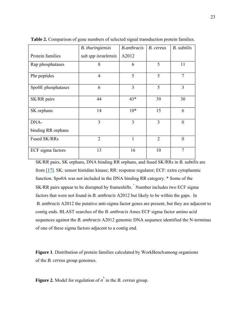
Table 2. Comparison of gene numbers of selected signal transduction protein families.
Protein families
B. thuringiensis
sub spp israelensis
B.anthracis
A2012
B. cereus B. subtilis
Rap phosphatases 8 6 5 11
Phr peptides 4 5 5 7
Spo0E phosphatases 6 3 5 3
SK/RR pairs 44 43* 39 30
SK orphans 14 10* 15 6
DNA-
binding RR orphans
3 3 3 0
Fused SK/RRs 2 1 2 0
ECF sigma factors 13 16 10 7
SK/RR pairs, SK orphans, DNA binding RR orphans, and fused SK/RRs in B. subtilis are
from [17]. SK: sensor histidine kinase; RR: response regulator; ECF: extra cytoplasmic
function. Spo0A was not included in the DNA binding RR category. * Some of the
SK/RR pairs appear to be disrupted by frameshifts. #
Number includes two ECF sigma
factors that were not found in B. anthracis A2012 but likely to be within the gaps. In
B. anthracis A2012 the putative anti-sigma factor genes are present, but they are adjacent to
contig ends. BLAST searches of the B. anthracis Ames ECF sigma factor amino acid
sequences against the B. anthracis A2012 genomic DNA sequence identified the N-terminus
of one of these sigma factors adjacent to a contig end.
Figure 1. Distribution of protein families calculated by WorkBench
among organisms
of the B. cereus group genomes.
Figure 2. Model for regulation of σB
in the B. cereus group.
24
A) During unstressed conditions, the phosphatase acting on RsbV is inactive, so RsbV is
phosphorylated and incapable of binding to the antisigma factor RsbW. RsbW is free to bind
to and inactivate σB
.
B) During stressful conditions, histidine kinases activate the phosphatase by phosphorylating
its response regulator receiver domain. RsbV is dephosphorylated and binds to RsbW, freeing
σB
to activate transcription of stress-regulated genes. Abbreviations: Pase, phosphatase;
RR, response regulator receiver domain.
Figure 3. Alignment of proposed autophosphorylating tyrosine kinases from the B. cereus
group organisms. The residue corresponding to the phosphorus-accepting histidine residue is
marked with an arrow above the alignment using ClustalW. Conserved amino acid residues
are marked with an asterisk; similar amino acid residues are marked with a colon; more
distantly similar amino acids are marked with a period.
Related Documents