Comparative evaluation of positive tests to Mycobacterium avium subsp. paratuberculosis in clinically healthy sheep and goats in South-West Greece using molecular techniques, serology, and culture John Ikonomopoulos a, * , Christos Balaskas a , Bagia Kantzoura a , Eirini Fragiadaki a , Ivo Pavlik b , Milan Bartos b , John C. Lukas c , Maria Gazouli a a Department of Anatomy-Physiology, Agricultural University, 18875 Athens, Greece b Veterinary Research Institute, 62132 Brno, Czech Republic c Department of Pharmacology, School of Medicine, Universidad del Pais Vasco, Leioa, 48016 Vizcaya, Spain Accepted 4 September 2006 Abstract Mycobacterium avium subsp. paratuberculosis (MAP) is the cause of paratuberculosis, which affects mainly ruminants although there is a growing concern about its possible implication in Crohn’s disease in humans especially in connection with environmental spread and risks to the food chain. Retail cheese may represent a significant source of human exposure to MAP and the aim of this study was to assess MAP status in clinically healthy sheep and goats in Greece, comparing techniques routinely used in the positive diagnosis of the disease. From a total of 30 flocks, 632 sheep and goats had faecal, serum, and whole-blood samples examined by culture, complement fixation test (CFT), and polymerase chain reaction (PCR) targeted at IS900, IS1245, and IS6110. PCR produced positive results in 21% of the animals tested, with 5.6%, 3.9%, and 11.5% being identified as MAP, Mycobacterium avium subsp. avium, and Mycobacterium tuberculosis complex, respectively. CFT produced positive and suspicious results in 4.4% and 14.4% of the cases. Faecal cultures were negative in all but a single case that was identified as restriction fragment length polymorphism (RFLP)-type BC1. Agreement between results obtained by PCR and CFT was poor with isolated cases although an assessment of the MAP positive tests produced similar results for both methods. The findings indicate the need for additional measures of control, although the costs may be substantial if public health protection justifies elimination of MAP from livestock. Ó 2006 Elsevier Ltd. All rights reserved. Keywords: Paratuberculosis; PCR; CFT; Culture 1. Introduction Paratuberculosis, also known as Johne’s disease, is a form of chronic ileocolitis that affects mainly ruminants. The disease is caused by Mycobacterium avium subsp. par- atuberculosis (MAP), and signs usually include chronic diarrhoea, weight loss, and emaciation. Diseased animals and asymptomatic carriers may excrete MAP in their fae- ces and milk and spread the infection, which can cause con- siderable financial losses due to premature culling and death (Ayele et al., 2001). Humans are not usually affected by MAP, although a number of reports have linked the pathogen with Crohn’s disease, a chronic inflammatory bowel disease of man with considerable similarity to paratuberculosis (Chiodini, 1989; Chamberlin et al., 2001). Concern about the potential sig- nificance of human exposure to MAP has focused attention on reports about its environmental spread and risks to the food chain (Chiodini and Hermon-Taylor, 1993; Grant et al., 1996; Stehman, 1996; Sung and Collins, 1998), but 1090-0233/$ - see front matter Ó 2006 Elsevier Ltd. All rights reserved. doi:10.1016/j.tvjl.2006.09.004 * Corresponding author. Tel.: +3010 945 484027; fax: +3010 5294383. E-mail addresses: [email protected] (J. Ikonomopoulos), mgazouli@ med.uoa.gr (M. Gazouli). www.elsevier.com/locate/tvjl The Veterinary Journal xxx (2006) xxx–xxx The Veterinary Journal ARTICLE IN PRESS Please cite this article in press as: Ikonomopoulos, J. et al., Comparative evaluation of positive tests to Mycobacterium avium subsp. ..., Vet. J. (2006), doi:10.1016/j.tvjl.2006.09.004

Welcome message from author
This document is posted to help you gain knowledge. Please leave a comment to let me know what you think about it! Share it to your friends and learn new things together.
Transcript

ARTICLE IN PRESS
www.elsevier.com/locate/tvjl
The Veterinary Journal xxx (2006) xxx–xxx
TheVeterinary Journal
Comparative evaluation of positive tests to Mycobacterium avium subsp.paratuberculosis in clinically healthy sheep and goats in
South-West Greece using molecular techniques, serology, and culture
John Ikonomopoulos a,*, Christos Balaskas a, Bagia Kantzoura a, Eirini Fragiadaki a,Ivo Pavlik b, Milan Bartos b, John C. Lukas c, Maria Gazouli a
a Department of Anatomy-Physiology, Agricultural University, 18875 Athens, Greeceb Veterinary Research Institute, 62132 Brno, Czech Republic
c Department of Pharmacology, School of Medicine, Universidad del Pais Vasco, Leioa, 48016 Vizcaya, Spain
Accepted 4 September 2006
Abstract
Mycobacterium avium subsp. paratuberculosis (MAP) is the cause of paratuberculosis, which affects mainly ruminants although thereis a growing concern about its possible implication in Crohn’s disease in humans especially in connection with environmental spread andrisks to the food chain. Retail cheese may represent a significant source of human exposure to MAP and the aim of this study was toassess MAP status in clinically healthy sheep and goats in Greece, comparing techniques routinely used in the positive diagnosis of thedisease. From a total of 30 flocks, 632 sheep and goats had faecal, serum, and whole-blood samples examined by culture, complementfixation test (CFT), and polymerase chain reaction (PCR) targeted at IS900, IS1245, and IS6110.
PCR produced positive results in 21% of the animals tested, with 5.6%, 3.9%, and 11.5% being identified as MAP, Mycobacterium
avium subsp. avium, and Mycobacterium tuberculosis complex, respectively. CFT produced positive and suspicious results in 4.4% and14.4% of the cases. Faecal cultures were negative in all but a single case that was identified as restriction fragment length polymorphism(RFLP)-type BC1. Agreement between results obtained by PCR and CFT was poor with isolated cases although an assessment of theMAP positive tests produced similar results for both methods. The findings indicate the need for additional measures of control,although the costs may be substantial if public health protection justifies elimination of MAP from livestock.� 2006 Elsevier Ltd. All rights reserved.
Keywords: Paratuberculosis; PCR; CFT; Culture
1. Introduction
Paratuberculosis, also known as Johne’s disease, is aform of chronic ileocolitis that affects mainly ruminants.The disease is caused by Mycobacterium avium subsp. par-
atuberculosis (MAP), and signs usually include chronicdiarrhoea, weight loss, and emaciation. Diseased animalsand asymptomatic carriers may excrete MAP in their fae-
1090-0233/$ - see front matter � 2006 Elsevier Ltd. All rights reserved.
doi:10.1016/j.tvjl.2006.09.004
* Corresponding author. Tel.: +3010 945 484027; fax: +3010 5294383.E-mail addresses: [email protected] (J. Ikonomopoulos), mgazouli@
med.uoa.gr (M. Gazouli).
Please cite this article in press as: Ikonomopoulos, J. et al., Compar..., Vet. J. (2006), doi:10.1016/j.tvjl.2006.09.004
ces and milk and spread the infection, which can cause con-siderable financial losses due to premature culling anddeath (Ayele et al., 2001).
Humans are not usually affected by MAP, although anumber of reports have linked the pathogen with Crohn’sdisease, a chronic inflammatory bowel disease of man withconsiderable similarity to paratuberculosis (Chiodini, 1989;Chamberlin et al., 2001). Concern about the potential sig-nificance of human exposure to MAP has focused attentionon reports about its environmental spread and risks to thefood chain (Chiodini and Hermon-Taylor, 1993; Grantet al., 1996; Stehman, 1996; Sung and Collins, 1998), but
ative evaluation of positive tests to Mycobacterium avium subsp.

2 J. Ikonomopoulos et al. / The Veterinary Journal xxx (2006) xxx–xxx
ARTICLE IN PRESS
there are practical problems associated with the control ofparatuberculosis, especially among animals with sub-clini-cal infection (Ridge et al., 1991; Hietala, 1992; Stehman,1996; Nielsen et al., 2001).
Recently we have demonstrated that retail cheese pro-duced from sheep and goats in Greece may represent asource of human exposure to MAP with >90% of somecommercial feta-cheese products testing positive (Ikonom-opoulos et al., 2005). In the present study, the methodscommonly used for the diagnostic investigation of paratu-berculosis were applied to clinically healthy sheep andgoats.
Our target population consisted of animals from a pre-dominantly agricultural part of Greece and samples wereexamined comparatively using the complement fixation test(CFT), culture, and polymerase chain reaction (PCR) todetect Mycobacterium tuberculosis complex and Mycobac-
terium avium subsp. avium in addition to MAP.
2. Materials and methods
2.1. Sample collection
One thousand eight hundred and ninety-six sampleswere collected from 632 sheep and goats (436 sheep; 196goats) selected from a total of 30 flocks (19 sheep and 11goat herds). These represented the total number of regis-tered flocks, accessible in the part of South-Western Greecestudied (namely, Nomos Thesprotias, Nomos Trikalon,and Nomos Karditsas) (Table 1). Based on the records cre-ated from all farms in the region, a flock-size of 10 animalswas taken as the threshold to divide large from small farms.On the smallest farms in this region, hygiene and controlmeasures are not applied in a professional or thoroughmanner and so flocks with 10 animals or less were excludedfrom our sampling plan in an effort to produce representa-tive prevalence estimates for each flock and maximize thenumber of flocks tested.
Serum, faecal, and heparinised whole-blood sampleswere examined. For practical reasons, part of the material
Table 1Polymerase chain reaction (PCR), complement fixation test (CFT), and culturpositive results)
Test results PCR
IS900* IS1245*
Positive Animals 23 (5.6%) [CI; 3.6–8.3%] 16 (3.9%) [CI; 2.3–6.3%]Flocks 14 (46.6%) [CI; 28–66%] 12 (40%) [CI; 23–59%]
Negative Animals 385 392Flocks 16 18
Dubious – –
Total Animals 408 408Flocks 30 30
* Target regions of PCR, specific for Mycobacterium avium subsp. paratube
bacterium tuberculosis complex (IS6110).
Please cite this article in press as: Ikonomopoulos, J. et al., Compar..., Vet. J. (2006), doi:10.1016/j.tvjl.2006.09.004
was not made available for evaluation, which decreasedthe number of samples that were finally tested by PCR andculture, to 408 and 481, respectively (Table 1). However,the sample size was considered adequate since, with an aver-age 20% prevalence of paratuberculosis in small ruminantsin Greece (Demareli-Malli et al., 1991a,b), the minimumsample size required for detecting a difference of at least20%, for a level of a = 0.05 (Type I error) and b = 0.10 (TypeII error) was estimated to be as high as n = 106. (The animalpopulation in the studied region was considered uniform).
Clinical material was collected from adult sheep andgoats corresponding to about 20% of the total number ofanimals registered on the farm at the time of sample collec-tion. The percentage was empirical due to limitations innumbers, but previous studies have provided 95% confi-dence for >5% prevalence. Selection within each flockwas randomly by ballot among the clinically healthy ani-mals and based on information provided by the relevantveterinary inspectors and disease records. Data on thecharacteristics of the animals (species, breed, gender, age,and disease record) and the flocks (location, size, speciesof animals, tuberculin testing, vaccination, and diseaserecord) were collected through questionnaires completedby the investigators on location during sample collection.
Each sample was marked with a coded number andstored at 4 �C, for no more than 48 h prior to examination.Faecal samples were used for detection of MAP by culture;serum samples were used for the detection of MAP-specificantibody by complement fixation test (CFT), and whole-blood samples were used for DNA extraction and PCR.
2.2. Culture
Culture for the isolation of MAP was as previouslydescribed (Pavlik et al., 2000a). In brief, approximately50 g of faeces were homogenized using a laboratory blen-der (Kleinfeld Labortechnik). For decontamination,10 mL of the homogenized product were incubated for20 h with an equal volume of 0.75% HPC (hexa-decyl-pyridinium chloride), in a dark room, at room temperature.
e results, of the animals and flocks that were tested (95% CI listed for the
CFT Culture
IS6110*
47 (11.5%) [CI; 8.6–15.0%] 28 (4.4%) [CI; 2.9–6.3%] 118 (60%) [CI; 40–77%] 13 (43.3%) [CI; 25.5–62.6%] 1
361 513 48012 2 29
– 91 –25
408 632 48130 30 30
rculosis (IS900), Mycobacterium avium subsp. avium (IS1245), and Myco-
ative evaluation of positive tests to Mycobacterium avium subsp.

J. Ikonomopoulos et al. / The Veterinary Journal xxx (2006) xxx–xxx 3
ARTICLE IN PRESS
After centrifugation the pellet was resuspended in 2 mL ofsterile distilled water and incubated in a water bath at37 �C for 1 h. Two hundred microlitres of the suspensionwere inoculated onto three tubes of Herrold’s egg yolkmedium (HEYM) containing 2 lg/mL Mycobactin J. Incu-bation was at 37 �C for up to 8 months.
2.3. DNA isolation
DNA isolation was performed on 300 lL of whole-blood samples using a Qiagen kit according to the manu-facturer’s instruction. Evaluation of the quality of theDNA extract with reference to quantity, purity, and integ-rity was performed with optical density (OD) counts andagarose gel electrophoresis as previously described (Iko-nomopoulos et al., 1999).
In order to assess the presence of PCR inhibitors in ourDNA preparations, 50% (selected randomly) were sub-jected to PCR assay, amplifying a 900 bp DNA fragmentof an in-house gene (cytochrome c) of sheep and goats(data not shown). DNA preparations of poor quality orthose that failed to produce the expected product withthe PCR assay mentioned above were discarded, andDNA isolation was repeated from the original sample.
2.4. Polymerase chain reaction
The detection and identification of MAP, M. tuberculo-
sis complex, and Mycobacterium avium subsp. avium, wasperformed, as previously described (Gazouli et al., 2002;Ikonomopoulos et al., 2004), with PCR assays targeted,respectively, to IS900, IS6110, and IS1245 (Table 2). TheMAP PCR assay used was a one-tube-nested variation(Registered Greek Patent: No. 20030100421, Internationalpatent application: PCT/GR2004/000051). In all cases,PCR was performed on 5 lL of the DNA extracted fromwhole-blood samples.
PCR products were analysed by submerged electropho-resis on a 2% agarose gel. Samples were evaluated and pro-cessed together with positive and negative controls thatconsisted of confirmed, PCR-positive, and negative,whole-blood, and DNA preparations. Test tubes, contain-ing only the reaction mixture and water of the same sourceused for the dilution of DNA, were also included in eachPCR assay, as negative controls. The specificity of DNA
Table 2Primer sequences, amplification products, and target regions of the PCR assayMycobacterium tuberculosis complex, and Mycobacterium avium subsp. avium
Target region Mycobacterialspecies detected
Primers
Forward
IS900 M. avium subsp.paratuberculosisa
GCATGGCCCACAGGACGTTG
GGGTGTGGCGTTTTCCTTCGIS1245 M. avium subsp. avium GTGGGCAATCTGCCCTGCACTIS6110 M. tuberculosis complex CGTGAGGGCATCGAGGTGGC
a One-tube nested PCR.
Please cite this article in press as: Ikonomopoulos, J. et al., Compar..., Vet. J. (2006), doi:10.1016/j.tvjl.2006.09.004
amplification was confirmed by nucleotide sequences ofthe PCR products. Sequencing was performed on bothstrands using the Dye terminator cycle sequencing kit sup-plied by ABI (Applied Biosystems) and an ABI PRISM 377automated DNA sequencer.
2.5. Complement fixation test
Blood serum antibodies to MAP were detected by CFTusing a kit produced by Bioveta, following the manufac-turer’s instructions. Results were evaluated according tostandard procedures (OIE, Manual of Standards for Diag-nostic Tests and Vaccines for Terrestrial Animals, 2002).Positive, suspicious, and negative results corresponded,respectively, to titres of 1/10 or higher, 1/6–1/10, and 0–1/5. The evaluation performed previously using the CFTmethod incorporated in this study had demonstrated thatalthough specificity was found to be 85%, this rangedbetween 11% and 85% for slightly and heavily infected flocks(Pavlik et al., 2000b).
2.6. Restriction fragment length polymorphism analysis(RFLP)
RFLP analysis as previously described (Pavlik et al.,1999) was used to type MAP isolated in culture. In brief,digestion was by restriction endonucleases PstI and BstEII;the restriction products were hybridized with a standardIS900 probe prepared by PCR, DNA fingerprints werescanned by CCD camera and analyzed using the softwareGel Compar (Applied Maths).
2.7. Statistical analysis
Initially, descriptive statistics were calculated includingcell frequencies of 2 · 2 tables and the percent agreementbetween different methods. Frequencies were obtained bycounting. Formal statistical evaluation was based on theFisher’s exact test, with the aid of S-plus v. 6.1 (InsightfulCorp.) and SPSS 11.0 (SPSS Inc.). The uncertainty (95%CI) of the percentage calculations in the flocks can beaffected by clustering within herds. However, the range inthe number of animals per herd (between 9 and 28) wasnarrow and our 95% ranges were robust, so clusteringwas not explicitly accounted for.
s used for the detection of Mycobacterium avium subsp. paratuberculosis,
Product size (bp)
Forward
AG CTACAACAAGAGCCGTGCCG
TCCTGGGCGCTGAGTTCCTC 257TCGG GCCCGCACGCTCACAGTTAAGCCGT 504
GCGTAGGCGTCGGTGACAAA 245
ative evaluation of positive tests to Mycobacterium avium subsp.
4 J. Ikonomopoulos et al. / The Veterinary Journal xxx (2006) xxx–xxx
ARTICLE IN PRESS
3. Results
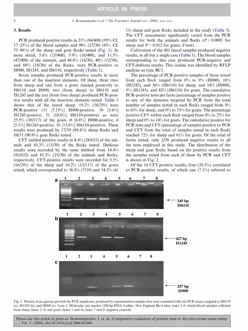
PCR produced positive results in 21% (86/408) (95% CI;17–25%) of the blood samples and 90% (27/30) (95% CI;73–98%) of the sheep and goat flocks tested (Fig. 1). Inmore detail, 5.6% (23/408), 3.9% (16/408), and 11.5%(47/408) of the animals, and 46.6% (14/30), 40% (12/30),and 60% (18/30) of the flocks, were PCR-positive toIS900, IS1245, and IS6110, respectively (Table 1).
Seven samples produced PCR-positive results in morethan one of the insertion elements. Of these, three (twofrom sheep and one from a goat) reacted positively toIS6110 and IS900, two (from sheep) to IS6110 andIS1245 and the rest (from four sheep) produced PCR-posi-tive results with all the insertion elements tested. Table 3shows that of the tested sheep, 19.2% (56/291) werePCR-positive (15 [5.15%] IS900-positive; 10 [3.4%]IS1245-positive; 31 [10.6%] IS6110-positive) as were25.5% (30/117) of the goats (8 [6.8%] IS900-positive, 6[5.1%] IS1245-positive, 16 [13.6%] IS6110-positive). Theseresults were produced by 17/19 (89.4%) sheep flocks and10/11 (90.9%) goat flocks tested.
CFT yielded positive results in 4.4% (28/632) of the ani-mals and 43.3% (13/30) of the flocks tested. Dubiousresults were recorded by the same method from 14.4%(91/632) and 83.3% (25/30) of the animals and flocks,respectively. CFT-positive results were recorded for 5.5%(16/291) of the sheep and 10.2% (12/117) of the goatstested, which corresponded to 36.8% (7/19) and 54.5% (6/
Fig. 1. Picture of an agarose gel with the PCR amplicons, produced by represen(a), IS1245 (b), and IS900 (c). Lane 1: Molecular size marker (100 bp DNA Lfrom sheep (lanes 2–4) and goats (lanes 5 and 6), lanes 7 and 8: negative cont
Please cite this article in press as: Ikonomopoulos, J. et al., Compar..., Vet. J. (2006), doi:10.1016/j.tvjl.2006.09.004
11) sheep and goat flocks included in the study (Table 3).The CFT assessments significantly varied from the PCRresults for both the animals and flocks (P < 0.0001 forsheep and P < 0.012 for goats; F-test).
Cultivation of the 481 faecal samples produced negativeresults, in all but a single case (Table 1). The blood samplescorresponding to this case produced PCR-negative andCFT-dubious results. This isolate was identified by RFLPanalysis as type BC1.
The percentage of PCR-positive samples of those testedfrom each flock ranged from 0% to 9% (IS900), 18%(IS1245), and 36% (IS6110) for sheep, and 14% (IS900),5% (IS1245), and 42% (IS6110) for goats. The cumulativePCR-positive tests per farm (percentage of samples positiveto any of the elements targeted by PCR from the totalnumber of samples tested in each flock) ranged from 0%to 63% for sheep, and 0% to 53% for goats. The percentagepositive CFT within each flock ranged from 0% to 25% forsheep and 0% to 14% for goats. The cumulative positive forPCR tests and CFT (percentage of samples positive to PCRand CFT from the total of samples tested in each flock)reached 72% for sheep and 61% for goats. Of the total offarms tested, only 2/30 produced negative results to allthe tests employed in this study. The distribution of thesheep and goat flocks based on the positive results fromthe samples tested from each of them by PCR and CFTis shown in Fig. 2.
Of the 14 CFT-positive results, four (28.5%) correlatedto PCR-positive results, of which one (7.1%) referred to
tative samples that were examined with our PCR assays targeted to IS6110adder, New England Bio-Labs), lanes 2–6: whole-blood samples collectedrols.
ative evaluation of positive tests to Mycobacterium avium subsp.
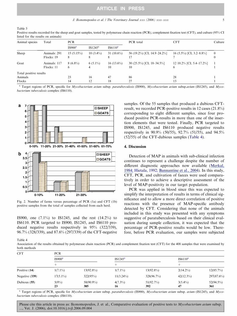
Table 3Positive results recorded for the sheep and goat samples, tested by polymerase chain reaction (PCR), complement fixation test (CFT), and culture (95% CIlisted for the results on animals)
Animal species Total PCR PCR total CFT Culture
IS900a IS1245a IS6110a
Sheep Animals: 291 15 (5.15%) 10 (3.4%) 31 (10.6%) 56 (19.2%) [CI; 14.9–24.2%] 16 (5.5%) [CI; 3.2–8.8%] 0Flocks: 19 8 8 8 17 7 0
Goat Animals: 117 8 (6.8%) 6 (5.1%) 16 (13.6%) 30 (25.5%) [CI; 18–34.5%] 12 10.2% [CI; 5.4–17.2%] 1Flocks: 11 6 4 10 10 6 1
Total positive resultsAnimals 23 16 47 86 28 1Flocks 14 12 18 27 13 1
a Target regions of PCR, specific for Mycobacterium avium subsp. paratuberculosis (IS900), Mycobacterium avium subsp.avium (IS1245), and Myco-
bacterium tuberculosis complex (IS6110).
Fig. 2. Number of farms versus percentage of PCR (1a) and CFT (1b)positive samples from the total of samples collected from each herd.
J. Ikonomopoulos et al. / The Veterinary Journal xxx (2006) xxx–xxx 5
ARTICLE IN PRESS
IS900, one (7.1%) to IS1245, and the rest (14.2%) toIS6110. PCR targeted to IS900, IS1245, and IS6110 pro-duced negative results respectively in 95% (322/339),96.7% (328/339), and 87.6% (297/339) of the CFT-negative
Table 4Association of the results obtained by polymerase chain reaction (PCR) and cboth methods
CFT PCR
IS900a IS1245
+ � +
Positive (14) 1(7.1%) 13(92.8%) 1(7.1%
Negative (339) 17(5.1%) 322(95%) 11(3.24
Dubious (55) 5(9%) 50(90.9%) 4(7.3%23 385 16
a Target regions of PCR, specific for Mycobacterium avium subsp. paratube
bacterium tuberculosis complex (IS6110).
Please cite this article in press as: Ikonomopoulos, J. et al., Compar..., Vet. J. (2006), doi:10.1016/j.tvjl.2006.09.004
samples. Of the 55 samples that produced a dubious CFT-result, we recorded PCR-positive results in 12 cases (21.8%)corresponding to eight different samples, since four pro-duced positive PCR-results in more than one of the inser-tion elements that were tested. Finally, PCR targeted toIS900, IS1245, and IS6110 produced negative resultsrespectively in 90.9% (50/55), 92.7% (51/55), and 94.5%(52/55) of the CFT-dubious samples (Table 4).
4. Discussion
Detection of MAP in animals with sub-clinical infectioncontinues to represent a challenge despite the number ofdifferent diagnostic approaches now available (Merkal,1984; Hietala, 1992; Bannantine et al., 2004). In this study,CFT, PCR, and cultivation of faeces were used compara-tively in order to achieve a descriptive assessment of thelevel of MAP-positivity in our target population.
PCR was applied in blood since this was expected tosimplify the interpretation of results in terms of clinical sig-nificance and to allow a more direct correlation of positivereactions with the presence of MAP-specific antibodydetected by CFT. Considering that none of the animalsincluded in this study was presented with any symptomssuggestive of paratuberculosis based on their clinical eval-uation during sample collection, it was expected that thepercentage of PCR-positive results would be low. There-fore, before PCR evaluation, our samples were subjected
omplement fixation test (CFT) for the 408 samples that were examined by
a IS6110a
� + �) 13(92.8%) 2(14.2%) 12(85.7%)
%) 328(96.7%) 42(12.3%) 297(87.6%)
) 51(92.7%) 3(5.4%) 52(94.5%)392 47 361
rculosis (IS900), Mycobacterium avium subsp. avium (IS1245), and Myco-
ative evaluation of positive tests to Mycobacterium avium subsp.
6 J. Ikonomopoulos et al. / The Veterinary Journal xxx (2006) xxx–xxx
ARTICLE IN PRESS
to a number of tests in order to minimise the impact offalse-negative PCR results generated by poor DNA qualityand PCR inhibitors. Similar precautions were taken toavoid false-positive results by incorporating a nestedPCR assay for the detection of MAP as performance hadbeen previously assessed following a thorough evaluationby intra-laboratory collaboration (Ikonomopoulos et al.,2004).
Despite these precautions, the PCR-positive results didnot correlate with CFT findings. This supports previousreports that indicated the inefficiency of CFT in the diag-nostic investigation of isolated cases of paratuberculosis,especially those with sub-clinical infection that usuallycarry inadequate amounts of complement-activating spe-cific antibodies in their serum (Ridge et al., 1991; Hietala,1992; Clarke and Little, 1996). This was also demonstratedby the results of the evaluation performed previously usingthe CFT method used here where, although specificity wasfound to be 85%, the sensitivity ranged between 11% and85% for slightly and heavily infected flocks (Pavlik et al.,2000b).
The failure of CFT to produce positive results from thePCR-positive samples could also be attributed to the anti-genic incompatibility of the MAP strains that weredetected by PCR and the antigen used in our CFT assay,although the latter was designed with reference to a RFLPtype B-C1 MAP strain, which was of the same type as thestrain we isolated from the tested faecal samples. Consider-ing there is no information available about the predomi-nant RFLP type of MAP strain found in sheep and goatsin Greece, we can only speculate on the impact of thisparameter on our results, although other studies on thecomparative sero-diagnosis of MAP have shown little var-iation in results with different antigens (Demareli-Malliet al., 1991a,b; Molina et al., 1991; Ridge et al., 1991; Niel-sen et al., 2001).
Interestingly, an assessment of MAP positive resultsusing CFT and PCR assays produced similar results bothto the animal population (4.4% vs. 5.6%) and the flocks(43.3% vs. 46.6%) studied. Although CFT costs less thanPCR, its use for the assessment of MAP infection in an ani-mal population must be questioned as PCR identifies posi-tive reactors in a highly specific manner allows immediateconfirmation of positive results.
The source of agreement between PCR targeted to IS900and CFT was obviously the consistency of the negativeresults recorded by both methods, which reached 95%(Table 4). However, an equally high percentage of IS900PCR-negative results corresponded to samples character-ized by CFT as positive (92.8%) or dubious (90.9%) (Table4). This could be attributed to the expected failure(Gwozdz et al., 1997) of PCR to detect all the positive reac-tors with a single blood sample, and may be associated withthe absence (or the low concentration) of MAP-circulatingantigen in the blood after production of adequate amountsof specific antibody (Ridge et al., 1991; Hietala, 1992;Clarke and Little, 1996).
Please cite this article in press as: Ikonomopoulos, J. et al., Compar..., Vet. J. (2006), doi:10.1016/j.tvjl.2006.09.004
The inconsistency between PCR-negative and CFT-positive results is often attributed to false positive CFT-results generated by cross-reactions (Bannantine et al.,2004). PCR evaluation of our blood samples was extendedto IS1245 and IS6110 in order to investigate whether myco-bacterial species other than MAP might cause CFT false-positive results due to cross-reactions – a hypothesis thatwas not supported by our findings since we did not recordany significant correlation between the IS6110 and IS1245PCR-positive and CFT-positive results (Table 4).
The failure of culture to produce more positive resultsfrom the faecal samples could be explained by the fact thatnone of the tested animals had clinical evidence of paratu-berculosis. Other workers have also reported that sheepand goats at certain stages of the disease produce negativefaecal cultures (Navarro et al., 1991; Clarke and Little,1996; Copra et al., 2002). However, it is probable thatour culture result was an underestimate of the true preva-lence of MAP among the animals tested. Indeed, samplesthat contain small numbers of MAP (especially whenfound in a condition of low viability) have been associatedwith false-negative culture-results generated by the neutral-isation of MAP cells during decontamination (Reddacliffet al., 2003). Because of this finding, which seems to bemore profound in connection with sheep and goat strainsof MAP than those with those isolated from bovines (Mer-kal, 1984), incubation of our samples with HPC was car-ried out for less than half the time recommended (OIE,Manual of Standards for Diagnostic Tests and Vaccinesfor Terrestrial Animals, 2002).
Surprisingly, based on the positive results recorded col-lectively by PCR targeted to the insertion elements tested, aconsiderable percentage (number of positive reactors fromthe total of animals in the tested herds) of the sheep(19.2%) (95% CI; 14.9–24.2%) and goats (25.6%) (95%CI; 18–34.5%) that covered >70% of the farms includedin this study were found to be carrying mycobacteria-spe-cific DNA sequences in their blood. This may reflect ani-mals incubating paratuberculosis or tuberculosis.However, at least with reference to the latter, they werenot consistent with the results of routine monitoring (byclinical evaluation of the livestock, post-mortem examina-tion in the relevant abattoirs, and tuberculin testing of sus-pected cases implemented by histopathology of grosslesions and culture) and the fact that (as opposed to para-tuberculosis) the incidence of tuberculosis in the area is rareand isolated (Demareli-Malli et al., 1991a,b). For this rea-son, tuberculin testing or vaccination with any member ofthe Mycobacterium spp. has not been practiced.
The positive results recorded by PCR on our sheep andgoat blood samples may indicate that the local small rumi-nant population is tolerant of certain types of mycobacte-rial infections that usually remain clinically silent. Thiscould account for the somewhat puzzling absence duringpost-mortem examination of any indication of mycobacte-rial infection in a considerable percentage of tuberculinreactors from various parts of the country.
ative evaluation of positive tests to Mycobacterium avium subsp.
J. Ikonomopoulos et al. / The Veterinary Journal xxx (2006) xxx–xxx 7
ARTICLE IN PRESS
5. Conclusion
There was considerable MAP positivity among clini-cally healthy sheep and goats in this region of Greece,suggesting that control will require constant alertnessand additional measures especially because these animalsmay be tolerant to certain types of mycobacterial infec-tions that could complicate diagnosis. This is likely topresent a problem with significant financial impact shouldthe protection of public health justify elimination of MAPfrom the livestock.
Acknowledgements
This work was partially supported by the Grant VENO-MYC (No. SSPE-CT-2004-501903, EU, Brussels) andGrant No. MZE 0002716201 of the Ministry of Agriculture(Czech Republic).
References
Ayele, W.Y., Machackova, M., Pavlik, I., 2001. The transmission andimpact of paratuberculosis infection in domestic and wild ruminants.Veterinary Medicine 46, 205–224.
Bannantine, J.P., Hansen, J.K., Paustian, M.L., Amonsin, A., Li, L.L.,Stabel, J.R., Kapur, V., 2004. Expression of immunogenicity ofproteins encoded by sequences specific to Mycobacterium avium subsp.paratuberculosis. Journal of Clinical Microbiology 42, 106–114.
Chamberlin, W., Graham, D.Y., Hulten, K., El-Zimaity, H.M., Schwartz,N., Shafran, I., El-Zaatari, F.A., 2001. Mycobacterium avium subsp.paratuberculosis one cause of Crohn’s disease. Alimentary Pharmacol-ogy and Therapeutics 15, 337–346.
Chiodini, R.J., 1989. Crohn’s disease and the mycobacterioses: a reviewand comparison of two disease entities. Clinical Microbiology Reviews2, 90–117.
Chiodini, R.J., Hermon-Taylor, J., 1993. The thermal resistance of M.
paratuberculosis in row milk under conditions simulating pasteuriza-tion. Journal of Veterinary Diagnostic Investigation 5, 629–631.
Clarke, C.J., Little, D., 1996. The pathology of ovine paratuberculosis:gross and histological changes in the intestines and other tissues.Journal of Comparative Pathology 114, 419–437.
Copra, J.M., Garrido, J., Garcia Marin, J.F., Perez, V., 2002. Classifi-cation of lesions observed in natural cases in paratuberculosis in goats.Journal of Comparative Pathology 122, 255–265.
Demareli-Malli, Z., Saris, K., Xenos, G., Papadopoulos, G., 1991a.Comparison of the ELISA, AGID, and CF tests for diagnosis ofcaprine paratuberculosis. The Paratuberculosis Newsletter 3, 32–33.
Demarelli-Malli, Z., Xenos, G., Argyroudis, S., Papadopoulos, O., 1991b.A survey of ovine and caprine paratuberculosis in the Thessalonikiarea of Greece. The Paratuberculosis Newsletter 3, 8–9.
Gazouli, M., Ikonomopoulos, J., Trigidou, R., Foteinou, M., Kittas, C.,Gorgoulis, V., 2002. Assessment of mycobacterial, propionibacterial,and human herpesvirus 8 DNA in tissues of Greek patients withsarcoidosis. Journal of Clinical Microbiology 40, 3060–3063.
Grant, I.R., Ball, H.J., Neill, S.D., Rowe, M.T., 1996. Inactivation ofMycobacterium paratuberculosis in cow’s milk at pasteurisation tem-peratures. Applied Environmental Microbiology 62, 631–636.
Please cite this article in press as: Ikonomopoulos, J. et al., Compar..., Vet. J. (2006), doi:10.1016/j.tvjl.2006.09.004
Gwozdz, J.M., Reichel, M.P., Murray, A., Manktelow, W., West, D.M.,Thompson, K.G., 1997. Detection of Mycobacterium avium subsp.paratuberculosis in ovine tissues and blood by the polymerase chainreaction. Veterinary Microbiology 57, 233–244.
Hietala, S.K., 1992. The options in diagnosing ruminant paratuberculosis.Veterinary Medicine 87, 1122–1131.
Ikonomopoulos, J., Gorgoulis, V.G., Zacharatos, P.V., Manolis, E.N.,Kanavaros, P., Rassidakis, A., Kittas, C., 1999. Multiplex polymerasechain reaction for the detection of mycobacterial DNA in cases oftuberculosis and sarcoidosis. Modern Pathology 12, 854–862.
Ikonomopoulos, J., Gazouli, M., Zacharatos, P., Xylouri, E., Gorgoulis,V., 2004. Comparative evaluation of PCR assays for the robustmolecular detection of Mycobacterium avium subsp. paratuberculosis.Journal of Microbiological Methods 56, 315–321.
Ikonomopoulos, J., Pavlik, I., Bartos, M., Svastova, P., Ayele, W.Y.,Roubal, P., Lukas, J., Cook, N., Gazouli, M., 2005. Detection ofMycobacterium avium subsp. paratuberculosis in retail cheeses fromGreece and the Czech Republic. Applied Environmental Microbiology71, 8934–8936.
Merkal, R.S., 1984. Paratuberculosis: advances in cultural, serologic andvaccination methods. American Journal of Veterinary Medical Asso-ciation 184, 939–943.
Molina, A., Morera, L., Llanes, D., 1991. Enzyme-linked immunosorbentassay for detection of antibodies against Mycobacterium paratubercu-
losis in goats. American Journal of Veterinary Research 52, 863–868.Navarro, J.A., Bernabe, A., Gomez, M.A., Sanchez, J., Gomez, S., 1991.
Mycobacterial antigen detection by immunohistochemistry and goatparatuberculosis. Journal of Veterinary Medicine 38, 231–237.
Nielsen, S.S., Houe, H., Thamsborg, S.M., Bitsch, V., 2001. Comparisonof two enzyme-linked immunosorbent assays for serologic diagnosis ofparatuberculosis (Johne’s disease) in cattle using different subspeciesstrains of Mycobacterium avium. Journal of Veterinary DiagnosticInvestigation 13, 164–166.
OIE, 2002. Manual of Standards for Diagnostic Tests and Vaccines forTerrestrial Animals, fourth ed. pp. 1–19.
Pavlik, I., Horvathova, A., Dvorska, L., Bartl, J., Svastova, P., du Maine,R., Rychlik, I., 1999. Standardization of restriction fragment poly-morphism analysis for Mycobacterium avium subspecies paratubercu-losis. Journal of Microbiological Methods 38, 155–167.
Pavlik, I., Matlova, L., Bartl, J., Svastova, P., Dvorska, L., Whitlock, R.,2000a. Parallel fecal and organ Mycobacterium avium subsp. paratu-
berculosis culture of different productivity types of cattle. VeterinaryMicrobiology 77, 309–324.
Pavlik, I., Rozsypalova, Z., Vesely, T., Bartl, J., Matlova, L., Vrbas, V.,Valent, L., Rajsky, D., Mracko, I., Hirko, M., Miskovic, P., 2000b.Control of paratuberculosis in five cattle farms by serological tests andfaecal culture during the period 1990–1999. Veterinarni Medicina 45,61–70.
Reddacliff, L.A., Vadali, A., Whittinghton, R.J., 2003. The effect ofdecontamination protocols on the numbers of sheep strain Mycobac-
terium avium subsp. paratuberculosis isolated from tissues and feces.Veterinary Microbiology 95, 271–282.
Ridge, S.E., Morgan, I.R., Sockett, D.C., Collins, M.T., Condron, R.J.,Skilbeck, N.W., Webber, J.J., 1991. Comparison of the Johne’sabsorbed EIA and the complement-fixation test for the diagnosis ofJohne’s disease in cattle. Australian Veterinary Journal 68, 253–257.
Stehman, S.M., 1996. Paratuberculosis in small ruminants, deer, andSouth American camelides. Veterinary Clinics of North America FoodAnimal Practice 12, 441–445.
Sung, N., Collins, M.T., 1998. Thermal tolerance of Mycobacterium
paratuberculosis. Applied Environmental Microbiology 64, 999–1005.
ative evaluation of positive tests to Mycobacterium avium subsp.
Related Documents