[Cell Cycle 5:9, e1-e8, EPUB Ahead of Print: http://www.landesbioscience.com/journals/cc/abstract.php?id=2715; 1 May 2006]; ©2006 Landes Bioscience e1 Cell Cycle 2006; Vol. 5 Issue 9 Frédéric M. Coquelle 1,6 Talia Levy 1 Sven Bergmann 1,2 Sharon Grayer Wolf 3 Daniela Bar-El 1 Tamar Sapir 1 Yehuda Brody 1 Irit Orr 4 Naama Barkai 1 Gregor Eichele 5 Orly Reiner 1, * 1 Department of Molecular Genetics; Weizmann Institute of Science; Rehovot, Israel 2 Department of Medical Genetics; University of Lausanne; Switzerland 3 Electron Microscopy Unit and 4 Department of Biological Services; Weizmann Institute of Science; Rehovot, Israel 5 Max-Planck Institute; Hannover, Germany † Present address: CNRS-UMR 6026; Université de Rennes 1; Equipe SDM; Campus de Beaulieu-Bat. 13; 35042 Rennes cedex, France *Correspondence to: Frederic M. Coquelle; Department of Molecular Genetics; Weizmann Institute of Science; Rehovot, Israel; Tel.: +972-8-9342319; Fax: +972-8-9344108; Email: [email protected] Original manuscript submitted: 02/21/06 Manuscript accepted: 03/18/06 This manuscript has been published online, prior to printing for Cell Cycle, Volume 5, Issue 9. Definitive page numbers have not been assigned. The current citation is: Cell Cycle 2006; 5(9): http://www.landesbioscience.com/journals/cc/abstract.php?id=2715 Once the issue is complete and page numbers have been assigned, the citation will change accordingly. KEY WORDS doublecortin, microtubules, protein family, cytoskeleton, actin, intracellular localization ACKNOWLEDGEMENTS See page e7. NOTE Supplementary Material can be found at: http://www.landesbioscience.com/journals/cc/ coquelleCC5-9-sup.pdf Report Common and Divergent Roles for Members of the Mouse DCX Superfamily ABSTRACT The doublecortin-like (DCX) domains serve as protein-interaction platforms. DCX tandem domains appear in the product of the X-linked doublecortin (DCX) gene, in retinitis pigmentosa-1 (RP1), as well as in other gene products. Mutations in the human DCX gene are associated with abnormal neuronal migration, epilepsy, and mental retardation; mutations in RP1 are associated with a form of inherited blindness, while DCDC2 has been associated with dyslectic reading disabilities. Motivated by the possible importance of this gene family, a thorough analysis to detect all family members in the mouse was conducted. The DCX-repeat gene superfamily is composed of eleven paralogs, and we cloned the DCX domains from nine different genes. Our study questioned which functions attributed to the DCX domain, are conserved among the different members. Our results suggest that the proteins with the DCX-domain have conserved and unique roles in microtubule regulation and signal transduction. All the tested proteins stimulated micro- tubule assembly in vitro. Proteins with tandem repeats stabilized the microtubule cytoskeleton in transfected cells, while those with single repeats localized to actin-rich subcellular structures, or the nucleus. All tested proteins interacted with components of the JNK/MAP-kinase pathway, while only a subset interacted with Neurabin 2, and a nonoverlapping group demonstrated actin association. The sub-specialization of some members due to confined intracellular localization, and protein interactions may explain the success of this superfamily. INTRODUCTION The existence of a shared defined protein domain among different proteins is usually accompanied with common functional activities. Although multiple algorithms define protein domains based on amino acid similarity, the exact definition of which functions are shared still needs to be demonstrated experimentally. The focus of our study is the evolutionary conserved doublecortin (DCX) domain. 1,2 Mutations in the X-linked gene DCX result in lissencephaly in males, and abnormal neuronal positioning (subcortical band heterotopia) in females. 3,4 The first function attributed to the DCX domain, which appears in tandem in DCX, was its interaction with the microtubule (MT) cytoskeleton. 5-7 This activity was detected also in a related gene product DCLK, 8,9 and in RP1 which contains two DCX domains. 10 RP1 is the protein product of retinitis pigmentosa-1 (RP1). 11,12 Mutations in this gene result in progressive blindness, not only in humans, but also in a mouse model. 13 Recently, an additional member of this protein family, doublecortin kinase 2, has been described, and found to posses MT binding activities. 14 The structure of the DCX domain is a ubiquitin-like fold, 15 different from that of classical MAPs. DCX is embedded in fenestrations localized between protofilaments from which MTs are built. 16 This is a previously uncharacterized MAP binding site, ideal for microtubule stabilization. DCX is not only a unique MAP, but it also interacts with multiple additional proteins (reviewed in refs. 17 and 18). Notably, DCX interacts with two scaffold proteins neurabin 2 and JIP1/2. Neurabin 2 connects DCX to actin microfilaments. 19,20 In addition, neurabin 2 binds to protein phosphates 1 (PP1), thereby regulating site-specific dephosphorylation of DCX (Shmueli et al., in press). JIP1/2 (JNK Interacting Protein 1 or 2) acts as a scaffold for both DCX and one of the kinases that phosphorylates it: JNK (cJun-N-terminal Kinase). 21 The phosphorylation state of DCX on JNK sites affects neuronal motility in vitro. In addition, JIP1 connects DCX to the molecular motor conventional kinesin, which mobilizes DCX in neurons. 21 We hypothesized that the selective recruitment of DCX to either a neurabin 2 complex or a JIP1 complex is important in regulating its activity (Shmueli at al, in press).

Welcome message from author
This document is posted to help you gain knowledge. Please leave a comment to let me know what you think about it! Share it to your friends and learn new things together.
Transcript

[Cell Cycle 5:9, e1-e8, EPUB Ahead of Print: http://www.landesbioscience.com/journals/cc/abstract.php?id=2715; 1 May 2006]; ©2006 Landes Bioscience
e1 Cell Cycle 2006; Vol. 5 Issue 9
Frédéric M. Coquelle1,6
Talia Levy1
Sven Bergmann1,2
Sharon Grayer Wolf3
Daniela Bar-El1
Tamar Sapir1
Yehuda Brody1
Irit Orr4
Naama Barkai1
Gregor Eichele5
Orly Reiner1,*1Department of Molecular Genetics; Weizmann Institute of Science; Rehovot, Israel
2Department of Medical Genetics; University of Lausanne; Switzerland
3Electron Microscopy Unit and 4Department of Biological Services; WeizmannInstitute of Science; Rehovot, Israel
5Max-Planck Institute; Hannover, Germany
†Present address: CNRS-UMR 6026; Université de Rennes 1; Equipe SDM; Campusde Beaulieu-Bat. 13; 35042 Rennes cedex, France
*Correspondence to: Frederic M. Coquelle; Department of Molecular Genetics;Weizmann Institute of Science; Rehovot, Israel; Tel.: +972-8-9342319; Fax:+972-8-9344108; Email: [email protected]
Original manuscript submitted: 02/21/06Manuscript accepted: 03/18/06
This manuscript has been published online, prior to printing for Cell Cycle, Volume 5,Issue 9. Definitive page numbers have not been assigned. The current citation is:Cell Cycle 2006; 5(9):http://www.landesbioscience.com/journals/cc/abstract.php?id=2715Once the issue is complete and page numbers have been assigned, the citation willchange accordingly.
KEY WORDS
doublecortin, microtubules, protein family,cytoskeleton, actin, intracellular localization
ACKNOWLEDGEMENTS
See page e7.
NOTE
Supplementary Material can be found at:http://www.landesbioscience.com/journals/cc/coquelleCC5-9-sup.pdf
Report
Common and Divergent Roles for Members of the Mouse DCXSuperfamily
ABSTRACTThe doublecortin-like (DCX) domains serve as protein-interaction platforms. DCX tandem
domains appear in the product of the X-linked doublecortin (DCX) gene, in retinitispigmentosa-1 (RP1), as well as in other gene products. Mutations in the human DCX geneare associated with abnormal neuronal migration, epilepsy, and mental retardation;mutations in RP1 are associated with a form of inherited blindness, while DCDC2 hasbeen associated with dyslectic reading disabilities. Motivated by the possible importanceof this gene family, a thorough analysis to detect all family members in the mouse wasconducted. The DCX-repeat gene superfamily is composed of eleven paralogs, and wecloned the DCX domains from nine different genes. Our study questioned which functionsattributed to the DCX domain, are conserved among the different members. Our resultssuggest that the proteins with the DCX-domain have conserved and unique roles inmicrotubule regulation and signal transduction. All the tested proteins stimulated micro-tubule assembly in vitro. Proteins with tandem repeats stabilized the microtubulecytoskeleton in transfected cells, while those with single repeats localized to actin-richsubcellular structures, or the nucleus. All tested proteins interacted with components of theJNK/MAP-kinase pathway, while only a subset interacted with Neurabin 2, and anonoverlapping group demonstrated actin association. The sub-specialization of somemembers due to confined intracellular localization, and protein interactions may explainthe success of this superfamily.
INTRODUCTIONThe existence of a shared defined protein domain among different proteins is usually
accompanied with common functional activities. Although multiple algorithms defineprotein domains based on amino acid similarity, the exact definition of which functionsare shared still needs to be demonstrated experimentally. The focus of our study is theevolutionary conserved doublecortin (DCX) domain.1,2 Mutations in the X-linked geneDCX result in lissencephaly in males, and abnormal neuronal positioning (subcorticalband heterotopia) in females.3,4 The first function attributed to the DCX domain, whichappears in tandem in DCX, was its interaction with the microtubule (MT) cytoskeleton.5-7
This activity was detected also in a related gene product DCLK,8,9 and in RP1 whichcontains two DCX domains.10 RP1 is the protein product of retinitis pigmentosa-1(RP1).11,12 Mutations in this gene result in progressive blindness, not only in humans, butalso in a mouse model.13 Recently, an additional member of this protein family, doublecortinkinase 2, has been described, and found to posses MT binding activities.14 The structureof the DCX domain is a ubiquitin-like fold,15 different from that of classical MAPs. DCXis embedded in fenestrations localized between protofilaments from which MTs are built.16
This is a previously uncharacterized MAP binding site, ideal for microtubule stabilization.DCX is not only a unique MAP, but it also interacts with multiple additional proteins
(reviewed in refs. 17 and 18). Notably, DCX interacts with two scaffold proteins neurabin 2and JIP1/2. Neurabin 2 connects DCX to actin microfilaments.19,20 In addition, neurabin 2binds to protein phosphates 1 (PP1), thereby regulating site-specific dephosphorylation ofDCX (Shmueli et al., in press). JIP1/2 (JNK Interacting Protein 1 or 2) acts as a scaffoldfor both DCX and one of the kinases that phosphorylates it: JNK (cJun-N-terminalKinase).21 The phosphorylation state of DCX on JNK sites affects neuronal motility invitro. In addition, JIP1 connects DCX to the molecular motor conventional kinesin,which mobilizes DCX in neurons.21 We hypothesized that the selective recruitment of DCXto either a neurabin 2 complex or a JIP1 complex is important in regulating its activity(Shmueli at al, in press).
www.landesbioscience.com Cell Cycle e2
Although it is clear that the DCX-domain proteins are an impor-tant group of proteins, the characterization of the group as a wholeis still lacking. So far no studies have addressed the possible commonfunctions among them. Here, we have undertaken to study proteinsinteractions, MAP activity, and the subcellular localization of nine ofthe superfamily members. Our study highlights common andunique features for various members of this superfamily, illustratingtheir widespread functional importance.
MATERIALS AND METHODSDatabase homology search. Our database similarity search initiated with
an NCBI blast search using the sequence of mouse doublecortin protein(NP_034155.2). In addition, all mouse proteins with the DCX domainwere retrieved from the CDD (Conserved Domain DB). In a similar fashion,DCX proteins from the EBI InterPro db, which were not detected in CDDwere extracted and added.
Plasmid construction. Sequences of the primers used in this study arefound in Supplementary Fig. 1. The ORF of BAC26042 was amplified byPCR from the RIKEN clone 4732421G10, accession number XM_489891.1(RIKEN® FANTOMTM CLONES, K.K. Dnaform, Ibaraki, Japan). TheN-terminus of Rp1 (amino acids 1-262, encompassing both DCX repeats),was amplified from the IMAGE clone 2123311, (Invitrogen Life Technologies,Paisley, Great Britain). The ORF of Dcdc2 was amplified by PCR from theIMAGE clone 6306251. A DNA fragment encoding the N-terminus ofDCDC2B (amino acids 1-83) was amplified by PCR from the RIKENclone E030003B04. A DNA fragment from DCLK2 (amino acids 41-295)was amplified by RTPCR from RNA extracted from E12 mouse brain. ADNA fragment encoding the N-terminus of RP1L1 (amino acids 31-261)was amplified by RTPCR from RNA extracted from P12 mouse eye. Aregion of FLJ46154 (amino acids 226-500) was amplified by RTPCR fromRNA extracted from adult mouse eye. PCR fragments were digested byEcoRI and XhoI (or EcoRI and SalI for FLJ46154), and cloned inpBluescript II KS(-) (Stratagene, USA), peGFP-C2 (Clonetech, USA),pcDNA3 (Invitrogen, USA) PCR-modified in order to encode the FLAGtag, and pGEX4T1 (Pharmacia, USA) at the EcoR1 and XhoI restrictionsites. All PCR products and all translation phases of the fusion proteins wereverified by sequencing. Vectors encoding FLAG-DCX, FLAG-DCLK,GFP-DCLK, DCX-DsRed1, GST-DCXpep1+2, GST-DCLK and GST-DCLwere described previously.1,5,8,22 Vectors encoding Myc-JNK2-MKK7 andMyc-JIP1/2 were described elsewhere.21 The plasmid encoding thecoiled-coil domain of Neurabin II was received from Dr. Miki Tsukada.GST fusion peptides were expressed in BL21 lysE strain of Escherichia coli,and purified using glutathione-agarose beads (Sigma, Israel).
Electron microscopy. Samples were prepared using tubulin in PEMbuffer (100 mM PIPES pH 6.9, 1 mM MgSO4 and 1 mM EGTA), at aconcentration of 20 µM (according to the monomer, around 1.1 mg.ml-1)with or without the addition of 0.5 or 2 µM of indicated proteins and incu-bated in the presence of 2 µM GTP (sodium salt, Sigma, Israel) for 30 minat 37˚C. Four to eight microliters of each sample was applied to a carbon-coated copper 400-mesh electron microscope grid, which had been previouslyglow-discharged for 30 sec. to render the carbon surface hydrophilic. Thesample was then stained with two successive 10 µl drops of uranyl acetatestain (1% in water). The grid was blotted after 1 minute. Samples wereexamined with an FEI T12 electron microscope operating at 120 kV. Imageswere acquired on a SIS MegaviewIII CCD camera.
Cell culture. Culturing of COS-7 and HEK-293 cells were performed asdescribed previously.23,24
Antibodies. Primary antibodies used in this study were anti α-tubulin(clone DM1A, Sigma, Israel), anti-actin (clone sc-40, Santa Cruz, USA),anti-FLAG (M2 clone from Sigma, Israel) and anti-myc (E910 clone fromSigma, Israel). In addition, anti-DCX antibodies,1 anti-DCLK antibodies,8
and anti-DCLK2 antibodies were used for Western blots. A synthetic peptidecorresponding to the unique C terminus of Mus musculus DCLK2 (aa 741–756) was conjugated to KLH (keyhole limpet hemocyanin) and used as
immunogen in rabbit. Rabbit antiserum was used in a 1:250 dilution forWestern blot analysis. Secondary antibodies used for the immunofluorescencestudies were made in goat and conjugated to CY3 (Jackson ImmunoResearchLaboratories, Inc., West Grove, PA) or ALEXA-488 (Molecular Probes).Secondary antibodies used for the immunoblots were peroxidase-conjugatedAffiPure Goat anti-mouse or anti-rabbit (Jackson ImmunoResearchLaboratories, Inc., West Grove, PA).
Immunostaining. Fourty-eight hours after transfection, COS-7 cells werefixed in 3% paraformaldehyde (Sigma, Israel), 0.05% glutaraldehyde(MERCK, Germany) in PHEM buffer (60 mM Pipes, 25 mM Hepes, 10 mMEGTA, 2mM magnesium acetate, pH6.9). They were washed three times5 min in phosphate-buffered saline (PBS) and permeabilized for 25 min in0.1% Triton X-100 in PBS. The cells were then treated twice with 1 mg.ml-1
NaBH4 in PBS, for 10 min, washed three times 5 min in PBS, blocked inPBS/BSA (PBS containing 0.1% BSA) and labeled with the monoclonalantibody DM1A raised against α-tubulin, or stained with rhodamine- orAlexa-488-phalloidine (Sigma, Israel) for 1 hour at 37˚C (humid atmosphere).After washing 3 times 10 min in PBS/BSA, in the case of tubulin staining,the cells were incubated with the secondary antibody for 45 min at 37˚C(humid atmosphere). Chromosomes were stained with 0.1 µg.ml-1 DAPI inPBS (Sigma, Israel) for 5 min. Coverslips were mounted using Immu-Mount(Thermo Shandon, USA). All antibodies were diluted in PBS/BSA. Allimages are maximal-intensity projections of X/Y optical section stacksacquired by a BioRad confocal microscope.
Immunoprecipitation assay. Immunoprecipitation was preformed fromtransfected-HEK293 cells using FLAG-M2 beads (Sigma, Israel) or A/Gbeads (Santa Cruz, USA) along with the c-myc 9E10 monoclonal antibody.Cells were cotransfected with each of the FLAG-doublecortin proteins andeach of the myc-tagged-DCX-interacting proteins that we investigated(see hereafter). Transfected-cell extracts were prepared in IP buffer(Tris-HCl 50 mM, pH 6.8 at room temperature, NaCl 0.15 M and TritonX-100 1%) supplemented with protease-inhibitors cocktail (Sigma, Israel).The samples were separated by SDS-PAGE and immunoblotted with thesuitable combination of antibodies [anti-FLAG (M2 clone) or anti-myc(E910 clone)].
GST pull-down assay. P1 mouse brain extract was prepared in T-Tbuffer (20 mM Tris-HCl pH 8, 100 mM NaCl, 1 mM EDTA and 0.1%Triton X-100), supplemented with protease-inhibitors cocktail (Sigma, Israel).1.5 mg protein-containing-brain extract were incubated with 10 µg ofGST-fusion proteins, at 4˚C for 3 hours. Glutathione beads (15 µl, Sigma,Israel) in 30 µl of T-T buffer were added to the protein mixture and rotatedfor 30 min at room temperature. After three washes with T-T buffer, 3Xsample buffer was added and beads were boiled and ran on SDS-PAGE gel.Immunoblot was performed with anti α-tubulin and anti actin antibodies.
Tubulin and MAPs purification. Brains from P7 ICR pups were retrievedand homogenized in cold PEM buffer (100 mM Pipes, 2 mM EGTA, 1 mMMgSO4) containing 10% Glycerol, 1 mM ATP and 1 mM PMSF) andultracetrefuged (100,000 x g) to obtain a clear lysate. To the lysate extra ATP(1 mM) GTP (1 mM) and Glycerol (30%) were added before incubation at34˚C for 45 minutes. Polymerized MTs were pelleted at 100,000 x g at34˚C and the pellet was resuspended in cold PEM containing 2 mM GTP,homogenized and depoymerized further by incubation of ice. Tubulin wasspun at 100,000 before loading onto a phosphocellulose column (Pharmacia).MAPs were eluted from the column using 0.4M NaCl.
Microtubule assembly. Tubulin from calf brain was purified as describedpreviously.25 The assembly rate of tubulin to form polymers was monitoredusing a light scattering assay.26,27 Purified tubulin was diluted in PEMbuffer to a final concentration of 20 µM (according to the monomer,around 1.1 mg.ml-1). GST-recombinant proteins were concentrated usingVivaspin columns (Vivascience, Germany) in EB buffer (100 mM Tris-HCl,120 mM NaCl, 10% glycerol). 0.5 or 2 µM recombinant proteins wereadded to the tubulin in a volume of EB buffer corresponding to 10% of thefinal volume, so that 1% glycerol is reached. Absorbance was measured at350 mm at 1 min intervals in a Uvicon spectrophotometer equipped withtemperature controlled cells. Switching the temperature to 37˚C inducedassembly, and to 4˚C the disassembly.
The DCX Superfamily
The DCX Superfamily
e3 Cell Cycle 2006; Vol. 5 Issue 9
RESULTSIdentification of mouse proteins containing a DCX domain. The mouse
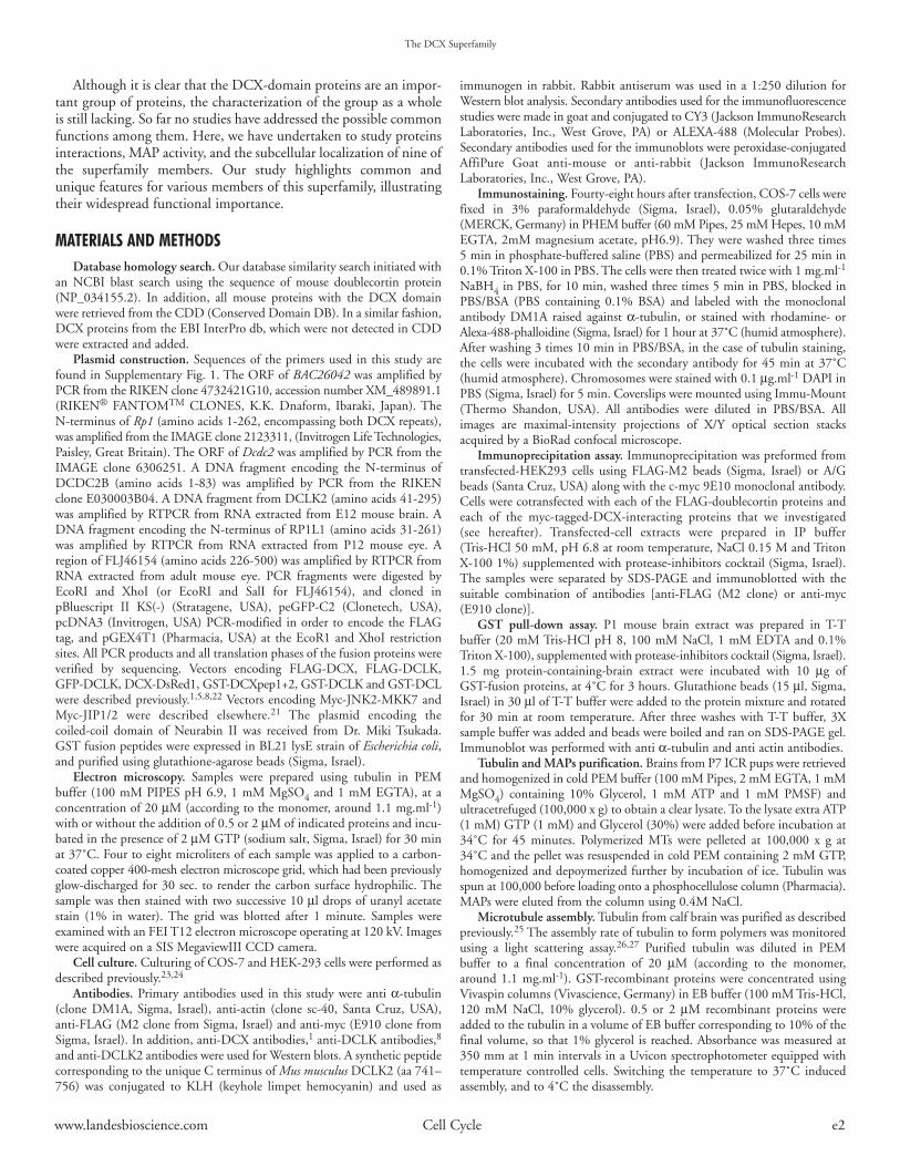
proteome was searched for sequences similar to that of the DCX domainresulting in a total of 11 proteins containing one or two DCX repeats. Aschematic presentation of the protein products is shown (Fig. 1). In additionto DCX domains, three proteins contained kinase domains, and onecontained a ricin lectin site, which is predicted to bind carbohydrates.28
Since several of the proteins contain a single DCX domain, we conductedanalysis for the seventeen individual DCX domains. Closer inspection of theprotein sequences of the DCX domains by multiple alignment, revealedseveral interesting features (Fig. 2). The sequences of the N-terminal DCXdomain in proteins containing a tandem domain are more similar to eachother than to the C-terminal DCX domain of the same protein. Most of theproteins with a single DCX domain are more similar to those from theN-terminal group and one is more similar to the C-terminal group.
The presence of the DCX domain in multiple different gene productsraised the question whether they sharing the same functions. Full lengthcDNA’s or fragments containing the corresponding DCX domains from
nine different gene products were cloned in different expression vectors(details in methods). Additional information regarding the chromosomalpositions of these genes, their accession numbers, and the number of exonsare found in (Supplementary Table 1).
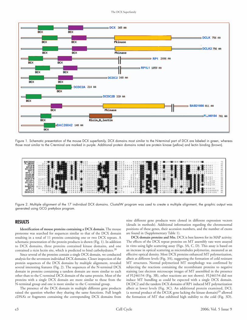
DCX-domain proteins and Mts. DCX is best known for its MAP activity.The effects of the DCX repeat proteins on MT assembly rate were assayedin vitro using light scattering assay (Figs. 3A, C, D). This assay is based onan increase in optical scattering as microtubules polymerize, measured as aneffective optical density. Most DCX proteins enhanced MT polymerization,albeit at different levels (Fig. 3A), suggesting the formation of cold resistantMT structures. Normal polymerized MT morphology was confirmed bysubjecting the reactions containing the recombinant proteins to negativestaining (see electron microscope images of MT assembled in the presenceof FLJ46154 (Fig. 3B), other reactions are not shown). FLJ46154 did notinduce MT bundling as could be expected with a single DCX domain.DCDC2 and the tandem DCX domains of RP1 induced MT polymerizationalbeit at lower levels (Fig. 3C). An additional protein examined, DCL(a normal product of the DCLK gene lacking the kinase domain)29 allowedthe formation of MT that exhibited high stability to the cold (Fig. 3D).
Figure 1. Schematic presentation of the mouse DCX superfamily. DCX domains most similar to the N-terminal part of DCX are labeled in green, whereasthose most similar to the C-terminal are marked in purple. Additional protein domains noted are protein kinase (yellow) and lectin binding (brown).
Figure 2. Multiple alignment of the 17 individual DCX domains. ClustalW program was used to create a multiple alignment, the graphic output wasgenerated using GCG prettybox program.
www.landesbioscience.com Cell Cycle e4
Electron microscopy of MTs assembled in the presence of DCL did notreveal any abnormal structures or bundling (data not shown).
Testing the effect of DCX-domain proteins in cells is expected to bemore indicative of their physiological functions. The expression of the tandemDCX domain is required to stabilize MTs in transfected cells.1 Therefore,this specific group of proteins was tested in transfected COS-7 cells usingGFP fusion proteins (Fig. 4), or Flag-tagged proteins (data not shown).Among the tested proteins, DCX, DCLK and RP1 are known MAPs5-10
(Fig. 4A1–8, B1–8 and D1–8). Here we show that the DCX domains ofDCLK2 (Fig. 4C1–8), and RP1L1 (Fig. 4E1–8) also localize along MTs. Inthe case of DCDC2 MT bundles were observed in some cells (Fig. 4F1–8),while most cells exhibited nuclear and nucleolar localization (SupplementaryFig. 2), thus suggesting a tight regulation of DCDC2 subcellular localization.DCDC2 and the DCX domains of RP1 and RP1L1, gave rise to thinner MTbundles than the other proteins (compare Fig. 4F1–8, D1–8 and E1–8),suggesting a gradation of MT affinity among the DCX domain proteins.Differences in the cellular distribution of MT bundles were also observed.While overexpressed DCX, DCLK, DCLK2 and RP1L1 provoked thedistribution of variable MT bundles, RP1 and, to some extent, DCDC2induced MTs in concentric circles around the nucleus (Fig. 4).
The relative stability of MTs in transfected cells was examined by treatingcells with 33 µM nocodazole, which usually results in disruption of the MTcytoskeleton (untransfected cells in Fig. 4 and Supplementary Fig. 2). MTbundles exhibited variable degrees of resistance to nocodazole treatment,however no correlation was observed between the initial thickness of thebundles and their stability. DCDC2, RP1 and RP1L1, that induced thinnerbundles, stabilized MTs with the same efficacy as DCX, DCLK andDCLK2 (compare Fig. 4F1–8, D1–8 and E1–8 to Fig. A1–8, B1–8 and
C1–8). Proteins with a single DCX repeat did not offer nocodazole protectionas expected (data not shown).
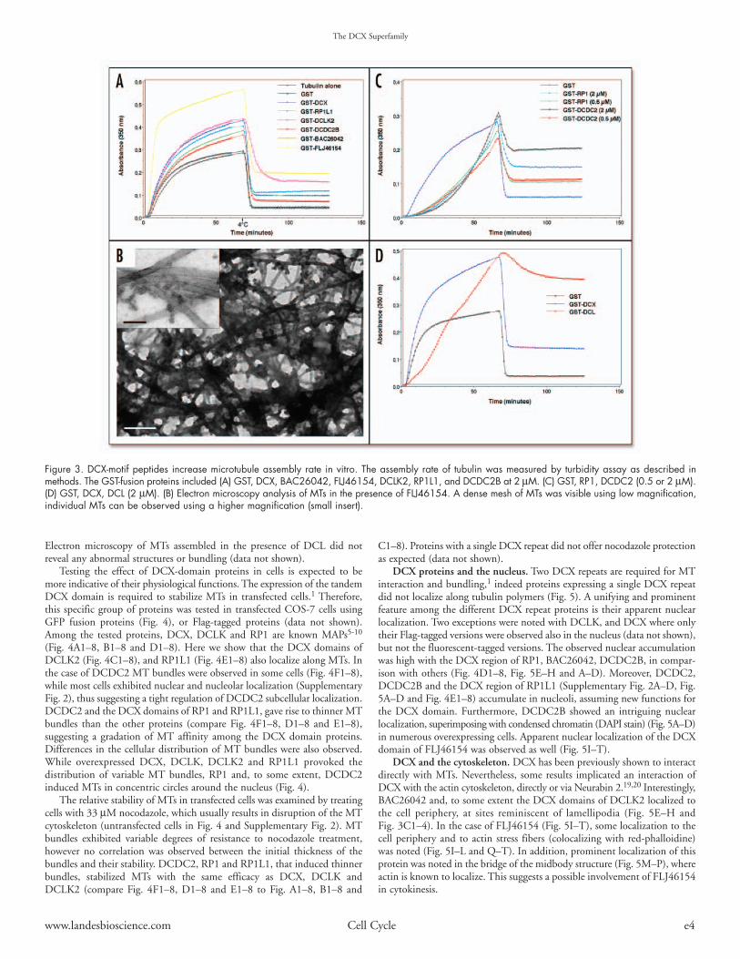
DCX proteins and the nucleus. Two DCX repeats are required for MTinteraction and bundling,1 indeed proteins expressing a single DCX repeatdid not localize along tubulin polymers (Fig. 5). A unifying and prominentfeature among the different DCX repeat proteins is their apparent nuclearlocalization. Two exceptions were noted with DCLK, and DCX where onlytheir Flag-tagged versions were observed also in the nucleus (data not shown),but not the fluorescent-tagged versions. The observed nuclear accumulationwas high with the DCX region of RP1, BAC26042, DCDC2B, in compar-ison with others (Fig. 4D1–8, Fig. 5E–H and A–D). Moreover, DCDC2,DCDC2B and the DCX region of RP1L1 (Supplementary Fig. 2A–D, Fig.5A–D and Fig. 4E1–8) accumulate in nucleoli, assuming new functions forthe DCX domain. Furthermore, DCDC2B showed an intriguing nuclearlocalization, superimposing with condensed chromatin (DAPI stain) (Fig. 5A–D)in numerous overexpressing cells. Apparent nuclear localization of the DCXdomain of FLJ46154 was observed as well (Fig. 5I–T).
DCX and the cytoskeleton. DCX has been previously shown to interactdirectly with MTs. Nevertheless, some results implicated an interaction ofDCX with the actin cytoskeleton, directly or via Neurabin 2.19,20 Interestingly,BAC26042 and, to some extent the DCX domains of DCLK2 localized tothe cell periphery, at sites reminiscent of lamellipodia (Fig. 5E–H andFig. 3C1–4). In the case of FLJ46154 (Fig. 5I–T), some localization to thecell periphery and to actin stress fibers (colocalizing with red-phalloidine)was noted (Fig. 5I–L and Q–T). In addition, prominent localization of thisprotein was noted in the bridge of the midbody structure (Fig. 5M–P), whereactin is known to localize. This suggests a possible involvement of FLJ46154in cytokinesis.
The DCX Superfamily
Figure 3. DCX-motif peptides increase microtubule assembly rate in vitro. The assembly rate of tubulin was measured by turbidity assay as described inmethods. The GST-fusion proteins included (A) GST, DCX, BAC26042, FLJ46154, DCLK2, RP1L1, and DCDC2B at 2 µM. (C) GST, RP1, DCDC2 (0.5 or 2 µM).(D) GST, DCX, DCL (2 µM). (B) Electron microscopy analysis of MTs in the presence of FLJ46154. A dense mesh of MTs was visible using low magnification,individual MTs can be observed using a higher magnification (small insert).

The DCX Superfamily
e5 Cell Cycle 2006; Vol. 5 Issue 9
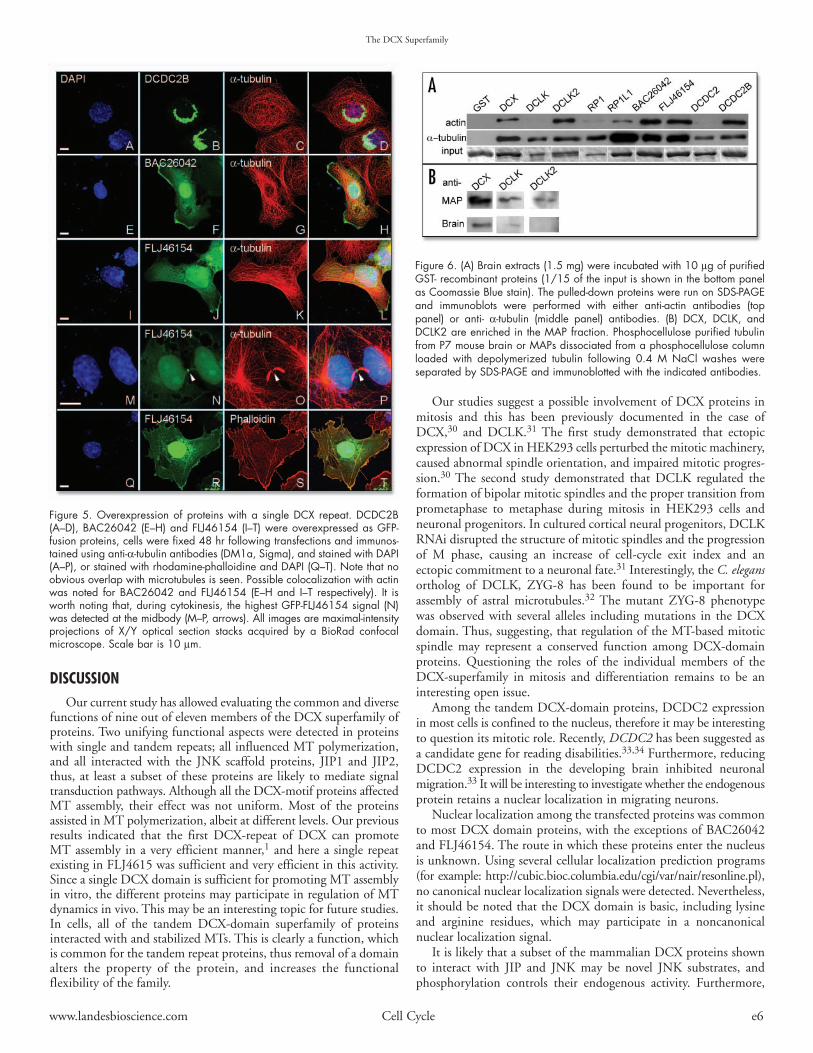
The possible association of recombinant DCX proteins with actin andtubulin in a newborn brain extract was assayed (Fig. 6A). All the DCX-motif proteins tested were capable of pulling down tubulin subunits, albeitat different levels. The capability of pulling-down actin differed significantly;positive results were obtained for DCX, DCLK2, RP1 (weak), RP1L1(weak), BAC26042, FLJ46154 and DCDC2B. No actin was observed inthe case of pull-down by DCLK, and DCDC2. In addition, we examinedwhether DCX proteins are detected in the MAP fraction for P7 mice (Fig. 6B).This experiment was possible only for proteins for which we developedantibodies for; DCX, DCLK, and DCLK2. Indeed, these three proteinswere enriched in the MAP fraction, as have been previously reported forDCX7 and DCLK.8,9
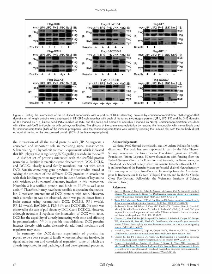
The DCX domain and protein-protein interactions. As mentionedabove, DCX interacts with the JIP1/2 scaffold protein, and with JNK. Thedomains of interactions were mapped within DCX to either conservedDCX domains, and within JIP to the PID (protein interaction domain),suggesting that the DCX domains function as a platform for protein-proteininteractions. In addition, we tested possible interactions with neurabin 2, ascaffold for actin and PP1. The interactions were investigated by cotrans-fecting HEK293 cells with Flag-tagged-DCX-domain proteins andmyc-tagged interacting protein (JIP1, JIP2, SH3-PID domain of JIP1, whichis highly similar to that of JIP2, kinase-dead JNK2, and the coiled-coildomain of neurabin 2). The assays involved reciprocal coimmunprecipitations
using myc- or Flag-antibodies (Fig. 7). Strikingly, a common feature wasnoted among all the DCX domain superfamily tested, which is their abilityto interact with JIP1 and JIP2. The interaction domain is including the PIDpart of JIP1. Only in the case of DCDC2B, a unidirectional interaction wasobserved, whereas in the case of all other proteins the coimmunoprecipitationassays worked in both directions. The interaction with JNK was less con-spicuous; DCX, DCLK2, FLJ46154 and BAC26042 interacted with JNKin both directions, whereas DCLK and DCDC2B interacted with JNK inone direction. In addition, the interaction with JNK was negative for RP1,RP1L1 and DCDC2. The immunoprecipitation of DCDC2 was conductedunder more stringent conditions, due to low extraction efficacy of the DCDC2protein, which is found mainly in the nucleus. Thus the results obtainedwith DCDC2 may not be conclusive. The interaction with neurabin 2 waspositive only for the most related members of this superfamily; DCX,DCLK, and DCLK2. Positive and negative controls for the immunoprecip-itation experiments were conducted in parallel (Supplementary Fig. 3),using Flag-tagged DCX (as a positive control), protein A/G beads as negativecontrol, and Flag-tagged C-terminal region of DCX, lacking the DCX repeats(as a negative control).
Figure 4. Overexpression of DCX motif proteins affects microtubule organization. DCX (A1–A8), the tandem DCX domains of DCLK (B1–B8), DCLK2(C1–C8), RP1 (D1–D8), RP1L1 (E1–E8), and full-length DCDC2 (F1–F8) were overexpressed as fluorescent-fusion proteins; each set of transfected cells wassubjected to nocodazole treatment (“+N”, A5–A8, B5–B8, C5–C8, D5–D8, E5–E8 and F5–F8). Cells were fixed 48 hr following transfections and immunostainedusing anti-α-tubulin antibodies (DM1a, Sigma; A3, A7, C3, C7, D3 D7, E3 and E7), and stained with DAPI (A1, A5, B1, B5, C1, C5, D1, D5, E1, E5,F1 and F5). Pictures A2, A6, B2, B6, C2, C6, D2, D6, E2, E6, F2 and F6 show the GFP signal. All images are maximal-intensity projections of X/Y opticalsection stacks acquired by a BioRad confocal microscope. Scale bar is 10 µm.
www.landesbioscience.com Cell Cycle e6
The DCX Superfamily
DISCUSSIONOur current study has allowed evaluating the common and diverse
functions of nine out of eleven members of the DCX superfamily ofproteins. Two unifying functional aspects were detected in proteinswith single and tandem repeats; all influenced MT polymerization,and all interacted with the JNK scaffold proteins, JIP1 and JIP2,thus, at least a subset of these proteins are likely to mediate signaltransduction pathways. Although all the DCX-motif proteins affectedMT assembly, their effect was not uniform. Most of the proteinsassisted in MT polymerization, albeit at different levels. Our previousresults indicated that the first DCX-repeat of DCX can promoteMT assembly in a very efficient manner,1 and here a single repeatexisting in FLJ4615 was sufficient and very efficient in this activity.Since a single DCX domain is sufficient for promoting MT assemblyin vitro, the different proteins may participate in regulation of MTdynamics in vivo. This may be an interesting topic for future studies.In cells, all of the tandem DCX-domain superfamily of proteinsinteracted with and stabilized MTs. This is clearly a function, whichis common for the tandem repeat proteins, thus removal of a domainalters the property of the protein, and increases the functionalflexibility of the family.
Our studies suggest a possible involvement of DCX proteins inmitosis and this has been previously documented in the case ofDCX,30 and DCLK.31 The first study demonstrated that ectopicexpression of DCX in HEK293 cells perturbed the mitotic machinery,caused abnormal spindle orientation, and impaired mitotic progres-sion.30 The second study demonstrated that DCLK regulated theformation of bipolar mitotic spindles and the proper transition fromprometaphase to metaphase during mitosis in HEK293 cells andneuronal progenitors. In cultured cortical neural progenitors, DCLKRNAi disrupted the structure of mitotic spindles and the progressionof M phase, causing an increase of cell-cycle exit index and anectopic commitment to a neuronal fate.31 Interestingly, the C. elegansortholog of DCLK, ZYG-8 has been found to be important forassembly of astral microtubules.32 The mutant ZYG-8 phenotypewas observed with several alleles including mutations in the DCXdomain. Thus, suggesting, that regulation of the MT-based mitoticspindle may represent a conserved function among DCX-domainproteins. Questioning the roles of the individual members of theDCX-superfamily in mitosis and differentiation remains to be aninteresting open issue.
Among the tandem DCX-domain proteins, DCDC2 expressionin most cells is confined to the nucleus, therefore it may be interestingto question its mitotic role. Recently, DCDC2 has been suggested asa candidate gene for reading disabilities.33,34 Furthermore, reducingDCDC2 expression in the developing brain inhibited neuronalmigration.33 It will be interesting to investigate whether the endogenousprotein retains a nuclear localization in migrating neurons.
Nuclear localization among the transfected proteins was commonto most DCX domain proteins, with the exceptions of BAC26042and FLJ46154. The route in which these proteins enter the nucleusis unknown. Using several cellular localization prediction programs(for example: http://cubic.bioc.columbia.edu/cgi/var/nair/resonline.pl),no canonical nuclear localization signals were detected. Nevertheless,it should be noted that the DCX domain is basic, including lysineand arginine residues, which may participate in a noncanonicalnuclear localization signal.
It is likely that a subset of the mammalian DCX proteins shownto interact with JIP and JNK may be novel JNK substrates, andphosphorylation controls their endogenous activity. Furthermore,
Figure 5. Overexpression of proteins with a single DCX repeat. DCDC2B(A–D), BAC26042 (E–H) and FLJ46154 (I–T) were overexpressed as GFP-fusion proteins, cells were fixed 48 hr following transfections and immunos-tained using anti-α-tubulin antibodies (DM1a, Sigma), and stained with DAPI(A–P), or stained with rhodamine-phalloidine and DAPI (Q–T). Note that noobvious overlap with microtubules is seen. Possible colocalization with actinwas noted for BAC26042 and FLJ46154 (E–H and I–T respectively). It isworth noting that, during cytokinesis, the highest GFP-FLJ46154 signal (N)was detected at the midbody (M–P, arrows). All images are maximal-intensityprojections of X/Y optical section stacks acquired by a BioRad confocalmicroscope. Scale bar is 10 µm.
Figure 6. (A) Brain extracts (1.5 mg) were incubated with 10 µg of purifiedGST- recombinant proteins (1/15 of the input is shown in the bottom panelas Coomassie Blue stain). The pulled-down proteins were run on SDS-PAGEand immunoblots were performed with either anti-actin antibodies (toppanel) or anti- α-tubulin (middle panel) antibodies. (B) DCX, DCLK, andDCLK2 are enriched in the MAP fraction. Phosphocellulose purified tubulinfrom P7 mouse brain or MAPs dissociated from a phosphocellulose columnloaded with depolymerized tubulin following 0.4 M NaCl washes wereseparated by SDS-PAGE and immunoblotted with the indicated antibodies.
The DCX Superfamily
e7 Cell Cycle 2006; Vol. 5 Issue 9
the interaction of all the tested proteins with JIP1/2 suggests aconserved and important role in mediating signal transduction.Substantiating this hypothesis are recent experiments which indicatedthat RP1 plays a role in regulating JNK signaling cascades in the eye.35
A distinct set of proteins interacted with the scaffold proteinneurabin 2. Positive interactions were observed with DCX, DCLK,and DCLK2, closely related family members, but not with otherDCX-domain containing gene products. Future studies aimed atsolving the structure of the different DCX proteins in associationwith their binding partners may assist in identification of key aminoacid residues, and structural elements, involved in this interaction.Neurabin 2 is a scaffold protein and binds to PP136 as well as toactin.37 Therefore, it may have been possible to speculate that neura-bin 2 mediates interactions of DCX proteins with actin. However,such a correlation was not observed. Actin was pulled down from abrain extract using recombinant DCX, DCLK2, RP1 (weak),RP1L1 (weak), BAC26042, FLJ46154 and DCDC2B. No actin wasobserved in the case of pull-down by DCLK, and DCDC2. However,although neurabin 2 regulates the interaction of DCX with actin,DCX has the capability of directly interacting with actin and affectingits polymerization.19,20 It is possible that additional DCX proteinsinteract directly with actin, alternatively additional mediators andregulators may exist.
In summary, the DCX-domain superfamily of proteins hasproven to be a very successful family whose members are involved insignal transduction and cytoskeletal regulation, some of which arealready implicated in and pathological and developmental processes.
AcknowledgementsWe thank Prof. Shmuel Pietrekovski, and Dr. Arlette Fellous for helpful
discussions. The work has been supported in part by the Fritz ThyseenStifung Foundation, the Israeli Science Foundation (grant no. 270/04),Foundation Jérôme Lejeune, Minerva foundation with funding from theFederal German Ministry for Education and Research, the Kekst center, theDavid and Fela Shapell Family Center for Genetic Disorders Research. O.R.is an Incumbent of the Berstein-Mason professorial chair of Neurochemistry.F.C. was supported by a Post-Doctoral Fellowship from the Associationpour la Recherche sur le Cancer (Villejuif, France), and by the Sir CharlesClore Post-Doctoral Fellowship, the Weizmann Institute of Science(Rehovot, Israel).
References1. Sapir T, Horesh D, Caspi M, Atlas R, Burgess HA, Grayer Wolf S, Francis F, Chelly J,
Elbaum M, Pietrokovski S, Reiner O. Doublecortin mutations cluster in evolutionaryconserved functional domains. Hum Mol Genet 2000; 5:703-712.
2. Taylor KR, Holzer AK, Bazan JF, Walsh CA, Gleeson JG. Patient mutations in doublecortindefine a repeated tubulin-binding domain. J Biol Chem 2000; 275:34442-50.
3. des Portes V, Pinard JM, Billuart P, Vinet MC, Koulakoff A, Carrie A, Gelot A, Dupuis E,Motte J, Berwald-Netter Y, Catala M, Kahn A, Beldjord C, Chelly J. A novel CNS generequired for neuronal migration and involved in X-linked subcortical laminar hetrotropiaand lissencephaly syndrome. Cell 1998; 92:51-61.
4. Gleeson JG, Allen KM, Fox JW, Lamperti ED, Berkovic S, Scheffer I, Cooper EC, DobynsWB, Minnerath SR, Ross ME, Walsh CA. Doublecortin, a brain-specific gene mutated inhuman X-linked lissencephaly and double cortex syndrome, encodes a putative signalingprotein. Cell 1998; 92:63-72.
5. Horesh D, Sapir T, Francis F, Caspi M, Grayer Wolf S, Elbaum M, Chelly J, Reiner O.Doublecortin, a stabilizer of microtubules. Hum Mol Genet 1999; 8:1599-1610.
6. Gleeson JG, Lin PT, Flanagan LA, Walsh CA. Doublecortin is a microtubule-associatedprotein and is expressed widely by migrating neurons. Neuron 1999; 23:257-71.
7. Francis F, Koulakoff A, Boucher D, Chafey P, Schaar B, Vinet MC, Friocourt G,McDonnell N, Reiner O, Kahn A, McConnell SK, Berwald-Netter Y, Denoulet P, ChellyJ. Doublecortin is a developmentally regulated, microtubule-associated protein expressed inmigrating and differentiating neurons. Neuron 1999; 23:247-56.
Figure 7. Testing the interactions of the DCX motif superfamily with a portion of DCX interacting proteins by coimmunoprecipitation. FLAG-tagged-DCXdomains or full-length proteins were expressed in HEK293 cells together with each of the tested myc-tagged partners (JIP1, JIP2, PID and the SH3 domainsof JIP1 marked as P+S, kinase dead JNK2 marked as JNK, and the coiled-coil domain of neurabin II marked as Ner2). Coimmunoprecipitation was donewith either anti-FLAG antibodies or with anti-myc antibodies. The efficacy of the coimmunoprecipitation by reacting the immuno-blot with the antibody usedfor immunoprecipitation (15% of the immuno-precipitate), and the coimmunoprecipitation was tested by reacting the immuno-blot with the antibody direct-ed against the tag of the coexpressed protein (85% of the immuno-precipitate).
www.landesbioscience.com Cell Cycle e8
8. Burgess HA, Reiner O. Doublecortin-like kinase, is a microtubule-associated proteinkinase expressed in growth cones. MCN 2000; 16.
9. Lin PT, Gleeson JG, Corbo JC, Flanagan L, Walsh CA. DCAMKL1 encodes a proteinkinase with homology to doublecortin that regulates microtubule polymerization. JNeurosci 2000; 20:9152-61.
10. Liu Q, Zuo J, Pierce EA. The retinitis pigmentosa 1 protein is a photoreceptor micro-tubule-associated protein. J Neurosci 2004; 24:6427-36.
11. Pierce EA, Quinn T, Meehan T, McGee TL, Berson EL, Dryja TP. Mutations in a geneencoding a new oxygen-regulated photoreceptor protein cause dominant retinitis pigmen-tosa. Nat Genet 1999; 22:248-54.
12. Sullivan LS, Heckenlively JR, Bowne SJ, Zuo J, Hide WA, Gal A, Denton M, InglehearnCF, Blanton SH, Daiger SP. Mutations in a novel retina-specific gene cause autosomal dom-inant retinitis pigmentosa. Nat Genet 1999; 22:255-9.
13. Gao J, Cheon K, Nusinowitz S, Liu Q, Bei D, Atkins K, Azimi A, Daiger SP, Farber DB,Heckenlively JR, Pierce EA, Sullivan LS, Zuo J. Progressive photoreceptor degeneration,outer segment dysplasia, and rhodopsin mislocalization in mice with targeted disruption ofthe retinitis pigmentosa-1 (Rp1) gene. Proc Natl Acad Sci USA 2002; 99:5698-703.
14. Edelman AM, Kim WY, Higgins D, Goldstein EG, Oberdoerster M, Sigurdson W.Doublecortin kinase-2, a novel doublecortin-related protein kinase associated with termi-nal segments of axons and dendrites. J Biol Chem 2005; 280:8531-43.
15. Kim MH, Cierpicki T, Derewenda U, Krowarsch D, Feng Y, Devedjiev Y, Dauter Z, WalshCA, Otlewski J, Bushweller JH, Derewenda ZS. The DCX-domain tandems of dou-blecortin and doublecortin-like kinase. Nat Struct Biol 2003; 10:324-33.
16. Moores CA, Perderiset M, Francis F, Chelly J, Houdusse A, Milligan RA. Mechanism ofmicrotubule stabilization by doublecortin. Mol Cell 2004; 14:833-9.
17. Reiner O, Coquelle FM. Missense mutations resulting in type 1 lissencephaly. Cell MolLife Sci 2005; 62:425-34.
18. Reiner O, Gdalyahu A, Ghosh I, Levy T, Sapoznik S, Nir R, Sapir T. DCX’s phosphoryla-tion by not just another kinase (JNK). Cell Cycle 2004; 3:747-51.
19. Tsukada M, Prokscha A, Oldekamp J, Eichele G. Identification of neurabin II as a noveldoublecortin interacting protein. Mech Dev 2003; 120:1033-43.
20. Tsukada M, Prokscha A, Ungewickell E, Eichele G. Doublecortin association with actin fil-aments is regulated by Neurabin II. J Biol Chem 2005; 280:11361-8.
21. Gdalyahu A, Ghosh I, Levy T, Sapir T, Sapoznik S, Fishler Y, Azoulai D, Reiner O. DCX,a new mediator of the JNK pathway. Embo J 2004; 23:823-832.
22. Burgess HA, Martinez S, Reiner O. KIAA0369, doublecortin-like kinase, is expressed dur-ing brain development. J Neurosci Res 1999; 58:567-575.
23. Dujardin D, Wacker UI, Moreau A, Schroer TA, Rickard JE, De Mey JR. Evidence for arole of CLIP-170 in the establishment of metaphase chromosome alignment. J Cell Biol1998; 141:849-62.
24. Coquelle FM, Caspi M, Cordelieres FP, Dompierre JP, Dujardin DL, Koifman C, MartinP, Hoogenraad CC, Akhmanova A, Galjart N, De Mey JR, Reiner O. LIS1, CLIP-170’s keyto the dynein/dynactin pathway. Mol Cell Biol 2002; 22:3089-102.
25. Sapir T, Elbaum M, Reiner O. Reduction of microtubule catastrophe events by LIS1,platelet-activating factor acetylhydrolase subunit. EMBO J 1997; 16:6977-6984.
26. Herzog W, Weber K. Fractionation of brain microtubule associated proteins. Isolation oftwo different proteins which stimulate tubulin polymerization in vitro. Eur J Biochem1978; 92:1-8.
27. Riederer BM, Pellier V, Antonsson B, Di Paolo G, Stimpson SA, Lutjens R, Catsicas S,Grenningloh G. Regulation of microtubule dynamics by the neuronal growth-associatedprotein SCG10. Proc Natl Acad Sci USA 1997; 94:741-5.
28. Liu Y, Chirino AJ, Misulovin Z, Leteux C, Feizi T, Nussenzweig MC, Bjorkman PJ. Crystalstructure of the cysteine-rich domain of mannose receptor complexed with a sulfated car-bohydrate ligand. J Exp Med 2000; 191:1105-16.
29. Burgess HA, Reiner O. Alternative splice variants of doublecortin-like kinase are differen-tially expressed and have different kinase activities. J Biol Chem 2002; 277:17696-705.
30. Couillard-Despres S, Uyanik G, Ploetz S, Karl C, Koch H, Winkler J, Aigner L. Mitoticimpairment by doublecortin is diminished by doublecortin mutations found in patients.Neurogenetics 2004.
31. Shu T, Tseng HC, Sapir T, Stern P, Zhou Y, Sanada K, Fischer A, Coquelle FM, Reiner O,Tsai LH. Doublecortin-like kinase controls neurogenesis by regulating mitotic spindles andM Phase progression. Neuron 2006; 49:25-39.
32. Gonczy P, Bellanger JM, Kirkham M, Pozniakowski A, Baumer K, Phillips JB, Hyman AA.zyg-8, a gene required for spindle positioning in C. elegans, encodes a doublecortin-relatedkinase that promotes microtubule assembly. Dev Cell 2001; 1:363-75.
33. Meng H, Smith SD, Hager K, Held M, Liu J, Olson RK, Pennington BF, Defries JC,Gelernter J, O’Reilly-Pol T, Somlo S, Skudlarski P, Shaywitz SE, Shaywitz BA, MarchioneK, Wang Y, Paramasivam M, Loturco JJ, Page GP, Gruen JR. DCDC2 is associated withreading disability and modulates neuronal development in the brain. Proc Natl Acad SciUSA 2005.
34. Schumacher J, Anthoni H, Dahdouh F, Konig IR, Hillmer AM, Kluck N, Manthey M,Plume E, Warnke A, Remschmidt H, Hulsmann J, Cichon S, Lindgren CM, Propping P,Zucchelli M, Ziegler A, Peyrard-Janvid M, Schulte-Korne G, Nothen MM, Kere J. Stronggenetic evidence of DCDC2 as a susceptibility gene for Dyslexia. Am J Hum Genet 2006;78:52-62.
35. Liu J, Huang Q, Higdon J, Liu W, Xie T, Yamashita T, Cheon K, Cheng C, Zuo J. Distinctgene expression profiles and reduced JNK signaling in retinitis pigmentosa caused by RP1mutations. Hum Mol Genet 2005; 14:2945-58.
36. MacMillan LB, Bass MA, Cheng N, Howard EF, Tamura M, Strack S, Wadzinski BE,Colbran RJ. Brain actin-associated protein phosphatase 1 holoenzymes containingspinophilin, neurabin, and selected catalytic subunit isoforms. J Biol Chem 1999; 274:35845-54.
37. Satoh A, Nakanishi H, Obaishi H, Wada M, Takahashi K, Satoh K, Hirao K, Nishioka H,Hata Y, Mizoguchi A, Takai Y. Neurabin-II/spinophilin. An actin filament-binding proteinwith one pdz domain localized at cadherin-based cell-cell adhesion sites. J Biol Chem 1998;273:3470-5.
The DCX Superfamily
Related Documents