International Journal of Pharmaceutics 340 (2007) 76–83 Combining SEM, TEM, and micro-Raman techniques to differentiate between the amorphous molecular level dispersions and nanodispersions of a poorly water-soluble drug within a polymer matrix Evangelos Karavas a,b , Manolis Georgarakis b , Aristides Docoslis c , Dimitrios Bikiaris d,∗ a Pharmathen S.A., Pharmaceutical Industry, Dervenakion Str. 6, Pallini Attikis, 153 51 Attiki, Greece b Section of Pharmaceutics and Drug Control, Department of Pharmacy, Aristotle University of Thessaloniki, 541 24 Thessaloniki, Greece c Department of Chemical Engineering, Queen’s University at Kingston, Kingston, Ont., Canada K7L 3N6 d Laboratory of Organic Chemical Technology, Department of Chemistry, Aristotle University of Thessaloniki, 541 24 Thessaloniki, Greece Received 31 October 2006; received in revised form 5 March 2007; accepted 19 March 2007 Available online 30 March 2007 Abstract The aim of the present study was to experimentally examine whether poorly water-soluble drugs dispersed in a polymeric matrix exist as amorphous nanodispersions or molecularly dispersed compounds. Felodipine (Felo) dispersed in PVP matrix (solid dispersion) was used as a model drug in this study. Drug/polymer ratios have an impact on the drug average particle size, morphology and dissolution profile while solid dispersions containing up to 50 wt% Felo are completely amorphous. SEM, TEM micrographs, and micro-Raman mapping reveal that Felo is dispersed in the form of nanoparticles into the PVP matrix. Due to the high spatial resolution of TEM, it was established that these nanoparticles are not uniform particles, but rather agglomerates of individual particles with sizes smaller than 5–10nm. Moreover, micro-Raman mapping allowed us to observe the size and spatial distribution of domains where the drug existed as molecularly or nanodispersed. Experimental evidence presented in this work contradicts the common belief that amorphous poorly water-soluble drugs exist only in the state of molecular dispersion inside a polymer matrix by showing that both types of dispersions (molecular-level and nanodispersions) can coexist. © 2007 Elsevier B.V. All rights reserved. Keywords: Poor water-soluble drugs; Solid dispersions; Amorphous nanodispersions; SEM; TEM; Raman 1. Introduction In pharmaceutical technology there exist numerous drug sub- stances, including new chemical entities, that in spite of their high therapeutic effectiveness, are characterized by poor water solubility. The latter limits their potential uses in formulating bioavailable pharmaceutical products. In all those cases, the rate limiting factor for drug absorption becomes the dissolu- tion rate of the active ingredient in the gastro-intestinal liquids (H¨ orter and Dressman, 2001). Therefore, the enhancement of oral bioavailability of such poor water-soluble drugs and the preparation of solid oral dosage forms is currently one of major objectives and greatest challenges in the area of new formula- tions development. ‘Solid dispersion’ is one of the earlier, yet ∗ Corresponding author. Tel.: +30 2310 997812; fax: +30 2310 997667. E-mail address: [email protected] (D. Bikiaris). still favorable, approaches for overcoming this limitation. Owing to its simplicity from the manufacturing and process scalabil- ity standpoints, solid dispersion has become one of the most active and promising research areas of great interest to phar- maceutical companies. Furthermore, such formulations possess considerable advantages over other commonly used techniques, especially micronization. Hence it is expected that the popular- ity of solid dispersions will grow rapidly (Serajuddin, 1999). The term solid dispersion refers to solid state mixtures, pre- pared through the dispersion, typically by solvent evaporation or melt mixing, of one or more active ingredients in an inert car- rier matrix (Chiou and Riegelmann, 1971). In these dispersions, the drug can be present in a fully crystalline state (in the form of coarse drug particles), in a semicrystalline state, and in fully amorphous state (in the form of a fine particle dispersion, or molecularly distributed within the carrier). Such systems prove to be very effective for enhancing the dissolution rate of low solubility drugs. 0378-5173/$ – see front matter © 2007 Elsevier B.V. All rights reserved. doi:10.1016/j.ijpharm.2007.03.037

Welcome message from author
This document is posted to help you gain knowledge. Please leave a comment to let me know what you think about it! Share it to your friends and learn new things together.
Transcript
A
amddnuip©
K
1
shsbrt(opot
0d
International Journal of Pharmaceutics 340 (2007) 76–83
Combining SEM, TEM, and micro-Raman techniques to differentiatebetween the amorphous molecular level dispersions and nanodispersions
of a poorly water-soluble drug within a polymer matrix
Evangelos Karavas a,b, Manolis Georgarakis b, Aristides Docoslis c, Dimitrios Bikiaris d,∗a Pharmathen S.A., Pharmaceutical Industry, Dervenakion Str. 6, Pallini Attikis, 153 51 Attiki, Greece
b Section of Pharmaceutics and Drug Control, Department of Pharmacy, Aristotle University of Thessaloniki, 541 24 Thessaloniki, Greecec Department of Chemical Engineering, Queen’s University at Kingston, Kingston, Ont., Canada K7L 3N6
d Laboratory of Organic Chemical Technology, Department of Chemistry, Aristotle University of Thessaloniki, 541 24 Thessaloniki, Greece
Received 31 October 2006; received in revised form 5 March 2007; accepted 19 March 2007Available online 30 March 2007
bstract
The aim of the present study was to experimentally examine whether poorly water-soluble drugs dispersed in a polymeric matrix exist asmorphous nanodispersions or molecularly dispersed compounds. Felodipine (Felo) dispersed in PVP matrix (solid dispersion) was used as aodel drug in this study. Drug/polymer ratios have an impact on the drug average particle size, morphology and dissolution profile while solid
ispersions containing up to 50 wt% Felo are completely amorphous. SEM, TEM micrographs, and micro-Raman mapping reveal that Felo isispersed in the form of nanoparticles into the PVP matrix. Due to the high spatial resolution of TEM, it was established that these nanoparticles areot uniform particles, but rather agglomerates of individual particles with sizes smaller than 5–10 nm. Moreover, micro-Raman mapping allowed
s to observe the size and spatial distribution of domains where the drug existed as molecularly or nanodispersed. Experimental evidence presentedn this work contradicts the common belief that amorphous poorly water-soluble drugs exist only in the state of molecular dispersion inside aolymer matrix by showing that both types of dispersions (molecular-level and nanodispersions) can coexist.2007 Elsevier B.V. All rights reserved.
rsions
stiamceiTpo
eywords: Poor water-soluble drugs; Solid dispersions; Amorphous nanodispe
. Introduction
In pharmaceutical technology there exist numerous drug sub-tances, including new chemical entities, that in spite of theirigh therapeutic effectiveness, are characterized by poor waterolubility. The latter limits their potential uses in formulatingioavailable pharmaceutical products. In all those cases, theate limiting factor for drug absorption becomes the dissolu-ion rate of the active ingredient in the gastro-intestinal liquidsHorter and Dressman, 2001). Therefore, the enhancement ofral bioavailability of such poor water-soluble drugs and the
reparation of solid oral dosage forms is currently one of majorbjectives and greatest challenges in the area of new formula-ions development. ‘Solid dispersion’ is one of the earlier, yet∗ Corresponding author. Tel.: +30 2310 997812; fax: +30 2310 997667.E-mail address: [email protected] (D. Bikiaris).
rtoamts
378-5173/$ – see front matter © 2007 Elsevier B.V. All rights reserved.oi:10.1016/j.ijpharm.2007.03.037
; SEM; TEM; Raman
till favorable, approaches for overcoming this limitation. Owingo its simplicity from the manufacturing and process scalabil-ty standpoints, solid dispersion has become one of the mostctive and promising research areas of great interest to phar-aceutical companies. Furthermore, such formulations possess
onsiderable advantages over other commonly used techniques,specially micronization. Hence it is expected that the popular-ty of solid dispersions will grow rapidly (Serajuddin, 1999).he term solid dispersion refers to solid state mixtures, pre-ared through the dispersion, typically by solvent evaporationr melt mixing, of one or more active ingredients in an inert car-ier matrix (Chiou and Riegelmann, 1971). In these dispersions,he drug can be present in a fully crystalline state (in the formf coarse drug particles), in a semicrystalline state, and in fully
morphous state (in the form of a fine particle dispersion, orolecularly distributed within the carrier). Such systems proveo be very effective for enhancing the dissolution rate of lowolubility drugs.
rnal o
(r22tctsae(sthnngtpopp
nm(qnwcicma
2
2
rFweohmAp
2
5ds
sa
2
stupwa
2
wpiwbcwoRhaitastt
2
pdfi
2
m(Ofi
2
E. Karavas et al. / International Jou
Poly(ethylene glycol) (PEG) and polyvinylpyrrolidonePVP) are the most used drug carriers for solid dispersion prepa-ations (Tantishaiyakul et al., 1999; Van dem Mooter et al.,001; Basit et al., 2001; Groves et al., 1984; Naima et al.,001; Oaya et al., 2003) due to their strong hydrophilic proper-ies and their capability to form molecular adducts with manyompounds. The presence of hydroxyl or carbonyl groups inhe repeat units of these polymers tend to increase the water-olubility (Trapani et al., 1999; Damian et al., 2002; Hideshind Hisakazu, 1997) and stability (Damian et al., 2002; Guptat al., 2005) of the drug and also improve its bioavailabilityKushida et al., 2002). It is accepted nowadays that the glassytate of a drug compound dispersed in such polymer matrices ishe most desired state, since it improves its dissolution rate andence drug absorption (Kaushal et al., 2004). Despite the largeumber of published studies on solid dispersions (600–700), theature of the drug amorphous state has been less well investi-ated, hence it is still not clear how the drug is dispersed withinhe matrix in the majority of cases (Craig, 2002). In most of theublished technical reports on amorphous dispersions, it wasnly surmised by the authors that the drug is molecularly dis-ersed in the matrix carrier, however, no clear evidence has beenrovided.
In the present study two microscopic techniques, scan-ing electron microscopy (SEM) and transmission electronicroscopy (TEM) were used in a complementary fashion
in conjunction with XRD), in order to precisely and ade-uately differentiate between drug molecular dispersions andanodispersions in amorphous solid dispersions of poorlyater-soluble drugs. Felodipine was used here as a model
ompound to represent a poorly water-soluble drug dispersedn polyvinylpyrrolidone (PVP) matrix. Since SEM and TEMan provide only optical evidence of the drug dispersion,icro-Raman spectroscopy was also employed for the chemical
nalysis of dispersed Felo into the PVP matrix.
. Experimental
.1. Materials
Felodipine (Felo) with an assay of 99.9% and particle sizeange 20–100 �m was obtained from PCAS (Longjumeau,rance). Felo has a melting point of 143–145 ◦C, solubility inater approximately equal to 0.5 mg/l, and it is freely soluble in
thanol. Polyvinylpyrrolidone (PVP), type Kollidon K30, wasbtained from BASF (Ludwigshafen, Germany). The PVP usedad molecular weight of 50,000–55,000, Tg = 167 ◦C (DSC),oisture content 1.95% (TGA) and bulk density of 0.410 g/cm3.ll other materials and reagents were of analytical grade ofurity.
.2. Preparation of solid dispersion systems
PVP/Felo solid dispersion systems of 90/10, 80/20, 70/30 and0/50 (w/w) were prepared by dissolution of accurately weighedry substances in appropriate quantities of absolute ethanol. Theolutions were mixed, ultrasonicated for 20 min, and then the
mfidu
f Pharmaceutics 340 (2007) 76–83 77
olvent was fully evaporated under vacuum in a rotary evaporatort 60 ◦C for 1 h.
.3. In vitro release profile
Release of Felo from the solid dispersion systems was mea-ured by a modified dissolution apparatus II (paddles) USP. Theest was performed in 37 ± 1 ◦C with a rotation speed of 100 rpmsing 500 ml of 0.1 M phosphate buffer pH 6.5 containing 2%olysorbate 20. The instrumentation used for the dissolution testas an HPLC apparatus type DISTEK 2100B equipped with an
uto sampler.
.4. Solubility in water at 37 ◦C
A dissolution apparatus II of USP (paddles) type DISTEKas used. Samples of each system equivalent to 50 mg FEL werelaced in the vessels into hard gelatin capsules under the follow-ng conditions: 500 ml H2O, 50 rpm, 37 ◦C, 36 h. The capsulesere placed into specific metallic nets in order to remain in theottom of the vessel so that the measurements are taken underonstant hydrodynamic conditions. After 36 h 50 ml per sampleas removed, filtered through 0.2 �m, and the water was evap-rated under vacuum on a rotary evaporator, BUCH Rotavapor200 equipped with a vacuum controller model V-800 and aeating bath model B-490. The residues were dissolved in 5 mlbsolute ethanol and assayed with the HPLC method describedn European pharmacopoeia. The analysis was performed on aype Shimadzu instrument model LC-20-10A, at 237 nm usingC18 column. The disintegration time of the hard gelatin cap-
ules cannot affect the results of the experiment, as it was lesshan 5 min. The experiment was performed in triplicate, whilehe R.S.D. per sample was found to be less than 1.5%.
.5. X-ray diffraction (XRD)
XRD analysis were performed on randomly oriented sam-les, scanned over the interval 5–55◦ 2θ, using a Philips PW1710iffractometer, with Bragg-Brentano geometry (θ, 2θ) and Ni-ltered Cu K� radiation.
.6. Scanning electron microscopy (SEM)
The morphology of the prepared solid dispersions was deter-ined by a scanning electron microscope (SEM), type JEOL
JMS-840) equipped with an energy-dispersive X-ray (EDX)xford ISIS 300 micro-analytical system. For this examination,
ractured samples of solid dispersions prepared by immersionn liquid nitrogen were used.
.7. Transmission electron microscopy (TEM)
Electron diffraction (ED) and transmission electron
icroscopy (TEM) investigations were performed on ultra thinlm samples of solid dispersions prepared by ultra-microtomeeposited on copper grids. TEM micrographs were obtainedsing a JEOL 120 CX microscope operating at 120 kV. To avoid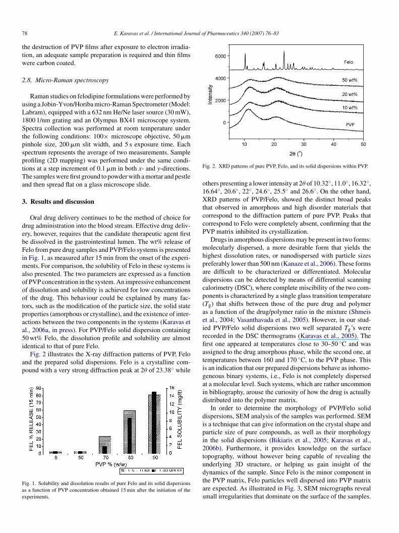
7 rnal of Pharmaceutics 340 (2007) 76–83
ttw
2
uL1StpsptTa
3
debFimaoootpaa5i
ap
Fae
F
o1XtccP
mhpadcp(aeir
8 E. Karavas et al. / International Jou
he destruction of PVP films after exposure to electron irradia-ion, an adequate sample preparation is required and thin filmsere carbon coated.
.8. Micro-Raman spectroscopy
Raman studies on felodipine formulations were performed bysing a Jobin-Yvon/Horiba micro-Raman Spectrometer (Model:abram), equipped with a 632 nm He/Ne laser source (30 mW),800 1/nm grating and an Olympus BX41 microscope system.pectra collection was performed at room temperature under
he following conditions: 100× microscope objective, 50 �minhole size, 200 �m slit width, and 5 s exposure time. Eachpectrum represents the average of two measurements. Samplerofiling (2D mapping) was performed under the same condi-ions at a step increment of 0.1 �m in both x- and y-directions.he samples were first ground to powder with a mortar and pestlend then spread flat on a glass microscope slide.
. Results and discussion
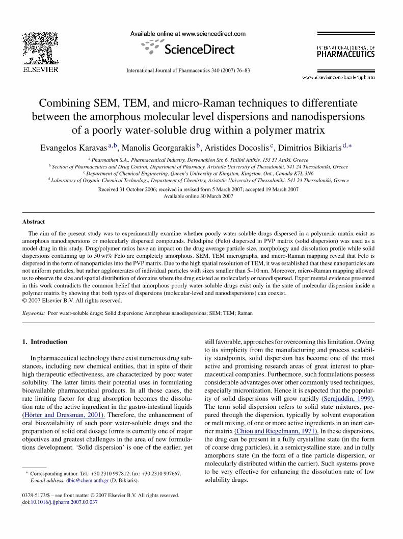
Oral drug delivery continues to be the method of choice forrug administration into the blood stream. Effective drug deliv-ry, however, requires that the candidate therapeutic agent firste dissolved in the gastrointestinal lumen. The wt% release ofelo from pure drug samples and PVP/Felo systems is presented
n Fig. 1, as measured after 15 min from the onset of the experi-ents. For comparison, the solubility of Felo in these systems is
lso presented. The two parameters are expressed as a functionf PVP concentration in the system. An impressive enhancementf dissolution and solubility is achieved for low concentrationsf the drug. This behaviour could be explained by many fac-ors, such as the modification of the particle size, the solid stateroperties (amorphous or crystalline), and the existence of inter-ctions between the two components in the systems (Karavas etl., 2006a, in press). For PVP/Felo solid dispersion containing0 wt% Felo, the dissolution profile and solubility are almost
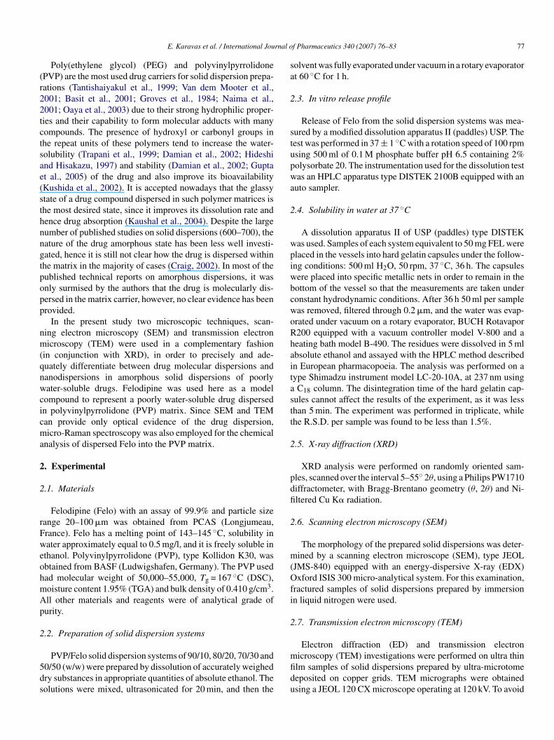
dentical to that of pure Felo.Fig. 2 illustrates the X-ray diffraction patterns of PVP, Felond the prepared solid dispersions. Felo is a crystalline com-ound with a very strong diffraction peak at 2θ of 23.38◦ while
ig. 1. Solubility and dissolution results of pure Felo and its solid dispersionss a function of PVP concentration obtained 15 min after the initiation of thexperiments.
fiatigaid
dipi2tudtas
ig. 2. XRD patterns of pure PVP, Felo, and its solid dispersions within PVP.
thers presenting a lower intensity at 2θ of 10.32◦, 11.0◦, 16.32◦,6.64◦, 20.6◦, 22◦, 24.6◦, 25.5◦ and 26.6◦. On the other hand,RD patterns of PVP/Felo, showed the distinct broad peaks
hat observed in amorphous and high disorder materials thatorrespond to the diffraction pattern of pure PVP. Peaks thatorrespond to Felo were completely absent, confirming that theVP matrix inhibited its crystallization.
Drugs in amorphous dispersions may be present in two forms:olecularly dispersed, a more desirable form that yields the
ighest dissolution rates, or nanodispersed with particle sizesreferably lower than 500 nm (Kanaze et al., 2006). These formsre difficult to be characterized or differentiated. Molecularispersions can be detected by means of differential scanningalorimetry (DSC), where complete miscibility of the two com-onents is characterized by a single glass transition temperatureTg) that shifts between those of the pure drug and polymers a function of the drug/polymer ratio in the mixture (Shmeist al., 2004; Vasanthavada et al., 2005). However, in our stud-ed PVP/Felo solid dispersions two well separated Tg’s wereecorded in the DSC thermograms (Karavas et al., 2005). Therst one appeared at temperatures close to 30–50 ◦C and wasssigned to the drug amorphous phase, while the second one, atemperatures between 160 and 170 ◦C, to the PVP phase. Thiss an indication that our prepared dispersions behave as inhomo-eneous binary systems, i.e., Felo is not completely dispersedt a molecular level. Such systems, which are rather uncommonn bibliography, arouse the curiosity of how the drug is actuallyistributed into the polymer matrix.
In order to determine the morphology of PVP/Felo solidispersions, SEM analysis of the samples was performed. SEMs a technique that can give information on the crystal shape andarticle size of pure compounds, as well as their morphologyn the solid dispersions (Bikiaris et al., 2005; Karavas et al.,006b). Furthermore, it provides knowledge on the surfaceopography, without however being capable of revealing thenderlying 3D structure, or helping us gain insight of the
ynamics of the sample. Since Felo is the minor component inhe PVP matrix, Felo particles well dispersed into PVP matrixre expected. As illustrated in Fig. 3, SEM micrographs revealmall irregularities that dominate on the surface of the samples.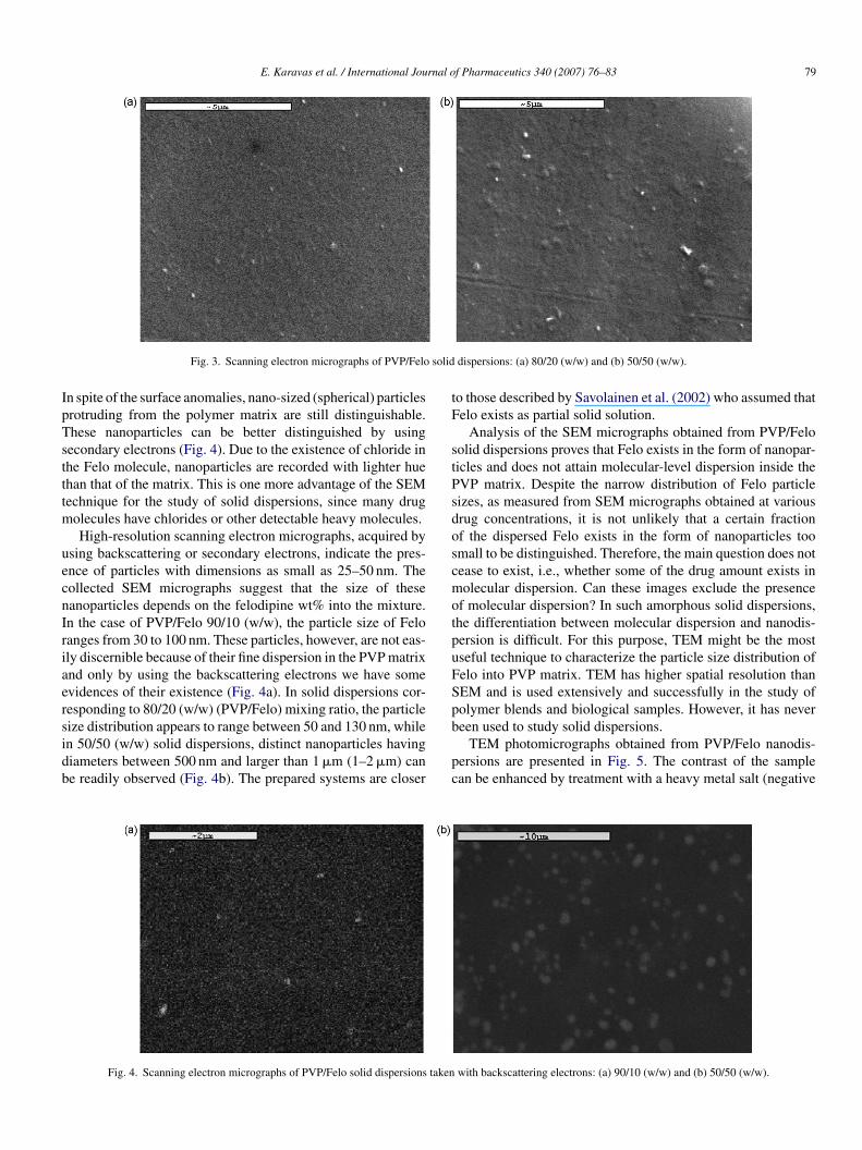
E. Karavas et al. / International Journal of Pharmaceutics 340 (2007) 76–83 79
solid
IpTstttm
uecnIriaersidb
tF
stPsdoscmotpuFSpb
Fig. 3. Scanning electron micrographs of PVP/Felo
n spite of the surface anomalies, nano-sized (spherical) particlesrotruding from the polymer matrix are still distinguishable.hese nanoparticles can be better distinguished by usingecondary electrons (Fig. 4). Due to the existence of chloride inhe Felo molecule, nanoparticles are recorded with lighter huehan that of the matrix. This is one more advantage of the SEMechnique for the study of solid dispersions, since many drug
olecules have chlorides or other detectable heavy molecules.High-resolution scanning electron micrographs, acquired by
sing backscattering or secondary electrons, indicate the pres-nce of particles with dimensions as small as 25–50 nm. Theollected SEM micrographs suggest that the size of theseanoparticles depends on the felodipine wt% into the mixture.n the case of PVP/Felo 90/10 (w/w), the particle size of Feloanges from 30 to 100 nm. These particles, however, are not eas-ly discernible because of their fine dispersion in the PVP matrixnd only by using the backscattering electrons we have somevidences of their existence (Fig. 4a). In solid dispersions cor-esponding to 80/20 (w/w) (PVP/Felo) mixing ratio, the particleize distribution appears to range between 50 and 130 nm, while
n 50/50 (w/w) solid dispersions, distinct nanoparticles havingiameters between 500 nm and larger than 1 �m (1–2 �m) cane readily observed (Fig. 4b). The prepared systems are closerpc
Fig. 4. Scanning electron micrographs of PVP/Felo solid dispersions taken
dispersions: (a) 80/20 (w/w) and (b) 50/50 (w/w).
o those described by Savolainen et al. (2002) who assumed thatelo exists as partial solid solution.
Analysis of the SEM micrographs obtained from PVP/Feloolid dispersions proves that Felo exists in the form of nanopar-icles and does not attain molecular-level dispersion inside theVP matrix. Despite the narrow distribution of Felo particleizes, as measured from SEM micrographs obtained at variousrug concentrations, it is not unlikely that a certain fractionf the dispersed Felo exists in the form of nanoparticles toomall to be distinguished. Therefore, the main question does notease to exist, i.e., whether some of the drug amount exists inolecular dispersion. Can these images exclude the presence
f molecular dispersion? In such amorphous solid dispersions,he differentiation between molecular dispersion and nanodis-ersion is difficult. For this purpose, TEM might be the mostseful technique to characterize the particle size distribution ofelo into PVP matrix. TEM has higher spatial resolution thanEM and is used extensively and successfully in the study ofolymer blends and biological samples. However, it has nevereen used to study solid dispersions.
TEM photomicrographs obtained from PVP/Felo nanodis-ersions are presented in Fig. 5. The contrast of the samplean be enhanced by treatment with a heavy metal salt (negative
with backscattering electrons: (a) 90/10 (w/w) and (b) 50/50 (w/w).
80 E. Karavas et al. / International Journal o
Fa
svcFbtrsfi(
p5ititttiuhtNeaeWcbmw
spnseo(osimselctsts(mats
ttsfef
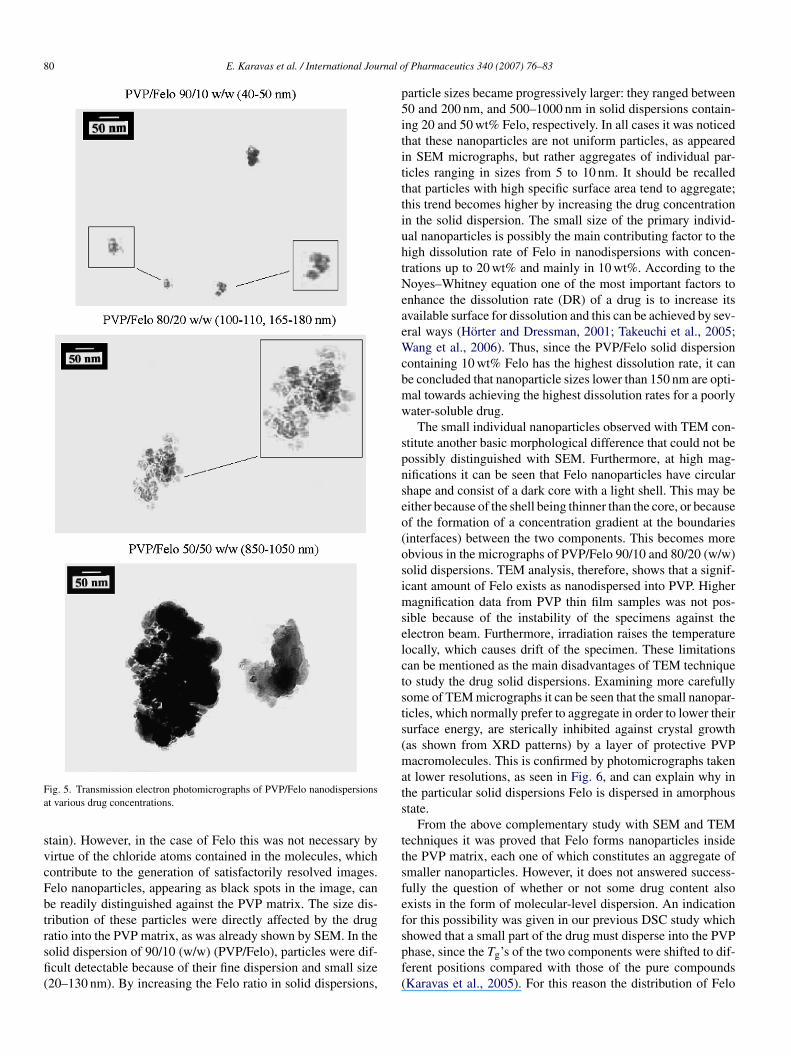
ig. 5. Transmission electron photomicrographs of PVP/Felo nanodispersionst various drug concentrations.
tain). However, in the case of Felo this was not necessary byirtue of the chloride atoms contained in the molecules, whichontribute to the generation of satisfactorily resolved images.elo nanoparticles, appearing as black spots in the image, cane readily distinguished against the PVP matrix. The size dis-ribution of these particles were directly affected by the drug
atio into the PVP matrix, as was already shown by SEM. In theolid dispersion of 90/10 (w/w) (PVP/Felo), particles were dif-cult detectable because of their fine dispersion and small size20–130 nm). By increasing the Felo ratio in solid dispersions,spf(
f Pharmaceutics 340 (2007) 76–83
article sizes became progressively larger: they ranged between0 and 200 nm, and 500–1000 nm in solid dispersions contain-ng 20 and 50 wt% Felo, respectively. In all cases it was noticedhat these nanoparticles are not uniform particles, as appearedn SEM micrographs, but rather aggregates of individual par-icles ranging in sizes from 5 to 10 nm. It should be recalledhat particles with high specific surface area tend to aggregate;his trend becomes higher by increasing the drug concentrationn the solid dispersion. The small size of the primary individ-al nanoparticles is possibly the main contributing factor to theigh dissolution rate of Felo in nanodispersions with concen-rations up to 20 wt% and mainly in 10 wt%. According to theoyes–Whitney equation one of the most important factors to
nhance the dissolution rate (DR) of a drug is to increase itsvailable surface for dissolution and this can be achieved by sev-ral ways (Horter and Dressman, 2001; Takeuchi et al., 2005;ang et al., 2006). Thus, since the PVP/Felo solid dispersion
ontaining 10 wt% Felo has the highest dissolution rate, it cane concluded that nanoparticle sizes lower than 150 nm are opti-al towards achieving the highest dissolution rates for a poorlyater-soluble drug.The small individual nanoparticles observed with TEM con-
titute another basic morphological difference that could not beossibly distinguished with SEM. Furthermore, at high mag-ifications it can be seen that Felo nanoparticles have circularhape and consist of a dark core with a light shell. This may beither because of the shell being thinner than the core, or becausef the formation of a concentration gradient at the boundariesinterfaces) between the two components. This becomes morebvious in the micrographs of PVP/Felo 90/10 and 80/20 (w/w)olid dispersions. TEM analysis, therefore, shows that a signif-cant amount of Felo exists as nanodispersed into PVP. Higher
agnification data from PVP thin film samples was not pos-ible because of the instability of the specimens against thelectron beam. Furthermore, irradiation raises the temperatureocally, which causes drift of the specimen. These limitationsan be mentioned as the main disadvantages of TEM techniqueo study the drug solid dispersions. Examining more carefullyome of TEM micrographs it can be seen that the small nanopar-icles, which normally prefer to aggregate in order to lower theirurface energy, are sterically inhibited against crystal growthas shown from XRD patterns) by a layer of protective PVPacromolecules. This is confirmed by photomicrographs taken
t lower resolutions, as seen in Fig. 6, and can explain why inhe particular solid dispersions Felo is dispersed in amorphoustate.
From the above complementary study with SEM and TEMechniques it was proved that Felo forms nanoparticles insidehe PVP matrix, each one of which constitutes an aggregate ofmaller nanoparticles. However, it does not answered success-ully the question of whether or not some drug content alsoxists in the form of molecular-level dispersion. An indicationor this possibility was given in our previous DSC study which
howed that a small part of the drug must disperse into the PVPhase, since the Tg’s of the two components were shifted to dif-erent positions compared with those of the pure compoundsKaravas et al., 2005). For this reason the distribution of Felo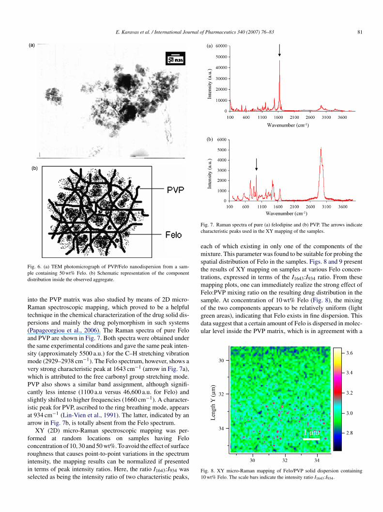
E. Karavas et al. / International Journal of Pharmaceutics 340 (2007) 76–83 81
Fpd
iRtp(atsmvwPcsiaa
fcriis
Fc
emsttmFsof the two components appears to be relatively uniform (lightgreen areas), indicating that Felo exists in fine dispersion. Thisdata suggest that a certain amount of Felo is dispersed in molec-ular level inside the PVP matrix, which is in agreement with a
ig. 6. (a) TEM photomicrograph of PVP/Felo nanodispersion from a sam-le containing 50 wt% Felo. (b) Schematic representation of the componentistribution inside the observed aggregate.
nto the PVP matrix was also studied by means of 2D micro-aman spectroscopic mapping, which proved to be a helpful
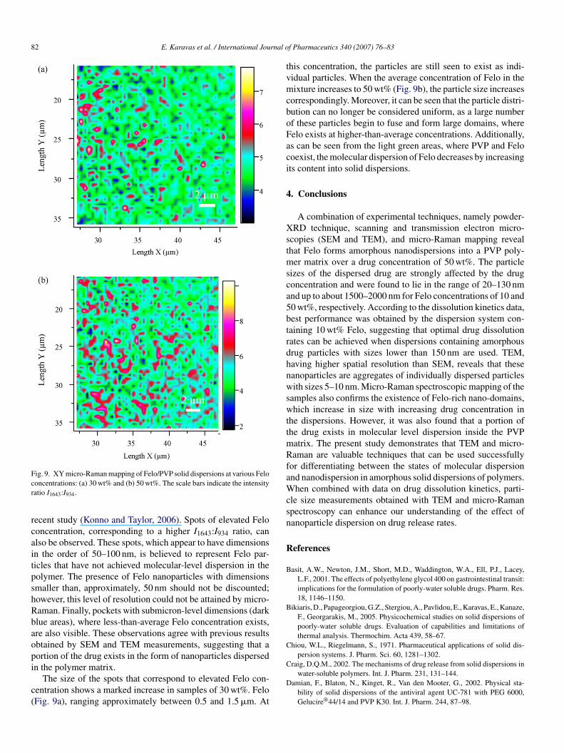
echnique in the chemical characterization of the drug solid dis-ersions and mainly the drug polymorphism in such systemsPapageorgiou et al., 2006). The Raman spectra of pure Felond PVP are shown in Fig. 7. Both spectra were obtained underhe same experimental conditions and gave the same peak inten-ity (approximately 5500 a.u.) for the C–H stretching vibrationode (2929–2938 cm−1). The Felo spectrum, however, shows a
ery strong characteristic peak at 1643 cm−1 (arrow in Fig. 7a),hich is attributed to the free carbonyl group stretching mode.VP also shows a similar band assignment, although signifi-antly less intense (1100 a.u versus 46,600 a.u. for Felo) andlightly shifted to higher frequencies (1660 cm−1). A character-stic peak for PVP, ascribed to the ring breathing mode, appearst 934 cm−1 (Lin-Vien et al., 1991). The latter, indicated by anrrow in Fig. 7b, is totally absent from the Felo spectrum.
XY (2D) micro-Raman spectroscopic mapping was per-ormed at random locations on samples having Felooncentration of 10, 30 and 50 wt%. To avoid the effect of surface
oughness that causes point-to-point variations in the spectrumntensity, the mapping results can be normalized if presentedn terms of peak intensity ratios. Here, the ratio I1643:I934 waselected as being the intensity ratio of two characteristic peaks,F1
ig. 7. Raman spectra of pure (a) felodipine and (b) PVP. The arrows indicateharacteristic peaks used in the XY mapping of the samples.
ach of which existing in only one of the components of theixture. This parameter was found to be suitable for probing the
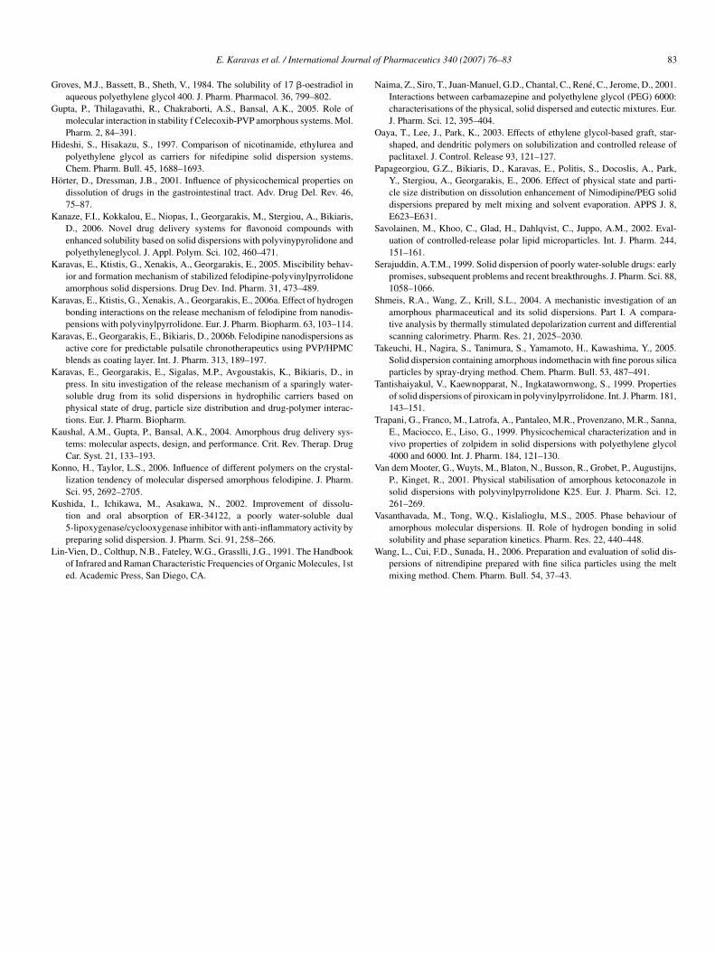
patial distribution of Felo in the samples. Figs. 8 and 9 presenthe results of XY mapping on samples at various Felo concen-rations, expressed in terms of the I1643:I934 ratio. From these
apping plots, one can immediately realize the strong effect ofelo:PVP mixing ratio on the resulting drug distribution in theample. At concentration of 10 wt% Felo (Fig. 8), the mixing
ig. 8. XY micro-Raman mapping of Felo/PVP solid dispersion containing0 wt% Felo. The scale bars indicate the intensity ratio I1643:I934.
82 E. Karavas et al. / International Journal o
Fcr
rcaitpshRbaopi
c(
tvmcboFaci
4
Xstmsca5btrdhnwswttmRfaWcsn
R
B
B
C
Craig, D.Q.M., 2002. The mechanisms of drug release from solid dispersions in
ig. 9. XY micro-Raman mapping of Felo/PVP solid dispersions at various Felooncentrations: (a) 30 wt% and (b) 50 wt%. The scale bars indicate the intensityatio I1643:I934.
ecent study (Konno and Taylor, 2006). Spots of elevated Felooncentration, corresponding to a higher I1643:I934 ratio, canlso be observed. These spots, which appear to have dimensionsn the order of 50–100 nm, is believed to represent Felo par-icles that have not achieved molecular-level dispersion in theolymer. The presence of Felo nanoparticles with dimensionsmaller than, approximately, 50 nm should not be discounted;owever, this level of resolution could not be attained by micro-aman. Finally, pockets with submicron-level dimensions (darklue areas), where less-than-average Felo concentration exists,re also visible. These observations agree with previous resultsbtained by SEM and TEM measurements, suggesting that aortion of the drug exists in the form of nanoparticles dispersed
n the polymer matrix.The size of the spots that correspond to elevated Felo con-entration shows a marked increase in samples of 30 wt%. FeloFig. 9a), ranging approximately between 0.5 and 1.5 �m. At
D
f Pharmaceutics 340 (2007) 76–83
his concentration, the particles are still seen to exist as indi-idual particles. When the average concentration of Felo in theixture increases to 50 wt% (Fig. 9b), the particle size increases
orrespondingly. Moreover, it can be seen that the particle distri-ution can no longer be considered uniform, as a large numberf these particles begin to fuse and form large domains, whereelo exists at higher-than-average concentrations. Additionally,s can be seen from the light green areas, where PVP and Felooexist, the molecular dispersion of Felo decreases by increasingts content into solid dispersions.
. Conclusions
A combination of experimental techniques, namely powder-RD technique, scanning and transmission electron micro-
copies (SEM and TEM), and micro-Raman mapping revealhat Felo forms amorphous nanodispersions into a PVP poly-
er matrix over a drug concentration of 50 wt%. The particleizes of the dispersed drug are strongly affected by the drugoncentration and were found to lie in the range of 20–130 nmnd up to about 1500–2000 nm for Felo concentrations of 10 and0 wt%, respectively. According to the dissolution kinetics data,est performance was obtained by the dispersion system con-aining 10 wt% Felo, suggesting that optimal drug dissolutionates can be achieved when dispersions containing amorphousrug particles with sizes lower than 150 nm are used. TEM,aving higher spatial resolution than SEM, reveals that theseanoparticles are aggregates of individually dispersed particlesith sizes 5–10 nm. Micro-Raman spectroscopic mapping of the
amples also confirms the existence of Felo-rich nano-domains,hich increase in size with increasing drug concentration in
he dispersions. However, it was also found that a portion ofhe drug exists in molecular level dispersion inside the PVP
atrix. The present study demonstrates that TEM and micro-aman are valuable techniques that can be used successfully
or differentiating between the states of molecular dispersionnd nanodispersion in amorphous solid dispersions of polymers.
hen combined with data on drug dissolution kinetics, parti-le size measurements obtained with TEM and micro-Ramanpectroscopy can enhance our understanding of the effect ofanoparticle dispersion on drug release rates.
eferences
asit, A.W., Newton, J.M., Short, M.D., Waddington, W.A., Ell, P.J., Lacey,L.F., 2001. The effects of polyethylene glycol 400 on gastrointestinal transit:implications for the formulation of poorly-water soluble drugs. Pharm. Res.18, 1146–1150.
ikiaris, D., Papageorgiou, G.Z., Stergiou, A., Pavlidou, E., Karavas, E., Kanaze,F., Georgarakis, M., 2005. Physicochemical studies on solid dispersions ofpoorly-water soluble drugs. Evaluation of capabilities and limitations ofthermal analysis. Thermochim. Acta 439, 58–67.
hiou, W.L., Riegelmann, S., 1971. Pharmaceutical applications of solid dis-persion systems. J. Pharm. Sci. 60, 1281–1302.
water-soluble polymers. Int. J. Pharm. 231, 131–144.amian, F., Blaton, N., Kinget, R., Van den Mooter, G., 2002. Physical sta-
bility of solid dispersions of the antiviral agent UC-781 with PEG 6000,Gelucire®44/14 and PVP K30. Int. J. Pharm. 244, 87–98.
rnal o
G
G
H
H
K
K
K
K
K
K
K
K
L
N
O
P
S
S
S
T
T
T
V
V
E. Karavas et al. / International Jou
roves, M.J., Bassett, B., Sheth, V., 1984. The solubility of 17 �-oestradiol inaqueous polyethylene glycol 400. J. Pharm. Pharmacol. 36, 799–802.
upta, P., Thilagavathi, R., Chakraborti, A.S., Bansal, A.K., 2005. Role ofmolecular interaction in stability f Celecoxib-PVP amorphous systems. Mol.Pharm. 2, 84–391.
ideshi, S., Hisakazu, S., 1997. Comparison of nicotinamide, ethylurea andpolyethylene glycol as carriers for nifedipine solid dispersion systems.Chem. Pharm. Bull. 45, 1688–1693.
orter, D., Dressman, J.B., 2001. Influence of physicochemical properties ondissolution of drugs in the gastrointestinal tract. Adv. Drug Del. Rev. 46,75–87.
anaze, F.I., Kokkalou, E., Niopas, I., Georgarakis, M., Stergiou, A., Bikiaris,D., 2006. Novel drug delivery systems for flavonoid compounds withenhanced solubility based on solid dispersions with polyvinypyrolidone andpolyethyleneglycol. J. Appl. Polym. Sci. 102, 460–471.
aravas, E., Ktistis, G., Xenakis, A., Georgarakis, E., 2005. Miscibility behav-ior and formation mechanism of stabilized felodipine-polyvinylpyrrolidoneamorphous solid dispersions. Drug Dev. Ind. Pharm. 31, 473–489.
aravas, E., Ktistis, G., Xenakis, A., Georgarakis, E., 2006a. Effect of hydrogenbonding interactions on the release mechanism of felodipine from nanodis-pensions with polyvinylpyrrolidone. Eur. J. Pharm. Biopharm. 63, 103–114.
aravas, E., Georgarakis, E., Bikiaris, D., 2006b. Felodipine nanodispersions asactive core for predictable pulsatile chronotherapeutics using PVP/HPMCblends as coating layer. Int. J. Pharm. 313, 189–197.
aravas, E., Georgarakis, E., Sigalas, M.P., Avgoustakis, K., Bikiaris, D., inpress. In situ investigation of the release mechanism of a sparingly water-soluble drug from its solid dispersions in hydrophilic carriers based onphysical state of drug, particle size distribution and drug-polymer interac-tions. Eur. J. Pharm. Biopharm.
aushal, A.M., Gupta, P., Bansal, A.K., 2004. Amorphous drug delivery sys-tems: molecular aspects, design, and performance. Crit. Rev. Therap. DrugCar. Syst. 21, 133–193.
onno, H., Taylor, L.S., 2006. Influence of different polymers on the crystal-lization tendency of molecular dispersed amorphous felodipine. J. Pharm.Sci. 95, 2692–2705.
ushida, I., Ichikawa, M., Asakawa, N., 2002. Improvement of dissolu-tion and oral absorption of ER-34122, a poorly water-soluble dual
5-lipoxygenase/cyclooxygenase inhibitor with anti-inflammatory activity bypreparing solid dispersion. J. Pharm. Sci. 91, 258–266.in-Vien, D., Colthup, N.B., Fateley, W.G., Grasslli, J.G., 1991. The Handbookof Infrared and Raman Characteristic Frequencies of Organic Molecules, 1sted. Academic Press, San Diego, CA.
W
f Pharmaceutics 340 (2007) 76–83 83
aima, Z., Siro, T., Juan-Manuel, G.D., Chantal, C., Rene, C., Jerome, D., 2001.Interactions between carbamazepine and polyethylene glycol (PEG) 6000:characterisations of the physical, solid dispersed and eutectic mixtures. Eur.J. Pharm. Sci. 12, 395–404.
aya, T., Lee, J., Park, K., 2003. Effects of ethylene glycol-based graft, star-shaped, and dendritic polymers on solubilization and controlled release ofpaclitaxel. J. Control. Release 93, 121–127.
apageorgiou, G.Z., Bikiaris, D., Karavas, E., Politis, S., Docoslis, A., Park,Y., Stergiou, A., Georgarakis, E., 2006. Effect of physical state and parti-cle size distribution on dissolution enhancement of Nimodipine/PEG soliddispersions prepared by melt mixing and solvent evaporation. APPS J. 8,E623–E631.
avolainen, M., Khoo, C., Glad, H., Dahlqvist, C., Juppo, A.M., 2002. Eval-uation of controlled-release polar lipid microparticles. Int. J. Pharm. 244,151–161.
erajuddin, A.T.M., 1999. Solid dispersion of poorly water-soluble drugs: earlypromises, subsequent problems and recent breakthroughs. J. Pharm. Sci. 88,1058–1066.
hmeis, R.A., Wang, Z., Krill, S.L., 2004. A mechanistic investigation of anamorphous pharmaceutical and its solid dispersions. Part I. A compara-tive analysis by thermally stimulated depolarization current and differentialscanning calorimetry. Pharm. Res. 21, 2025–2030.
akeuchi, H., Nagira, S., Tanimura, S., Yamamoto, H., Kawashima, Y., 2005.Solid dispersion containing amorphous indomethacin with fine porous silicaparticles by spray-drying method. Chem. Pharm. Bull. 53, 487–491.
antishaiyakul, V., Kaewnopparat, N., Ingkatawornwong, S., 1999. Propertiesof solid dispersions of piroxicam in polyvinylpyrrolidone. Int. J. Pharm. 181,143–151.
rapani, G., Franco, M., Latrofa, A., Pantaleo, M.R., Provenzano, M.R., Sanna,E., Maciocco, E., Liso, G., 1999. Physicochemical characterization and invivo properties of zolpidem in solid dispersions with polyethylene glycol4000 and 6000. Int. J. Pharm. 184, 121–130.
an dem Mooter, G., Wuyts, M., Blaton, N., Busson, R., Grobet, P., Augustijns,P., Kinget, R., 2001. Physical stabilisation of amorphous ketoconazole insolid dispersions with polyvinylpyrrolidone K25. Eur. J. Pharm. Sci. 12,261–269.
asanthavada, M., Tong, W.Q., Kislalioglu, M.S., 2005. Phase behaviour of
amorphous molecular dispersions. II. Role of hydrogen bonding in solidsolubility and phase separation kinetics. Pharm. Res. 22, 440–448.ang, L., Cui, F.D., Sunada, H., 2006. Preparation and evaluation of solid dis-persions of nitrendipine prepared with fine silica particles using the meltmixing method. Chem. Pharm. Bull. 54, 37–43.
Related Documents


