Antonie van Leeuwenhoek 54:509-520 (1988) t~) Kluwer Academic Publishers, Dordrecht - Printed in the Netherlands Columnaris infection among cultured Nile tilapia Oreochromis niloticus N.E. AMIN 1, I.S. ABDALLAH 2, M. FAISAL 3., M. EL-S. EASA 4, T. ALAWAY 2 & S.A. ALYAN 2 Faculty of Veterinary Medicine, i University of Zagazig; 2 University of Assiut; 3 University of Alexandria(present address: Dept. of Microbiology and lmmunology (A7-078), UCLA -Schoolof Medicine, Los Angeles, CA 90024, USA); 4 University of Cairo, Egypt (* requests for offprints) Accepted in revised form 14 March 1988 Key words: Columnaris disease, Flexibacter columnaris, tilapia Abstract. Flexibacter columnar& was isolated from 13 cultured Oreochrom& niloticus showing respiratory disorders. The isolates developed typical swarming rhizoid colonies on Cytophaga agar medium. Antibiotic sensitivity test revealed the susceptibility of F. columnaris isolated to oxytetracycline, chloramphenicol and erythromycin. A marked difference in the pathogenicity of seven tested isolates was observed: two were highly virulent, one was moderately virulent and four were avirulent. No experimental infection could be induced with the highly virulent isolates except after injuring one of the natural barriers of the fish body. The severity of the disease and the increased median death time shortened by keeping infected fishes with injured gills in water containing ammonia. In naturally infected O. niloticus, the disease became chronic as indicated by the presence of excessive proliferative and necrotic changes. On the other hand, severe dilatation of branchial blood vessel, oedema and round cell infiltration proved that, the disease among experimentally infected tilapias was acute. Introduction Pathological gill changes have a detrimental effect on the health of fish and threaten their survival. Bacteria belonging to the genus Flexibacter have been incriminated by various authors as the main cause of gill affections among cultured fish allover the world (Popp 1980; Richards et al. 1985). Snieszko (1981) differentiated between columnaris disease caused by Flexibacter colum- naris and bacterial gill disease caused by other flexibacteria. The former is characterized by gill necrosis while proliferation of gill epithelium is usually associated with the latter. The reports of Avault et al. (1968); Balarin & Hatton (1979) and Roberts & Sommerville (1982) on the presence of filamentous bacteria in cultured tilapia suffering from gill affections urged us to investigate the role of Flexibacter spp.

Welcome message from author
This document is posted to help you gain knowledge. Please leave a comment to let me know what you think about it! Share it to your friends and learn new things together.
Transcript
Antonie van Leeuwenhoek 54:509-520 (1988) t~) Kluwer Academic Publishers, Dordrecht - Printed in the Netherlands
Columnaris infection among cultured Nile tilapia Oreochromis niloticus
N . E . A M I N 1, I .S . A B D A L L A H 2, M. F A I S A L 3., M. EL-S . E A S A 4, T. A L A W A Y 2 & S .A . A L Y A N 2 Faculty of Veterinary Medicine, i University of Zagazig; 2 University of Assiut; 3 University of Alexandria(present address: Dept. of Microbiology and lmmunology (A7-078), UCLA -Schoolof Medicine, Los Angeles, CA 90024, USA); 4 University of Cairo, Egypt (* requests for offprints)
Accepted in revised form 14 March 1988
Key words: Columnaris disease, Flexibacter columnaris, tilapia
Abstract. Flexibacter columnar& was isolated from 13 cultured Oreochrom& niloticus showing respiratory disorders. The isolates developed typical swarming rhizoid colonies on Cytophaga agar medium. Antibiotic sensitivity test revealed the susceptibility of F. columnaris isolated to oxytetracycline, chloramphenicol and erythromycin. A marked difference in the pathogenicity of seven tested isolates was observed: two were highly virulent, one was moderately virulent and four were avirulent. No experimental infection could be induced with the highly virulent isolates except after injuring one of the natural barriers of the fish body. The severity of the disease and the increased median death time shortened by keeping infected fishes with injured gills in water containing ammonia.
In naturally infected O. niloticus, the disease became chronic as indicated by the presence of excessive proliferative and necrotic changes. On the other hand, severe dilatation of branchial blood vessel, oedema and round cell infiltration proved that, the disease among experimentally infected tilapias was acute.
Introduct ion
Pa tho log i ca l gill changes have a d e t r i m e n t a l effect on the hea l th of fish and
t h r e a t e n the i r survival . B a c t e r i a be long ing to the genus Flexibacter have been
i n c r i m i n a t e d by va r ious au tho r s as the ma in cause of gill a f fec t ions a m o n g
c u l t u r e d fish a l love r the wor ld ( P o p p 1980; R i c h a r d s et al. 1985). Sn ieszko
(1981) d i f f e r e n t i a t e d b e t w e e n co lumna r i s d i sease caused by Flexibacter co lum-
naris and bac t e r i a l gill d i sease caused by o t h e r f l ex ibac te r ia . The f o r m e r is
c h a r a c t e r i z e d by gill necros i s whi le p ro l i f e r a t i on of gill ep i t he l i um is usual ly a s soc i a t ed with the la t te r .
T h e r epo r t s o f A v a u l t e t al. (1968); Ba la r in & H a t t o n (1979) and R o b e r t s &
S o m m e r v i l l e (1982) on the p r e s e n c e of f i l amen tous bac t e r i a in cu l tu red t i l ap ia
suffer ing f rom gill a f fec t ions u rged us to inves t iga te the role of Flexibacter spp.
510
in causing losses among the most popular fish Oreochromis niloticus in Egypt.
Materials and methods
Fish Both gills of 416 one year old cultured Nile tilapia Oreochromis niloticus were examined clinically and parasitologically throughout the course of this study. The fish were obtained from EI-Serow fish farm, Menia, Upper Egypt which has a history of repeated mortalities associated with respiratory disorders.
Bacteriological examination A total of 165 gill smears from fish suffering from respiratory disorders were streaked on Cytophaga agar medium (Anacker & Ordal 1959), that was composed of tryptone 0.05%, yeast extract 0.05%; beef extract 0.02%; sodi- um acetate 0.02% and Agar (Difco) 1.1% and adjusted to pH 7.2-7.4. In case of primary isolation from affected gill tissue or reisolation from gills and internal organs of freshly dead fish, neomycin and polymyxin B were added to a final concentration of 5/zg/l as recommended by Fij an (1969). The suspected colonies (swarming, rhizoid) were picked and inoculated into different solid media like, Nutrient, 5% sheep blood, MacConkey and SS agar (Difco). Incubation was tried at 3 different temperatures; 10, 22 and 37 ~ C.
Colonial, morphological (in Gram stained preparations) as well as some biochemical characteristics were studied on purified isolates according to the methods described by Cruickshank et al. (1975).
To perform the antibiogram test, 7 isolates were cultured for 5 h at 22 ~ C in Cytophaga broth, adjusted to a turbidity that is equivalent to 0.5 McFarland (1) standard. The test was done in sensitivity agar (Difco) using standard disk stars (Oxoid) of 10 antibiotics as described by Treagan and Pulliam (1982). The results were interpreted according to Acar & Goldstein (1986).
Experimental infection This was carried out on groups (3 each) of O. niloticus fingerlings (12 cm + 1). No Flexibacter columnaris could be isolated from the gills or internal organs (kidney, liver, spleen and'heart) of the experimental fish. Purified colonies of 7 isolates were subcultured on Cytophaga broth for 24 h at 22 ~ C, adjusted to the desired bacterial count as predetermined by the optical density at 540 nm wave length with Bauschal and Lomb spectronic 20. To confirm the applied bacte- rial number, contaminated water or inoculum were titrated on colony count agar (Difco).
Fish infection was performed as follows:
511
Group A. Fish were bathed for one hour in 101 water containing 2 x 10³ living bacterial cells/ml of isolates 1-7. The aquarium was filled gradually with additional water up to the desired volume.
Group B. Installation of 2 • 10 4 living bacterial cell of isolates 1-7/ fish suspended in 1 ml saline on scarified gills of one side. Following installation, the fish were held in a horizontal position for 2 min.
Group C. Fish were injected intramuscularly i cm below the dorsal fin with 2.16 • 10 4 living bacterial cells/fish of isolates 3 & 7 suspended in 1 ml saline.
Group D. This group was treated similarly to group B. However, the fish were kept after installation in water containing 168 mg NH4CI/1 so as to resemble the effect of accumulation of organic matter according to the method de- scribed by Waiters & Plumb (1980) and modified by Soliman (1984).
Following infection, fish groups were kept in 2001 glass aquaria (1 group/ aquarium) supplied with aerated autoclaved water, that was changed every three days, over an observation period of 2 weeks.
Reisolation of the inoculated bacteria from the gills and organs (kidney, liver and spleen) of experimentally infected tilapia was conducted on Cytopha- ga agar media supplemented with antibiotics.
The median death time (MDT) was calculated as suggested by Sch~iperclaus (1978) according to the formula:
MDT in h =
(No. of fish (No. of fish died a tXh) . X + died a t Y h )
Total number of dead fish
. y
Histopathological technique Gills of naturally and experimentaly infected fish were paraffinized, sectioned (5-7 tz), stained with haematoxylin and eosin (H&E) and examined micro- scopically for histopathological alterations.
Results
Natural infection Out of 416 fish examined, 165 (39.66%) animals were suffering from respira- tory disorders. The highest incidence was recorded during summer (47%) and autumn (43.2%) during which 13 bacterial isolates were recovered from
512
diseased fish on cytophaga agar (Table 1). The diseased fish swam near the water surface at its inlet with rapid opercular movements and abnormal swimming movements like whirling and jumping. The gill filaments were covered with a thick film of turbid mucous.
Bac ter io log ica l ident i f ica t ion
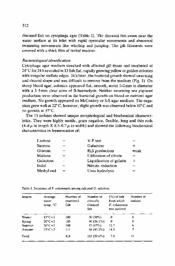
Cytophaga agar medium streaked with affected gill tissue and incubated at 28 ~ C for 24 h revealed in 13 fish flat, rapidly growing yellow or golden colonies
with irregular stellate edges. 24 h later, the bacterial growth showed swarming and rhizoid shape and was difficult to remove from the medium (Fig. 1). On sheep blood agar, colonies appeared flat, smooth, moist 1-2 mm in diameter with a 3-5 mm clear zone of B-haemolysis. Neither swarming nor pigment production were observed in the bacterial growth on blood or nutrient agar medium. No growth appeared on McConkey or S-S agar medium. The orga- nism grew well at 22 ~ C, however, slight growth was observed below 10 ~ C and no growth at 37 ~ C.
The 13 isolates showed unique morphological and biochemical character- istics. They were highly motile, gram negative, flexible, long and thin rods (4-8/z in length X 0.5-0.7/z in width) and showed the following biochemical characteristics in fermentation of:
Lactose - V-P test - Sucrose - Galactose +
Glucose - H2S production weak Maltose - Utilization of citrate - Galactose - Liquification of gelatin +
Indol - Nitrate reduction - Methyl red - Urea hydrolysis -
Table I. Incidence of F. columnaris among cultured O. niloticus.
Season Average Number of Number of (%) of fish Number of water examined clinically from which isolates temp. ~ C fish diseased F. columnaris
fish was isolated
Winter Spring Summer Autumn
Total
15~ 100 30 (30%) 0 0 20~ C+2 105 40 (38.1%) 0 0 26 ~ C+2 100 47 (47%) 12.7 6 23~ C+2 111 48 (43.2%) 14.5 7
416 165 (39.6%) 7.8 13
513
Fig. 1. F. colurnnaris on cytophaga agar medium, notice: bacterial growth showed swarming and rhizoid shape.
The antibiotic sensitivity of obtained isolates revealed that the seven tested isolates reacted in somewhat different manner to the used antibiotic discs. Thus, almost all strains were highly sensitive to Oxytetracycline, chloram- phenicol and erythromycin while were moderately sensitive to streptomycin (with the exception of isolates 3 and 7). On the other hand, they were all resistant to baciteracin. The isolates were also less sensitive to colistin sul- phate, mandelamine, novobiocin, vancomycin and ampicillin (Table 2).
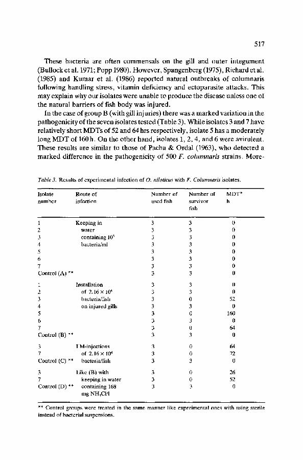
Results of experimental infection Experimental infection failed to occur without damaging the fish natural barrier (group A). Isolates 3, 5 and 7 induced mortality of all infected fish but varied in their MDT; The MDT for isolates 3 and 7 was prolonged by in- tramuscular injection and shortened by keeping the infected fish in water containing 168 mg NH4CI/1 (Table 3). On cytophaga agar the organism could be reisolated from the internal organs and gill smear of fish succumbed to experimental infection.
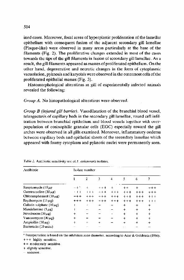
Results of histopathological examination Histological examination of stained gill sections of naturally infected fish revealed dilatation of the blood vessels in gill arches, primary gill filaments and capillary beds within the secondary gill lamellae (Telangectasis) in all exam-
514
ined cases. M o r e o v e r , focal a reas o f hype rp l a s t i c p ro l i f e r a t i on of the l ame l l a r
ep i t he l i um with c o n s e q u e n t fusion of the a d j a c e n t s e c o n d a r y gill l ame l l ae
(P l aque - l i ke ) we re o b s e r v e d in m a n y a reas pa r t i cu la r ly at the base of the
f i l aments (Fig. 2). The p ro l i f e ra t ive changes e x t e n d e d in mos t o f the cases
t owards the t ips of the gill f i l aments in fusion of s e c o n d a r y gill l amel lae . A s a
resul t , the gill f i l aments a p p e a r e d as masses of p ro l i f e r a t ed ep i the l ium. O n the
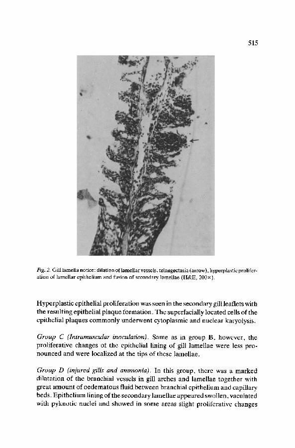
o t h e r h a n d , d e g e n e r a t i v e and nec ro t i c changes in the fo rm of cy top lasmic
vacuo la t i on , pyknos i s and karyols i s were o b s e r v e d in the o u t e r m o s t cells of the
p r o l i f e r a t e d ep i the l i a l masses (Fig. 3).
H i s t o m o r p h o l o g i c a l a l t e ra t ions in gill o f e x p e r i m e n t a l l y in fec ted an imals
r e v e a l e d the fo l lowing:
Group A. N o h i s topa tho log i ca l a l t e ra t ions were obse rved .
Group B (injured gill barrier). V a s o d i l a t a t i o n of the b ranch ia l b l o o d vessel ,
t e langec tas i s of cap i l l a ry beds in the s e c o n d a r y gill l ame l l ae , r o u n d cell infil-
t r a t i on b e t w e e n b ranch ia l ep i t he l i um and b l o o d vessels t o g e t h e r with over -
p o p u l a t i o n of eos inoph i l i c g r anu la r cells ( E G C ) espec ia l ly t o w a r d the gill
a rches were o b s e r v e d in all gills e x a m i n e d . M o r e o v e r , i n f l a m m a t o r y o e d e m a
b e t w e e n cap i l l a ry beds and ep i the l i a l shee ts of the s e c o n d a r y l ame l l ae which
a p p e a r e d with f o a m y c y t o p l a s m and pykno t i c nucle i were p e r m a n e n t l y seen.
Table 2. Antibiotic sensitivity test of F. columnaris isolates.
Antibiotic Isolate number
1 2 3 4 5 6 7
Streptomycin (10/xg ++* + + + + + ++ + + + + Oxtetracycline (30/~g) + + + + + + + + + + + + + + + + + + + + + Chloramphenicol (30/xg) + + + + + + + + + + + + + + + + + + + + + Erythomycin (15/zg) + + + + + + + + + + + + + + + + + + + + + Colistin sulphate (10/zg) + + + + + + + Mandelamine (5 p.g) + + + + + + + Novobiocin (30 ttg) + + + + + + + Vanocomycin (30/zg) + + + + + + + Ampicillin (10/~g) + + + + + + + Bacteracin (10 units) . . . . . . .
* Interpretation is based on the inhibition zone diameter, according to Acar & + + + highly sensitive. ++ moderately sensitive. + slightly sensitive. - resistant.
Goldstein (1986).
515
Fig. 2. Gill lamella notice: dilation of lamellar vessels, telangectasia (arrow), hyperplastic prolifer- ation of lamellar epithelium and fusion of secondary lamellae (H&E, 200 x).
Hyperplastic epithelial proliferation was seen in the secondary gill leaflets with the resulting epithelial plaque formation. The superfacially located cells of the epithelial plaques commonly underwent cytoplasmic and nuclear karyolysis.
Group C (Intramuscular inoculation). Same as in group B, however, the proliferative changes of the epithelial lining of gill lamellae were less pro- nounced and were localized at the tips of these lamellae.
Group D (injured gills and ammonia). In this group, there was a marked dilatation of the branchial vessels in gill arches and lamellae together with great amount of oedematous fluid between branchial epithelium and capillary beds. Epithelium lining of the secondary lamellae appeared swollen, vaculated with pyknotic nuclei and showed in some areas slight proliferative changes
516
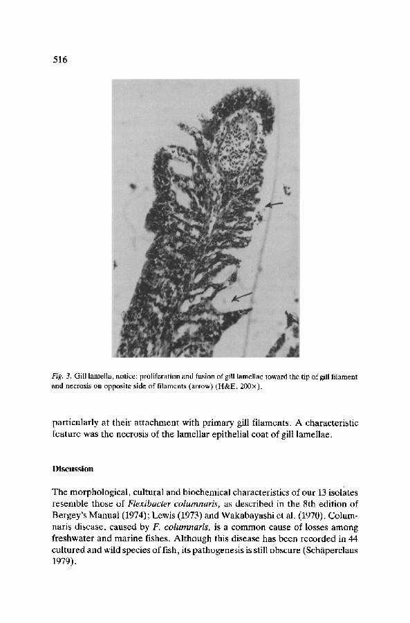
Fig. 3. Gill lamella, notice: proliferation and fusion of gill lamellae toward the tip of gill filament and necrosis on opposite side of filaments (arrow) (H&E, 200x).
particularly at their attachment with primary gill filaments. A characteristic feature was the necrosis of the lamellar epithelial coat of gill lamellae.
Discussion
The morphological, cultural and biochemical characteristics of our 13 isolates resemble those of Flexibacter colurnnaris, as described in the 8th edition of Bergey's Manual (1974); Lewis (1973) and Wakabayashi et al. (1970). Colum- naris disease, caused by F. columnaris, is a common cause of losses among freshwater and marine fishes. Although this disease has been recorded in 44 cultured and wild species of fish, its pathogenesis is still obscure (Sch/iperclaus 1979).
517
These bacter ia are of ten commensa l s on the gill and ou te r i n t e g u m e n t
(Bul lock et al. 1971; Popp 1980). However , Spangenbe rg (1975), Richard et al.
(1985) and K u m a r et al. (1986) repor ted na tu ra l ou tb reaks of co lumnar i s
fol lowing hand l ing stress, v i t amin deficiency and ectoparasi te attacks. This
may expla in why our isolates were unab le to produce the disease unless one of
the na tu ra l barr iers of fish body was in jured .
In the case of group B (with gill in jur ies) there was a marked var ia t ion in the
pa thogenic i ty of the seven isolates tes ted (Table 3). Whi le isolates 3 and 7 have
relat ively short M D T s of 52 and 64 hrs respectively, isolate 5 has a modera te ly
long M D T of 160h. O n the o ther hand , isolates 1, 2, 4, and 6 were avirulent .
These results are similar to those of Pacha & Orda l (1963), who detec ted a
m a r k e d difference in the pa thogenic i ty of 500 F. c o l u m n a r i s strains. More-
Table 3. Results of experimental infection of O. niloticus with F. Columnaris isolates.
Isolate Route of Number of Number of MDT* number infection used f i s h survivor h
fish
1 Keeping in 3 3 0 2 water 3 3 0 3 containing 103 3 3 0 4 bacteria/ml 3 3 0 5 3 3 0 6 3 3 0 7 3 3 0 Control (A) ** 3 3 0
1 Installation 3 3 0 2 of 2.16 x 10⁴ 3 3 0 3 bacteria/fish 3 0 52 4 on injured gills 3 3 0 5 3 0 160 6 3 3 0 7 3 0 64 Control (B) ** 3 3 0
3 I M-injections 3 0 64 7 of 2.16 x 10⁴ 3 0 72 Control (C) ** bacteria/fish 3 3 0
3 Like (B) with 3 0 26 7 keeping in water 3 0 52 Control (D) ** containing 168 3 3 0
mg NH4CI/1
** Control groups were treated in the same manner like experimental ones with using sterile instead of bacterial suspensions.
518
over, serological comparisons indicated differences between pathogenic and non-pathogenic F. columnaris strains (Pacha & Porter 1968). Some authors considered a strain of F. columnaris to be highly virulent when it can cause the death of infected salmon within 24 h (Buxton & Fraserm 1977) or 48 h (Amend 1970), while low virulence is indicated by the death after several days. Simi- larly, on the basis of MDT isolate 3 (MDT = 52 h) is considered the most virulent of those tested.
The gill pathology, death of the fishes and reisolation of F. columnaris from the internal organs following intramuscular inoculation proved, that the na- ture of this disease in O. niloticus is systemic. However, the longer MDT of group C (intramuscular injection) than B (injured gills) after infection with isolates 3 and 7 proves the importance of the gills in the establishment and development of columnaris infection. It is therefore worthy of mention, that Flexibacter spp. were reported to have a tropism for ectodermal tissues with obscure chemical basis (Munro 1982). Moreover, in acute cases of columnaris disease, the gills are the only organ with gross lesions (Snieszko & Bullock 1976).
In naturally infected O. niloticus, the disease became chronic, as indicated by the occurrence of excessive proliferative and necrotic changes. On the other hand, the severe dilation of the branchial blood vessels, oedema, and round cell infiltration indicated that the disease among experimentally infected tila- pias was acute. The dilatation of branchial blood vessel and the hyperplasia of lamellar epithelium in naturally as well as in experimentally infected fishes were similar to those observed by Fish & Rucker (1943), Pacha & Ordal (1967) and Wakabayashi et al. (1970). The proliferative changes of the lamellar epithelium were distributed randomly and did not start at the distal ends of the filaments, as described by Davis (1952). The observed necrosis of the epithelial plaques distinguishes columnaris infection from bacterial gill disease (BGD), which is characterized by proliferative changes only (Snieszko 1981). The Proliferative epithelial plaques on the gill lamellae and necrosis of the gill tissue affected a great part of the respiratory surfaces, which could lead to impairment of gas exchange and the secretory and excretory functions of the gills with consequent moralities. This may explain the respiratory difficulties among our infected O. niloticus.
The MDT was shortened markedly when the infected fishes with injured gills were kept in water containing ammonia (Table 3). This may be attributed partially to the extensive dilation of branchial vessels with the resulting extra- vasation of blood plasma and osmotic disbalance and partially to the stress induced by the chemical changes in the surrounding environment (Plumb & Waiters 1980). Keeping tilapias with injured gills in water with high ammonia concentration may resemble what is happening in tilapia ponds during summer and autumn in Egypt (Soliman 1984). Tilapia ponds are stocked in the early
519
spring and harvested during the autumn, when the biomass is maximum. Meanwhile parasitological examinations of cultured tilapia in the same study indicated high incidence of infection with monogenetic trematodes (Cichlid- ogyrus spp.), which reached a peak of 48 percent during the same period (Alyan et al. 1985). Infection of tilapia with Cichlidogyrus spp. was reported to cause severe injury to the branchial blood vessels, allowing the entry of fish pathogens (Faisal et al. 1984, 1985).
The antibiogram of our isolates revealed their sensitivity to erythromycin, oxytetracycline, and chloramphenicol. This should be considered when choos- ing a therapy against columnaris infection in order to avoid the development of antibiotic resistant strains. It was also interesting to observe the resistance of tested isolates to the antibiotic bacitracin. However, its use as a purifying agent in the isolation media requires further study.
Our results highlight the implication of Flexibacteria in causing losses among cultured O. niloticus. Although the percent of F. columnaris at this time is considered relatively low (7.8%), the virulent isolates might represent a potential hazard when the fish are stressed. Further studies are needed on the epizootology of columnaris infection in the Egyptian fishes and the role of the environmental factors in the establishment of infection and determination of the disease course.
References
Acar JF & Goldstein FW (1986) Disk susceptibility test. In: Lorian V (Ed) Antibiotic in Lab- oratory Medicine. Williams & Wilkins, Baltimore
Alyan SA, Amin NE, Abdallah IS, Easa M El-S, Imam EA, Rizk MH & Alawy T (1985) Monogenetic trematode from gills of Sarotherodon niloticus in Upper Egypt. J. Egypt. Vet. Med. Assoc. 45:117-126
Amend DF (1970) Myxobacterial infections of salmonids: prevention and treatment. In: Symposi- um of Diseases of fish and shellfish. Spec. Publ. Am. Fish. Soc. 5:258-265
Anacker RL & Ordal EJ (1959) Studies of the myxobacterium Chondrococcus columnaris. I. Serological typing. J. Bacteriol. 78:25-32
Avault JW, Shell EW & Smitherman RO (1968) Procedures for overwintering tilapia. FAO Fish. Rep. 44:343--345
Balarin JD & Hatton Y (1979) Tilapia: A guide to their biology and culture in Africa (pp 1-79) University of Stirling Press
Bergey's manual of determinative bacteriology. 8th ed. Williams & Wilkins Co., Baltimore Bullock GL, Conroy DA & Snieszko SF (1971) Bacterial diseases of fishes. Book 2A. In: Sniesako
SF & Axelrod HR (Eds) Diseases of Fish (p 151) T.F.H. Publications, Inc. Neptune City, New Jersey
Buxton A & Fraser M (1977) Animal Microbiology (pp 327-336) Oxford London-Edinburgh- Melbourne
Cruickshank R, Duguid JP, Marmion BP & Swain RH (1975) Textbook of Medical Microbiology. 12th edn. Churchill Livingstone, Edinburgh-London-New York
520
Davis HS (1953) Culture and disease of game fishes. Berkeley, University of California Press Faisal M, Abd Elhamid H, Torky HA, Soliman MK & Avu Elwafaa N (1984) Distribution of
Aeromonas hydrophila in organs and blood of naturally and experimentaly infected Aeromonas hydrophila in organs and blood of naturally and experimently infected Oreochrmomis niloticus. J. Egypt. Vet. Med. Assoc. 44:11-20
Faisal M, Ashmawy KI & Rizk MH (1985) A contribution on Dactylogyrosis among some freshwater fishes in Egypt. J. Egypt. Vet. Med. Assoc. 45:95-105
Fijan N (1969) Antibiotic additive for the isolation of Chondrococcus columnaris from fish. Appl. Microbiol. 17:333-339
Fish FF & Rucker RR (1943) Columnaris as a disease of cold water fishes. Trans. Amer. Fish. Soc. 73:32
Kumar D, Suresh K, Dey RK & Mishra BK (1986) Stress mediated columnaris disease in rohu, Labeo rohita (Hamiton). J. Fish. Dis. 9:87-89
Lewis DH (1973) Predominant Aerobic Bacteria of Fish and Shellfish. Texas A and M University, p 102
Munro ALS (1982) The pathogenesis of bacterial diseases of fishes. In: Roberts RJ (Ed) Microbial Diseases of Fish (pp 131-141) Academic Press, London
Pacha RE & Ordal EJ (1963) Epidemiology of columnaris disease in salmon. Bacteriol. Proc. 63:3 Pacha RE & Ordal EJ (1967) Histopathology of experimental columnaris in young salmon. J.
Comp. Path. 77:419--423 Pacha RE & Porter S (1968) Characteristics if isolated myxobacteria from the surface of fresh
water fish. App. Microbiol. 16:1901-1906 Popp W (1980) Bakterien als Erreger infekti6ser Fishkrankheiten. In: Reichenbach-Kilnke H-H
(Ed) Krankheiten und Sch~idigungen der Fische (pp 106-134) Gustav Fischer Verlag. Stuttgart- New York
Richards RH, Roberts RJ & Schiotfeldt HJ (1985) Bakterielle Erkrankungen der Knochenfische. In: Roberts RJ & Schotfeldt HJ (Eds) (pp 174-220) Grundlagen der Fischpathologie. Verlag Paul Parey-Berlin-Hamburg
Roberts RJ & Sommerville LE (1982) Diseases of Tilapia. In: Pullin RSV & McConnel L (Eds) The Biology and Culture of Tilapias (pp 247-263) ICLARM Conference Proceedings 7. International Center for Living Aquatic Resources Management, Manila, Philippines
Sch~iperclaus (1979) Fischkrankheiten. 4th edn. Akademie Verlag, Berlin Sniesko SF (1981) Bacterial gill disease of freshwater fishes. Fish Wildl. Serv., Fish Disease
Leaflet 62: pp 11 Sniesko SF & Bullock GL (1976) Columnaris disease of fishes (p 10) U.S. Fish Wildl. Serv., Fish
Diseases Leaflet 45 Soliman MK (1984) Some ecological factors and their relations to the development and elim-
ination of Aeromonosis in fish. Master of Vet. Sci., University of Alexandria. Spangenberg R (1975) Orientierende Untersuchungen fiber das Vorkommen von Myxobakterien
bei der Kiemennekrose des Karpfens. Z. Binnenfisch. DDR 22:121-127 Treagan L & Pulliam L (1982) Medical Microbiology Procedures. W.B. Saunders Company,
Philadelphia, pp 235 Wakabayashi H, Kira K & Egusa S (1970) Studies on columnaris disease of pond-cultured eels. II.
The relation between gill disease and Chondrococcus columnaris. Bull. Jap. Soc. Fish. 36: 147-155
Waiters WR & Plumb JA (1980) Environmental stress and bacterial infection in channel catfish. Ictalurus punctatus Rafinesque. J. Fish. Biol. 17:177-185
Related Documents