Januar 2015 Collodion baby SWISS SOCIETY OF NEONATOLOGY

Welcome message from author
This document is posted to help you gain knowledge. Please leave a comment to let me know what you think about it! Share it to your friends and learn new things together.
Transcript
Januar 2015
Collodion baby
SWISS SOCIETY OF NEONATOLOGY
Soroken C, Scerba F, Calza AM, Karam O, Neonatal Inten-
sive Care Unit, University Hospitals of Geneva, Switzerland
Title figure: Wikipedia
© Swiss Society of Neonatology, Thomas M Berger, Webmaster
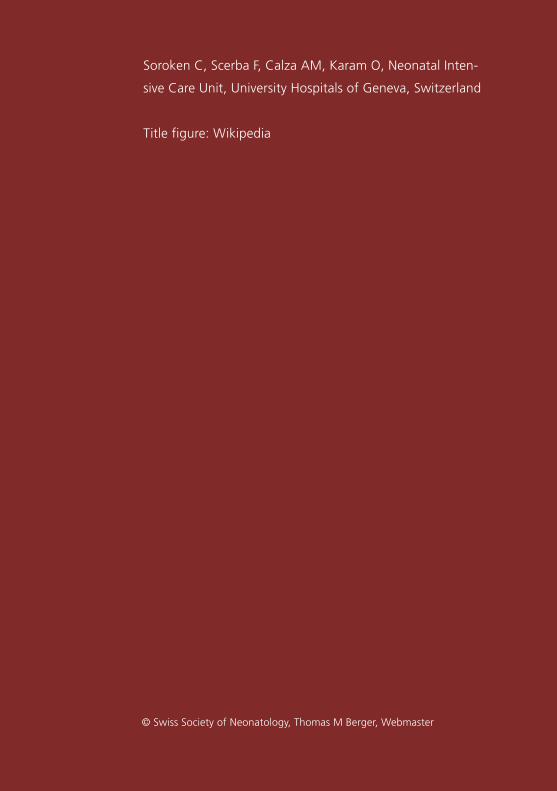
Collodion baby (CB) is rare, estimated to occur once in
50'000 to 100'000 deliveries (1). Less than 300 cases
of collodion babies (CB) have been reported in the
literature. Here, we present a CB with favorable out-
come.
INTRODUCTION
CASE REPORT
3
This female patient was born at 37 weeks of gestation
via vaginal delivery after an uneventful pregnancy.
She was the first child of the family, and the parents
were not consanguineous. The mother had a medical
history of skin disorders: desquamative erythrodermia
in infancy and Lyell syndrome secondary to the admi-
nistration of various medications (non-steroidal anti-
inflammatory drugs, myorelaxants).
Postnatal adaptation was excellent with Apgar scores
of 9, 10, 10 at 1, 5 and 10 minutes, respectively. The
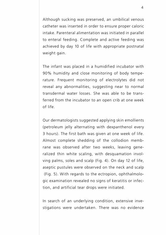
first impression was that the newborn was covered
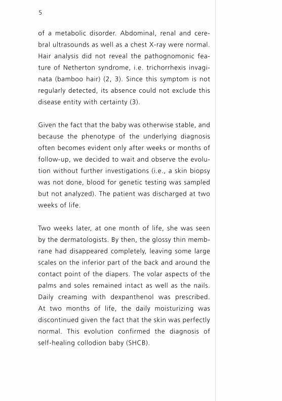
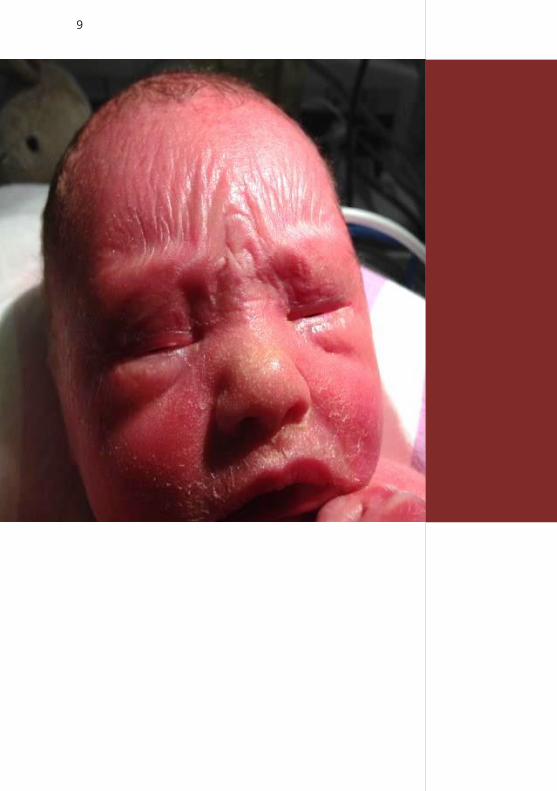
by a thin parchment-like glossy skin (Fig. 1). The face
showed discrete bilateral ectropion, absence of eye
brows and small eyelids (Fig. 2). In addition, there was
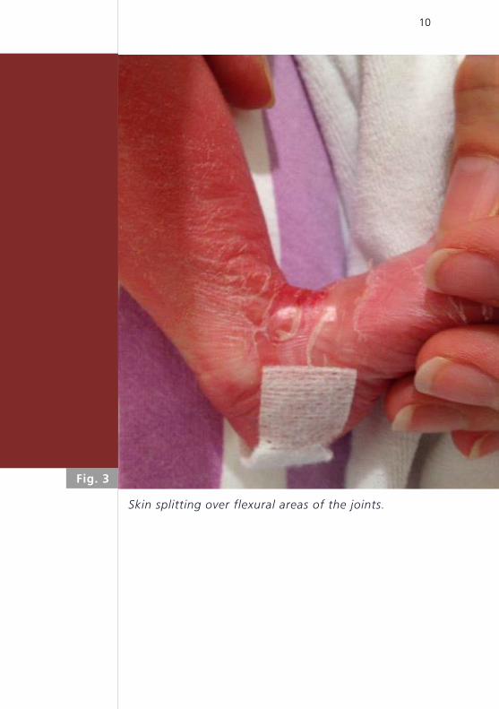
sparse hair, discrete eclabion, wrinkled ears, and skin
splitting localized in flexural areas (Fig. 3). Mobility of
all joints was preserved. There were no extracutaneous
symptoms except for intrauterine growth restriction:
birth weight was 2320 g (P 5 – 10), head circumference
33 cm (P 10 – 25), length 43 cm (< P3).
4
Although sucking was preserved, an umbilical venous
catheter was inserted in order to ensure proper caloric
intake. Parenteral alimentation was initiated in parallel
to enteral feeding. Complete and active feeding was
achieved by day 10 of life with appropriate postnatal
weight gain.
The infant was placed in a humidified incubator with
90% humidity and close monitoring of body tempe-
rature. Frequent monitoring of electrolytes did not
reveal any abnormalities, suggesting near to normal
transdermal water losses. She was able to be trans-
ferred from the incubator to an open crib at one week
of life.
Our dermatologists suggested applying skin emollients
(petroleum jelly alternating with dexpanthenol every
3 hours). The first bath was given at one week of life.
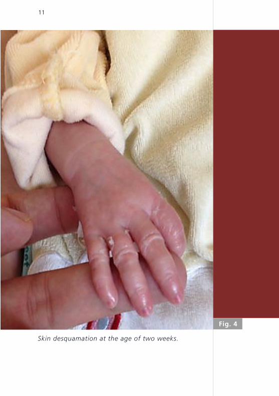
Almost complete shedding of the collodion memb-
rane was observed after two weeks, leaving gene-
ralized thin white scaling, with desquamation invol-
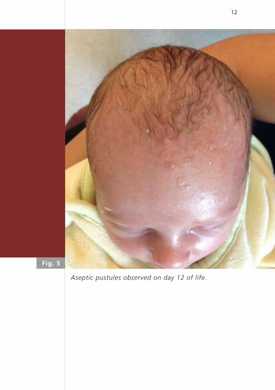
ving palms, soles and scalp (Fig. 4). On day 12 of life,
aseptic pustules were observed on the neck and scalp
(Fig. 5). With regards to the ectropion, ophthalmolo-
gic examination revealed no signs of keratitis or infec-
tion, and artificial tear drops were initiated.
In search of an underlying condition, extensive inve-
stigations were undertaken. There was no evidence
5
of a metabolic disorder. Abdominal, renal and cere-
bral ultrasounds as well as a chest X-ray were normal.
Hair analysis did not reveal the pathognomonic fea-
ture of Netherton syndrome, i.e. trichorrhexis invagi-
nata (bamboo hair) (2, 3). Since this symptom is not
regularly detected, its absence could not exclude this
disease entity with certainty (3).
Given the fact that the baby was otherwise stable, and
because the phenotype of the underlying diagnosis
often becomes evident only after weeks or months of
follow-up, we decided to wait and observe the evolu-
tion without further investigations (i.e., a skin biopsy
was not done, blood for genetic testing was sampled
but not analyzed). The patient was discharged at two
weeks of life.
Two weeks later, at one month of life, she was seen
by the dermatologists. By then, the glossy thin memb-
rane had disappeared completely, leaving some large
scales on the inferior part of the back and around the
contact point of the diapers. The volar aspects of the
palms and soles remained intact as well as the nails.
Daily creaming with dexpanthenol was prescribed.
At two months of life, the daily moisturizing was
discontinued given the fact that the skin was perfectly
normal. This evolution confirmed the diagnosis of
self-healing collodion baby (SHCB).
6
Collodion membrane with fine scaling.
Fig. 1
7

8
Discrete bilateral ectropion, absence of eye brows
and small eyelids, sparse hair, discrete eclabion.
Fig. 2
9
10
Skin splitting over flexural areas of the joints.
Fig. 3
11
Skin desquamation at the age of two weeks.
Fig. 4
12
Aseptic pustules observed on day 12 of life.
Fig. 5
13
DISCUSSIONAlthough the collodion membrane is only an evane-
scent phenomenon in the newborn, neonatal compli-
cations can occur in 45% of all CBs, leading to a mor-
tality rate of up to 11% in the first few weeks of life
(2). Common complications are marked temperature
instability (hypothermia), increased insensible water
loss predisposing to hypernatremic dehydration, cuta-
neous infections and septicemia (1– 4).
Given the disruption of the skin barrier, the initial
management of these patients includes:
• Nursing of the newborn in a highly humidified
incubator and frequent monitoring of electrolyte
balance
• Application of greasy emollients several times a day
• Close supervision for signs of cutaneous or syste-
mic infections
• Ophthalmologic evaluation and follow-up
The diagnosis of CB is made clinically; it is characte-
rized by a shiny tough transparent membrane, resem-
bling cellophane wrapping stretched over the skin.
The neonatal presentation differs greatly from the
later mature phenotype because of the differences
between the wet, intrauterine environment and the
dry, postnatal environment.
More than 60% of infants born with a collodion
membrane eventually develop ichthyosis (4, 5). The
term «ichthyosis», from the Greek word for fish, is
14
used for those disorders sharing generalized scaling
of the skin. Differentiation of ichthyosis subtypes in
the neonatal period is difficult. Skin histology in the
first few weeks of life is not specific, and therefore
not helpful. The final diagnosis emerges after weeks
or months of follow-up and depends on the genetic
analysis.
In 2009, the first ichthyosis consensus conference was
held and established an international nomenclature
and classification of inherited ichthyoses: syndromic
versus non-syndromic forms (3, 6). Six major distinct
clinical subtypes of hereditary autosomal recessive
non-syndromic ichthyoses were identified: Harlequin
ichthyosis (the most severe form), lamellar ichthyosis
(LI), non-bullous congenital ichthyosiform erythro-
derma (NBIE), epidermolytic ichthyosis (EI), recessive
X-linked ichthyosis and ichthyosis vulgaris (IV) (6).
Based on this classification, once the clinical subtype
is suspected, the results of genetic analyses will help
to provide proper treatment and genetic counselling.
In severe congenital ichthyosis, DNA-based prenatal
diagnosis is possible (3, 7).
More than 60% of affected CB will develop one of
the following two subtypes later in life (2, 4, 8): LI
(classic clinical findings are large, dark plate-like sca-
les with little or no erythema) or NBIE (fine scale with
prominent erythema). In approximately 10 – 20% of
CB cases (as in our case), the ichthyosis phenotype
15
may improve spontaneously within first three months
of life, leaving nearly normal-appearing skin (1 – 3, 5,
8). This entity has been described as self-healing CB
(SHCB) (1, 2, 4, 8). Because many of these patients,
when re-examined later in childhood or as adults,
have a variable degree of anhidrosis, heat intolerance
and mild signs of ichthyosis such as xerosis and fine
desquamation, particularly in the axillary and neck
region, the term self-improving collodion ichthyosis
may be more appropriate (8).
Histological analysis of skin biopsy specimens taken
in the first weeks of life usually show nonspecific
patterns and are therefore not helpful in differenti-
ating between the various forms of ichthyosis. Some
findings on electron microscopy have been reported
to help predict whether a baby will ultimately have
normal skin or ichthyosis. However, these findings may
be misleading (9). To date, mutations in eleven genes
have been identified to cause ichthyosis in human
patients (6). In the case of SHCB, mutations can be
found in the TGM1, ALOXE3 or ALOX12B genes enco-
ding, respectively, for transglutaminase 1 (involved
in the cornification of the stratum corneum) and for
arachidonate 3 and 12 lipoxygenase (involved in lipid
metabolism) (1, 3, 7 – 10).
CONCLUSION
16
Ichthyosis is a rare condition that requires significant
attention in the neonatal period. Successful manage-
ment in the newborn period requires an interdisci-
plinary approach. Families should be offered proper
genetic counselling, psychological support and receive
information about relevant patient organization and
foundations (see: www.ichthyose.ch) (3, 7).
1. Prado R, Ellis LZ, Gamble R, et al. Collodion baby: An update
with a focus on practical management. J Am Acad Dermatol
2012;67:1362-1374 (Abstract)
2. Chiavérini C. Congenital ichthyosis. Ann Dermatol Venereol
2009;136:923-934 (Abstract)
3. Oji V, Tadini G, Akiyama M, et al. Revised nomenclature
and classification of inherited ichthyoses: results of the First
Ichthyosis Consensus Conference in Sorèze 2009. J Am Acad
Dermatol 2010;63:607-641 (Abstract)
4. Larrègue M, Ottavy N, Bressieux JM, et al. Collodion baby: 32
new case reports. Ann Dermatol Venereol 1986;113:773-785
(Abstract)
5. Aradhya SS, Srinivas SM, Hiremagalore R, Shanmukappa AG.
Clinical outcome of collodion baby: a retrospective review.
Indian J Dermatol Venereol Leprol 2013;79:553
6. Akiyama M. Updated molecular genetics and pathogenesis of
ichthyoses. Nagoya J Med Sci 2011;73:79-90 (Abstract)
7. Rodríguez-Pazos L, Ginarte M, Vega A, Toribio J. Autoso-
mal recessive congenital ichthyosis. Actas Dermosifiliogr
2013;104:270-284 (Abstract)
8. Vahlquist A, Bygum A, Gånemo A, et al. Genotypic and clinical
spectrum of self-improving collodion ichthyosis: ALOX12B,
ALOXE3, and TGM1 mutations in Scandinavian patients. J
Invest Dermatol 2010;130:438-443 (Abstract)
9. Harting M, Brunetti-Pierri N, Chan CS, et al. Self-healing collo-
dion membrane and mild non-bullous congenital ichthyosiform
erythroderma due to 2 novel mutations in the ALOX12B gene.
Arch Dermatol Mar 2008;144:351-356 (Abstract)
REFERENCES
17

SUPPORTED BY
CONTACT
10. Raghunat M, Hennis HC, Ahvazi B, et al. Self-healing collodion
baby: a dynamic phenotype explained by a particular trans-
glutaminase 1 mutation. J Inv Dermatol 2003;120:224-228
(Abstract)
18
SUPPORTED BY
CONTACT
Swiss Society of Neonatology
www.neonet.ch
con
cep
t &
des
ign
by
mes
ch.c
h
Related Documents