biology Review Cnidarian Immunity and the Repertoire of Defense Mechanisms in Anthozoans Maria Giovanna Parisi 1, * , Daniela Parrinello 1 , Loredana Stabili 2 and Matteo Cammarata 1, * 1 Department of Earth and Marine Sciences, University of Palermo, 90128 Palermo, Italy; [email protected] 2 Department of Biological and Environmental Sciences and Technologies, University of Salento, 73100 Lecce, Italy; [email protected] * Correspondence: [email protected] (M.G.P.); [email protected] (M.C.) Received: 10 August 2020; Accepted: 4 September 2020; Published: 11 September 2020 Abstract: Anthozoa is the most specious class of the phylum Cnidaria that is phylogenetically basal within the Metazoa. It is an interesting group for studying the evolution of mutualisms and immunity, for despite their morphological simplicity, Anthozoans are unexpectedly immunologically complex, with large genomes and gene families similar to those of the Bilateria. Evidence indicates that the Anthozoan innate immune system is not only involved in the disruption of harmful microorganisms, but is also crucial in structuring tissue-associated microbial communities that are essential components of the cnidarian holobiont and useful to the animal’s health for several functions including metabolism, immune defense, development, and behavior. Here, we report on the current state of the art of Anthozoan immunity. Like other invertebrates, Anthozoans possess immune mechanisms based on self/non-self-recognition. Although lacking adaptive immunity, they use a diverse repertoire of immune receptor signaling pathways (PRRs) to recognize a broad array of conserved microorganism-associated molecular patterns (MAMP). The intracellular signaling cascades lead to gene transcription up to endpoints of release of molecules that kill the pathogens, defend the self by maintaining homeostasis, and modulate the wound repair process. The cells play a fundamental role in immunity, as they display phagocytic activities and secrete mucus, which acts as a physicochemical barrier preventing or slowing down the proliferation of potential invaders. Finally, we describe the current state of knowledge of some immune effectors in Anthozoan species, including the potential role of toxins and the inflammatory response in the Mediterranean Anthozoan Anemonia viridis following injection of various foreign particles differing in type and dimensions, including pathogenetic bacteria. Keywords: cnidarians; innate immunity; Anthozoan; bioactive molecules; inflammatory response 1. Introduction Cnidarians are phylogenetically basal aquatic animals within the Metazoa, with radial symmetry and the first level of tissue organization. They are evolutionarily early-diverged Metazoa, some of whom can live for hundreds of years, which suggests that they are potentially exposed to some pathogens on many occasions during their lifespans [1]. It is difficult to explain how these long-lived organisms have done so well with only an innate immune system as the protective mechanism against infectious agents. Cnidarians are of great interest since they apply many of the same cellular pathways involved in innate immunity in mammals. They have a surprising amount of immune complexity, as they contain innate immune components that are lacking in other basal invertebrate groups [2]. Biology 2020, 9, 283; doi:10.3390/biology9090283 www.mdpi.com/journal/biology

Welcome message from author
This document is posted to help you gain knowledge. Please leave a comment to let me know what you think about it! Share it to your friends and learn new things together.
Transcript

biology
Review
Cnidarian Immunity and the Repertoire of DefenseMechanisms in Anthozoans
Maria Giovanna Parisi 1,* , Daniela Parrinello 1, Loredana Stabili 2 and Matteo Cammarata 1,*1 Department of Earth and Marine Sciences, University of Palermo, 90128 Palermo, Italy;
[email protected] Department of Biological and Environmental Sciences and Technologies, University of Salento, 73100 Lecce,
Italy; [email protected]* Correspondence: [email protected] (M.G.P.); [email protected] (M.C.)
Received: 10 August 2020; Accepted: 4 September 2020; Published: 11 September 2020�����������������
Abstract: Anthozoa is the most specious class of the phylum Cnidaria that is phylogeneticallybasal within the Metazoa. It is an interesting group for studying the evolution of mutualisms andimmunity, for despite their morphological simplicity, Anthozoans are unexpectedly immunologicallycomplex, with large genomes and gene families similar to those of the Bilateria. Evidence indicatesthat the Anthozoan innate immune system is not only involved in the disruption of harmfulmicroorganisms, but is also crucial in structuring tissue-associated microbial communities that areessential components of the cnidarian holobiont and useful to the animal’s health for several functionsincluding metabolism, immune defense, development, and behavior. Here, we report on the currentstate of the art of Anthozoan immunity. Like other invertebrates, Anthozoans possess immunemechanisms based on self/non-self-recognition. Although lacking adaptive immunity, they usea diverse repertoire of immune receptor signaling pathways (PRRs) to recognize a broad arrayof conserved microorganism-associated molecular patterns (MAMP). The intracellular signalingcascades lead to gene transcription up to endpoints of release of molecules that kill the pathogens,defend the self by maintaining homeostasis, and modulate the wound repair process. The cells play afundamental role in immunity, as they display phagocytic activities and secrete mucus, which actsas a physicochemical barrier preventing or slowing down the proliferation of potential invaders.Finally, we describe the current state of knowledge of some immune effectors in Anthozoan species,including the potential role of toxins and the inflammatory response in the Mediterranean AnthozoanAnemonia viridis following injection of various foreign particles differing in type and dimensions,including pathogenetic bacteria.
Keywords: cnidarians; innate immunity; Anthozoan; bioactive molecules; inflammatory response
1. Introduction
Cnidarians are phylogenetically basal aquatic animals within the Metazoa, with radial symmetryand the first level of tissue organization. They are evolutionarily early-diverged Metazoa, some ofwhom can live for hundreds of years, which suggests that they are potentially exposed to somepathogens on many occasions during their lifespans [1]. It is difficult to explain how these long-livedorganisms have done so well with only an innate immune system as the protective mechanism againstinfectious agents.
Cnidarians are of great interest since they apply many of the same cellular pathways involved ininnate immunity in mammals. They have a surprising amount of immune complexity, as they containinnate immune components that are lacking in other basal invertebrate groups [2].
Biology 2020, 9, 283; doi:10.3390/biology9090283 www.mdpi.com/journal/biology
Biology 2020, 9, 283 2 of 26
In addition to pathogen recognition, the cnidarian innate immune system has a role in managingbeneficial microbes and supporting mutualistic microbial symbioses. It regulates the maintenanceof symbiosis and takes on the task of discerning between pathogens that need to be cleared versusbeneficial symbiotic microbes [3].
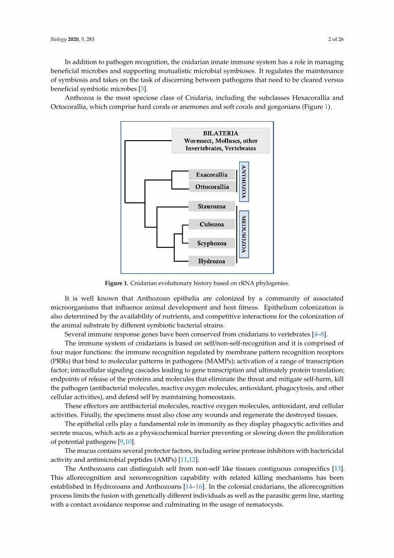
Anthozoa is the most speciose class of Cnidaria, including the subclasses Hexacorallia andOctocorallia, which comprise hard corals or anemones and soft corals and gorgonians (Figure 1).
Biology 2020, 9, x 2 of 25
In addition to pathogen recognition, the cnidarian innate immune system has a role in managing beneficial microbes and supporting mutualistic microbial symbioses. It regulates the maintenance of symbiosis and takes on the task of discerning between pathogens that need to be cleared versus beneficial symbiotic microbes [3].
Anthozoa is the most speciose class of Cnidaria, including the subclasses Hexacorallia and Octocorallia, which comprise hard corals or anemones and soft corals and gorgonians (Figure 1).
Figure 1. Cnidarian evolutionary history based on rRNA phylogenies.
It is well known that Anthozoan epithelia are colonized by a community of associated microorganisms that influence animal development and host fitness. Epithelium colonization is also determined by the availability of nutrients, and competitive interactions for the colonization of the animal substrate by different symbiotic bacterial strains.
Several immune response genes have been conserved from cnidarians to vertebrates [4–8]. The immune system of cnidarians is based on self/non-self-recognition and it is comprised of four
major functions: the immune recognition regulated by membrane pattern recognition receptors (PRRs) that bind to molecular patterns in pathogens (MAMPs); activation of a range of transcription factor; intracellular signaling cascades leading to gene transcription and ultimately protein translation; endpoints of release of the proteins and molecules that eliminate the threat and mitigate self-harm, kill the pathogen (antibacterial molecules, reactive oxygen molecules, antioxidant, phagocytosis, and other cellular activities), and defend self by maintaining homeostasis.
These effectors are antibacterial molecules, reactive oxygen molecules, antioxidant, and cellular activities. Finally, the specimens must also close any wounds and regenerate the destroyed tissues.
The epithelial cells play a fundamental role in immunity as they display phagocytic activities and secrete mucus, which acts as a physicochemical barrier preventing or slowing down the proliferation of potential pathogens [9,10].
The mucus contains several protector factors, including serine protease inhibitors with bactericidal activity and antimicrobial peptides (AMPs) [11,12].
The Anthozoans can distinguish self from non-self like tissues contiguous conspecifics [13]. This allorecognition and xenorecognition capability with related killing mechanisms has been established in Hydrozoans and Anthozoans [14–16]. In the colonial cnidarians, the allorecognition process limits the fusion with genetically different individuals as well as the parasitic germ line, starting with a contact avoidance response and culminating in the usage of nematocysts.
As invertebrates who rely on the innate component of immunity, they should not be able to develop resistance toward a particular pathogen [17]. However, a form of immunological memory has long been
Figure 1. Cnidarian evolutionary history based on rRNA phylogenies.
It is well known that Anthozoan epithelia are colonized by a community of associatedmicroorganisms that influence animal development and host fitness. Epithelium colonization isalso determined by the availability of nutrients, and competitive interactions for the colonization ofthe animal substrate by different symbiotic bacterial strains.
Several immune response genes have been conserved from cnidarians to vertebrates [4–8].The immune system of cnidarians is based on self/non-self-recognition and it is comprised of
four major functions: the immune recognition regulated by membrane pattern recognition receptors(PRRs) that bind to molecular patterns in pathogens (MAMPs); activation of a range of transcriptionfactor; intracellular signaling cascades leading to gene transcription and ultimately protein translation;endpoints of release of the proteins and molecules that eliminate the threat and mitigate self-harm, killthe pathogen (antibacterial molecules, reactive oxygen molecules, antioxidant, phagocytosis, and othercellular activities), and defend self by maintaining homeostasis.
These effectors are antibacterial molecules, reactive oxygen molecules, antioxidant, and cellularactivities. Finally, the specimens must also close any wounds and regenerate the destroyed tissues.
The epithelial cells play a fundamental role in immunity as they display phagocytic activities andsecrete mucus, which acts as a physicochemical barrier preventing or slowing down the proliferationof potential pathogens [9,10].
The mucus contains several protector factors, including serine protease inhibitors with bactericidalactivity and antimicrobial peptides (AMPs) [11,12].
The Anthozoans can distinguish self from non-self like tissues contiguous conspecifics [13].This allorecognition and xenorecognition capability with related killing mechanisms has beenestablished in Hydrozoans and Anthozoans [14–16]. In the colonial cnidarians, the allorecognitionprocess limits the fusion with genetically different individuals as well as the parasitic germ line, startingwith a contact avoidance response and culminating in the usage of nematocysts.

Biology 2020, 9, 283 3 of 26
As invertebrates who rely on the innate component of immunity, they should not be able todevelop resistance toward a particular pathogen [17]. However, a form of immunological memoryhas long been suggested in cnidarians, based on tissue allo- and xeno- transplants [18]. It is truethat the mechanisms underlying recognition may differ from those involved in the functioning of thedefense system.
Further evidence of immunological memory derives from classes of receptors that recognizemolecular models of different pathogens [19]. This means that Anthozoans can recognize the differencebetween pathogenic types, responding with specific binding receptors to acquire memory for aspecific pathogen.
Here, we reported the current state of knowledge about cellular and molecular immune repertoirein Anthozoan species and the inflammatory responses in Anemonia viridis (Cnidaria: Anthozoa)following bacterial injection and pathogenetic invasions with bacteria such as Escherichia coli andVibrio alginolyticus. These belong to different genera and are of diverse shape and dimension.
The currently accepted taxonomic scheme subdivides Cnidaria into two main assemblages:Anthozoa (Hexacorallia and Octocorallia) with a reproductive polyp and the absence of a medusa stageand Medusozoa (Cubozoa, Hydrozoa, Scyphozoa, and Staurozoa) that usually possess a reproductivemedusa stage.
2. Allorecognition in Anthozoans
In marine colonial invertebrates (including members of the phylum Chordata), allorecognitionmay be essential for survival as a response to selection pressures [18]. In vertebrates, allorecognitionand self-tolerance are the result of a major histocompatibility complex (MHC)-based recognition event,critical to adaptive immunity.
In many invertebrate colonies that share alleles of specific histocompatibility, loci (Alr 1, Alr 2)generally merge to create a chimeric colony, while individuals who do not participate in this sharingare aggressively rejected. In cnidarians, the Alr1 and Alr2 loci encode membrane-bound moleculeswith immunoglobulin-like domains not homologous to those found in basal chordate [20].
In Anthozoans, such as Montipora verrucosa, branches within a colony can easily join, while branchesof genetically diverse individuals do not fuse [21]. Hydractinia symbiolongicarpus is a cnidarian modelstudied in the context of allorecognition—a hydroid feeding on gastropods inhabited by hermit crabsalong the intertidal areas of the east coast of North America. In this species, fusion occurs when thecolonies share one of two, or both, alleles of two loci. By contrast, the rejection reaction, characterizedby the non-adhesion and the discharge of the nematocysts, occurs when the matched partners do notshare any of these alleles. The Hydractinia colonies are composed of polyps, the bowels of which areconnected to each other by a network of gastrovascular channels. These channels then extend outof the carpet to grow along the surface of the shell, forming stolons. When the stolons encounter anincompatible colony, they differentiate into “hyperplastics”, loaded with cnidocytes, growing on theopposite colony and releasing cnidocytes until the death of the incompatible colony. In the case ofcompatibility, a chimeric colony is formed [22]. The Alr1–Alr1 and Alr2–Alr2 interactions modulateself/non-self-discrimination, and their long cytoplasmic tails (200–250 amino acids) contain motifsreminiscent of sites for kinase-mediated phosphorylation.
In Hydractinia, Mokady & Buss (1996) showed that these two closely related loci for recognitionare contained in a single chromosomal region [23,24]. Genomic analysis of the region correspondingto the Alr2 locus led to the identification of a transmembrane protein, CDS7, which has threeextracellular polymorphic domains [25]. The CDS7 domain polymorphism represents an element ofhistocompatibility as a probable candidate responsible for the recognition in basal metazoans. Researchefforts on the identification of molecules involved in recognition in solitary cnidarians, such as Hydra,were not successful thus far [26].

Biology 2020, 9, 283 4 of 26
3. Symbiosis and Immunity
The association of sea anemones with photosynthetic algae of the genus Symbiodinium, collectivelycalled zooxanthellae, provides mutualistic relationships [27]. The cnidarians supply nutrients to thezooxanthellae, as well as carbon dioxide derived from the digestion of the preys; in turn, the symbiontphotosynthetic zooxanthellae produce sugars, lipids, and oxygen that support energy production forthe metabolic activity of the Anthozoan, associated with microbial communities [28]. The unicellularalgae are in gastrodermal cells within host-derived membrane vesicles called symbiosomes that ariseduring the symbiotic process of the cnidarian host [29]. The symbiotic relationship is useful forstudying the evolution of the various groups. Some symbionts’ taxa have a notably wide geographicaldistribution and are associated with multiple species of hosts. Others, however, show a restrictedgeographical distribution [30,31].
Transcriptomic studies have shown that homologs of genes involved in the defense system andinflammation as mediators of oxidative stress, serine protease, or immunity transcription factor NF-κBare downregulated by the establishment of symbiosis and upregulated when lacking in symbioticcnidarians [32].
Symbiosis is recognized as a non-harmful infection, and host defense responses are controlleduntil environmental conditions are optimal for the survival of autotrophic and heterotrophic organisms;the role of bacterial communities has yet to be explained [33].
Bleaching is a host-innate immune response to a compromised symbiont, much like innate immuneresponses in other host–microbe interactions [34].
Although the downregulation of immunity appears to be a necessary condition to symbioses,evidence from certain bleached corals suggests that immunity genes are less expressed [35].In this respect, some Anthozoan specimens losing symbionts resulted in increased susceptibilityto pathogen-induced diseases greater than that of symbiotic specimens. Pollution induces thesymbiosis breakdown and the zooxanthellae can be expelled, exposing the specimens to the greatestrisk of mortality.
For some time, people have been wondering if autophagy and apoptosis have been workingtogether to induce loss of symbiosis during heat stress-induced bleaching [36]. It has been proposed thatthe Rab GTPase, a regulating lysosome–phagosome fusion gene family, plays key roles in maintainingsymbiosis with host gastrodermal cells [37].
The gene encoding Rab32, a regulator of the lysosomal enzyme recruitment to phagosome, isinvolved in the exclusion and maintenance of symbionts [38]. In Aiptasia pulchella, the upregulation ofAp-Rab7 during heat stress induces the phagosomal–lysosomal fusion of the Symbiodiniaceae-containingvacuoles, leading to the loss of symbiosis.
The symbiotic relationship is useful for studying the evolution and biogeography of the variouszoological groups. Some symbionts taxa in fact have a wide geographical distribution and areassociated with multiple species of hosts [39].
4. Interaction with Microbial Communities
Cnidarians host a variety of microbes including many bacterial species and viruses [40,41].Identification of the unique role of the microbiota in eukaryotic host development, and their responseto environmental perturbations, has led to the definition of a “holobiont”. For Anthozoans, too,it describes a complex organism in which multiple components can evolve together as symbioticentity [42]. These multi-partite symbiotic organisms are formed by polyp animals, zooxantellae,and microbial assemblages associated with polyps and photosynthetic symbionts.
There is increasing evidence to indicate that the cnidarian innate immune system is not onlyinvolved in the disruption of harmful microorganisms, but is also crucial in structuring tissue-associatedmicrobial communities that are essential components of the holobiont, and useful to the animal’shealth [43].

Biology 2020, 9, 283 5 of 26
Cnidarians have many microorganisms associated (epibiotic or symbiotic) with their tissues.As reported by Tinta et al. [44], early reports on microbes associated with cnidarians resulted ascorollary observations, whereas primary targets of research were cnidarians. Later studies, focusingon the relationships between microbes and their host organisms, addressed more specific issues onthe composition and ecological role of cnidarian-associated microbial communities. These studiesdealt with issues on the role, ecology, and composition of microbial communities associated withcnidarians, the mechanisms at the base of these interactions, and the nature of the relationshipsestablished between cnidarians and their associated microbiome. On account of these considerations,researchers were focused on the microbial counterpart of the jellyfish–microbe associations, in order toassess the diversity of microbial communities associated with different jellyfish species from variousecosystems and with their different life stages and body compartments. Further studies concernedbacteria associated to outer surfaces of cnidarian epithelia in different taxa and life stages showingtheir involvement in a number of important potential roles including antibiotics synthesis, nitrogenfixation, organic compounds decomposition, primary defense against pathogens, or modulation ofcontractile activities.
The microbial communities associated with the semeostome jellyfish Aurelia aurita, the rhizostomejellyfish species Mastigias papua, Cotylorhiza tuberculata, and Rhizostoma pulmo and the box jellyfishTripedalia cf. cystophora were analyzed in different studies, identifying jellyfish as a host of bacterialassociates. The microbiome associated with different life stages of A. aurita (polyp, strobila, ephyrae,juvenile, and adult medusae) was examined by Weiland-Bräuer et al. [45]. The authors also considereddifferent compartments of the adult medusae (exumbrella, mucus, and gastric cavity) and comparedthe microbiome associated with specimens (polyp stage) collected in different geographic sites.In particular, the microbial community of jellyfish A. aurita seems to be strictly host-specific anddifferent from the bacterioplankton suspended in the surrounding water column. In A. aurita adultmedusa, the microbiota of different compartments exhibit significant differences, showing bodypart-specific bacterial colonization with a mucus-associated bacterial composition that is more variablecompared to bacteria living in the gastric cavity, likely thanks to trapping properties of mannoseand mucine glycan components of mucus. Moreover, Kos Kramar et al. [46] checked the bacterialcommunity associated with the moon jellyfish Aurelia solida. In particular, different body parts(exumbrella surface, oral arms, and gastric cavity) were analyzed for the bacterial community diversity.Furthermore probable differences in medusa-associated bacterial community structure during thejellyfish population peak and the senescent stage when the bloom ended were evaluated. The authorsconcluded that microbiota associated with moon jellyfish were different from microbial assemblagesin the surrounding seawater and differed between different body compartments. The microbiotain the gastral cavity of medusa Betaproteobacteria (Burkholderia, Cupriavidus, and Achromobacter)prevailed, by contrast, over those on the ‘outer’ body parts of Alphaproteobacteria (Phaeobacter,Ruegeria) and Gammaproteobacteria (Stenotrophomonas, Alteromonas, Pseudoalteromonas and Vibrio).During the senescent phase, at the end of the jellyfish bloom, the bacterial community resultedchanged in the structure with an increase of Gammaproteobacteria, wholly Vibrio. On the basis ofthese results, it was hypothesized that the jellyfish-associated bacterial community might play animportant role for the host. Analyzing the composition of the microbiome associated with jellyfishmucus, Tinta et al. [44] concluded that Gammaproteobacteria (mainly Pseudoalteromonas and Vibrio)are abundant, but to some extent also Alphaproteobacteria (Phaeobacter, Rugeria, and Roseovarius).These bacteria, due to their capability to synthesize antimicrobial compounds when attached to live orinert surfaces, were previously recognized as significant players in the host defense towards pathogensand fouling organisms from the surrounding environment. Rhizostoma pulmo-associated microbiotawere investigated in three distinct compartments, namely umbrella, oral arms, and the mucussecretion [47,48]. Actinobacteria, Bacteroidetes, Chlamydiae, Cyanobacteria, Deinococcus-Thermus,Firmicutes, Fusobacteria, Planctomycetes, Proteobacteria, Rhodothermaeota, Spirochaetes, Tenericutes,and Thaumarchaeota were the phyla isolated from all the three R. pulmo compartments in the
Biology 2020, 9, 283 6 of 26
sampling times. In particular, the main genera Mycoplasma and Spiroplasma, belonging to theclass Mollicutes (phylum Tenericutes), have been identified in all three jellyfish compartments.Microorganisms associated with mucus are characterized by great diversity and the mucus appears tobe the compartment mainly rich in bacteria compared to the oral arms and the umbrella.
In Anthozoans microbial agents fulfill several functions, including regulation of metabolism,immune defense, development, and behavior. Bacteria associated with Anthozoan tissues canindeed fix nitrogen, digest complex polysaccharides, and produce antibiotics to prevent infectionwith pathogens. In turn, Symbiodinium produces dimethylsulfoniopropionate (DMSP) as osmolite,antioxidant agents [49], and a nutrient source for associated bacteria [50].
A certain specificity was also found in the Anthozoan-bacteria association; Porporato et al. [51],for example, described the bacterial communities associated with Pennatula phosphorea andPteroeides spinosum. Results from this study showed the occurrence of species-specific coral-associatedbacteria since P. phosphorea and P. spinosum host distinct bacterial communities. Moreover, since inthe same pennatulid species, only a few phylotypes were shared between mucus and tissues, it washypothesized that there might exist a microhabitat partitioning between the associated microbialcommunities as already described for jellyfish. In the case of P. phosphorea, the communities associatedwith both tissues and mucus were characterized by the predominance of Alphaproteobacteria.Conversely, the Alphaproteobacteria prevailed in the mucus layer of P. spinosum and the tissue librarywas dominated by the Gammaproteobacteria and Mollicutes. Bacterial isolates belonging to Vibrio spp.,mainly obtained from coral mucus, showed an antibacterial activity against indicator organisms,indicating a protective function of the coral-associated bacterial communities as in the case of jellyfish.
On account of these evidences the ability of cnidarians to control production and composition of amucosal matrix and its associated bacteria can represent an important part of immunity [52]. Mucusindeed contains many microorganisms and bacteria, and the Vibrio genus particularly prevails in thecultivable bacterial isolates from the mucus of several other Anthozoans [53].
Some mucus bacteria producing antimicrobial compounds contribute to competition in space andnutrition with the host potential pathogenic bacteria. Due to these features, mucus and its componentshave interesting biotechnological implications.
The specific interactions of the microbial colonization of mucosal surfaces are still unknown.The “Coral Probiotic Hypothesis” [54] affirms that Pseudoalteromonas sp. can be considered as“probiotic” to corals, taking part in the coral holobiont defense against bacteria, placing a differentcritical perspective on the host–microbe interactions.
Calow [55] indicated that the differences in mucus biochemical composition influence the attack ofseveral microbial agents that use exoenzymes to degrade mucoid polymers. Microbes themselves cantransform dissolved and particulate matter into living matter, attracting other predatory organisms.
It also appears that change in the density of bacterial communities may influence animal healthand, consequently, their sensitivity to disease [54].
Recent reports of the succession of the microbial communities associated with the developmentalstages of Porites astreoides [56] and the discovery of the potentially beneficial functions inα-Proteobacteriaand strains of Marinobacter [16] lend further support to the hologenome evolution hypothesis indicatingthat in colonial symbiotic organisms, the ‘hologenome’, deriving from all the members of the holobiont,acts as a single unit of evolution, with faster evolving micro-organisms providing the plasticity toadapt to the changing environment [57].
Controversies still surround this aspect of the Anthozoans, as it is not yet clear how some coralsare not susceptible to the pathogens that have caused diseases in the past [58]. Furthermore, the naturalselection for disease-resistant coral genotypes seems to have acted, which could explain the loss ofvirulence by the bacterial specimens. On the other hand, disease has been increasing over the last fewdecades in response to elevated sea surface temperatures and anthropogenic stressors causing bleachingand eventual death of the tissue [9,59]. Coral bleaching, for example, occurs when the balance betweenthe different components of the holobiont is destroyed and immune cascades are activated, which lead
Biology 2020, 9, 283 7 of 26
to Nuclear factor-κB (NF-κB) protein levels up-production, involved in an elaborate system that enablesa response to environmental changes. The elimination of the symbiont from Anthozoans tissues isregulated by a variety of mechanisms such as exocytosis, host cell detachment, and apoptosis [60].
5. Repertoire of Immune Receptors Signaling Pathways (PRRs)
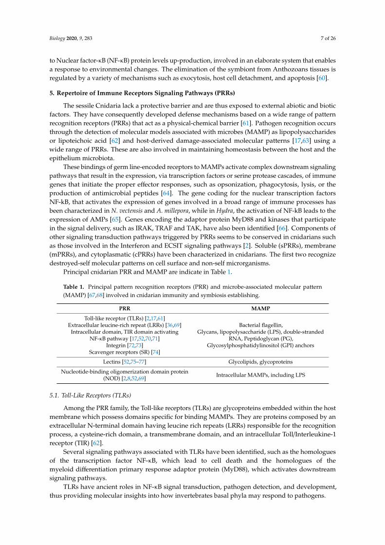
The sessile Cnidaria lack a protective barrier and are thus exposed to external abiotic and bioticfactors. They have consequently developed defense mechanisms based on a wide range of patternrecognition receptors (PRRs) that act as a physical-chemical barrier [61]. Pathogen recognition occursthrough the detection of molecular models associated with microbes (MAMP) as lipopolysaccharidesor lipoteichoic acid [62] and host-derived damage-associated molecular patterns [17,63] using awide range of PRRs. These are also involved in maintaining homeostasis between the host and theepithelium microbiota.
These bindings of germ line-encoded receptors to MAMPs activate complex downstream signalingpathways that result in the expression, via transcription factors or serine protease cascades, of immunegenes that initiate the proper effector responses, such as opsonization, phagocytosis, lysis, or theproduction of antimicrobial peptides [64]. The gene coding for the nuclear transcription factorsNF-kB, that activates the expression of genes involved in a broad range of immune processes hasbeen characterized in N. vectensis and A. millepora, while in Hydra, the activation of NF-kB leads to theexpression of AMPs [65]. Genes encoding the adaptor protein MyD88 and kinases that participatein the signal delivery, such as IRAK, TRAF and TAK, have also been identified [66]. Components ofother signaling transduction pathways triggered by PRRs seems to be conserved in cnidarians suchas those involved in the Interferon and ECSIT signaling pathways [2]. Soluble (sPRRs), membrane(mPRRs), and cytoplasmatic (cPRRs) have been characterized in cnidarians. The first two recognizedestroyed-self molecular patterns on cell surface and non-self microrganisms.
Principal cnidarian PRR and MAMP are indicate in Table 1.
Table 1. Principal pattern recognition receptors (PRR) and microbe-associated molecular pattern(MAMP) [67,68] involved in cnidarian immunity and symbiosis establishing.
PRR MAMP
Toll-like receptor (TLRs) [2,17,61]Extracellular leucine-rich repeat (LRRs) [36,69]
Intracellular domain, TIR domain activatingNF-κB pathway [17,52,70,71]
Integrin [72,73]Scavenger receptors (SR) [74]
Bacterial flagellin,Glycans, lipopolysaccharide (LPS), double-stranded
RNA, Peptidoglycan (PG),Glycosylphosphatidylinositol (GPI) anchors
Lectins [52,75–77] Glycolipids, glycoproteins
Nucleotide-binding oligomerization domain protein(NOD) [2,8,52,69] Intracellular MAMPs, including LPS
5.1. Toll-Like Receptors (TLRs)
Among the PRR family, the Toll-like receptors (TLRs) are glycoproteins embedded within the hostmembrane which possess domains specific for binding MAMPs. They are proteins composed by anextracellular N-terminal domain having leucine rich repeats (LRRs) responsible for the recognitionprocess, a cysteine-rich domain, a transmembrane domain, and an intracellular Toll/Interleukine-1receptor (TIR) [62].
Several signaling pathways associated with TLRs have been identified, such as the homologuesof the transcription factor NF-κB, which lead to cell death and the homologues of themyeloid differentiation primary response adaptor protein (MyD88), which activates downstreamsignaling pathways.
TLRs have ancient roles in NF-κB signal transduction, pathogen detection, and development,thus providing molecular insights into how invertebrates basal phyla may respond to pathogens.
Biology 2020, 9, 283 8 of 26
In addition to NF-κB signaling, TLR receptors can activate mitogen-activated protein kinase(MAPK) resulting in activation of the immune signaling pathways and appropriate effectorresponses [63]. The known pathway homology indicates that the Toll/TlR receptors are highly conserved.
Since the invertebrate immunity is innate and non-specific, the effector responses can be activatedin relation to serine protease cascades and redox signaling [64] without gene transcription.
In Hydra magnipapillata, four TLR-domain proteins HmMyD88-1 and HyMyD88-2 are related tothe downstream MyD88. The other two Hydra TIR-domain proteins, HyTRR-1 and HyTRR2, are likelyto be pathway initiator receptors, and they lack the LRR-domains [2].
Among the Anthozoa, transcriptomic and genomic studies have resulted in the discovery of manymore putative TLRs proteins in Nematostella vectensis [2] and Gorgonia ventalina [78]. One N. vectensisTLR (Nv-TLR) is also expressed in some cnidocytes, and it is able to express Nv-NF-κB, another innateimmune pathway to engulf the coral pathogen Vibrio coralliilyticus [61].
Five TLR-domain proteins have been identified from the predicted structures inNematostella vectensis. The structure of NvMyD88 appears similar to the HmMyD88-1 and 2 andinitiates the transmission of intracellular signals leading to the translocation of transcription factorsfrom the NF-kB family [79].
Other identified predicted TLR-domain protein structures include immunoglobulin (Ig) domainswhich act in cell–cell recognition, resulting in similar architecture to mammalian interleukin receptors(ILR) but form a distinct clade away from the higher vertebrate structures.
In Acropora digitifera, several TLRs and ILRs have been identified, suggesting the immunerecognition repertoire in this coral species is different than that of Nematostella. Downstream componentsof the Toll/TLR pathways were also described from the EST/genome analysis, such as those associatedwith the c-Jun N-terminal kinases (JNK)/Mitogenactivated protein kinase (MAPK) pathway and NFkBtranscription that can lead to cell death [80].
5.2. NOD-Like Receptors (NLRs)
The structure of NLRs consists of a C terminal LRR domain for microbe pattern recognition,an intermediary NOD domain for nucleotide binding and modulation of NLR activity, and anN-terminal effector domain. The family of NOD-like receptors (NLRs) that function as cytosolicreceptors form a signaling scaffold to activate an inflammatory cascade. In the corals Acropora digitiferaand Pseudodiploria strigos, an array of NOD-like receptor proteins has been detected. Like TRLs, theNLR activation triggers multiple proinflammatory signaling pathways, which result in the inhibitionof the pathogens [81,82].
PRRs can also recognize damage-associated molecular patterns (DAMPs), which are self-moleculesor debris from altered cells, and they can trigger the response mediated by the immunological effectors.These are substances inside the cells which are not easily detected by the surrounding cells, but arereleased in the case of danger or injury to signal the presence of a threat to the healthy cells formingthe surrounding tissue.
5.3. Lectins
Lectins act as PRRs. A variety of lectins have been described in cnidarians [81,83,84] involvedin opsonization activity and activation of the complement cascade. The complement lectin pathway,in fact, has been detected among the cnidarians, in which lectin binds to the sugar present on thepathogen surface, activating mannose-binding lectin associated serine proteases (MASPs), C2 andC4-like proteins up to formation of the C3 complex. After the complex formation, membrane attackcomplex perforin (MACPF) effector proteins are secreted to realize a hole in the microbial membrane.The resulting effector is the lysis of the pathogen. One lectin common to several groups of cnidariansis the tachylectin [85], initially isolated from Tachypleus tridentatus, which has experimentally shownantimicrobial activity and recognition of MAMP as LPS and peptidoglycans.
MAMP-PRR interactions occur during the establishing or in the maintenance of symbiosis.

Biology 2020, 9, 283 9 of 26
Cnidarian lectins that could participate in MAMP-PRR interactions in symbiosis establishmenthave been identified by a genomic approach. A d-Galactose-binding lectin, SLL-2, was purified fromthe octocoral Sinularia lochomodes, sequenced, and found by immunolocalization to occur surroundingsymbiotic dinoflagellates in the gastrodermis. Lectin-binding patterns varied between differentSymbiodinium types, suggesting a complex surface glycome that varies between types. N-Acetyl andmannose residues are well-characterized MAMPs that bind to mannose-binding lectins and ficolins,respectively [86].
Millectin, involved in both symbiont recognition and an innate immune response to bacteria, hasbeen described as primary PRR in the coral Acropora millepora and it is expressed in both nematocystsand the gastroderm. This lectin is expressed around intracellular symbiont cells in vivo and somedomains are similar to the vertebrate mannose binding lectin (MBL) [75,83].
Hydractinia symbiolongicarpus and other cnidarians express one rhamnose-binding lectin (RBL)gene, rhamnospondin, which contains multiple PRR-type domains with probable opsonization oragglutination activity [87].
5.4. Integrin
Integrins are trans-membrane alpha beta heterodimers that mediate the interactions between cellsand the extracellular matrix ECM [72]. They are involved in multiple immunological cellular processessuch as cell migration, differentiation, signal transduction, and wound repair. ECM engagement leadsto the activation of focal adhesion kinase (FAK) and to the tyrosine kinases. Three integrin have beenidentified in N. vectensis and two beta subunits have been identified in the hard coral A. millepora [2,73].
5.5. Other PRRs
A putative immune recognition repertoire of the cnidarians, involved in recognition and responseto microbial infection, in addition to the categories discussed, through a variety of molecular methods,has been identified. These are specifically leucine rich repeat (LRR) identified in expressed sequencetags (ESTs) [74], and a transforming growth factor β (TGF-β) receptor [88].
A family of PRRs that recognize LPS from Gram-negative bacteria, leading to the activation ofNF-kB pathway, is the lipopolysaccharide (LPS)-binding proteins (LBPs) identified in the genomes ofH. magnipapillata and N. vectensis [2].
The scavenger receptors (SR), characterized by the presence of scavenger receptor cysteine-rich(SRCR) domains, which recognize a wide variety of molecular patterns, have been identified throughEST analysis in the corals M. faveolata and A. palmata [74].
6. Molecular Signaling
After the receptors’ activation, the intracellular domains propagation of the signal though thecell is pivotal to the activation of the immune responses. The multitiered kinase pathway have beenidentified in cnidarians as a component of TLR pathways [2]. They consist of mitogen-activatedprotein kinases (MAPK) and extracellular signal-regulated kinases (ERK). A derived group of GTPases,a family of hydrolase enzymes, known as GIMAPS, have been identified in the transcriptome of theAcropora millepora, where they are upregulated when challenged with bacterial and viral PAMPs [89].In addition to these signaling pathways, the complement and phenoloxidase-like systems have beendocumented in Anthozoans.
6.1. Melanin Synthesis Pathways Activation
Melanin synthesis is a critical component of invertebrate immunity during pathogen encapsulationand wound healing process. In arthropods, melanin synthesis is initiated by PRRs that trigger theactivation of serine protease cascades, up to the cleavage of the prophenoloxidase (PPO) and thesubsequent formation of active phenoloxidase (PO) enzymes [64]. The following proteolytic cascadeslead to melanin production. PPOs and POs exist in various isoforms as components of melanin

Biology 2020, 9, 283 10 of 26
synthesis types including tyrosinase and laccase [12,90], involved in defense from infection and incuticle formation respectively [91].
The receptors and mechanisms involved in melanin synthesis pathway activation have notbeen elucidated well for cnidarians [92]. However, genes homologous to those involved in melaninsynthesis in other invertebrates have been identified within the genomes or transcriptomes of severalAnthozoans [64] and deposits of melanin have been detected biochemically and/or histologically inAnthozoans tissues.
In Pocillopora damicornis, two genes of the prophenoloxidase pathway (prophenoloxidase activatingenzyme and laccase) resulted up-regulated after bacteria challenge [93] while in the genomes ofNematostella vectensis and Hydra magnipapillata, tyrosinase genes have been identified [94].
PO-like activity also seems to be involved in symbiosis; it is upregulated in the coralMontipora aequituberculata when subjected to heat stress [95]. Similarly, naturally bleached specieshad higher ProPO-like activity compared to both healthy and diseased symbiotic corals. Anthozoansalso use cellular defense activities to respond to fungal infection during a stressful temperature event.In the sea fan Gorgonia ventalina, the granular acidophilic amoebocytes, normally involved in woundrepair and histocompatibility, participate in pathogenic response. In this species, injected with thefungal pathogen Aspergillus sydowii, an increase in granular amoebocytes has been shown to be adjacentto infection with respect to other areas of mesoglea [96]. However, in naturally infected sea fans,the melanosomes were found to be adjacent to a protective melanin band in relation to the level ofinfected tissues with fungal hyphae.
Granular amoebocytes containing melanin have been characterized within hard corals. In recentyears, the involvement of cells in a post-injury recovery process and an increase in melanin productionin Porites cylindrica [97]. This suggests the capacity of this coral to respond to localized stressors [90]and indicates the prominent role of increase in melanin cell density in coral immunity.
6.2. Complement System
The complement system is a basal effector mechanism in animals functioning in cytotoxicity,opsonization, regulation of inflammatory responses, and bacterial lysis [98]. The complementsystem is activated by three parallel proteolytic cascades, known as the classical pathway,the mannan-binding lectin (MB-lectin) pathway, and the alternative pathway. The MB pathwayis triggered by mannan-binding lectin, a normal serum constituent that binds some encapsulatedbacteria. The alternative pathway is regulated directly by pathogen surfaces, which converge in thecleavage of the Complement component 3 (C3) to generate inflammatory factors and bacterial lysis.
The central molecule in the pathway C3 serves as an opsonin that is recognized by complementreceptors to initiate phagocytosis, and activates an inflammatory response [99]. The result is theformation of the membrane attack complex (MAC) that results in microbe lysis and destruction [98].Complement C3 plays a pivotal role in the innate immune system of mammals. C3 has a uniqueintra-chain thioester bond that is shared by some complement and non-complement proteins forminga thioester protein (TEP) family [100]. Phylogenetic analysis of TEP family genes in both vertebratesand invertebrates revealed that the TEP family is divided into two subfamilies, the C3 subfamily andthe alpha-2-macroglobulin (A2M) subfamily. The establishment of the TEP genes and differentiation ofthem into the C3 and A2M subfamilies occurred prior to the divergence of Cnidaria and Bilateria, in acommon ancestor of Eumetazoa more than 600 MYA [99].
The sequencing of the Nematostella vectensis genome revealed two C3, two alternative pathwayserine protease Factor B and one mannan-binding protein-associated serine protease (MASP) genes [101].The discovery of MASP in a cnidarian genome was a surprise, as this gene had previously only beenfound in a few deuterostome species [102]. C3 has also been reported in several other coral and anemonespecies including Swiftia exserta, Acropora millepora, Acropora digitifera, Haliplanella lineata, Porites lobata,and Anemonia viridis [103,104]. There are complement or precursors of the complement pathways,in the form of C3-like thiolester-containing proteins (TEP) that predate the protostome-deuterostome

Biology 2020, 9, 283 11 of 26
split identified in cnidarians [2,103]. The first C3-like, TEP cDNA to be identified from the gorgoniancoral, Swiftia exserta, had high conservation to vertebrate C3. There was an overall similarity withmammalian C3, C4, and C5 sequences. Miller et al. [2] described a suite of predicted proteins witha membrane attack complex (MAC) and perforin domains associated with the final phase of thecomplement cascade, indicating that multiple components from different stages of the complementcascade pathways exist in the cnidarians.
7. Effector Responses
After binding of PRRs-MAMPs, the signaling molecules and transcription factors communicatethe signal within the cell, activating the intracellular signaling component of immunity. The endpointsof the signaling cascades are the immunological effectors, proteins and molecules that kill the pathogens(Figure 2). A diversity of effector responses has been identified in Anthozoans. These are a surfacemucus layer (SML), a polysaccharide protein lipid that limits the pathogens invasion, the antimicrobialpeptides, and the reactive oxygen species (ROS) released as byproducts of host and symbiont metabolicprocesses. The specimens possess also circulating populations of amoebocytes that can migrate to sitesof injury or infection and phagocytose foreign cells resulting in release of proteolytic enzymes andfree radicals.Biology 2020, 9, x 11 of 25
Figure 2. General scheme of the main invertebrate immunity components identified within Anthozoans. TLR, TOLL-like receptor; C3 Complement protein; IL-1Rs, Interleuchin-like; MyD88, myeloid differentiation primary-response protein 88; transcription factors NF-κB; C3, Complement protein; MASPs, mannose binding lectin-associated serine proteases; MACPF, Membrane-attack complex–perforin protein; FAK, focal adhesion kinases; AMPs, antimicrobial peptides; ROS, reactive oxygen species.
7.1. The Powerful Role of Mucus
The first line of defense against pathogens in Anthozoans is mucus, a dynamic polysaccharide protein lipid complex, which covers the animal's body.
Mucus is involved in several biological processes such as locomotion, structural support, food particle trapping and defense against multiple environmental stresses, predators, as well as infectious agents, parasites, and pathogens and environmental stress.
The ectodermal cells on the animal's eyelashes contribute to the mucus transport of foreign particles towards the mouth of the polyp, where they are digested by swallowing.
Cytotoxic substances may also be present in the mucus, acting defensively and aggressively towards organisms of the same genus [105].
An active cytolysin and aliphatic antibiotic compounds have been isolated from the mucus of Heteractis magnica. The mucus from A. equina is composed of water, proteins, carbohydrates, lipids and inorganic matter [12,105,106] and in vitro, it deploys several activities including cytotoxicity vs. rabbit erythrocytes and tumor cell line k562. An antibacterial lysozyme-like activity has also been found. Lysozyme is an antimicrobial enzyme involved in internal innate defense systems. As a glycoside hydrolase, it catalyzes the hydrolysis of 1, 4-beta-linkages between N-acetylmuramic acid and N-acetyl-D-glucosamine in peptidoglycans component of Gram-positive bacteria compromising the integrity of bacterial pathogens through the lysis of the cell wall. The presence of an antibacterial activity together with a hemolytic and cytotoxic activity in A. equina mucus indicates its involvement in the defense system of this species against pathogenic invasion. Lysozyme-like proteins have already been found in other sea anemones [107].
Figure 2. General scheme of the main invertebrate immunity components identified withinAnthozoans. TLR, TOLL-like receptor; C3 Complement protein; IL-1Rs, Interleuchin-like; MyD88,myeloid differentiation primary-response protein 88; transcription factors NF-κB; C3, Complementprotein; MASPs, mannose binding lectin-associated serine proteases; MACPF, Membrane-attackcomplex–perforin protein; FAK, focal adhesion kinases; AMPs, antimicrobial peptides; ROS, reactiveoxygen species.
7.1. The Powerful Role of Mucus
The first line of defense against pathogens in Anthozoans is mucus, a dynamic polysaccharideprotein lipid complex, which covers the animal’s body.
Mucus is involved in several biological processes such as locomotion, structural support, foodparticle trapping and defense against multiple environmental stresses, predators, as well as infectiousagents, parasites, and pathogens and environmental stress.

Biology 2020, 9, 283 12 of 26
The ectodermal cells on the animal’s eyelashes contribute to the mucus transport of foreignparticles towards the mouth of the polyp, where they are digested by swallowing.
Cytotoxic substances may also be present in the mucus, acting defensively and aggressivelytowards organisms of the same genus [105].
An active cytolysin and aliphatic antibiotic compounds have been isolated from the mucus ofHeteractis magnica. The mucus from A. equina is composed of water, proteins, carbohydrates, lipidsand inorganic matter [12,105,106] and in vitro, it deploys several activities including cytotoxicity vs.rabbit erythrocytes and tumor cell line k562. An antibacterial lysozyme-like activity has also been found.Lysozyme is an antimicrobial enzyme involved in internal innate defense systems. As a glycoside hydrolase,it catalyzes the hydrolysis of 1, 4-beta-linkages between N-acetylmuramic acid and N-acetyl-D-glucosaminein peptidoglycans component of Gram-positive bacteria compromising the integrity of bacterial pathogensthrough the lysis of the cell wall. The presence of an antibacterial activity together with a hemolytic andcytotoxic activity in A. equina mucus indicates its involvement in the defense system of this species againstpathogenic invasion. Lysozyme-like proteins have already been found in other sea anemones [107].
7.2. Cellular Responses
7.2.1. Phagocytosis
Circulating invertebrate immune cells are capable of phagocytosis, engulfing and destroyingforeign invaders. Innate immune systems also include recognition proteins that bind to molecules ofthe bacterial wall, fungi and other pathogens, and recognition of foreign agents triggers the productionof antimicrobial peptides stored in the cells [104]. Within the Cnidaria, cells with phagocytic activitytoward potential pathogen substances were found in the sea anemone A. equina and in the soft coralS. exserta [108,109].
Cells specialized in the phagocytosis process against the Aspergillus sydowii fungal pathogen havebeen identified in the Gorgonia ventalina [110]. In the gorgonaceae cells, phagocytosis is activated posttrauma and thermal stress [111]. Olano & Bigger [109] classified phagocytes in granular amoebocytes,epidermal cells, sclerocytes, mesogleal cells, and gastrodermal cells.
7.2.2. Cytotoxicity
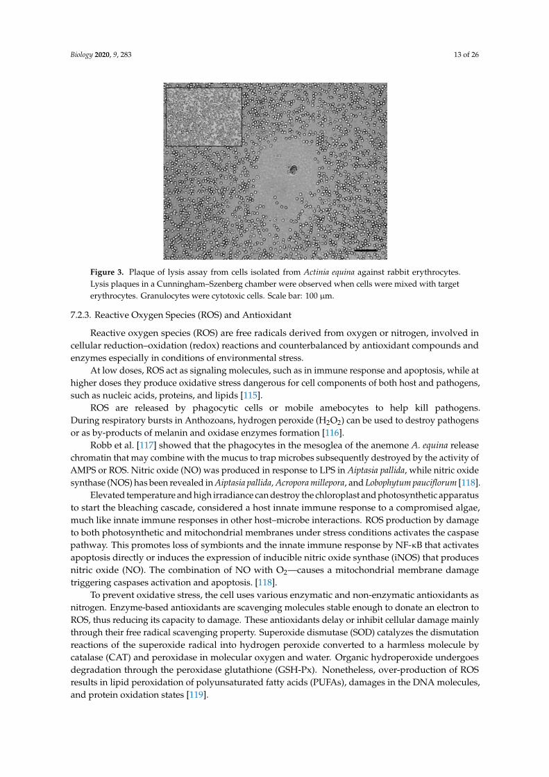
A. equina toxins have been found in specific marginal outgrowths of the body-wall (acrorhagi) andin cellular components of gastrodermal fluid [108] such as equinatoxins and sodium and potassiumchannel peptide toxins [112,113]. From internal liquid in A. equina specimens, six cell categorieshave been characterized and their lytic activity against several mammals’ erythrocytes types wasshown [114]. In Figure 3, the plaque lysis assay of cells of separated bands from a Percoll gradient,applied on a cellular suspension of A. equina, is shown by phase-contrast microscope.
Contrary to the belief that nematocysts are the only organ capable of releasing toxins with specificbiological activity, some types of cells identified as granulocytes in A. equina contain lytic molecules.In some cnidarian species, amebocytes participate in the wound healing process [110] as with thehard-coral P. cylindrical. Coagulation leading to clot formation occurs via the degranulation of immunecells and the incorporation of cellular debris and extracellular matrix followed by infiltration of immunecells into tissues in the wound area.
In some invertebrates, a phase characterized by the infiltration of amoebocytes and the presenceof fibroblasts able to control extracellular matrix production and collagen release was identified.The epithelial cells proliferate and migrate to regulate the re-epithelialization process [111].
Biology 2020, 9, 283 13 of 26
Biology 2020, 9, x 12 of 25
7.2. Cellular Responses
7.2.1. Phagocytosis
Circulating invertebrate immune cells are capable of phagocytosis, engulfing and destroying foreign invaders. Innate immune systems also include recognition proteins that bind to molecules of the bacterial wall, fungi and other pathogens, and recognition of foreign agents triggers the production of antimicrobial peptides stored in the cells [104]. Within the Cnidaria, cells with phagocytic activity toward potential pathogen substances were found in the sea anemone A. equina and in the soft coral S. exserta [108,109].
Cells specialized in the phagocytosis process against the Aspergillus sydowii fungal pathogen have been identified in the Gorgonia ventalina [110]. In the gorgonaceae cells, phagocytosis is activated post trauma and thermal stress [111]. Olano & Bigger [109] classified phagocytes in granular amoebocytes, epidermal cells, sclerocytes, mesogleal cells, and gastrodermal cells.
7.2.2. Cytotoxicity
A. equina toxins have been found in specific marginal outgrowths of the body-wall (acrorhagi) and in cellular components of gastrodermal fluid [108] such as equinatoxins and sodium and potassium channel peptide toxins [112,113]. From internal liquid in A. equina specimens, six cell categories have been characterized and their lytic activity against several mammals’ erythrocytes types was shown [114]. In Figure 3, the plaque lysis assay of cells of separated bands from a Percoll gradient, applied on a cellular suspension of A. equina, is shown by phase-contrast microscope.
Contrary to the belief that nematocysts are the only organ capable of releasing toxins with specific biological activity, some types of cells identified as granulocytes in A. equina contain lytic molecules. In some cnidarian species, amebocytes participate in the wound healing process [110] as with the hard-coral P. cylindrical. Coagulation leading to clot formation occurs via the degranulation of immune cells and the incorporation of cellular debris and extracellular matrix followed by infiltration of immune cells into tissues in the wound area.
In some invertebrates, a phase characterized by the infiltration of amoebocytes and the presence of fibroblasts able to control extracellular matrix production and collagen release was identified. The epithelial cells proliferate and migrate to regulate the re-epithelialization process [111].
Figure 3. Plaque of lysis assay from cells isolated from Actinia equina against rabbit erythrocytes. Lysis plaques in a Cunningham–Szenberg chamber were observed when cells were mixed with target erythrocytes. Granulocytes were cytotoxic cells. Scale bar: 100 µm.
Figure 3. Plaque of lysis assay from cells isolated from Actinia equina against rabbit erythrocytes.Lysis plaques in a Cunningham–Szenberg chamber were observed when cells were mixed with targeterythrocytes. Granulocytes were cytotoxic cells. Scale bar: 100 µm.
7.2.3. Reactive Oxygen Species (ROS) and Antioxidant
Reactive oxygen species (ROS) are free radicals derived from oxygen or nitrogen, involved incellular reduction–oxidation (redox) reactions and counterbalanced by antioxidant compounds andenzymes especially in conditions of environmental stress.
At low doses, ROS act as signaling molecules, such as in immune response and apoptosis, while athigher doses they produce oxidative stress dangerous for cell components of both host and pathogens,such as nucleic acids, proteins, and lipids [115].
ROS are released by phagocytic cells or mobile amebocytes to help kill pathogens.During respiratory bursts in Anthozoans, hydrogen peroxide (H2O2) can be used to destroy pathogensor as by-products of melanin and oxidase enzymes formation [116].
Robb et al. [117] showed that the phagocytes in the mesoglea of the anemone A. equina releasechromatin that may combine with the mucus to trap microbes subsequently destroyed by the activity ofAMPS or ROS. Nitric oxide (NO) was produced in response to LPS in Aiptasia pallida, while nitric oxidesynthase (NOS) has been revealed in Aiptasia pallida, Acropora millepora, and Lobophytum pauciflorum [118].
Elevated temperature and high irradiance can destroy the chloroplast and photosynthetic apparatusto start the bleaching cascade, considered a host innate immune response to a compromised algae,much like innate immune responses in other host–microbe interactions. ROS production by damageto both photosynthetic and mitochondrial membranes under stress conditions activates the caspasepathway. This promotes loss of symbionts and the innate immune response by NF-κB that activatesapoptosis directly or induces the expression of inducible nitric oxide synthase (iNOS) that producesnitric oxide (NO). The combination of NO with O2—causes a mitochondrial membrane damagetriggering caspases activation and apoptosis. [118].
To prevent oxidative stress, the cell uses various enzymatic and non-enzymatic antioxidants asnitrogen. Enzyme-based antioxidants are scavenging molecules stable enough to donate an electron toROS, thus reducing its capacity to damage. These antioxidants delay or inhibit cellular damage mainlythrough their free radical scavenging property. Superoxide dismutase (SOD) catalyzes the dismutationreactions of the superoxide radical into hydrogen peroxide converted to a harmless molecule bycatalase (CAT) and peroxidase in molecular oxygen and water. Organic hydroperoxide undergoesdegradation through the peroxidase glutathione (GSH-Px). Nonetheless, over-production of ROSresults in lipid peroxidation of polyunsaturated fatty acids (PUFAs), damages in the DNA molecules,and protein oxidation states [119].
Biology 2020, 9, 283 14 of 26
Anthozoans possess many types of enzymatic antioxidants, peroxidases [120], SOD [121] andCAT [122]. This antioxidant activity varies with alteration in environmental conditions and particularlyduring bleaching or in response to both injury and infection.
Anemonia viridis has two kinds of SOD (CuZnSOD and MnSOD) enzymes, both of which are foundin the ectoderm and the endoderm of this anemone. In this species, as in the coral Goniopora stokesi,the antioxidant molecules are associated with granular vesicles and accumulation bodies in theperoxisomes [123].
Wounds and alteration to the environmental temperature modulate a production of antioxidantenzymes in Gorgonians species such as P. elisabethae, P. americana, Eunicea fusca and L. chilensis. Thus,enzymes act as biomarkers for environmental stress assessment [124].
In Stylophora pistillata [125], the production of antioxidant enzymes is correlated with thebleaching process, demonstrating the strong involvement of light radiation and thermal stress in theROS activation.
The symbiotic zooxantellae of hermatypic or reef-building corals such as Seriatopora hystrix andStylophora pistillata produce a large amount of ROS. This production is followed by the activation of thecaspase cascade up to apoptosis and bleaching of the polyp host [126].
The family of peroxidase enzymes, a subgroup of oxidoreductase, is involved in a number ofdefense and metabolic processes. In addition to the relationship with the formation of ROS, they attendin the detoxification of oxygen forms, phagocytosis activity, and melanin synthesis [127,128].
In G. ventalina, five peroxidase isoforms production is induced by inflammatory conditions dueto infection with fungal pathogens, as demonstrated by Mydlarz and Harvell [120]. Peroxidasewas localized in granular amoebocytes during the process of phagocytosis in the gorgonianSwiftia exserta [109]. In anemones, there are other molecules that contribute to antioxidant systems,but these are not scavengers—rather, they are fluorescent proteins (FPs). These are involved inphotoprotection and eliminate hydrogen peroxide [129].
8. Inflammatory Processes in Model Anemonia viridis
Actiniaria is one of the most successful and diverse taxa of Anthozoa. Sea anemones occupyall marine habitats, depths and latitudes, despite their body simplicity [130]. The group comprises48 families, 269 valid genera and more than 1000 species [131,132].
Anemonia viridis is a symbiotic sea anemone from a wide geographical range of temperate areas,Its immense ecological success is due to a symbiotic association based on nutritional exchanges withzooxanthellae that live within endodermal cells facing the gastroderm and separated from the externalenvironment by the ectodermal layer and the mesoglea. The tentacles are lined with venomous stingingcells called spirocysts used to paralyze the preys while the movement in the tentacles brings the foodthrough the mouth for extracellular digestion (Figure 4).
From June through August, the oviparous snakelocks anemone reproduces sexually. In addition,longitudinal fission occurs: a literal sea anemone cleavage which starts from the basal disk. The wholeprocess lasts from five minutes up to two hours [133].
A. viridis is a model for studies on symbiosis and environmental stress (temperature, light,symbiosis breakdown), power of acclimatization to climate change, evolution of innate immunity,and inflammatory responses [134,135].
Different host genetic lineages, differing in their associated symbiont populations, have beenidentified. The species A. viridis is present in the Mediterranean with the following color morphsbased on pigment content: rustica, vulgaris, viridis, smaragdina and rufescens [136]. These not only differfor morphology, reproduction modality and protein composition, but also with the presence of twogreen fluorescent pigments (gp499; gp522), an orange fluorescent pigment (OP) and a red-pigment(RP) non-fluorescent in the ectoderm with UV protection function (Figure 5).

Biology 2020, 9, 283 15 of 26Biology 2020, 9, x 15 of 25
Figure 4. Anatomy of Anemonia viridis. Gomory stain of tentacles histological section (M: Mesoglea, Sy: Symbiont, Sp: Spyrocysts, Muc: Mucocytes, Ci: Cilia, MF: Muscular fiber). Bar: 10 µm.
Figure 5. A. viridis color morphs based on pigment content. Specimens collected along the North Sicilian coast and maintained in the laboratory. The red (rustica variety) and green (viridis variety) pigment leakage is detectable after irradiation with ultraviolet light.
Removal of microbial agents is carried out via phagocytosis and enzymatic activation to circumscribe the area of infection and inflammation [141]. This follows the activation of effective molecules by transduction of intracellular signals.
Sea anemones also use antimicrobial molecules from mucus as the host defense mechanisms against infectious disease. Their functional cellular immune system is analogous to vertebrate immune mechanisms [142].
Gene expression studies also suggest a conserved function for cnidarian innate immune pathways. In the coral Acropora cervicornis, several immunity related transcripts were shown to be upregulated in animals with white band disease including a C-type lectin, collectin, and a TLR-2 like gene [143]. In addition, challenge of the coral A. millepora with molecules of both viral and bacterial origins showed differences in expression of several immune genes and challenge with live pathogens lead to an increase in C3 expression [67]. Therefore, there is evidence for the involvement of cnidarian innate immune pathways, including the complement system, in the defense against pathogens.
Stress events or even natural contact with other individuals in the environment causes the rich mucus secretion of molecules with cytotoxic activity [144].
Furthermore, mucus from Acropora palmata inhibits water column marine bacteria [145]. Stronger antibacterial activity versus Bacillus subtilis, Staphylococcus aureus, Salmonella typhimurium and Serratia marcescens has been detected in specimens from populations living in stable environments and not stressed by the alteration of chemical and physical factors, especially temperature, than in samples isolated from bleached or stressed colonies [146].
Figure 4. Anatomy of Anemonia viridis. Gomory stain of tentacles histological section (M: Mesoglea, Sy:Symbiont, Sp: Spyrocysts, Muc: Mucocytes, Ci: Cilia, MF: Muscular fiber). Bar: 10 µm.
Biology 2020, 9, x 15 of 25
Figure 4. Anatomy of Anemonia viridis. Gomory stain of tentacles histological section (M: Mesoglea, Sy: Symbiont, Sp: Spyrocysts, Muc: Mucocytes, Ci: Cilia, MF: Muscular fiber). Bar: 10 µm.
Figure 5. A. viridis color morphs based on pigment content. Specimens collected along the North Sicilian coast and maintained in the laboratory. The red (rustica variety) and green (viridis variety) pigment leakage is detectable after irradiation with ultraviolet light.
Removal of microbial agents is carried out via phagocytosis and enzymatic activation to circumscribe the area of infection and inflammation [141]. This follows the activation of effective molecules by transduction of intracellular signals.
Sea anemones also use antimicrobial molecules from mucus as the host defense mechanisms against infectious disease. Their functional cellular immune system is analogous to vertebrate immune mechanisms [142].
Gene expression studies also suggest a conserved function for cnidarian innate immune pathways. In the coral Acropora cervicornis, several immunity related transcripts were shown to be upregulated in animals with white band disease including a C-type lectin, collectin, and a TLR-2 like gene [143]. In addition, challenge of the coral A. millepora with molecules of both viral and bacterial origins showed differences in expression of several immune genes and challenge with live pathogens lead to an increase in C3 expression [67]. Therefore, there is evidence for the involvement of cnidarian innate immune pathways, including the complement system, in the defense against pathogens.
Stress events or even natural contact with other individuals in the environment causes the rich mucus secretion of molecules with cytotoxic activity [144].
Furthermore, mucus from Acropora palmata inhibits water column marine bacteria [145]. Stronger antibacterial activity versus Bacillus subtilis, Staphylococcus aureus, Salmonella typhimurium and Serratia marcescens has been detected in specimens from populations living in stable environments and not stressed by the alteration of chemical and physical factors, especially temperature, than in samples isolated from bleached or stressed colonies [146].
Figure 5. A. viridis color morphs based on pigment content. Specimens collected along the NorthSicilian coast and maintained in the laboratory. The red (rustica variety) and green (viridis variety)pigment leakage is detectable after irradiation with ultraviolet light.
These differences reflect an adaptation to environmental conditions, although they can alsocomprise explicit genetic differences divergent in the context of speciation phenomena.
In the northern coastal area of Sicily, we have previously characterized subtidal specimens thatshowed less pigment than infralittoral organisms, due to dysfunction or breakdown of symbiosis withzooxantellae in frequent swirling events, during which the temperature can increase considerably [137].In anemone species, as with corals, bleaching is a stress response to environmental perturbation,including changes in salinity, increased visible and/or ultraviolet radiations, increased sediments,nutrients, or pollutants [138].
Studies have focused on identification of the etiological agents of coral diseases [139], but littlework has focused on the cellular mechanisms that cnidarians possess to defend against pathogenicmicrobes. Anthozoans are potentially damaged by infections, anthropogenic stress, disease, and factorsof climate change. In the last decade, the knowledge of recovery mechanisms after inflammatoryevents or injuries has increased. During prey catching, the Anthozoans species with soft tentacles,such as the Mediterranean Sea anemone A. viridis, can be subject to tentacle breakage, becomingvulnerable to pathogenic infections present in the environment. The immune humoral and cellulareffectors trigger various inflammatory processes, which ensure the survival of the species in theirnatural environment [140].
Removal of microbial agents is carried out via phagocytosis and enzymatic activation tocircumscribe the area of infection and inflammation [141]. This follows the activation of effectivemolecules by transduction of intracellular signals.

Biology 2020, 9, 283 16 of 26
Sea anemones also use antimicrobial molecules from mucus as the host defense mechanismsagainst infectious disease. Their functional cellular immune system is analogous to vertebrate immunemechanisms [142].
Gene expression studies also suggest a conserved function for cnidarian innate immune pathways.In the coral Acropora cervicornis, several immunity related transcripts were shown to be upregulatedin animals with white band disease including a C-type lectin, collectin, and a TLR-2 like gene [143].In addition, challenge of the coral A. millepora with molecules of both viral and bacterial origins showeddifferences in expression of several immune genes and challenge with live pathogens lead to an increasein C3 expression [67]. Therefore, there is evidence for the involvement of cnidarian innate immunepathways, including the complement system, in the defense against pathogens.
Stress events or even natural contact with other individuals in the environment causes the richmucus secretion of molecules with cytotoxic activity [144].
Furthermore, mucus from Acropora palmata inhibits water column marine bacteria [145]. Strongerantibacterial activity versus Bacillus subtilis, Staphylococcus aureus, Salmonella typhimurium andSerratia marcescens has been detected in specimens from populations living in stable environments andnot stressed by the alteration of chemical and physical factors, especially temperature, than in samplesisolated from bleached or stressed colonies [146].
Within scleractinian corals, several types of immune cells have also been identified, includinggranular amoebocytes, melanin-containing cells, chromophore cells, agranular (hyaline) cells andfibroblast-like cells in response to injury [147].
The protection role of amebocytes from injuries and infections in A. equina is demonstrated fromingestion of the gram-negative bacterium Psychrobacter [141]. In addition, granular amebocytes flow tothe sites of fungal infections. In the soft coral S. exserta, phagocytic activity is dependent on the type oftrauma, and normally the amebocytes are concentrated around inflicted wounds in order to clean upcell debris [105].
Palmer and Traylor-Knowles [148] described a study about the Caribbean gorgonianPlexaurella fusifera subjected to injury, during which amoebocytes and the photosynthetic endosymbiontform an epithelial front. The amebocytes, increased in number at the injured area, extrude mesoglealconnective fibers to promote the regeneration process, while the zooxanthellae provide the energyused for the repair processes.
In Anthozoa sea fan corals, a localized inflammatory reaction to fungal infection and a systemicresponse to environmental stress (such as seawater warming) triggered by an increase of granularacidophilic amebocytes normally involved in injury repair and a self/non-self-recognition system wasdescribed. Furthermore, along with the systemic cellular response, a related presence of melanin in seafan tissue was detected.
Particularly in Anthozoan tissues, reactive oxygen species increased following injury and infectionevents, and studying these can be useful for understanding the physiological dynamics during a shiftfrom a functional to a not active host–symbiont interaction.
Disturbances such as anchoring, sedimentation, predation, and algal overgrowth result inwounding and compromised integrity of Anthozoan integrity. Disturbances that create wounds enablethe entry of microbes into the tissues. It is thus important to determine how the species respond towounding and physical damage and understand their capacity to regenerate the wounded areas.
Even for Anthozoans, the danger model theory can justify a series of humoral and cellularresponses, such as inflammatory, so their immune system can distinguish between harmful andself-bodies. In soft corals and anemones, after an injury, the mesoglea swells and cells infiltrate fromthe mesoglea to the point of injury. Thereafter, a process of re-epithelialization begins from the marginof the wound.
In the sea anemone Anthopleura elegantissima, the repair area was detected at 72 h after thermalstressor treatment, and the movement of cells from the epithelium cells around and from the mesogleawas observed [149]. In the soft coral P. fusifera, re-epithelialization was detected 24 h after the onset

Biology 2020, 9, 283 17 of 26
of a wound [150]. Conversely, Vargas [151] did not find any specific reaction in Montipora capitataspecimens, due to the narrow with of mesoglea.
Our research A. viridis innate responses after injection of various bacterial and non-pathogenicsubstances of varying size showed the reaction of a swelling of the pedal disc and the overlying portionof the body, as well as the formation of a small yellowish portion.
By histological analysis in the section of the infected animals, a specific reaction and extrusionin the pedal disk was observed, and mesenteric filaments oocytes were found to be coming from thepores of the aconzie extrusion. The reaction zone, formed after injection, decreased over time until thespecimens had completely recovered (Figure 6).
Biology 2020, 9, x 17 of 25
In A. viridis, we analyzed the protease, phosphatase, and esterase activity following the injection of different bacterial strains. Results suggested a relationship between inflammatory process and modulation of enzymatic activities and in particular, a strong correlation between the animal's inflammatory response and increased phosphatase and esterase in the tentacles and body extract infected with E. coli and V. alginolitycus.
Figure 6. Morphology variation after bacterial infection in A. viridis. (A), Schematic model of anatomy and injection site, swelling and reaction (B), Reaction zone (C), rejection and swelling of animal body 24 h after injection of suspensions of various heat-killed bacteria in (inset) the reaction after E. coli injection (D), A. viridis Gomori stain histological section (E), The original figure was produced for the study published by Trapani et al., [135]. The modified figure is consistent with the topic of the review.
9. Conclusions
Cnidarians possess components of the main pathways of invertebrate immunity. Receptors and pathways already identified indicate that these basal invertebrates are far from “simple” in their dealing with potential invading microbes and pathogens. However, there are numerous gaps in current knowledge, with a serious lack of functional component studies that can play a crucial role in immune responses.
The need to recognize and distinguish between symbiont and pathogenic microorganisms may be a selective force driving this diversity of PRRs in cnidarians. Immunological signaling cascades in the Cnidaria are among the most complex components and highly conserved within invertebrate innate immunity. Some of them are homologous with mammalian cascades.
The onset and maintenance of symbiosis in host cnidarians are associated with the downregulation of immune-related pathways host immunity. In fact, bleached Anthozoan specimens are less susceptible to pathogen infection, supporting the idea that reduced immunity is part of obligate symbioses with zooxantellae. Symbiodiniaceae downregulates host immunity to allow its survival in the host, similar to what occurs in other symbiotic mutualisms.
The chemical arsenal of Anthozoans represents the optimal strategy for survival, mainly through the production of neurotoxins, but also with the use of antimicrobial defense molecules.Multifunctionality alone can represent an optimal survival strategy, allowing these animals to be active predators through
Figure 6. Morphology variation after bacterial infection in A. viridis. (A), Schematic model of anatomyand injection site, swelling and reaction (B), Reaction zone (C), rejection and swelling of animal body24 h after injection of suspensions of various heat-killed bacteria in (inset) the reaction after E. coliinjection (D), A. viridis Gomori stain histological section (E), The original figure was produced for thestudy published by Trapani et al., [135]. The modified figure is consistent with the topic of the review.
During the early responses of the Anthozoans to pathogen agents or environmental stress,enzymatic activity (i.e., peroxidase or hydrolase) plays a focal role. In addition, differentiation of cellsand nematocysts are controlled by a modification of the alkaline and acid phosphatase levels. Amongthese, only the peroxidase has been widely studied as a marker of regeneration in Hydra [152].
Mydlardz et al., [110] examined the role and inducibility of defense responses in theGorgonia ventalina peroxidase enzyme superfamily to an A. sydowii fungal pathogen infection. Severalisoforms of peroxidase in healthy colonies were found indicating the involvement of peroxidase in aplurality of physiological functions, such as the activation of signal cascade mechanisms of transduction,or activity against the fungal type employed.
In A. viridis, we analyzed the protease, phosphatase, and esterase activity following the injectionof different bacterial strains. Results suggested a relationship between inflammatory process andmodulation of enzymatic activities and in particular, a strong correlation between the animal’sinflammatory response and increased phosphatase and esterase in the tentacles and body extractinfected with E. coli and V. alginolitycus.
Biology 2020, 9, 283 18 of 26
9. Conclusions
Cnidarians possess components of the main pathways of invertebrate immunity. Receptors andpathways already identified indicate that these basal invertebrates are far from “simple” in theirdealing with potential invading microbes and pathogens. However, there are numerous gaps incurrent knowledge, with a serious lack of functional component studies that can play a crucial role inimmune responses.
The need to recognize and distinguish between symbiont and pathogenic microorganisms may bea selective force driving this diversity of PRRs in cnidarians. Immunological signaling cascades in theCnidaria are among the most complex components and highly conserved within invertebrate innateimmunity. Some of them are homologous with mammalian cascades.
The onset and maintenance of symbiosis in host cnidarians are associated with the downregulationof immune-related pathways host immunity. In fact, bleached Anthozoan specimens are less susceptibleto pathogen infection, supporting the idea that reduced immunity is part of obligate symbioses withzooxantellae. Symbiodiniaceae downregulates host immunity to allow its survival in the host, similarto what occurs in other symbiotic mutualisms.
The chemical arsenal of Anthozoans represents the optimal strategy for survival, mainly through theproduction of neurotoxins, but also with the use of antimicrobial defense molecules. Multifunctionalityalone can represent an optimal survival strategy, allowing these animals to be active predators throughthe production of neurotoxins and to resist bacterial infections caused by the possible breakage of thetentacles through the functionality of antimicrobial peptides.
This work aims to expand knowledge related not only to the characterization of the effectors ofinnate immunity of Anthozoans, and to the various and multiple activities of antimicrobial toxins andpeptides, but also to analyze the inflammatory response in Anthozoans as metazoan model systems.This may flesh out the scenario of the first stages of the evolution of immunity.
The study of the inflammatory response, which occurred following the inoculation of varioussubstances in A. viridis, enabled the observation of specific reactions, especially following the inoculationof bacteria such as E. coli. This specific response, which ends in the appearance of a rejection zone,suggests an extrusion of the damaged material by the animal, probably via the pores of extrusion ofthe acoustical present in the pedal disc, a defense mechanism not yet found in other cnidarians.
A further study was also carried out in the form of a histological analysis for the characterizationof the tissues and the rejection area.
Enzymatic activity, specifically protease, phosphatase, and esterase activity, was also analyzed,showing how the inoculation of the different bacterial strains, in particular E. coli, alters the expressionof these enzymes. These data agree with the results of the studies on inflammation, suggestinga correlation between the appearance of the inflammatory reaction and the modification of theenzymatic activity.
Despite progress over the past decade in learning about cnidarian immunity, many studies arestill needed to understand how key processes are translated from receptor to effector.
We are evaluating the possibility of studying empirically responses of hosts to specific stressors,such as pathogenic challenge.
Cnidarians are also important to study from an evolutionary perspective because they possessa surprising amount of complexity in their genomes and share many pathways with vertebratesthat are not present in other invertebrate groups. Information on innate immunity pathways incnidarians can provide insight into the evolutionary origins of mechanisms vertebrates use to respondto foreign microbes.
Author Contributions: M.G.P. and M.C. wrote the manuscript, conceived the idea, and supervised the wholestudy. L.S. and D.P. participated in the experimental part and revised the manuscript. All authors have read andagreed to the published version of the manuscript.
Funding: This study was funded by the FFR-Cammarata (PJ_RIC_FFABR_2017_004312 MC) and FFR-Parisi(PJ_RIC_FFABR_2017_161753 MGP) for Scientific Research from the University of Palermo, Italy.

Biology 2020, 9, 283 19 of 26
Acknowledgments: We are grateful to M. Dara that realized the Figure 2 picture and Claudia la Corte for thesupport in the histological study.
Conflicts of Interest: The authors declare no conflict of interest.
References
1. Petralia, R.S.; Mattson, M.P.; Yao, P.J. Aging and longevity in the simplest animals and the quest forimmortality. Ageing Res. Rev. 2014, 16, 66–82. [CrossRef] [PubMed]
2. Miller, D.J.; Hemmrich, G.; Ball, E.E.; Hayward, D.C.; Khalturin, K.; Funayama, N.; Agata, K.; Bosch, T.C.G.The innate immune repertoire in cnidaria–ancestral complexity and stochastic gene loss. Genome Biol. 2007,8, R59. [CrossRef] [PubMed]
3. McFall-Ngai, M.; Hadfield, M.G.; Bosch, T.C.G.; Carey, H.V.; Domazet-Lošo, T.; Douglas, A.E.; Dubilier, N.;Eberl, G.; Fukami, T.; Gilbert, S.F.; et al. Animals in a bacterial world, a new imperative for the life sciences.Proc. Natl. Acad. Sci. USA 2013, 110, 3229–3236. [CrossRef] [PubMed]
4. Soza-Ried, J.; Hotz-Wagenblatt, A.; Glatting, K.-H.; del Val, C.; Fellenberg, K.; Bode, H.R.; Frank, U.;Hoheisel, J.D.; Frohme, M. The transcriptome of the colonial marine hydroid Hydractinia echinata. FEBS J.2010, 277, 197–209. [CrossRef] [PubMed]
5. Chapman, J.A.; Kirkness, E.F.; Simakov, O.; Hampson, S.E.; Mitros, T.; Weinmaier, T.; Rattei, T.;Balasubramanian, P.G.; Borman, J.; Busam, D.; et al. The dynamic genome of Hydra. Nature 2010,464, 592–596. [CrossRef]
6. Wenger, Y.; Galliot, B. RNAseq versus genome-predicted transcriptomes: A large population of noveltranscripts identified in an Illumina-454 Hydra transcriptome. BMC Genom. 2013, 14, 204. [CrossRef]
7. Putnam, N.H.; Srivastava, M.; Hellsten, U.; Dirks, B.; Chapman, J.; Salamov, A.; Terry, A.; Shapiro, H.;Lindquist, E.; Kapitonov, V.V.; et al. Sea anemone genome reveals ancestral eumetazoan gene repertoire andgenomic organization. Science 2007, 317, 86–94. [CrossRef]
8. Buchmann, K. Evolution of Innate Immunity: Clues from Invertebrates via Fish to Mammals. Front. Immunol.2014, 5, 459. [CrossRef]
9. Vidal-Dupiol, J.; Adjeroud, M.; Roger, E.; Foure, L.; Duval, D.; Mone, Y.; Ferrier-Pages, C.; Tambutte, E.;Tambutte, S.; Zoccola, D.; et al. Coral bleaching under thermal stress: Putative involvement of host/symbiontrecognition mechanisms. BMC Physiol. 2009, 9, 14. [CrossRef]
10. Augustin, R.; Bosch, T.C.G. Cnidarian immunity: A tale of two barriers. Adv. Exp. Med. Biol. 2010, 708, 1–16.[CrossRef]
11. Augustin, R.; Siebert, S.; Bosch, T.C.G. Identification of a kazal-type serine protease inhibitor with potentanti-staphylococcal activity as part of Hydra’s innate immune system. Dev. Comp. Immunol. 2009, 33,830–837. [CrossRef] [PubMed]
12. Stabili, L.; Schirosi, R.; Parisi, M.G.; Piraino, S.; Cammarata, M. The mucus of Actinia equina (Anthozoa,Cnidaria): An unexplored resource for potential applicative purposes. Mar. Drugs 2015, 13, 5276–5296.[CrossRef] [PubMed]
13. Rinkevich, B. Neglected Biological Features in Cnidarians Self-Nonself Recognition. Adv. Exp. Med. Biol.2012, 738, 46–59. [CrossRef] [PubMed]
14. Campbell, R.D.; Bibb, C. Transplantation in coelenterates. Transplant. Proc. 1970, 2, 202–211. [PubMed]15. Hildemann, W.H.; Jokiel, P.L.; Bigger, C.H.; Johnston, I.S. Allogeneic polymorphism and alloimmune memory
in the coral, Montipora verrucosa. Transplantation 1980, 30, 297–301. [CrossRef]16. Lubbock, R. Clone-specific cellular recognition in a sea anemone. Proc. Natl. Acad. Sci. USA 1980, 77,
6667–6669. [CrossRef] [PubMed]17. Palmer, C.V.; Traylor-Knowles, N. Towards an integrated network of coral immune mechanisms.
Proc. Biol. Sci. 2012, 279, 4106–4114. [CrossRef]18. Rinkevich, B. Allorecognition and xenorecognition in reef corals: A decade of interactions. Hydrobiologia
2004, 530, 443–450. [CrossRef]19. Netea, M.G.; Quintin, J.; van der Meer, J.W.M. Trained immunity: A memory for innate host defense.
Cell Host Microbe 2011, 9, 355–361. [CrossRef]20. Karadge, U.B.; Gosto, M.; Nicotra, M.L. Allorecognition proteins in an invertebrate exhibit homophilic
interactions. Curr. Biol. 2015, 25, 2845–2850. [CrossRef]
Biology 2020, 9, 283 20 of 26
21. Frank, U.; Rinkevich, B. Alloimmune memory is absent in the Red Sea hydrocoral Millepora dichotoma.J. Exp. Zool. 2001, 291, 25–29. [CrossRef] [PubMed]
22. Chadwick-Furman, N.; Rinkevich, B. A complex allorecognition system in a reef-building coral: Delayedresponses, reversals and nontransitive hierarchies. Coral Reefs 1994, 13, 57–63. [CrossRef]
23. Nicotra, M.L. Invertebrate allorecognition. Curr. Biol. 2019, 29, R463–R467. [CrossRef]24. Cadavid, L.F.; Powell, A.E.; Nicotra, M.L.; Moreno, M.; Buss, L.W. An invertebrate histocompatibility
complex. Genetics 2004, 167, 357–365. [CrossRef] [PubMed]25. Dishaw, L.J.; Litman, G.W. Invertebrate Allorecognition: The Origins of Histocompatibility. Curr. Biol. 2009,
19, 6–10. [CrossRef]26. Kuznetsov, S.G.; Bosch, T.C.G. Self/nonself recognition in Cnidaria: Contact to allogeneic tissue does not
result in elimination of nonself cells in Hydra vulgaris. Zoology 2003, 106, 1–8. [CrossRef]27. Tremblay, P.; Grover, R.; Maguer, J.F.; Legendre, L.; Ferrier-Pagès, C. Autotrophic carbon budget in coral
tissue: A new 13C-based model of photosynthate translocation. J. Exp. Biol. 2012, 215, 1384–1393. [CrossRef]28. Allemand, D.; Furla, P.; Tambutté, S. Mechanisms of carbon acquisition for endosymbiont photosynthesis in
Anthozoa. Can. J. Bot. 2011, 76, 925–941. [CrossRef]29. Santos, S.R.; Taylor, D.J.; Kinzie, R.A.; Hidaka, M.; Sakai, K.; Coffroth, M.A. Molecular phylogeny of symbiotic
dinoflagellates inferred from partial chloroplast large subunit (23S)-rDNA sequences. Mol. Phylogenet. Evol.2002, 23, 97–111. [CrossRef]
30. LaJeunesse, T.C.; Parkinson, J.E.; Gabrielson, P.W.; Jeong, H.J.; Reimer, J.D.; Voolstra, C.R.; Santos, S.R.Systematic Revision of Symbiodiniaceae Highlights the Antiquity and Diversity of Coral Endosymbionts.Curr. Biol. 2018, 28, 2570–2580.e6. [CrossRef]
31. Burns, J.A.; Zhang, H.; Hill, E.; Kim, E.; Kerney, R. Transcriptome analysis illuminates the nature of theintracellular interaction in a vertebrate-algal symbiosis. Elife 2017, 6. [CrossRef]
32. Mansfield, K.M.; Carter, N.M.; Nguyen, L.; Cleves, P.A.; Alshanbayeva, A.; Williams, L.M.; Crowder, C.;Penvose, A.R.; Finnerty, J.R.; Weis, V.M.; et al. Transcription factor NF-κB is modulated by symbiotic statusin a sea anemone model of cnidarian bleaching. Sci. Rep. 2017, 7, 16025. [CrossRef] [PubMed]
33. Pinzón, J.H.; Kamel, B.; Burge, C.A.; Harvell, C.D.; Medina, M.; Weil, E.; Mydlarz, L.D. Whole transcriptomeanalysis reveals changes in expression of immune-related genes during and after bleaching in a reef-buildingcoral. R. Soc. Open Sci. 2015, 2. [CrossRef]
34. Caroselli, E.; Goffredo, S. Population Dynamics of Temperate Corals in a Changing Climate. In The Cnidaria,Past, Present and Future. The World of Medusa and Her Sisters; Springer: Berlin, Germany, 2016; pp. 317–328.
35. Merselis, D.G.; Lirman, D.; Rodriguez-Lanetty, M. Symbiotic immuno-suppression: Is disease susceptibilitythe price of bleaching resistance? PeerJ 2018, 6, e4494. [CrossRef] [PubMed]
36. Dunn, S.R.; Schnitzler, C.E.; Weis, V.M. Apoptosis and autophagy as mechanisms of dinoflagellate symbiontrelease during cnidarian bleaching: Every which way you lose. Proc. Biol. Sci. 2007, 274, 3079–3085.[CrossRef]
37. Brumell, J.H.; Scidmore, M.A. Manipulation of rab GTPase function by intracellular bacterial pathogens.Microbiol. Mol. Biol. Rev. 2007, 71, 636–652. [CrossRef]
38. Chen, M.C.; Hong, M.C.; Huang, Y.S.; Liu, M.C.; Cheng, Y.M.; Fang, L.S. ApRab11, a cnidarian homologueof the recycling regulatory protein Rab11, is involved in the establishment and maintenance of theAiptasia-Symbiodinium endosymbiosis. Biochem. Biophys. Res. Commun. 2005, 338, 1607–1616. [CrossRef][PubMed]
39. Rohwer, F.; Seguritan, V.; Knowlton, N.; Rohwer, F.; Seguritan, V.; Azam, F.; Knowlton, N. Diversity anddistribution of coral-associated bacteria. Mar. Ecol. Prog. Ser. 2002, 243, 1–10. [CrossRef]
40. Vega Thurber, R.; Correa, A. Viruses of reef-building scleractinian corals. J. Exp. Mar. Biol. Ecol. 2011, 408,102–113. [CrossRef]
41. Rivera, H.; Thompson, J.; Medina, M.; Closek, C. Microbes in the coral holobiont: Partners through evolution,development, and ecological interactions. Front. Cell. Infect. Microbiol. 2015, 4. [CrossRef]
42. Deschaseaux, E.S.M.; Jones, G.B.; Deseo, M.A.; Shepherd, K.M.; Kiene, R.P.; Swan, H.B.; Harrison, P.L.;Eyre, B.D. Effects of environmental factors on dimethylated sulfur compounds and their potential role in theantioxidant system of the coral holobiont. Limnol. Oceanogr. 2014, 59, 758–768. [CrossRef]

Biology 2020, 9, 283 21 of 26
43. Cróquer, A.; Bastidas, C.; Elliott, A.; Sweet, M. Bacterial assemblages shifts from healthy to yellow banddisease states in the dominant reef coral Montastraea faveolata. Environ. Microbiol. Rep. 2013, 5, 90–96.[CrossRef] [PubMed]
44. Tinta, T.; Kogovšek, T.; Klun, K.; Malej, A.; Herndl, G.J.; Turk, V. Jellyfish-Associated Microbiome in theMarine Environment: Exploring Its Biotechnological Potential. Mar. Drugs 2019, 17, 94. [CrossRef] [PubMed]
45. Weiland-Bräuer, N.; Neulinger, S.C.; Pinnow, N.; Künzel, S.; Baines, J.F.; Schmitz, R.A. Composition ofbacterial communities associated with Aurelia aurita changes with compartment, life stage, and population.Appl. Environ. Microbiol. 2015, 81, 6038–6052. [CrossRef] [PubMed]
46. Kos Kramar, M.; Tinta, T.; Lucic, D.; Malej, A.; Turk, V. Bacteria associated with moon jellyfish during bloomand post-bloom periods in the Gulf of Trieste (northern Adriatic). PLoS ONE 2019, 14, e0198056. [CrossRef]
47. Basso, L.; Rizzo, L.; Marzano, M.; Intranuovo, M.; Fosso, B.; Pesole, G.; Piraino, S.; Stabili, L. Jellyfish summeroutbreaks as bacterial vectors and potential hazards for marine animals and humans health? The case ofRhizostoma pulmo (Scyphozoa, Cnidaria). Sci. Total Environ. 2019, 692, 305–318. [CrossRef] [PubMed]
48. Stabili, L.; Rizzo, L.; Basso, L.; Marzano, M.; Fosso, B.; Pesole, G.; Piraino, S. The Microbial CommunityAssociated with Rhizostoma pulmo: Ecological Significance and Potential Consequences for Marine Organismsand Human Health. Mar. Drugs 2020, 18, 437. [CrossRef]
49. Lawson, C.; Raina, J.; Kahlke, T.; Seymour, J.; Suggett, D. Defining the Core Microbiome of the SymbioticDinoflagellate, Symbiodinium. Environ. Microbiol. Rep. 2017, 10. [CrossRef]
50. Rosenberg, E.; Kushmaro, A.; Kramarsky-Winter, E.; Banin, E.; Yossi, L. The role of microorganisms in coralbleaching. ISME 2009, 3, 139–146. [CrossRef]
51. Porporato, E.M.; Lo Giudice, A.; Michaud, L.; De Domenico, E.; Spanò, N. Diversity and antibacterialactivity of the bacterial communities associated with two Mediterranean sea pens, Pennatula phosphorea andPteroeides spinosum (Anthozoa: Octocorallia). Microb. Ecol. 2013, 66, 701–714. [CrossRef]
52. Ocampo, I.D.; Cadavid, L.F. Mechanisms of immune responses in Cnidarians. Acta Biol. Colomb. 2015, 20,5–11. [CrossRef]
53. Chimetto, L.A.; Cleenwerck, I.; Alves, N., Jr.; Silva, B.S.; Brocchi, M.; Willems, A.; De Vos, P.; Thompson, F.L.Vibrio communis sp. nov., isolated from the marine animals Mussismilia hispida, Phyllogorgia dilatata,Palythoa caribaeorum, Palythoa variabilis and Litopenaeus vannamei. Int. J. Syst. Evol. Microbiol. 2011,61, 362–368. [CrossRef] [PubMed]
54. Reshef, L.; Koren, O.; Loya, Y.; Zilber-Rosenberg, I.; Rosenberg, E. The coral probiotic hypothesis.Environ. Microbiol. 2006, 8, 2068–2073. [CrossRef]
55. Calow, P. Why some metazoan mucus secretions are more susceptible to microbial attack than others. Am. Nat.1979, 114, 149–152. [CrossRef]
56. Sharp, K.H.; Distel, D.; Paul, V.J. Diversity and dynamics of bacterial communities in early life stages of theCaribbean coral Porites astreoides. ISME 2012, 6, 790–801. [CrossRef] [PubMed]
57. Rosenberg, E.; Zilber-Rosenberg, I. Symbiosis and development: The hologenome concept. Birth Defects Res.C Embryo Today 2011, 93, 56–66. [CrossRef] [PubMed]
58. Ainsworth, T.D.; Fine, M.; Roff, G.; Hoegh-Guldberg, O. Bacteria are not the primary cause of bleaching inthe Mediterranean coral Oculina patagonica. ISME 2008, J2, 67–73. [CrossRef]
59. Miller, A.; Richardson, L. Emerging coral diseases: A temperature-driven process? Mar. Ecol. 2014, 36.[CrossRef]
60. Weis, V. Cellular mechanisms of Cnidarian bleaching: Stress causes the collapse of symbiosis. J. Exp. Biol.2008, 211, 3059–3066. [CrossRef]
61. Brennan, J.J.; Messerschmidt, J.L.; Williams, L.M.; Matthews, B.J.; Reynoso, M.; Gilmore, T.D. Sea anemonemodel has a single Toll-like receptor that can function in pathogen detection, NF-κB signal transduction,and development. Proc. Natl. Acad. Sci. USA 2017, 114, E10122–E10131. [CrossRef]
62. O’Neill, L.; Golenbock, D.; Bowie, A. The history of Toll-like receptors—Redefining innate immunity.Nat. Rev. Immunol. 2013, 13, 453–460. [CrossRef] [PubMed]
63. Medzhitov, R.; Janeway, C. Innate immune recognition: Mechanisms and pathways. Immunol. Rev. 2000, 173,89–97. [CrossRef]
64. Cerenius, L.; Kawabata, S.; Lee, B.; Nonaka, M.; Soderhall, K. Proteolytic cascades and their involvement ininvertebrate immunity. Trends Biochem. Sci. 2010, 35, 575–583. [CrossRef]

Biology 2020, 9, 283 22 of 26
65. Augustin, R.; Fraune, S.; Franzenburg, S.; Bosch, T. Where simplicity meets complexity: Hydra, a model forhostmicrobe interactions. Adv. Exp. Med. Biol. 2012, 710, 71–80. [CrossRef]
66. Palmer, C.; Bythell, J.; Willis, B. Enzyme activity demonstrates multiple pathways of innate immunity inIndo-Pacific anthozoans. Proc. R. Soc. B Biol. Sci. 2012, 279, 3879–3887. [CrossRef]
67. Janeway, C.; Medzhitov, R. Innate immune recognition. Annu. Rev. Immunol. 2002, 20, 197–216. [CrossRef][PubMed]
68. Hemmrich, G.; Miller, D.; Bosch, T. The evolution of immunity: A low-life perspective. Trends Immunol. 2007,28, 449–454. [CrossRef] [PubMed]
69. Van der Burg, C.; Prentis, P.; Surm, J.; Pavasovic, A. Insights into the innate immunome of actiniarians usinga comparative genomic approach. BMC Genom. 2016, 17, 850. [CrossRef]
70. Baumgarten, S.; Simakov, O.; Esherick, L.Y.; Leiw, Y.J.; Lehnert, E.M.; Michell, C.; Li, Y.; Hambleton, E.A.;Guse, A.; Oates, M.E.; et al. The genome of Aiptasia, a sea anemone model for coral symbiosis. Proc. Natl.Acad. Sci. USA 2015, 22, 11893–11898. [CrossRef]
71. Reitzel, A.; Sullivan, J.; Traylor-Knowles, N.; Finnerty, J. Genomic survey of candidatestress-response genesin the esturine anemone Nematostella vectensis. Biol Bull. 2008, 214, 233–254. [CrossRef]
72. Armulik, A.; Velling, T.; Johansson, S. The integrin beta1 subunit transmembrane domainregulates phosphatidylinositol 3-kinase-dependent tyrosine phosphorylation of Crk-associated substrate.Mol. Biol. Cell 2004, 15, 2558–2567. [CrossRef] [PubMed]
73. Knack, B.; Iguchi, A.; Shinzato, C.; Hayward, D.; Ball, E.; Miller, D. Unexpected diversity of cnidarianintegrins: Expression during coral gastrulation. BMC Evol. Biol. 2008, 8, 136. [CrossRef]
74. Schwarz, J.; Brokstein, P.; Voolstra, C.; Terry, A.; Miller, D.; Szmant, A.; Coffroth, M.; Medina, M. Coral lifehistory and symbiosis: Functional genomic resources for two reef building Caribbean corals, Acropora palmataand Montastraea faveolata. BMC Genom. 2008, 97, 1471–2164.
75. Kvennefors, E.C.E.; Leggat, W.; Hoegh-Guldberg, O.; Degnan, B.M.; Barnes, A.C. An ancient andvariable mannose-binding lectin from the coral Acropora millepora binds both pathogens and symbionts.Dev. Comp. Immunol. 2008, 32, 1582–1592. [CrossRef]
76. Desalvo, M.; Voolstra, C.; Sunagawa, S.; Schwarz, J.; Stillman, J.; Coffroth, M.; Szmant, A.; Medina, M.Differential gene expression during thermal stress and bleaching in the Caribbean coral Montastraea faveolata.Mol. Ecol. 2008, 17, 3952–3971. [CrossRef]
77. Elisha, M.W.-C.; Virginia, W.V. The diversity of C-Type lectin in the genome of a basal metazoan nematostellavectensis. Dev. Comp. Immunol. 2014, 33, 881–889.
78. Burge, C.; Mouchka, M.; Harvell, C.; Roberts, C. Immune response of the Caribbean sea fan, Gorgonia ventalina,exposed to an Aplanochytrium parasite as revealed by transcriptome sequencing. Front. Physiol. 2013, 4, 180.[CrossRef] [PubMed]
79. Bosch, T.; Augustin, R.; Anton-Erxleben, F.; Fraune, S.; Hemmrich, G.; Zill, H.; Rosenstiel, P.; Jacobs, G.;Schreiber, S.; Leippe, M.; et al. Uncovering the evolutionary history of innate immunity: The simple metazoanHydra uses epithelial cells for host defence. Dev. Comp. Immunol. 2009, 33, 559–569. [CrossRef] [PubMed]
80. Shinzato, C.; Shoguchi, E.; Kawashima, T.; Hamada, M.; Hisata, K.; Tanaka, M.; Fujie, M.; Fujiwara, M.;Koyanagi, R.; Ikuta, T.; et al. Using the Acropora digitifera genome to understand coral responses toenvironmental change. Nature 2011, 476, 320–323. [CrossRef]
81. Hamada, M.; Shoguchi, E.; Shinzato, C.; Kawashima, T.; Miller, D.J.; Satoh, N. The complex NOD-likereceptor repertoire of the coral Acropora digitifera includes novel domain combinations. Mol. Biol. Evol. 2013,30, 167–176. [CrossRef]
82. Leclerc, M. Humoral Factors in Marine Invertebrates. In Invertebrate Immnology. Progress in Molecularand Subcellular Biology; Rinkevich, B., Müller, W.E.G., Eds.; Springer: Berlin/Heidelberg, Germany, 1996;Volume 15, ISBN 9783642797378.
83. Kvennefors, E.C.E.; Leggat, W.; Kerr, C.C.; Ainsworth, T.D.; Hoegh-Guldberg, O.; Barnes, A.C.Analysis of evolutionarily conserved innate immune components in coral links immunity and symbiosis.Dev. Comp. Immunol. 2010, 34, 1219–1229. [CrossRef] [PubMed]
84. Dunn, S.R. Immunorecognition and immunoreceptors in the Cnidaria. ISJ 2009, 6, 7–14.85. Hayes, M.; Eytan, R.I.; Hellberg, M. High amino acid diversity and positive selection at a putative coral
immunity gene (tachylectin-2). BMC Evol. Biol. 2010, 10, 150. [CrossRef] [PubMed]

Biology 2020, 9, 283 23 of 26
86. Mcguinness, D.; Dehal, P.; Pleass, R. Pattern recognition molecules and innate immunity to parasites.Trends Parasitol. 2003, 19, 312–319. [CrossRef]
87. Lopez, J.; Fain, M.; Cadavid, L. The evolution of the immunetype gene family Rhamnospondin in cnidarians.Gene 2011, 473, 119–124. [CrossRef]
88. Detournay, O.; Schnitzler, C.; Poole, A.; Weis, V. Regulation of cnidarian–dinoflagellate mutualisms:Evidence that activation of a host TGFβ innate immune pathway promotes tolerance of the symbiont.Dev. Comp. Immunol. 2012, 38, 525–537. [CrossRef]
89. Weiss, Y.; Foret, S.; Hayward, D.; Ainsworth, T.; King, R.; Ball, E.; Miller, D.J. The acute transcriptionalresponse of the coral Acropora millepora to immune challenge: Expression of GiMAP/IAN genes links theinnate immune responses of corals with those of mammals and plants. BMC Genom. 2013, 14, 400. [CrossRef]
90. Palmer, C.V.; Mydlarz, L.D.; Willis, B.L. Evidence of an inflammatory-like response in non-normallypigmented tissues of two scleractinian corals. Proc. R. Soc. B Biol. Sci. 2008, 275, 2687–2693. [CrossRef]
91. Luna-Acosta, A.; Rosenfeld, E.; Amari, M.; Fruitier-Arnaudin, I.; Bustamante, P.; Thomas-Guyon, H. Firstevidence of laccase activity in the Pacific oyster Crassostrea gigas. Fish Shellfish Immunol. 2010, 28, 719–726.[CrossRef]
92. Takahashi, D.; Garcia, B.L.; Kanost, M.R. Initiating protease with modular domains interacts with β-glucanrecognition protein to trigger innate immune response in insects. Proc. Natl. Acad. Sci. USA 2015, 112,13856–13861. [CrossRef]
93. Vidal-Dupiol, J.; Dheilly, N.; Rondon, R.; Grunau, C.; Cosseau, C.; Smith, K.; Freitag, M.; Adjeroud, M.; Mitta, G.Thermal stress triggers broad Pocillopora damicornis transcriptomic remodeling, while Vibrio coralliilyticusinfection induces a more targeted immunosuppression response. PLoS ONE 2014, 9, e107672. [CrossRef]
94. Esposito, R.; D’Aniello, S.; Squarzoni, P.; Pezzotti, M.; Ristoratore, F.; Spagnuolo, A. New insights into theevolution of Metazoan tyrosinase gene family. PLoS ONE 2012, 7, e35731. [CrossRef] [PubMed]
95. Mydlarz, L.D.; Jones, L.E.; Harvell, C.D. Innate Immunity, Environmental Drivers, and Disease Ecology ofMarine and Freshwater Invertebrates. Annu. Rev. Ecol. Evol. Syst. 2006, 37, 251–288. [CrossRef]
96. Schultz, G.S.; Chin, G.A.; Moldawer, L.; Diegelmann, R.F. Principles of Wound Healing. In Mechanisms ofVascular Disease: A Reference Book for Vascular Specialists; Fitridge, R., Thompson, M., Eds.; University ofAdelaide Press: Adelaide, Australia, 2011; ISBN 9780987171825.
97. Fitt, W.; Gates, R.; Hoegh-Guldberg, O.; Bythell, J.C.; Grottoli, A.; Gomez Cabrera, K.-L.; Fisher, P.L.;Pantos, O.; Iglesias-Prieto, R.; Rodrigues, L.; et al. Response of two species of Indo-Pacific corals, Poritescylindrica and Stylophora pistillata, to short-term thermal stress: The host does matter in determining thetolerance of corals to bleaching. J. Exp. Mar. Biol. Ecol. 2009, 373, 102–110. [CrossRef]
98. Sarma, J.V.; Ward, P.A. The complement system. Cell Tissue Res. 2011, 343, 227–2358. [CrossRef] [PubMed]99. Noris, M.; Remuzzi, G. Overview of complement activation and regulation. Semin. Nephrol. 2013, 33, 479–492.
[CrossRef] [PubMed]100. Nonaka, M. The complement C3 protein family in invertebrates. Invertebr. Surviv. J. 2011, 8, 21–32.101. Endo, Y.; Nonaka, M.; Saiga, H.; Kakinuma, Y.; Matsushita, A.; Takahashi, M.; Matsushita, M.; Fujita, T.
Origin of mannose-binding lectin-associated serine protease (MASP)-1 and MASP-3 involved in the lectincomplement pathway traced back to the invertebrate, amphioxus. J. Immunol. 2003, 170, 4701–4707.[CrossRef]
102. Kimura, A.; Sakaguchi, E.; Nonaka, M. Multi-component complement system of Cnidaria: C3, Bf, and MASPgenes expressed in the endodermal tissues of a sea anemone, Nematostella vectensis. Immunobiology 2009, 214,165–178. [CrossRef]
103. Dishaw, L.J.; Smith, S.L.; Bigger, C.H. Characterization of a C3-like cDNA in a coral: Phylogenetic implications.Immunogenetics 2005, 57, 535–548. [CrossRef]
104. Fujito, N.T.; Sugimoto, S.; Nonaka, M. Evolution of thioester-containing proteins revealed by cloning andcharacterization of their genes from a cnidarian sea anemone, Haliplanella lineate. Dev. Comp. Immunol. 2010,34, 775–784. [CrossRef] [PubMed]
105. Frazão, B.; Vasconcelos, V.; Antunes, A. Sea anemone (Cnidaria, Anthozoa, Actiniaria) toxins: An overview.Mar. Drugs 2012, 10, 1812–1851. [CrossRef] [PubMed]
106. Castañeda, O.; Harvey, A.L. Discovery and characterization of cnidarian peptide toxins that affect neuronalpotassium ion channels. Toxicon 2009, 54, 1119–1124. [CrossRef] [PubMed]
Biology 2020, 9, 283 24 of 26
107. Chi, V.; Pennington, M.W.; Norton, R.S.; Tarcha, E.J.; Londono, L.M.; Sims-Fahey, B.; Upadhyay, S.K.;Lakey, J.T.; Iadonato, S.; Wulff, H.; et al. Development of a sea anemone toxin as an immunomodulator fortherapy of autoimmune diseases. Toxicon 2012, 59, 529–546. [CrossRef] [PubMed]
108. Parisi, M.G.; Cammarata, M. Granulocytes of sea anemone Actinia equina (Linnaeus, 1758) body fluid containand release cytolysins forming plaques of lysis Abstract the Cnidaria phylum includes organisms that areamong the most poisonous animals. The exact composit. Invertebr. Surviv. J. 2014, 11, 39–46.
109. Olano, C.T.; Bigger, C.H. Phagocytic activities of the gorgonian coral Swiftia exserta. J. Invertebr. Pathol. 2000,76, 176–184. [CrossRef]
110. Mydlarz, L.D.; Holthouse, S.F.; Peters, E.C.; Harvell, C.D. Cellular responses in sea fan corals: Granularamoebocytes react to pathogen and climate stressors. PLoS ONE 2008, 3, e1811. [CrossRef]
111. Toledo-hernandez, C.; Ruiz-Diaz, C. The immune responses of the coral. Invertebr. Surviv. J. 2014, 11, 319–328.112. Honma, T.; Shiomi, K. Peptide toxins in sea anemones: Structural and functional aspects. Mar. Biotechnol.
2006, 8, 1–10. [CrossRef]113. Honma, T.; Minagawa, S.; Nagai, H.; Ishida, M.; Nagashima, Y.; Shiomi, K. Novel peptide toxins from
acrorhagi, aggressive organs of the sea anemone Actinia equina. Toxicon Off. J. Int. Soc. Toxinology 2005, 46,768–774. [CrossRef]
114. Parisi, M.G.; Trapani, M.R.; Cardinale, L.; Cammarata, M. Evidence of cytotoxic activity against mammalianred blood cell of Na+ channel neurotoxin (Ae1) from sea anemone (Actinia equina). Invertebr. Surviv. J.2016, 13. [CrossRef]
115. Evans, P.; Halliwell, B. Free radicals and hearing. Cause, consequence, and criteria. Ann. NY Acad. Sci. 1999,884, 19–40. [CrossRef] [PubMed]
116. Mydlarz, L.; Jacobs, R. An inducible release of reactive oxygen radicals in four species of gorgonian corals.Mar. Freshw. Behav. Physiol. 2006, 39, 143–152. [CrossRef]
117. Robb, C.; Dyrynda, E.; Gray, R.; Rossi, A.; Smith, V. Invertebrate extracellular phagocyte traps show thatchromatin is an ancient defence weapon. Nat. Commun. 2014, 5, 4627. [CrossRef] [PubMed]
118. Perez, S.; Weis, V. Nitric oxide and cnidarian bleaching: An eviction notice mediates breakdown of asymbiosis. J. Exp. Biol. 2006, 209, 2804–2810. [CrossRef]
119. Birben, E.; Sahiner, U.M.; Sackesen, C.; Erzurum, S.; Kalayci, O. Oxidative stress and antioxidant defense.World Allergy Organ. J. 2012, 5, 9–19. [CrossRef]
120. Mydlarz, L.D.; Harvell, C.D. Peroxidase activity and inducibility in the sea fan coral exposed to a fungalpathogen. Comp. Biochem. Physiol. A Mol. Integr. Physiol. 2007, 146, 54–62. [CrossRef]
121. Diaz, J.M.; Hansel, C.M.; Apprill, A.; Brighi, C.; Zhang, T.; Weber, L.; McNally, S.; Xun, L. Species-specificcontrol of external superoxide levels by the coral holobiont during a natural bleaching event. Nat. Commun.2016, 7, 13801. [CrossRef]
122. Merle, P.-L.; Sabourault, C.; Richier, S.; Allemand, D.; Furla, P. Catalase characterization and implication inbleaching of a symbiotic sea anemone. Free Radic. Biol. Med. 2007, 42, 236–246. [CrossRef]
123. Hawkridge, J.M.; Pipe, R.K.; Brown, B.E. Localisation of antioxidant enzymes in the cnidarians Anemonia viridisand Goniopora stokesi. Mar. Biol. 2000, 137, 1–9. [CrossRef]
124. Goulet, T.L.; Shirur, K.P.; Ramsby, B.D.; Iglesias-Prieto, R. The effects of elevated seawater temperatures onCaribbean gorgonian corals and their algal symbionts, Symbiodinium spp. PLoS ONE 2017, 12, e0171032.[CrossRef] [PubMed]
125. Saragosti, E.; Tchernov, D.; Katsir, A.; Shaked, Y. Extracellular production and degradation of superoxide inthe coral Stylophora pistillata and cultured Symbiodinium. PLoS ONE 2010, 5, e12508. [CrossRef] [PubMed]
126. Tchernov, D.; Kvitt, H.; Haramaty, L.; Bibby, T.S.; Gorbunov, M.Y.; Rosenfeld, H.; Falkowski, P.G. Apoptosisand the selective survival of host animals following thermal bleaching in zooxanthellate corals. Proc. Natl.Acad. Sci. USA 2011, 108, 9905–9909. [CrossRef]
127. Downs, C.A.; Fauth, J.; Halas, J.; Dustan, P.; Bemiss, J.; Woodley, C.M. Oxidative stress and seasonal coralbleaching. Free Radic. Biol. Med. 2002, 33, 533–543. [CrossRef]
128. Lesser, M.P. Oxidative stress in marine environments: Biochemistry and physiological ecology.Annu. Rev. Physiol. 2006, 68, 253–278. [CrossRef]
129. Palmer, C.V.; Modi, C.K.; Mydlarz, L.D. Coral fluorescent proteins as antioxidants. PLoS ONE 2009, 4, e7298.[CrossRef]
Biology 2020, 9, 283 25 of 26
130. Daly, M.; Chaudhuri, A.; Gusmão, L.; Rodríguez, E. Phylogenetic relationships among sea anemones(Cnidaria: Anthozoa: Actiniaria). Mol. Phylogenet. Evol. 2008, 48, 292–301. [CrossRef]
131. Daly, M.; Brugler, M.; Cartwright, P.; Collins, A.; Dawson, M.; Fautin, D.; France, S.; McFadden, C.;Opresko, D.; Rodriguez, E.; et al. The Phylum Cnidaria: A Review of Phylogenetic Patterns and Diversity300 Years After Linnaeus. Zootaxa 2006, 1668. [CrossRef]
132. Fautin, D.G.; Malarky, L.; Soberón, J. Latitudinal diversity of sea anemones (Cnidaria: Actiniaria). Biol. Bull.2013, 224, 89–98. [CrossRef]
133. Geller, J.; Fitzgerald, L.; King, C. Fission in Sea Anemones: Integrative Studies of Life Cycle Evolution.Integr. Comp. Biol. 2005, 45, 615–622. [CrossRef]
134. Ganot, P.; Moya, A.; Magnone, V.; Allemand, D.; Furla, P.; Sabourault, C. Adaptations to endosymbiosis in aCnidarian-Dinoflagellate association: Differential gene expression and specific gene duplications. PLoS Genet.2011, 7. [CrossRef] [PubMed]
135. Trapani, M.R.; Parisi, M.G.; Parrinello, D.; Sanfratello, M.A.; Benenati, G.; Palla, F.; Cammarata, M. Specificinflammatory response of Anemonia sulcata (Cnidaria) after bacterial injection causes tissue reaction andenzymatic activity alteration. J. Invertebr. Pathol. 2016, 135, 15–21. [CrossRef] [PubMed]
136. Wiedenmann, J.; Elke, C.; Spindler, K.D.; Funke, W. Cracks in the beta-can: Fluorescent proteins fromAnemonia sulcata (Anthozoa, Actinaria). Proc. Natl. Acad. Sci. USA 2000, 97, 14091–14096. [CrossRef][PubMed]
137. Parisi, M.G.; Lentini, A.; Cammarata, M. Seasonal changes in morpho-functional aspects of twoAnemonia sulcata (Pennant, 1777) wild populations. Mar. Biodivers. 2017, 47, 561–573. [CrossRef]
138. Xu, H.; Feng, B.; Xie, M.; Ren, Y.; Xia, J.; Zhang, Y.; Wang, A.; Li, X. Physiological Characteristics andEnvironment Adaptability of Reef-Building Corals at the Wuzhizhou Island of South China Sea. Front. Physiol.2020, 11, 390. [CrossRef]
139. Bruckner, A.W. History of coral disease research. Diseases of Coral. In Diseases of Coral, 1st ed.; Woodley, C.M.,Downs, C.A., Eds.; Wiley: New York, NY, USA, 2015; pp. 52–84. [CrossRef]
140. Reed, K.C.; Muller, E.M.; Van Woesik, R. Coral immunology and resistance to disease. Dis. Aquat. Organ.2010, 90, 85–92. [CrossRef]
141. Hutton, D.; Smith, V.J. Antibacterial properties of isolated amoebocytes from the sea anemone Actinia equina.Biol. Bull. 1996, 191, 441–451. [CrossRef]
142. Kortschak, R.; Samuel, G.; Saint, R.; Miller, D. EST analysis of the Cnidarian Acropora millepora revealsextensive gene loss and rapid sequence divergence in the model invertebrates. Curr. Biol. 2003, 13, 2190–2195.[CrossRef]
143. Libro, S.; Kaluziak, S.; Vollmer, S. RNA-seq Profiles of Immune Related Genes in the Staghorn CoralAcropora cervicornis Infected with White Band Disease. PLoS ONE 2013, 8, e81821. [CrossRef]
144. Wright, R.M.; Kenkel, C.D.; Dunn, C.E.; Shilling, E.N.; Bay, L.K.; Matz, M.V. Intraspecific differences inmolecular stress responses and coral pathobiome contribute to mortality under bacterial challenge inAcropora millepora. Sci. Rep. 2017, 7. [CrossRef]
145. Ritchie, K. Regulation of microbial populations by coral surface mucus and mucus-associated bacteria.Mar. Ecol. Prog. Ser. 2006, 322, 1–14. [CrossRef]
146. Frydenborg, B.R.; Krediet, C.J.; Teplitski, M.; Ritchie, K.B. Temperature-dependent inhibition of opportunisticVibrio pathogens by native coral commensal bacteria. Microb. Ecol. 2014, 67, 392–401. [CrossRef] [PubMed]
147. Vargas-Angel, B.; Peters, E.C.; Kramarsky-Winter, E.; Gilliam, D.S.; Dodge, R.E. Cellular reactions tosedimentation and temperature stress in the Caribbean coral Montastraea cavernosa. J. Invertebr. Pathol. 2007,95, 140–145. [CrossRef] [PubMed]
148. Palmer, C.V.; Traylor-Knowles, N.G.; Willis, B.L.; Bythell, J.C. Corals use similar immune cells andwound-healing processes as those of higher organisms. PLoS ONE 2011, 6, 1–11. [CrossRef]
149. Patterson, M.; Landolt, M. Cellular reaction to injury in the anthozoan Anthopleura elegantissima.J. Invertebr. Pathol. 1979, 33, 189–196. [CrossRef]
150. Meszaros, A.; Bigger, C.H. Qualitative and quantitative study of wound healing 1018 processes in thecoelenterate, Plexaurella fusifera: Spatial, temporal, and environmental (light attenuation) influences.J. Invert. Pathol. 1999, 73, 321–331. [CrossRef]

Biology 2020, 9, 283 26 of 26
151. Vargas-Angel, B. Coral health and disease assessment in the US Pacific Remote Island Areas. Bull. Mar. Sci.2009, 84, 211–227.
152. Hoffmeister, S. Isolation and characterization of two new morpho-genetically active peptides fromHydra vulgaris. Development 1996, 122, 1941–1948.
© 2020 by the authors. Licensee MDPI, Basel, Switzerland. This article is an open accessarticle distributed under the terms and conditions of the Creative Commons Attribution(CC BY) license (http://creativecommons.org/licenses/by/4.0/).
Related Documents