April 2014 ⎪ Vol. 24 ⎪ No. 4 J. Microbiol. Biotechnol. (2014), 24(4), 447–452 http://dx.doi.org/10.4014/jmb.1310.10121 Research Article jmb Cloning and Characterization of a Novel α-Amylase from a Fecal Microbial Metagenome Bo Xu 1,2,3,4† , Fuya Yang 1† , Caiyun Xiong 1 , Junjun Li 1,2,3,4 , Xianghua Tang 1,2,3,4 , Junpei Zhou 1,2,3,4 , Zhenrong Xie 1 , Junmei Ding 1,2,3,4 , Yunjuan Yang 1,2,3,4 , and Zunxi Huang 1,2,3,4 * 1 School of Life Science, Yunnan Normal University, Kunming 650500, China 2 Engineering Research Center of Sustainable Development and Utilization of Biomass Energy, Ministry of Education, Kunming 650500, China 3 Key Laboratory of Yunnan for Biomass Energy and Biotechnology of Environment, Kunming 650500, China 4 Key Laboratory of Enzyme Engineering, Yunnan Normal University, Kunming 650500, China Gastrointestinal microbes possess numerous hydrolases that mediate hydrolysis and subsequent fermentation of ingested foods. Thus, the intestinal microbiome has been recognized as a rich source of enzymes. However, various molecular analyses have confirmed that >99% of microorganisms cannot be cultured by conventional methods [17]. This unexplored microbial diversity represents an untapped source of potentially novel and unique enzymatic activities and metabolic pathways. To search for new or enhanced enzymes from unculturable intestinal biota, total gut microbial metagenomes have been extracted to construct metagenomic libraries or metagenomic shotgun sequences. Previous studies have shown that this strategy generates a large pool of novel hydrolases, such as lipases/esterases [4, 5, 23], amylases [4, 18, 20, 24], cellulase [3, 6, 14, 21], xylanase [14, 15, 24], and glucosidase [1, 7], from the gastrointestinal tracts of humans and animals. These studies have provided further evidence that the gut microbiome is a rich source of glycosyl hydrolases containing currently unculturable microbes. Previous sequence-based metagenomic analyses have revealed the extensive diversity of glycoside hydrolase (GH) families in pygmy loris feces [22]. Thus, this study aims to search for hydrolases in a fosmid library constructed from pygmy loris fecal samples. In this present study, we describe the construction of a metagenomic library and function-based screening of this library for amylase. One novel amylase gene (amyPL) has been identified, recombinantly expressed, and characterized from the constructed library. Fresh fecal samples from pygmy loris were collected Received: November 5, 2013 Revised: December 25, 2013 Accepted: January 2, 2014 First published online January 7, 2014 *Corresponding author Phone: +86-871-65920830; Fax: +86-871-65920952; E-mail: [email protected] † These authors contributed equally to this work. upplementary data for this paper are available on-line only at http://jmb.or.kr. pISSN 1017-7825, eISSN 1738-8872 Copyright © 2014 by The Korean Society for Microbiology and Biotechnology To isolate novel and useful microbial enzymes from uncultured gastrointestinal microorganisms, a fecal microbial metagenomic library of the pygmy loris was constructed. The library was screened for amylolytic activity, and 8 of 50,000 recombinant clones showed amylolytic activity. Subcloning and sequence analysis of a positive clone led to the identification a novel gene (amyPL) coding for α-amylase. AmyPL was expressed in Escherichia coli BL21 (DE3) and the purified AmyPL was enzymatically characterized. This study is the first to report the molecular and biochemical characterization of a novel α-amylase from a gastrointestinal metagenomic library. Keywords: Amylase, metagenome, function-based screening, gastrointestinal microorganisms, cloning, characterization S S

Welcome message from author
This document is posted to help you gain knowledge. Please leave a comment to let me know what you think about it! Share it to your friends and learn new things together.
Transcript

April 2014⎪Vol. 24⎪No. 4
J. Microbiol. Biotechnol. (2014), 24(4), 447–452http://dx.doi.org/10.4014/jmb.1310.10121 Research Article jmbCloning and Characterization of a Novel α-Amylase from a FecalMicrobial MetagenomeBo Xu1,2,3,4†, Fuya Yang1†, Caiyun Xiong1, Junjun Li1,2,3,4, Xianghua Tang1,2,3,4, Junpei Zhou1,2,3,4, Zhenrong Xie1,
Junmei Ding1,2,3,4, Yunjuan Yang1,2,3,4, and Zunxi Huang1,2,3,4*
1School of Life Science, Yunnan Normal University, Kunming 650500, China2Engineering Research Center of Sustainable Development and Utilization of Biomass Energy, Ministry of Education, Kunming
650500, China3Key Laboratory of Yunnan for Biomass Energy and Biotechnology of Environment, Kunming 650500, China4Key Laboratory of Enzyme Engineering, Yunnan Normal University, Kunming 650500, China
Gastrointestinal microbes possess numerous hydrolases
that mediate hydrolysis and subsequent fermentation of
ingested foods. Thus, the intestinal microbiome has been
recognized as a rich source of enzymes. However, various
molecular analyses have confirmed that >99% of microorganisms
cannot be cultured by conventional methods [17]. This
unexplored microbial diversity represents an untapped
source of potentially novel and unique enzymatic activities
and metabolic pathways. To search for new or enhanced
enzymes from unculturable intestinal biota, total gut
microbial metagenomes have been extracted to construct
metagenomic libraries or metagenomic shotgun sequences.
Previous studies have shown that this strategy generates a
large pool of novel hydrolases, such as lipases/esterases [4,
5, 23], amylases [4, 18, 20, 24], cellulase [3, 6, 14, 21],
xylanase [14, 15, 24], and glucosidase [1, 7], from the
gastrointestinal tracts of humans and animals. These
studies have provided further evidence that the gut
microbiome is a rich source of glycosyl hydrolases containing
currently unculturable microbes. Previous sequence-based
metagenomic analyses have revealed the extensive diversity
of glycoside hydrolase (GH) families in pygmy loris feces
[22]. Thus, this study aims to search for hydrolases in a
fosmid library constructed from pygmy loris fecal samples.
In this present study, we describe the construction of a
metagenomic library and function-based screening of this
library for amylase. One novel amylase gene (amyPL) has
been identified, recombinantly expressed, and characterized
from the constructed library.
Fresh fecal samples from pygmy loris were collected
Received: November 5, 2013
Revised: December 25, 2013
Accepted: January 2, 2014
First published online
January 7, 2014
*Corresponding author
Phone: +86-871-65920830;
Fax: +86-871-65920952;
E-mail: [email protected]
†These authors contributed
equally to this work.
upplementary data for this
paper are available on-line only at
http://jmb.or.kr.
pISSN 1017-7825, eISSN 1738-8872
Copyright© 2014 by
The Korean Society for Microbiology
and Biotechnology
To isolate novel and useful microbial enzymes from uncultured gastrointestinal
microorganisms, a fecal microbial metagenomic library of the pygmy loris was constructed.
The library was screened for amylolytic activity, and 8 of 50,000 recombinant clones showed
amylolytic activity. Subcloning and sequence analysis of a positive clone led to the
identification a novel gene (amyPL) coding for α-amylase. AmyPL was expressed in Escherichia
coli BL21 (DE3) and the purified AmyPL was enzymatically characterized. This study is the
first to report the molecular and biochemical characterization of a novel α-amylase from a
gastrointestinal metagenomic library.
Keywords: Amylase, metagenome, function-based screening, gastrointestinal microorganisms,
cloning, characterization
S
S
448 Xu et al.
J. Microbiol. Biotechnol.
from the Daweishan Nature Reserve of Pingbian, Yunnan
Province, China. High-molecular-weight DNA extraction
from the fecal samples was performed using the protocol
previously described by Morita et al. [13]. A metagenomic
library containing 2.36 × 10⁵ clones was constructed using a
CopyControl Fosmid Library Production Kit (Epicentre,
USA), according to the manufacturer’s instructions. The
library was replicated in agar plates containing 0.1% (w/v)
soluble starch for detecting amylase activity [18]. From a
screening of 50,000 clones, we identified 8 that exhibited
amylase activity.
The most active amylase-positive fosmid clone was
subcloned into the pUC118 vector and screened for amylolytic
activity. Positive clones were sequenced, and the open
reading frames (ORFs) were identified by ORF Finder
(Open Reading Frame Finder, http://www.ncbi.nlm.nih.gov/
gorf/gorf.html). Sequence manipulation was performed
using Vector NTI 11.5.1 software (InforMax, USA). The
signal sequence was predicted using SignalP 4.1 (http://
www.cbs.dtu.dk/services/SignalP/). It suggested that a
~1.5 kb DNA fragment derived from this clone encodes a
gene possessing amylase activity. The amyPL gene was
1,539 bp long, and the molecular mass of the translated
protein was estimated to be 55.4 kDa. The deduced AmyPL
polypeptide was 512 amino acid residues in length,
including the first 27 residues forming a putative signal
peptide (Fig. S1). The cleavage site of the signal peptide was
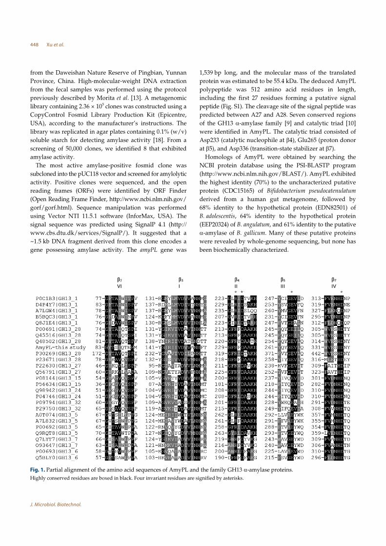
predicted between A27 and A28. Seven conserved regions
of the GH13 α-amylase family [9] and catalytic triad [10]
were identified in AmyPL. The catalytic triad consisted of
Asp233 (catalytic nucleophile at β4), Glu265 (proton donor
at β5), and Asp336 (transition-state stabilizer at β7).
Homologs of AmyPL were obtained by searching the
NCBI protein database using the PSI-BLASTP program
(http://www.ncbi.nlm.nih.gov/BLAST/). AmyPL exhibited
the highest identity (70%) to the uncharacterized putative
protein (CDC15165) of Bifidobacterium pseudocatenulatum
derived from a human gut metagenome, followed by
68% identity to the hypothetical protein (EDN82501) of
B. adolescentis, 64% identity to the hypothetical protein
(EEP20324) of B. angulatum, and 61% identity to the putative
α-amylase of B. gallicum. Many of these putative proteins
were revealed by whole-genome sequencing, but none has
been biochemically characterized.
Fig. 1. Partial alignment of the amino acid sequences of AmyPL and the family GH13 α-amylase proteins.
Highly conserved residues are boxed in black. Four invariant residues are signified by asterisks.
Amylase from Fecal Microbial Metagenome 449
April 2014⎪Vol. 24⎪No. 4
Based on the previously reported classification of α-
amylases in GH13 [2, 8, 16, 19], we chose 26 amino acid
sequences of known α-amylases to represent the 9 established
subfamilies. These proteins, together with AmyPL, were
used for analyzing the sequence features (Fig. 1) and
determining the phylogenetic tree (Fig. 2). Alignment of
the amino acid sequences of AmyPL with other enzymes
belonging to the GH13 α-amylase family revealed that
AmyPL had five conserved regions and four amino acid
residues necessary for activity in the α-amylase family [11]
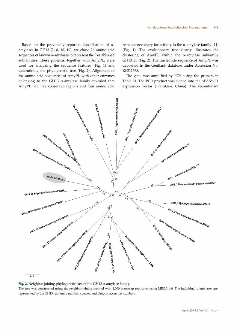
(Fig. 1). The evolutionary tree clearly illustrates the
clustering of AmyPL within the α-amylase subfamily
GH13_28 (Fig. 2). The nucleotide sequence of AmyPL was
deposited in the GenBank database under Accession No.
KF311768.
The gene was amplified by PCR using the primers in
Table S1. The PCR product was cloned into the pEASY-E1
expression vector (TransGen, China). The recombinant
Fig. 2. Neighbor-joining phylogenetic tree of the GH13 α-amylase family.
The tree was constructed using the neighbor-joining method with 1,000 bootstrap replicates using MEGA 4.0. The individual α-amylases are
represented by the GH13 subfamily number, species, and Uniprot accession numbers.
450 Xu et al.
J. Microbiol. Biotechnol.
plasmid (pEasy-E1-AmyPL) was then transformed into
Trans1-T1-phage-resistant chemically competent cells
(TransGen, China). Forward positive clones were screened
by PCR using vector primer T7 and primer plAR (Table S1),
and further confirmed by nucleotide sequencing. The valid
recombinant plasmid (pEasy-E1- AmyPL) was transformed
into E. coli BL21 (DE3) competent cells. Induction of the
recombinant enzyme and enzyme purification were performed
as previously described by Zhou et al. [25]. The molecular
mass of the purified enzyme was close to the calculated
value of AmyPL (55.4 kDa) (Fig. S2).
The α-amylase activity was determined by measuring the
amount of reducing sugar using 3,5-dinitrosalicylic acid
[12]. Through an assay using soluble starch as the substrate,
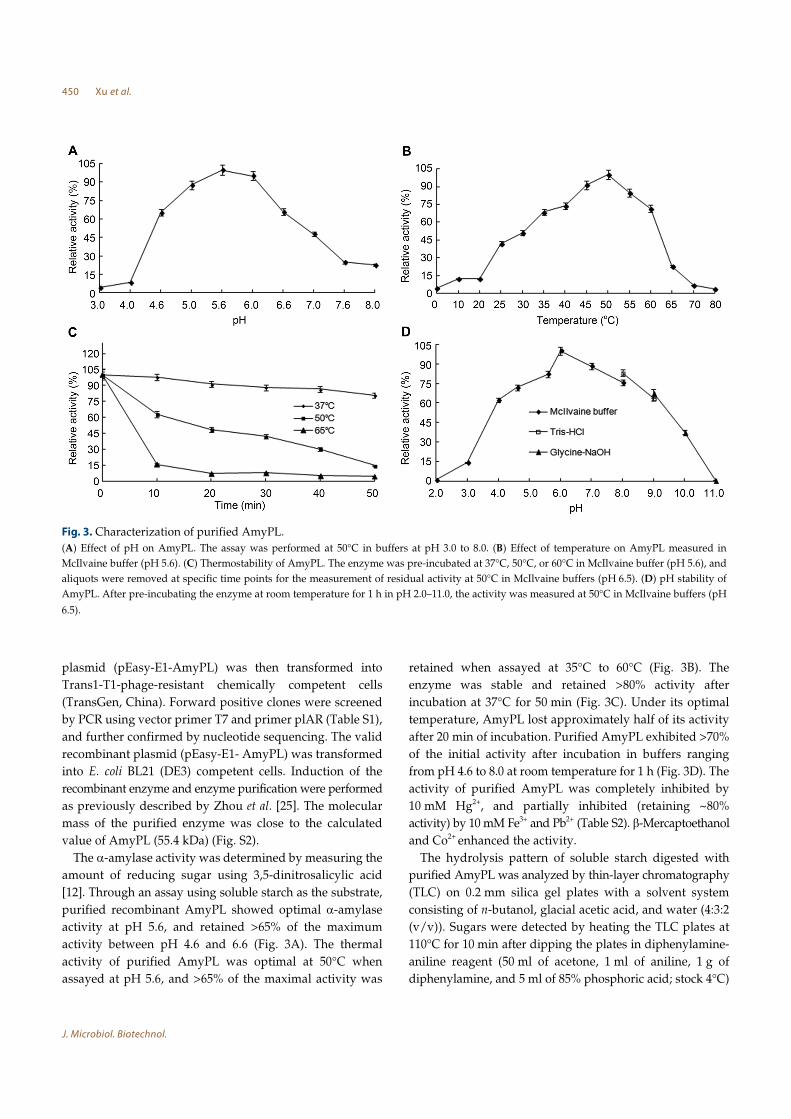
purified recombinant AmyPL showed optimal α-amylase
activity at pH 5.6, and retained >65% of the maximum
activity between pH 4.6 and 6.6 (Fig. 3A). The thermal
activity of purified AmyPL was optimal at 50°C when
assayed at pH 5.6, and >65% of the maximal activity was
retained when assayed at 35°C to 60°C (Fig. 3B). The
enzyme was stable and retained >80% activity after
incubation at 37°C for 50 min (Fig. 3C). Under its optimal
temperature, AmyPL lost approximately half of its activity
after 20 min of incubation. Purified AmyPL exhibited >70%
of the initial activity after incubation in buffers ranging
from pH 4.6 to 8.0 at room temperature for 1 h (Fig. 3D). The
activity of purified AmyPL was completely inhibited by
10 mM Hg2+, and partially inhibited (retaining ~80%
activity) by 10 mM Fe3+ and Pb2+ (Table S2). β-Mercaptoethanol
and Co2+ enhanced the activity.
The hydrolysis pattern of soluble starch digested with
purified AmyPL was analyzed by thin-layer chromatography
(TLC) on 0.2 mm silica gel plates with a solvent system
consisting of n-butanol, glacial acetic acid, and water (4:3:2
(v/v)). Sugars were detected by heating the TLC plates at
110°C for 10 min after dipping the plates in diphenylamine-
aniline reagent (50 ml of acetone, 1 ml of aniline, 1 g of
diphenylamine, and 5 ml of 85% phosphoric acid; stock 4°C)
Fig. 3. Characterization of purified AmyPL.
(A) Effect of pH on AmyPL. The assay was performed at 50°C in buffers at pH 3.0 to 8.0. (B) Effect of temperature on AmyPL measured in
McIlvaine buffer (pH 5.6). (C) Thermostability of AmyPL. The enzyme was pre-incubated at 37°C, 50°C, or 60°C in McIlvaine buffer (pH 5.6), and
aliquots were removed at specific time points for the measurement of residual activity at 50°C in McIlvaine buffers (pH 6.5). (D) pH stability of
AmyPL. After pre-incubating the enzyme at room temperature for 1 h in pH 2.0–11.0, the activity was measured at 50°C in McIlvaine buffers (pH
6.5).
Amylase from Fecal Microbial Metagenome 451
April 2014⎪Vol. 24⎪No. 4
(Fig. S3). Maltose and maltotriose were formed in great
amounts at the early stage of hydrolysis. On further
incubation, maltotriose subsequently hydrolyzed to
accumulate glucose and maltose.
In this study, a total of eight amylolytic-active positive
fosmid clones were screened using an activity-based method
from a fecal microbial metagenomic library of pygmy loris,
and a novel α-amylase gene, amyPL, was identified. To the
best of our knowledge, AmyPL is the first α-amylase
isolated from a gastrointestinal metagenomic library that
has been biochemically characterized. This study contributes
to knowledge of the diversity of amylolytic genes and
demonstrates that gut microorganisms are a very important
source of novel hydrolase genes. Future studies should
include further cloning and characterization of the other
amylolytic-active positive clones detected in this study and
screening of other hydrolases from this metagenomic
library.
Acknowledgments
This work was supported by the National Natural
Science Foundation of China (30960165, 31360268, and
31160229).
References
1. Bao L, Huang Q, Chang L, Sun Q, Zhou J, Lu H. 2012.Cloning and characterization of two β-glucosidase/xylosidaseenzymes from yak rumen metagenome. Appl. Biochem.
Biotechnol. 166: 72-86. 2. Da Lage JL, Feller G, Jane ek Š. 2004. Horizontal gene
transfer from Eukarya to bacteria and domain shuffling: theα-amylase model. Cell Mol. Life Sci. 61: 97-109.
3. Feng Y, Duan CJ, Pang H, Mo XC, Wu CF, Yu Y, et al. 2007.Cloning and identification of novel cellulase genes fromuncultured microorganisms in rabbit cecum and characterizationof the expressed cellulases. Appl. Microbiol. Biotechnol. 75:
319-328.4. Ferrer M, Golyshina OV, Chernikova TN, Khachane AN,
Reyes-Duarte D, Santos VA, et al. 2005. Novel hydrolasediversity retrieved from a metagenome library of bovinerumen microflora. Environ. Microbiol. 7: 1996-2010.
5. Ferrer M, Ghazi A, Beloqui A, Vieites JM, López-Cortés N,Marín-Navarro J, et al. 2012. Functional metagenomics unveilsa multifunctional glycosyl hydrolase from the family 43catalysing the breakdown of plant polymers in the calfrumen. PLoS One 7: e38134.
6. Gong X, Gruninger RJ, Qi M, Paterson L, Forster RJ, TeatherRM, McAllister TA. 2012. Cloning and identification ofnovel hydrolase genes from a dairy cow rumen metagenomic
library and characterization of a cellulase gene. BMC Res.
Notes 5: 566.7. Guo H, Feng Y, Mo X, Duan C, Tang J, Feng J. 2008.
Cloning and expression of a beta-glucosidase gene umcel3G
from metagenome of buffalo rumen and characterization ofthe translated product. Sheng Wu Gong Cheng Xue Bao
(Chinese) 24: 232-238.8. Hostinová E, Jane ek Š, Gašper k J. 2010. Gene sequence,
bioinformatics and enzymatic characterization of α-amylasefrom Saccharomycopsis fibuligera KZ. Protein J. 29: 355-364.
9. Janecek S. 2002. How many conserved sequence regions arethere in the α-amylase family? Biologia 57(Suppl. 11): 29-41.
10. MacGregor EA, Janecek S, Svensson B. 2001. Relationship ofsequence and structure to specificity in the alpha-amylasefamily of enzymes. Biochim. Biophys. Acta 1546: 1-20.
11. Machovi M, Jane ek Š. 2003. The invariant residues in theα-amylase family: just the catalytic triad. Biologia (Bratisl) 58:
1127-1132.12. Miller GL. 1959. Use of dinitrosalicylic acid reagent for
determination of reducing sugar. Anal. Chem. 32: 426-428.13. Morita H, Kuwahara T, Ohshima K, Sasamoto H, Itoh K,
Hattori M, et al. 2007. An improved DNA isolation methodfor metagenomic analysis of the microbial flora of thehuman intestine. Microb. Environ. 22: 214-222.
14. Nimchua T, Thongaram T, Uengwetwanit T, PongpattanakitshoteS, Eurwilaichitr L. 2012. Metagenomic analysis of novellignocellulose-degrading enzymes from higher termite gutsinhabiting microbes. J. Microbiol. Biotechnol. 22: 462-469.
15. Rashamuse KJ, Visser DF, Hennessy F, Kemp J, Roux-vander Merwe MP, Badenhorst J, et al. 2013. Characterisation oftwo bifunctional cellulase-xylanase enzymes isolated from abovine rumen metagenome library. Curr. Microbiol. 66: 145-151.
16. Stam MR, Danchin EGJ, Rancurel C, Coutinho PM,Henrissat B. 2006. Dividing the large glycoside hydrolasefamily 13 into subfamilies: towards improved functionalannotations of α-amylase-related proteins. Protein Eng. Des.
Sel. 19: 555-562.17. Streit WR, Schmitz RA. 2004. Metagenomics: the key to the
uncultured microbes. Curr. Opin. Biotechnol. 7: 492-498.18. Tasse L, Bercovici J, Pizzut-Serin S, Robe P, Tap J, Klopp C,
et al. 2010. Functional metagenomics to mine the human gutmicrobiome for dietary fiber catabolic enzymes. Genome Res.
20: 1605-1612.19. Van der Kaaij RM, Jane ek Š, van der Maarel MJEC,
Dijkhuizen L. 2007. Phylogenetic and biochemical characterizationof a novel cluster of intracellular fungal α-amylase enzymes.Microbiology 153: 4003-4015.
20. Walter J, Mangold M, Tannock GW. 2005. Construction,analysis, and β-glucanase screening of a bacterial artificialchromosome library from the large-bowel microbiota ofmice. Appl. Environ. Microbiol. 71: 2347-2354.
21. Warnecke F, Luginbühl P, Ivanova N, Ghassemian M,
c
ê
c
ê
ró
c
ê
c
ê
c
ê
452 Xu et al.
J. Microbiol. Biotechnol.
Richardson TH, Stege JT, et al. 2007. Metagenomic andfunctional analysis of hindgut microbiota of a wood-feedinghigher termite. Nature 450: 560-565.
22. Xu B, Xu W, Yang F, Li J, Yang Y, Tang X, et al. 2013.Metagenomic analysis of the pygmy loris fecal microbiomereveals unique functional capacity related to metabolism ofaromatic compounds. PLoS One 8: e56565.
23. Zhao S, Wang J, Liu K, Zhu Y, Bu D, Li D, Yu P. 2009.Screening and characterization of lipase from a metagenomelibrary of dairy rumen microflora. Sheng Wu Gong Cheng
Xue Bao (Chinese) 25: 869-874.
24. Zhao S, Wang J, Bu D, Liu K, Zhu Y, Dong Z, Yu Z. 2010.Novel glycoside hydrolases identified by screening a ChineseHolstein dairy cow rumen-derived metagenome library.Appl. Environ. Microbiol. 76: 6701-6705.
25. Zhou J, Dong Y, Li J, Zhang R, Tang X, Mu Y, et al. 2012.Cloning, heterologous expression, and characterization ofnovel protease-resistant α-galactosidase from new Sphingomonas
strain. J. Microbiol. Biotechnol. 22: 1532-1539.
Related Documents