Cloning and Characterization of 5E6(Ly-49C), A Receptor Molecule Expressed on a Subset of Murine Natural Killer Cells By Earl R. Stoneman,*~ Michael Bennett,* Jiaban An,* Kye A. Chesnut,S Edward K. Wakeland,SJulia B. Scheerer,[I Michael J. Siciliano, l[ Vinay Kumar,* and PoruneUoor A. Mathew* From the *Laboratory of Molecular Pathology, Department of Pathology, *Immunology Graduate Program, University of Texas Southwestern Medical Center at Dallas, Dallas, Texas 75235; gCenter for Mammalian Genetics, College of Medicine, University of Florida, Gainesville, Florida 32610; and ItDepartment of Molecular Genetics, University of Texas M.D Anderson Cancer Center, Houston, Texas 77030 Summal~ 5E6 is a cell surface molecule expressed on a subpopulation of murine natural killer (NK) cells that are involved in the specific rejection of H-2 a or H-2 f (hemopoietic histocompatibility determinant 2) bone marrow cell grafts. Here, we isolated and cloned the gene encoding 5E6 and determined the nucleotide sequence of the eDNA. 5E6 is nearly identical to Ly-49C; the deduced amino acid sequence reveals a polypeptide of 266 amino acids with a molecular weight of 31,284 that contains multiple cysteine residues to explain its disulfide-linked homodimer structure and five potential N-linked glycosylation sites. 5E6 is a type II integral membrane protein with an extracellular carbohydrate recognition domain characteristic of C-type (Ca2+-dependent) animal lectins. Chromosomal mapping indicates that 5E6 is located within the NK gene complex on chromosome 6. The sequence of 5E6 mRNA and the degree of glycosylation of 5E6 protein are under genetic control. Immunoprecipitation before removal of N-linked sugars reveals different size molecules. There are several nucleotide differences among BALB/c, B6, and NZB mRNAs; however, none of them would be expected to affect N-glycosylation. Of particular interest are two findings: (a) BALB/c, B6, and (BALB/c x B6)F1 5E6 reduced molecules are m65, 54, and 54 kD, and (b) the eDNA sequence of (BALB/c x B6)F1 is identical to B6. Thus, there appears to be allelic exclusion of 5E6 expression that may be related to the ability of F1 hybrid mice to reject parental H-2 a bone marrow cell grafts. N 'K cells comprise a small population of lymphocytes that have a large granular morphology and are distinct from mature T and B cells and from cells of the myeloid lineage (1, 2). NK cells are defined as a functionally important im- mune effector population that display "natural" cytolytic ac- tivity against certain tumor cells and vitally infected cells in a manner that is not MHC restricted (3, 4). NK cells also mediate the rejection of incompatible bone marrow cell al- lografts in lethally irradiated mice, possibly by the specific recognition of recessively inherited hematopoietic histocom- patibility i (Hh-1)1 antigens, a phenomenon known as "hy- brid resistance" (5-7). The mechanism by which NK cells 1 Abbreviationsused in thispaper: A-LAK, adherentlymphokine-activated killer (cells); DGGE, denaturing gradientgel electrophoresis; Hh-1, hema- topoietichistocompatibility 1; RI, recombinant inbred;UT, untranslated. recognize bone marrow cells in a highly specifc manner and mediate hybrid resistance is not known. However, it has been postulated that specific receptors are present on subsets of NK cells that allow for this recognition. Indeed, NK-specific molecules have been reported in humans (8-12), rats (13, 14), and mice (15-22). Recently, several murine NK cell receptor-like molecules have been cloned and categorized into two genetically linked families termed the NKR-P1 and Ly-49 gene families. These gene families share some structural features and are located in close proximity on mouse chromosome 6 in an area that has been termed the NK gene complex (23, 24). These two families of molecules have been suggested to deliver opposing signals, with NKR-P1 molecules activating natural killer and Ly-49 molecules delivering inhibitory signals to NK cells when they encounter certain MHC class I molecules on target cells (23, 25). 305 J. Exp. Med. 9 The Rockefeller UniversityPress 9 0022-1007/95/08/0305/09 $2.00 Volume 182 August 1995 305-313 on August 12, 2015 jem.rupress.org Downloaded from Published August 1, 1995

Welcome message from author
This document is posted to help you gain knowledge. Please leave a comment to let me know what you think about it! Share it to your friends and learn new things together.
Transcript

Cloning and Characterization of 5E6(Ly-49C), A Receptor Molecule Expressed on a Subset of Murine Natural Killer Cells By Earl R. Stoneman,*~ Michael Bennett,* Jiaban An,* Kye A. Chesnut,S Edward K. Wakeland,S Julia B. Scheerer,[I Michael J. Siciliano, l[ Vinay Kumar,* and PoruneUoor A. Mathew*
From the *Laboratory of Molecular Pathology, Department of Pathology, *Immunology Graduate Program, University of Texas Southwestern Medical Center at Dallas, Dallas, Texas 75235; gCenter for Mammalian Genetics, College of Medicine, University of Florida, Gainesville, Florida 32610; and ItDepartment of Molecular Genetics, University of Texas M.D Anderson Cancer Center, Houston, Texas 77030
Summal~ 5E6 is a cell surface molecule expressed on a subpopulation of murine natural killer (NK) cells that are involved in the specific rejection of H-2 a or H-2 f (hemopoietic histocompatibility determinant 2) bone marrow cell grafts. Here, we isolated and cloned the gene encoding 5E6 and determined the nucleotide sequence of the eDNA. 5E6 is nearly identical to Ly-49C; the deduced amino acid sequence reveals a polypeptide of 266 amino acids with a molecular weight of 31,284 that contains multiple cysteine residues to explain its disulfide-linked homodimer structure and five potential N-linked glycosylation sites. 5E6 is a type II integral membrane protein with an extracellular carbohydrate recognition domain characteristic of C-type (Ca2+-dependent) animal lectins. Chromosomal mapping indicates that 5E6 is located within the NK gene complex on chromosome 6. The sequence of 5E6 mRNA and the degree of glycosylation of 5E6 protein are under genetic control. Immunoprecipitation before removal of N-linked sugars reveals different size molecules. There are several nucleotide differences among BALB/c, B6, and NZB mRNAs; however, none of them would be expected to affect N-glycosylation. Of particular interest are two findings: (a) BALB/c, B6, and (BALB/c x B6)F1 5E6 reduced molecules are m65, 54, and 54 kD, and (b) the eDNA sequence of (BALB/c x B6)F1 is identical to B6. Thus, there appears to be allelic exclusion of 5E6 expression that may be related to the ability of F1 hybrid mice to reject parental H-2 a bone marrow cell grafts.
N 'K cells comprise a small population of lymphocytes that have a large granular morphology and are distinct from
mature T and B cells and from cells of the myeloid lineage (1, 2). NK cells are defined as a functionally important im- mune effector population that display "natural" cytolytic ac- tivity against certain tumor cells and vitally infected cells in a manner that is not MHC restricted (3, 4). NK cells also mediate the rejection of incompatible bone marrow cell al- lografts in lethally irradiated mice, possibly by the specific recognition of recessively inherited hematopoietic histocom- patibility i (Hh-1)1 antigens, a phenomenon known as "hy- brid resistance" (5-7). The mechanism by which NK cells
1 Abbreviations used in this paper: A-LAK, adherent lymphokine-activated killer (cells); DGGE, denaturing gradient gel electrophoresis; Hh-1, hema- topoietic histocompatibility 1; RI, recombinant inbred; UT, untranslated.
recognize bone marrow cells in a highly specifc manner and mediate hybrid resistance is not known. However, it has been postulated that specific receptors are present on subsets of NK cells that allow for this recognition. Indeed, NK-specific molecules have been reported in humans (8-12), rats (13, 14), and mice (15-22).
Recently, several murine NK cell receptor-like molecules have been cloned and categorized into two genetically linked families termed the NKR-P1 and Ly-49 gene families. These gene families share some structural features and are located in close proximity on mouse chromosome 6 in an area that has been termed the NK gene complex (23, 24). These two families of molecules have been suggested to deliver opposing signals, with NKR-P1 molecules activating natural killer and Ly-49 molecules delivering inhibitory signals to NK cells when they encounter certain MHC class I molecules on target cells (23, 25).
305 J. Exp. Med. �9 The Rockefeller University Press �9 0022-1007/95/08/0305/09 $2.00 Volume 182 August 1995 305-313
on August 12, 2015
jem.rupress.org
Dow
nloaded from
Published August 1, 1995
The mAb SW5E6 has been described as binding to a popu- lation of '~50% of murine NK cells of most strains tested. Previous experiments in this laboratory have shown that treat- ment of recipient mice with mAb SW5E6 eliminates the ability to reject H-2 d but not H-2 b bone marrow cell grafts (26, 27). In this study, we report the cloning, chromosomal mapping, and molecular characteristics of the 5E6 gene, as well as its unique expression in different strains of mice.
Materials and Methods
Cell Purification and Cell Culture. Murine cells were cultured in RPMI 1640 (GIBCO BRL, Gaithersburg, MD) supplemented with 10% FCS (Hyclone Laboratories, Logan, UT), 2 mM t-gluta- mine, 100 U/ml penicillin, 100/~g/ml streptomycin (Irvine Scientific, Santa Ana, CA), 1 mM sodium pyruvate, and 0.1 mM nonessen- tial amino acids (GIBCO BRL) (complete RPMI). COS-7 cells have been described (28) and were cultured in DMEM (GIBCO BRL) with the same supplements (complete DMEM). Adherent lymphokine-activated killer (A-LAK) cells were prepared as previ- ously described (29) with minor modifications. Briefly, A-LAK cells were generated by culturing nylon wool nonadherent spleen cells in T25 flasks (Corning Inc., Coming, NY) in RPMI 1640 com- plete media (RPMI 1640 [GIBCO BILL] supplemented with 10% FCS [Hyclone Laboratories], 2 mM L-glutamine, 100 U/ml peni- cillin, 100/~g/ml streptomycin [Irvine Scientific], 1 mM sodium pyruvate, and 0.1 mM nonessential amino acids [GIBCO BRL]) containing 22.5/~M B-mercaptoethanol (Sigma Immunochemicals, St. Louis, MO), 1/~g/ml indomethacin (Sigma), 50/~g/ml gen- tamicin (GIBCO BRL), and 500 or 1,000 U/ml human rlL-2 (a kind gift from the Cetus Corporation, Emeryville, CA) (LAK cul- turing media) for 3 d at 37~ and 5% CO2. On day 3 of culture, the nonadherent cells were removed by gentle washing with warm complete RPMI. The adherent cells were further cultured and fed every 2-3 d with LAK culturing media, maintaining Ib2 at a con- centration of 500 or 1,000 U/ml. Cells were harvested at 8-9 d of culture. LAK cells from C.B-17 scid/scid bone marrow were pre- pared in the same manner, with the exception of the unnecessary step of removing the nonadherent T cells on day 3.
Construction ofcDNA Library. Total cytoplasmic RNA was pre- pared from day 9 C.B-17 scid/scid LAK cells by the acid-gnanidine- phenol-chloroform method (30). Poly (A) + RNA was prepared by oligo (dT)-cellulose chromatography using an mRNA separator system (Clontech, Palo Alto, CA) as instructed by the manufac- turer. The cDNA library was constructed using the superscript system for cDNA synthesis and cloning (BILL-Life Technologies, Inc., Gaithersburg, MD). Briefly, first-strand cDNA was synthe- sized from 5 #g of poly (A) + RNA using a modified Moloney murine leukemia virus (M-MLV) reverse transcriptase, Superscript RT, (GIBCO BRL) and a NotI primer adapter 5'-pGACTAGTT- CTAGATCGCGAGCGGCCGCCC(T)1s-3', which provided direc- tionality to the cDNA. Second-strand synthesis was by nick trans- lational replacement of the mRNA, as first described by Okayama and Berg (31). SalI adapters were ligated to the 5' end of the cDNA followed by digestion with NotI to yield cDNA with 5' SalI and 3' NotI ends. The cDNA was then size-fractionated by passage over a Sephacryl S-500 HR column. The resulting cDNA was then ligated to XhoI-NotI-digested pME18S vector (kindly provided by Dr. Kevin Moore, DNAX Research Institute, Palo Alto, CA). The pME18S vector is a powerful mammalian expression vector containing the SRo~ promoter (composed of the SV-40 early pro-
motet along with the R segment and part of the U5 sequence (R-U5') of the long terminal repeat of human T cell leukemia virus I (32), the SV-40 small t intron, the SV-40 poly(A) and splice signals, as well as the pUC 18 origin and the ampicillin-resistance gene. The ligated cDNA was introduced into MC1061 bacteria by elec- troporation. Approximately 2 x 10⁷ independent clones were ob- tained with an average size ofl.6 kb and a range of 0.8 to >3.5 kb.
Isolation and Characterization of cDNA Clone. The cDNA library was screened by multiple rounds of transient expression in COS-7 cells and immunoselection by panning as described by Seed and Aruffo (33). In the first round, COS-7 cells were transfected by the DEAE-dextran method (34). Selection was by panning with sheep anti-mouse IgG (Jackson ImmunoResearch Laboratories, Inc., West Grove, PA)-coated plates and mAb SW5E6. Episomal DNA was recovered by the method of Hirt (35) and used to transform Escherichia coli MC1061 by electroporation. The bacteria were amplified in liquid media and the plasmid DNA was reintroduced into COS-7 cells by spheroplast fusion according to the method of Sandri-Goldin et al. (36). After one additional round of sphero- plast fusion, panning, recovery ofplasmid DNA, and transforma- tion, the bacteria were plated so as to obtain individual colonies. Plasmid DNA was prepared from 18 individual colonies and used to transfect COS-7 cells by the DEAE-dextran method, and 48 h later the cells were recovered, stained with mAb SWSE6, and ana- lyzed by flow cytometry using a FACScan | flow cytometer.(Becton Dickinson & Co., Mountain View, CA). Three of the clones showed positive staining for 5E6 and had inserts of '~1.3 kb. Each of the clones were subcloned into the multifunctional vector Gemscript (a gift from David Crawford, University of Texas Southwestern Medical Center, Dallas, TX). The largest of these clones, p5E6GS.16, was used to prepare' a set of nested deletions from each end of the cDNA using exonuclease III (37). Clones that were "~150 bp different in size were isolated and used for sequence analysis by the dideoxynucleotide chain termination method (38) using the Sequenase system (United States Biochemical Corp., Cleveland, OH). The clone was sequenced completely on both strands. Se- quence analysis and database comparison were performed using the Genetics Computer Group, Inc. (Madison, WI) suite of programs.
Reverse Transcription PCR Amplification of 5E6 eDNA from D~'fferent Mouse Strains. Poly(A) + RNA was isolated from NK cells of C57BL/6, BALB/c, BALB.B, CB6F1, and NZB by using the Fast Track mRNA isolation kit (InVitrogen, San Diego, CA). First- strand cDNA was synthesized from 1 #g of poly (A) + RNA using the modified reverse transcriptase, superscript RT (GIBCO BRL), and a random primer. A 1.1-kb fragment of 5E6 that con- tains the complete coding region, as well as 145 bp 5' untranslated (UT) and 157 bp 3' UT sequences, was amplified using the fol- lowing two primers: 5E6-20(5'-ATCATGAGGTTGAGTATCACC-Y) and 3' primer, 5E6-1D2 (5'-GCAAAGCGTGCCCATTCAC-3'). The PCR mixture contained 1/~M of each primer, 200/~M dNTPs, 1 x PCR buffer, 1 U Ampli Taq DNA polymerase, 5 #I ofcDNA reaction product and 2 mM magnesium chloride in a reaction volume of 50 #1. PCR conditions were 94~ 4 min, 35 cycles of 94~ 1 min, 55~ 1 min, 72~ 1 min followed by extension of 72~ for 7 rain. 5 #1 of the PCR products were run on 1.2% agarose gel and visualized by staining with ethidium bromide. A portion of the product (1/~1) was used for TA cloning according to the instructions provided by the supplier (InVitrogen). The cloned genes were sequenced by using specific primers based on the sequence of 5E6 from BALB/c using the Sequenase system.
Surface Iodination, Immunoprecipitation, Removal of N-linked Car- bohydrates, and SDS-PAGE. Transiently transfected COS-7 cells
306 Cloning of 5E6(Ly-49C), a Natural Killer Cell-associated Receptor Molecule
on August 12, 2015
jem.rupress.org
Dow
nloaded from
Published August 1, 1995
or A-LAK cells from various strains of mice were labeled with Na12SI (Amersham Corp., Arlington Heights, IL) using lac- toperoxidase as described (39). Cells were washed three times in PBS and lysed in 50 mM Tris, pH 8.0, 1% NP-40 (Sigma) con- taining 1 mM EDTA, 0.5/zM leupeptin, 0.7/zM pepstatin, and 150 /zg/ml PMSF (Boehringer Mannheim Biochemicals, Indi- anapolis, IN) for 10 rain on ice. The nuclei were removed by cen- trifugation at 12,000 g for 10 min. The lysates were precleared with rabbit anti-mouse IgG (Jackson ImmunoResearch Laboratories) coated protein A-Sepharose (Pharmacia Fine Chemicals, Piscataway, NJ) for 30 rain at 4~ while rocking. Predeared lysates were in- cubated with mAb SWSE6 or isotype control mAb 22B5 for 2 h at 4~ while rocking. Protein A-Sepharose coated with rabbit anti-mouse IgG was added for an additional 45 rain at 4~ while rocking. The beads were recovered by centrifugation and washed three times with NET gel (50 mM Tris-HC1, pH 7.5, 150 mM NaC1, 0.1% NP-40, 1 mM EDTA, 0.25% gelatin, 0.02% sodium azide). The bound proteins were eluted by boiling for 5 min in SDS sample buffer for SDS-PAGE analysis or in antigen elution buffer (100 mM Tris-HC1, pH 7.5, 1% SDS, 1%/3-mercaptoethanol) for removal of N-linked carbohydrates. Endoglycosidase F diges- tion was performed by removing the supernatant from the beads and adding endoglycosidase F buffer (0.1 M potassium phosphate, pH 6.1, 1% Triton X-100, 0.1% SDS, 45 mM EDTA, 1% B-mer- captoethanol) to a final volume of 1 ml. 1 U of endoglycosidase F/N-glycosidase F (Boehringer Mannheim Biochemicals) was added and the samples were allowed to digest overnight at 37~ The protein was precipitated with TCA and washed three times with ice cold acetone followed by addition of I x SDS sample buffer and boiling for 5 rain. Samples were analyzed on 12 or 7.5% slab gels according to the method of Laemmli (40). Gels were fixed, dried, and exposed to Hyperfilm-MP (Amersham Corp.) at -70~
Genetic Mapping of 5E6(Ly-49C). Mapping of 5E6(Ly-49C) was performed using a panel of 26 genomic DNAs representing the complete C57BL/6 x DBA/2 (B x D) recombinant inbred strain collection. Candidate primer sequences for the amplification of genomic fragments from 5E6(Ly-49C) were initially identified using the computer program Primer (kindly provided by Eric Lander, Massachusetts Institute of Technology, Cambridge, MA) to ana- lyze the cDNA sequence of 5E6(Ly-49C). The primer sequences ultimately selected were: 5' TTCCCTGATTAATTTTCCAACC and 3' TCTTTCTTGTGTTAAGACTTCCG. These sequences were from the 3' end of 5E6(Ly-49C) and amplified a 278-bp genomic fragment that had minimal homology with sequences in Ly-49. Polymorphisms between C57BL/6 and DBA/2 in this amplified fragment were identified by automated nucleotide sequence anal- ysis using an automated DNA sequencer (model 373; Applied Bio- systems, Inc., Foster City, CA) as previously described (41). Allelic forms of the amplified fragment were subsequently distinguished by denaturing gradient gel electrophoresis (DGGE) as previously described (42). Preliminary analysis using perpendicular gradients established that optimal variation in DGGE mobility occurred in a denaturing gradient of 30-70% for 5E6(Ly-49C) alleles from C57BL/6 and DBA/2. The genotypes of all 26 B x D recom- binants strains were determined for 5E6(Ly-49C) by standard ana- lytical techniques (43) as described previously (44) using the MAP MANAGER software package and database (kindly provided by K. Manly, Roswell Park Cancer Institute, Buffalo, NY).
Chromosomal Mapping by PCR Analysis of Somatic Cell Hybrid DNA. Based on the 5E6 cDNA sequence, primers were designed to specifically amplify a 3' portion of the 5E6 gene. The sense primer
corresponds to nucleotides 925-947 (5' ATTCCCTACTACTGT- ATTTGTGG 3') and the antisense primer corresponds to nucleo- tides 1,134-1,116 (5' GCAAAGCGTGCCCATTCAC 3'). A PCR mixture of 0.5/zM primers, 200/~M dNTPs, and 1• Amersham Hot Tub buffer, plus an Ampliwax "gem" (Perkin Elmer Corp., Norwalk, CT) was put through a "hot start" of 75~ for 15 min followed by cooling to 20~ DNA (100 rig) and Amersham Hot Tub Taq polymerase (1.25 U) were then added to each reaction mix. PCR conditions were 94~ 5 min, 35 cycles: 94~ 1 min, 62~ 1 min, and 72~ I min, followed by extension at 72~ for 7 min and storage at 4~ A portion of the PCR product (9 #1 of the 50/zl total) of each sample was electrophoresed on a 2% agarose gel and visualized by staining with ethidium bromide.
Results Isolation and Characterization of 5E6 cDNA Clon~ A cDNA
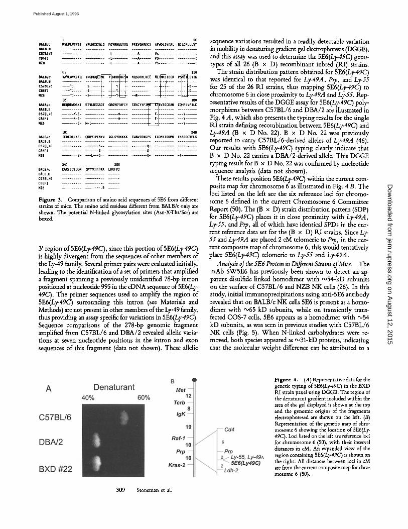
library was constructed in the mammalian expression vector pME18S using mRNA isolated from IL-2-propagated C.B- 17 (scid/scid) bone marrow cells. The library was transfected into COS-7 cells and these cells were then screened for sur- face expression of the 5E6 molecule using the transient expression-immunoselection procedure of Seed and Aruffo (33). After three rounds of transfection and selection by pan- ning using the mAb SW5E6, 18 bacterial colonies were iso- lated at random for further analysis. Three of these plasmids contained inserts of "~ 1.2 kb and each of these clones directed the surface expression of 5E6 protein on transfected COS-7 cells as determined by staining with the 5E6 mAb and flow cytometry analysis (Fig. 1). Each of these three clones were identical at the 3' end and differed only in the length of the 5' untranslated region. The nucleotide sequence of the largest clone is shown in Fig. 2. Comparison of the predicted amino acid sequence of 5E6 to the databanks revealed 64% identity to the Ly-49A molecule (75% identity at the nucleotide level) (data not shown). The 5E6 polypeptide also contains an ex- tracellular carbohydrate recognition domain characteristic of Ca2+-dependent lectins (45) and members of the Ly-49 gene family (23, 24). Comparison of the carbohydrate recogni- tion domains of 5E6 and several other known C-type lectins shows that multiple residues are conserved and that 5E6 is a member of this group of molecules. The 5E6 cDNA was found to be identical to a previously reported molecule, Ly- 49C (17), with the following exceptions. The 5E6 cDNA is 84 nucleotides longer at the 5' UT sequence and has a 78-bp deletion at nucleotide 995 in the 3' untranslated region cor- responding to nucleotides 912-989 of the reported Ly-49C sequence. There are also two nucleotide differences at 279 and 280. We report a cytosine followed by an adenine, while Ly-49C is reported to have an adenine followed by a thymi- dine. This results in a change from asparagine in the 5E6 peptide to tyrosine in the Ly-49C peptide (data not shown). Comparison of amino acid sequence of 5E6 from different strains of mice is shown in Fig. 3.
The 5E6 cDNA Reveals Significant Polymorphism. Southern blots of HindlII-, BamHI-, and EcoRI-digested DNA from
307 Stoneman et al.
on August 12, 2015
jem.rupress.org
Dow
nloaded from
Published August 1, 1995
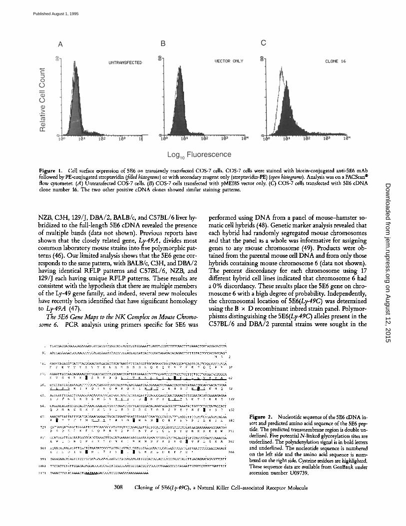
Figure 1. Cell surface expression of 5E6 on transiently transfected COS-7 cells. COS-7 cells were stained with biotin-conjugated anti-5E6 mAb followed by PE-conjugated streptavidin (filled histograms) or with secondary reagent only (streptavidin-PE) (open histograms). Analysis was on a FACScan | flow cytometer. (.4) Untransfected COS-7 cells. (B) COS-7 cells transfected with pME18S vector only. (C) COS-7 cells transfected with 5E6 c D N A clone number 16. The two other positive c D N A clones showed similar staining patterns.
NZB, C3H, 129/J, DBA/2, BALB/c, and C57BL/6 liver hy- bridized to the full-length 5E6 cDNA revealed the presence of multiple bands (data not shown). Previous reports have shown that the closely related gene, Ly-49A, divides most common laboratory mouse strains into five polymorphic pat- terns (46). Our limited analysis shows that the 5E6 gene cor- responds to the same pattern, with BALB/c, C3H, and DBA/2 having identical RFLP patterns and C57BL/6, NZB, and 129/J each having unique RFLP patterns. These results are consistent with the hypothesis that there are multiple members of the Ly-49 gene family, and indeed, several new molecules have recently been identified that have significant homology to Ly-49A (47).
The 5E6 Gene Maps to the NK Complex on Mouse Chromo- some 6. PCR analysis using primers specific for 5E6 was
performed using DNA from a panel of mouse-hamster so- matic cell hybrids (48). Genetic marker analysis revealed that each hybrid had randomly segregated mouse chromosomes and that the panel as a whole was informative for assigning genes to any mouse chromosome (49). Products were ob- tained from the parental mouse cell DNA and from only those hybrids containing mouse chromosome 6 (data not shown). The percent discordancy for each chromosome using 17 different hybrid cell lines indicated that chromosome 6 had a 0% discordancy. These results place the 5E6 gene on chro- mosome 6 with a high degree of probability. Independently, the chromosomal location of 5E6(Ly-49C) was determined using the B x D recombinant inbred strain panel. Polymor- phisms distinguishing the 5E6(Ly-49C) alleles present in the C57BL/6 and DBA/2 parental strains were sought in the
1
91
181
271
361
451
541
631
721
811
901
991
1081
1171
TGA~GAGGAGGGG~AGA~AAT~ATGAGGTTGAGTATCACCCGGTGGAAATTTAGTTCCGT~TTT~TTTTGAAACTCGTAGGAGATCTA
~CCAGAAAA~GCC~GTTTCAGA~AAATTTT~C~TCcA~CAG~AT~ACTCCGGTAGAGACACAGACTT~TTGTACT~C~GATGAGT MS
GAG~CAGAGGT~A~TTA~TC~TGTGAGA~TT~AT~GTCTTCAGGGTTGCAGAAAC~GT~GGCATGAGGAGACTC~GGG~c~AGA E P E TY T V R HK SG Q K Q V R H ET G P R
G~GTTGG~CAGAAAGTGTT~AGCA~C~TGG~CT~ATTGTGAAAGCTCTTGG~TCCTCTGTTTCCTTCTTcTTGT~CAGTTGCA E V G R K ~ S A P QL VK L G I L ~ F L~ T ~
GTGTTGGCAGTAAAGATTTTTCAGTAT~TC~CACAAAC~GAAATC~TGAAACTCTAAACCATCACCAT~CTGCAGC~CATGC~ V ~ A KI Q Y N Q H K E I ~ L N H H H N m N M Q
AGTGATTTC~CTTAAAGG~AAATGTTGACAAAT~GTCTATAGATTGTAGGCC~GC~TGAAA~TCTGG~TATATCAAAAGAG~ S D F LK E M L NK ID R P S N E ~ L E Y K R E
CAGGACAGATGGGACAGTAA~CAAA~ACTGTTTTAGATTCCTCACGGGACACAGGCAGAGGTGTTAAATACTGGTTCTGCTATAGTACT Q D R DS T K T LD SR T G R G V K WF Y S T
/~ATGTTATTATTTCATCATG~CA~CTACATGGAGTGGATGTAAAGCG~CTGCCAGCATTTTAGCGTTCCCATTCTG~GATAG~ K B Y F ~ N K T w s w K ~ ~ e H ~ S V P ~ K ~ E
GATG~GATG~CTG/ZAATTCCTTC~CGCCATGTTATTCCAGAG~TTACTGGATTGGATTGTCTTATGAT~GA~GG~TGG D E D LK L Q R VI EN W I G L S Y D K K K E W
GCATGGATTGAC~TGGCCCATCT/~CTTGACATG~AAT~GG~u~AATG~CTTT~GTCTAGAGGATGTGTATTTTTATCTAAAGCA A W l NG S K L D M K RK N F K S R G ~ V F S K A
AG~TAG~GATATTGACTGT~TATTCCCTACTA~TGTATTTGTGGG~GAAACTGGAT/~ATTC~CTGATT~TTTTCC~CCAGAGT R ! E ID N I P Y~ ~G K L D K F P
TAAAG~CATAGTCTCCTGTG~GAGAAACAAAGCTGC~G~CATTGGGACTGTACTCTCCTATCTATCTTGACAG~CAGAGGTCATT
TTCTATCCTGTTGGAGAGAGGACGCATCTACTCGGGTG~TGGGCACGCTTTGCCCTAAAGCCTTCAG~TTGTGTTCTTTCTGATTTcT
TAAACTCCCATAAAACTGAATAAAGAGTCCTCCCT
2
32
62
92
122
152
182
212
242
266
Figure 2. Nucleotide sequence of the 5E6 c D N A in- sert and predicted amino acid sequence of the 5E6 pep- tide. The predicted transmembrane region is double un- derlined. Five potential N-linked glycosylation sites are underlined. The polyadenylation signal is in bold letters and underlined. The nucleotide sequence is numbered on the left side and the amino acid sequence is num- bered on the right side Cysteine residues are highlighted. These sequence data are available from GenBank under accession number U09739.
308 Cloning of 5E6(Ly-49C), a Natural Killer Cell-associated Receptor Molecule
on August 12, 2015
jem.rupress.org
Dow
nloaded from
Published August 1, 1995
B A L B / c
B A L B . B
C 5 7 B L / 6
C B 6 F 1
N Z B
B A L B / c
B A L B . B
C 5 7 B L / 6
C B S F I
N Z B
B A L B / c
B A L B . B
C 5 7 B L / 6
CBSF1
N Z B
B A L B / c
B A L B . B
C 5 7 B L / 6
C B 6 F 1
N Z B
8 A L B / c
B A L B , 8
C57BL/6 CBSF1
N Z B
1 S0 H S E P E V T Y S T VRLHKSSGLQ KQVRHEETQG PREVGNRKC$ A P W Q L I V K A L G I L C F L L L V T . . . . . . . . . . . . . . . . . . . . . . . . . . . . . . . . . . . . . . . . . . . . . . . . . . . . . . . . . . . .
. . . . . . . . . . . . . . . . . . . . . L . . . . . . . . . . . A - . . . . . VS . . . . . . . . . . . . . . . . . I
. . . . . . . . . . . . . . . . . . . . . L . . . . . . . . . . . A . . . . . . VS . . . . . . . . . . . . . . . . . 1
. . . . . . . . . . . . . . . . . . . . . L . . . . . . . . . . . A . . . . . . V $ . . . . . . . . . . . . . . . . . I
6 1 1 2 0
x : c I l x x . . . . . . . . . . . . . . . . . . . . x l . . . . . S . . . . . . k : _~ - - v - - I ~ -
1 2 1 1 8 0
. . . . . . N - E . . . . . . . . . . . . . . . . H . . . . . . . . . . . . ~ - E ~ . . . . . . . . . . Y . . . . . . / /
. . . . . . N - E . . . . . . . . . . . . . . . . H . . . . . . . . . . . . - ~ E - ~ . . . . . . . . . . q . . . . . . i /
. . . . . . . . E - N - I . . . . . . . . . . . . . . . . . . . . . . . . . ~ . . . . . . . . . . Y . . . . . .
181 240 I E D E D E L K F L QRHVIPENYW I G L S Y D K K K K EWAWIDNGPS K L D R K I R K R N FKSRECVFLS . . . . . . . . . . . . . . . . . . . . . . . . . . . . . . . . . . . . . . . . . . . . . . . . . . . . . . . . . . . .
. . . . . . . . . . . . . . . . . S . . . . . . . . . . . . . . . . . . . . Q . . . . . . . . . . . . . . . . . . . . .
. . . . . . . . . . . . . . . . . S . . . . . . . . . . . . . . . . . . . . Q . . . . . . . . . . . . . . . . . . . . .
. . . . . . . . S . . . . L - - - S . . . . . . . . . . . . . . . . . . . . O . . . . . . . . . . . . T . . . . . . . .
2 4 1 266 K A R I E O I D C N I P Y Y C Z C G K K LDKFPD . . . . . . . . . . . . . . . . . . . . . . . . . .
. . . . . . . . . . . . . . . . . . . . . . . . . .
. . . . . . . . . . . . . . . . . . . . . . . . . .
. . . . . . . . . . . . . . . . . . . R . . . . . .
Figure 3. Comparison of amino acid sequences of 5E6 from different strains of mice. The amino acid residues different from BALB/c only are shown. The potential N-linked glycosylation sites (Asn-X-Thr/Ser) are boxed.
3' region of 5E6(Ly-49C), since this portion of 5E6(Ly-49C) is highly divergent from the sequences of other members of the Ly-49 family. Several primer pairs were evaluated initially, leading to the identification of a set of primers that amplified a fragment spanning a previously unidentified 78-bp intron positioned at nucleotide 995 in the cDNA sequence of 5E6(Ly- 49C). The primer sequences used to amplify the region of 5E6(Ly-49C) surrounding this intron (see Materials and Methods) are not present in other members of the Ly-49 family, thus providing an assay specific for variations in 5E6(Ly-49C). Sequence comparisons of the 278-bp genomic fragment amplified from C57BL/6 and DBA/2 revealed allelic varia- tions at seven nucleotide positions in the intron and exon sequences of this fragment (data not shown). These allelic
sequence variations resulted in a readily detectable variation in mobility in denaturing gradient gel electrophoresis (DGGE), and this assay was used to determine the 5E6(Ly-49C) geno- types of all 26 (B x D) recombinant inbred (RI) strains.
The strain distribution pattern obtained for 5E6(Ly-49C) was identical to that reported for Ly-49A, Prp, and Ly-55 for 25 of the 26 ILl strains, thus mapping 5E6(Ly-49C) to chromosome 6 in close proximity to Ly-49A and Ly-55. Rep- resentative results of the DGGE assay for 5E6(Ly-49C) poly- morphisms between C57BL/6 and DBA/2 are illustrated in Fig. 4 A, which also presents the typing results for the single RI strain defining recombination between 5E6(Ly-49C) and Ly-49A (B x D No. 22). B x D No. 22 was previously reported to carry C57BL/6-derived alleles of Ly-49A (46). Our results with 5E6(Ly-49C) typing clearly indicate that B x D No. 22 carries a DBA/2oderived allele. This DGGE typing result for B x D No. 22 was confirmed by nucleotide sequence analysis (data not shown).
These results position 5E6(Ly-49C) within the current com- posite map for chromosome 6 as illustrated in Fig. 4 B. The loci listed on the left are the six reference loci for chromo- some 6 defined in the current Chromosome 6 Committee Report (50). The (B x D) strain distribution pattern (SDP) for 5E6(Ly-49C) places it in close proximity with Ly-49A, Ly-55, and Prp, all of which have identical SPDs in the cur- rent reference data set for the (B x D) ILl strains. Since Ly- 55 and Ly-49A are placed 2 cM telomeric to Prp, in the cur- rent composite map of chromosome 6, this would tentatively place 5E6(Ly-49C) telomeric to Ly-55 and Ly-49A.
Analysis of the 5E6 Protein in Different Strains of Mice. The mAb SW5E6 has previously been shown to detect an ap- parent disulfide linked homodimer with "~54-kD subunits on the surface of C57BL/6 and NZB NK cells (26). In this study, initial immunoprecipitations using anti-5E6 antibody revealed that on BALB/c NK cells 5E6 is present as a homo- dimer with '~,65 kD subunits, while on transiently trans- fected COS-7 cells, 5E6 appears as a homodimer with '-54 kD subunits, as was seen in previous studies with C57BL/6 NK cells (Fig. 5). When N-linked carbohydrates were re- moved, both species appeared as ~31-kD proteins, indicating that the molecular weight difference can be attributed to a
Figure 4. (.4) Representative data for the genetic typing of 5E6(Ly-49C) in the BXD RI strain panel using DGGE. The region of the denaturant gradient included within the area of the gel displayed is shown at the top and the genomic origins of the fragments electrophoresed are shown on the left. (B) Representation of the genetic map of chro- mosome 6 showing the location of 5E6(Ly- 49C). Loci listed on the left are reference loci for chromosome 6 (50), with their interval distances in cM. An expanded view of the region containing 5E6(Ly-49C) is shown on the right. All distances between loci in cM are from the current composite map for chro- mosome 6 (50).
309 Stoneman et al.
on August 12, 2015
jem.rupress.org
Dow
nloaded from
Published August 1, 1995
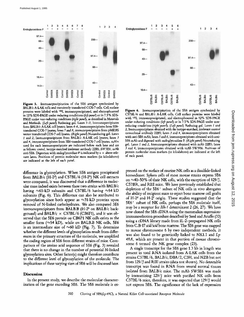
Figure 5. Immunoprecipitation of the 5E6 antigen synthesized by BALB/c A-LAK cells and transiently transfected COS-7 cells. Cell surface proteins were labeled with 12sI, immunoprecipitated, and electrophoresed in 12% SDS-PAGE under reducing conditions (left panel) or in 7.5% SDS- PAGE under non-reducing conditions (right panel), as described in Materials and Methods. (Lefipanel) Reducing gal. Lanes I-3, Immunoprecipitates from BALB/c A-LAK cell lysates; lanes 4-6, immunoprecipitates from 5E6- transfected COS-7 lysates; lanes 7 and 8, immunoprecipitates from pME18S vector transfected COS-7 cell lysates. (Right panel) Nonreducing gel. Lanes 1 and 2, Immunoprecipitates from BALB/c A-LAK cell lysates; lanes 3 and 4, immunoprecipitates from 5E6-transfected COS-7 cell lysates, mAbs used for each immunoprecipitate are indicated below each lane and are as follows: control, isotype-matched irrelevant antibody 2285; SW5E6, mAb anti-5E6. Digestion with endoglycosidase-F is indicated by a + above rele- vant lanes. Positions of protein molecular mass markers (in kilodaltons) are indicated at the left of each panel.
Figure 6. Immunoprecipitation of the 5E6 antigen synthesized by C57BL/6 and BALB/c A-LAK cells. Cell surface proteins were labeled with 12si, immunoprecipitated, and electrophoresed in 12% SDS-PAGE under reducing conditions (left panel) or in 7.5% SDS-PAGE under non- reducing conditions (right panel). (Left panel) Reducing gel. Lanes I and 2, Immunoprecipitates obtained with the isotype-matched, irrelevant control monoclonal antibody 2285; lanes 3 and 4, immunoprecipitates obtained with anti-5E6 mAb; lanes 5 and 6, immunoprecipitates obtained with anti- 5E6 mAb and digested with endoglycosidase E (Right panel) Nonreducing gel. Lanes 1 and 2, Immunoprecipitates obtained with mAb 2285; lanes 3 and 4, immunoprecipitates obtained with mAb SW5E6. Positions of protein molecular mass markers (in kilodaltons) are indicated at the left of each pand.
difference in glycosylation. When 5E6 antigen precipitated from BALB/c (H-2 a) and C57BL/6 (H-2 b) NK cell extracts were compared, it was discovered that a difference in molec- ular mass indeed exists between these two strains with BALB/c having "~65-kD subunits and C57BL/6 having ~54 kD subunits (Fig. 6). This difference can also be attributed to glycosylation since both appear as '~31-kD proteins upon removal of N-linked carbohydrates. We also compared 5E6 immunoprecipitates from BALB.B (H-2 b on BALB/c back- ground) and BALB/c x C57BL/6 (CB6F1), and it was ob- served that the 5E6 protein on CB6F1 NK cells exists as the smaller form ("~54 kD), while on BALB.B NK cells, 5E6 is an intermediate size of "~60 kD (Fig. 7). To determine whether the different levels of glycosylation result from differ- ences in the primary structure of the molecule, we amplified the coding region of 5E6 from different strains of mice. Com- parison of the amino acid sequence of 5E6 (Fig. 3) revealed that there is no change in the number of potential N-linked glycosylation sites. Other factor(s) might therefore contribute to the different level of glycosylation of the molecule. The implications of these varying sizes of 5E6 will be discussed later.
Discussion In the present study, we describe the molecular character-
ization of the gene encoding 5E6. The 5E6 molecule is ex-
pressed on the surface of murine NK cells as a disulfide-linked homodimer. Spleen cells of most mouse strains express 5E6 on "~40-50% of their NK cells, with the exception of 129/J, C57BR, and Ril l mice. We have previously established that depletion of the 5E6 § subset of NK cells in vivo abrogates the ability of recipient mice to reject bone marrow cell grafts of H-2 a and H-2 f origin. These studies suggested that the 5E6 § subset of NK cells, perhaps the 5E6 molecule itself, may be a receptor for Hh-1 determinant 2 (26, 27). We have now cloned the 5E6 cDNA using the mammalian expression- immunoselection procedure described by Seed and Aruffo (33) using a cDNA library made from IL-2-propagated NK cells from C.B-17 scid/scid bone marrow. The 5E6 gene was mapped to mouse chromosome 6 by two independent methods. It was also found to be genetically linked to NKI.1 and Ly- 49,4, which are present in that portion of mouse chromo- some 6 termed the NK gene complex (23).
A single transcript for the 5E6 gene 1.3 kb in length was present in total RNA isolated from A-LAK cells from the strains C57BL/6, BALB/c, DBA/2, C3H, and NZB but not from 129/J and Ril l strains (data not shown). No detectable transcript was found in RNA from several normal tissues isolated from BALB/c mice. The mAb SW5E6 was made by immunizing 129/J mice with purified NK cells from C57BL/6 mice, therefore, it was expected that 129/J would not express 5E6. The significance of the lack of expression
310 Cloning of 5E6(Ly-49C), a Natural Killer Cell-associated Receptor Molecule
on August 12, 2015
jem.rupress.org
Dow
nloaded from
Published August 1, 1995
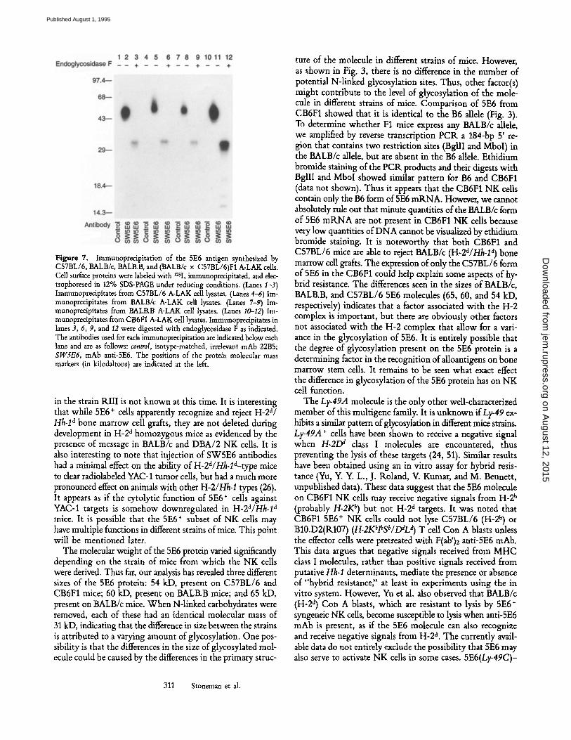
Figure 7. Immunoprecipitation of the 5E6 antigen synthesized by C57BL/6, BALB/c, BALB.B, and (BALB/c x C57BL/6)F1 A-LAK cells. Cell surface proteins were labeled with 125I, immunoprecipitated, and dec- trophoresed in 12% SDS-PAGE under reducing conditions. (Lanes I-3) Immunoprecipitates from C57BL/6 A-LAK cell lysates. (Lanes 4-6) Im- munoprecipitates from BALB/c A-LAK cell lysates. (Lanes 7-9) Im- munoprecipitates from BALB.B A-LAK cell lysates. (Lanes I0-12) Im- munoprecipitates from CB6FI A-LAK cell lysates. Immunopredpitates in lanes 3, 6, 9, and 12 were digested with endoglycosidase F as indicated. The antibodies used for each immunoprecipitation are indicated below each lane and are as follows: control, isotype-matched, irrelevant mAb 22B5; SW5E6, mAb anti-SE6. The positions of the protein molecular mass markers (in kilodaltons) are indicated at the left.
in the strain Ril l is not known at this time. It is interesting that while 5E6 + cells apparently recognize and reject H-2a/ Hh-1 a bone marrow cell grafts, they are not deleted during development in H-2 a homozygous mice as evidenced by the presence of message in BALB/c and DBA/2 NK cells. It is also interesting to note that injection of SW5E6 antibodies had a minimal effect on the ability of H-2a/Hh-la-type mice to clear radiolabeled YAC-1 tumor cells, but had a much more pronounced effect on animals with other H-2/Hh-1 types (26). It appears as if the cytolytic function of 5E6 + cells against YAC-1 targets is somehow downregulated in H-2d/Hh-1 a mice. It is possible that the 5E6 § subset of NK cells may have multiple functions in different strains of mice, This point will be mentioned later.
The molecular weight of the 5E6 protein varied significantly depending on the strain of mice from which the NK cells were derived. Thus far, our analysis has revealed three different sizes of the 5E6 protein: 54 kD, present on C57BL/6 and CB6F1 mice; 60 kD, present on BALB.B mice; and 65 kD, present on BALB/c mice. When N-linked carbohydrates were removed, each of these had an identical molecular mass of 31 kD, indicating that the difference in size between the strains is attributed to a varying amount of glycosylation. One pos- sibility is that the differences in the size of glycosylated mol- ecule could be caused by the differences in the primary struc-
ture of the molecule in different strains of mice. However, as shown in Fig. 3, there is no difference in the number of potential N-linked glycosylation sites. Thus, other factor(s) might contribute to the level of glycosylation of the mole- cule in different strains of mice. Comparison of 5E6 from CB6F1 showed that it is identical to the B6 allele (Fig. 3). To determine whether F1 mice express any BALB/c allele, we amplified by reverse transcription PCK a 184-bp 5' re- gion that contains two restriction sites (BgllI and MboI) in the BALB/c allele, but are absent in the B6 allele. Ethidium bromide staining of the PCK products and their digests with BgllI and MboI showed similar pattern for B6 and CB6F1 (data not shown). Thus it appears that the CB6F1 NK cells contain only the B6 form of 5E6 mKNA. However, we cannot absolutely rule out that minute quantities of the BALB/c form of 5E6 mRNA are not present in CB6F1 NK cells because very low quantities of DNA cannot be visualized by ethidium bromide staining. It is noteworthy that both CB6F1 and C57BL/6 mice are able to reject BALB/c (H-2a/Hh-1 a) bone marrow cell grafts. The expression of only the C57BL/6 form of 5E6 in the CB6F1 could help explain some aspects of hy- brid resistance, The differences seen in the sizes of BALB/c, BALB.B, and C57BL/6 5E6 molecules (65, 60, and 54 kD, respectively) indicates that a factor associated with the H-2 complex is important, but there are obviously other factors not associated with the H-2 complex that allow for a vari- ance in the glycosylation of 5E6. It is entirely possible that the degree of glycosylation present on the 5E6 protein is a determining factor in the recognition of alloantigens on bone marrow stem cells. It remains to be seen what exact effect the difference in glycosylation of the 5E6 protein has on NK cell function.
The Ly-49A molecule is the only other well-characterized member of this multigene family. It is unknown ifLy-49 ex- hibits a similar pattern of glycosylation in different mice strains. Ly-49A § cells have been shown to receive a negative signal when H-21M class I molecules are encountered, thus preventing the lysis of these targets (24, 51). Similar results have been obtained using an in vitro assay for hybrid resis- tance (Yu, Y. Y. L., J. Roland, V. Kumar, and M. Bennett, unpublished data). These data suggest that the 5E6 molecule on CB6F1 NK cells may receive negative signals from H-2 b (probably H-2K b) but not H-2 a targets. It was noted that CB6F1 5E6 + NK cells could not lyse C57BL/6 (H-2 b) or B10.D2(K107) (H-2KblbS~/DaL~ T cell Con A blasts unless the effector cells were pretreated with P(ab')2 anti-5E6 mAb. This data argues that negative signals received from MHC class I molecules, rather than positive signals received from putative Hh-1 determinants, mediate the presence or absence of "hybrid resistance" at least in experiments using the in vitro system. However, Yu et al. also observed that BALB/c (H-2 a) Con A blasts, which are resistant to lysis by 5E6 + syngeneic NK cells, become susceptible to lysis when anti-5E6 mAb is present, as if the 5E6 molecule can also recognize and receive negative signals from H-2 a. The currently avail- able data do not entirely exclude the possibility that 5E6 may also serve to activate NK cells in some cases. 5E6(Ly-49C)-
311 Stoneman et al.
on August 12, 2015
jem.rupress.org
Dow
nloaded from
Published August 1, 1995
transfected cells were recently shown to bind to various class I molecules (52). It is conceivable that the 5E6 molecule may function as an activating molecule when H-2 a targets are en- countered by H-2 b or H-2 bxa NK cells, but as a negative sig- nalling receptor with respect to H-2 b targets. This provides an attractive explanation for the differences seen in the glyco- sylation of 5E6 in H-2 a homozygous versus other strains of mice.
It is also noteworthy that NK cells of NZB (H-2 a) origin express the smaller (54 kD subunit) form of 5E6, as opposed to BALB/c (H-2a), which expresses 5E6 with 65-kD sub- units (data not shown). This is particularly interesting in view of the fact that treatment of recipient C57BL/6 mice with anti-5E6 mAb had no effect on their ability to reject NZB bone marrow cells (53). Therefore, we cannot be absolutely certain that NZB bone marrow cells express Hh-1 determinant 2. Indeed, one study maps the gene for hybrid resistance to NZB bone marrow cells 32 cM telomeric of H-2 instead of near H-2, as with other Hh-I determinants (54).
Another recent report (Murphy, W. J., A. Kaziuddin, L. Mason, V. Kumar, M. Bennett, and D. L. Longo, manuscript submitted for publication) demonstrate that 5E6 + and 5E6- subsets of NK cells can function differently in the regulation of hematopoiesis in different strains of mice. They observed that in H-2 a mice, 5E6 § cells produce more GM-CSF than IFN-'r and stimulate hematopoiesis, while 5E6- cells pro- duce more INF-y than GM-CSF and inhibit hematopoiesis. In contrast the reverse is true in H-2 b mice: 5E6 + cells make more INF-3' than GM-CSF and inhibit hematopoiesis, while 5E6- cells make more GM-CSF than INF- 7 and stimulate hematopoiesis. These differences in cytokine secretion pat- terns in 5E6 + cells of H-2 b and H-2 a strains of mice may also be related in some manner to the variation we report in the glycosylation of 5E6,
Note added in proof Further PCR analysis has detected low levels of the BALB/c form of 5E6 mRNA in CB6F1 NK cells.
The authors would like to thank Thomas Moore for flow cytometry analysis.
This work was supported by grants CA-36921, CA-36922, AI-20451, and CA-09082 from the National Institutes of Health, and by a grant from the Texas Department of Ladies' Auxiliary, Veterans of Foreign Wars.
Address correspondence to Porunelloor A. Mathew, Department of Pathology, University of Texas South- western Medical Center, 5323 Harry Hines Boulevard, Dallas, TX 75235.
Received for publication 4 August
References
1. Trinchieri, G. 1989. Biology of natural killer cells. Adv. Im- munol. 47:187-376.
2. Tutt, M.M., W.A. Kuziel, J. Hackett, Jr., M. Bennett, P.W. Tucker, and V. Kumar. 1986. Murine natural killer cells do not express functional transcripts of the c~-, 3-, or 3,-chain genes of the T cell receptor. J. Immunol. 137:2298-3001.
3. Herberman, R.B. 1982. NK Cells and Other Natural Effector Cells. Academic Press, New York. p. 1566.
4. Reynolds, C.W., and R.H. Wiltrout. 1989. Functions of the Natural Immune System. Plenum Press, New York. p. 485.
5. Bennett, M. 1987. Biology and genetics of hybrid resistance. Adv. Immunol. 41:333-445.
6. Murphy, W.J., V. Kumar, and M. Bennett. 1987. Rejection of bone marrow allografts by mice with severe combined im- mune deficiency (SCID): evidence that natural killer cells can mediate the specificity of marrow graft rejection.J. Exp. Med. 165:1212-1217.
7. Yu, Y.Y.L., V. Kumar, and M. Bennett. 1992. Murine natural killer cells and marrow graft rejection. Annu. Rev. Immunol. 10:189-213.
8. Harris, D.T., L. Jaso-Friedmann, R.B. Devlin, S.H. Koren, and D.L. Evans. 1991. Identification of an evolutionary conserved function-associated molecule on human natural killer cells. Proa Natl. Acad. Sci. USA. 88:3009-3013.
9. Frey, J.L., T. Bino, R.R.S. Kantor, D.M. Sega, S.L. Giardina, J. Roder, S. Anderson, and J.R. Ortaldo. 1991. Mechanism of target cell recognition by natural killer cells: characteriza- tion of a novel triggering molecule restricted to CD3- large granular lymphocytes. J. Exp. Med. 174:1527-1536.
10. Ciccone, E., D. Pende, O. Viale, C. Di Donato, G. Tripodi, A.M. Orengo, J. Guardiola, A. Moretta, and L. Moretta. 1992.
1994 and in revised form 23 February 1995.
Evidence of a natural killer (NK) cell repertoire for (Allo) an- tigen recognition: definition of five distinct NK-determined allospecificities in humans. J. Exl~ Med. 175:709-718.
11. Moretta, L., E. Ciccone, A. Moretta, P. H6glund, C. (~hl6n, and K. K~irre. 1992. Allorecognition by NK cells: nonself or no self? Immunol. Today. 13:300-306.
12. Houchins, J.P., T. Yabe, C. McSherry, and F.H. Bach. 1991. DNA sequence analysis of NKG2, a family of related cDNA clones encoding type II integral membrane proteins on human natural killer cells. J. Exl~ Med. 173:1017-1020.
13. Chambers, W.H., N.L. Vujanovic, A.B. DeLeo, M.W. O1- szowy, K.B. Herberman, andJ.C. Hiserodt. 1989. Monoclonal antibody to a triggering structure expressed on rat natural killer cells and adherent lymphokine-activated killer cells.J. Exl~ Med. 169:1373-1389.
14. Giorda, R., W.A. Rudert, C. Vavassori, W.H. Chambers, J.C. Hiseerodt, and M. Trucco. 1990. NKR-P1, a signal transduc- tion molecule on natural killer cells. Science (Wash. DC). 249:1298-1300.
15. Chart, P., and F. Takei. 1989. Molecular cloning and charac- terization of a novel murine T cell surface antigen, YE1/48. J. Immunol. 142:1727-1736.
16. Yokoyama, W.M., L.B. Jacobs, O. Kanagawa, E.M. Shevach, and D.I. Cohen. 1989. A murine T lymphocyte antigen be- longs to a supergene family of type II integral membrane pro- teins. J. Immunol. 143:1379-1386.
17. Wong, S., J.D. Freeman, C. Kelleher, D. Mager, and F. Takei. 1991. Ly-49 multigene family: new members of a superfamily of type II membrane proteins with lectin-like domains.J. Im- munol. 147:1417-1423.
18. Giorda, R., and M. Trucco. 1991. Mouse NKR-P1. A family
on August 12, 2015
jem.rupress.org
Dow
nloaded from
Published August 1, 1995
of genes selectively coexpressed in adherent lymphokine- activated killer cells. J. Immunol. 147:1701-1708.
19. Yokoyama, W.M., J.C. Ryan, J.J. Hunter, H.R.C. Smith, M. Stark, and W.E. Seaman. 1991. cDNA cloning of mouse NKR- P1 and genetic linkage with Ly-49. Identification of a natural killer cell gene complex on mouse chromosome 6.J. Immunol. 147:3229-3236.
20. Ryan, J.C., J. Turck, E.C. Niemi, W.M. Yokoyama, and W.E. Seaman. 1992. Molecular cloning of the NKI.1 antigen, a member of the NKR-P1 family of natural killer cell activation molecules. J. Immunol. 149:1631-1635.
21. Mathew, P.A., B.A. Garni-Wagner, K. land, A. Takashima, E. Stoneman, M. Bennett, and V. Kumar. 1993. Cloning and characterization of the 2B4 gene encoding a molecule associated with non-MHC-restricted killing mediated by activated nat- ural killer cells and T cells. J. Immunol. 151:5328-5337.
22. Yokoyama, W.M. 1993. Recognition structures on natural killer cells. Cu~ Opin. Immunol. 5:67-73.
23. Yokoyama, W.M., and W.E. Seaman. 1993. The Ly-49 and NKK-P1 gene families encoding lectin-like receptors on nat- ural killer cells: the NK gene complex. Annu. Rev. Immunol. 11:613-635.
24. Chambers, W.H., T. Adamkiewicz, andJ.P. Houchins. 1993. Type II integral membrane proteins with characteristics of C-type animal lectins expressed by natural killer (NK) cells. Glycobiology. 3:9-14.
25. Karlhofer, F.M., R.K. Ribaudo, and W.M. Yokoyama. 1992. MHC class I alloantigen specificity of Ly-49 + IL-2-activated natural killer cells. Nature (Lond.). 358:66-70.
26. Sentman, C.L., J. Hackett, Jr., V. Kumar, and M. Bennett. 1989. Identification of a subset of routine natural killer cells that mediates rejection of Hh-1 d but not Hh-1 b bone marrow grafts. J. Exp. Med. 170:191-202.
27. Sentman, C.L., V. Kumar, and M. Bennett. 1991. Rejection of bone marrow cell allografts by natural killer cell subsets: 5E6 + cell specificity for Hh-1 determinant 2 shared by H-2 a and H-2 f. Eur. J. Immunol. 21:2821-2828.
28. Gluzman, Y. 1981. SV40-transformed simian cells support the replication of early SV40 mutants. Cell. 23:175-182.
29. Gunjii, Y., N.L. Vujanovic, J.C. Hiseordt, R.B. Herberman, and E. Gorelik. 1989. Generation and characterization of purified adherent lymphokine-activated killer cells in mice. J. Immunol. 142:1748-1754.
30. Chomczynski, P., and N. Sacchi. 1987. Single-step method of RNA isolation by acid guanidinium thiocyanate-phenol- chloroform extraction. Anal. Biochem. 162:156-159.
31. Okayama, H., and P. Berg. 1982. High-affinity cloning of full- length cDNA. Mol. Cell. Biol. 2:161-170.
32. Takebe, Y., M. Seiki, J. Fujisawa, P. Hoy, K. Yokota, K. Arai, M. Yoshida, and N. Arai. 1988. SRc~ promoter: an efficient and versatile mammalian cDNA expression system composed of the simian virus 40 early promoter and the R-U5 segment of human T-cell leukemia virus type 1 long terminal repeat. Mol. Cell. Biol. 8:466-472.
33. Seed, B., and A. Aruffo. 1987. Molecular cloning of the CD2 antigen, the T-cell erythrocyte receptor, by a rapid immunoselec- tion procedure. Proc. Natl. Acad. Sci. USA. 84:3365-3369.
34. Sambrook, J., E.F. Fritsch, and T. Maniatis. 1989. Molecular Cloning: A Laboratory Manual. Cold Spring Harbor labora- tory Press, Cold Spring Harbor, New York. pp. 1641-1646.
35. Hirt, B. 1967. Selective extraction of polyoma DNA from in- fected mouse cell cultures. J. Mol. Biol. 26:365-369.
36. Sandri-Goldin, R.M., A.L. Goldin, M. Levine, and J.C. Glorioso. 1981. High-frequency transfer of cloned herpes sim- plex virus type I sequences to mammalian cells by protoplast
fusion. Mol. Cell. Biol. 1:743-752. 37. Henikoff, S. 1987. Unidirectional digestion with exonuclease
III in DNA sequence analysis. Methods Enzymol. 155:156-165. 38. Sanger, F., S. Nicklen, and A.R. Coulson. 1977. DNA se-
quencing with chain-terminating inhihitors. Pro~ Natl. Acad. Sci. USA. 74:5463-5467.
39. Coligan, J.E., A.M. Kruisbeek, D.H. Margulies, E.M. Shevach, and W. Strober. 1992. Current Protocols in Immunology. John Wiley & Sons, Inc., New York. pp. 8111-8114.
40. laemmli, U.K. 1970. Cleavage of structural proteins during the assembly of the head of bacteriophage T4. Nature (Lond.). 227:680-685.
41. Chestnut, K., J.-X. She, I. Cheng, K. Muralidharan, and E.K. Wakeland. 1993. Characterizations of the candidate genes for IDD susceptibility from the diabetes-prone NOD mouse strain. Mature. Genome. 4:549-554.
42. She, J.X., M.M. Bui, X.H. Tian, A. Muir, E.K. Wakeland, B. Zorovich, L.P. Zhang, M.C. Liu, G. Thomson, and N.K. Maclaren. 1994. Additive susceptibility to insulin-dependent diabetes conferred by HLA-DQB1 and insulin genes. Autoim- munity. 18:195-201.
43. Bailey, D.W. 1981. Recombinant inbred strains and bilineal con- genic strains. In The Mouse in Biomedical Research. Volume I: History, Genetics, and Wild Mice. H.L. Foster, J.D. Small, and J.G. Fox, editors. Academic Press, New York. pp. 223-240.
44. Seigelman, M.H., I.C. Cheng, I.L. Weissman, and E.K. Wakeland. 1990. The mouse lymph node homing receptor is identical with the lymphocyte cell surface marker Ly-22: role of the EGF domain in endothelial binding. Cell. 61:611-622.
45. Drickamer, K. 1988. Two distinct classes of carbohydrate- recognition domains in animal lectins.J. BioL Chem. 263:9557- 9560.
46. Yokoyama, W.M., P.A. Kehn, D.I. Cohen, and E.M. Shevach. 1990. Chromosomal location of the Ly-49 (A1, YE1/48) mul- tigene family: genetic association with the NKI.1 antigen. J. Immunol. 145:2353-2358.
47. Smith, H.K.C., F.M. Karlhofer, and W.M. Yokoyama. 1994. Ly-49 multigene family expressed by I1-,2 activated NK cells. J. Immunol. 153:1068-1079.
48. Shawlot, W., M.J. Siciliano, K.L. Stallings, and P.A. Over- beek. 1989. Insertional inactivation of the downless gene in a family of transgenic mice. Mol. Biol. Med. 6:299-307.
49. Deng, Z., P. Lui, P. Marlton, D.F. Claxton, S. Lane, D.F. CaUen, F.S. Collins, and M.J. Siciliano. 1993. Smooth muscle myosin heavy chain locus (MYH11) maps to 16p13.13-p13.12 and es- tablishes a new region of conserved synteny between human 16p and mouse 16. Genomics. 18:156-159.
50. Moore, K.J., and R.W. Elliott. 1993. Mouse chromosome 6. Mamm. Genome. 4(Suppl.):88-109.
51. Yokoyama, W.M., B.F. Daniels, H.R.C. Smith, W.E. Seaman, D.H. Margulies, and F.M. Karlhofer. 1994. The Ly-49 family of murine NK cell receptors. Nat. Immun. 13:195. (Abstr.).
52. Brennan, J., D. Mager, W. Jefferies, and F. Takei. 1994. Ex- pression of different members of the Ly-49 gene family defines distinct natural killer cell subsets and cell adhesion properties. J. Exp. Med. 180:2287-2295.
53. Bennett, M., R.M. Rembecki, C.L. Sentman, W.J. Murphy, Y.Y.L. Yu, C. Davenport, and V. Kumar. 1993. Bone marrow transplantation and natural killer (NK) cells in mice. In Nat- ural Immunity to Normal Hemopoietic Cells. B. Rolstad, editor. CRC Press, Inc., Boca Raton, FL. pp. 33-84.
54. LotzovL E., and G. Cudkowicz. 1973. Resistance of irradi- ated F1 hybrid and allogeneic mice to bone marrow grafts of NZB donors. J. Immunol. 110:791-800.
on August 12, 2015
jem.rupress.org
Dow
nloaded from
Published August 1, 1995
Related Documents