Clinicopathologic Correlation and Genetic Analysis in a Case of Posterior Polymorphous Corneal Dystrophy SAYOKO E. MOROI, MD, PHD, PARAG A. GOKHALE, MD, MIRIAM T. SCHTEINGART, MD, ALAN SUGAR, MD, CATHERINE A. DOWNS, MS, SATOKO SHIMIZU, MD, CHARLES KRAFCHAK, MPH, NOBUO FUSE, MD, SUSAN G. ELNER, MD, VICTOR M. ELNER, MD, PHD, ANDREW FLINT, MD, MICHAEL P. EPSTEIN, PHD, MICHAEL BOEHNKE, PHD, AND JULIA E. RICHARDS, PHD ● PURPOSE: To evaluate the clinical history, histopathol- ogy, and genetics of posterior polymorphous corneal dystrophy (PPMD) in a woman with a prominent retro- corneal membrane. ● DESIGN: Observational case report and genetic analysis of her family, UM:139. ● METHODS: Records were reviewed from a case and associated family members. The diagnosis of PPMD was based on clinical examination, immunohistochemical staining, electron microscopy, and screening of genetic markers from regions previously reported to be associated with PPMD. ● RESULTS: Over 17 years, the proband with PPMD had 25 ocular procedures performed for glaucoma, cataract, cornea, retina, and postoperative problems. A prominent retrocorneal membrane grew onto the crystalline lens and intraocular lens (IOL). Histopathology revealed strati- fied epithelial-like cells on iris from an iridectomy and stratified corneal endothelium on a corneal button. Elec- tron microscopy on the cornea revealed microvilli, tono- filaments, and desmosomes consistent with endothelial transformation, which was confirmed by positive anticy- tokeratin (CK) AE1/AE3 and CAM 5.2 immunoreactiv- ity. Negative immunoreactivity in epithelium and positive in endothelium with anti-CK 7 supported the diagnosis of PPMD rather than epithelial downgrowth. Multiple relatives were affected with PPMD with appar- ent autosomal dominant inheritance, but surprisingly, the PPMD, congenital hereditary endothelial dystrophy 1 (CHED1) and CHED2 loci on chromosome 20 and the collagen, type VIII, -2 (COL8A2) gene were excluded by linkage and haplotype analyses. ● CONCLUSIONS: We are unaware of previous PPMD reports describing the unusual feature of a retrocorneal membrane extending onto the crystalline lens and IOL. In addition, this family suggests another PPMD locus. (Am J Ophthalmol 2003;135:461– 470. © 2003 by Elsevier Science Inc. All rights reserved.) P OSTERIOR POLYMORPHOUS DYSTROPHY (PPMD), which is also referred to in the literature as posterior polymorphous corneal dystrophy (PPCD and PPD), is an autosomal dominant bilateral corneal dystrophy characterized by transformation of the endothelium into epithelial-like cells. 1 There is a wide clinical spectrum ranging from a benign clinical course of asymptomatic nonprogressive disease to relentless progression with seri- ous visual disability from corneal edema and secondary glaucoma. 2 It is estimated that up to 40% of patients with PPMD have elevated intraocular pressure (IOP). 3 Clinical findings include corneal endothelial vesicles, bands, diffuse Accepted for publication Oct 9, 2002. InternetAdvance publication at ajo.com Oct 22, 2002. From the Department of Ophthalmology and Visual Sciences, W. K. Kellogg Eye Center, University of Michigan, Ann Arbor, Michigan (S.E.M., A.S., C.A.D., S.S., C.K., N.F., S.G.E., V.M.E., J.E.R.); Depart- ment of Ophthalmology, Medical College of Georgia, Augusta, Georgia (P.A.G.); Andersen Eye Center, Saginaw, Michigan (M.T.S.); Depart- ment of Ophthalmology, Teikyo University, Tokyo, Japan (S.S.); De- partment of Ophthalmology, Tohoku University, Sendai, Japan (N.F.); Department of Pathology, University of Michigan, Ann Arbor, Michigan (V.M.E., A.F.); Department of Biostatistics, University of Michigan, Ann Arbor, Michigan (M.P.E., M.B.); Department of Human Genetics, Emory University, Atlanta, Georgia (M.P.E.); Department of Epidemiology, University of Michigan, Ann Arbor, Michigan (J.E.R.). This work was supported by NIH EY00353 (S.E.M.), NIH EY11671 (J.E.R.), NIH EY07003 (core grant supported Mitchell S. Gillett, BS, Department of Ophthalmology and Visual Sciences, University of Mich- igan, to create Figures 1–5), and NIH T32 HG00040 (M.P.E.), University of Michigan Rackham Predoctoral Fellowship (M.P.E.), the Lew R. Wasserman Award from Research to Prevent Blindness, Inc. (J.E.R.), Career Development Award from Research to Prevent Blindness, Inc. (S.E.M.), and an unrestricted grant from Research to Prevent Blindness, Inc., New York, NY. Inquiries to Sayoko E. Moroi, MD, PhD, Department of Ophthalmol- ogy & Visual Sciences, University of Michigan, 1000 Wall Street, Ann Arbor, MI 48105; fax: (734) 615-0542; e-mail: [email protected] © 2003 BY ELSEVIER SCIENCE INC.ALL RIGHTS RESERVED. 0002-9394/03/$30.00 461 PII S0002-9394(02)02032-9

Welcome message from author
This document is posted to help you gain knowledge. Please leave a comment to let me know what you think about it! Share it to your friends and learn new things together.
Transcript
Clinicopathologic Correlation and GeneticAnalysis in a Case of Posterior Polymorphous
Corneal Dystrophy
SAYOKO E. MOROI, MD, PHD, PARAG A. GOKHALE, MD,MIRIAM T. SCHTEINGART, MD, ALAN SUGAR, MD, CATHERINE A. DOWNS, MS,
SATOKO SHIMIZU, MD, CHARLES KRAFCHAK, MPH, NOBUO FUSE, MD,SUSAN G. ELNER, MD, VICTOR M. ELNER, MD, PHD, ANDREW FLINT, MD,
MICHAEL P. EPSTEIN, PHD, MICHAEL BOEHNKE, PHD, AND JULIA E. RICHARDS, PHD
● PURPOSE: To evaluate the clinical history, histopathol-ogy, and genetics of posterior polymorphous cornealdystrophy (PPMD) in a woman with a prominent retro-corneal membrane.● DESIGN: Observational case report and genetic analysisof her family, UM:139.● METHODS: Records were reviewed from a case andassociated family members. The diagnosis of PPMD wasbased on clinical examination, immunohistochemicalstaining, electron microscopy, and screening of geneticmarkers from regions previously reported to be associatedwith PPMD.● RESULTS: Over 17 years, the proband with PPMD had25 ocular procedures performed for glaucoma, cataract,cornea, retina, and postoperative problems. A prominent
retrocorneal membrane grew onto the crystalline lens andintraocular lens (IOL). Histopathology revealed strati-fied epithelial-like cells on iris from an iridectomy andstratified corneal endothelium on a corneal button. Elec-tron microscopy on the cornea revealed microvilli, tono-filaments, and desmosomes consistent with endothelialtransformation, which was confirmed by positive anticy-tokeratin (CK) AE1/AE3 and CAM 5.2 immunoreactiv-ity. Negative immunoreactivity in epithelium andpositive in endothelium with anti-CK 7 supported thediagnosis of PPMD rather than epithelial downgrowth.Multiple relatives were affected with PPMD with appar-ent autosomal dominant inheritance, but surprisingly,the PPMD, congenital hereditary endothelial dystrophy1 (CHED1) and CHED2 loci on chromosome 20 and thecollagen, type VIII, �-2 (COL8A2) gene were excludedby linkage and haplotype analyses.● CONCLUSIONS: We are unaware of previous PPMDreports describing the unusual feature of a retrocornealmembrane extending onto the crystalline lens and IOL.In addition, this family suggests another PPMD locus.(Am J Ophthalmol 2003;135:461–470. © 2003 byElsevier Science Inc. All rights reserved.)
P OSTERIOR POLYMORPHOUS DYSTROPHY (PPMD),
which is also referred to in the literature as posteriorpolymorphous corneal dystrophy (PPCD and PPD),
is an autosomal dominant bilateral corneal dystrophycharacterized by transformation of the endothelium intoepithelial-like cells.1 There is a wide clinical spectrumranging from a benign clinical course of asymptomaticnonprogressive disease to relentless progression with seri-ous visual disability from corneal edema and secondaryglaucoma.2 It is estimated that up to 40% of patients withPPMD have elevated intraocular pressure (IOP).3 Clinicalfindings include corneal endothelial vesicles, bands, diffuse
Accepted for publication Oct 9, 2002.InternetAdvance publication at ajo.com Oct 22, 2002.From the Department of Ophthalmology and Visual Sciences, W. K.
Kellogg Eye Center, University of Michigan, Ann Arbor, Michigan(S.E.M., A.S., C.A.D., S.S., C.K., N.F., S.G.E., V.M.E., J.E.R.); Depart-ment of Ophthalmology, Medical College of Georgia, Augusta, Georgia(P.A.G.); Andersen Eye Center, Saginaw, Michigan (M.T.S.); Depart-ment of Ophthalmology, Teikyo University, Tokyo, Japan (S.S.); De-partment of Ophthalmology, Tohoku University, Sendai, Japan (N.F.);Department of Pathology, University of Michigan, Ann Arbor, Michigan(V.M.E., A.F.); Department of Biostatistics, University of Michigan, AnnArbor, Michigan (M.P.E., M.B.); Department of Human Genetics, EmoryUniversity, Atlanta, Georgia (M.P.E.); Department of Epidemiology,University of Michigan, Ann Arbor, Michigan (J.E.R.).
This work was supported by NIH EY00353 (S.E.M.), NIH EY11671(J.E.R.), NIH EY07003 (core grant supported Mitchell S. Gillett, BS,Department of Ophthalmology and Visual Sciences, University of Mich-igan, to create Figures 1–5), and NIH T32 HG00040 (M.P.E.), Universityof Michigan Rackham Predoctoral Fellowship (M.P.E.), the Lew R.Wasserman Award from Research to Prevent Blindness, Inc. (J.E.R.),Career Development Award from Research to Prevent Blindness, Inc.(S.E.M.), and an unrestricted grant from Research to Prevent Blindness,Inc., New York, NY.
Inquiries to Sayoko E. Moroi, MD, PhD, Department of Ophthalmol-ogy & Visual Sciences, University of Michigan, 1000 Wall Street, AnnArbor, MI 48105; fax: (734) 615-0542; e-mail: [email protected]
© 2003 BY ELSEVIER SCIENCE INC. ALL RIGHTS RESERVED.0002-9394/03/$30.00 461PII S0002-9394(02)02032-9

haze, iridocorneal adhesions, corectopia, and pupillaryectropion.3 Retrocorneal “glass-like” membranes havebeen described, and these membranes may extend acrossthe angle and onto the iris.1,3–6 We describe a 52-year-oldproband with PPMD who has the unusual complication ofa retrocorneal membrane proliferating on the anteriorsurface of a posterior chamber intraocular lens (IOL) andcrystalline lens. We also present evidence that PPMD inher family is not linked to the known PPMD (see OnlineMendelian Inheritance in Man number, or OMIM#,122000, which may be accessed at http://www.ncbi.nlm.nih.gov/), congenital hereditary endothelial dystrophy 1(CHED1, OMIM# 121700) or CHED2 (OMIM# 217700)loci on chromosome 20,7 nor to the candidate PPMDgenes, visual system homeobox gene 1 (VSX1, OMIM#605020), which is localized within the PPMD locus,8 orcollagen, type VIII, �-2 (COL8A2, OMIM# 120252),which is localized to 1p34.3–p32.3.9
METHODS
OPHTHALMOLOGIC EXAMINATION OF MEMBERS OF FAMILY
UM:139 was performed as part of a protocol approved bythe Institutional Review Board of the University of Mich-igan Medical Center. Blood samples from 26 family mem-bers were obtained after informed consent. Twenty-threeof these family members were examined by at least one ofthe authors (S.E.M., M.T.S., or A.S.), and the medicalrecords were obtained from the other three individuals’ophthalmologist. Participants were examined for the pres-ence of unequivocally characteristic corneal endothelialabnormalities of PPMD.3,10 Subjects were classified ashaving PPMD if they exhibited any of the followingcorneal findings: vesicular, geographic or band-like lesionsat the level of the Descemet membrane or posteriorvesicles in at least one eye. For the three individuals notexamined at our institution, the affected status was basedon the clinical diagnosis of PPMD by the primary ophthal-mologist. In addition, all examined subjects were evaluatedfor the presence of guttae based on excrescences visible onspecular reflection of the posterior corneal surface. Theclinicians who assigned the affected status had no genotypeinformation.
Formalin-fixed paraffin-embedded sections of two surgi-cal specimens from the left eye, the iris (February 27,1992), and corneal button (September 17, 1998) werestained with hematoxylin–eosin. A portion of the corneawas prepared for transmission electron microscopy. Immu-nohistochemistry was performed on the cornea usinganticytokeratin (CK) AE1/AE3 (Boehringer Mannheim,Indianapolis, Indiana, USA) and anti-CK CAM 5.2 (Bec-tin Dickinson, San Jose, California, USA) according tothe manufacturers’ instructions. Immunoperoxidase reac-tivity was detected by diaminobenzidine tetrahydrochlo-ride according to a standard protocol. In addition, the
anti-CK7 monoclonal antibody (DAKO-CK7, cloneOV-TL 12/30, DAKO Corp., Carpinteria, California,USA) was used.11 The anti-CK7 monoclonal antibody wasdiluted 1:400, warmed to 60 C for 15 minutes, and thenapplied to the section for 1 hour at room temperature. Thesection was washed three times with DAKO AntibodyDiluent (DAKO Corp.). The corneal sections were incu-bated with the secondary antibody conjugated to peroxi-dase using the Vectastain Elite ABC Kit (VectorLaboratories, Inc., Burlingame, California, USA) accord-ing to the manufacturer’s instructions. Immunoperoxidaseactivity was detected with 3-amino-9-ethylcarbazole ac-cording to a standard protocol.
Genomic DNA was isolated from blood samples from 26family members using Puregene (Gentra Systems, Minne-apolis, Minneapolis, USA) according to the manufactur-er’s instructions. Microsatellite repeat markers wereassayed by polymerase chain reaction using methods andparameters described previously.7,12–17 To evaluate linkageto the previously reported PPMD locus on chromosome20q, eight markers were evaluated from D20S98 toD20S119, a region that contains the entire reported 19.73cM PPMD interval between D20S98 and D20S108.7
Screening of these markers allowed us to evaluate linkageto the autosomal dominant CHED1 locus (also calledadCHED) previously suggested to be allelic to the PPMDlocus.14 The CHED1 locus lies in a 2.7 cM interval(D20S48–D20S471)14 located between D20S118 andD20S912. The VSX1 gene is reported to be mutated in tworelatives with PPMD and lies between D20S912 andD20S195.8,15–17 Screening of the interval from D20S199 toD20S95 allowed us to evaluate linkage to the autosomalrecessive CHED2 locus (also called arCHED) previouslyreported as mapping to a 6 cM interval between D20S113and D20S882 on chromosome 20p.18 In addition, wescreened D1S234 and D1S255, which flank COL8A2, toevaluate whether PPMD results from mutations inCOL8A2. This gene is relevant because a Gln455Lysmutation has been reported in Fuchs endothelial cornealdystrophy and has been found in a woman and her fatherwho both have PPMD.9
Single-point linkage analysis was performed by themethod of log of the odds (LOD) scores,19 which comparesthe probability of the data assuming that the two loci arelinked to each other at a hypothesized distance to theprobability of the data if the two loci are actually unlinked.This project used the accepted standard value of a LODscore less than or equal to �2.0 as significant evidenceagainst linkage and a LOD score greater than or equal to� 3.0 as significant evidence in favor of linkage. Thecomputer program MENDEL was used to carry out two-point linkage calculations to evaluate whether a singlegenetic marker is linked to a PPMD disease locus in thisfamily.20 Multipoint linkage analysis, which makes use ofdata from multiple genetic markers in a region simulta-
AMERICAN JOURNAL OF OPHTHALMOLOGY462 APRIL 2003
neously, was performed using the Monte Carlo Markovchain method of Sobel and Lange,21 as implemented in thecomputer program SIMWALK2.22 Assumptions included1% sporadic rate and 90% penetrance. Two differentdisease allele frequencies, 0.01 and 0.001, were tested with0.001 presumed as the relevant disease allele frequency inthe case of autosomal dominant inheritance and 0.01presumed as the relevant disease allele frequency in thecase of autosomal recessive inheritance. The autosomaldominant model was tested for both regions because of theobserved mode of inheritance in this family and the modeof inheritance of the first reported PPMD locus7 andCHED1.14 We also tested a less likely model of autosomalrecessive inheritance of prevalent recessive CHED218 al-lele(s), presuming that the apparent autosomal dominanttransmission of PPMD in this family could result from an
unusually high number of marriages of homozygous af-fected individuals to carriers. Marker allele frequencieswere estimated by the method of Boehnke.23 Lack ofgenotyping incompatibilities was determined throughuse of the method of O’Connell and Weeks24 with theuse of the program PedCheck and by the Monte CarloMarkov chain method of Sobel and Lange,21 as imple-mented in SIMWALK2.22
For purposes of analysis, individuals were designatedaffected if they were clinically diagnosed with PPMD, asdefined in methods, or unaffected if they had a normalophthalmologic examination. Individuals with other oph-thalmologic findings, such as guttae (individuals IV-2,IV-3, IV-15, and IV-17) and primary open-angle glaucoma(individual III-11) were considered to have an undesig-nated phenotype.
FIGURE 1. Photographs highlighting the 17-year history of our patient with posterior polymorphous corneal dystrophy (taken byour ophthalmic photography unit formerly directed by Professor Csaba Martonyi). (A) Vesicular lesion (white arrow) at the levelof the Descemet membrane in the left eye. (B) Broad–based peripheral anterior synechiae of the nasal angle of the left eye showing1 to 2 clock hours of open angle. (C, D) Progressive corectopia and ectropion uveae of the right eye approximately between the 1and 4-o’clock hours (between the white arrows) from 1985 (C) to 1989 (D). (E) “Glasslike” membrane (curved white line) on theleft iris and associated ectropion uvea from the 7 and 2-o’clock positions. (F) Two months following the first surgery, atrabeculectomy, on the left eye, this membrane proliferated on the crystalline lens. (G) Retrocorneal membrane nearly covers thesurface of a posterior chamber intraocular lens in the right eye. (H) Proliferative vitreoretinopathy caused a tractional retinaldetachment (left) with associated choroidal detachments (right) in the left eye.
CORRELATION AND ANALYSIS OF PPMDVOL. 135, NO. 4 463

RESULTS
THE 52-YEAR-OLD PROBAND WAS FIRST EVALUATED AT THE
University of Michigan in 1985 at the age of 38 years withan 18-month history of difficulty with vision and a9-month history of corectopia in her left eye. Both eyesrevealed corneal endothelial vesicles (Figure 1A), corec-topia, iridocorneal adhesions, and broad-based peripheralanterior synechiae (PAS) (Figure 1B). Over the next 4years, the corectopia and ectropion uveae progressed (Fig-ure 1C, D), PAS increased, and IOP rose, requiringtreatment with glaucoma medications. In February 1992,she underwent a trabeculectomy for secondary angle-closure glaucoma with an IOP of 45 mm Hg in the left eye.The peripheral iridectomy specimen was sent for histopa-thology. The filter failed, and subsequent surgical proce-dures are summarized in Table 1. Before the first surgery, aprominent retrocorneal membrane extended onto the leftiris (Figure 1E) and 2 months following the trabeculec-tomy, the membrane was seen on the crystalline lens(Figure 1F). In December 1997, 3 months after uneventfulcataract surgery and posterior chamber intraocular lens(IOL) placement in the right eye, a prominent membranewas visible on the anterior surface of the posterior chamber
IOL (Figure 1G). A “membranotomy” was performed twicewith a neodymium:yttrium–aluminum–garnet (Nd:YAG)laser, but the membrane regrew, and no further laserprocedures were advised.
Given the complicated glaucoma and retrocornealmembrane growth onto the IOL in the right eye, visual
TABLE 1. Surgical Interventions in Proband to Manage Posterior Polymorphous Corneal Dystrophy, Secondary Glaucoma, andOther Ocular Complications
Date Eye Procedure
2/27/92 Left Trabeculectomy and postoperative subconjunctival 5-FU injections
5/22/92 Left Trabeculectomy with mitomycin C (0.4 mg/ml, 3.5 min)
12/22/92 Left Phacoemulsification, no IOL placed
1/5/93 Left Trabeculectomy with mitomycin C (0.5 mg/ml, 2.5 min)
2/23/93 Left Molteno double plate implant
3/12/93 Left Tube revision due to endothelial touch
3/30/93 Left Pars plana vitrectomy for aqueous misdirection
11/24/93 Left Lacrimal gland biopsy for nonspecific orbital inflammation
1/24/94 Left Molteno implant removed due to orbital inflammation
2/8/94 Left Nd:YAG contact transcleral laser cyclophotocoagulation
5/22/94 Left Upper lid ptosis correction
10/4/95 Left Argon/Nd:YAG laser peripheral iridotomy for iris bombe
12/12/95 Left Repeat argon/Nd:YAG laser peripheral iridotomy for iris bombe
7/10/96 Left Nd:YAG laser peripheral iridotomy and capsulotomy
11/7/96 Left Pars plana vitrectomy and lysis of anterior chamber adhesions
4/24/97 Right Thermal sclerostomy and postoperative 5-FU injections
5/22/97 Right Trabeculectomy with mitomycin C (0.33 mg/ml, 3 min) and postoperative 5-FU injections
9/10/97 Right Phacoemulsification with posterior chamber IOL, ab interno bleb revision, and postoperative 5-FU injections
10/15/97 Right Nd:YAG posterior capsulotomy
12/16/97 Right Trabeculectomy revision and postoperative 5-FU injections
2/24/98 Right Baerveldt tube (350 mm2) with scleral patch graft
5/20/98 Right Nd:YAG “polishing” of anterior membrane on posterior chamber IOL
7/27/98 Right Nd:YAG “polishing” of anterior membrane on posterior chamber IOL
9/17/98 Left Penetrating keratoplasty
1/7/99 Left Pars plana vitrectomy and membrane peeling
5-FU � 5-fluorouracil; IOL � intraocular lens; Nd:YAG � neodymium:yttrium–aluminum–garnet laser.
FIGURE 2. Stratified epithelial-like cells on anterior surface ofleft peripheral iridectomy specimen from the first surgery(hematoxylin–eosin; bar is 100 �m).
AMERICAN JOURNAL OF OPHTHALMOLOGY464 APRIL 2003

rehabilitation was pursued in the left eye, which was anaphakic eye and had bullous keratopathy and an epiretinalmembrane. In September 1998, the proband had anuncomplicated penetrating keratoplasty. Two monthspostoperatively, she complained of a scotoma and had anIOP of 9 mm Hg on timolol 0.5% twice daily anddorzolamide 2% twice daily. There were peripheral cho-roidal detachments and a tractional macular retinal de-tachment due to progression of the epiretinal membrane(Figure 1H). The choroidal detachments did not resolveafter treatment with systemic prednisone and stoppingglaucoma medications. In January 1999, a repeat pars planavitrectomy and membrane peeling was performed. Unliketypical proliferative vitreoretinopathy, this membrane wasextremely tenacious and required a peripheral 360-degreeretinotomy to separate it from the retina. The membranespanned from the posterior segment to the inferior ciliarybody and the posterior iris surface.
Seven months postoperatively, she had an episode ofbilateral anterior uveitis with hypotony (4 mm Hg, righteye and 0–5 mm Hg, left eye) and nongranulomatousinflammation, which responded to topical and systemic
corticosteroids. In May 2002, her visual acuity was 20/100right eye and light perception left eye. Her IOP was 5 mmHg in the right eye without glaucoma medications. Theanterior segment examinations were unchanged, and theright retina was attached.
An iris biopsy obtained from the first surgical interven-tion, trabeculectomy of the left eye, revealed stratifiedepithelial-like cells on the anterior surface of the iris(Figure 2). Given this histopathologic appearance, it wasnecessary to discriminate between epithelial downgrowthand PPMD. The left penetrating keratoplasty specimenobtained in September 1998 allowed us to resolve thisimportant issue. This cornea revealed areas of stratifiedendothelium, which is a typical feature of PPMD (Figure3A). On transmission electron microscopy, the endothe-lium revealed microvilli projecting into the anterior cham-ber and desmosomes with associated tonofilamentsconsistent with epithelial-like transformation (Figure 3B).
To further discriminate the epithelial-like corneal en-dothelium from epithelium, an immunohistochemicalanalysis was performed using anti-CK antibodies. Cytoker-atins are water-insoluble proteins that form intracellular
FIGURE 3. Light and transmission electron microscopy of left corneal button. (A) Light microscopy revealed area of the Descemetmembrane covered by stratified endothelial cells (hematoxylin–eosin; bar is 100 �m). Endothelial surface is oriented down.) (B)Transmission electron microscopy revealed stratified endothelial cells with nuclei (N), microvilli (arrow), and desmosomalattachments (asterisk) (final magnification, 7625�).
CORRELATION AND ANALYSIS OF PPMDVOL. 135, NO. 4 465

intermediate filaments in epidermis and most epithelialtissues,25 including normal corneal epithelium, but not inendothelium.26,27 In contrast, corneas with PPMD stainpositively in the endothelium with anti-CK.26 Our caseillustrated positive immunoreactivity for anti-CK AE1/AE3 and CAM 5.2 in both epithelium and endothelium(Figure 4). Although this result is consistent with PPMD,it does not eliminate the possibility of epithelial down-growth in which the associated intraocular proliferativemembrane would stain similar to the ocular surface epi-thelia. A marker that is expressed in corneal endothelium,but not epithelium, is desirable. Two such markers havebeen identified: 2B4.14.1,28 a monoclonal antibody againsthuman corneal endothelium and CK7, a 54-kD CK iden-tified in an ovarian carcinoma cell line.29 The 2B4.14.1antibody stains normal corneal endothelium and notepithelium, but inconsistently stains the epithelial-likeendothelium in PPMD.28 In contrast, a comparative studyof normal and diseased corneas, including PPMD, illus-
trated that a monoclonal anti-CK7 antibody stains onlythe endothelium of corneas and not the epithelium.11 Inour case, the corneal epithelium stained negatively,whereas the endothelium stained positively with anti-CK7antibodies, supporting the diagnosis of PPMD rather thanepithelial downgrowth (Figure 5).
The histopathology of the proliferative vitreoretinopa-thy membrane obtained from January 1999 revealed fibro-cellular connective tissue containing pigmented epithelialcells, fibroblast-like cells, lens capsule, and stratified epi-thelial-like cells (not shown). Evaluation of anti-CK im-munohistochemical staining was not possible due to thelimited specimen.
A detailed family history covering six generations indi-cated that the proband is Caucasian with ancestry fromboth western and eastern Europe. A diagnosis of PPMDwas confirmed in 12 relatives of the proband in threegenerations through ophthalmologic examination or eval-uation of clinical records (Figure 6). Pedigree analysis offamily UM:139 (Figure 6) supports an autosomal dominantinheritance with high, but incomplete penetrance andexcludes X-linked and mitochondrial inheritance. In ad-dition, the lack of any history of hereditary nephritis inUM:139 family clearly eliminates the possibility of the rareoccurrence of PPMD associated with X-linked Alportsyndrome.30
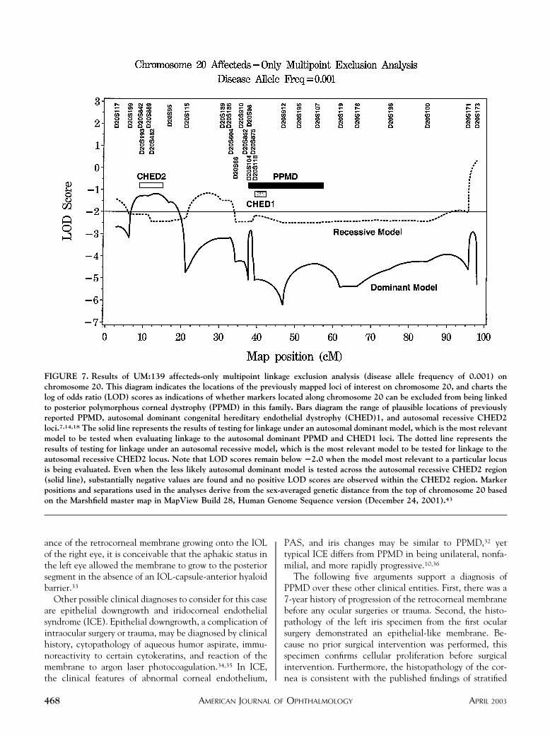
Multipoint analysis revealed formal exclusion of theentire PPMD interval and both the CHED1 locus andVSX1 gene contained within it (Figure 7). Under anautosomal dominant model with disease allele frequency of
FIGURE 4. Anticytokeratin (CK) AE1/AE3 and CAM 5.2immunoperoxidase staining with diaminobenzidine of left cor-neal specimen. (A) Corneal epithelium (top) and endothelium(bottom) stained positively for anti-CKs (bar is 100 �m). (B)Higher magnification of corneal endothelium (bar is 100 �m).(C) Negative control with no endothelial anti-CK staining (baris 100 �m).
FIGURE 5. Anticytokeratin (CK)7 immunoperoxidase stain-ing with 3-amino-9-ethylcarbazole of left corneal button (finalmagnification 2375�; stained by Zong-Mei Bian, MD). (A)Epithelium revealed no staining. (B) Endothelium stainedpositively, as evident with reddish reaction product.
AMERICAN JOURNAL OF OPHTHALMOLOGY466 APRIL 2003
0.001, an “affecteds”-only multipoint analysis yielded LODscores between �2.85 and �6.22, where values below�2.0 are normally considered formal proof of exclusion.19
Other combinations of models and parameters tested allresulted in LOD scores below �2.0 across the entirePPMD interval.
Although no previous linkage of PPMD to the CHED2locus has been reported, we also tested it because of themodel proposed for a genetic relationship between CHED1and PPMD.14,31 The most relevant combination of param-eters for evaluating linkage to the CHED2 locus (autoso-mal recessive inheritance, disease allele frequency of 0.01,affecteds-only analysis) resulted in formal exclusion of theCHED2 locus with LOD scores ranging from �3.17 to�3.38. Most other model-parameter combinations testedalso resulted in formal exclusion with LOD scores below�2.0, except for the test of autosomal recessive inheri-tance using information from the whole family with diseaseallele frequency of 0.001, which yielded LOD scores from�1.76 to �2.07.
Because of a report that two affected members of onePPMD family have a mutation in COL8A2, we also testedfor linkage to the relevant region of chromosome 1.9 Whendata from the whole UM:139 family were used under amodel of autosomal dominant inheritance with diseaseallele frequency of 0.001, multipoint analysis excluded theregion containing COL8A2 with LOD scores ranging from�4.35 to �6.02. Although evidence against involvementof COL8A2 was less strong when considering data fromaffected individuals only, the LOD scores ranging from
�1.65 to �2.20 provide evidence against linkage of PPMDin this family to the region containing COL8A2.
For no combination of models and parameters tested wasevidence found to favor linkage of PPMD in this family tothe PPMD/CHED1 interval, which contains the VSX1gene, the CHED2 interval, or the region containing theCOL8A2 gene. Haplotype analysis, which allowed us todetermine which alleles were likely to be traveling to-gether on the same chromosome, supported our conclusionthat alleles of PPMD, CHED1, CHED2, or COL8A2 areunlikely to be the cause of PPMD in this family.
DISCUSSION
ALTHOUGH PPMD IS USUALLY NONPROGRESSIVE, THERE
have been several reported cases of aggressive disease, as inthis case with retrocorneal membranes and glaucoma,which required penetrating keratoplasty and glaucomaprocedures.1,5,6,32 Pathology specimens in previous reportshave demonstrated the presence of retrocorneal mem-branes extending onto the anterior surface of the iris.1,4–6
We are unaware of previous PPMD reports of a retrocor-neal membrane growing onto the anterior surface of thecrystalline lens and the anterior surface of a posteriorchamber IOL. It is striking that the patient had multiplesurgeries in both eyes (see Table 1) with apparentlyaccelerated growth of this membrane after each surgery. Itis conceivable that inflammation during the postoperativeperiod stimulated membrane growth. Given the appear-
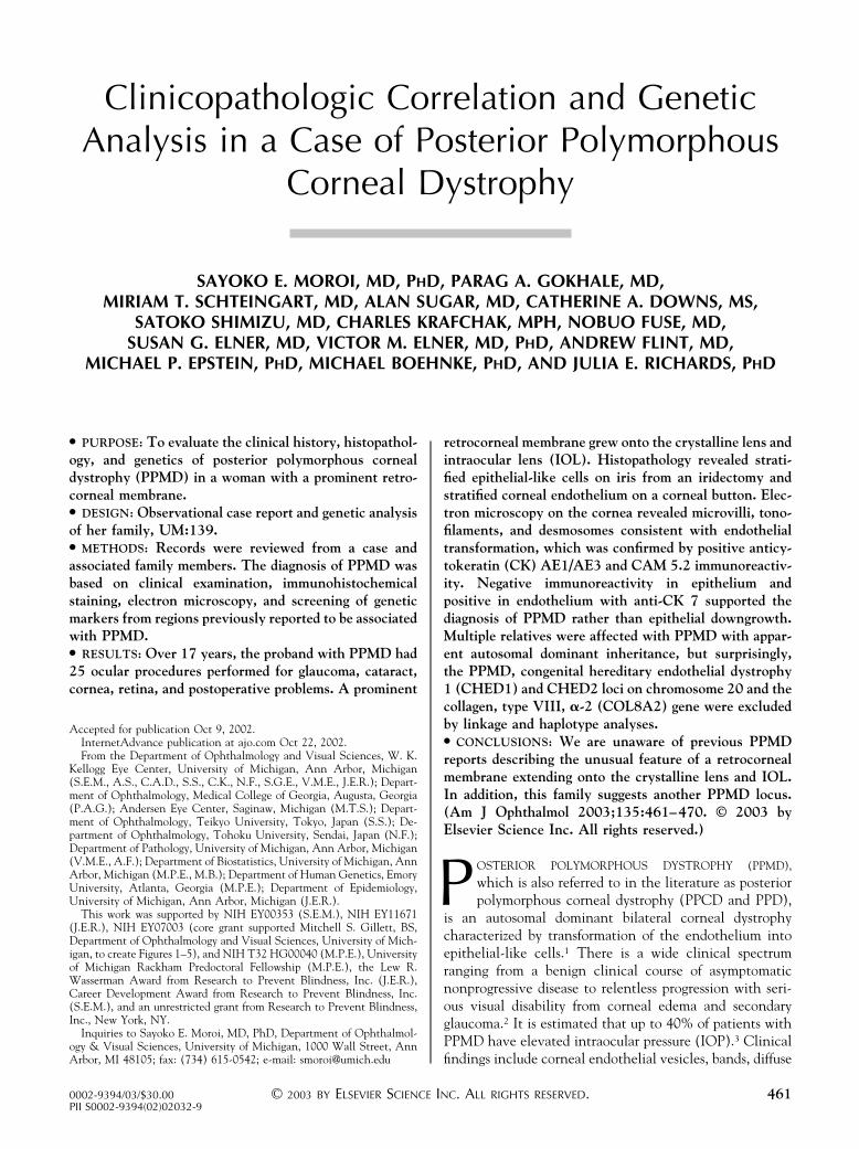
FIGURE 6. Pedigree of Family UM:139. Among those individuals for whom molecular data were generated for use in the analysis(marked with �), filled symbols are individuals with posterior polymorphous corneal dystrophy (PPMD), open symbols areindividuals deemed unaffected with PPMD, and a cross within a symbol indicates individuals who were scored as having anindeterminate PPMD status for purposes of analysis. Open symbols not marked with a � are individuals for whom molecular datawere not generated; they are free of PPMD by family report, but were not examined by us. Arrow indicates proband. Squaresrepresent males; circles represent females. A diagonal line through a symbol indicates the individual is deceased. Other significanteye disease in the family includes angle-closure glaucoma in the proband (IV-10), primary open-angle glaucoma (III-11), and familyreport of glaucoma (II-5); blindness in one eye due to accident (I-1) and blindness in one eye due to cataract (III-3).
CORRELATION AND ANALYSIS OF PPMDVOL. 135, NO. 4 467
ance of the retrocorneal membrane growing onto the IOLof the right eye, it is conceivable that the aphakic status inthe left eye allowed the membrane to grow to the posteriorsegment in the absence of an IOL-capsule-anterior hyaloidbarrier.33
Other possible clinical diagnoses to consider for this caseare epithelial downgrowth and iridocorneal endothelialsyndrome (ICE). Epithelial downgrowth, a complication ofintraocular surgery or trauma, may be diagnosed by clinicalhistory, cytopathology of aqueous humor aspirate, immu-noreactivity to certain cytokeratins, and reaction of themembrane to argon laser photocoagulation.34,35 In ICE,the clinical features of abnormal corneal endothelium,
PAS, and iris changes may be similar to PPMD,32 yettypical ICE differs from PPMD in being unilateral, nonfa-milial, and more rapidly progressive.10,36
The following five arguments support a diagnosis ofPPMD over these other clinical entities. First, there was a7-year history of progression of the retrocorneal membranebefore any ocular surgeries or trauma. Second, the histo-pathology of the left iris specimen from the first ocularsurgery demonstrated an epithelial-like membrane. Be-cause no prior surgical intervention was performed, thisspecimen confirms cellular proliferation before surgicalintervention. Furthermore, the histopathology of the cor-nea is consistent with the published findings of stratified
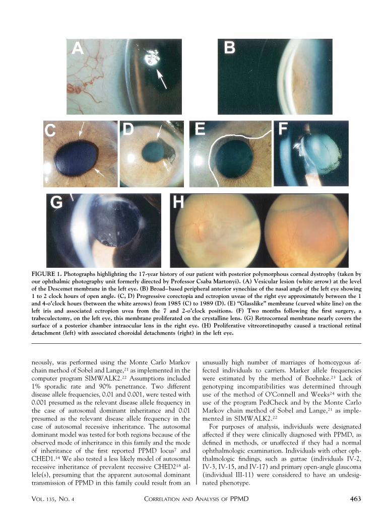
FIGURE 7. Results of UM:139 affecteds-only multipoint linkage exclusion analysis (disease allele frequency of 0.001) onchromosome 20. This diagram indicates the locations of the previously mapped loci of interest on chromosome 20, and charts thelog of odds ratio (LOD) scores as indications of whether markers located along chromosome 20 can be excluded from being linkedto posterior polymorphous corneal dystrophy (PPMD) in this family. Bars diagram the range of plausible locations of previouslyreported PPMD, autosomal dominant congenital hereditary endothelial dystrophy (CHED)1, and autosomal recessive CHED2loci.7,14,18 The solid line represents the results of testing for linkage under an autosomal dominant model, which is the most relevantmodel to be tested when evaluating linkage to the autosomal dominant PPMD and CHED1 loci. The dotted line represents theresults of testing for linkage under an autosomal recessive model, which is the most relevant model to be tested for linkage to theautosomal recessive CHED2 locus. Note that LOD scores remain below �2.0 when the model most relevant to a particular locusis being evaluated. Even when the less likely autosomal dominant model is tested across the autosomal recessive CHED2 region(solid line), substantially negative values are found and no positive LOD scores are observed within the CHED2 region. Markerpositions and separations used in the analyses derive from the sex-averaged genetic distance from the top of chromosome 20 basedon the Marshfield master map in MapView Build 28, Human Genome Sequence version (December 24, 2001).43
AMERICAN JOURNAL OF OPHTHALMOLOGY468 APRIL 2003

endothelium, microvilli, desmosomal attachments, andtonofilaments in PPMD.1,37–39 Third, this disease wasbilateral. Fourth, the immunohistochemistry staining ofher cornea demonstrated that the corneal endotheliumexpressed epithelial-like keratin markers (Figure 4).26,28
More important, however, the keratin marker CK7, whichis positive for endothelium and negative for epithelium11
was positive in our patient’s corneal endothelium andnegative in epithelium (Figure 5). Fifth, 13 family mem-bers in three generations were affected with PPMD indi-cating the expected autosomal dominant pattern ofinheritance. Hence, the clinical course, family history,histopathology, and immunohistochemistry support thediagnosis of PPMD in our patient and not epithelialdowngrowth or ICE.
Surprisingly, although our observation of autosomaldominant inheritance of PPMD in the UM:139 family wasconsistent with the previously reported PPMD locus onchromosome 20q,7 multipoint linkage analysis excludedthis PPMD locus. Specifically, screening of chromosome 20markers suggests that PPMD in the UM:139 family is notthe result of disease-influencing allele(s) located at thePPMD locus, the CHED1 locus, or the CHED2 locus. Inaddition, the PPMD in this family is apparently not causedby the COL8A2 mutation previously reported in a womanand her father who both had PPMD.9 Exclusion of theseloci in a family whose pedigree is strongly consistent withan autosomal dominant form of PPMD does not detractfrom the PPMD diagnosis but does suggest the existence ofanother PPMD locus, which could be identified through agenome scan.
In summary, the proband demonstrated a prominentretrocorneal membrane that extended onto the crystallinelens and anterior surface of the IOL, which has not beenpreviously reported for PPMD. The genetic analysis of herfamily, UM:139, illustrates the genetic heterogeneity ofPPMD. Insights into the pathogenesis of PPMD willundoubtedly result from genetic studies of this autosomaldominant corneal disease, as with recent genetic studies ofother corneal dystrophies.40–42 Identification of the PPMDgenes will provide valuable tools with which to study theabnormal biology of the corneal endothelium in PPMDand to investigate the phenotypic variability of this cor-neal dystrophy.
REFERENCES
1. Krachmer JH. Posterior polymorphous corneal dystrophy: adisease characterized by epithelial-like endothelial cellswhich influence management and prognosis. Trans AmOphthalmol Soc 1985;83:413–475.
2. Weisenthal RW, Streeten BW. Posterior membrane dystro-phies. Krachmer JH, Mannis MJ, Holland EJ, eds. Cornea,Corneal and external disease: clinical diagnosis and manage-ment. St. Louis, MO: Mosby-Year Book, 1997;II: Chapter90:1063–1090.
3. Cibis GW, Krachmer JA, Phelps CD, Weingeist TA. Theclinical spectrum of posterior polymorphous dystrophy. ArchOphthalmol 1977;95:1529–1537.
4. Threlkeld AB, Green WR, Quigley HA, de la Cruz Z, StarkWJ. A clinicopathologic study of posterior polymorphousdystrophy: implications for pathogenetic mechanism of theassociated glaucoma. Trans Am Ophthalmol Soc 1994;92:133–165.
5. Rodrigues MM, Phelps CD, Krachmer JH, Cibis GW,Weingeist TA. Glaucoma due to endothelialization of theanterior chamber angle. A comparison of posterior polymor-phous dystrophy of the cornea and Chandler’s syndrome.Arch Ophthalmol 1980;98:688–696.
6. Bourgeois J, Shields MB, Thresher R. Open-angle glaucomaassociated with posterior polymorphous dystrophy. A clini-copathologic study. Ophthalmology 1984;91:420–423.
7. Heon E, Mathers WD, Alward WL, et al. Linkage ofposterior polymorphous corneal dystrophy to 20q11. HumMol Genet 1995;4:485–488.
8. Heon E, Greenberg A, Kopp KK, et al. VSX1: A gene forposterior polymorphous dystrophy and keratoconus. HumMol Genet 2002;11:1029–1036.
9. Biswas S, Munier FL, Yardley J, et al. Missense mutations inCOL8A2, the gene encoding the alpha2 chain of type VIIIcollagen, cause two forms of corneal endothelial dystrophy.Hum Mol Genet 2001;10:2415–2423.
10. Miller CA, Krachmer JH. Endothelial dystrophies. KaufmanHE, Barron BA, McDonald MB, eds. The cornea. 2nd ed.Boston, MA: Butterworth-Heinemann, 1997:453–475.
11. Cockerham GC, Hidayat AA. Posterior polymorphous dys-trophy. An immunohistochemical and ultrastructural studyof four cases. Invest Ophthalmol Vis Sci 1999;40:S331.
12. Richards JE, Lichter PR, Boehnke M, et al. Mapping of agene for autosomal dominant juvenile-onset open-angleglaucoma to chromosome 1q. Am J Hum Genet 1994;54:62–70.
13. Johnson AT, Richards JE, Boehnke M, et al. Clinicalphenotype of juvenile-onset primary open-angle glaucomalinked to chromosome 1q. Ophthalmology 1996;103:808–814.
14. Toma NM, Ebenezer ND, Inglehearn CF, Plant C, FickerLA, Bhattacharya SS. Linkage of congenital hereditaryendothelial dystrophy to chromosome 20. Hum Mol Genet1995;4:2395–2398.
15. UCSC Human Genome Project Working Draft. http://genome.cse.ucsc.edu/. Accessed April, 2002 (web page).
16. Lander ES, Linton LM, Birren B, et al. Initial sequencing andanalysis of the human genome. Nature 2001;409:860–921.
17. Kent WJ, Sugnet CW, Furey TS, et al. The human genomebrowser at UCSC. Genome Res 2002;12:996–1006.
18. Hand CK, Harmon DL, Kennedy SM, FitzSimon JS, CollumLM, Parfrey NA. Localization of the gene for autosomalrecessive congenital hereditary endothelial dystrophy(CHED2) to chromosome 20 by homozygosity mapping.Genomics 1999;61:1–4.
19. Morton NE. Sequential tests for the detection of linkage.Am J Hum Genet 1955;7:277–318.
20. Lange K, Weeks D, Boehnke M. Programs for pedigreeanalysis. MENDEL, FISHER, and dGENE. Genet Epidemiol1988;5:471–472.
21. Sobel E, Lange K. Descent graphs in pedigree analysis:applications to haplotyping, location scores, and marker-sharing statistics. Am J Hum Genet 1996;58:1323–1337.
22. Sobel E, Papp JC, Lange K. Detection and integration ofgenotyping errors in statistical genetics. Am J Hum Genet2002;70:496–508.
CORRELATION AND ANALYSIS OF PPMDVOL. 135, NO. 4 469

23. Boehnke M. Allele frequency estimation from data onrelatives. Am J Hum Genet 1991;48:22–25.
24. O’Connell JR, Weeks DE. PedCheck: a program for identi-fication of genotype incompatibilities in linkage analysis.Am J Hum Genet 1998;63:259–266.
25. Moll R. Cytokeratins as markers of differentiation in thediagnosis of epithelial tumors. Subcell Biochem 1998;31:205–262.
26. Rodrigues MM, Sun TT, Krachmer J, Newsome D. Epithe-lialization of the corneal endothelium in posterior polymor-phous dystrophy. Invest Ophthalmol Vis Sci 1980;19:832–835.
27. Kivela T, Uusitalo M. Structure, development, and functionof cytoskeletal elements in non-neuronal cells of the humaneye. Prog Retin Eye Res 1998;17:385–428.
28. Ross JR, Foulks GN, Sanfilippo FP, Howell DN. Immuno-histochemical analysis of the pathogenesis of posterior poly-morphous dystrophy. Arch Ophthalmol 1995;113:340–345.
29. Poels LG, Jap PH, Ramaekers FF, et al. Characterization of ahormone-producing ovarian carcinoma cell line. GynecolOncol 1989;32:203–214.
30. Colville DJ, Savige J. Alport syndrome. A review of theocular manifestations. Ophthalmic Paediatr Genet 1997;18:161–173.
31. Kanis AB, Al-Rajhi AA, Taylor CM, et al. Exclusion ofAR-CHED from the chromosome 20 region containing thePPMD and AD-CHED loci. Ophthalmic Paediatr Genet1999;20:243–249.
32. Anderson NJ, Badawi DY, Grossniklaus HE, Stulting RD.Posterior polymorphous membranous dystrophy with over-lapping features of iridocorneal endothelial syndrome. ArchOphthalmol 2001;119:624–625.
33. Smith RT, Moscoso WE, Trokel S, Auran J. The barrier
function in neodymium–YAG laser capsulotomy. Arch Oph-thalmol 1995;113:645–652.
34. Rodrigues MM, Krachmer JH, Sun TT. Clinical, electronmicroscopic, and monoclonal antibody studies of intraocularepithelial downgrowth. Trans Am Ophthalmol Soc 1986;84:146–169.
35. Lee BL, Gaton DD, Weinreb RN. Epithelial downgrowthfollowing phacoemulsification through a clear cornea. ArchOphthalmol 1999;117:283.
36. Wilson MC, Shields MB. A comparison of the clinicalvariations of the iridocorneal endothelial syndrome. ArchOphthalmol 1989;107:1465–1468.
37. Boruchoff SA, Kuwabara T. Electron microscopy of posteriorpolymorphous degeneration. Am J Ophthalmol 1971;72:879–887.
38. Henriquez AS, Kenyon KR, Dohlman CH, et al. Morpho-logic characteristics of posterior polymorphous dystrophy. Astudy of nine corneas and review of the literature. SurvOphthalmol 1984;29:139–147.
39. Levy SG, Moss J, Noble BA, McCartney AC. Early-onsetposterior polymorphous dystrophy. Arch Ophthalmol 1996;114:1265–1268.
40. Bron AJ. Genetics of the corneal dystrophies: what we havelearned in the past twenty-five years. Cornea 2000;19:699–711.
41. Klintworth GK. Advances in the molecular genetics ofcorneal dystrophies. Am J Ophthalmol 1999;128:747–754.
42. Munier FL, Frueh BE, Othenin-Girard P, et al. BIGH3mutation spectrum in corneal dystrophies. Invest Ophthal-mol Vis Sci 2002;43:949–954.
43. Broman KW, Murray JC, Sheffield VC, White RL, Weber JL.Comprehensive human genetic maps: individual and sex-specific variation in recombination. Am J Hum Genet1998;63:861–869.
AMERICAN JOURNAL OF OPHTHALMOLOGY470 APRIL 2003
Related Documents