Anatomic Pathology / OVARIAN METASTASES FROM COLORECTAL CARCINOMA Ovarian Metastases From Colorectal Carcinoma Clinicopathologic Profile, Immunophenotype, and Karyotype Analysis Adriana Dionigi, MD, 1 Carla Facco, MD, 1 Maria Grazia Tibiletti, BD, 2 Barbara Bernasconi, BD, 1 Cristina Riva, MD, 2 and Carlo Capella, MD 1 Key Words: FISH; Fluorescence in situ hybridization; Chromosome 13; Immunohistochemistry; Ovarian metastasis; Colon carcinoma Abstract Ovarian metastases from colorectal carcinoma frequently mimic primary ovarian carcinomas. The present study was performed to identify possible criteria helpful in differential diagnosis. Twenty-three ovarian metastases from colorectal carcinomas and 23 primary ovarian carcinomas were evaluated clinico- pathologically and immunostained with antigastric M1 antigen, cathepsin E, CA125, vimentin, estrogen and progesterone receptors, cytokeratins 7 and 20, and alpha-inhibin antibodies. We performed a conventional and molecular cytogenetic study on 5 ovarian metastases from colorectal carcinoma using direct preparation, Q banding techniques, and fluorescence in situ hybridization. Integration of clinicopathologic, immunohistochemical, and cytogenetic features is helpful for the differential diagnosis of metastases of colorectal carcinomas from primary ovarian carcinomas. Bilaterality, extrapelvic spreading, high mitotic index, and cytokeratin 20 immunoreactivity, and lack of M1, CA125, and cytokeratin 7 immunoreactivity favor the diagnosis of ovarian metastases from colon carcinomas. The identification of 13q gain as a peculiar, sensitive, and specific marker of colorectal carcinomas seems relevant. From 5% to 10% of malignant tumors involving the ovary are metastatic 1 and frequently are mistaken for primary ovarian carcinomas. 2 The distinction of metastatic ovarian carcinoma from a primary malignant ovarian neoplasm is crucial to its subsequent management, 3 and diagnostic misinterpretation may have important adverse consequences for the patient. 4 Most ovarian metastases arise from the gastrointestinal tract, and colonic adenocarcinomas account for 37% to 45% of all metastatic ovarian tumors. 5 Metastatic colonic carcinomas may simulate endometrioid carcinomas or mucinous carcinomas on gross and histologic examination. Previous studies have suggested morphologic and immunohistochemical distinguishing criteria. The identifica- tion of a specific subtype of necrosis, the so-called dirty necrosis, in conjunction with a garland or cribriform pattern of tumor glands has been suggested as a useful histologic feature to identify colonic carcinomas metastatic to the ovary. 3 Among tumor markers, CA125 is known to be an impor- tant indicator of ovarian carcinomas in tumor cells 6 and in the serum, 7 but it is absent in mucinous tumors. 7 Carcinoembry- onic antigen is often positive in colorectal carcinomas, 5 whereas endometrioid carcinomas of the ovary are less commonly positive for carcinoembryonic antigen. 8 Subgroups of cytokeratins (CKs) also have been used to differentiate primary from metastatic carcinomas. CK7 has been shown to be ubiquitously present in primary ovarian carcinomas but not in colorectal carcinomas. 9 In contrast, CK20 is well expressed in colorectal carcinomas and their metastases, but it usually is absent in primary ovarian carcinomas. 10,11 As far as we know, no studies are reported in the litera- ture that deal with the cytogenetic characteristics of metastatic ovarian neoplasms. The purpose of the present study was to identify and clarify some possible morphologic, Am J Clin Pathol 2000;114:111-122 111 © American Society of Clinical Pathologists

Welcome message from author
This document is posted to help you gain knowledge. Please leave a comment to let me know what you think about it! Share it to your friends and learn new things together.
Transcript
Anatomic Pathology / OVARIAN METASTASES FROM COLORECTAL CARCINOMA
Ovarian Metastases From Colorectal Carcinoma
Clinicopathologic Profile, Immunophenotype, and KaryotypeAnalysis
Adriana Dionigi, MD,1 Carla Facco, MD,1 Maria Grazia Tibiletti, BD,2 Barbara Bernasconi, BD,1
Cristina Riva, MD,2 and Carlo Capella, MD1
Key Words: FISH; Fluorescence in situ hybridization; Chromosome 13; Immunohistochemistry; Ovarian metastasis; Colon carcinoma
A b s t r a c t
Ovarian metastases from colorectal carcinomafrequently mimic primary ovarian carcinomas. Thepresent study was performed to identify possiblecriteria helpful in differential diagnosis. Twenty-threeovarian metastases from colorectal carcinomas and 23primary ovarian carcinomas were evaluated clinico-pathologically and immunostained with antigastric M1antigen, cathepsin E, CA125, vimentin, estrogen andprogesterone receptors, cytokeratins 7 and 20, andalpha-inhibin antibodies. We performed a conventionaland molecular cytogenetic study on 5 ovarianmetastases from colorectal carcinoma using directpreparation, Q banding techniques, and fluorescence insitu hybridization. Integration of clinicopathologic,immunohistochemical, and cytogenetic features ishelpful for the differential diagnosis of metastases ofcolorectal carcinomas from primary ovariancarcinomas. Bilaterality, extrapelvic spreading, highmitotic index, and cytokeratin 20 immunoreactivity, andlack of M1, CA125, and cytokeratin 7 immunoreactivityfavor the diagnosis of ovarian metastases from coloncarcinomas. The identification of 13q gain as apeculiar, sensitive, and specific marker of colorectalcarcinomas seems relevant.
From 5% to 10% of malignant tumors involving the ovaryare metastatic1 and frequently are mistaken for primary ovariancarcinomas.2 The distinction of metastatic ovarian carcinomafrom a primary malignant ovarian neoplasm is crucial to itssubsequent management,3 and diagnostic misinterpretationmay have important adverse consequences for the patient.4
Most ovarian metastases arise from the gastrointestinal tract,and colonic adenocarcinomas account for 37% to 45% of allmetastatic ovarian tumors.5 Metastatic colonic carcinomas maysimulate endometrioid carcinomas or mucinous carcinomas ongross and histologic examination.
Previous studies have suggested morphologic andimmunohistochemical distinguishing criteria. The identifica-tion of a specific subtype of necrosis, the so-called dirtynecrosis, in conjunction with a garland or cribriform pattern oftumor glands has been suggested as a useful histologic featureto identify colonic carcinomas metastatic to the ovary.3
Among tumor markers, CA125 is known to be an impor-tant indicator of ovarian carcinomas in tumor cells6 and in theserum,7 but it is absent in mucinous tumors.7 Carcinoembry-onic antigen is often positive in colorectal carcinomas,5
whereas endometrioid carcinomas of the ovary are lesscommonly positive for carcinoembryonic antigen.8 Subgroupsof cytokeratins (CKs) also have been used to differentiateprimary from metastatic carcinomas. CK7 has been shown tobe ubiquitously present in primary ovarian carcinomas but notin colorectal carcinomas.9 In contrast, CK20 is well expressedin colorectal carcinomas and their metastases, but it usually isabsent in primary ovarian carcinomas.10,11
As far as we know, no studies are reported in the litera-ture that deal with the cytogenetic characteristics ofmetastatic ovarian neoplasms. The purpose of the presentstudy was to identify and clarify some possible morphologic,
Am J Clin Pathol 2000;114:111-122 111© American Society of Clinical Pathologists

Dionigi et al / OVARIAN METASTASES FROM COLORECTAL CARCINOMA
immunohistochemical, and cytogenetic criteria helpful fordifferentiating primary ovarian carcinomas and ovarian metas-tases from colorectal carcinomas.
Materials and Methods
Clinicopathologic and Immunohistochemical Studies
We studied 23 ovarian metastases from colorectal carci-nomas and, for comparison, 23 primary ovarian carcinomas,including 16 mucinous carcinomas and 7 endometrioid carci-nomas. The cases were obtained from the surgical pathologyfiles of the Department of Clinical and Biological Sciences,University of Insubria, Varese, Italy. Clinicopathologic infor-mation about patient age, tumor site and size, evidence oflocal invasion, and signs of recurrence or distant metastaseswas obtained from patient records or by contacting clinicians.The gross appearance of the involved ovaries was recorded.
Formalin-fixed paraffin-embedded sections of the tissueswere stained routinely with H&E. Microscopic slides of all46 cases were reviewed, and the following histologic parame-ters were analyzed: histologic type, grade, mitotic index,cytologic atypia, and type and extent of necrosis. Primaryovarian carcinomas were classified histologically accordingto the 1995 International Federation of Gynecology andObstetrics criteria, whereas the metastases were classifiedinto different patterns according to Young and Scully.1 Toassess the mitotic index, at least 10 microscopic fields in eachcase were examined under high power (×400), and thenumber of mitoses per 10 high-power fields (HPF) wascounted. Nuclear atypia was scored as follows: mild (+),almost all nuclei were regular; moderate (++), nuclei showedcytologic features intermediate between mild and severe; or
severe (+++), nuclei were markedly hyperchromatic andirregular in size. Dirty necrosis (scarce, 1+; moderate, 2+;abundant, 3+) was defined as the presence of eosinophilic andcoarsely granular necrotic material containing abundant kary-orrhectic debris5 and exhibiting several patterns (intraluminal,segmental, and confluent). The presence of garland formationand cribriform pattern was noted.
A panel of immunohistochemical stains was applied to allcases. Paraffin sections were deparaffinized, hydrated in agraded alcohol series, quenched with 3% hydrogen peroxidefor 10 minutes, and incubated with primary antibodies ❚Table
1❚ at 4°C for 18 to 20 hours, followed by the avidin-biotincomplex procedure according to Hsu et al.12 Sections stainedfor CA125 were pretreated for 10 minutes with 0.003% subtil-isin (protease type XXVII or Nagarse protease, P4789, Sigma,St Louis, MO) in a 0.05-mol/L concentration of tris(hydrox-ymethyl)aminomethane (Tris)-buffered saline, pH 7.4.Sections stained for M1 were pretreated with 0.05% trypsin ina 0.05-mol/L concentration of Tris-buffered saline, pH 7.4, for20 minutes. Finally, paraffin sections for estrogen receptors,progesterone receptors, CK7,10 CK20, and alpha-inhibin13
were pretreated for 10 minutes with a 0.01-mol/L concentra-tion of citrate buffer, pH 6.0, in a microwave oven at 650 W.Specificity controls included omission of the first layer anduse of control tissues with or without the pertinent antigen.Immunoreactivity was evaluated semiquantitatively as thepercentage of positive cells. Only tumors with more than 5%of immunoreactive cells were considered positive to the anti-bodies tested.
Cytogenetic and Fluorescence In Situ HybridizationAnalysis
We obtained only 5 fresh samples from 34 samples ofovarian metastases (2 samples were bilateral metastases
112 Am J Clin Pathol 2000;114:111-122 © American Society of Clinical Pathologists
❚Table 1❚Antibodies and Antisera Used
Immunoreactivity Dilution Clonality Antibody Catalog No. Source
M1 Gastric foveolar 1:32,000 Monoclonal 1-13M1,2-11M1,9-13M1,58M1 Gift Dr P. Burtin,epithelium Villejuif, France
Cathepsin E Gastric foveolar 1:2,000 Polyclonal — Gift Dr I.M.epithelium Samloff,
Sepulveda, CA CA125 Coelomic epithelium Undiluted Monoclonal HIS-125II-AB-I(OC125) Cis Diagnostici, Gif
Sur Yvette, France Vimentin Mesenchymal cell 1:10 Monoclonal M725 DAKO, Carpinteria,
CA Estrogen Human estrogen 1:100 Monoclonal 220M Biomeda, Fosterreceptor receptor City, CA
Progesterone Human progesterone 1:20 Monoclonal MCR202 Ylem, Rome, Italyreceptor receptor
Cytokeratin 7 Bile and pancreatic duct 1:200 Monoclonal M7018 DAKO Cytokeratin 20 Gastrointestinal epithelium 1:100 Monoclonal M7019 DAKO alpha-Inhibin Human follicle cell 1:100 Monoclonal MCA9515 Serotec, Oxford,
England

Anatomic Pathology / ORIGINAL ARTICLE
from the same patient) and 18 fresh samples of primarycolorectal carcinoma after surgical removal under sterileconditions. In the remaining 29 samples of ovarian metas-tases, only material fixed in formalin was obtained, and as aresult direct preparations could not be made. The tumorkaryotype was determined on direct preparations asdescribed by Tibiletti et al.14,15 Briefly the suspension ofsmall pieces obtained by mincing the tumor was incubatedfor 48 or 72 hours at 37°C with 5% carbon dioxide andharvested after overnight treatment with colchicin(Colcemid, Celbio, Milan, Italy) (0.02 µg/mL).
Karyotype evaluation was performed using Q and Gbanding techniques. Chromosome abnormalities weredescribed according to the Cancer Cytogenetic Supplementrecommendations.16 In particular, only clonal abnormalitieswere considered in the description of the tumor karyotype;more specifically, the same structural rearrangement orchromosomal gain had to be present in at least 2metaphases, whereas loss of a chromosome had to bedetected in at least 3 metaphases. When different tumor cellpopulations were identified, the chromosome complementof each population was described. A diploid cell line wasdefined when the chromosome number ranged from 35 to57, while a triploid cell line was defined when the chromo-some number ranged from 58 to 80.
Fluorescence in situ hybridization (FISH) using wholechromosome painting for chromosomes 1, 3, 6, 13, and 14(painting for chromosomes, Cambio, Cambridge, England)was performed following the method of Pinkel et al17 withmodifications. The chromosomal DNA was denatured in70% formamide, 2× standard saline citrate (SSC; pH 7.0)at 72°C for 2 minutes. Painting DNA probes were dena-tured at 65°C for 10 minutes and preannealed for 1 hour at37°C. Hybridization was done at 42°C overnight in ahumid chamber. Washes were performed twice at 42°C for10 minutes in 50% formamide/2× SSC and 3 times for 5minutes in 2× SSC at 42°C. Hybridized probes weredetected by using a fluorescein-avidin detection kit(Oncor, Gaithersburg, MD). Preparations were mounted inantifade solution containing propidium iodide (PI) or 4´-6-diamino-2-phenylindole (DAPI) and observed with a fluo-rescence microscope (Leica DMR, Leica, Wetzlar,Germany) under single-band fluorescein isothiocyanate ortriple-band filters.
Statistical Analysis
Statistical significance of results was tested by usingthe Fisher exact test. A P value of less than .05 was inter-preted as the meaningful criterion. The sensitivity andspecificity for separating metastases from primary ovariancarcinomas were calculated for some immunohistochem-ical and for cytogenetic markers.
Results
Ovarian Metastases
Clinical Findings
The age of the patients ranged from 33 to 84 years (mean,58 years) at the time of diagnosis of ovarian metastases. In 16of 23 cases, the ovarian tumor and the large bowel carcinomawere discovered synchronously; 5 cases of ovarian metastaseswere metachronous; and in 1 case, ovarian metastasis repre-sented the first manifestation of the disease. Of the primaryintestinal carcinomas, 9 cases occurred in the rectosigmoidcolon. The ascending and descending colon were involvedwith a similar frequency (8 and 5 cases, respectively). In 18patients, lymph nodal (14 cases), omental (6 cases), hepatic (4cases), small intestinal (1 case), dermal (1 case), and uterine (1case) metastases also were present. In 3 cases, informationwas not available.
Pathologic FindingsGross Features.—Secondary ovarian neoplasms ranged
in size from 1 to 22 cm (median, 10.5 cm) in greatestdimension. Gross examination revealed that 10 tumorswere solid while 12 were predominantly cystic, closelysimulating primary ovarian carcinomas. Bilaterality waspresent in 11 of 23 cases. Unilateral metastasis was morefrequent in the right ovary.
Microscopic Features.—Histologic examination ❚Table 2❚
revealed that 16 of 34 ovarian metastases were classified,according to the method of Young and Scully,1 as pseudoen-dometrioid, 16 as mucinous, and 2 as mixed. All cases showeda prevalent garland pattern with cystic glandular structurescontaining necrotic debris ❚Image 1❚. In cystic spaces, papil-lary projections and cribriform aspects were present. Glan-dular lining often was interrupted by segmental and confluentintraluminal necrosis, sometimes so prominent that it obscuredthe majority of the tumor. A peculiar feature was the presenceof dirty necrosis, which was noted in 15 of the 16 pseudoen-dometrioid metastases and in 13 of 16 mucinous metastaseswith a variable degree of extent (1+, 2+, or 3+). Mucinousneoplasms showed abundant extracellular mucus.
The glands were lined by stratified, tall, columnar,absorptive-like cells intermingled with a variable proportion ofgoblet cells. The hyperchromatic nuclei appeared cigar-shapedor rounded and were oriented basally. Cytologic atypia wasprominent (2+ or 3+) in 24 of the 34 ovarian metastases❚Image 2❚. Mitoses were abundant in all evaluated cases. Inparticular, pseudoendometrioid and mucinous metastasesdisplayed a very high mitotic index (range, 5-116; median, 34per 10 HPFs), including bizarre and haphazard mitoticfeatures even in the architecturally well-differentiated areas.
Am J Clin Pathol 2000;114:111-122 113© American Society of Clinical Pathologists
Dionigi et al / OVARIAN METASTASES FROM COLORECTAL CARCINOMA
114 Am J Clin Pathol 2000;114:111-122 © American Society of Clinical Pathologists
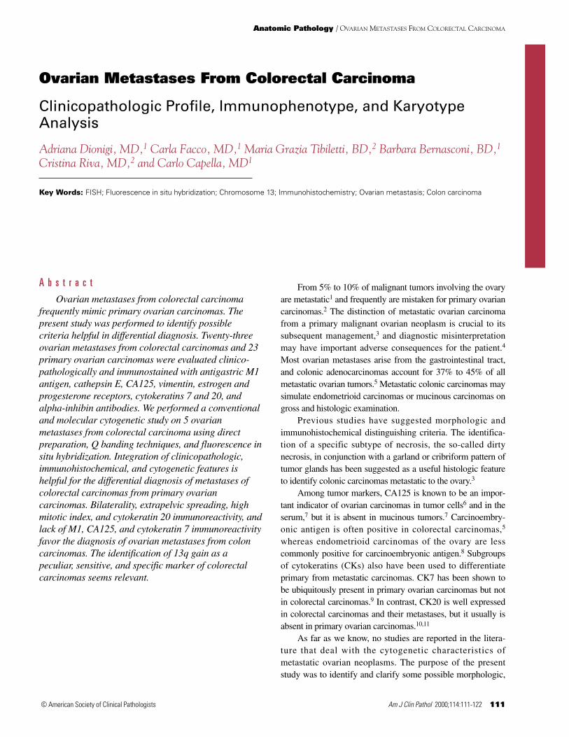
❚Table 2❚Clinicopathologic Results*
Case Age Pattern or No. (y) Site Histotype† Architecture‡ Cytology‡
Ovarian metastasis 1a 57 Right Pseudoendometrioid Cystic Columnar 1b 58 Left Pseudoendometrioid Solid-cystic Columnar-goblet 2 71 Right Pseudoendometrioid Cystic-solid Columnar 3 57 Right Pseudoendometrioid Cystic-solid Columnar 4a 33 Left Mucinous-pseudoendometrioid Cystic Columnar-goblet 4b 33 Right Mucinous Solid-cystic Columnar 5 81 Right Pseudoendometrioid Solid-cystic Columnar 6a 49 Right Pseudoendometrioid Solid-cystic Columnar 6b Left Pseudoendometrioid Solid-cystic Columnar 7 56 Right Mucinous Cystic Columnar-goblet 8 50 Right Pseudoendometrioid Cystic Columnar 9a 47 Right Pseudoendometrioid Solid Columnar 9b Left Pseudoendometrioid Cystic Columnar 10a 54 Right Pseudoendometrioid Solid Columnar 10b Left Pseudoendometrioid Solid-cystic Columnar 11 67 Left Mucinous Solid-cystic Columnar-goblet 12 56 Right Pseudoendometrioid Cystic Columnar-goblet 13 72 Right Pseudoendometrioid-mucinous Solid-cystic Columnar 14a 46 Right Mucinous Solid-cystic Columnar 14b Left Mucinous Solid-cystic Columnar 15a 72 Right Mucinous Cystic Columnar-goblet 15b Left Mucinous Cystic Columnar 16a 71 Right Mucinous Solid Columnar 16b Left Mucinous Solid Columnar 17a 48 Right Mucinous Solid-cystic Columnar-goblet 17b Left Mucinous Solid-cystic Columnar 18 65 Left Pseudoendometrioid Solid-cystic Columnar 19a 75 Right Mucinous Cystic Columnar-goblet 19b Left Mucinous Cystic Columnar-goblet 20a 56 Right Mucinous Cystic Columnar-goblet 20b Left Mucinous Cystic Columnar-goblet 21 43 Left Pseudoendometrioid Solid Columnar 22 55 Right Pseudoendometrioid Cystic-solid Columnar 23 84 Right Mucinous Solid Columnar
Primary ovarian carcinoma 24 51 Right Mucinous Solid-cystic Columnar; endocervical-goblet 25 62 Left Mucinous Solid-cystic Columnar-goblet 26 57 Right/left Mucinous Cystic-solid Columnar; endocervical 27 77 Left Mucinous Cystic Columnar; endocervical-goblet 28 56 NE Mucinous Cystic-solid Columnar; endocervical-goblet 29 65 NE Mucinous Cystic-solid Columnar 30 45 Left Mucinous Cystic Columnar; endocervical-goblet 31 40 Right Mucinous Cystic-solid Columnar; endocervical-goblet32 24 Left Mucinous Cystic Columnar; endocervical 33 35 Right Mucinous Solid Columnar34 68 Left Mucinous Cystic Columnar; endocervical-goblet 35 53 Right Mucinous Cystic Columnar; endocervical-goblet 36 87 Right Mucinous Cystic-solid Columnar-goblet 37 50 Left Mucinous Cystic Columnar; endocervical-goblet38 58 Left Mucinous Cystic-solid Columnar-goblet39 81 Right/left Mucinous Solid-cystic Columnar-goblet 40 43 Right Endometrioid Cystic-solid Columnar 41 59 Left Endometrioid Solid-cystic Columnar-squamous 42 68 Right Endometrioid Solid Polygonal-clear squamous 43 90 NE Endometrioid Solid Polygonal-clear 44 85 Right Endometrioid Solid-cystic Columnar-squamous 45 49 Right Endometrioid Cystic-solid Columnar 46 51 Left Endometrioid Solid-cystic Columnar-squamous
NE, not evaluated.*Scoring is as follows: nucleoli: +, small; ++, prominent; atypia: +, mild; ++, moderate; +++, severe; extent of necrosis: +, scarce; ++, moderate; +++, abundant; and stromal
desmoplasia: +, scarce; ++, moderate; +++, abundant; –, negative.† Pattern for ovarian metastases; histotype for primary ovarian carcinomas.‡ The order of the terms reflects decreasing prevalence.

Anatomic Pathology / ORIGINAL ARTICLE
Am J Clin Pathol 2000;114:111-122 115© American Society of Clinical Pathologists
❚Table 2❚ (cont)Clinicopathologic Results*
Stromal Nucleli Nucleoli Atypia Mitoses Extent Type Desmoplasia
Ovarian metastasis Round, hyperchromatic + +++ 28 +++ Dirty – Round, hyperchromatic + +++ 47 +++ Dirty + Cigar-shaped, hyperchromatic + ++ 7 + Dirty + Cigar-shaped + ++ 14 +++ Dirty + Cigar-shaped, hyperchromatic + + 16 +++ Dirty + Cigar-shaped, hyperchromatic ++ ++ 24 +++ Dirty + Round, hyperchromatic, vesicular + ++ 80 +++ Dirty + Cigar-shaped, hyperchromatic + ++ 68 +++ Dirty + Cigar-shaped, hyperchromatic + ++ 65 +++ Dirty + Cigar-shaped, hyperchromatic + ++ 47 +++ Dirty ++ Cigar-shaped, hyperchromatic, vesicular + ++ 116 +++ Dirty – Round, vesicular + ++ NE ++ Dirty ++ Round, hyperchromatic + ++ 48 +++ Dirty – Round, vesicular + ++ NE + Dirty + Round, hyperchromatic + ++ 19 +++ Dirty – Round, vesicular ++ +++ 33 +++ Dirty + Round, vesicular – + 23 + Dirty – Round, hyperchromatic + ++ 36 +++ Dirty + Round, vesicular + + 27 + Dirty ++ Round, hyperchromatic + + 15 ++ Dirty ++ Round, vesicular + + 18 – – Round, vesicular + + 15 ++ Dirty – Round, hyperchromatic + ++ 26 + Dirty – Round, hyperchromatic ++ ++ 32 + Dirty +++ Round, hyperchromatic + ++ 28 ++ Dirty ++ Round, hyperchromatic + ++ 23 + Dirty + Round, hyperchromatic + ++ 51 +++ Dirty – Round, hyperchromatic + ++ 25 + Dirty – Round, hyperchromatic + ++ 23 ++ Dirty + Cigar-shaped, hyperchromatic + + 38 + Dirty – Cigar-shaped, hyperchromatic + ++ 26 – + Round, vesicular + + 16 – + Cigar-shaped, hyperchromatic, vesicular ++ ++ 46 +++ Dirty – Round, hyperchromatic, vesicular + ++ 5 – +
Primary ovarian carcinoma Round, hyperchromatic + + 13 ++ Dirty – Round, hyperchromatic + +++ 27 + Dirty + Round, hyperchromatic + ++ 26 ++ Dirty – Cigar-shaped, vesicular + + 6 + Ischemic – Cigar-shaped, hyperchromatic + ++ 20 – – Round, vesicular + + 6 +++ Ischemic – Cigar-shaped, vesicular + + 7 + Dirty – Cigar-shaped, vesicular + + 0 ++ Ischemic + Cigar-shaped, vesicular + + 0 – – Cigar-shaped, vesicular + ++ 8 + Ischemic ++ Cigar-shaped, hyperchromatic ++ ++ 7 +++ Dirty – Cigar-shaped, vesicular + + 2 – – Cigar-shaped, vesicular + ++ 24 + Ischemic ++ Cigar-shaped, vesicular + + 15 – – Cigar-shaped, vesicular + ++ 20 – – Cigar-shaped, vesicular + + 20 – ++ Cigar-shaped, hyperchromatic + ++ 24 +++ Dirty – Cigar-shaped, vesicular + ++ 11 + Dirty ++ Round, vesicular ++ ++ 23 + Ischemic ++ Round, hyperchromatic ++ ++ 32 + Dirty +++ Round, hyperchromatic + ++ NE ++ Ischemic +++ Round, vesicular + ++ 24 + Dirty + Cigar-shaped, vesicular + + NE ++ Dirty +++
Necrosis

Dionigi et al / OVARIAN METASTASES FROM COLORECTAL CARCINOMA
Besides the obvious cytologically malignant mucinous epithe-lium, 8 cases showed areas composed of a monostratifiedmucinous well-differentiated epithelium. None of the caseshad areas of squamous differentiation. The stroma varied fromnegligible to abundant. It was desmoplastic in 22 of the cases,but sometimes it was edematous or mucoid. Stromal luteiniza-tion was observed focally in few cases.
Immunohistochemical FeaturesOur data ❚Table 3❚ showed that only 6 of 27 ovarian
metastases tested contained extracellular and intracellularmucus immunoreactive for M1. In 19 ovarian metastases, fewand scattered cathepsin E–staining cells were present along theluminal margins and the apical cytoplasm of the epithelia ofthe glands. In only 4 ovarian metastases a weak CA125 posi-tivity was found in rare cells. Results for vimentin andestrogen and progesterone receptors were consistently nega-tive in tumor cells. Metastatic tumors were strongly anddiffusely positive for CK20 ❚Image 3❚ in all ovarian metastasestested; weak CK7 positivity was observed in 4 of 22 ovarianmetastases. Positivity for alpha-inhibin was present in thestromal cells of metastatic tumors in 10 of the 17 ovarianmetastases tested.
Cytogenetic and FISH ResultsThe tumor karyotypes of 5 samples of ovarian metas-
tases from large bowel were defined ❚Table 4❚. Threesamples showed 2 cell lines, 1 diploid and 1 triploid (cases20a, 20b, and 22), while the other 2 samples showed only atriploid complement (cases 18 and 21). The abnormalitiesmore often represented were chromosome markers, as
shown in Table 4. In particular, m1 and m2 were present in4 of 5 samples. Gains and losses of different chromosomeswere present in all cases, while chromosome rearrange-ments were less frequent and identified by FISH as a deriv-ative of chromosome 1 (cases 18, 20a, and 20b), a deriva-tive of chromosome 3 (cases 21 and 22), and a derivative ofchromosome 6 (case 20a). It is noteworthy that the 2samples obtained from bilateral ovarian metastases showedsimilar chromosome anomalies: derivatives of 1p and 1q,gains and losses of the same chromosomes.
116 Am J Clin Pathol 2000;114:111-122 © American Society of Clinical Pathologists
❚Image 1❚ Ovarian metastasis from colorectal carcinoma.Garland pattern and confluent intraluminal “dirty” necrosis(H&E, ×200).
❚Image 2❚ Prominent cytologic atypia and abundant mitosesin ovarian metastasis from colorectal carcinoma (H&E,×400).
❚Image 3❚ Ovarian metastasis from colorectal carcinoma.Cytokeratin 20 immunoreactivity (immunoperoxidase, ×200).

Anatomic Pathology / ORIGINAL ARTICLE
Interestingly, at the molecular level FISH analysisidentified (in cases 18 and 21) m1 and m2 as derivatives oftranslocations between chromosome 13 and an unknownchromosome (m1) and between 2 chromosomes 13 (m2)giving in all cases gains of this chromosome. FISH with
whole chromosome painting for chromosome 13 confirmedthe gain of this chromosome also in case 22. In the samecases, FISH analysis with whole chromosome painting forchromosome 6 and 14 confirmed the conventional cytogeneticresults. In summary, gains of chromosome 13 such as
Am J Clin Pathol 2000;114:111-122 117© American Society of Clinical Pathologists
❚Table 3❚Immunohistochemical Results*
Case Pattern or No. Histotype† Site M1 Cathepsin E CA125 Vimentin Estrogen Progesterone 7 20 Epithelium Stroma
Ovarian metastasis 1a Pseudoendometrioid Right 0 0 0 0 0 0 0 60 0 10 1b Pseudoendometrioid Left 0 5 0 0 0 0 0 80 0 60 2 Pseudoendometrioid Right 0 30 0 0 0 0 0 100 30 0 3 Pseudoendometrioid Right 0 0 0 0 0 0 0 20 0 0 4a Mucinous- Left 20 30 0 0 0 0 0 20 NE NE
pseudoendometrioid 4b Mucinous Right 0 0 5 0 0 0 0 100 NE NE 5 Pseudoendometrioid Right 0 5 0 0 0 0 0 40 NE NE 6a Pseudoendometrioid Right 0 0 0 0 0 0 6b Pseudoendometrioid Left 0 5 0 0 0 0 0 30 NE NE 8 Pseudoendometrioid Right 0 0 0 0 0 0 0 40 NE NE 9 Pseudoendometrioid Left 0 5 0 0 0 0 0 20 0 30 10 Pseudoendometrioid Left 0 5 0 0 0 0 0 40 0 20 11 Mucinous Left 0 0 5 0 0 0 0 30 0 0 13 Pseudoendometrioid- Right 0 5 0 0 0 0 0 30 0 0
mucinous 14a Mucinous Right 0 20 0 0 0 0 14b Mucinous Left 0 10 0 0 0 0 30 40 0 0 15a Mucinous Right 0 20 0 0 NE 0 0 50 15b Mucinous Left 10 10 0 0 NE 0 0 40 NE NE 17 Mucinous Right 0 10 1 0 0 0 5 5 0 0 18 Pseudoendometrioid Left 0 0 0 0 0 0 NE NE NE NE 19a Mucinous Right 10 30 0 0 0 0 NE NE NE NE 19b Mucinous Left 5 10 0 0 0 0 0 40 0 50 20a Mucinous Right 20 20 0 0 0 0 0 70 0 60 20b Mucinous Left 10 30 0 0 0 0 0 90 0 5 21 Pseudoendometrioid Left 0 15 10 0 0 0 10 80 0 0 22 Pseudoendometrioid Right 0 0 0 0 0 0 0 50 0 60 23 Mucinous Right 0 50 0 0 0 0 40 5 0 20
Primary ovarian carcinoma 24 Mucinous Right 90 80 0 0 0 0 100 20 NE NE 25 Mucinous Left 60 0 0 0 0 0 0 10 NE NE 26 Mucinous Right 30 30 0 0 0 0 90 30 NE NE 27 Mucinous Left 10 80 0 0 0 0 100 80 NE NE 28 Mucinous NE 40 90 0 0 0 0 100 40 NE NE 29 Mucinous NE 0 10 0 0 0 0 80 40 NE NE 30 Mucinous Left 50 80 0 0 0 0 80 40 NE NE 31 Mucinous Right 60 60 0 0 0 0 90 10 NE NE 32 Mucinous Left 10 80 0 0 0 0 80 0 NE NE 33 Mucinous Right 5 10 0 0 0 0 80 70 NE NE 34 Mucinous Left 0 0 0 0 0 0 0 10 NE NE 35 Mucinous Right 0 20 0 0 0 0 40 0 NE NE 36 Mucinous Right 0 90 0 0 0 0 0 70 0 20 37 Mucinous Left 100 NE 0 NE 0 0 90 80 0 50 38 Mucinous Left 30 NE 0 NE 0 0 100 0 0 10 39 Mucinous Left 60 80 0 NE 0 0 5 50 10 0 40 Endometrioid Right 0 0 0 0 0 0 0 10 NE NE 41 Endometrioid Left 0 0 20 80 80 90 90 0 NE NE 42 Endometrioid Right 0 0 60 NE 0 0 100 0 NE NE 43 Endometrioid NE 0 0 20 0 0 0 80 0 0 0 44 Endometrioid Right 0 NE 0 NE 0 40 80 0 0 20 45 Endometrioid Right 0 NE 0 NE 0 20 20 0 0 20 46 Endometrioid Left 40 0 60 50 10 20 40 0 40 10
NE, not evaluated.* Numbers indicate percent of positive cells.† Pattern for ovarian metastases; histotype for primary ovarian carcinomas.
Hormone Receptors Cytokeratin alpha-Inhibin

Dionigi et al / OVARIAN METASTASES FROM COLORECTAL CARCINOMA
trisomies (case 22) or derivatives of a translocation (m1 andm2 in cases 18 and 21) were frequent cytogenetic findingsin ovarian metastases from colorectal carcinomas.
For comparison, we also cytogenetically studied aseries of primary colorectal carcinomas. The tumor kary-otypes are listed in ❚Table 5❚. Interestingly, both primarycolorectal carcinoma (case 6 in Table 5) and ovarian metas-tasis (case 21 in Table 4) were analyzed, and the sametriploid complement, markers m1 and m2, ring chromo-some, and gains and losses, were found. In summary, gainsof chromosome 13 (trisomy or derivative) also were themore frequent anomaly observed in primary colorectalcarcinomas.
Primary Ovarian Carcinomas
Clinical Features
Mucinous carcinomas occurred most frequently in patientswho were between the ages of 24 and 87 years (median, 56years), whereas the patients with endometrioid carcinomasranged in age from 43 to 90 years (mean age, 64 years).
Pathologic FeaturesGross Findings.—Mucinous carcinomas were grossly
cystic, multiloculated neoplasms (7/16) ranging in diameterfrom 1 to 33 cm (median, 12.7 cm). Solid areas and papillaryendocystic projections were frequent. Twelve of 14 mucinous
118 Am J Clin Pathol 2000;114:111-122 © American Society of Clinical Pathologists
❚Table 4❚Tumor Karyotypes in Ovarian Metastases From Colorectal Carcinomas*
Case No. Tumor Karyotype
18 58-74,XX,–X,+ish ins(D;1)(wcp1–;wcp1+),+2,–3,–4,–5,–6,–7,–8,–9,–12,–13,–14,–15,–16,–17,–18,–19, –20,–21,–22,+m1.ish der(13;?)(wcp13+;wcp13–),+m2.ish der(13;13)(wcp13+,wcp13+),+m3, +m4,+m5,1-9mar[cp 9]
21 61-71,XXX,+2,+3,+ish der(3)t(3;?)(wcp3+;wcp3–),–4,–6,–8,–9,–10,–11,–12,–14,–16,–17,–18,+20 –22,+m1.ish(wcp13–;wcp13–),+m2.ishder(13;13)(wcp13+,wcp13+),+m6,+m8,+m10,+r,2–3mar [cp12]
20a 44-56,XX,–1,der(1p).ishder(1)(wcp1+),der(1q).ishder(1)(wcp1+),+2,+3,+ish der(6)t(6q;?)(wcp6+;wcp6–),+7,–8,+11,–13,–15,–17,+19,+20,+21,+22,+m1.ish(wcp13–;wcp13–), 1-2mar[cp4]/59-71,XX,–X,der(1p),der(1q),del(1p),–4,–5,–6,+7,–8,–10,–11,–13,–14,–15,–16,–17,–18,+19,+21,+m1.ish(wcp13–;wcp13–),+m2.ish(wcp13–;wcp13–),3-5mar[cp7]
20b 39-53,XX,1-3mar[cp3]/59-74,XX,–X,der(1p)x2,der(1q)x2,del(1p),–4,–5,+6,+7,–8,–9,–10,–12,–13,–14,–15,+16,–17,–18,+19,+21,+22,+m1.ish(wcp13–;wcp13–),+m2.ish(wcp13–;wcp13–),+m4, 2-5mar[cp7]
22 50-57,XXX,+X,+1,add(3q+),+7,+10,+13,+13.ish(wcp13+x3,x4,x5),+15,+17,+17, +18,+21,+22[cp 4]/59-61,XXX,–3,add(3q+),–9,–11,+13,–18,–20[cp 4]
* In cases 20a and 20b, fluorescence in situ hybridization analysis failed to demonstrate m1 and m2 as a derivative of chromosome 13.
❚Table 5❚Tumor Karyotypes in Colorectal Carcinomas*
Case No. Tumor Karyotype
1 46,XX[4]/83-86,XX,–X,–5,–6,–7,–8,–11,+13,–14,–16,–17,–18,+19,–20,–21,–22,+m1.ishder(13;?)(wcp13+;wcp13–),+m2.ishder(13;13)(wcp13+,wcp13+),+m6,+m7[cp 6]
2 68-72,XXXX,+X,+1,+3,–5,+7,–8,+9,–10,–12,+13,+14,–15,+16,–18,–19,+20,+21,–22,+m1.ishder(13;?)(wcp13+;wcp13–),+m4,+m6[cp 7]
3 43-48,XX,+1,+2,–5,–10,–11,+13,–14,–15,–17,–18,+m1.ishder(13;?)(wcp13+;wcp13–),+m6,+m8,+1–5mar[cp 9] 4 43-48,XX,–14,–15,+m1.ish der(13;?)(wcp13+;wcp13–),m7,+1-5mar[cp 4] 5 46,XX[4] 6 63-69,XXXX,+X,i(1p),+2,–3,–4,del(6)(q26–qter),–11,–12,–14,–15,–16,–17,–18,+20,+21,–22,+m1.ishder(13;?)(wcp13+;wcp13–),
+m2.ishder(13;13)(wcp13+,wcp13+),+m10,+m11,+r[cp11] 7 43-47,XY,del(6)(q25–qter),+12,+m2.ish der(13;13)(wcp13+,wcp13+)[cp 10] 8 35-45,X,–X,–5,–6,del(6)(q23–qter),–7,–8,–10,+11,add(11p),+13,–14,–17,–18,–19,+20,+m1.ish
der(13;?)(wcp13+;wcp13–)x2,+dm[cp 11] 9 Fragmented 10 36-44,XY,–4,–5,del(6)(q24–qter),+7,–13,–14,–15,–17,–18,–19,–20,–21,–22,m1.ishder(13;?)(wcp13+;wcp13–),+m9,+m14,
+m15,+m16,+m17,+r[cp 10] 11 46,XX[cp4] 12 53-57,XXY,+X,+3,+4,–8,+9,+13,+14,+16,+20,+21,+22,+m1.ish der(13;?)(wcp13+;wcp13–),+m2.ish der(13;13)(wcp13+,wcp13+),
+m2,+m4[cp 9] 13 39-46,XY,–14,–22,+m1.ishder(13;?)(wcp13+;wcp13–)[cp 9] 14 47,XY,–13,–15,–17,+m1,+m7.ish der(1)t(1;?)(wcp1+;wcp1–),+m8,+1mar[cp6] 15 59-63,XXX,–4,–5,–6,–12,–13,–14,–15,–17,–18,+19,–21,+m1,+m7,+1–5mar[cp 5] 16 46-47,XX,+3,–10,–15,–16,+m1,+m6x2,+m7,+m9,+2–3mar[cp 4]/83–90,XXXX,–2,+3,–6,–8,–10, –12,–14,–15,–16,–17,–18,–20,
–22,+m1,+m6x2,+m7,+m8,+m9,+3-16mar[cp 3] 17 39-46,XYY,+Y,add(1)(?),–2,–5,+7,–9,–10,–12,–17,–18,–19,–20,–22,+m1[cp 10] 18 36-47,X,–X,del(1)(q21–qter),–5,+del(9),–15,–17,add(18),+20,+21[cp7]
* In cases 14 through 18, fluorescence in situ hybridization analysis was not performed because few metaphases were available.
Anatomic Pathology / ORIGINAL ARTICLE
neoplasms were unilateral. Four of 7 endometrioid carcinomaswere cystic, ranging in size from 8 to 30 cm (median, 15 cm).Friable, soft, or papillary structures partly filled the lumen in 2of 7 cases. All endometrioid carcinomas with available datawere unilateral.
Microscopic Findings.—Mucinous carcinomas (Table2) showed glandular cystic features mixed with microglan-dular cribriform aspects. Tubular glands, sometimes ectatic,were lined by tall columnar mucin-producing cells, intermin-gled with goblet cells and absorptive-like cells; occasionallya few scattered endocrine cells were present. Mucouscolumnar cells displayed rounded vesicular nuclei. Atypiawas generally mild or moderate. Mitoses ranged from few tonumerous (range, 0-27 per 10 HPFs). Eight of 16 mucinouscarcinomas showed benign and borderline histologic areasalong with obvious stromal invasion. In 2/3 of our cases, wealso found 2 patterns of necrosis: dirty and ischemic, butnecrotic areas were usually only focal. Five of 16 mucinouscarcinomas exhibited foci of stromal desmoplasia accompa-nied by broken glands and extracellular mucus. Examinationof endometrioid carcinomas showed the prevalent presenceof a mixed glandular-cystic and microglandular-solid archi-tecture accompanied by irregular and crowded tubularglands. The epithelium displayed stratified and atypicalendometrial-like cells with vesicular nuclei (4/7) andenlarged nucleoli. The mitotic index was high (range, 0-32;median, 17 per 10 HPFs). Limited foci of centroglandulardirty necrosis were found in 5 of 7 cases and of ischemicnecrosis in 2 of 7 cases. In 6 of 7 endometrioid carcinomas,focal and/or diffuse stromal desmoplasia (2+ or 3+) wasdetected. Squamous metaplasia was detected in 5 of 7 casesof endometrioid carcinomas.
Immunohistochemical Features
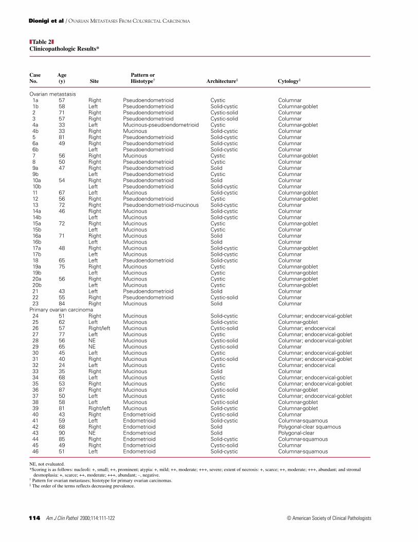
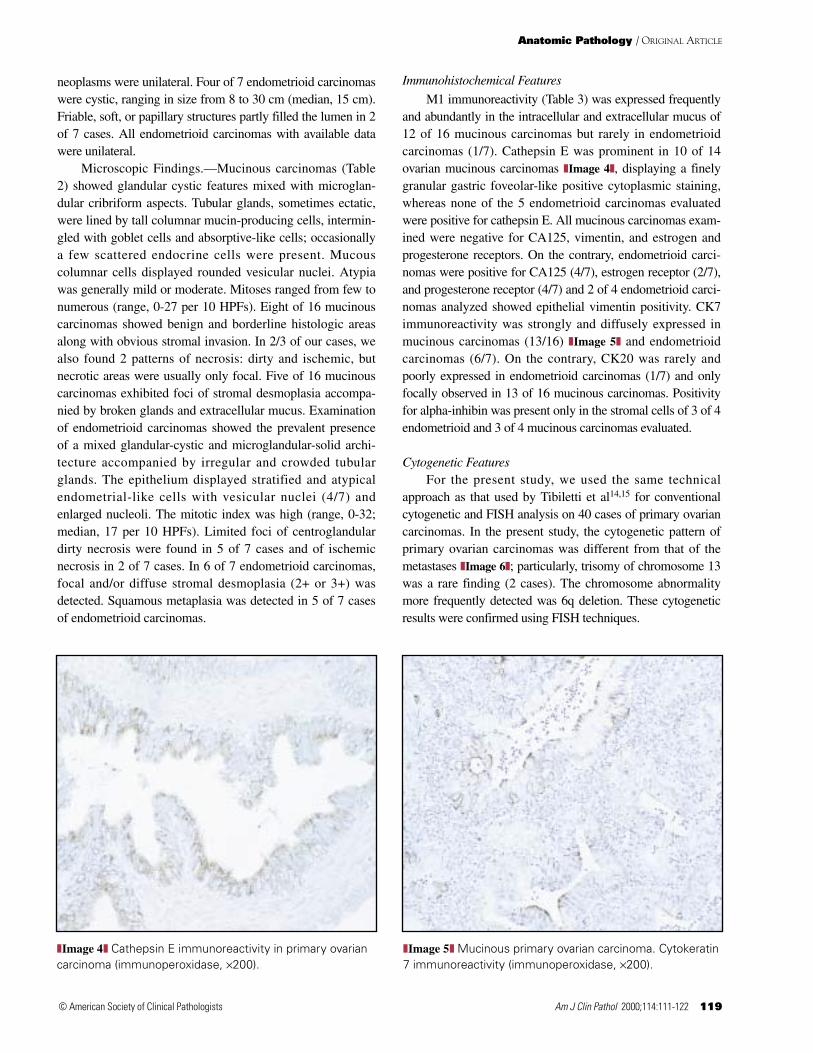
M1 immunoreactivity (Table 3) was expressed frequentlyand abundantly in the intracellular and extracellular mucus of12 of 16 mucinous carcinomas but rarely in endometrioidcarcinomas (1/7). Cathepsin E was prominent in 10 of 14ovarian mucinous carcinomas ❚Image 4❚, displaying a finelygranular gastric foveolar-like positive cytoplasmic staining,whereas none of the 5 endometrioid carcinomas evaluatedwere positive for cathepsin E. All mucinous carcinomas exam-ined were negative for CA125, vimentin, and estrogen andprogesterone receptors. On the contrary, endometrioid carci-nomas were positive for CA125 (4/7), estrogen receptor (2/7),and progesterone receptor (4/7) and 2 of 4 endometrioid carci-nomas analyzed showed epithelial vimentin positivity. CK7immunoreactivity was strongly and diffusely expressed inmucinous carcinomas (13/16) ❚Image 5❚ and endometrioidcarcinomas (6/7). On the contrary, CK20 was rarely andpoorly expressed in endometrioid carcinomas (1/7) and onlyfocally observed in 13 of 16 mucinous carcinomas. Positivityfor alpha-inhibin was present only in the stromal cells of 3 of 4endometrioid and 3 of 4 mucinous carcinomas evaluated.
Cytogenetic FeaturesFor the present study, we used the same technical
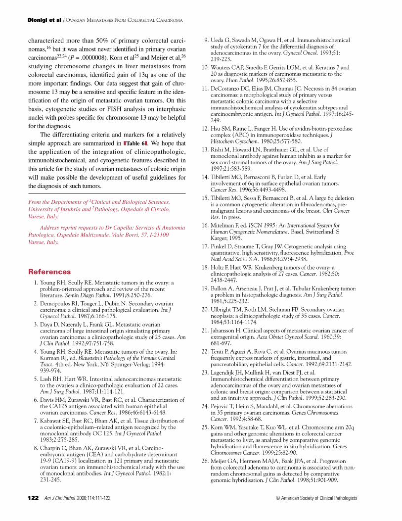
approach as that used by Tibiletti et al14,15 for conventionalcytogenetic and FISH analysis on 40 cases of primary ovariancarcinomas. In the present study, the cytogenetic pattern ofprimary ovarian carcinomas was different from that of themetastases ❚Image 6❚; particularly, trisomy of chromosome 13was a rare finding (2 cases). The chromosome abnormalitymore frequently detected was 6q deletion. These cytogeneticresults were confirmed using FISH techniques.
Am J Clin Pathol 2000;114:111-122 119© American Society of Clinical Pathologists
❚Image 4❚ Cathepsin E immunoreactivity in primary ovariancarcinoma (immunoperoxidase, ×200).
❚Image 5❚ Mucinous primary ovarian carcinoma. Cytokeratin7 immunoreactivity (immunoperoxidase, ×200).

Dionigi et al / OVARIAN METASTASES FROM COLORECTAL CARCINOMA
Statistical Analysis
The Fisher exact test revealed that among clinicopatho-logic features, bilaterality (P = .007) and dirty necrosis (P= .009) were criteria favoring metastases. CK7 negativity(P = .00001) and CK20 positivity (P = .0009) were themost significant immunohistochemical markers of metas-tases, while there were no significant differences regardingexpression of cathepsin E and CA125 between metastasesand primary ovarian carcinomas. Gains of chromosome 13were significantly more frequent in colorectal carcinomasand in their ovarian metastases than in primary ovariancarcinomas (P = .0000008).
Among immunohistochemical markers of metastases,CK20 positivity showed a great degree of sensitivity (100%)but a very low specificity (39%), while CK7 negativityshowed high specificity (83%) and sensitivity (82%). In addi-tion, gains of chromosome 13 displayed high specificity(95%) and moderate sensitivity (77%).
Discussion
The ovary is a common site for spread of carcinomas fromthe gastrointestinal tract. Most of the literature on this subjecthas concentrated on metastatic signet-ring cell carcinomas ofgastric origin (Krukenberg tumors).5,18,19 Few studies haveaddressed the more common problem of accurate diagnosis ofmetastatic colonic adenocarcinoma.5,20 A thorough historyfacilitates the diagnosis, but occasionally, a complete clinicalhistory is not available or the ovarian tumor may be the firstindication of an unsuspected primary carcinoma of the colon.
Patients with intestinal adenocarcinoma metastatic to theovary may be classified as follows: (1) those in whom anintestinal carcinoma precedes the detection of ovarian tumor(8 of 23 of our cases); (2) those in whom the ovarian involve-ment is detected synchronously at the time of operation forlarge bowel carcinoma (17 of 23 of our series), and (3) thosewith initial manifestations of an ovarian tumor (1 of 23 of ourcases).3,21
The presence of extraovarian disease at certain sitesmerits consideration by the surgeon and pathologist in distin-guishing between primary and metastatic ovarian tumor.Omental, hepatic parenchymal metastases, a prominentinvolvement of the peritoneal surface and/or of mesentericlymph nodes (18 of 20 of our cases) favor an intestinalprimary site. Our present findings confirm that extrapelvicspread is a helpful feature in the distinction of ovarian metas-tasis from colon carcinoma. Bilaterality favors a metastatictumor; although bilaterality is a feature of many primaryovarian carcinomas, it occurs with much greater frequency (11of 23 of our cases) in metastatic neoplasms with a statisticallysignificant difference (P = .007).
Colonic metastases to the ovary often produce cystic orpartly cystic tumors that may grossly simulate primaryepithelial ovarian carcinoma. Histologic differentiationbetween colonic adenocarcinoma and ovarian carcinomas ofthe mucinous and endometrioid types may be particularlydifficult. Because of the tumors’ capability to reproducedifferent morphologic patterns, Young and Scully1 intro-duced a classification of histologic aspects of metastaticovarian carcinomas. They observed a prevalence of the glan-dular endometrioid-like pattern and of the mucinous-like
120 Am J Clin Pathol 2000;114:111-122 © American Society of Clinical Pathologists
A B
❚Image 6❚ Fluorescence in situ hybridization with whole chromosome painting for chromosome 13 (green signals). A,Metaphase of ovarian metastasis from colorectal carcinoma. B, Metaphase of the corresponding primary colorectal carci-noma. Both metaphases show gains and the same rearrangements involving chromosome 13.

Anatomic Pathology / ORIGINAL ARTICLE
pattern. We had similar findings in our series of metastaticcolonic cancers that equally mimicked endometrioid carci-nomas and mucinous carcinomas. According to Lash andHart5 and Daya et al,3 the key to recognizing metastaticcolonic adenocarcinoma and its distinction from primaryovarian carcinoma lies in its characteristic histologic appear-ance. Those studies showed that metastatic tumors almostalways contain prominent garland and cribriform structureswith intraluminal dirty necrosis, often accompanied by aggre-gates of rounded glands with segmental necrosis of their walls.
These criteria seemed easy to apply and useful, becauseall but 2 of 23 patients with metastatic colonic carcinomasshowed an abundant dirty necrosis, with the presence ofeosinophilic and coarsely granular necrotic materialcontaining karyorrhectic debris and exhibiting several differentpatterns (intraluminal, segmental, and confluent), and theformation of garland or cribriform gland aspects. On thecontrary, in our cases of primary ovarian carcinomas, mainlyof the endometrioid type, dirty necrosis was not alwayspresent; in addition, it was very focal and of mild extent. Thesignificance of this parameter has been confirmed by statisticalanalysis (P = .0009).
Classic cytologic criteria, such as the marked cytologicatypia (2+ or 3+) and the high mitotic index may be consid-ered helpful in the definition of a correct diagnosis of ovarianmetastases. In addition, in metastatic endometrioid-liketumors, absence of squamous metaplasia is a distinguishingfeature from endometrioid carcinomas. Mucinous-like tumorscan mimic mucinous carcinomas because of the presence ofbland pseudobenign areas.
Our immunohistochemical results indicated that gastricM1 antigen and cathepsin E were expressed in primary andmetastatic ovarian tumors. In the ovarian metastases, theirimmunoreactivity was only focal and limited to scatteredcells, while primary ovarian carcinomas, especially muci-nous, showed a prominent and diffuse M1 and cathepsin E
positivity, which was often coexpressed in the same tumorareas, mimicking the distribution of these antigens in epithelialgastric foveolar cells. This pattern has been described by Tentiet al.22 This different distribution and cathepsin E negativity ofendometrioid carcinomas can help differentiate ovarian metas-tases from primary ovarian carcinomas.
The lack of CA125 was not surprising in ovarian metas-tases of colonic carcinomas, because this antigen generally isabsent in tumors arising in extragynecologic sites. Asexpected, CK7 was expressed especially in mucinous carci-nomas and endometrioid primary carcinomas, whereas it wasrarely and scarcely present in colonic metastases (P =.00001). On the contrary, CK7 negativity was a highly sensi-tive and specific marker of colonic metastases, and thephenotype CK7-negative and CK20-positive was expressedin most of the metastatic colonic neoplasms that we exam-ined, in agreement with the previous findings.11,23 Vimentincan be useful for the differential diagnosis, since it waspresent in 2 of 4 endometrioid primary ovarian carcinomas,but it was never found in our cases of ovarian metastases. Onthe basis of our immunohistochemical findings, we suggestthat immunoreactivity for CK20 and lack of CA125, CK7,and vimentin favor the diagnosis of ovarian metastases fromcolonic carcinoma.
Stroma of colonic ovarian metastases expressed somealpha-inhibin–positive cells showing the same immunoreac-tivity pattern observed in primary ovarian carcinomas, espe-cially the mucinous type. This result seems to be of greatbiologic interest, suggesting that sometimes the tumor stromacan retain phenotypic features of implantation tissue.
The conventional and molecular cytogenetic investiga-tions of ovarian metastases from colorectal carcinomas indi-cate that gain of chromosome 13, as a trisomy or as a deriva-tive from a translocation, is a peculiar feature of theseneoplasms and of primary colon carcinomas. Gain of chromo-some 13, as shown by our data and reported in the literature,
Am J Clin Pathol 2000;114:111-122 121© American Society of Clinical Pathologists
❚Table 6❚Differential Criteria and Markers*
Primary Ovarian Carcinoma Ovarian Metastasis of Colonic Origin
Bilaterality + +++ Extrapelvic spread + ++ “Dirty” necrosis + +++ Cytologic atypia + ++/+++ Mitotic index + +++ M1 + ± Cathepsin E + ± CA125 ± – Cytokeratin 7 + – Cytokeratin 20 ± + Chromosome assessment No gain of chromosome 13 Gain of chromosome 13
* Scoring is as follows: +, mild; ++, moderate; +++, high; ±, scarce; –, negative; ++/+++, intermediate between moderate and high.
Dionigi et al / OVARIAN METASTASES FROM COLORECTAL CARCINOMA
characterized more than 50% of primary colorectal carci-nomas,16 but it was almost never identified in primary ovariancarcinomas22,24 (P = .0000008). Korn et al25 and Meijer et al,26
studying chromosome changes in liver metastases fromcolorectal carcinomas, identified gain of 13q as one of themore important findings. Our data suggest that gain of chro-mosome 13 may be a sensitive and specific feature in the iden-tification of the origin of metastatic ovarian tumors. On thisbasis, cytogenetic studies or FISH analysis on interphasicnuclei with probes specific for chromosome 13 may be helpfulfor the diagnosis.
The differentiating criteria and markers for a relativelysimple approach are summarized in ❚Table 6❚. We hope thatthe application of the integration of clinicopathologic,immunohistochemical, and cytogenetic features described inthis article for the study of ovarian metastases of colonic originwill make possible the development of useful guidelines forthe diagnosis of such tumors.
From the Departments of 1Clinical and Biological Sciences,University of Insubria and 2Pathology, Ospedale di Circolo,Varese, Italy.
Address reprint requests to Dr Capella: Servizio di AnatomiaPatologica, Ospedale Multizonale, Viale Borri, 57, I-21100Varese, Italy.
References1. Young RH, Scully RE. Metastatic tumors in the ovary: a
problem-oriented approach and review of the recentliterature. Semin Diagn Pathol. 1991;8:250-276.
2. Demopoulos RI, Touger L, Dubin N. Secondary ovariancarcinoma: a clinical and pathological evaluation. Int JGynecol Pathol. 1987;6:166-175.
3. Daya D, Nazeraly L, Frank GL. Metastatic ovariancarcinoma of large intestinal origin simulating primaryovarian carcinoma: a clinicopathologic study of 25 cases. AmJ Clin Pathol. 1992;97:751-758.
4. Young RH, Scully RE. Metastatic tumors of the ovary. In:Kurman RJ, ed. Blaustein’s Pathology of the Female GenitalTract. 4th ed. New York, NY: Springer-Verlag; 1994:939-974.
5. Lash RH, Hart WR. Intestinal adenocarcinomas metastaticto the ovaries: a clinico-pathologic evaluation of 22 cases.Am J Surg Pathol. 1987;11:114-121.
6. Davis HM, Zurawski VR, Bast RC, et al. Characterization ofthe CA125 antigen associated with human epithelialovarian carcinomas. Cancer Res. 1986;46:6143-6148.
7. Kabawat SE, Bast RC, Bhan AK, et al. Tissue distribution ofa coelomic-epithelium–related antigen recognized by themonoclonal antibody OC 125. Int J Gynecol Pathol.1983;2:275-285.
8. Charpin C, Bhan AK, Zurawski VR, et al. Carcino-embryonic antigen (CEA) and carbohydrate determinant19-9 (CA19-9) localization in 121 primary and metastaticovarian tumors: an immunohistochemical study with the useof monoclonal antibodies. Int J Gynecol Pathol. 1982;1:231-245.
9. Ueda G, Sawada M, Ogawa H, et al. Immunohistochemicalstudy of cytokeratin 7 for the differential diagnosis ofadenocarcinomas in the ovary. Gynecol Oncol. 1993;51:219-223.
10. Wauters CAP, Smedts F, Gerrits LGM, et al. Keratins 7 and20 as diagnostic markers of carcinomas metastatic to theovary. Hum Pathol. 1995;26:852-855.
11. DeCostanzo DC, Elias JM, Chumas JC. Necrosis in 84 ovariancarcinomas: a morphological study of primary versusmetastatic colonic carcinoma with a selectiveimmunohistochemical analysis of cytokeratin subtypes andcarcinoembryonic antigen. Int J Gynecol Pathol. 1997;16:245-249.
12. Hsu SM, Raine L, Fanger H. Use of avidin-biotin-peroxidasecomplex (ABC) in immunoperoxidase techniques. JHistochem Cytochem. 1980;25:577-580.
13. Rishi M, Howard LN, Bratthauer GL, et al. Use ofmonoclonal antibody against human inhibin as a marker forsex cord-stromal tumors of the ovary. Am J Surg Pathol.1997;21:583-589.
14. Tibiletti MG, Bernasconi B, Furlan D, et al. Earlyinvolvement of 6q in surface epithelial ovarian tumors.Cancer Res. 1996;56:4493-4498.
15. Tibiletti MG, Sessa F, Bernasconi B, et al. A large 6q deletionis a common cytogenetic alteration in fibroadenomas, pre-malignant lesions and carcinomas of the breast. Clin CancerRes. In press.
16. Mitelman F, ed. ISCN 1995: An International System forHuman Cytogenetic Nomenclature. Basel, Switzerland: SKarger; 1995.
17. Pinkel D, Straume T, Gray JW. Cytogenetic analysis usingquantitative, high sensitivity, fluorescence hybridization. ProcNatl Acad Sci U S A. 1986;83:2934-2938.
18. Holtz F, Hart WR. Krukenberg tumors of the ovary: aclinicopathologic analysis of 27 cases. Cancer. 1982;50:2438-2447.
19. Bullon A, Arseneau J, Prat J, et al. Tubular Krukenberg tumor:a problem in histopathologic diagnosis. Am J Surg Pathol.1981;5:225-232.
20. Ulbright TM, Roth LM, Stehman FB. Secondary ovarianneoplasia: a clinicopathologic study of 35 cases. Cancer.1984;53:1164-1174.
21. Jahansson H. Clinical aspects of metastatic ovarian cancer ofextragenital origin. Acta Obstet Gynecol Scand. 1960;39:681-697.
22. Tenti P, Aguzzi A, Riva C, et al. Ovarian mucinous tumorsfrequently express markers of gastric, intestinal, andpancreatobiliary epithelial cells. Cancer. 1992;69:2131-2142.
23. Lagendijk JH, Mullink H, van Diest PJ, et al.Immunohistochemical differentiation between primaryadenocarcinomas of the ovary and ovarian metastases ofcolonic and breast origin: comparison between a statisticaland an intuitive approach. J Clin Pathol. 1999;52:283-290.
24. Pejovic T, Heim S, Mandahl, et al. Chromosome aberrationsin 35 primary ovarian carcinomas. Genes ChromosomesCancer. 1992;4:58-68.
25. Korn WM, Yasutake T, Kuo WL, et al. Chromosome arm 20qgains and other genomic alterations in colorectal cancermetastatic to liver, as analyzed by comparative genomichybridization and fluorescence in situ hybridization. GenesChromosomes Cancer. 1999;25:82-90.
26. Meijer GA, Hermsen MAJA, Baak JPA, et al. Progressionfrom colorectal adenoma to carcinoma is associated with non-random chromosomal gains as detected by comparativegenomic hybridisation. J Clin Pathol. 1998;51:901-909.
122 Am J Clin Pathol 2000;114:111-122 © American Society of Clinical Pathologists
Related Documents