Clinical terms • Dwarfism – Achondroplasia • Abnormal projection from a bone due to bony overgrowth – Bony spur • Pain in bone – Ostealgia • Bone inflammation – Osteitis • Inflammation of bone and bone marrow caused by pus-forming bacteria – Osteomyalitis • Bone cancer in a long bone of limb – Osteosarcoma • Fracture in diseased bone involving slight (coughing or quick turn) – Pathological fracture • Placing sustained tension on a body region to keep fractured bone in alignment – Traction 1

Clinical terms Dwarfism –Achondroplasia Abnormal projection from a bone due to bony overgrowth –Bony spur Pain in bone –Ostealgia Bone inflammation –Osteitis.
Dec 25, 2015
Welcome message from author
This document is posted to help you gain knowledge. Please leave a comment to let me know what you think about it! Share it to your friends and learn new things together.
Transcript

Clinical terms• Dwarfism
– Achondroplasia• Abnormal projection from a bone due to bony overgrowth
– Bony spur• Pain in bone
– Ostealgia• Bone inflammation
– Osteitis• Inflammation of bone and bone marrow caused by pus-forming
bacteria– Osteomyalitis
• Bone cancer in a long bone of limb– Osteosarcoma
• Fracture in diseased bone involving slight (coughing or quick turn)– Pathological fracture
• Placing sustained tension on a body region to keep fractured bone in alignment– Traction 1

Epiphyseal Zones• Name the 5 zones
2
Resting cartilage
Proliferation
Hypertrophy
Erosion (Calcification)
Diaphysis (ossification)

Resting Cartilage• Identify characteristics of this zone
1. Chondro’s resting in Lacunae
2. Not mitotically dividing
3. Not actively contributing to bone growth
4. Near epiphysis5. Scattered chondro’s
• What is the Role of this zone?
1. To anchor the rest of the epiphyseal plate to the epiphysis
3
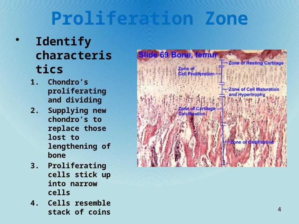
Proliferation Zone• Identify
characteristics1. Chondro’s
proliferating and dividing
2. Supplying new chondro’s to replace those lost to lengthening of bone
3. Proliferating cells stick up into narrow cells
4. Cells resemble stack of coins
4

Hypertrophy (Maturing) Zone• Identify
characteristics1. Cellular breakdown
2. Chondro’s begin to break down and produce alkaline phosphatase
3. Enzyme facilitates calcification of extra cellular matrix
4. Large chondro’s arranged in columns
5

Erosion Zone (calcification)
• Identify1. Calcium salts deposited
in cartilage matrix by osteoids
2. Osteogenesis, osteoblasts laying down the matrix
3. Only a few cells thick4. Dead cells because the
matrix around them became calcified
5. Calcified matrix destroyed by osteoclasts (acids and enzymes), then invaded by osteoblasts and capillaries from diaphysis
6. Diaphyseal border of the plate firmly cemented to the bone of the diaphysis
6

Diaphysis Zone (ossification)
• Identify1. Long
spiracles (tuberculae) that form spongy bone produced
7
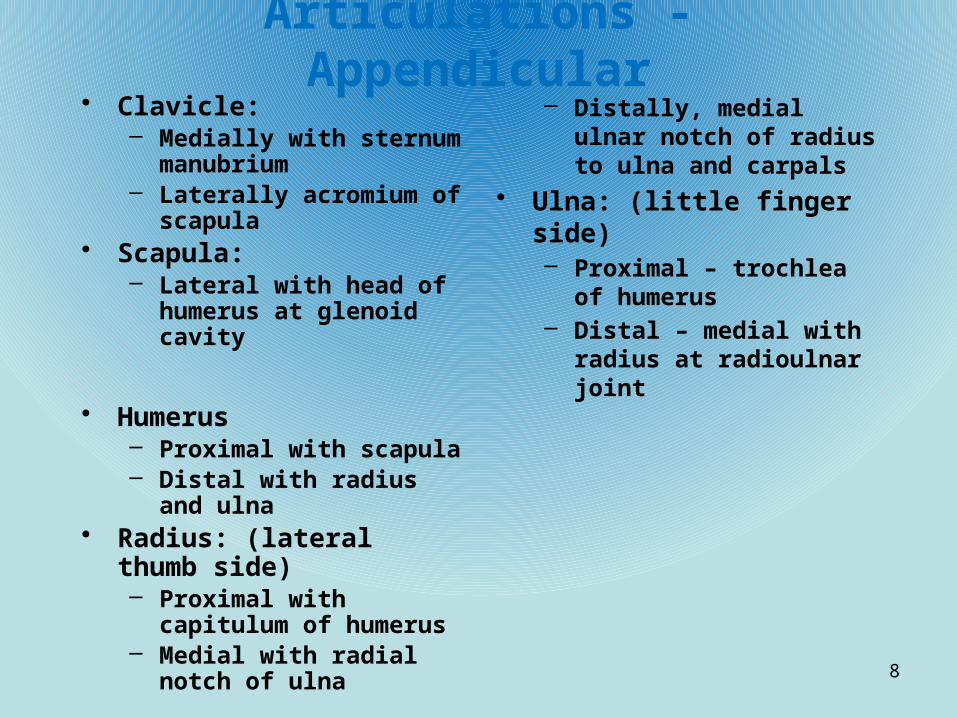
Articulations - Appendicular• Clavicle:
– Medially with sternum manubrium
– Laterally acromium of scapula
• Scapula:– Lateral with head of
humerus at glenoid cavity
• Humerus– Proximal with scapula– Distal with radius and ulna
• Radius: (lateral thumb side)– Proximal with capitulum of
humerus– Medial with radial notch of
ulna
– Distally, medial ulnar notch of radius to ulna and carpals
• Ulna: (little finger side)– Proximal – trochlea of
humerus– Distal – medial with radius at
radioulnar joint
8

Osteological Terms - Processes• A rounded articulating
process– Condyle
• Any bony projection– Process
• A projection located above a condyle– Epicondyle
• A large rounded or irregular process– Tuberosity
• A small rounded process– Tubercle
• A very large, often blunt process– trochanter
• A sharp, slender process– Spine
• A hook-shaped process– Hamulus
• A very slight ridge of bone– Line
• A prominent ridge of bone– Crest
• A smooth flattened articulating surface– Facet
9

Osteological terms - Fosae• A hole in a bone through which nerves and blood pass
– Foramen• A tunnel-like passage through a bone
– Meatus or canal• A cavity within a bone
– Sinus• A furrow on a bone’s surface
– Sulcus or Groove• A slit-like opening in a bone
– Fissure• A shallow depression
– Fovea
10

Identify parts of the bone
• Tubular shaft of the long bone– Diaphysis
• Part of long bone where growth occurs– Epiphysis
• Irregularly arranged lamellae and osteocytes interconnected by canaliculi– Spongy bone (Cancellous,
Trabeculae bone)• Marrow cavity in the shaft of
long bone– Medullary cavity
• Hematopoietic tissue found within trabecular cavities of spongy bone– Red marrow
• Fat that fills cavities of bones– Yellow marrow
• Membrane covering internal bone surfaces– Endosteum
• Highly vascularized membrane covering the exterior of the diaphysis– Periosteum
• Disc of hyaline cartilage that grows during childhood to lengthen the bone– Epiphyseal plate 11
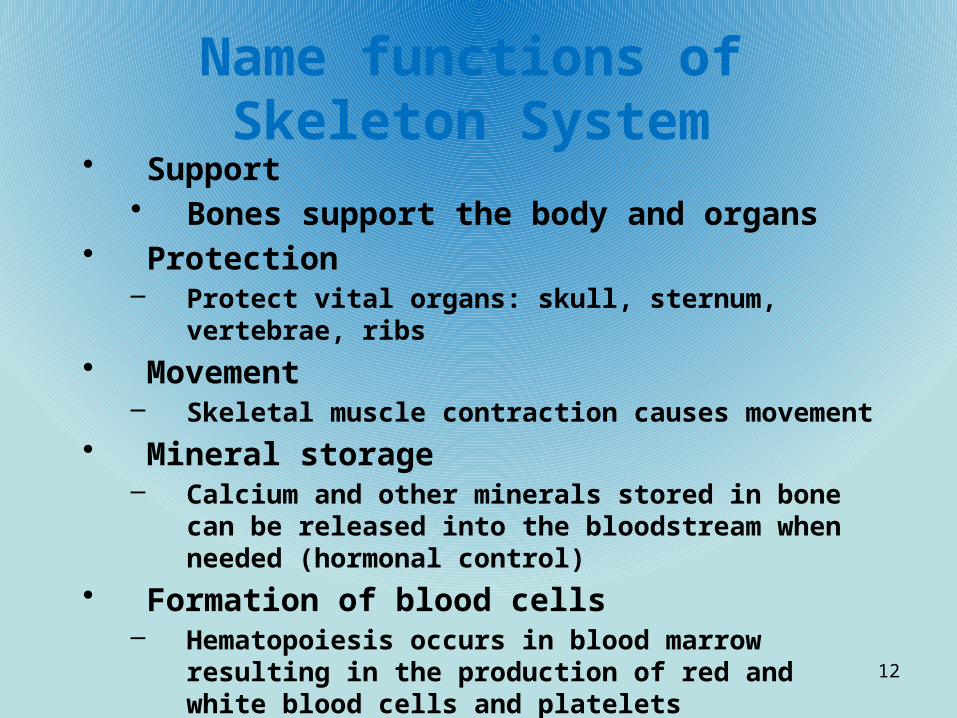
Name functions of Skeleton System
• Support• Bones support the body and organs
• Protection– Protect vital organs: skull, sternum, vertebrae, ribs
• Movement– Skeletal muscle contraction causes movement
• Mineral storage– Calcium and other minerals stored in bone can be released
into the bloodstream when needed (hormonal control)
• Formation of blood cells– Hematopoiesis occurs in blood marrow resulting in the
production of red and white blood cells and platelets
12

What are the 2 types of Osseous Tissue
• Compact bone– Smooth, compact, with little air space– Haversian systems
• Spongy bone (cancellous, trabecular)– Small pieces of bone surrounded by open spaces
filled with red or yellow marrow
13

Name the 4 types of bone• Long bone
– Shaft with a wide portion at both ends– Primarily compact bone with spongy at the widened areas– Humerus, radius, ulna, femur, tibia, fibula, phalanges
• Short bones– Cube shaped, mostly cancellous bone– Wrist, ankle– Sesamoid – short bones embedded within tendon (patella)
• Flat bone– Thin and flat– 2 layers of compact with spongy between– Sternum, ribs, skull
• Irregular bone– Spongy with odd shape– Vertebrae and hip
14

Compact bone• Composed of?
– Osteons or Haversion systems• Interspersed with?
– Blood, lymphatic vessels and nerves• Concentric rings of compact bone called ____________ surround hollow
passageways called ____________________– Lamella, Haversion canals
• What connects Haversion canals?– Volkman’s canals
• Canals are lined with what tissue?– Endosteum
• Where do osteocytes reside?– Lacunae
• What connects the lacunae?– Canaliculi
• What are the canaliculi’s function?– Permit nutrients and oxygen to pass between osteocytes– Remove wastes
• What are interstitial lamellae?– Layers of bone that fill gaps between osteons
• What are circumferential lamellae?– Large rings of bone extending around the entire shaft of the bone
15

Compact bone structure
16
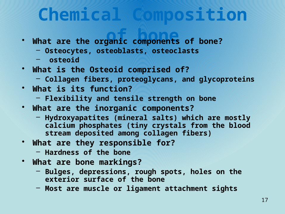
Chemical Composition of bone• What are the organic components of bone?
– Osteocytes, osteoblasts, osteoclasts– osteoid
• What is the Osteoid comprised of?– Collagen fibers, proteoglycans, and glycoproteins
• What is its function?– Flexibility and tensile strength on bone
• What are the inorganic components?– Hydroxyapatites (mineral salts) which are mostly calcium phosphates
(tiny crystals from the blood stream deposited among collagen fibers)• What are they responsible for?
– Hardness of the bone• What are bone markings?
– Bulges, depressions, rough spots, holes on the exterior surface of the bone
– Most are muscle or ligament attachment sights
17
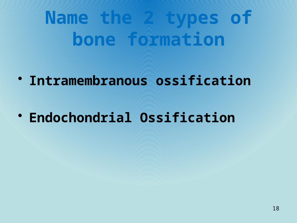
Name the 2 types of bone formation
• Intramembranous ossification
• Endochondrial Ossification
18

Intramembranous Ossification?• What is it?– The process of bone developing from a fibrous tissue
• What bones are formed this way?– Flat bones such as the skull and clavicles
• Explain the process:– Fibrous connective tissue in developing fetus become?
– scattered with osteoblasts that begin secreting organic matrix. – Osteoid is?
– then mineralized and osteoblasts become osteocytes– Osteoid accumulates in?
– small networks of collagen fibers called trabeculae. – This early bone formation is called?
– woven bone. – The periosteum forms around the?
– woven bone.– Trabeculae thickens until they become?
– plates of bone. – Eventually these plates of woven bone are replaced by?
– compact bone. – Spongy bone remains in the center of the bone and the vascular
tissue within it?– differentiates into red marrow (formation of diploe)
19

Intramembranous Ossification 1
20

Intramembranous 2
21

Intramembranous 3
22
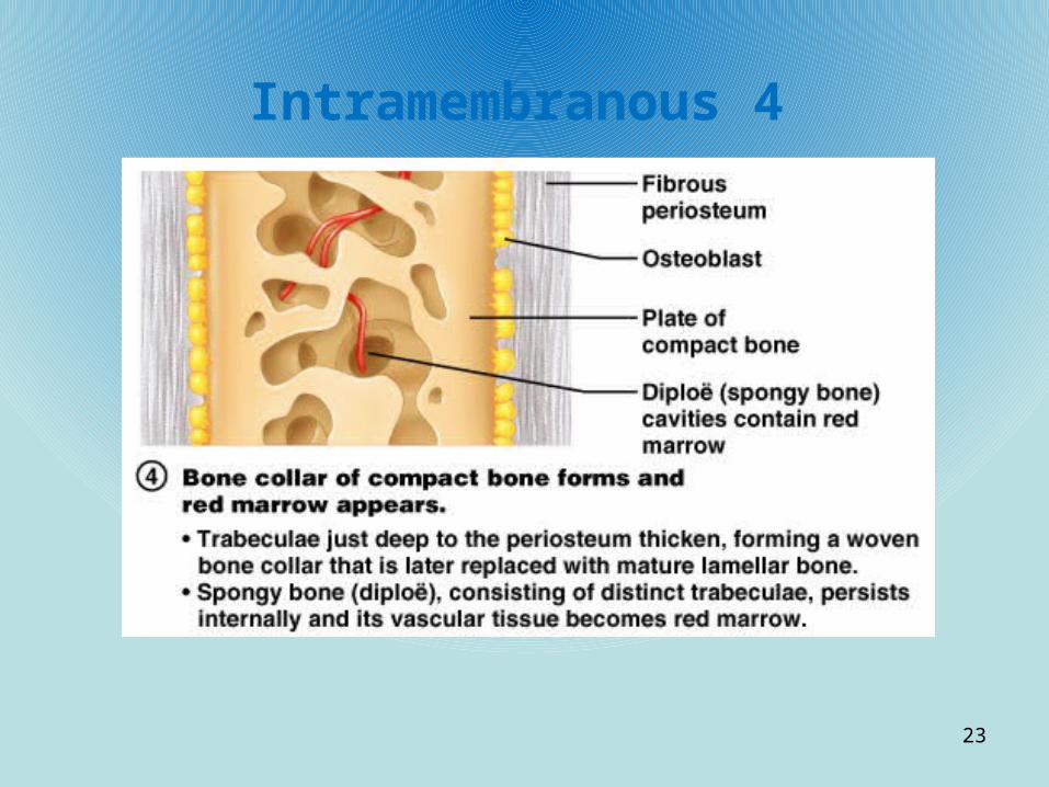
Intramembranous 4
23
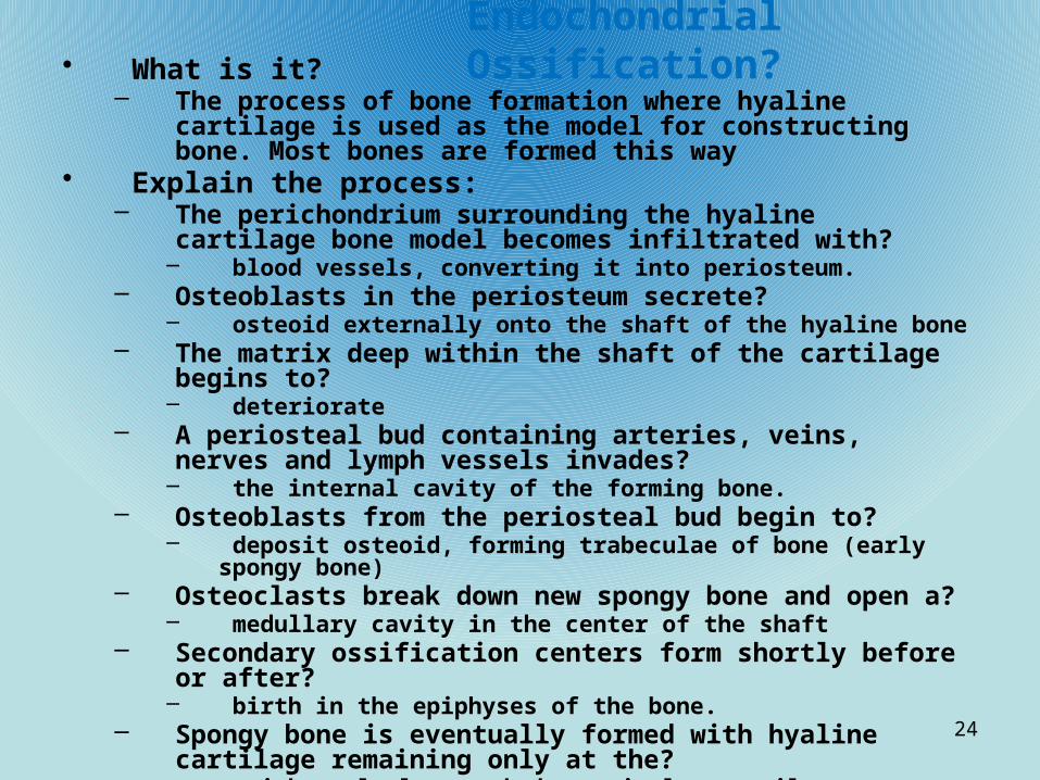
Endochondrial Ossification?• What is it?
– The process of bone formation where hyaline cartilage is used as the model for constructing bone. Most bones are formed this way
• Explain the process:– The perichondrium surrounding the hyaline cartilage bone model
becomes infiltrated with?– blood vessels, converting it into periosteum.
– Osteoblasts in the periosteum secrete?– osteoid externally onto the shaft of the hyaline bone
– The matrix deep within the shaft of the cartilage begins to?– deteriorate
– A periosteal bud containing arteries, veins, nerves and lymph vessels invades?
– the internal cavity of the forming bone. – Osteoblasts from the periosteal bud begin to?
– deposit osteoid, forming trabeculae of bone (early spongy bone)– Osteoclasts break down new spongy bone and open a?
– medullary cavity in the center of the shaft– Secondary ossification centers form shortly before or after?
– birth in the epiphyses of the bone. – Spongy bone is eventually formed with hyaline cartilage remaining
only at the?– epiphyseal plate and the articular cartilages
24

Endochondral Ossification
25
Formation of bone collar around hyaline cartilage model.
Cavitation of the hyaline cartilage within the cartilage model.
Invasion of internal cavities by the periosteal bud and spongy bone formation.
Ossification of the epiphyses; when completed, hyaline cartilage remains only in the epiphyseal plates and articular cartilages
Formation of the medullary cavity as ossification continues; appearance of secondary ossification centers in the epiphyses in preparation for stage 5.
Hyaline cartilage
Primary ossification center
Bone collar
Deteriorating cartilage matrix
Spongy bone formation
Blood vessel of periosteal bud
Secondary ossification center
Epiphyseal blood vessel
Medullary cavity
Epiphyseal plate cartilage
Spongy bone
Articular cartilage

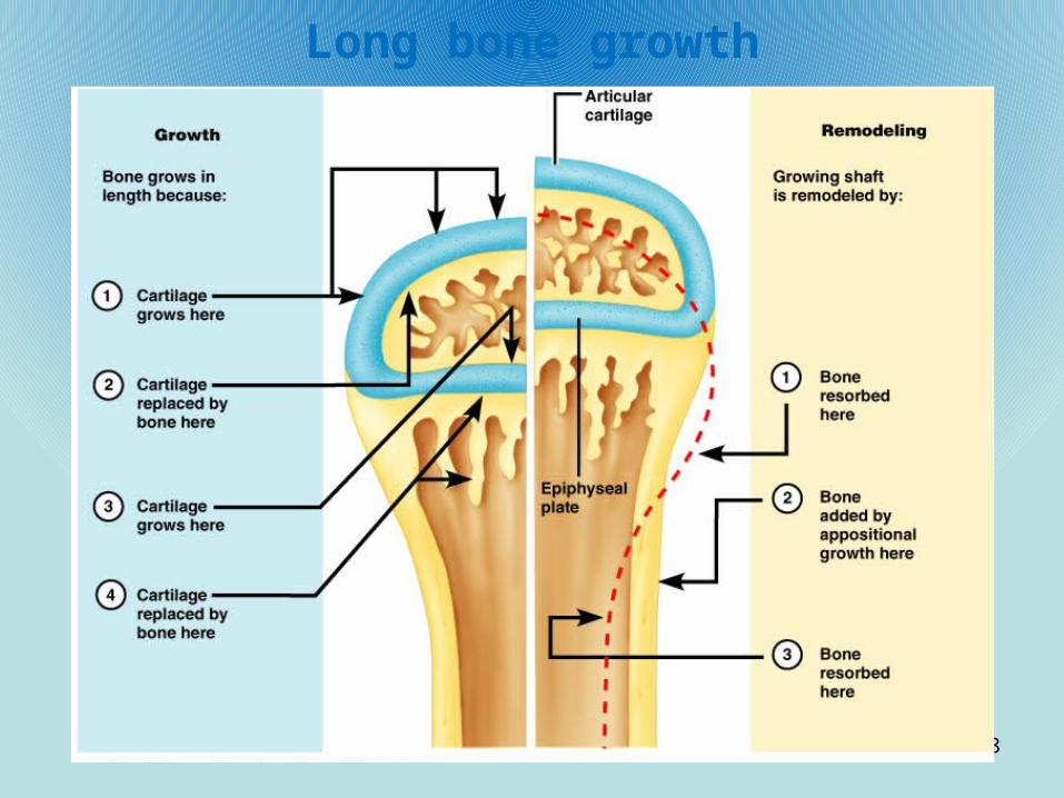
Bone growth in long bones• How do long bones grow in children?
– Entirely by interstitial growth of the epiphyseal plates
• How do bones grow in thickness?– By appositional growth
• The widening of bones
26

Epiphyseal bone growth• The top layer (epi side) of hyaline cartilage plate contains actively
dividing chondrocytes that are pushed towards the ________________?• Diaphysis
• What does this do?• Causes the entire bone to lengthen
• What happens to the chondrocytes as they get closer to the shaft?• They enlarge and eventually die
• What occurs at the junction of the epiphysis and the diaphysis?• Osteoblasts begin to secrete bone matrix and form small spicules of bone
• What promotes the adolescent growth spurt and eventually leads to the closure of epiphyseal plate?
• Sex hormones• What happens as adolescence ends?
• The epiphyseal plate becomes smaller and is entirely replaced by bone tissue• What is the result of this occurring?
• Bone growth ends
27
Long bone growth
28

Structure of long bone
29

What is appositional growth
• The process used to?– Widen bones
• _____________ beneath the _____________ form new ______________ systems on the outer surface of the bone– Osteoblasts, periosteum, Haversian
• ________________ on the ________________ break down bone to enlarge the ______________ cavity.– Osteoclasts, endosteum, medullary
30

Appositional growth
31
Osteoblasts beneath the periosteum secrete bone matrix, forming ridges that follow the course of periosteal blood vessels.
As the bony ridges enlarge and meet, the groove containing the blood vessel becomes a tunnel.
The periosteum lining the tunnel is transformed into an endosteum and the osteoblasts just deep to the tunnel endosteum secrete bone matrix, narrowing the canal.
As the osteoblasts beneath the endosteum form new lamellae, a new osteon is created. Meanwhile new circumferential lamellae are elaborated beneath the periosteum and the process is repeated, continuing to enlarge bone diameter.
Artery Periosteum Penetrating canalCentral canal of osteonPeriosteal ridge
1 2 3 4

Bone Remodeling• What is bone remodeling?
– Process where bone is resorbed and deposited at periosteal or endosteal surfaces
• What should the rate of bone resorption be in healthy adults?– The same rate as deposition so the total mass remains constant
• What is bone deposit?– Osteoblasts laying down new osteoid which is later mineralized into bone
• How is bone resorption carried out?– by osteoclasts secreting enzymes onto the bone that digest organic matrix. The
osteoclasts also secrete acids that help to make the calcium salts more soluble• What is PTH?
– Parathyroid hormone– Hormone produced by the parathyroid gland in response to low blood calcium
levels. – It stimulates bone resorption so that calcium is released and put back into the
blood• What is Calcitonin?
– A protein produced by specialized “C” cells in the thyroid and secreted when blood calcium levels rise
– Inhibits bone resorption and enhances calcium deposit in the bone matrix
32

Bone remodeling• How much of our bone mass is recycled every week?
– 5 to 7%• How often is spongy bone replaced? Compact bone
– Every 3 to 4 years– Every 10 years
• What are remodeling units?– Packets of adjacent osteoblasts and osteoclasts that coordinate bone
remodeling• What is bone deposit?
– Added bone for injured or strength of bone• What is an osteoid seam?
– The marking of new matrix deposits by osteocytes (band of gauzy looking bone matrix)
• What helps trigger calcification?– Calcium and phosphate ions
33

Bone remodeling (cont)• What happens when calcium and phosphate mix reach a certain
level?– Tiny crystals of hydroxyapatite form and catalyze further
crystallization of calcium salts in the area• What are other factors involved?
– Matrix proteins binding and concentrating calcium– Alkaline phosphatase mineralizing
• When the conditions are met, what happens?– Calcium salts are deposited all at once and with great precision
throughout the mature matrix
34

Bone resorption• What accomplishes this?
– Osteoclasts• As they move along the bone surface what do they do?
– Dig grooves called resorption bays and break down bony matrix• How does the area of bone destruction seal off?
– By osteoclasts that touch the bone forming a ruffled membrane that clings to the bone, sealing it off
• What does the ruffled border secrete and what does this do?– Lysosomal enzymes – digest organic matrix– Hydrocholoric acid – converts calcium salts into soluble forms in
order to make them pass easily into solution• What happens to these products?
– They are endocytosed and then released into the blood• What cells are important in this process?
– T cells
35

Remodeling control• What regulates remodeling?
– 2 control loops:• Negative feedback that maintains Ca2 Homeostasis in the blood• Gravitational forces acting on skeleton
• Why is calcium so important for the body?– Nerve impulses– Muscle contractions– Blood coagulation– Gland and nerve cell secretions– Cell division
• Where is 99% of calcium in the body?– Bone minerals
• What range does the hormonal loop keep calcium?– 9-11 mg per 100 ml of blood
• Where is calcium absorbed from?– Intestine
36

Hormonal Mechanism• What does the hormonal mechanism involve?
– PTH – parathyroid hormone – parathyroid gland– Calcitonin – produced by parafollicular cells (C-cells) of thyroid
gland• When is PTH released?
– When calcium levels decline• What does it do?
– Stimulates osteoclasts to resorb bone, release calcium into blood• What happens?
– Osteoclasts break down old and new matrix• What escaped digestion and why?
– Osteoid– Because it lacks calcium salts
• When does the stimulus for PTH end?– When blood concentrations of calcium rise
37

Hormonal mechanisms (cont)• What is secreted when calcium levels rise?
– Calcitonin• What is its function?
– Inhibit resorption, encourage deposition– Reduce blood calcium
• What happens when blood calcium levels fall?– Calcitonin release wanes
• Are these responses to preserve bone strength?– No. They are for maintaining blood calcium homeostasis
• What happens if levels are low for a long time?– Bones demineralize and develop large, punched out holes
38

Bone Remodeling
39

Mechanical Stress• What is Wolff’s law?
– Bones grow and remodel in response to the demands placed on it?
• What are other observations of Wolff’s law?– Long bones thickest midway along diaphysis (where stress is
greatest)– Curved bones thickest where most likely to buckle– Trabeculae form trusses along lines of compression– Large bony projections occur where active heavy muscles
attach• How do forces communicate with remodeling cells?
– Electrical signaling• What are hormonal loops function in remodeling?
– Whether and when remodeling occur• What are mechanical stresses functions?
– Where remodeling will occur
40

Fracture Classification• Location of bone after fracture:
– Non-displaced – normal end positions– Displaced – out of normal alignment
• Completeness of break:– Complete – all the way through– Incomplete – not all the way
• Orientation:– Linear – parallel of long axis– Transverse – perpendicular to axis
• Skin penetration:– Open – penetrates the skin– Closed – skin not penetrated
41
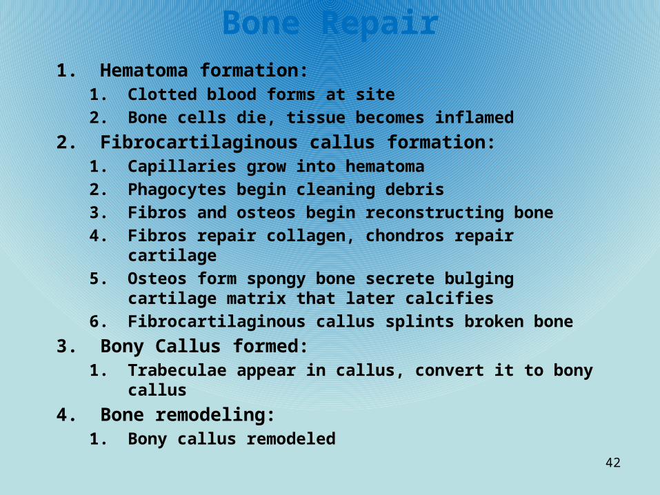
Bone Repair1. Hematoma formation:
1. Clotted blood forms at site
2. Bone cells die, tissue becomes inflamed
2. Fibrocartilaginous callus formation:1. Capillaries grow into hematoma
2. Phagocytes begin cleaning debris
3. Fibros and osteos begin reconstructing bone
4. Fibros repair collagen, chondros repair cartilage
5. Osteos form spongy bone secrete bulging cartilage matrix that later calcifies
6. Fibrocartilaginous callus splints broken bone
3. Bony Callus formed:1. Trabeculae appear in callus, convert it to bony callus
4. Bone remodeling:1. Bony callus remodeled
42

Common Fractures• Comminuted:
– Fragments into 3 or more pieces (aged, brittle boned)
• Compression:– Crushed
• Spiral:– Ragged break, excessive twisting (sports)
• Epiphyseal:– Epiphysis separated from diaphysis
• Depressed:– Broken inward
• Greenstick:– Incomplete break on one side, other side bends
43
Clinical advances in bone repair• What does electrical stimulation do to a fracture?
– Dramatically increases healing time• What is the piezo electric effect?
– Minute electrical currents are produced when minerals are stressed• What happens in regions of negative electrical charge? Positive?
– Negative – bone deposited– Positive – bone resorbed
• What effect does ultrasound treatment produce on fractures?– Reduce healing times 35 to 45%
• What is VEGF? Function?– Vascular endothelial growth factor– Stimulates blood vessel growth
44
Bone Imbalances - Osteoporosis
• What is osteoporosis?– Group of diseases in which bone resorption occurs more than bone
deposit• What are the results of the disease?
– Reduced bone mass• Which bones are most susceptible?
– Vertebrae, neck of the femur• Who is most likely to have this disease?
– Postmenopausal women• Why?
– Estrogen loss• What are some contributors?
– Insufficient exercise– Poor calcium intake– Vitamin D or Calcitonin metabolism problems– Smoking, drinking– Immobility
45

Bone Imbalances - Osteomalacia
• What is it?– Inadequate bone mineralization– Osteoid deposited but calcium salts are not
• Weight bearing bones?– Fracture, bend, deform
• What are Rickets?– Bowing of the legs and deformed pelvis
• What causes this?– Insufficient calcium or vitamin D intake
46

Bone Imbalances – Paget’s Disease• What is Paget’s Disease?
– Excessive, abnormal bone formation and resorption
• What causes this?– High ratio of woven bone to compact bone and bone
mineralization reduction
• What is the result?– Soft, weak bones
47

Skeleton• What % of body mass is the skeleton?
– 20%
• How many bones?– 206
• What are ligaments?– Fibrous tissue that connect bones at joints
48

What are the 2 groups of skeleton?• Axial: • How many bones?
– 80
• Which bones make up axial?– Skull, vertebrae, rib cage
• Appendicular:• How many bones?
– 126
• Which make up appendicular?– Upper, lower limbs, shoulders, hips
49

Skull• How many bones?
– 22 including cranial and facial• What kind, how do they articulate?
– Flat, sutures• What is the top, lateral and posterior of skull called?
– The Calvaria• What are the 3 base regions?
– Anterior, middle, posterior cranial fossae• What are the 4 pair of cavities called and what are they?
– Paranasal sinuses - Sphenoid, Ethmoid, Maxillary, Frontal– Lighten the skull and give resonance to the voice
• What is each cavity lined with?– Mucous membranes that form the mucus that drains into the nasal
cavity• What are Orbits?
– Eye cavities– Surrounded by fatty tissue– Formed by 7 bones
50

Skull (cont)• What is the Nasal cavity composed of?
– Bone and hyaline cartilage• What is the roof, superior/lateral walls and superior nasal septum
formed by?– Ethmoid bone
• What is the floor formed by?– Anteriorly – maxilla– Posteriorly – palatine bones
• What is the cavity divided by?– Nasal septum
• What is the Nasal septum formed by?– Perpendicular plate of ethmoid, vomer, and septal cartilage
• Where is the nasal concha and what does it do?– Lateral walls– Increase turbulence of air to help trap particles in mucus
51

Skull (cont)• What are the conchae and septum lined with and what
does it do?– Mucosa– Helps lighten air and secrete mucus
• What is the Cribriform plate and what does it do?– Part of ethmoid bone in roof of nasal cavity– Has small openings for olefactor (smell) nerves to pass through on
their way to the brain
52

Vertebral column• How many vertebrae?
– 33 , some fused in adults• What separates them?
– Fibrocartilage called intervertebral disc– Inner portion of disc contain jelly-like sub for elasticity
• What is a herniated disc?– Jelly-like sub is pushed out by pressure putting pressure on spinal
nerve• What are the curvatures of the vertebrae?
– Thoracic – bow out– Lumbar – curve in
53

Appendicular Skeleton• What makes up the appendicular skeleton?
– Pectoral and pelvic girdle– Upper and lower limbs
• What is the only attachment of arms to body?– Scapula
• Does the fibula articulate with the femur?– No. Only with the Tibia
• What do both the fibula and tibia articulate with distally?– Talus bone to form the ankle
54

Axial Skeleton
Skull, Vertebral Column, Bony Thorax
55
Skull
• What is body’s most complex structure?– Skull
• What bones form it? How many?– Cranial and facial bones– 22
• What are the functions of the facial bones?– Form framework for face– Contain cavities for sense organs for sight, taste, smell– Air and food passageways– Secure teeth– Anchor expression muscles
• What kind of bones are found in the skull?– Flat (except mandible)
• What unites these bones?– Interlocking sutures
• What are the major sutures?– Coronal– Sagittal– Squamous– Lambdoid
56

Skull (cont)
• What is the cranial vault?– The Calvaria– The superior, lateral and posterior aspects of the head including the
forehead• What is the cranial base or floor?
– Inferior aspects of skull• What are the 3 internal fossae of the base of the skull?
– Anterior, middle and posterior cranial fossae• What sits in these fossae?
– The brain• What are the smaller cavities of the skull?
– Middle, inner ear– Nasal– Orbits
• How many openings are in the skull?– 85
• What are their functions?– Spinal cord passage– Blood vessels– nerves 57

Cranium
• How many bones?– 8
• Name them:– Parietal (pair)– Frontal– Occipital– Sphenoid– Ethmoid– Temporal (pair)
• What do these bones form?– The helmet around brain
58

Frontal bone• What is medical term for forehead?
– Frontal squama• What are the supraorbital margins?
– Thick, margin under eyebrows• What does the anterior cranial fossa support?
– Lobes of the brain• What allows the supraorbital artery and nerve to pass to the
forehead?– Supraorbital foramen
• What is the smooth portion between the eyes called?– Glabella
• What are the nasal sutures called?– Frontonasal sutures
• What is riddled around the glabella?– Frontal sinuses
59

Parietal bones
• Where are these bones?– Superior and lateral skull
• They form the bulk of the cranial ________?– Vault
• What occurs where the parietal bones articulate with other cranial bones?– The four largest sutures
60

Main Sutures
• Where parietal meets frontal anteriorly?– Coronal
• Where parietal meets superiorly at cranial midline?– Sagittal
• Where parietal meets occipital?– Lambdoid
• Where parietal meets temporal?– squamous
61
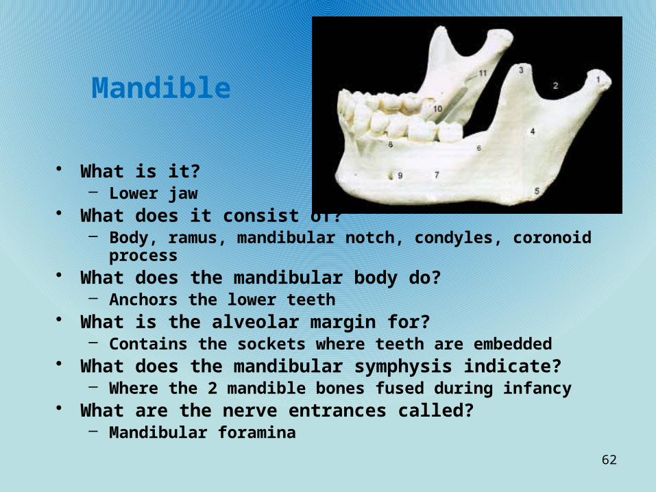
Mandible
• What is it?– Lower jaw
• What does it consist of?– Body, ramus, mandibular notch, condyles, coronoid process
• What does the mandibular body do?– Anchors the lower teeth
• What is the alveolar margin for?– Contains the sockets where teeth are embedded
• What does the mandibular symphysis indicate?– Where the 2 mandible bones fused during infancy
• What are the nerve entrances called?– Mandibular foramina
62

Mandible functions• Coronoid
– Insertion for temporalis muscle • Condyles
– Articulate with temporal bones for movement of jaw• Symphysis
– Fusion point of mandible• Alveoli
– Teeth sockets• Foramina
– Admit inferior alveolar nerve• Mental foramen
– Blood vessel and nerves to chin and lower lip
63

Maxillary bones• What area of face?
– Upper jaw, central portion• What articulates with it?
– All facial bones except mandible• Why is it keystone of face?
– All bones articulate with it. (- mandible)• What part of maxilla carry upper teeth?
– Alveolar margin• What is the function of incisive fossa?
– Passage for blood vessels, nerves• What articulates with the maxilla laterally?
– Zygomatic process• What forms the bony roof of the mouth?
– Palatine processes• What is at the junction of maxilla and greater wing of the
sphenoid?– Inferior orbital fissure
64

Maxilla functions• Alveoli
– Tooth socket• Zygomatic processes
– Form zygo arch• Palatine
– Form anterior hard palate• Frontal
– Form lateral part of nose bridge• Incisive fossa
– Admit blood vessels, nerves through hard palate• Inferior orbital fissure
– Admit maxillary branch of cranial nerve V, zygo nerve, blood vessels• Infraorbital foramen
– Nerve to skin of face
65

Articulation of other face bones• Zygomatic:• Articulation:
– Zygomatic processes of temporal posteriorly– Zygomatic processes of frontal superiorly– Zygomatic processes of maxilla anteriorly
• Nasal bones:• Articulation:
– Frontal bone superiorly– Maxilla laterally– Perpendicular plate posteriorly– Inferiorly to cartilage that form external nose
• Lacrimal bones:• Articulation:
– Frontal superiorly– Ethmoid posteriorly– Maxilla anteriorly
66

Articulation of other face bones
• Palatine bones• Articulation:• What are the important plates?
– Horizontal, perpendicular• What are the 3 processes?
– Pyramidal– Sphenoidal– Orbital
67

Orbits
68
Supraorbital foramen
SuperiorOrbital fissure Optic canal
Medial wall:
Sphenoid body
Orbital plate of ethmoid
Frontal process of maxilla
Lacrimal
Nasal bone
Floor of orbit:
Orbital process of palantine
Orbital surface of maxillaryZygomatic boneInfraorbital foramen
Zygomatic boneInfraorbital grooveInfraorbital fissure
Orbital surface of zygo
Greater wing of sphenoid
Zygo process of frontal
Orbital plate of frontal
Lesser wing of sphenoid

Nasal Cavity• What is it constructed of?
– Bone and hyaline cartilage• What is the roof formed by?
– Cribriform plate of ethmoid• What shapes the lateral walls?
– Superior, middle conchae of ethmoid, perpendicular of palatine • What are the depressions under the conchae?
– Superior, middle, inferior meatus• What is the floor formed by?
– Palatine processes of maxillae and palatine bones• What divides the nasal cavity?
– Septum• What is the bony part of septum?
– Vomer• What cartilage completes the septum anteriorly?
– Septal cartilage
69

Nasal Cavity
70
Frontal sinus
Superior nasal concha
Middle nasal concha
Inferior nasal concha
Nasal bone
Anterior nasal spineMaxillary bone(Palatine process)
Palatine bonehorizontal
Palatine bonePerpendicular
Pterygoid process
Sphenoid sinus
Superior, middle, andInferior meatus

Paranasal sinuses
71

Hyoid bone • Where?– Just inferior to mandible
in the neck• What is unique about this
bone?– Does not articulate
directly with any other bone
• What is anchored by?– Stylohyoid ligaments
• To What?– Styloid processes of
temporal• What is its function?
– Move tongue– Attach muscles that
raise/lower larynx for speech, swallowing
72

Anterior Skull 1. Frontal Bone2. Supra-Orbital Foramen3. Orbit (Orbital Cavity)4. Superior Orbital Fissure5. Inferior Orbital Fissure6. Zygomatic Bone7. Infra-Orbital Foramen8. Maxilla9. Mandible10. Mental Foramen11. Incisive Fossa12. Symphysis13. Vomer14. Inferior Nasal Concha15. Middle Nasal Concha16. Perpendicular Plate of
Ethmoid17. Nasal Bone18. Lacrimal Bone
73

Skull - Lateral 1. Parietal Bone2. Coronal Suture3. Frontal Bone4. Nasal Bone5. Vomer6. Lacrimal Bone7. Orbital Part of Ethmoid8. Zygomatic Bone9. Maxilla10. Body of Mandible11. Ramus of Mandible12. Coronoid Process13. Mandibular Condyle14. Mental Foramen15. Styloid Process16. External Acoustic Meatus17. Mastoid Process18. Zygomatic Process19. Temporal Bone20. Greater Wing of Sphenoid21. Inferior Temporal Line22. Superior Temporal Line23. Squamosal Suture24. Lambdoidal Suture25. Occipital Bone
74

Skull Posterior
1. Parietal Bone
2. Sagittal Suture
3. Lambdoid Suture
4. Occipital Bone
5. External Occipital Protruberance
6. Superior Nuchal Line
7. Inferior Nuchal Line
75

Occipital bone• Where is this bone?
– Posterior wall and base of skull• What sutures connect it to the temporal and parietal bones?
– Lambdoid– Occipitomastoid
• What does the internal walls of occipital form?– Posterior cranial fossa
• What foramen is at the base of occipital?– Foramen magnum
• What are the occipital condyles?– Rocker like condyles that articulate with c1 and allow nodding of
head• What does the external occipital crest secure?
– Ligamente nuchae• What do nuchal lines and bony regions do?
– Anchor many back muscles
76

Occipital Parts functions
• Foramen magnum – Spinal cord passage from brain stem to vertebral canal
• Hypoglossal canal– Passage of hypoglossal nerve (cranial nerve XII)
• Occipital condyles– Atlas articulation
• External protuberence/nuchal lines– Muscle attachments
• External Crest– Ligamente nuchae
77

Skull - superior
1. Occipital Bone
2. Lambdoidal Suture
3. Parietal Bone
4. Sagittal Suture
5. Coronal Suture
6. Frontal Bone
78

Superior Skull1. Frontal Sinus2. Foramen Cecum3. Crista Galli4. Cribriform Plate5. Anterior Cranial Fossa6. Lesser Wing of Sphenoid7. Chiasmatic Groove8. Hypophyseal Fossa9. Dorsum Sella10. Optic Canal11. Anterior Clinoid Process12. Foramen Rotundum13. Foramen Ovale14. Foramen Spinosum15. Squamous Part of Temporal16. Petrous Part of Temporal17. Groove for Transverse Sinus18. Posterior Cranial Fossa19. Foramen Magnum20. Hypoglossal Canal21. Jugular Foramen22. Internal Acoustic Meatus23. Posterior Clinoid Process24. Foramen Lacerum25. Superior Orbital Fissure
79

Mandible1. Mandibular
Condyle
2. Mandibular Notch
3. Coronoid Process
4. Ramus
5. Angle
6. Oblique Line
7. Body
8. Alveolar Process
9. Mental Foramen
10. Mylohyoid Line
11. Mandibular Foramen
80

Anterior skull
81
Frontal squamaOf frontal bone
Nasal bone
Sphenoid
Parietal bone
Temporal
Ethmoid
LacrimalZygomatic
Infraorbital foramen
Maxilla
MandibleMentalForamen
Mandibular Symphysis
Frontal
Glabella
Frontonasal suture
Supraorbital foramenSupraorbital marginSuperior Orbital fissure
Optic Canal
Inferior orbital fissure
Middle nasal conchaPerpendicular plateInferior nasal concha
Vomer

Lateral Skull
82
Frontal bone
Sphenoid bone
Ethmoid boneLacrimal boneLacrimal fossa
Nasal bone
Zygomatic bone
Maxilla
Alveolar margins
MandibleMental foramen
Coronoid ProcessMandibular angle
Mandibular ramusMandibular notch
Mandibular condyleStyloid process
Mastoid process
External auditory meatusOccipitomastoid suture
Zygomatic processOccipital bone
Squamous suture
Lambdoid suture
Temporal bone
Parietal bone
Coronal suture

Posterior Skull
83
Sagittal suture
Parietal bone
Mastoid process
InferiorNuchal line
Occipitalcondyle
External Occipitalcrest
Occipitomastoidsuture
External Occipitalprotuberance
Superior nuchalline
Occipital bone
Lambdoidsuture
Wormian bone

Midsagittal Internal left side of skull
84
Coronal suture
Frontal bone
Sphenoid bone
Frontal sinusCrista galliNasal boneSphenoid sinusEthmoid bone
Vomer boneIncisive fossaMaxilla
Alveolar margins
Mandible
Palatine process
Palatine bone
Mandibular foramen
PterygoidProcess of Sphenoid
Sella Turcica of sphenoid
Internal acoustic meatus
External occipital Pro-tuberence
Occipitomastoid suture
Occipital bone
Lambdoid suture
Temporal bone
Squamous suture
Parietal bone

Inferior skull
85
Incisive fossaMedial palantine sutureInfraorbital foramenMaxilla
Sphenoid bone
Foramen ovaleForamen lacerum
Carotid canalExternal acoustic meatusStylomastoid foramen
Jugular foramen
Occipital condyleInferior nuchal line
Superior nuchal line
Foramen magnum
External Occipitalprotuberence
External OccipitalCrest
Parietal bone
Pharyngeal Tubercle of basoccipital
Temporal boneMastoid process
Styloid process
Mandibular fossa
Vomer
Temporal boneZygomatic bone
Palatine bone
Maxilla

Temporal bone
86
Squamous region
Zygomatic process
Mandibular fossa
Tympanic regionStyloid process
Mastoid process
Mastoid region
External Acousticmeatus

Temporal bones
• How many? Where are they on skull?– 2 (One on each side - inferior to parietal bones)
• What are the temporal’s four major regions?– Squamous,Tympanic, Mastoid, Petrous
• Functions of parts?• Zygomatic process –
– helps form prominence of cheek• External acoustic meatus –
– for hearing• Styloid process –
– attach hyoid and neck muscles• Mastoid process –
– attach neck and tongue muscles• Carotid canal –
– passage of internal carotid artery• Jugular foramen –
– passage of internal jugular vein and cranial nerves IX, X, XI
87

Superior sphenoid
88
Optic canal
Chiasmatic groove
Lesser wing
Greater wing
Anterior clinoid processForamen rotundumForamen ovale
Foramen spinosum
Dorsum Sellae
Body of sphenoid
Posterior clinoidprocess
HypophysealFossa of sella turcica
Greater wing

Posterior sphenoid
89
Body of sphenoidPosterior clinoid process
Superior orbital fissure
Foramen rotundum
Pterygoid process
Pterygoidplates
Greater wing

Sphenoid• Where?
– Spans the middle cranial fossa• Why is it considered the keystone of cranium?
– Because it forms a central wedge that articulates with all other cranial bones
• What does it consist of?– Central body, 3 pairs of wings– Greater, lesser, pterygoid processes
• What is the sella turcica?– Area where pituitary gland is located
• What is the function of the anterior clinoid processes?– Anchor the brain to the skull
• What are the pterygoid processes functions?– Anchor pyterygoid muscles used for chewing
• What are the optic canals for?– They allow optic nerves to pass to the eyes
• What is the superior orbital fissure’s function?– It allows cranial nerves that control eye movements to enter the orbit
90

Sphenoid parts functions
• Sella turcica– Seat of pituitary gland
• Optic canal– Passage of cranial nerve II, opthamolic arteries
• Superior Orbital fissures– Cranial nerves III, IV, part of V, opthamolic vein
• Foramen rotundum– Passage of maxillary division of cranial nerve V
• Ovale– Pass mandibular division of nerve V
• Spinosum– Pass middle meningeal artery
91

Ethmoid
• Where?– Between sphenoid and nasal
bones (between nasal and orbits)• What forms roof of nasal cavity
and floor of anterior cranial fossa?– Cribriform plate
• What are the tiny holes in cribriform that allow smell nerves to pass to the brain?– Olfactory foramina
• What is the crista galli’s function?– Attach falx cerbri
• What are some other parts of the ethmoid?– Inferior, middle nasal concha– Perpendicular plate– Orbital plates
92

Ethmoid
93
Crista galli
Cribriform plate
Left lateral mass
Middle nasal concha
Perpendicular plate
Ethmoid sinuses
Orbital plate
Olfactory foramina
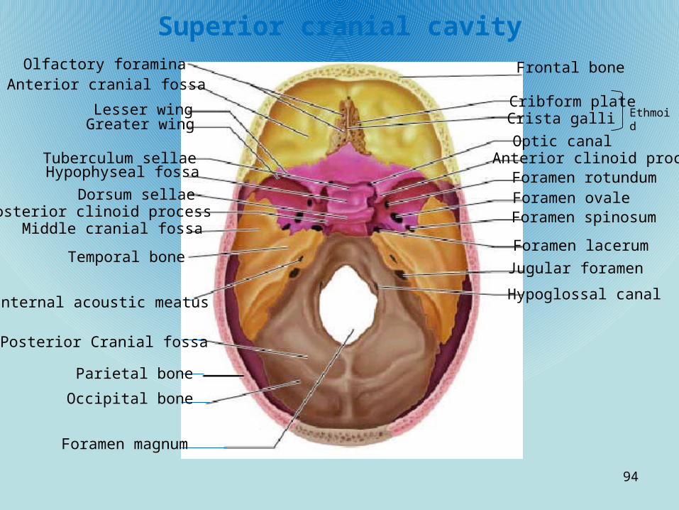
Superior cranial cavity
94
Frontal bone
Cribform plateCrista galli Ethmoid
Optic canalAnterior clinoid processForamen rotundumForamen ovaleForamen spinosum
Foramen lacerum
Jugular foramen
Hypoglossal canal
Foramen magnum
Occipital bone
Parietal bone
Posterior Cranial fossa
Internal acoustic meatus
Temporal bone
Middle cranial fossaPosterior clinoid process
Dorsum sellae
Hypophyseal fossaTuberculum sellae
Greater wingLesser wing
Anterior cranial fossaOlfactory foramina

Vertebrae• How many bones? What kind of bones?
– 26, irregular• Name some functions of vertebrae?
– Transmits weight of trunk to lower limbs– Surround, protect spinal cord– Attachment site for ribs and muscles of back and neck
• How many in fetus?– 33 separate bones
• How many eventually fuse and what do they become?– 9– Sacrum, coccyx
• What are the divisions? How many bones in each?– Cervical – 7– Thoracic – 12– Lumbar – 5– Sacrum– Coccyx
95

Vertebrae (cont)
• What are the four curvatures?– Cervical, lumbar – concave posteriorly– Thoracic, sacrum – convex posteriorly
• What are some abnormal curvature disorders?– Scoliosis – Thoracic twisted– Kyphosis – hunchback
• What are ligaments?– Bands of fibrous tissue that connect and support bones
• What are the ligs of the vertebrae?– Anterior, posterior longitudinal ligaments
• What are the intervertebral discs functions?– Shock absorbers during walking, jumping and running
96

General structure of Vertebrae
• What is the common structure of all vertebrae?– Body anteriorly– Vertebral arch posteriorly– Vertebral foramen for vertebral canal (spinal cord)
• What forms vertebral arch?– Pedicles, laminae
• What are pedicles?– Short, bony pillars that form side of arch (project posteriorly)
• What are laminae?– Flat plates that fuse in median plane (posterior)
• How many processes project from vertebral arch?– 7
• What are they?– Spinous – median, at junction of 2 laminae– Transverse – lateral from each side of arch– Superior, inferior articular processes – smooth joints called facets (covered
with hyaline cartilage) form movable joints with processes of vertebrae below and above them
97
Facet(ThoracicOnly)
Demifacet(ThoracicOnly

Vertebrae structure
98

99
Cervical Thoracic Lumbar

100

101
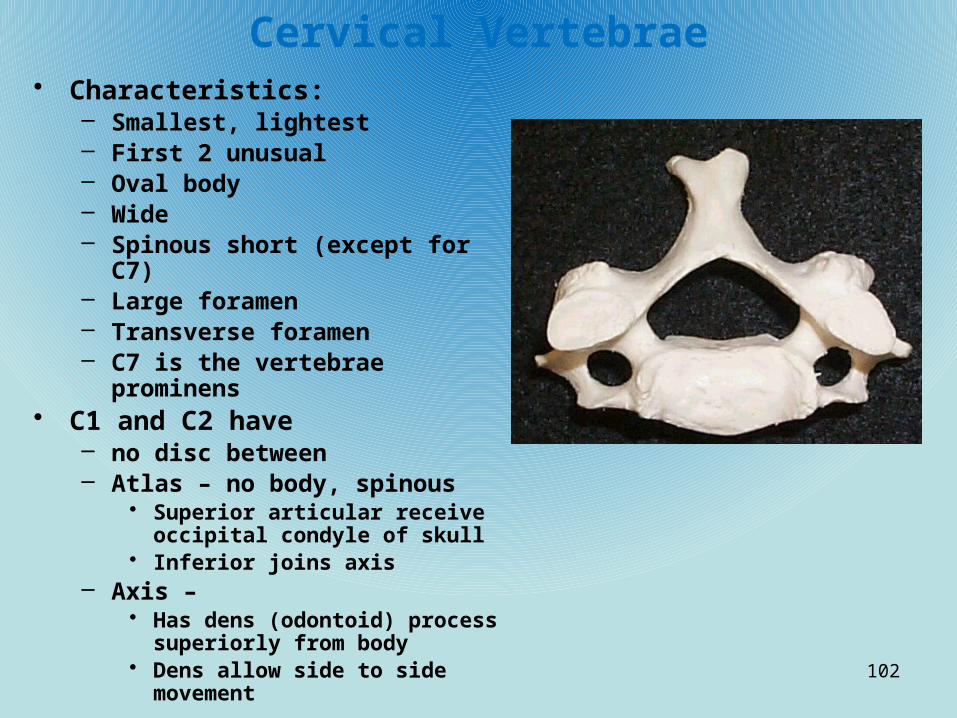
Cervical Vertebrae• Characteristics:
– Smallest, lightest– First 2 unusual– Oval body– Wide– Spinous short (except for C7)– Large foramen– Transverse foramen– C7 is the vertebrae prominens
• C1 and C2 have – no disc between– Atlas – no body, spinous
• Superior articular receive occipital condyle of skull
• Inferior joins axis– Axis –
• Has dens (odontoid) process superiorly from body
• Dens allow side to side movement
102
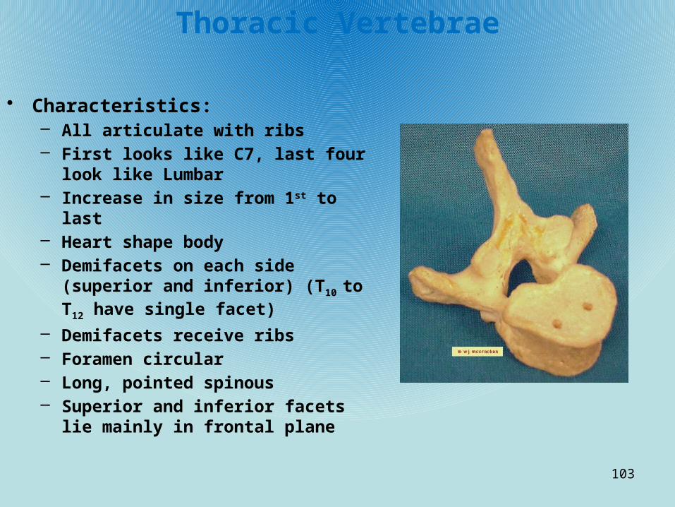
Thoracic Vertebrae
• Characteristics:– All articulate with ribs– First looks like C7, last four look like
Lumbar– Increase in size from 1st to last– Heart shape body– Demifacets on each side (superior and
inferior) (T10 to T12 have single facet)
– Demifacets receive ribs– Foramen circular– Long, pointed spinous– Superior and inferior facets lie mainly in
frontal plane
103
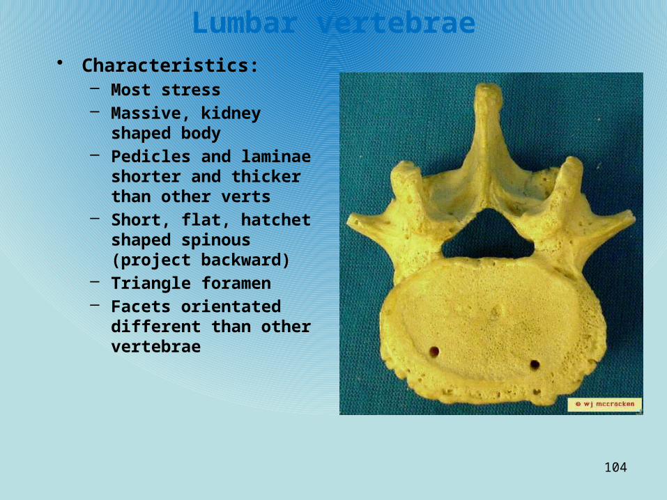
Lumbar vertebrae• Characteristics:
– Most stress– Massive, kidney shaped body– Pedicles and laminae shorter
and thicker than other verts– Short, flat, hatchet shaped
spinous (project backward)– Triangle foramen– Facets orientated different
than other vertebrae
104
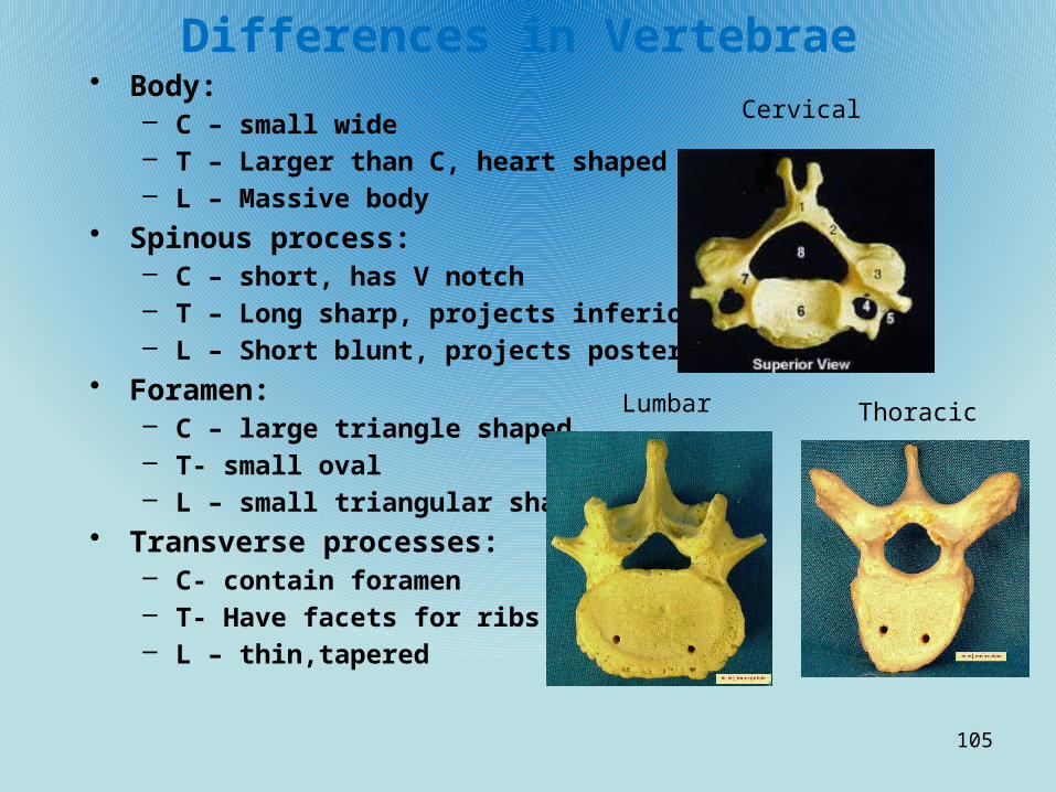
Differences in Vertebrae• Body:
– C – small wide– T – Larger than C, heart shaped– L – Massive body
• Spinous process:– C – short, has V notch– T – Long sharp, projects inferiorly– L – Short blunt, projects posteriorly
• Foramen:– C – large triangle shaped– T- small oval– L – small triangular shape
• Transverse processes:– C- contain foramen– T- Have facets for ribs– L – thin,tapered
105
Cervical
Lumbar Thoracic
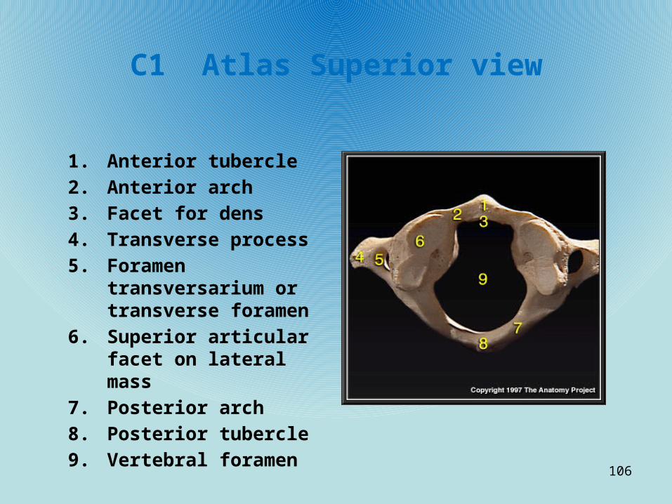
C1 Atlas Superior view
1. Anterior tubercle
2. Anterior arch
3. Facet for dens
4. Transverse process
5. Foramen transversarium or transverse foramen
6. Superior articular facet on lateral mass
7. Posterior arch
8. Posterior tubercle
9. Vertebral foramen
106

Cervical, Thoracic, Lumbar, Sacrum
1. Body 2. Vertebral foramen 3. Anterior tubercle 4. Posterior tubercle 5. Foramen
transversarium or transverse foramen
6. Demifacet for head of rib
7. Superior articular process
8. Pedicle 9. Lamina 10. Transverse process 11. Spinous process or
spine 12. Lateral mass 13. Posterior sacral
foramina 14. Coccyx 15. Sacral hiatus
107

Cervical, Thoracic, Lumbar, sacrum
1. Body
2. Facet for head of rib
3. Superior articular process
4. Superior vertebral notch
5. Pedicle
6. Transverse process
7. Inferior vertebral notch
8. Inferior articular process
9. Spinous process or spine
108

Cervical Vertebrae 1. C1 or atlas 2. C2 or axis 3. C3 4. C4 5. C5 6. C6 7. C7 8. Body 9. Vertebral
foramen 10. Bifid spinous
process or spine 11. Transverse
process 12. Foramen
transversarium or transverse foramen
13. Superior articular facet
109

Vertebrae - Atlas
1. Superior Articular Surface
2. Transverse Foramen
3. Transverse Process
4. Odontoid (Dens) Facet
5. Vertebral Foramen
6. Inferior Articular Surface
110

Axis1. Spinous Process
2. Lamina
3. Transverse Process
4. Pedicle
5. Superior Articular Surface
6. Odontoid Process (Dens)
7. Body
8. Vertebral Foramen
9. Inferior Articular Surface
111

Cervical Vertebrae
1. Spinous Process
2. Lamina
3. Superior Articular Surface
4. Transverse Foramen
5. Transverse Process
6. Body
7. Pedicle
8. Vertebral Foramen
112

Thoracic
1. Spinous Process
2. Lamina
3. Superior Articular Surface
4. Transverse Process
5. Pedicle
6. Body
7. Vertebral Foramen
8. Articular Facet for Rib
9. Inferior Articular Surface
113

Lumbar
1. Spinous Process
2. Lamina
3. Superior Articular Surface
4. Transverse Process
5. Pedicle
6. Body
7. Vertebral Foramen
8. Inferior Articular Surface
114

Sacrum
• Characteristics:– Formed by 5 fused verts– Articulates superiorly with L5 – inferiorly with coccyx, laterally with
hip bone (sacroiliac joint)– Sacral promontory anterosuperior– Four ridges: The transverse lines cross concave anterior aspect,
marking lines of fusion of sacral vertebrae– Ventral sacral foramina penetrate sacrum at lateral ends of
transverse lines to transmit blood vessels and nerves– Lateral to these and expanding superior are the alae– Dorsal midline – median sacral crest– Dorsal sacral foramina– Lateral sacral crests– Vertebral canal – sacral canal– Sacral hiatus – opening where vert failed to fuse
115

Sacrum1. Promontory2. Transverse
Ridges (lines)3. Coccyx4. Body of Sacrum5. Sacral Canal6. Superior
Articular Surface
7. Median Sacral Crest
8. Sacrum to Ilium Articular Surface
9. Dorsal Sacral Foramina
10. Sacral Hiatus
116
alae
Lateral sacralcrest
Ventral Sacral foramina

Rib and Vertebrae
1. Articular Facet of Rib
2. Interarticular Crest3. Neck4. Articular Portion of
Tubercle5. Nonarticular Portion
of Tubercle6. Angle of Rib7. Costal Groove8. Body (shaft) of Rib9. Articular Facet of
Transverse Process10. Transverse Process11. Spinous Process12. Lamina13. Vertebral Foramen
117
Sternal edge

Sternum
1. Jugular Notch
2. Manubrium
3. Sternal Angle
4. Body (Gladiolus)
5. Xiphoid Process
118
Clavicular Notch
Costal facet

Bony Thorax
119

Bony Thorax
• What is included in bony thorax?– Thoracic vertebrae dorsally, ribs laterally, sternum and costal
cartilage anteriorly• How many ribs?
– 12 pairs• What do they articulate with?
– Posteriorly – thoracic vertebrae– Anteriorly – superior 7 rib pairs – directly to sternum (true)– 5 (false) – indirectly to sternum or no sternal– Ribs 8-10 connect to cartilage joining to rib above– Ribs 11-12 – floating – no anterior attachment –
• Characteristics of ribs:– Bowed flat bone– Superior smooth, inferior sharp and thin– Costal groove – lodges nerves and blood vessels– Head and tubercle – join to the body and transverse process of vert– Tubercle is posterior and superior
120

Appendicular Skeleton
Pectoral & Pelvic Girdle, Upper & Lower Limbs
121
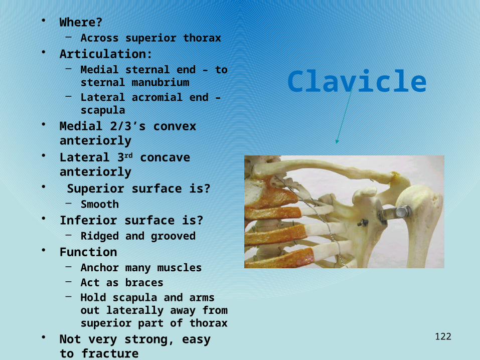
Clavicle
• Where?– Across superior thorax
• Articulation:– Medial sternal end – to sternal
manubrium– Lateral acromial end – scapula
• Medial 2/3’s convex anteriorly• Lateral 3rd concave anteriorly• Superior surface is?
– Smooth• Inferior surface is?
– Ridged and grooved• Function
– Anchor many muscles– Act as braces– Hold scapula and arms out
laterally away from superior part of thorax
• Not very strong, easy to fracture
122

Clavicle1. sternal head 2. superior surface 3. sternal head 4. inferior surface 5. groove for subclavious
muscle6. conoid tubercle7. trapezoid line8. acromial head
• Identification aspects:– Conoid tubercle always
posterior/inferior– Medial surface always
convex, lateral concave– Acromial - lateral
123

Scapula Posterior1. Coracoid Process
2. Scapular Notch
3. Superior Border
4. Supraspinous Fossa
5. Superior Angle
6. Scapular Spine
7. Vertebral Margin
8. Infraspinous Fossa
9. Inferior Angle
10. Lateral Border
11. Glenoid Cavity Margin (lateral angle)
12. Acromion Process• Identification:
– Spine-posterior– Glenoid - lateral
124

Scapula (Ventral)1. superior angle
2. vertebral (medial) border
3. inferior angle
4. subscapular fossa
5. infraglenoid tubercle
6. glenoid fossa
7. coracoid process
8. Acromion
9. suprascapular notch
125

Scapula lateral
1. Coracoid process2. Glenoid cavity3. Scapular spine4. Acromion process5. Infraspinous Fossa6. Inferior Angle7. Axillary Margin
126

Scapula • Where?
– Dorsal surface of rib cage, between ribs 2 and 7• What are the 3 borders?
– Superior – shortest, sharpest– Medial (vertebral) – parallels vertebral column– Lateral (axillary) – abuts the armpit and ends superiorly in a small
shallow fossa, the glenoid cavity • Articulation
– Glenoid cavity articulates with humerus of the arm• What are the 3 angles? Where do they meet?
– Superior scapular border meets medial border at superior angle and lateral border at the lateral angle
– Medial and lateral borders join at the inferior angle
127

Scapula (cont)• Features:• Anterior surface is?
– Concave and relatively featureless
• Posterior surface?– Prominent spine easily felt
through skin– Ends laterally in enlarged,
roughened triangular projection called?
• Acromion– Acromion articulates with?
• Acromial end of clavicle– Which forms the?
• Acromioclavicle joint• Projecting anteriorly from superior
scapular border is what?– Coracoid process
• What is the coracoid process’ function?– Helps anchor the biceps of the
arm
• Bounded by?– Suprascapular notch
medially and glenoid cavity laterally
• Several large fossae appear on both sides of scapula. They are?– Infraspinous and
supraspinous fossae– Named for their location– Inferior and superior to the
spine• What is the subscapular
fossa?– Shallow concavity formed by
the entire anterior scapula surface
128

Proximal Humerus1. Head2. Anatomical neck3. Lesser Tubercle4. Intertubercular Groove5. Greater Tubercle6. Surgical Neck7. Deltoid Neck
(tuberosity)• Identify:
• Head – medial• Shallow, distal coronoid
fossa – anterior• Capitulum – anterior
lateral
129

Humerus Distal
1. Radial Fossa
2. Lateral Epicondyle
3. Capitulum
4. Trochlea
5. Medial Epicondyle
6. Coronoid Fossa
7. Olecranon Fossa
130

Humerus – proximal to middle of the bone
• Location– Upper arm
• Articulation?– proximal to the scapula (glenoid
cavity)– distal to the radius and ulna
• What is at the proximal end?– Smooth hemispherical head
• What does it fit into to?– Glenoid cavity
• What is immediately inferior to the head?– Anatomical neck
• What tubercles are just inferior to the neck?– Lateral greater tubercle– Medial lesser tubercle
• What separates these tubercles?– Intertubercular (bicipital) groove
• What are tubercles function?– Sites where muscles attach
• What is distal to tubercles?– Surgical neck
• What is midway down shaft laterally?– Deltoid tuberosity (roughened
deltoid muscle attachment site)• What runs obliquely down the
posterior aspect of shaft marking the course of radial nerve?– Radial groove
131

Humerus - Distal• What are the 2 condyles at distal
end?– Medial trochlea – looks like hour
glass tipped on side– Lateral capitulum – ball-like
• What do these articulate with?– Ulna and radius
• What flanks these condyles?– Medial, lateral epicondyles (muscle
attachment sites)• What is directly above these
condyles?– Supracondyle ridges
• What is responsible for the tingling feeling when “funny bone” is hit?’– Ulnar nerves that run behind the
medial epicondyle• Where is the coronoid fossa?
– Superior to the trochlea on the anterior surface
• Where is the Olecranon fossa?– Posterior to the coronoid fossa
132

Humerus – distal
• What is the function of the coronoid and olecranon fossae?– They allow the corresponding processes of the ulna to
move freely when the elbow is flexed and extended
• What receives the head of the radius when the elbow is flexed?– Radial fossa
133

Radius – Right/anterior
1. Head of radius
2. Neck of radius
3. Radial Tuberosity
4. Radius (Shaft)
5. Styloid Process
6. Ulnar Notch• Recognition aspects:
– Tuberosity – anterior/medial
– Styloid – lateral– Concave surface above
styloid - anterior
134
Radial TuberosityAnterior medial
Always anterior in Anatomical position

Forearm- antebrachium• Location:
– Lower arm• Bones:
– Radius, ulna• Articulation
– Proximal end with humerus– Distal end forms joints with the wrist– Radius and ulna articulate with each other both proximally and
distally at small radioulnar joints• What connects the radius and ulnar across their entire length?
– Flexible, interosseous membrane• What is the position of radius and ulna in anatomical postion?
– Radius – lateral on thumb side– Ulna – medial on little finger side
• What happens in the prone position?– Distal end of the radius crosses over the ulna and form an “X”
135

Ulna-right/ proximal/distal 1. Olecranon process
2. Trochlear notch
3. Coronoid process
4. Tuberosity
5. Radial notch
6. Ulna shaft
7. Head of ulna
8. Styloid process
• Identification aspects:– Radial notch always
lateral– Styloid process-medial
136

Ulna• Main responsibility?
– Forming elbow joint with humerus
• What are the 2 main processes at proximal end?– Olecranon, coronoid
processes• What separates these?
– Trochlear notch• What does the locking of the
olecranon process and olecranon fossa do?– Keeps the forearm from
moving posteriorly beyond the elbow joint
• Where does the ulna articulate with the head of the radius?
– Radial notch• Where is the ulnar head?
– At the distal end of the bone by wrist
• What is medial to the ulnar head?– Styloid process
• What separates the ulnar from the carpals?– A disc of fibrocartilage
• Does it have any role in hand movement?– Little to none at all
137
Radius• What is the superior surface of the head – convex or
concave?– Concave
• Articulation– Head proximal with the capitulum of the humerus– Medially with the radial notch of the ulna– Distal where the radius expands, medial ulnar notch with the
ulna– Carpal bones
• The ulna contributes heavily to?– The elbow
• The radius contributes to?– Wrist
• What happens when the radius moves?– The hand moves with it
138

Hand – Right Dorsal
1. Styloid process of radius
2. Navicular (Scaphoid)
3. Lunate
4. Triquetral
5. Pisiform
6. Trapezium
7. Trapezoid
8. Capitate
9. Hamate
10. Metacarpal
11. Proximal Phalange
12. Middle Phalange
13. Distal Phalange
14. Styloid Process of Ulna139

Hand – Right palmar
1. Navicular (Scaphoid)
2. Lunate
3. Triquetral
4. Pisiform
5. Trapezium
6. Trapezoid
7. Capitate
8. Hamate
9. Metacarpal
10. Proximal Phalange
11. Middle Phalange
12. Distal Phalange
140
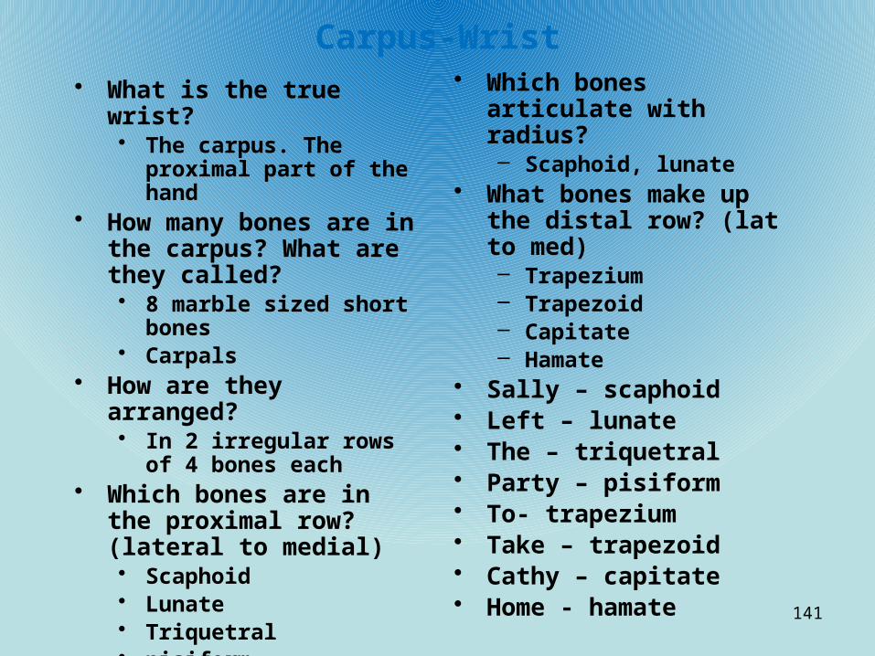
Carpus-Wrist• What is the true wrist?
• The carpus. The proximal part of the hand
• How many bones are in the carpus? What are they called?• 8 marble sized short bones• Carpals
• How are they arranged?• In 2 irregular rows of 4
bones each• Which bones are in the
proximal row? (lateral to medial)• Scaphoid• Lunate• Triquetral• pisiform
• Which bones articulate with radius?– Scaphoid, lunate
• What bones make up the distal row? (lat to med)– Trapezium– Trapezoid– Capitate– Hamate
• Sally – scaphoid• Left – lunate• The – triquetral• Party – pisiform• To- trapezium• Take – trapezoid• Cathy – capitate• Home - hamate
141

Metacarpus - palm• How many? What are they named?
– 5, No name. They are numbered from 1 to 5 from thumb to little finger
• Articulation– Bases with carpals proximally– Each other medially and laterally– Heads with proximal phalanges distally
• What is meta #1 and why is it different from the rest?– The thumb– Does not have a middle phalanx
142

Phalanges - Fingers• What are other names for the phalanges?
– Fingers, digits• How are they numbered?
– From 1 to 5 beginning with pollex (thumb)• How many phalanges on each hand?
– 14• What are the parts named?
– Proximal– Middle– Distal
• Thumb no middle
143

Os Coxa – Left lateral
1. Anterior Superior Spine2. Iliac Crest3. Posterior Superior Spine4. Posterior Inferior Spine5. Greater Sciatic Notch6. Body of Ilium7. Ischial Spine8. Lesser Sciatic Notch9. Body of Ischium10. Ischial Tuberosity11. Obturator Foramen12. Inferior Ramus of Ischium13. Inferior Ramus of Pubis14. Body of Pubis15. Acetabulum16. Anterior Inferior Spine
144

Os Coxa – Left,medial
1. Iliac Fossa2. Anterior Superior Spine3. Anterior Inferior Spine4. Arcuate Line5. Obturator Foramen6. Symphysis Pubis
Articulating Surface7. Ischial Tuberosity8. Lesser Sciatic Notch9. Ischial Spine10. Greater Sciatic Notch11. Sacrum Articulating
Surface12. Posterior Inferior Spine13. Posterior Superior Spine14. Iliac Crest 145
Identification:• Acetabulum – lateral• Pubis – anterior• Ischial- posterior

Pelvic girdle - Hip• What are its functions?
– Attach lower limbs to axial skeleton
– Transmit upper body weight to lower limbs
– Support visceral organs of the pelvis
• What are the hip bones named?– Os coxae
• Articulation– Each other anteriorly– Sacrum posteriorly
• What is the hip, sacrum and coccyx called?– Bony pelvis
• What are the 3 bones of the pelvis?– Ilium– Ischium– Pubis
• Are they separate?– In childhood. Fuse in
adults• What is the name of
the deep lateral socket on the pelvis?– Acetabulum
• What is its function? What is the joint called?
1. Receives head of femur2. Hip joint
146
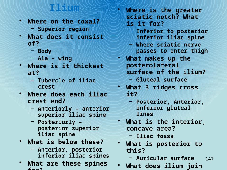
Ilium• Where on the coxal?
– Superior region• What does it consist of?
– Body– Ala – wing
• Where is it thickest at?– Tubercle of iliac crest
• Where does each iliac crest end?– Anteriorly – anterior
superior iliac spine– Posteriorly – posterior
superior iliac spine• What is below these?
– Anterior, posterior inferior iliac spines
• What are these spines for?– Attachment points for
muscles of trunk, hip, thigh
• Where is the greater sciatic notch? What is it for?– Inferior to posterior inferior
iliac spine– Where sciatic nerve passes to
enter thigh• What makes up the
posterolateral surface of the ilium?– Gluteal surface
• What 3 ridges cross it?– Posterior, Anterior, inferior
gluteal lines• What is the interior, concave
area?– Iliac fossa
• What is posterior to this?– Auricular surface
• What does ilium join anteriorly?– Ischium, pubis
147

Ischium• What part of hip bone?
– Posteroinferior• Articulation
– Body – ilium– Ramus – pubis anteriorly
• What are the 3 major markings?– Ischial spine – medially into pelvic cavity– Lesser sciatic notch – just inferior to ischial spine– Ischial tuberosity – strongest part of hip bone. Inferior surface of
ischial body• What helps hold the pelvis together?
– Sacrotuberous ligament – massive ligament from sacrum to ischial tuberosity
148

Pubis• Where?
– Anterior of hip bone• V shaped consisting of?
– Superior, inferior rami– Medial body
• Articulation– Medial to ischium– Inferior to ilium
• What does the anterior border form?– Pubic crest
• What is at the lateral end of pubic crest?– Pubic tubercle
• What is the large opening of the hip bone?– Obturator foramen
• What is the name of the fibrocartilage joining the 2 pubic bones?– Pubic symphysis
• What is the arch that differentiates male and female pelve?– Pubic arch
149

Pelvis - anterior 1. anterior superior iliac spine
2. superior pubic ramus
3. ischial tuberosity
4. inferior pubic ramus
5. pubic symphysis
6. pubic tubercle7. Acetabulum8. anterior
inferior iliac spine
9. iliac fossa
150

Pelvis - posterior1. iliac crest
2. greater sciatic foramen
3. ischial spine
4. ischial tuberosity
5. Coccyx
6. sacrum (dorsum)
7. posterior superior iliac spine
151

Pelvic structure/childbearing• What is the difference
between female and male pelvis?– Female
• wider, shallower, lighter and rounder
• Tilted forward• What are the false and
true pelvis?– False
• superior to the pelvic brim
• Bounded by alae laterally; lumbar vertebrae posteriorly
• Part of abdomen• Does not restrict
childbirth– True
• Inferior to pelvis brim
• Forms deep bowl containing pelvic organs
• Dimensions critical for childbirth
• What are the pelvic inlet and outlet?– Inlet – pelvic brim– Outlet – inferior margin
of true pelvis
152

Femur-proximal end/left1. Head2. Neck3. Greater Trochanter4. Intertrochanteric
Line5. Lesser Trochanter6. Shaft of Femur7. Gluteal Tuberosity8. Intertrochanteric
Crest9. Linea Aspera• Identification:
• Distal Patellar Surface – anterior
• Intercondyle notch - posterior
153
FoveaCapitis

Left femur-distal
1. Medial Condyle
2. Lateral Condyle
3. Intercondylar Fossa
154
Intercondylenotch
Lateral Epicondyle Medial Epicondyle
Adductor tubercle

Femur• Location
– Upper leg• Articulation
– Proximal – hip bone/ acetabulum
– Distal – tibia• What is the name of the small
central pit at the head?– Fovea capitis
• What is at the junction of the shaft and neck?– Greater and lesser
trochanters• What connects these
trochanters?– Intertrochanteric line
anteriorly– Intertrochanteric crest
posteriorly
• What does the gluteal tuberosity blend into?– Linea aspera inferiorly
• What does the linea aspera diverge into?– Medial and lateral
suprachondyle lines• What are all of these
markings?– Muscle attachment sites
• Distally, the femur widens into?– Lateral and medial condyles
• What flanks the condyles superiorly?– Medial and lateral
epicondyles
155

Femur (cont)
• What is the patellar surface?– Articulation site for the
patella• What is the deep U-shape
on the distal posterior aspect of femur?– Intercondyle notch
• What is superior to this?– Smooth popliteal
surface• What is the patella?
– Triangular, sesamoid bone enclosed in quadriceps that secures anterior thigh muscles to the tibia
156

Left Tibia-proximal/distal/anterior
1. Intercondylar Eminence
2. Lateral Condyle
3. Tibial Tuberosity
4. Anterior Crest
5. Medial Condyle
6. Anterior Surface
7. Medial Malleolus• Identify aspects:
– Tibial tuberosity always anterior
– Flattened side always lateral
– Medial malleolus always medial
157
Proximal
Tibiofibular
joint
Distal
Tibiofibular
joint

Tibia• Location– Anterior shin bone
• Articulation– Proximal – femur – tibia condyles to the femur condyles– Distally – talus of the foot– Lateral to the fibula (tibiofibular joint)
• Parts– Broad proximal end
• Medial and lateral condyles• Intercondyle imminence
– Inferior to this• Tibial tuberosity – anterior• Lateral tibial condyle – proximal tibiofibular joint
– Middle of bone• Anterior crest
– Distal end• Medial malleolus• Distal tibiofibular joint• Articular surface
158

Left Fibula-proximal/distal/anterior
1. Head of Fibula
2. Neck of Fibula
3. Anterior Crest
4. Lateral Malleolus• Identifying aspects:
– Lateral malleolus always lateral
159

Fibula• Location– Posterior, lateral shin
• Articulation– Proximal with the tibia– Distal with the tibia medially and the talus distally
• Name of proximal and distal ends?– Proximal – head– Distal – lateral malleolus (ankle bulge)
• Does the fibula bear weight?– No
160

Left foot superior
1. Calcaneus
2. Talus
3. Navicular
4. Cuboid
5. Cuneiform, Middle
6. Cuneiform, Intermediate
7. Cuneiform, Lateral
8. Metatarsal
9. Proximal Phalange
10. Middle Phalange
11. Distal Phalange
161

Tarsus• How many bones? Name them?– 7– Calcaneus– Talus– Navicular– Cuboid– 1st Cuneiform (Hallux), 2nd Cuneiform, 3rd Cuneiform
• What 2 tarsals carry most body weight?– Talus, calcaneus
• What does the Achilles tendon attach to?– Posterior surface of calcaneus
• What part of calcaneus touches the ground?– Tuber calcanei, calcaneal tuberosity, sustentaculum tali
• Name the remaining tarsals:– Lateral cuboid– Medial navicular– Intermediate, lateral cuneiform
• What do the cuboid and cuneiform bones articulate with?– Metatarsal bones anteriorly
162

Left footlateral
1. Calcaneus
2. Talus
3. Navicular
4. Cuboid
5. Cuneiform, First
6. Cuneiform, Second
7. Cuneiform, Third
8. Metatarsal
163

Metatarsus• How many?
– 5 small bones• Articulation
– Distal – proximal phalanges of the toes– Proximal – tarsals
• What is the first metatarsal?– Great toe (Hallux)
• How are they identified?– By number laterally from big toe
164

Phalanges-Toes• How many?
– 14• 3 in each digit except the?
– Hallux (big toe)
165
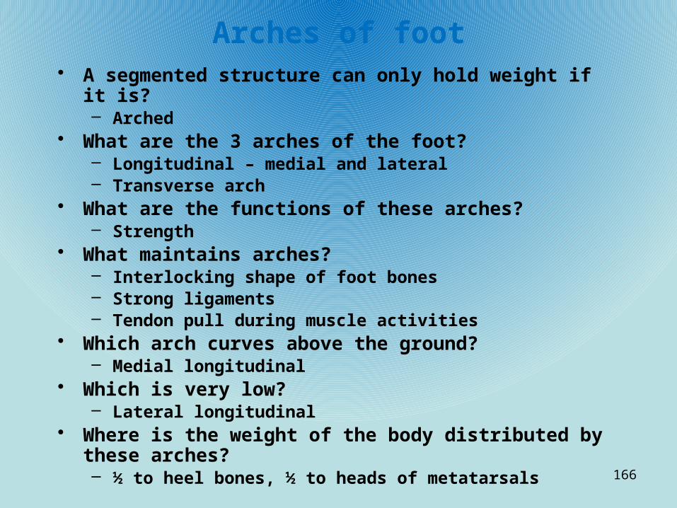
Arches of foot• A segmented structure can only hold weight if it is?
– Arched• What are the 3 arches of the foot?
– Longitudinal – medial and lateral– Transverse arch
• What are the functions of these arches?– Strength
• What maintains arches?– Interlocking shape of foot bones– Strong ligaments– Tendon pull during muscle activities
• Which arch curves above the ground?– Medial longitudinal
• Which is very low?– Lateral longitudinal
• Where is the weight of the body distributed by these arches?– ½ to heel bones, ½ to heads of metatarsals
166

Knee-anterior/posterior
1. Tibial Collateral Ligament
2. Medial Condyle of Femur
3. Posterior Cruciate Ligament
4. Anterior Cruciate Ligament
5. Lateral Condyle of Femur
6. Fibular Collateral Ligament
7. Lateral Condyle of Tibia
8. Lateral Meniscus
9. Medial Meniscus
10. Medial Condyle of Tibia
11. Tibia
12. Fibula
13. Transverse Ligament
167

168
Basic structure, types, location
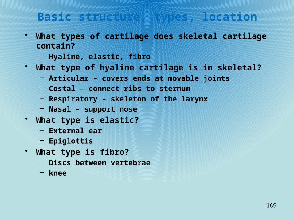
• What types of cartilage does skeletal cartilage contain?– Hyaline, elastic, fibro
• What type of hyaline cartilage is in skeletal?– Articular – covers ends at movable joints– Costal – connect ribs to sternum– Respiratory – skeleton of the larynx– Nasal – support nose
• What type is elastic?– External ear– Epiglottis
• What type is fibro?– Discs between vertebrae– knee
169

What are the 2 ways cartilage grows?
• Appositional– Cartilage-forming cells secrete new matrix against external
face of existing cartilage
• Interstitial– Chondrocytes divide and secrete new matrix– Expand from within
• When does growth end?– During adolescence
• Is calcified cartilage bone?– No. Cartilage and bone are always distinct tissues
170

Classification of Bone
• What are the 2 groups?– Axial– Appendicular
• What is the axial?– Skull, vertebrae, ribs
• Appendicular?– Upper/lower limbs, girdles (hips,shoulders)
171

Structure of long bone• Diaphysis
– Shaft– Surrounds medullary cavity. Adults – yellow bone marrow
• Epiphysis– Bone ends– Interior spongy bone– Joint surface – articular cartilage– Epiphyseal line – between the diaphysis and epiphysis of adult bone; remnant
of epiphyseal plate (hyaline that grows during childhood) Also called metaphysis
• Membranes– Glistening white outer cover?
• periosteum– Where do nerve, blood, lymph enter diaphysis?
• Nutrient foramen– Periosteum secured to bone by?
• Sharpey’s fibers
• What covers internal bone surfaces?– endosteum
172

Structure of short bone
• What design do all short, irregular and flat bones share?– Thin plates of periosteum-covered compact bone
sandwiching spongy bone in the middle
• Short bones have no what?– Shaft or epiphysis
• What is the spongy bone called?– Diploe
173

Hemopoietic tissue-red marrow
• Where is it found?– Within trabecular cavities in long bone– Diploe of flat bone
• What are those cavities called?– Red marrow cavities
• Where does blood production occur in adult long bone?– Only in head of femur and humerus
• What sites are usually more active and used for obtaining red marrow samples?– Diploe, irregular sites of flat bones (sternum, hip bone)
174

Compact bone• What do canaliculi do?
– Tie all the osteocytes in osteon together– Permit nutrients and wastes to enter and exit– Maintain bone matrix
• What is interstitial lamellae?– Incomplete lamellae that fill the gaps between forming osteons
• What are circumferential lamellae?– Lamellae just deep to the periosteum and superficial to the
endosteum, that extend around the entire circumference of the diaphysis and resist twisting of the bone
175

Formation of skeleton• What is the skeleton of human embryo made of before week 8?
– Entirely fibrous membranes and hyaline cartilage• What is it called when a bone develops from fibrous membrane?
– Intramembranous ossification• What is the bone of this type of formation called?
– Membrane bone• What is it called when bone is formed by replacing hyaline
cartilage?– Endochondral Ossification
• What is this bone called?– Cartilage or endochondral bone
176

Intramembranous Ossification• What bones are formed this way?
– Skull– Clavicle
• What type of bones formed this way?– Flat bones
• What are the 4 major steps?Step One:– What appears in the fibrous tissue?
• Ossification center– What do mesenchymal cells do at this stage?
• Cluster• Differentiate into osteoblasts• Form the ossification center
Step Two:– What do the osteoblasts do at this stage?
• Secrete the osteoid– What do trapped osteoblasts become?
• osteocytes
177

Intramembranous Ossification (cont)Step 3:– What is formed in step 3?
• Woven bone• Periosteum
– What is a random network?• Accumulated osteoid laid down between blood vessels
– What does this form?• Trabeculae (spongy bone)
– What does vascularized mesenchyme condensing on the external face of woven bone become?
• The periosteum
Step 4:– What does thickened trabeculae just deep to the periosteum
form?• Woven bone collar
– What is this replaced by?• Mature lamellar bone
– What is a diploe?• Compact bone sandwiching spongy bone
178

Endochondral Ossification• What bones are formed this way?
– All bones of the skeleton below the base of the skull• What is the model this process uses?
– Hyaline cartilage model• When does the process begin?
– 2nd month of fetal development• What is the primary ossification center?
– The region where long bone formation usually begins• What sets the stage for this process to begin?
– Perichondrium infiltrated by blood vessels converting it to periosteum– Mesenchymal cells specialize into osteoblasts
179
Endochondral Ossification (cont)Step 1:• What do the osteoblasts secretions against hyaline cartilage do?
– Encase it in a bone collar
Step 2:• What does cartilage in the center of diaphysis do?
– Calcifies, cavitates (creates cavities)• What do chondrocytes do at this stage?
– Hypertrophy (enlarge)– Signal matrix to calcify
• What happens to the chondrocytes and the matrix at this stage?– They die– Matrix deteriorates
• What happens to cartilage elsewhere?– Remains healthy– Grows briskly
• What does this cause the cartilage model to do?– Elongate
180

Endochondral Ossification (cont)
Step 3:• What is the periosteal bud?
– Collection of elements• Nutrient artery• Vein• Lymphatics• Nerve fibers• Red marrow elements• Osteoblasts, osteoclasts
• When does it appear?– The 3rd month of development
• What are the osteoclasts and osteoblasts doing at this stage?– Clasts – eroding calcified matrix– Blasts – secreting osteoid around remaining cartilage
• What is this forming?– Trabeculae (earliest version of spongy bone)
181

Endochondral Ossification (cont)• Step 4:• What forms in stage 4?
– Medullary cavity
182
Joints
183
Classification of joints
• What are the 2 ways to classify joints?– Functional– Structural
• Functional:– Synarthroses
• Immovable (skull)– Amphiarthroses
• Slightly movable (intervertebral, pubic symph)– Diarthroses
• Freely movable (limbs, joints of limbs)
• Structural:– Fibrous
• Bones joined by fibrous tissue – no joint cavity• Most immovable or slightly movable
184

Fibrous joints (cont)
• 3 types of fibrous joints:• Sutures:
– Rigid splices of interlocking bone – Where’s the only place they occur?
• Skull
• Syndesmoses:– Joints where bones connect by cord or sheet of fibrous tissue called a
ligament • Gomphoses:
– Resembles peg or socket (teeth)
185

Cartilaginous Joints
• Bones connected by cartilage with no joint cavity• 2 types:• Synchondroses-
– Plate of hyaline cartilage connects the bones– Epiphyseal plates of long bones, joint of 1st rib and manubrium
• Symphyses-– Bones covered with articular hyaline cartilage fused to
fibrocartilage plate.– All amphiarthritic (intervertebral, pubic symphysis)
186

Synovial Joints
• Articulating bones separated by fluid filled joint
• All have:– Hyaline both bony
surfaces– Joint cavity– 2 layer joint capsule with
outer fibrous layer– Synovial membrane lining
fibrous capsule– All internal joint surfaces
not covered with cartilage– Synovial fluid fills joint
capsule• May have:
– Wedges of fibro separating
– Sacs lined with synovial membrane
187

6 types of synovial• Plane
– Flat articular surface
• Hinge– Similar to door hinge
188

Pivot joints• Rotation of bone on
axis• Ulna• atlantoaxial
189

Condyloid joints
• Oval surface of one fits into concave of other
190

Saddle joints• Each bone has concave
and convex surface• Carpometacarpal of
thumb
191

Ball and Socket
• Round head of one fits into concave of other
• Humerus• femur
192

Joint Injuries• Sprain
– Overstretched, torn ligaments
• Cartilage– Torn or overused. Does not heal. Must remove
• Dislocation– Bones forced out of position
• Bursitis– Inflamed bursae, tendon sheath. Overuse, stress,
bacterial, friction
• Arthritis– Diseases that damage joints. Wear, bacteria, immune
193
Related Documents