Page 1 of 18 King Edward Memorial Hospital Obstetrics & Gynaecology Contents Antenatal Clinic Flowchart for Diagnosis & Management of IUGR .......... 3 Flow chart for Suspected SGA ................................................................ 4 Flow chart for Confirmed IUGR ............................................................... 5 Suspected Small for gestational age fetus: MFAU QRG ......................... 6 Criteria for Referral ................................................................................................. 6 Assessment ............................................................................................................ 6 Subsequent Visits for Confirmed SGA .................................................................... 6 Ultrasound Assessment .......................................................................................... 6 CTG monitoring....................................................................................................... 7 Management ........................................................................................................... 7 Intrauterine Growth Restriction ............................................................... 8 Aim.......................................................................................................................... 8 Background Information .......................................................................................... 8 Key Points ............................................................................................................... 9 Screening and Diagnosis ........................................................................................ 9 Determination of Gestational Age ........................................................................... 9 Abdominal Palpatation .......................................................................................... 10 Fundal - Symphysis Measurements ...................................................................... 10 Ultrasound examination ........................................................................................ 10 Management ......................................................................................................... 10 Assess for causes of IUGR ................................................................................... 10 Ultrasound Surveillance ........................................................................................ 11 CTG Monitoring..................................................................................................... 11 Anticipated Preterm Birth ...................................................................................... 11 CLINICAL PRACTICE GUIDELINE Small for Gestational Age and Intrauterine Growth Restriction: Management of This document should be read in conjunction with the Disclaimer

Welcome message from author
This document is posted to help you gain knowledge. Please leave a comment to let me know what you think about it! Share it to your friends and learn new things together.
Transcript

Page 1 of 18
King Edward Memorial Hospital
Obstetrics & Gynaecology
King Edward Memorial Hospital
Obstetrics & Gynaecology
Contents
Antenatal Clinic Flowchart for Diagnosis & Management of IUGR .......... 3
Flow chart for Suspected SGA ................................................................ 4
Flow chart for Confirmed IUGR ............................................................... 5
Suspected Small for gestational age fetus: MFAU QRG ......................... 6
Criteria for Referral ................................................................................................. 6
Assessment ............................................................................................................ 6
Subsequent Visits for Confirmed SGA .................................................................... 6
Ultrasound Assessment .......................................................................................... 6
CTG monitoring ....................................................................................................... 7
Management ........................................................................................................... 7
Intrauterine Growth Restriction ............................................................... 8
Aim .......................................................................................................................... 8
Background Information .......................................................................................... 8
Key Points ............................................................................................................... 9
Screening and Diagnosis ........................................................................................ 9
Determination of Gestational Age ........................................................................... 9
Abdominal Palpatation .......................................................................................... 10
Fundal - Symphysis Measurements ...................................................................... 10
Ultrasound examination ........................................................................................ 10
Management ......................................................................................................... 10
Assess for causes of IUGR ................................................................................... 10
Ultrasound Surveillance ........................................................................................ 11
CTG Monitoring ..................................................................................................... 11
Anticipated Preterm Birth ...................................................................................... 11
CLINICAL PRACTICE GUIDELINE
Small for Gestational Age and Intrauterine Growth Restriction: Management of
This document should be read in conjunction with the Disclaimer

SGA & IUGR
Page 2 of 18
Obstetrics & Gynaecology
Timing of Delivery ................................................................................................. 11
Intrapartum management ...................................................................................... 12
Small for Gestational Age Fetus ........................................................... 13
Aim ........................................................................................................................ 13
Background Information ........................................................................................ 13
Key Points ............................................................................................................. 14
Diagnosis .............................................................................................................. 14
Management ......................................................................................................... 14
Risks for IUGR/SGA ............................................................................................. 14
Assess for causes of the SGA and/or the IUGR fetus ........................................... 15
Fetal Surveillance ................................................................................................. 15
Cardiotocograph monitoring (CTG) ....................................................................... 16
Medical review and Antenatal Care ...................................................... 16
References ........................................................................................... 17

SGA & IUGR
Page 3 of 18
Obstetrics & Gynaecology
Antenatal Clinic Flowchart for Diagnosis & Management of IUGR
Assess
Risk factors at booking
FSH at every visit
Are measurements small for dates?
AND/OR are IUGR risk factors present?(1 major or 3
minor)
Check gestational age is correct(dating scan)
Review anatomy scan /FTS/possible causes.
Discuss with the obstetric team if measuring small for
dates
Document the plan in the antenatal record and
MR004 Obstetric Instruction Sheet
Diagnose
Arrange ultrasounds
· AFI/Dopplers/fetal biometry/BPP
· Anatomy (if not already performed)
AC or EFW < 10th centile or
Serial ultrasound indicates SGA/IUGR?
Manage
· Serial ultrasounds
· Schedule ultrasounds and antenatal visits on the
same day
· If SGA is confirmed but not IUGR and there is no
fetal compromise, document an individualised
care plan
Routine Care
Normal
UA Doppler
Fortnightly
· UA Doppler
· MCA Doppler
· AC & EFW
Birth
· Offer by 37 weeks-timing d/w consultant
· Recommended by > 34 weeks if
Static growth over 34 weeks
MCA Doppler PI <5th centile
· Consider steroids if CS birth & appropriate
Abnormal
UA DopplerAREDV
Daily
UA Doppler
DV Doppler
CTG > 32/40
PI or RI >2SD
EDV present
Twice weekly
UA Doppler
CTG > 32/40
Birth
· Recommended by 32 weeks, after
steroids.
· Consider 30-32 weeks
· Recommended < 32 weeks after
steroids if abnormal DV Doppler &/or
CTG
> 24 weeks & EFW >500g
No Yes
SGA & IUGR
Page 4 of 18
Obstetrics & Gynaecology
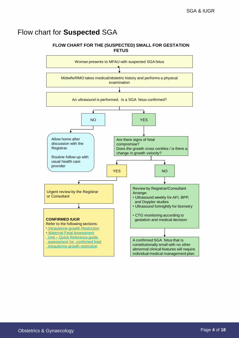
Flow chart for Suspected SGA
FLOW CHART FOR THE (SUSPECTED) SMALL FOR GESTATION
FETUS
Midwife/RMO takes medical/obstetric history and performs a physical
examination
An ultrasound is performed. Is a SGA fetus confirmed?
NO YES
Allow home after
discussion with the
Registrar.
Routine follow up with
usual health care
provider
Are there signs of fetal
compromise?
Does the growth cross centiles / is there a
change in growth velocity?
YES NO
Urgent review by the Registrar
or Consultant
Review by Registrar/Consultant
Arrange:
• Ultrasound weekly for AFI, BPP,
and Doppler studies
• Ultrasound fortnightly for biometry
• CTG monitoring according to
gestation and medical decision
A confirmed SGA fetus that is
constitutionally small with no other
abnormal clinical features will require
individual medical management plan.
CONFIRMED IUGR
Refer to the following sections:
• Intrauterine growth Restriction
• Maternal Fetal Assessment
Unit – Quick Reference guide
assessment for confirmed fetal
intrauterine growth restriction
Woman presents to MFAU with suspected SGA fetus

SGA & IUGR
Page 5 of 18
Obstetrics & Gynaecology
Flow chart for Confirmed IUGR
FLOW CHART FOR MANAGEMENT OF CONFIRMED INTRA-UTERINE GROWTH
RESTRICTION
Woman presents to MFAU with confirmed IUGR
Are all the measurements/tests within the normal limits?
Midwife/RMO reviews all maternal and fetal assessments
Midwife/RMO performs assessment as outlined in the Quick Reference Guide
YES NO
ABNORMAL RESULTS INCLUDE:
CTGs
• two or more Non Reactive
• features suggestive of fetal compromise
Ultrasound results
• AFI persistently low or decreasing
• Abnormal Doppler studies
• Fetal growth plateau or declining
• No fetal movements observed during
the scan
Fetal movements
• Maternal reporting of decreased fetal
movements
Inform the Obstetric
team of the results.
Arrange review in the
antenatal clinic or MFAU
as appropriate.
Inform the Level 3
Obstetric team Registrar
/ Consultant and
arrange urgent review.
Arrange continued
weekly or biweekly
assessment and
review in MFAU.
Team Consultant review
as required.

SGA & IUGR
Page 6 of 18
Obstetrics & Gynaecology
Suspected small for gestational age fetus: MFAU QRG
Criteria for Referral
Antenatal women for whom there is clinical suspicion of a suspected ‘small for
gestational age’ (SGA) fetus at or more than 24 weeks gestation.
Assessment
1. Confirm the gestational age by the woman’s dating ultrasound or last
menstrual period dates. Ensure a copy of the ultrasound report is available in
the medical records.
2. Review the result of the First Trimester and Second Trimester Screen if
available. Ensure a copy of the result is in the woman’s medical records.
3. Document the medical and obstetric history. Note any risk factors that may
contribute to a SGA fetus.
4. Palpate the abdomen as appropriate to determine:
· Symphysis fundal height
· Lie
· Presentation
5. Arrange an ultrasound scan for fetal biometry, amniotic fluid index (AFI),
umbilical artery (UA) Doppler velocities.
6. On confirmation of SGA diagnosis:
· If more than 32 weeks gestation, commence cardiotocography (CTG)
monitoring.
· If less than 32 weeks gestation discuss with Registrar or Consultant if CTG
monitoring is required.
Subsequent Visits for Confirmed SGA
Ultrasound and CTG monitoring management will be altered according to the clinical
picture and the medical management plan.
See the section on Intrauterine growth restriction for antenatal management of the
SGA fetus confirmed as intrauterine growth restricted.
Ultrasound Assessment
Fortnightly ultrasound assessment for biophysical profile, AFI and UA Doppler
velocities.
· Increased to twice weekly if abnormality in UA Doppler, or daily if
absent/reversed end diastolic velocity.
· Fortnightly fetal biometry.
· Increased to weekly if UA Doppler abnormality.

SGA & IUGR
Page 7 of 18
Obstetrics & Gynaecology
CTG monitoring
The frequency of CTG monitoring will depend on the fetal gestation and clinical
picture.
Management
· Inform the obstetric team of all results before the woman is discharged home. A
management plan is formulated prior to discharge.
· Document test results and management plan for future follow-up management in
MFAU and the antenatal clinic.
· Attempt where possible to arrange appointments in MFAU to coincide with the
antenatal clinic appointments. This allows review of the results by her team during
clinic appointments.
· The frequency of antenatal clinic appointments will depend on the clinical picture
and medical consultation
· Consider administering Betamethasone if pre term birth is anticipated.

SGA & IUGR
Page 8 of 18
Obstetrics & Gynaecology
Intrauterine growth restriction (IUGR)
Aim
To inform clinicians of the screening, management and obstetric birth considerations
for pregnancies complicated with fetal intrauterine growth restriction (IUGR).
Background Information
50-70% of the Small-for-Gestation Age (SGA) fetuses are constitutionally small but
healthy1. Approximately 10-15% of SGA fetuses are classified to be ‘true’ IUGR
cases, and another 5-10% are associated with chromosomal/structural anomalies,
or chronic intrauterine infection.2
A fetus is considered to have intrauterine growth restriction when the ultrasound fetal
measurements, particularly the abdominal circumference or serial weight
measurements, are below what is considered normal for that age and gestation.3
This is usually below the 5th or 10th centile when compared to the normal growth and
gestational age by ultrasound measurements.4 The IUGR infant has not reached
their genetic growth potential due to a pathological reason or event in utero causing
placental dysfunction.5 The IUGR fetus is associated with an increased risk of
perinatal mortality and morbidity and long term health consequences for survivors.2, 6,
7 Current evidence suggests long term consequences for IUGR infants are that they
are prone to heart disease, type 2 diabetes, strokes, hypertension and even
osteoporosis later in life.4
The Growth Restriction Intervention Trial (GRIT) concluded that generally if the fetus
is less than 31 weeks gestation it is best to delay delivery if there is uncertainty about
need for intervention, rather than immediate delivery. Evidence to date indicates that
by delivering the fetus early to pre-empt severe hypoxia and acidosis does not
reduce adverse outcomes.2, 8
Umbilical artery (UA) Doppler measurement is a tool used to identify if the SGA fetus
is affected by placental dysfunction which occurs with the IUGR fetus.9 With
worsening severity of placental insufficiency there is higher placental resistance
which can lead to absent or reversed end-diastolic flow velocities. This is associated
with poorer perinatal outcomes and mortality.1, 7 Fetal circulatory redistribution due
to placental insufficiency leads to abnormal Doppler indices in the cerebral and
umbilical arteries10 providing valuable information to assist decision making regarding
timing of birth. Doppler abnormalities have been shown to deteriorate before
biophysical profile scores (BPS) in the preterm fetus with IUGR prior to 32 weeks
gestation.10
In 2013, identification of babies with IUGR birthed >40wks formed Indicator 8 for
clinical audit. See: Indicator 8: IUGR, in RANZCOG/ACHS Obstetric Clinical
Indicators 2011.

SGA & IUGR
Page 9 of 18
Obstetrics & Gynaecology
Causes and risk factors for IUGR 3, 11
Maternal Fetal Placental
Hypertensive disorders Aneuploidy Anatomical conditions
Autoimmune disease Malformations Vascular conditions
Certain medications Abnormal genetic
imprinting
Chromosomal conditions
syndromes
Severe malnutrition,
anaemia
Viral or protozoan
infections
Morphological
abnormalities
Maternal lifestyle e.g.
smoking
Preterm birth
alcohol abuse, substance
abuse
Multiple gestation
Key Points
1. An accurate expected delivery date (EDD) is a critical component to allow
monitoring, assessment and optimal timing of delivery.
2. Management of the IUGR fetus must include a balance of the risks of intra-
uterine chronic hypoxia with preterm delivery and its associated risks.
3. Fetal Doppler studies provide the most accurate non-invasive assessment for
placental function. Absent or reversed UA Doppler’s are associated with poor
perinatal outcome and high perinatal mortality.12
Screening and Diagnosis
Screening and diagnosis for IUGR includes13:
1. Accurate determination of the gestational age.
2. Abdominal palpation to determine fundal height during each antenatal visit.
3. Fundal symphysis height measurements.
4. Ultrasound examination of a suspected SGA fetus.
5. Assessment of fetal well-being when an SGA fetus or IUGR fetus is
diagnosed. This includes biophysical profile (BPP), Doppler studies, and
cardiotocography monitoring (CTG) depending on gestation.
6. Crossing centiles or a change in growth velocity.
Determination of Gestational Age
A dating ultrasound in the first trimester provides the most accurate method to
determine gestational age.13 If the earliest ultrasound was between 13 and 24
weeks of pregnancy and the last menstrual period (LMP) is certain, with regular
menstruation, and there is a difference of less than 10 days between LMP &
ultrasound, use the LMP estimate.14 If the LMP is uncertain or irregular
menstruation, use the ultrasound EDD.14

SGA & IUGR
Page 10 of 18
Obstetrics & Gynaecology
Abdominal Palpation
· The ability to detect fetal weight by palpation is limited.9 If there is suspicion of
SGA, or IUGR, management should be discussed with the obstetric team. A
follow up ultrasound examination may be required.9, 13
· Document a management plan on the MR 004 ‘Obstetric Special Instruction
Sheet’ after consultation with the Obstetric team if a SGA or IUGR fetus is
suspected from palpation.
Fundal - Symphysis Measurements
· See Clinical Guideline,Measuring Fundal Height with a Tape Measure.
· If SGA or IUGR is suspected by abnormal fundal-symphysis measurements,
ultrasound examination may be required after obstetric team consultation.
Ultrasound examination
If there is suspicion of SGA or IUGR ultrasound examination should be performed to
assess:
· Biometry – assessment of growth requires at least 2 measurements two weeks
apart.1 Three weeks apart reduces false positive rate.9
· Doppler studies – Doppler studies are a valuable tool to differentiate the SGA
fetus that is healthy, and the true IUGR fetus.1, 9
· Amniotic Fluid Volume (AFV)
· Fetal well-being – Biophysical profile (BPP)
· Anatomy examination - if an anatomy scan has not been done or is unavailable,
this scan is required to exclude fetal anomalies, and fetal aneuploidy.9, 15
Management
1. Frequency of fetal surveillance is assessed at each visit, and management plan
adjusted by Obstetric team according to fetal and maternal clinical condition.
2. Antenatal surveillance may be conducted with antenatal clinic visits and by
outpatient review in the Maternal Fetal Assessment Unit (MFAU). If the
maternal or fetal clinical condition requires more intensive surveillance in-patient
hospitalisation should be considered in consultation with the team Obstetrician.
3. All ultrasound examinations, CTGs, and BPP must be reviewed and
documented by the Registrar or Consultant prior to discharge of a woman.
4. Document the assessment and test results at each visit to MFAU on the
Maternal Fetal Assessment Outpatient form MR 226.
Assess for causes of IUGR
1. Review the medical and pregnancy history to determine the cause of the IUGR
e.g. accurate delivery date, normal anatomy scan, and if any history of infection15.
2. Ensure a ‘hard copy’ of the antenatal testing and the results are available in
the medical records.

SGA & IUGR
Page 11 of 18
Obstetrics & Gynaecology
Ultrasound Surveillance
1. Amniotic fluid volume (AFV) and Doppler studies
· If normal at the initial visit: continue fortnightly assessment of AFV and
UA/ MCA Doppler studies.9
· If abnormal at the initial visit:
If end diastolic velocities (EDV) present/ pulsatility index (PI) or
resistance index (RI) >2SD: Arrange twice-weekly assessment of
AFV and Doppler studies, or more frequent surveillance if the clinical
condition requires closer monitoring.9
If absent / reversed end diastolic velocities (AREDV): Repeat UA and
DV Doppler daily.9 Discuss with Obstetric Consultant/ refer for fetal
medicine specialist opinion.9
2. Fetal Biometry- Abdominal circumference (AC) and estimated fetal weight
(EFW):
· If normal Doppler, arrange fetal biometry fortnightly.9, 15
· If abnormal Doppler, arrange weekly.9
CTG MONITORING
If the gestation is more than 32 weeks:
· Arrange a weekly CTG in MFAU on the woman’s Obstetric Team day on duty in
the antenatal clinic.
· If abnormal AFI or Doppler’s arrange bi-weekly CTG monitoring in MFAU.
· If abnormal Doppler with AREDV attend daily CTG.9
If the gestation is less than 32 weeks gestation discuss with the Registrar and
Consultant if CTG monitoring is required.
Anticipated Preterm Birth
· Consider a course of corticosteroids if pre-term birth ≤ 36+6 weeks gestation is
anticipated.1, 9
· Arrange Paediatric consultation if the gestation is less than 32 weeks.
Timing of Delivery
Delivery is indicated when risk of fetal death or morbidity is greater than the risk of
prematurity.
IUGR with end diastolic flow
· If other surveillance findings and maternal condition are normal delivery may
be delayed until 37 weeks.9
· Recommend birth >34weeks if:
· Static growth over 3-4 weeks
· MCA Doppler PI <5th centile
Consider steroids if caesarean birth. 9,16
SGA & IUGR
Page 12 of 18
Obstetrics & Gynaecology
IUGR associated with absent or reversed flow
· Admit for close surveillance16.
· Administration of steroids is recommended if preterm birth16 expected ≤36+6
weeks, if the clinical condition allows time17. See guideline Corticosteroids: Use of
· If other surveillance results are abnormal delivery is indicated.9,16
Intrapartum management
· Early admission in spontaneous labour.9
· Apply continuous CTG monitoring from onset of uterine contractions.9
· Caesarean birth is recommended in the IUGR fetus with UA AREDV.9
· Induction of labour can be offered where normal UA Doppler or abnormal UA
PI with EDV present.9

SGA & IUGR
Page 13 of 18
Obstetrics & Gynaecology
Small for gestational age fetus
AIM
· To inform clinicians of the assessment and pregnancy management of the woman with a suspected small for gestational age fetus.
Background Information
The term ‘small for gestational age ‘(SGA) refers to the fetus that has failed to reach a
specific biometry or estimated weight threshold by a specific gestational age.1, 2 It is
estimated that 50-70% of fetuses born weighing less than the 10th centile for
gestational age are constitutionally small, with the growth appropriate for the parental
size and ethnicity. The outcome is usually associated with normal placental function
and normal outcomes. SGA fetuses with a birth weight less than the 50th centile for
gestational age have a greater likelihood of intrauterine growth restriction (IUGR).1
SGA fetuses are at greater risk for stillbirth, birth hypoxia, neonatal complications,
impaired neurodevelopment, and possibly Type 2 diabetes and hypertension in adult
life, although the high incidence of adverse perinatal outcomes maybe contributed to
the IUGR foetuses in this group. The majority of term SGA infants have no
appreciable morbidity or mortality.2
Biometric tests used to assess fetal size assist diagnosis of SGA, while biophysical
tests are used to detect fetal wellbeing and are more indicative of IUGR.2 The use of
the customised fundal height chart has been demonstrated to improve the accuracy to
predict a SGA fetus, but ultrasound measurements of the abdominal circumference
and estimated fetal weight provide the most accurate way to predict SGA.2 Symphysis
fundal height (SFH) measurements may improve sensitivity and specificity for
predicting SGA, whilst abdominal palpation alone has limited accuracy for identification
of a SGA fetus2. The impact on perinatal outcomes of SFH measurement, compared
to abdominal palpation, is uncertain with a Cochrane systematic review finding only
one controlled trial that showed SFH measurements did not significantly change
perinatal outcomes.3 Continuation of SFH measurement at each antenatal
appointment has been recommended.2, 3
Assessment of fetal growth, abdominal circumference (AC) and estimated fetal weight
(EFW), requires two ultrasound measurements at least three weeks apart, which will
differentiate normally growing fetuses from those with IUGR.2 More frequent scanning
may be required by the Obstetric team where awareness of EFW would assist in
obstetric management, for reasons other than SGA diagnosis. 2 Routine biometry is
not justified in third trimester as it does not reduce the risk of SGA and does not
improve perinatal outcomes2. Measurements only provide limited information to assist
decision making for management for timing of delivery. Associated antenatal
surveillance techniques assist in clinical judgement for timing of delivery. These
techniques differentiate between a SGA fetus with a predicted normal outcome, and
the fetus which is growth restricted resulting in adverse perinatal morbidity and
mortality.1, 2 Umbilical artery (UA) Doppler measurements can identify if a confirmed
SGA fetus is affected by placental dysfunction, with end-diastolic flow velocity results
providing valuable information on risk for perinatal mortality and morbidity.1, 2, 4

SGA & IUGR
Page 14 of 18
Obstetrics & Gynaecology
Key Points
1. SGA describes the fetus that has failed to reach the normal biometric weight
by a specific gestational age. This does not always indicate a fetus is growth
restricted.
2. The use of ultrasound biometry and biophysical tests can assist differentiation
between the SGA with no expected perinatal morbidity or mortality risk, and
the IUGR fetus with predicted poor perinatal outcomes.
3. To evaluate fetal growth over time at least two subsequent measurements two
weeks apart should be performed.5 A three week interval further reduces
false positive results.2
4. Management is individualised according to gestation, fetal wellbeing and any
compounding maternal or fetal health factors.2
Diagnosis
Most methods to detect SGA require an accurate estimation of gestation as a
prerequisite.
Methods to detect SGA include:
· Measurement of symphysis pubis fundal height –recommended at each
antenatal appointment from 24 weeks to improve prediction of SGA fetus2.
· Abdominal palpation – has a limited diagnostic ability to predict the SGA
fetus.2 If a SGA fetus is suspected, diagnosis should be supplemented by
ultrasound biometry.
· Ultrasound biometry (AC or EFW <10th centile).
· Biophysical tests.
Management
At booking identify those needing increased monitoring:
· Where SFH is less accurate (large uterine fibroids, >BMI) = serial growth
ultrasounds2.
· One major or three minor risk factors present (see below).2
Consider preventative interventions in high risk groups (smoking cessation advice,
antiplatelet agents in women at high risk of pre-eclampsia).2
Risks for IUGR/SGA:
· Maternal age >35, >40 2
· Nulliparity2
· BMI2 <20
· IVF single pregnancy2
· Daily vigorous exercise2
· Low fruit intake pre-pregnancy2
· Low maternal weight gain2

SGA & IUGR
Page 15 of 18
Obstetrics & Gynaecology
· Previous stillbirth2
· Pre- eclampsia2 (previous pregnancy or this pregnancy)
· Maternal or paternal SGA2
· Pregnancy interval (<6months or >60months) 2
· Heavy bleeding2 (threatened miscarriage), unexplained APH2, or Placental
abruption2
· Echogenic fetal bowel2
· Caffeine >300mg/day in third trimester2
· PAPP- A < 0.4 MoM2
· Smoking
· Multiple pregnancy
Assess for causes of the SGA and/or the IUGR fetus
· Constitutionally small mothers2, 6, 7
· Poor maternal nutrition leading to a malnourished and underweight mother6, 7
· Previous birth of an SGA baby increases risk in a subsequent pregnancy2, 6
· Fetal structural abnormalities and congenital malformations6-8
· Fetal chromosomal abnormalities6-8
· Multiple pregnancy - a twin pregnancy is associated with a 10% increased
chance of IUGR6, 8
· Life style factors e.g. smoking2, 6, 8 (>11/day)2, alcohol and substance abuse7, 8
(cocaine)2
· Fetal infections e.g. cytomegalovirus, malaria, parvovirus, rubella6-8
· Maternal disease or disorders e.g. pregnancy induced hypertension2, 6
(mild/ severe); diabetes2; vascular disease2; chronic HTN2
· Disorders of cartilage and bone7
· Teratogens7
· Renal disease2, 7
· Chronic hypoxia7
· Placental and cord abnormalities7, 8
· Antiphospholipid Antibody Syndrome2, 7
Note: Factors in bold represent major risk factors for IUGR
Fetal Surveillance
Ultrasound scans
1. If severe SGA identified on anatomy scan (from external results), arrange
detailed anatomical ultrasound and uterine artery Doppler2 with fetal medicine
sonographer.

SGA & IUGR
Page 16 of 18
Obstetrics & Gynaecology
· Offer karyotyping in severe SGA with structural anomalies, those
before 23 weeks gestation, particularly if UA Doppler normal2
2. Arrange ultrasound assessment if a SGA fetus is suspected – biometry,
amniotic fluid index (AFI), umbilical artery (UA) Doppler velocities, and fetal
wellbeing.
3. If SGA is confirmed organise serial assessment of fetal size and umbilical
artery (UA) Doppler2:
· Weekly ultrasounds including AFI and UA Doppler’s. UA Doppler is the
primary surveillance tool in SGA2.
If normal UA Doppler flow: may be repeated every 14 days
More frequently in severe SGA
If abnormal UA Doppler flow indices and birth not indicated
repeat
· Twice weekly if end-diastolic velocities present
· Daily if absent/reversed end-diastolic frequencies).2
· Fortnightly fetal biometry and fetal well-being.
4. In the preterm SGA fetus with abnormal UA Doppler, the Ductus venous
Doppler should be used to assist in timing birth.2
5. In the term SGA fetus with normal UA Doppler, the middle cerebral artery
(MCA) Doppler should be used to assist in timing birth. 2
Cardiotocograph monitoring (CTG)
· If SGA is confirmed perform a CTG if the fetus is > 32 weeks gestation.
· If SGA is confirmed and the fetus is < 32 weeks gestation – discuss
management with the obstetric team Consultant if CTG monitoring is required
in correlation with ultrasound findings.
· Frequency of follow-up CTG monitoring in MFAU will be weekly or bi-weekly
depending on the biophysical profile and the UA Doppler studies. The
Consultant or Senior Registrar will make this decision.
· The CTG should be used in conjunction with other fetal monitoring for the
SGA fetus 2.
Medical review and antenatal care
SGA IS NOT CONFIRMED
If the ultrasound examination does not confirm SGA:
· Discuss with the team registrar or Consultant.
· Allow routine follow-up with the usual health care provider.
CONFIRMED SGA
1. Abnormalities of ultrasound examination or CTG monitoring should have
urgent review by the Consultant or the Senior Registrar.

SGA & IUGR
Page 17 of 18
Obstetrics & Gynaecology
2. Document a management plan on the MR 004 ‘Obstetric Special Instruction Sheet’.
3. Organise ultrasound follow-up appointments in the Maternal Fetal Assessment Unit
(MFAU).
4. Organise CTG monitoring according to gestation and medical management plan.
5. Arrange obstetric team antenatal clinic appointments weekly for medical review.
Ideally the appointments should be made to coincide with appointments in MFAU.
6. If SGA is confirmed but serial ultrasound biometry and UA Doppler do not indicate
IUGR or fetal compromise an individualised management plan should be
documented.
CONFIRMED IUGR
1. If IUGR is diagnosed refer to Section Intrauterine Growth Restriction
2. Consider administration of corticosteroids if pre-term delivery is anticipated.2
References
1. Alberry M, Soothill P. Management of growth restriction. Archives Disease and Childhood, Fetal and Neonatal Edition. 2007;72(1):F62-F7.
2. Sheridan C. Intrauterine growth restriction. Australian Family Physician. 2005;34(9):717-23. 3. Maulik D. Fetal Growth Compromise: Definitions, Standards, and Classification. Clinical Obstetrics
and Gynecology. 2006;49(2):214-8. 4. Sifianou P. Small and growth-restricted babies: Drawing the distinction. Acta Paediatrica.
2006;95:1620-4. 5. Bamburg C, Kalache KD. Prenatal diagnosis of fetal growth restriction. Seminars in Fetal &
Neonatal Medicine. 2004;9(5):387-94. 6. Illanes S, Soothill P. Management of fetal growth restriction. Seminars in Fetal & Neonatal
Medicine. 2004;9(5):395-401. 7. Marsal K. Obstetric management of intrauterine growth restriction. Best Practice & Research
Clinical Obstetrics and Gynaecology. 2009;23:857-70. 8. The GRIT study group. Infant wellbeing at 2 years of age in the Growth Restriction Intervention Trial
(GRIT): multicentred randomised controlled trial. The Lancet. 2004;364:513-20. 9. Royal College of Obstetricians and Gynaecologists. Green-top guideline No. 31: The investigation
and management of the small for gestational age fetus. 2nd ed. UK: RCOG; 2013. 10. Miller J, Turan S, Baschat AA. Fetal Growth Restriction. Seminars in Perinatology. 2008;32:274-80. 11. Pairman S, Tracy S, Thorogood C, Pincombe J. Midwifery: Preparation for practice. 2nd ed.
Chatswood, NSW: Elsevier Australia; 2010. 12. Chauhan SP, Gupta LM, Hendrix NW, et al. Intrauterine growth restriction: comparison of American
College of Obstetricians and Gynecologists practice bulletin with other national guidelines. American Journal of Obstetrics and Gynecology. 2009;409:e1-e6.
13. Haram K, Softeland E, Bukowski R. Intrauterine growth restriction. International Journal of Gynecology and Obstetrics. 2006;93:5-12.
14. Australian Health Ministers' Advisory Council. Clinical practice guidelines: Antenatal care- Module 1. Canberra: Australian Government Department of Health and Ageing; 2012. Available from: http://www.health.gov.au/antenatal.
15. Kinzler WL, Vinzileos AM. Fetal growth restriction: a modern approach. Current Opinion in Obstetrics and Gynecology. 2008;20:125-31.
16. Royal College of Obstetricians and Gynaecologists. Green-top guideline No. 7: Antenatal corticosteroids to reduce neonatal morbidity and mortality. 4th ed. UK: RCOG; 2010.

SGA & IUGR
Page 18 of 18
Obstetrics & Gynaecology
Related WNHS policies, procedures and guidelines
Keywords: SGA, IUGR,CTG, corticosteroids, ultrasound, AFI, fundal height, fetal compromise, Doppler, small for gestation, intrauterine growth restriction
Document owner: OGCCU
Author / Reviewer: Evidence Based Clinical Guidelines Co-ordinator
Date first issued: April 2008
Last reviewed: Oct 2016 Next review date: Oct 2019
Endorsed by: Maternity Services Management Committee Date: 18.10.16
Standards Applicable: NSQHS Standards: 1 Clinical Care is Guided by Current Best Practice
9 Clinical Deterioration,
Printed or personally saved electronic copies of this document are considered uncontrolled.
Access the current version from the WNHS website.
Related Documents