BRITISH ASSOCIATION OF ORAL AND MAXILLOFACIAL SURGEONS CLINICAL EFFECTIVENESS MATTERS 2010 The purpose of Clinical Effectiveness Matters is to provide an opportunity for the various regions to disseminate audit-related material for reflection on current practice and provide a source of discussion. The reports reflect a range of issues. Some of the submissions have had to be truncated so apologies if any key data has been deleted in this process. Please don’t hesitate to contact the authors or Regional Coordinator should you need more information. The intention is to produce Clinical Effective Matters each year so please take this opportunity to discuss with your Regional Coordinator your regions contribution for next autumn. It has been a tremendously busy year for the Regional Co-ordinators and members of the subcommittee. Three national audits in support of revalidation have been produced. This has involved a steep learning curve and still there are still several issues that need to be resolved before we have a system fit for purpose. A huge thank you for all those who have taken part and I hope it is the sort of information that is helpful in your portfolio of evidence for revalidation. The audits are web-based so some of you might need to make sure that BAOMS have an up to date email address. As you are probably aware the first was on subspeciality interests, audits and databases. There is more information on the BAOMS website and there is a short communication in BJOMS. The second survey was on bisphosphonate related osteonecrosis of the jaw with the purpose of gaining an indication of expected number of cases and local audits in place. The 3rd audit was wisdom teeth and involved 10 patient records and the whether removal of the teeth fitted with national guidelines. There was a process problem with this audit and I think this led to a much lower response rate. The 4th audit will be on trauma and it expected to take place in the Spring of 2011. Finally the 2-year bisphosphonate new case registration project is drawing to a close with final registrations in May this year and a need to get the full data in over the two-year period. Thank you all for your support in this as I appreciate how busy you and your departments are. It is only with your support that this project will provide any clinically meaningful data. Simon Rogers Chairman of the BAOMS Clinical Effectiveness Committee Region Name Armed forces Andrew Gibbons * Beds Herts Bucks Chi-Hwa Chan * Mersey Simon Rogers * Northern Mark Greenwood * Northern Ireland Dermot Pierse North West Stuart Clark North West Thames Bhavin Visavadia * Oxford Nad Saeed Scotland Ian Holland South Thames Jeremy Collyer South West Peter Revington Trent Iain McVicar * Wales Steven Key Wessex Steve Walsh West Midlands Bernie Speculand * Yorkshire Kelvin Mizen *Subcommittee members 1

Welcome message from author
This document is posted to help you gain knowledge. Please leave a comment to let me know what you think about it! Share it to your friends and learn new things together.
Transcript

BRITISH ASSOCIATION OF ORAL AND MAXILLOFACIAL SURGEONS CLINICAL EFFECTIVENESS MATTERS 2010 The purpose of Clinical Effectiveness Matters is to provide an opportunity for the various regions to disseminate audit-related material for reflection on current practice and provide a source of discussion. The reports reflect a range of issues. Some of the submissions have had to be truncated so apologies if any key data has been deleted in this process. Please don’t hesitate to
contact the authors or Regional Coordinator should you need more information. The intention is to produce Clinical Effective Matters each year so please take this opportunity to discuss with your Regional Coordinator your regions contribution for next autumn. It has been a tremendously busy year for the Regional Co-ordinators and members of the subcommittee. Three national audits in support of revalidation have been produced. This has involved a steep learning curve and still there are still several issues that need to be resolved before we have a system fit for purpose. A huge thank you for all those who have taken part and I hope it is the sort of information that is helpful in your portfolio of evidence for revalidation. The audits are web-based so some of you might need to make sure that BAOMS have an up to date email address. As you are probably aware the first was on subspeciality interests, audits and databases. There is more information on the BAOMS website and there is a short communication in BJOMS. The second survey was on bisphosphonate related osteonecrosis of the jaw with the purpose of gaining an indication of expected number of cases and local audits in place. The 3rd audit was wisdom teeth and involved 10 patient records and the whether removal of the teeth fitted with national guidelines. There was a process problem with this audit and I think this led to a much lower response rate. The 4th audit will be on trauma and it expected to take place in the Spring of 2011. Finally the 2-year bisphosphonate new case registration project is drawing to a close with final registrations in May this year and a need to get the full data in over the two-year period. Thank you all for your support in this as I appreciate how busy you and your departments are. It is only with your support that this project will provide any clinically meaningful data. Simon Rogers Chairman of the BAOMS Clinical Effectiveness Committee Region Name Armed forces Andrew Gibbons * Beds Herts Bucks Chi-Hwa Chan * Mersey Simon Rogers * Northern Mark Greenwood * Northern Ireland Dermot Pierse North West Stuart Clark North West Thames Bhavin Visavadia * Oxford Nad Saeed Scotland Ian Holland South Thames Jeremy Collyer South West Peter Revington Trent Iain McVicar * Wales Steven Key Wessex Steve Walsh West Midlands Bernie Speculand * Yorkshire Kelvin Mizen *Subcommittee members
1

Armed forces Mandible fractures in British Military Personnel secondary to blast trauma sustained in Iraq and Afghanistan Andrew Gibbons Introduction Blast trauma is the main cause of maxillofacial injury sustained by British servicemen on deployment. The mandible is the most common maxillofacial fracture sustained in combat but there is little in the literature describing the effect of blast trauma on the mandible. Aim To identify all mandible fractures sustained by British servicemen secondary to blast injury between 01 January 2004 and 30 September 2009 and assess patterns of injury and possible protection. Method The Military Joint Theatre Trauma Registry was used to identify patient with mandibular fractures. These patients were matched to their corresponding hospital notes from the Royal Centre for Defence Medicine (RCDM) for those evacuated servicemen and autopsy records for those who died of wounds. Results 60 mandible fractures were identified, of which 22 servicemen were evacuated to RCDM and the remaining 38 died from wounds. Symphyseal (38%) and body fractures (29%) were found more commonly than angle (26%) and condylar fractures (7%). The overall distribution of mandible fractures was different to civilian blunt trauma in that the number of condylar fractures was low. However when the injuries were subdivided into those more likely to be due to the blast wave versus those likely to be due to shrapnel, the blast injuries group resembled that of civilian blunt trauma. Bilateral fractures were also more common in the blast injury group than in the shrapnel group and the site of fractures distribution more closely resembled that seen in civilian blunt trauma patients. 83% of mandible fractures sustained when servicemen were inside vehicles resembled injuries caused by blunt trauma rather than penetrating trauma. Fragmentation (shrapnel) injuries were responsible for all mandible fractures found in servicemen positioned in the turret of vehicles Conclusions (recommendations) The civilian blunt trauma pattern found in those servicemen within vehicles injured by explosions supports the requirement for mandatory wearing of seat belts. For personnel exposed on top cover in the turrets of vehicles, many of the mandibular injuries sustained could potentially be prevented by the adoption of simple facial protection such as that used in modern motorcycle helmets. This baseline audit has established data for mandibular fractures due to blast against which future studies can be assessed.
2

Bed Herts Bucks
Effectiveness of mandibular advancement devices for the treatment of sleep apnoea – a clinical audit Ziba Cunningham, Y. Szyszko, D. Von Arx Background In 2006 a sleep clinic was established at Lister Hospital to treat people with sleep disorders Sleep Apnoea patients are referred from sleep clinic to OMFS to be fitted with MAD However there has been a marked increase in the number of patients referred to OMFS department for MAD From 3 patients in 2006 to over 100 in 2009. Aim To evaluate the effectiveness of Repositioning the mandible appliance (MAD) in treatment of Sleep Apnoea patients at Lister hospital To provide answers to the following questions: 1.How effective is the treatment of OSA by MAD? 2.In which cases is the treatment more effective? Methodology Retrospective study.147patient notes were randomly selected from a list of all patients referred from the Sleep clinic to OMFS to be fitted with MAD The frequency of apnoea/hypopnoea hourly is used to assess the severity of OSA, this is termed the Apnoeas/Hypopnoea Index AHI. OSA classification:Mild: AHI 5-14/hr, Moderate: AHI 15-30/hr, Severe: AHI >30/hr. Period of referral 2006-2009. In order to assess the effectiveness of the MAD, only the patients who satisfied the following criteria were considered:Had taken part in pre appliance sleep study (sleep clinic), They were dentally fit (OMFS), Had taken part in post appliance sleep study (sleep clinic),Their case had been reviewed (OMFS) Of 147 notes only 51 (35%) notes satisfied this criteria. Results Referral date : Nov 2006 - July 2009. 51 Patients : 37 Male 14 Female Mild category : 30 patients. Moderate category : 21 patients . Mean average age : 43.6 years . Mean average BMI : 30.1 Kg/ M2 . Mean average weight : 99.2 Kg
3

In which cases is the treatment more effective? Impact of severity of OSA on effectiveness of appliance Pre appliance
Mean AHI Post appliance Mean AHI
Percentage improvement
Mild OSA
21
13
39%
Moderate OSA
35
20
45%
Conclusions
1. MAD are an effective treatment of OSA • Objective review: AHI scores improved in X% • Subjective review: Patients felt the appliance improved their symptoms in
76% 2. Cannot conclude which cases the MAD is more effective in as they have been
wrongly classified at point of referral Future Plans
• Implement better communication between respiratory medics and OMFS to aid better quality of care for these patients with a MPT.
• Looked at the long term use of the appliance • Gain feedback from patient to assess if they have continued to use the appliance by
means of a questionnaire
4

Maxfax uses of botulinum toxin—five year audit N Fanaras, R Challa, S Saraf, A Majumdar Introduction Botolinum toxins are exotoxins of Clostridium botulinum, a gram positive, anaerobic organism. Botulinum toxin type A is used for the treatment of spastic conditions of the head and neck such as oromandibular dystonia and for temporomandibular disorders, bruxism and masseter hypertrophy. A regular three monthly Botulinum toxin clinic was commenced in 2004 to treat patients with predominantly bruxism associated myofascial pain dysfunction syndrome which had proved resistant to conventional means of treatment. A patient questionnaire was used to analyse the response to the treatment. Protocol for the use of botulinum toxin A for masseteric hypertrophy The patient is seated in an upright position and asked to clench the teeth. The greatest area of bulge of the masseter is palpated and 2.5 to 5 units are injected into the muscle bulge in four different sites 1cm apart. The total dose given is 10 to 20 units per side. The patient is reviewed in 3 months for further treatment. Results Data collected from 23 patients’ notes revealed that: The commonest symptoms on presentation were grinding/clenching, morning fatigue, pain on muscle palpation and clicking. 65% underwent conservative treatment, 26% pharmacological and 22% surgical with limited or no response before the botulinum toxin A injections. 87% of the patients had unilateral or bilateral injection of 20 units of botulinum toxin A. The majority of the patients (44%) had between 8 to 16 appointments for treatment in the above clinic. The questionnaire completed by these 23 patients was analysed. 74% of the patients reported improvement of the pain at rest and 65% at chewing. The eating ability was improved for the 56% of the patients. The efficiency of the treatment was rated as good/excellent by 87% of the patients. The degree of tolerance of the treatment was good/excellent for the 96% of the patients. 61% of the patients reported that the pain relief lasted for 2-3 months. 23% of the patients suffered from chronic pain and were seen in pain clinics as well Discussion and conclusions Improvement of pain at rest and at chewing and eating ability is comparable to the results of relevant literature, especially considering that some of the patients in the study did not have treatment for pain (e.g. for blepharospasm). The therapeutic effect lasts for 2 to 3 months for the majority of the patients and then a repeat injection is necessary. Pain relief starts 1 to 2 weeks post treatment, pain in the injection sites in the post-treatment period rarely occurs and the treatment is excellently tolerated. Botulinum toxin A represents a useful option to the group of patients with failed conservative treatment for myofascial pain secondary to bruxism, but further research with randomised control trials on appropriate samples is required. References: 1. L Guarda-Nardini et al. Efficacy of Botulinum toxin in treating myofascial pain in bruxers: acontrolled placebo study. J Craniomand Pract: 2008; 26, 2: 126-135 2. JJ von Lindern et al. Type A Botulinum toxin in the treatment of chronic facial pain associated with masticatory hyperactivity. J Oral Maxillofac Surg 2003; 61:774-778
5

Merseyside Blood components request and utilisation in maxillofacial surgery MW Ho, J Gorry and SN Rogers Introduction This audit was carried out in response to a report by transfusion services that there were several cases of inappropriate blood products requests by the maxillofacial team in June 2009. This was highlighted as there was potential for blood product shortage in anticipation of the H1N1 flu pandemic in the winter of 2009. Aims of audit This audit was carried out to assess blood products request by junior members of the maxillofacial team assessing compliance with the Maximum Surgical Blood Order Schedule(MSBOS) prepared by the trust clinical governance team and identify areas for development. Patients and methods This was a retrospective audit of all blood products requested and utilised based on records provided by the transfusion service lab from 29 April 2009-30 June 2009. Results Out of a total of fifty blood products requests, seven (14%) were rejected by the lab as being non compliant to the MSBOS and six (12%) non-compliant requests were processed by the lab. Thirty three percent (41/124) of red blood cells units requested(mainly for composite free flap reconstructions and post-surgical anaemia) , and 57% (4/7) of platelets units requested were utilised. This has prompted a modification of the MSBOS guidelines: Surgical procedure Current MSBOS
guidelines Proposed change
Neck dissection G & S G & S Fracture of jaw G & S None Fracture of middle third of face
G & S G & S
Mandibular osteotomy G & S G & S Maxillary osteotomy G & S G & S Bimaxillary osteotomy G & S G & S Pectoralis major flaps G & S G & S Salivary gland excision G & S None Microvascular free flaps G & S G & S Composite free flaps 2 units cross match 2 units cross match Conclusion This audit has identified aspects of the MSBOS guidelines that required updating and also allowed opportunity to revisit this aspect of practice with junior members of the maxillofacial team. A re-audit to complete the cycle is due in the autumn/winter 2010.
6

Effect of additional trauma operating lists on trauma patient waiting times MW Ho, P Magennis, SA Parikh and SN Rogers Introduction Acute maxillofacial trauma operating has been carried out in the emergency operating theatres complex, while some non-urgent (e.g. fractured zygomatic complex) trauma operating have been listed on elective lists prior to 26 February 2010. Unpredictability of trauma workload and lack of access to emergency operating theatres on weekends have in numerous occasions led to a backlog of acute trauma cases waiting for operations. The utilisation of elective lists for non-urgent trauma impacts of elective waiting lists and breaches. In order to address this, two all day (2 sessions) maxillofacial operating lists have been introduced in the emergency theatre complex, as an interim measure. Aim of audit This retrospective audit was carried out to evaluate the effect of introducing additional trauma operating lists on trauma patient waiting times for surgery and assess whether it has reduced the use of elective list slots for non-elective trauma operations. Patients and methods All acute and non-urgent trauma cases that required surgery were included in the audit during the four weeks immediately before and after the introduction of additional trauma operating lists. Results Time period in relation to introduction of additional trauma lists
Before (29/1/2010‐26/2/2010)
After (26/02/2010‐25/03/2010)
Number of cases 63 91 Lists operated on: Emergency theatre complex (%) Elective theatres (%)
44 (70) 19 (30)
84 (92) 7 (8)
Number of cases operated on additional lists (%)
‐ 48/84 (57)
Time from admission to surgery for emergency trauma(days)
1 + 0.7 (0‐2)
0 + 0.6 (0‐2)
Time from clinic appt to surgery for elective trauma(days)
2 + 1.9 (1‐8)
2 + 2.9 (1‐15)
Conclusion The introduction of additional trauma lists has allowed the Aintree Regional Maxillofacial Unit to maintain trauma patient waiting times whilst dealing with 44% increase in workload and reduce utilisation of elective list slots for non-urgent trauma operating. In view of this, provision of additional operating lists now forms part of the future Regional Maxillofacial trauma service reconfiguration.
7

Northern Ridge augmentation using mandibular ramus bone grafts for the placement of dental implants: Timing, Outcomes and Osseointegration Dr D. Hand Aims To measure the success of autogenous ramus grafts carried out at Newcastle General Hospital (NGH) . To measure the success of implant placement and osseointegration in NGH/NDH sites. To apply principles of the audit cycle. Gold Standards:
• 100% of patients will have no profound morbidity following a ramus graft procedure • 100% of ramus grafts should be successful • 97% of implants will osseointegrate • 100% of patients will receive implants 3-6 months post graft surgery
Materials and Methods
• January 08 to August 09 – notes via Mermaid/PAS • 20 ramus bone grafts carried out at NGH, 6 females, 14 males • Average age 29.1 years • 19 anterior maxillary deficiencies • 1 posterior maxillary defect • 30 implant placements in total
Results
• 1 bone graft lost of anterior maxilla after placement – patient had self-discharged the same day and worn a partial denture
• 17 patients successfully had implants placed in graft sites • 2 patients “lost in system” after successful grafts • 1 graft failure due to non-compliance • 100% of all implants placed (25) successfully oseointegrated • Mild pain and swelling post-operatively • No paraesthesia/damaged teeth • Only 1 case of slight wound dehiscence in right ramus region • 1 graft procedure failed (successful second time) • 100% of implants placed successfully oseointegrated into graft site • Mean time for placement of implants – 93 days after graft (range 49-176) • 2 patients “unaccounted for in NHS system” after graft surgery
Conclusions
• Two of the gold standards were met with a 100% success rate in the outcomes measured
• No patients suffered any morbidity or failure of implants placed • 95% of all ramus grafts were successful • 89% of patients had implants placed in the graft sites within six months post-surgery
8

Investigation of estimated blood loss experienced by patients during orthognathic surgery
Dr H. Marshall, Dr Farhid, Dr Ghandi Aims
• To assess blood loss experienced by patients during orthognathic surgery • To assess the consistency, reliability and necessity of perioperative blood tests
Objectives
• To establish if orthognathic patients are having the appropriate peri-operative blood tests
• To evaluate the difference between pre-operative and post-operative haemoglobin and haematocrit values
• To assess whether a significant anaemia has been produced which would require intervention with iron supplementation or blood transfusion
• 133 patients who had orthognathic surgery under the care of Mr Langford between December 2002 and December 2009
Method • Pre-operative and post-operative Hb and HCT levels were recorded • Pre-operative clotting screens were recorded • Methodology was chosen to be consistent with rules from the Haematology Dept of
James Cook University Hospital. Intervention was recommended as blood transfusion if Hb less than 7 g/dl and transfusion if the haematocrit was less than 0.18.
Results For Hb and HCT percentage available pre-operatively = 88.7% percentage available post-operatively = 70.4%. Percentage available both pre- and post operatively 69.9%. 133 patients mandibular osteotomy = 51 patients, maxillary osteotomy = 21 patients, bimaxillary osteotomy = 61 patients.
• 12 patients (12.8%) of all 94 patients with available results had Hb between 7 g/dl and 10 g/dl.
• Of this group 11 patients had bimaxillary osteotomies and 1 patient had a maxillary osteostomy.
• 23 patients (24.5%) of all 94 patients with available results had an HCT between 0.18 and 0.3.
Clotting screens were not taken in 12.8% of the total of patients. Nothing abnormal was detected in 69.8% of patients. 35 (30.2%) of patients required further investigation. Conclusion Not all patients had the blood test required as per the present protocol.
• No patient undergoing orthognathic surgery from December 2002 to October 2009 had blood loss which required a transfusion on haematological parameters.
• One patient was found to have a significant clotting abnormality (von Willebrand’s Disease).
9

Northern Ireland REGIONAL AUDIT SUMMARY – 2010 FOR NORTHERN IRELAND
Annually there are 3 Regional Audits held in Northern Ireland: at the Ulster Hospital – Dundonald, Royal Hospital – Belfast and Altnagelvin Hospital in Londonderry. A number of interesting Audits were presented during the Regional OMFS Audit Meeting on 19/3/10 in Altnagelvin Area Hospital.
Miss Laura McLaughlin: An Audit on pre-hospital management of orofacial infections prior to their presentation in the Hospital environment.
Mr Ryan Higgins: An Audit of the letters of referral for apicectomies. Mr Derek Falls: An Audit of paraesthesia following lower wisdom tooth removal. Mr Indika Ratnatilake: An Audit on the pathological excision margins following excision of BCC’s. Miss Ashleigh Nocher: An Audit of preliminary results of a prospective Audit of facial sensory loss following orthognathic surgery. Mr Padraig O’Fearraigh: A breakdown of causes of mandibular fractures. Miss Ashleigh Nocher: The introduction of a diagram to aid patients with elastic placement following orthognathic surgery and follow-up patient satisfaction survey. In this Audit Miss Nocher formulated a visual aid to help patients place their elastics post orthognathic surgery. She then went on to assess patient satisfaction with this. All patients involved in the study found the visual aid very helpful in the placing of their elastics post discharge. This Audit was accepted by BAOMS as post presentation for the Annual Conference in Manchester. Mr Padraig O’Fearraigh: An Audit of delays to theatre following facial trauma.
10
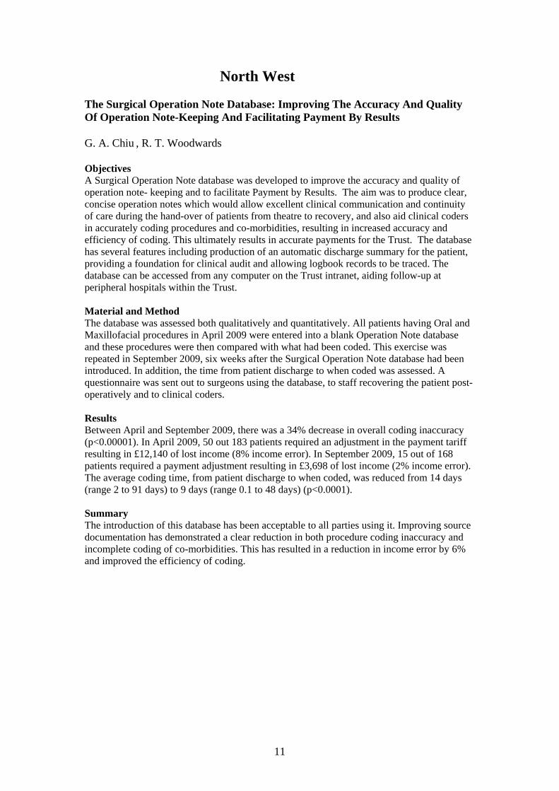
North West The Surgical Operation Note Database: Improving The Accuracy And Quality Of Operation Note-Keeping And Facilitating Payment By Results G. A. Chiu , R. T. Woodwards Objectives A Surgical Operation Note database was developed to improve the accuracy and quality of operation note- keeping and to facilitate Payment by Results. The aim was to produce clear, concise operation notes which would allow excellent clinical communication and continuity of care during the hand-over of patients from theatre to recovery, and also aid clinical coders in accurately coding procedures and co-morbidities, resulting in increased accuracy and efficiency of coding. This ultimately results in accurate payments for the Trust. The database has several features including production of an automatic discharge summary for the patient, providing a foundation for clinical audit and allowing logbook records to be traced. The database can be accessed from any computer on the Trust intranet, aiding follow-up at peripheral hospitals within the Trust. Material and Method The database was assessed both qualitatively and quantitatively. All patients having Oral and Maxillofacial procedures in April 2009 were entered into a blank Operation Note database and these procedures were then compared with what had been coded. This exercise was repeated in September 2009, six weeks after the Surgical Operation Note database had been introduced. In addition, the time from patient discharge to when coded was assessed. A questionnaire was sent out to surgeons using the database, to staff recovering the patient post-operatively and to clinical coders. Results Between April and September 2009, there was a 34% decrease in overall coding inaccuracy (p<0.00001). In April 2009, 50 out 183 patients required an adjustment in the payment tariff resulting in £12,140 of lost income (8% income error). In September 2009, 15 out of 168 patients required a payment adjustment resulting in £3,698 of lost income (2% income error). The average coding time, from patient discharge to when coded, was reduced from 14 days (range 2 to 91 days) to 9 days (range 0.1 to 48 days) (p<0.0001). Summary The introduction of this database has been acceptable to all parties using it. Improving source documentation has demonstrated a clear reduction in both procedure coding inaccuracy and incomplete coding of co-morbidities. This has resulted in a reduction in income error by 6% and improved the efficiency of coding.
11

North West Thames Blood glucose levels of patients admitted with odontogenic infections Farhan janjua, Haroon khan Background Odontogenic infections are common occurrences in most maxillofacial units. Patients with diabetes usually have a more severe clinical course and have problems with glucose control during their acute infections. They may require an increase in their current therapy, and/or review by the diabetologists. Aim The aim of the audit was to look at a cohort of patients admitted to the department with odontogenic infections over a 3 month period (retrospectively) and check if routine blood glucose was being measured. Results A total of 33 patients were identified, with 18% having pre-existing diabetes. 55% of patients did not have their blood glucose monitored during their admission. Of those patients who did have readings that were abnormal, no mention was made in there discharge summaries. Conclusion The results were highlighted to A/E and nursing staff and our junior doctors and changes were implemented to improve blood glucose monitoring. The Audit will be re-visited in the near future with the standard of having all our patients undergoing BM monitoring during their admission. Clinical Audit to assess the incidence of nerve injury during surgical removal of lower wisdom teeth . Ushita shah, Manas Mishra Extraction of wisdom teeth is one of the most common surgical procedures undertaken in the UK. Our audit is designed to check the incidence of nerve injuries during removal of lower wisdom teeth. The most important factor that can be used to assess the risk of nerve injury is the radiographic relationship of the root apices of the lower wisdom teeth to the ID canal. The current quoted incidence of nerve injury is as follows: Injury to the inferior alveolar nerve:
• Frequency of immediate post-operative dysaesthesia after wisdom tooth extraction: 0.4-5.5 %
• Frequency of permanent dysaesthesia after wisdom tooth extraction: 0-2.2 %).
Injury to the lingual nerve: • frequency of immediate post-operative sensory disturbances related to wisdom tooth
extraction: 0.06-11.5 • frequency of permanent sensory disturbances related to wisdom tooth extraction:
0-2 %
Aim The aim of the was to audit the incidence of ID nerve and lingual nerve damage during lower wisdom teeth extractions. To investigate factors leading to ID nerve and lingual nerve damage during lower wisdom teeth extractions.
12

A randomised prospective audit of the first 108 patients who had their wisdom teeth removed under a general anaesthetic this year was carried out. The OPT were assessed prior to the procedure to determine the type of impaction and the relationship of the root apices of the wisdom teeth to the ID canal. The details of the surgical procedures involved including the need for lingual retraction was also recorded . All the procedures were carried out by the same surgeon. Post-operative review was carried out two weeks following the procedures. The patients were telephoned and asked if they had developed any numbness of the lower lip, chin and/or tongue, and whether this was on the right or left side. This was recorded and any patients who reported numbness were telephoned 2 months following the first review to determine if the numbness was persistent. All the post-operative reviews were carried out by the same investigator, who did not operate on the patients. The investigator was not aware of the site (left or right) of the wisdom tooth extraction, the difficulty of extraction, the relationship to the ID nerve, the surgical procedure or any other factors concerning the patient or the tooth. Results Overall the total incidence of temporary numbness was 3% and 0% for permanent numbness. Conclusion Our clinical audit showed that, overall excellent results are being achieved by our department and the incidence of nerve damage following lower wisdom teeth extractions is minimal. However, it is important to continually carry out clinical audits to ensure that the high standards achieved are not only maintained, but are also continually improved upon. Future audits/ongoing Maxillofacial Trauma Audit The primary aims are to look at,
• the length of time patients wait for surgical intervention, • the types (trauma list, CEPOD) and location (hub or spoke hospital) of the theatre that
the patient is operated on, • the surgical experience of the operating surgeon.
It will also include basic social demographic data about the patients and trauma. There are two additional questions. The first on the suspicion of “non accidental injuries” in children. The second on the availability of imaging, which is a common problem when dealing with hub and spoke systems. The Audit will be carried out prospectively.
13

Oxford Complications of Orthognathic Surgery – Regional Audit This was a retrospective Regional re-audit involving 3 centres, of which 2 had been involved in the previous audit. The aim was to assess complications experienced by orthognathic patients and to assess the quality of post-operative records, to ensure all complications were well documented and to compare to previous results. Inclusion criteria were orthognathic surgery patients between July 2006 to October 2009 treated at Reading, Oxford or Buckinghamshire. Patients with craniofacial and cleft anomalies were excluded, as were patients who had genioplasty only or SARPE only. Data Capture The audit looked at pre-surgical, surgical and post-surgical phases and used case notes to help with data collection. Results 24 patients were identified from Reading, 63 from Oxford and 43 from Buckinghamshire. In Reading the pre-surgical orthodontics took 35 months with the number of weeks between pre-surgical records and surgery being 7 weeks. 75% of the patients had had pre-molars removed prior to surgery. Intra-operative complications occurred in only 1 patient with an unfavourable split (4%). Post-operative complications in this group of patients included significant bleeding in 1 patient (4%) and return to theatre in 2 patients (8%). The occlusion at 6 weeks post-operatively included centre line discrepancy in 1 patient (4%) an open bite in 3 patients (12.5%) and a cross bite in 3 patients (12.5%). The average for post-surgical orthodontics was 9 months. Numbness and paraesthesia at 6 weeks post-op was noted in 58% of patients, with 36 confirming an improvement. 3 patients (12.5%) had incomplete records and 1 patient (4%) had numbness at his last post-operative visit. The pre-surgical orthodontic phase in Oxford was 31 months and in Buckinghamshire 19 months. The average number of weeks between pre-surgical records and surgery in Oxford was 4 weeks and in Buckinghamshire was 9 weeks. Third molar removal prior to surgery in Oxford was 27% and in Buckinghamshire was 19%. Intra-operative complications included debonded brackets in 2 patients in Oxford (3%) and 4 patients in Buckinghamshire (9%) with unfavourable splits in 6 patients in Oxford (9%) and 3 patients in Buckinghamshire (7%). IMF was required in 2 patients in Oxford (3%) and 5 patients in Buckinghamshire (11%). In terms of surgical complications, there were 2 patient with significant bleeding from Oxford (3%) and 1 from Buckinghamshire (2%). There was infection in 1 patient from Oxford (2%) and 4 patients in Buckinghamshire (9%). The average post-surgical orthodontic period for both Buckinghamshire and Oxford was 6 months. The occlusal outcomes at the 6 week review appointment included centre line discrepancy in 5 patients in Oxford (8%), 7 patients in Buckinghamshire (16%), open bite 14 patients in Oxford (22%), 3 patients in Buckinghamshire (7%). There was a cross bite in 4 patients in Oxford (6%) and 1 patient in Buckinghamshire (2%). In terms of numbness, at the 6 week review, 30% of patients in Oxford reported numbness with 10% reporting it as very slight. In Buckinghamshire there was 28%. There were incomplete records for 6 patients in Oxford (9%) and 2 patients in Buckinghamshire (5%). At the last review appointment 4 patients in Oxford still complained of numbness (6%) and 3 in Buckinghamshire (7%). Previous study In the previous study 48 procedures had been carried out in a 12 months period and 70% of the surgery was bimaxillary. 21% had had third molars removed pre-operatively and the mean pre-surgical orthodontic phase had been 27 months. 8% of patients had had debonded brackets. IMF had been needed in 6% of patients and 10% had significant numbness at 6
14

weeks. Occlusal discrepancy included centre lines being wrong in 15%, open bite wrong in 23% and cross bite present in 10%. The post-surgical orthodontic phase had been 5 months and incomplete records had been found in 31% of patients. Comparison with previous audit The previous audit had only looked at Buckinghamshire and Oxfordshire, so Reading has been added as a new unit. Compared with the previous audit, 106 procedures had been performed in Buckinghamshire and Oxfordshire, compared to 48 in the previous study. In addition, 24 had been carried out on Reading patients. In the past, 70% of procedures had been by bimaxillary osteotomy but now 54% were by bimaxillary osteotomy. The third molar removal rate was similar in both studies, as was the mean pre-surgical orthodontic phase. The pre-surgical orthodontic phase for Reading was high at 35 months. The debonded rate was similar as was the unfavourable split rate at 6% in 2004 and 8% in the new study. Conclusions and Recommendations These figures are appropriate in comparison to other Nationally reported figures in the literature. We should advise patients that orthognathic treatment is taking approximately 30 months. We need to improve record keeping and standardise immediate occlusal summary post-op. Complete data collection should follow BAOMS guidelines. Plate Removal Following Fixation of Mandibular Fractures Aim To compare the plate removal rate of mandibular fractures treated at the John Radcliffe Hospital in Oxford and with figures available in literature (Cardiff 7.3%, Birmingham 10.3%, Vienna Austria 6.3%). Method All patients admitted for mandibular fractures between 1st March and 31st September 2009 and received operative intervention were identified by the computerised theatre system. The general notes were reviewed to check co-morbidity and whether plates were removed. The minimum follow-up period was 11 months with a maximum of 17 months. It was felt that this period of review would be adequate since a previous study in Birmingham had shown that most complications occur within the first year. For each patient their basic demographic details were collected, as well as the type of trauma, the area of mandibular fracture, the area of plating and the number of plates. Any co-morbidities were noted, any plates removed were noted and the reason for their removal. Results 77 consecutive patients were identified with 162 fractures treated with open reduction internal fixation. 47% had been admitted through the Royal Berkshire Hospital, 32% through the John Radcliffe Hospital, 9% through Stoke Mandeville Hospital, 4% through High Wycombe Hospital and 8% through the other hospitals via the neurosurgical maxillofacial craniofacial acceptance criteria. The most commonly treated fracture site was the angle of the mandible, closely followed by the parasymphysis and then the body. Although condyle fractures were common, many were treated conservatively. The causes of trauma include RTA, sports injuries and falls, with 62% being due to assault. Complications relating to plate removal were found in 6 patients. In all patients, at the time of plate removal there was pain and loosening of the plate with obvious infection in 3 patients. Conclusion Our plate removal rate of 7.8% is comparable to other studies, with plate removal rates of 6.3% to 10.3% but clearly could be improved. In the 6 cases of plate removal, 4 of the 6
15

patients had smoking habits and 1 patient had diabetes. 3 of the 6 patients had plates removed near dubious teeth, which were extracted at the time of plate removal. The age of fracture (days after injury) does not seem to correlate to future fixation failure and delays in operating were acceptable, with one particular outlier. Additional morbidity was not collected but there was documentation showing persistent paraesthesia in some cases and one case of marginal mandibular nerve weakness.
Scotland Audit activity across Scottish OMFS units is presented at SOMS, the annual meeting of the Scottish Oral & Maxillofacial Society. This year’s meeting was in Stirling and had many audits presented. In addition to the annual meeting local units have regular clinical governance meetings throughout the year to Topics covered related to cleft lip and palate, trauma, oncology, orthognathic surgery education and social issues. A survey of Midwives’ awareness of cleft lip and palate and associated problems across Scotland. E Chalmers(Oral Surgery, SHO), V Sood (Oral and Maxillofacial Surgery, Registrar), M Devlin(Cleft and Maxillofacial Surgery, Consultant) , A Crawford (Cleft Nurse Specialist), S Wallace (Cleft Nurse Specialist), A Ray Aim To assess Midwives’ experiences and knowledge of cleft lip and or palate, confidence counselling families and to identify areas for potential input from The Cleft Team. Method Questionnaires were sent to maternity units throughout Scotland to assess; midwifes experience of delivering babies with cleft lip and or palate (CL+/-P), knowledge, confidence in supporting the families and in giving feeding advice. The midwives were also asked what input they would like their Cleft Team to provide. Results Results from the study (n=206) were as follows; 41% of the midwives had delivered a baby with CL+/-P. 23% knew the incidence of CL+/-P. 33% were aware of the stages in treating CL+/- P. 99% knew feeding difficulties is a potential complication. When asked about counseling the families 70% were not confident and 60% were not confident at giving feeding advice. 65% would like a training day, 45% small group teaching, 5% would like a website and 57% would like printed literature. Conclusion Our recommendations are to implement training days to undergraduate and graduate midwives along with supporting websites.
16

South Thames South Thames regional audit summary 2010 Units: Chichester, Guildford, East Grinstead, St Georges, Brighton/Eastbourne, Canterbury 2 Regional based audits were run in 2010 Interval time surgery to radiotherapy in head and neck cancer patients
(host unit - St Georges - May 2010) Blood transfusion in head and neck cancer
(host unit - Chichester - November 2010) Radiotherapy Interval This session started with an excellent lecture from Dr Kate Newbold (oncologist Marsden) explaining the increased efficacy for post op RT commencing within 6 weeks of surgery. This was followed by the results of a first stage audit looking at data collected from 5 participating units. 80 patients met the inclusion criteria (surgery + RT), the majority of these cases were SCCs from a variety of sites. 69 % involved mucosal primary. 42/80 patients received their radiotherapy within 6 weeks of surgery. Of those patients delayed over 8 weeks the reasons recorded were: Post operative medical complications (n=1), Long in-patient stay (n=1), Cancelled appointments (n=3), Flap failure (n=1), Administrative delay (n=5), Insufficient planning & treatment capacity (n=7) MBOS This session started with an excellent lecture from Ruth O’Donnell (transfusion practitioner) on the process, costs and hazards of transfusion. This was followed by the first stage audit looking at data from 6 participating units. Inclusion criteria looked at crossmatch for 20 head and neck oncology patients from each of the 6 units. In neck dissection a total of 18 units were cross matched for 13 patients - no patients had transfusion. In neck dissection + resection + soft tissue free flap 145 units were cross matched and only 36 transfused. Most units did not have an MBOS specific for head and neck surgery. The recommendation of the audit is: Neck Dissection: Group and Save only Tongue Resection +/- Neck Dissection: Group and Save only. Resection + Neck Dissection + Soft Tissue Free Flap: Crossmatch 2 units This has been circulated to transfusion departments at all units involved and it is our intention to carry out a second stage to the audit within 2 years.
South West The focus in the SW during the last year has been clefts, in that PJR was involved in completing the national outcome study on alveolar bone grafts. this was published in December 2010 ( Alveolar Bone Grafting: Results of a National Outcome Study.Revington P., Deacon, S. Shah, H, Macnamara C & Pereira E. Ann R Coll Surg Engl 2010; 92:643-686). We have in addition developed a questionnaire in conjucntion with Nick Baker, in an attempt to address the frquency of serious complications associated with orthognathic surgery. This is currently being piloted in the SW, to assess its suitabilty for wider usage.
17
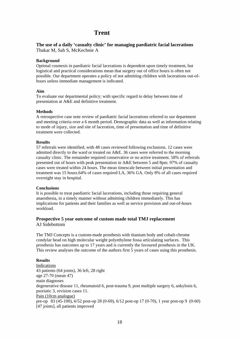
Trent The use of a daily ‘casualty clinic’ for managing paediatric facial lacerations Thakar M, Sah S, McKechnie A Background Optimal cosmesis in paediatric facial lacerations is dependent upon timely treatment, but logistical and practical considerations mean that surgery out of office hours is often not possible. Our department operates a policy of not admitting children with lacerations out-of-hours unless immediate management is indicated. Aim To evaluate our departmental policy; with specific regard to delay between time of presentation at A&E and definitive treatment. Methods A retrospective case note review of paediatric facial lacerations referred to our department and meeting criteria over a 6 month period. Demographic data as well as information relating to mode of injury, size and site of laceration, time of presentation and time of definitive treatment were collected. Results 57 referrals were identified, with 48 cases reviewed following exclusions. 12 cases were admitted directly to the ward or treated on A&E. 36 cases were referred to the morning casualty clinic. The remainder required conservative or no active treatment. 58% of referrals presented out of hours with peak presentation in A&E between 5 and 8pm. 97% of casualty cases were treated within 24 hours. The mean timescale between initial presentation and treatment was 15 hours.64% of cases required LA, 36% GA. Only 8% of all cases required overnight stay in hospital. Conclusions It is possible to treat paediatric facial lacerations, including those requiring general anaesthesia, in a timely manner without admitting children immediately. This has implications for patients and their families as well as service provision and out-of-hours workload. Prospective 5 year outcome of custom made total TMJ replacement AJ Sidebottom The TMJ Concepts is a custom-made prosthesis with titanium body and cobalt-chrome condylar head on high molecular weight polyethylene fossa articulating surfaces. This prosthesis has outcomes up to 17 years and is currently the favoured prosthesis in the UK. This review analyses the outcome of the authors first 5 years of cases using this prosthesis. Results Indications 43 patients (64 joints), 36 left, 28 right age 27-70 (mean 47) main diagnoses degenerative disease 11, rheumatoid 6, post-trauma 9, post multiple surgery 6, ankylosis 6, psoriatic 3, revision cases 11. Pain (10cm analogue) pre-op 83 (45-100), 6/52 post-op 28 (0-69), 6/12 post-op 17 (0-70), 1 year post-op 9 (0-60) [47 joints], all patients improved
18

Opening (mm) Pre-op 24 (3-44)mm , 6/52 post-op [42 patients] 27 (15-41)mm, 6 months [35 patients] 31 (15-43)mm, 1 year post-op [31 patients] 32 (15-45)mm All patients with reduced opening showed improvement. Dietary scores (10cm analogue liquid to solid) Pre-op 41 (1-100), Post-op 1 year 93 (25-100) All patients improved. Revisions Case 1. Acute dental infection 3 days post-op. Dentist delayed treatment for 6 weeks. Biofilm infection of the prosthesis required joint removal and placement of gentamicin impregnated acrylic spacer for 3 months. Now 3 years post revision prosthesis with no ongoing problems. Case 2. 6 weeks post-op developed otitis externa with secondary cellulitis of face and secondary joint infection. Joint removed, awaiting replacement as gets recurrent otitis. Conclusion TMJ replacement provides an excellent mode of reconstruction of the irreparably damaged TMJ. Other audits Claire Barrett: Patient Compliance with Pre-operative Sedation Instructions Mr Tom Barry: Temporal Artery Biopsy: A New Service: 1 Year on. David Houghton: The Outcome of the first 43 Total TMJ replacements P Hole: Reliability in microvascular surgery: is quality dependent on quantity? M Thakar: The use of a daily ‘casualty clinic’ for managing paediatric facial lacerations Mr Tom Barry: Audit of the Chesterfield/Rotherham Unit of North Trent Cancer Network Khaled Borghol: Radial Forearm free flap surgery: A survey of the practice Pankaj Taneja: Use of X-Rays post ORIF of Fractured mandibles Dyar Gahir: PEG’s at Leicester Mr Tom Barry: Audit of Head and Neck Ultrasound since the set-up of the 'One Stop' Lump Clinic David Houghton: Documentation of Facial Nerve Morbidity following Parotidectomy -a 5 year retrospective audit of 53 cases Gozie Idaboh: A waiting time audit on A and E referrals to the Oral and Maxillofacial Surgery Unit Sunil Sah: Management of oral cancer by laser surgery: relation between rate of recurrence and histological features Dr Rob McCann: Deep Neck Space Infections Pankaj Taneja: Audit of Use of Leeches Sunil Sah: Management of oral cancer by laser surgery: relation between rate of recurrence and histological features Nabeela Ahmed : A Six Year Post-Operative Review of Secondary Alveolar Bone Grafting in the Trent Region Mark Buah : Clinical Aptitude of Maxillofacial SHO’s
19

Wessex A regional Audit pooling data from 6 units (Chichester/ Portsmouth, Brighton, Guildford, East Grinstead, St Georges and Kent and Canterbury Tom Aldridge , Stephen Walsh Aim To look at blood transfusion requirements in 120 patients (each unit provided the data on 20 patients) who underwent a substantial head and neck surgical oncology procedure. Results We crossmatched (as a region) 266 units and transfused only 72 ( a ratio of 3.7:1 - Ideal is 2:1) – returning 192 units unused. Only 24% of our group of patients were transfused. No department had an MBOS (Maximum Blood Ordering Schedule) specifically for Oral & Maxillofacial Surgery. No patient who had a neck dissection had a blood transfusion and the following recommendations were made: Neck Dissection: Group and Save only. Tongue Resection +/‐ Neck Dissection: Group and Save only. Resection + Neck Dissection + Soft Tissue Free Flap: Crossmatch 2 units. Bony free flap surgery was excluded for comment as there were too many variables in the group and the numbers relatively small. We also agreed to feed the above recommendations into each hospitals transfusion committee so that an official MBOS could be agreed upon for maxillofacial surgery. National Antibiotic Audit Mr Mark Singh, Mr Gerard Surendakumar,
Introduction Currently there is increasing pressure on clinicians to ensure shorter hospital inpatient stays with less morbidity. One of the driving pressures includes the financial impact of long stays and the treatment of complications in hospital. Monitoring of performance is also an essential tool in modern NHS service provision and thus within the field of Oral and Maxillofacial Surgery (OMFS) the use of adjuncts to improve such factors need to be considered. Plowman1 et al in 2000 estimated the cost of a hospital acquired infection to be in the region of £3000. We embarked on looking at the use of prophylactic antibiotics in OMFS departments across the country. Prophylactic antibiotic treatment is defined as the use of antibiotics before, during or after a diagnostic, therapeutic, or surgical procedure to prevent infectious complications Method Postal surveys were sent to 100 OMFS units throughout the country. We had a 56% response rate. No reminders were sent and the results were analysed. Results Pre-operatively most units prescribed antibiotics for Trauma (hard tissue) 90%, Oncology 85% and Orthognathic 75%. Post-operatively most units followed this prescribing practice. Prescribing for skin and aesthetics was less than 25%. The most common duration of
20

prescribing was an induction dose. However this was by less than half of our responders. This was followed by 5 day courses of antibiotics. Few gave 2 post op doses that was the third most common. The drug of choice was co-amoxiclav, followed by metronidazole then cefuroxime. The biggest influence on choice of regime was the type of procedure followed by microbiology input. Discussion Scottish Intercollegiate Guidelines Network published the first guide in 2000 to provide evidence based recommendations to reduce inappropriate prophylactic antibiotic prescribing2. The recommendations include – Antibiotic to cover expected pathogens (Staphylococcus and Streptococcus Sp.). Intravenous route <30 minutes before skin incised and single therapeutic doses within first 24 hours. Clinical Practise Outcome We used to prescribe cefuroxime and metronidazole for 5 days for major cases. We now use IV Augmentin at induction We consider high-risk patients for longer courses (5 days) Use for trauma, oncology and orthognathic not dentoalveolar Consider auditing our post operative infection rates Obtain a better response rate using a telephone questionnaire References
1. Plowman R et al. The socioeconomic burden of hospital acquired infections. Public health laboratory service. 2000
2. SIGN Guidelines. Antibiotic prophylaxis in Surgery. 2000
West Midlands Pattern of In-patients referrals to maxillofacial speciality E .Beshara , T. Hassan , J.Mathur , Mr. S. Parmar Mr.T. Martin Introduction The Maxillofacial speciality has offered the service of reviewing patients admitted under other specialities when they require dental assessment for different reasons i.e. undergoing Cardiac Surgery or Organ transplant, prior to Chemotherapy or Radiotherapy, Oro-facial infections develops during hospital stay, endocarditis and brain abscess. Aims To assess the pattern of in-patients referrals to the Oral and Maxillofacial speciality and determine the sources of referrals, types of problems referred the level of urgency and efficiency of management of these referrals. Methods Prospective study from September 2009 to August 2010 at Queen Elizabeth and Selly Oak hospitals. A gold standard was set that only appropriate referrals should be made referrals should be dealt with efficiently.Audit proformas were filled by the on-call SHO when referrals were received. Trauma cases were excluded. 17 interviews with SHOs and SPRs were carried out at the end of the audit process to find out any concerns with providing this service.
21

Results All the referrals received over the phone and then transferred to the proforma. The quality of data given over the phone was clear for 52 patients, however( 6 )referrals were judged to be unclear.Information was available for 63 patients of which male to female ratio 3:2. Referring departments included Cardiology (n=16), Neurosurgery (n=7), Oncology (n=7), ITU and anaesthetics(n=6), Medical team (n=5), Liver (n=3) and others.The most common reasons of referrals were dental assessment (42), facial swellings (10)and mouth lesions(8). Although 13 patients requested to be seen urgently, 31 to be seen soon , yet most of these referrals 8, 21 respectively only required dental assessment. However the rest of these referrals included 6 facial swellings, 5 mouth lesions , 2 dislodged crowns and 1 avulsed tooth.Further analysis of the diagnosis of those who referred for dental assessment, showed majority of patients to have chronic asymptomatic dental problems i.e. carious teeth and remaining roots (14), chronic periodontitis and teeth mobility(4) or even no abnormality for 6 patients including (2) fully edentulous patients . The interviews with the (12) SHO and (6)SPRs heighted the difficulty of examining patients on the ward because of the lack of light, suction and equipments. The majority (9) of junior staff preferred to have a second opinion from a senior members most of the times, as there are no guidelines available to specify the dental treatment required for Cardiology patients prior to their surgery and also due to their complex medical history . Conclusion Reviewing in-patients with acute dental conditions should take place in Maxillofacial outpatients clinics, where the equipment and senior advice are available. However none acute cases should be seen prospectively by general dental practitioners Regional and national protocols need to be developed to clarify the necessary intervention required for the cardiology and Oncology patients prior to surgery, radiotherapy and chemotherapy. Auditing cross matching in head and neck cancer patients. Beshara E, Zhao J, Tanday AK , Parmar S, Martin T Aim To audit cross-matching for head and neck cancer patients undergoing surgery in order to reduce unnecessary cross-matching and associated costs. Method Retrospective analysis of cross matching and blood transfusion for the head and neck cancer patients at Queen Elizabeth hospital was done using theatre logs, the head and neck cancer database, and blood bank records. Numbers of units of blood used were compared to those that were cross-matched. Gold standards of the number of units to be cross-matched were then set after discussion with the anaesthetists and the surgical teams. Junior medical staff were advised to only cross-match patients undergoing bilateral neck dissection or composite flaps reconstruction for 4 units of blood but otherwise grouping and saving for unilateral neck dissectionwith or without soft tissue flaps reconstruction. A further comparison of cross-matching practice to the gold standards then took place. Results The first stage included 74 patients who underwent complex surgery between April 2007 to October 2008 compared to 36 patients that were operated between April 2009 to December 2009. In the first stage all the patients 100% were cross-matched for 563 blood units but only 31% of these patients had transfusion of 197 blood units (35% of the total cross-matched units), compared to 26 patients 72% in the second stage were cross-matched for 142 units and only
22

33% of these patients required transfusion of 35 blood units 24% of the total cross-matched blood units). The patients who underwent unilateral neck dissection(UND) (n38) with soft tissue flaps (n26) Radial Forearm free flaps (RFFF), (n2) Anterolateral Thigh flaps (ALT) in the first stage were compared to (n31)(UND), (n17) (RFFF) in the second stage.100% of this group were cross matched in the first stage but only 23% required blood transfusion compared to 47% cross matching and 12% transfusion respectively in the second stage. The compliance of the junior team to the new protocol for this group was 41%. On the other hand 38 patients underwent bony resection and or bony flap reconstruction(n22) in the first stage were compared against 14 patients out of which 11 had bony flaps in the second stage. 51% in the first stage and 64% in the second stage required blood transfusion respectively. The majority of patients 72 %in the first stage and 81% in the second stage respectively who needed transfusion did not require more than 2 units of blood. An improvement was noticed in the number of patients having had more than 4 units cross-matched in that it dropped from 34% in the first stage to 13% in the second stage, as well the habit of cross-matching unilateral neck dissection and soft tissue flaps from 100% to 47%. Conclusion Routine pre operative cross matching of blood is unnecessary for certain head and neck cancer procedures. The changes we implemented in cross-matching proved to be effective. Old New
Pt number (complex surgery) 74 36 (out of 82, rest are
Total x-m 563 142
Units transfused 197 35% 35 24%
% of pt had x-m 100% 72%
% pt needed transfusion 31% 33%
x-m > 4 units 34% 13%
% of < 2 units transfusion 72% 81%
38 , 35 resection, 22 recons pt 14 pt 11 flaps
176 x-m 60 x-m
Bony resection or reconstruction
60 units transfused 20 transfused
100% x-matched 47% Unilateral N.D + S.T
39% transfusion 12%
13 pt no 5 pt no
52 x-m 20 x-m
BILATERAL N.D
27 transfused units 4 transfused units
Overall compliance is 61%
23

Yorkshire Are the correct antibiotics being prescribed to treat dog bite wounds to the head and neck region Introduction NHS statistics indicate the number of people presenting to A & E with dog bites has increased by more than 40% in the last 4 years to nearly 3,800 per year. Dog bite wounds require specific antibiotic treatment to allow satisfactory healing and prevent infection and there are guidelines for the management of these injuries: The BNF (2009) states for dog bites drug protocol should include:
• Thorough cleaning of wound • Tetanus immunoglobulin depending on immunisation history and risk of infection • Co-amoxiclav alone • OR doxycycline and metronidazole if penicillin-allergic
Aim To audit the management of patients with dog bite injuries to the head and neck region attending for treatment at the Mid Yorkshire Hospitals NHS Trust. Method A retrospective audit was conducted to establish if the current guidelines for the management of dog bit injuries to the head and neck region were being followed. All A & E attendances were recorded at three sites over a 6 month period and the results were recorded in a proforma Results Results demonstrated that the majority of patients received thorough wound debridement and were prescribed antibiotics for the treatment of their injuries. Tetanus status was established in 80 % of patients but only 78 % of patients were prescribed the antibiotics suggested by current guidelines. Discussion Our experience is suggestive of an increased incidence of dog bit injuries to the head and neck region. There are national guidelines for the management of these injuries but experience in our unit suggested that a minority of patients were not prescribed the correct prophylactic antibiotic regimen. It is essential that all hospital staff are made aware of current protocols in order to optimise patient care. Surgical Clearance margins of basal cell carcinomas of the head and neck Background Rates of the incomplete surgical excision of basal cell carcinoma (BCC) has been reported at rates ranging from 4% to 16.6%. Objective The objective was to determine the clearance rates of BCC’s surgically removed from the head and neck. This was a prospective study of 200 consecutive BCC’s excised from patients treated under Mr Telfer’s care, between March and August 2008, at both York and Harrogate district
24

hospitals. Exclusions were recurrent BCC’s or the removal of previously incompletely excised BCC’s Results Age, sex, BCC location and size, method of repair, involvement of lateral or deep surgical margins and measurement of clearance were collected on a pro-forma then analysed. The mean age of the 176 patients ( 114 male, 62 female) was 76 years (range 40 to 97) Location of the BCC’s was; nose 20%, cheek 16%, forehead 15%, temple 15%, scalp 8%, ear 8%, lip 6%, neck 6% and medial canthus 5%. Reconstructions carried out were flap repair 65%, full thickness skin graft 34%, and primary closure 1%. Complete excisions occurred in 96.5% of BCC’s. Of the incomplete excisions, 4 were positive lateral margins, 1 positive deep margin and 2 cases both lateral and deep positive margins. Conclusion This reported histological clearance rate compares extremely favourably to that of other studies.
25
Related Documents