Original Article J Clin Med Res • 2011;3(3):124-131 ress Elmer Articles © The authors | Journal compilation © J Clin Med Res and Elmer Press™ | www.jocmr.org Clinical Comparison of Conventional Testicular Sperm Extraction and Microdissection Techniques for Non-Obstructive Azoospermia Ibrahim Fathi Ghalayini a, d , Mohammed A. Al-Ghazo a , Osama Bani Hani a , Rami Al-Azab a , Ibrahim Bani-Hani a , Faheem Zayed b , Yazan Haddad c Abstract Background: We compared the efficacy of microdissection tes- ticular sperm extraction (microdissection TESE) and conventional TESE in patients with non-obstructive azoospermia (NOA) and related the positive sperm recovery to certain variables: follicle- stimulating hormone (FSH) and luteinizing hormone (LH) levels, testicular volume and histology. Methods: Sperm retrieval rates (SRR) in patients with NOA who underwent microdissection TESE (n = 65) or conventional TESE (n = 68) were compared and related to the different variables. Results: SRR by microdissection TESE (56.9%) was significantly higher than conventional TESE (38.2%). There was a positive rela- tion between the SRR and increased testicular volume or decreased FSH levels. No effect of Testosterone or Prolactin levels on SRR by using either technique was observed. Sperm were recovered from those with hypospermatogenesis in 84% and 92.9% by convention- al and microdissection TESE, respectively (P = 0.3). In cases of maturation arrest the SRR was 27.3% and 36.4%, respectively (P = 0.6). In cases of Sertoli-cell-only syndrome (SCOS) the SRR was 6.2% and 26.9%, respectively (P = 0.03). No major operative com- plications occurred in any patient in either group, and no patient re- quired post-operative hormone replacement to treat hypogonadism. Conclusions: Microdissection TESE significantly had twice bet- ter probability of success of SRR when compared to conventional TESE. No secure pre-operative prognostic elements of sperm re- covery exist for NOA patients. Microdissection TESE appears to be recommendable in cases of atrophied testicles, high FSH concentra- tion, or when SCOS with high FSH concentration can be predicted. Keywords: Microdissection TESE; Sperm retrieval; Non-obstruc- tive azoospermia; Histopathology; FSH concentration; Orchidom- etry Introduction Non-obstructive azoospermia (NOA) refers to absence of spermatozoa in semen analysis due to minimal or no produc- tion of fully developed spermatozoa in the testicles. Etiolo- gies for testicular failure include genetic disorders such as sexual chromosomal abnormalities, translocations and mi- crodeletions of the Y chromosome, cryptorchidism, testicu- lar torsion, radiation and toxins [1-4]. Approximately 1% of all men and 10% of infertile men are affected by testicular failure as a result of NOA [5]. Tes- ticular spermatozoa can be retrieved in some NOA men de- spite the absence of ejaculated spermatozoa in their semen, because of the existence of isolated foci of active spermato- genesis. Testicular spermatozoa can be retrieved successful- ly by the testicular sperm extraction (TESE) procedure and used for intracytoplasmic sperm injection (ICSI) in cases of NOA [6]. Such cases used to be treated with conventional TESE, including multiple biopsy samples of the testis. At present, in many centers this treatment has been replaced by microdissection TESE. Direct vision with the operating microscope in microdis- section TESE is of great advantage as larger, more opaque, whitish tubules, presumably containing more germ cells with active spermatogenesis, can be identified. This procedure is currently the best method for the certain identification of sperm, resulting in a high spermatozoa retrieval rate (SRR) and minimal postoperative complications. Histological find- ings are important in any comparison, since a relationship between SRR and testicular histopathology has been report- ed in the context of conventional TESE [5, 7, 8]. Manuscript accepted for publication March 29, 2011 a Urology Division, King Abdullah University Hospital, Jordan University of Science and Technology, Jordan b Department of Obstetrics and Gynecology, King Abdullah University Hospital, Jordan University of Science and Technology, Jordan c Princess Haya Biotechnology Center, Jordan University of Science and Technology, Jordan d Corresponding author: Ibrahim Fathi Ghalayini, P.O. Box 940165, Amman 11194, Jordan. Email: [email protected] doi:10.4021/jocmr542w 124

Welcome message from author
This document is posted to help you gain knowledge. Please leave a comment to let me know what you think about it! Share it to your friends and learn new things together.
Transcript
Original Article J Clin Med Res • 2011;3(3):124-131
ressElmer
Articles © The authors | Journal compilation © J Clin Med Res and Elmer Press™ | www.jocmr.org
Clinical Comparison of Conventional Testicular Sperm Extraction and Microdissection Techniques for
Non-Obstructive AzoospermiaIbrahim Fathi Ghalayinia, d, Mohammed A. Al-Ghazoa, Osama Bani Hania,
Rami Al-Azaba, Ibrahim Bani-Hania, Faheem Zayedb, Yazan Haddadc
Abstract
Background: We compared the efficacy of microdissection tes-ticular sperm extraction (microdissection TESE) and conventional TESE in patients with non-obstructive azoospermia (NOA) and related the positive sperm recovery to certain variables: follicle-stimulating hormone (FSH) and luteinizing hormone (LH) levels, testicular volume and histology.
Methods: Sperm retrieval rates (SRR) in patients with NOA who underwent microdissection TESE (n = 65) or conventional TESE (n = 68) were compared and related to the different variables.
Results: SRR by microdissection TESE (56.9%) was significantly higher than conventional TESE (38.2%). There was a positive rela-tion between the SRR and increased testicular volume or decreased FSH levels. No effect of Testosterone or Prolactin levels on SRR by using either technique was observed. Sperm were recovered from those with hypospermatogenesis in 84% and 92.9% by convention-al and microdissection TESE, respectively (P = 0.3). In cases of maturation arrest the SRR was 27.3% and 36.4%, respectively (P = 0.6). In cases of Sertoli-cell-only syndrome (SCOS) the SRR was 6.2% and 26.9%, respectively (P = 0.03). No major operative com-plications occurred in any patient in either group, and no patient re-quired post-operative hormone replacement to treat hypogonadism.
Conclusions: Microdissection TESE significantly had twice bet-ter probability of success of SRR when compared to conventional
TESE. No secure pre-operative prognostic elements of sperm re-covery exist for NOA patients. Microdissection TESE appears to be recommendable in cases of atrophied testicles, high FSH concentra-tion, or when SCOS with high FSH concentration can be predicted.
Keywords: Microdissection TESE; Sperm retrieval; Non-obstruc-tive azoospermia; Histopathology; FSH concentration; Orchidom-etry
Introduction
Non-obstructive azoospermia (NOA) refers to absence of spermatozoa in semen analysis due to minimal or no produc-tion of fully developed spermatozoa in the testicles. Etiolo-gies for testicular failure include genetic disorders such as sexual chromosomal abnormalities, translocations and mi-crodeletions of the Y chromosome, cryptorchidism, testicu-lar torsion, radiation and toxins [1-4].
Approximately 1% of all men and 10% of infertile men are affected by testicular failure as a result of NOA [5]. Tes-ticular spermatozoa can be retrieved in some NOA men de-spite the absence of ejaculated spermatozoa in their semen, because of the existence of isolated foci of active spermato-genesis. Testicular spermatozoa can be retrieved successful-ly by the testicular sperm extraction (TESE) procedure and used for intracytoplasmic sperm injection (ICSI) in cases of NOA [6]. Such cases used to be treated with conventional TESE, including multiple biopsy samples of the testis. At present, in many centers this treatment has been replaced by microdissection TESE.
Direct vision with the operating microscope in microdis-section TESE is of great advantage as larger, more opaque, whitish tubules, presumably containing more germ cells with active spermatogenesis, can be identified. This procedure is currently the best method for the certain identification of sperm, resulting in a high spermatozoa retrieval rate (SRR) and minimal postoperative complications. Histological find-ings are important in any comparison, since a relationship between SRR and testicular histopathology has been report-ed in the context of conventional TESE [5, 7, 8].
Manuscript accepted for publication March 29, 2011
aUrology Division, King Abdullah University Hospital, Jordan University of Science and Technology, JordanbDepartment of Obstetrics and Gynecology, King Abdullah University Hospital, Jordan University of Science and Technology, JordancPrincess Haya Biotechnology Center, Jordan University of Science and Technology, JordandCorresponding author: Ibrahim Fathi Ghalayini, P.O. Box 940165, Amman 11194, Jordan. Email: [email protected]
doi:10.4021/jocmr542w
124 125

J Clin Med Res • 2011;3(3):124-131Division et al
Articles © The authors | Journal compilation © J Clin Med Res and Elmer Press™ | www.jocmr.org
In our present study we compared SRR by microdis-section TESE with that obtained by conventional multiple TESE in patients with histological findings including hypo-spermatogenesis, maturation arrest (MA), and Sertoli cell-only syndrome (SCOS). Diagnostic biopsy specimens were reviewed in all cases. As far as is known, there are few stud-ies that compare conventional and microdissection TESE and relate the positive sperm recovery to certain variables, FSH and LH concentrations, testicular volume and testicular histology, which are all clinically relevant for NOA patients. We hope our study would add some information to support the previous reports.
Materials and Methods
Patients
A consecutive series of 133 men referred with a diagnosis of azoospermia were involved in the current study. All patients were diagnosed with NOA on the basis of a complete history, physical examination, endocrine profile, and chromosomal analysis before being scheduled for TESE with sperm freez-ing. Those with abnormal karyotyping were excluded from analysis. All patients underwent ejaculated semen examina-tion at least 3 times before surgery. Patients also underwent careful evaluation by urologists concerning the duration of sterility, medical history, sexual function and results of a gynecologic evaluation of the spouse. Ultrasonography was performed to measure testicular volume and to determine the status of the epididymis and testis. Serum follicle-stim-ulating hormone (FSH) and luteinizing hormone (LH) were measured by immunoradiometric assay, while testosterone was measured by radioimmunoassay. Patients included in the study were allocated, according to the waiting list on the basis of the general operative theatre plan, after informed consent including explanations about results in the literature and invasiveness of the two procedures. Every operated tes-ticle was classified according to the following variables: (i) testicular volume (V), categorized according to volume (≤ 8 ml, 9 - 12 ml, and ≥ 13 ml, i.e., normal); (ii) FSH concentra-tion, categorized into two groups according to multiples of the normal range (N) (N and 2N: 1 - 24 mIU/mL, and > 3N: > 24 mIU/mL); (iii) testicular histology based on the most advanced pattern of spermatogenesis present such as hypo-spermatogenesis, maturation arrest (MA) and Sertoli cell-only syndrome (SCOS). Institutional Review Board (IRB) approval was obtained.
Surgical approach
Conventional TESE
Conventional multiple TESE ordinarily was performed un-
der general or local anesthesia. Through a small vertical inci-sion in the median scrotal raphe, the skin, dartos muscle, and tunica vaginalis were opened to expose the tunica albuginea. The tunica albuginea was incised for about 4 mm at the up-per pole near the head of the epididymis. If no sperm were seen in the initial sample, subsequent samples were taken from other locations, in the middle of the testis and at the lower pole opposite the rete testis, and subsequently from the contralateral testis. The procedure was terminated when sufficient spermatozoa were retrieved.
Microdissection TESE
Microdissection TESE was performed under general anes-thesia according to the procedure reported previously [9, 10]. After the tunica albuginea was opened widely along the antimesenteric border, direct examination of the testicular parenchyma was performed under the operating microscope. An attempt was made to identify individual seminiferous tu-bules that were larger, more opaque and whiter than other tubules in the testicular parenchyma, which were consid-ered to contain spermatozoa. The procedure was terminated when sperm were retrieved or further biopsy was thought likely to jeopardize the blood supply of the testis. If all tu-bules were seen to have an identical morphological appear-ance, at least three samples (upper, middle, and lower) that were equivalent to those from multiple TESE were obtained. The procedure was terminated when a sufficient volume of spermatozoa had been retrieved for ICSI. At the same time of testicular intervention in both procedures, a small tissue specimen was placed in Bouin’s solution and sent for histo-pathological examination.
Sperm retrieval
Each sample was placed in a Petri dish filled with 0.5 ml of sperm wash media (Frederick-Maryland, USA), minced and shredded using sterile glass slides. Then, each sample was examined immediately by placing a small droplet of dispersed tissue suspension on the slide under a phase mi-croscope using x 200 magnification for the presence of the testicular sperm. Further, all testicular samples for ICSI with 5 ml of media were subjected to centrifugation at 2000 x gravity for 10 minutes with careful, extended examination to determine the presence of even a single sperm after TESE procedure.
Statistical analysis
The results were statistically evaluated using Mann-Whitney and Pearson’s Chi Square tests for comparison of the base-line data. A binary logistic regression was used to assess the adjusted odd ratios for the success of sperm retrieval in both methods of treatment. P-values less than 0.05 were consid-
124 125
J Clin Med Res • 2011;3(3):124-131 Conventional and Microdissection Testicular Sperm Extraction
Articles © The authors | Journal compilation © J Clin Med Res and Elmer Press™ | www.jocmr.org
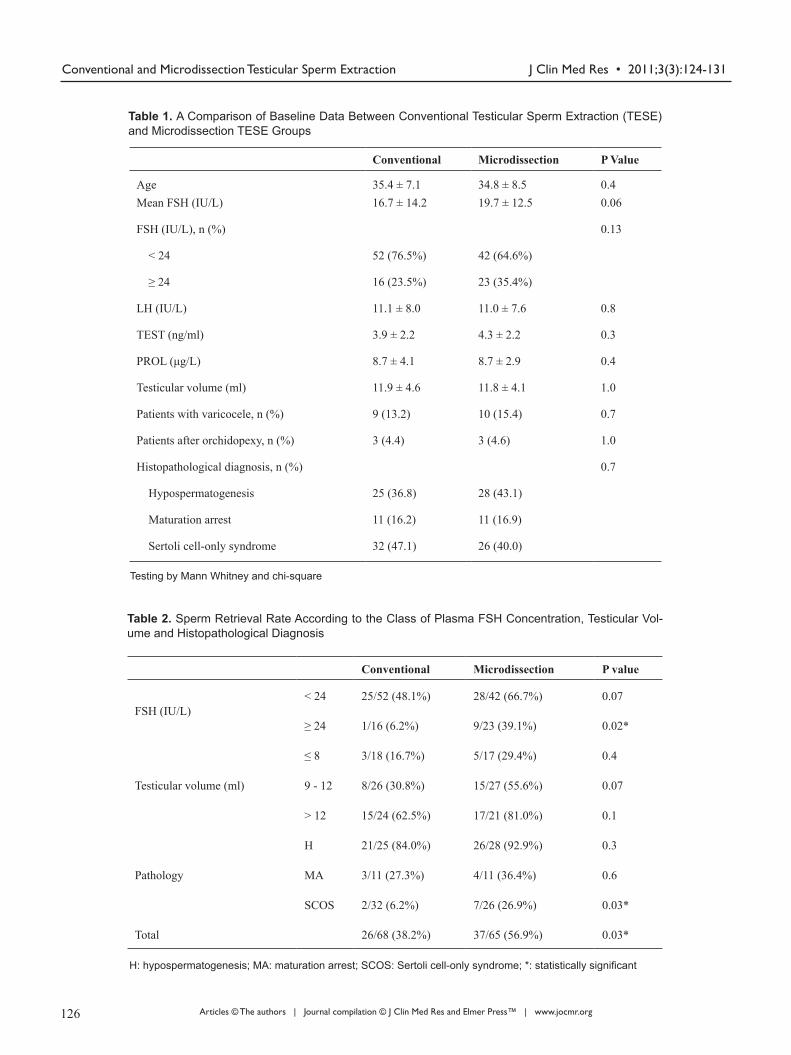
Table 1. A Comparison of Baseline Data Between Conventional Testicular Sperm Extraction (TESE) and Microdissection TESE Groups
Table 2. Sperm Retrieval Rate According to the Class of Plasma FSH Concentration, Testicular Vol-ume and Histopathological Diagnosis
Testing by Mann Whitney and chi-square
H: hypospermatogenesis; MA: maturation arrest; SCOS: Sertoli cell-only syndrome; *: statistically significant
Conventional Microdissection P Value
Age 35.4 ± 7.1 34.8 ± 8.5 0.4Mean FSH (IU/L) 16.7 ± 14.2 19.7 ± 12.5 0.06
FSH (IU/L), n (%) 0.13
< 24 52 (76.5%) 42 (64.6%)
≥ 24 16 (23.5%) 23 (35.4%)
LH (IU/L) 11.1 ± 8.0 11.0 ± 7.6 0.8
TEST (ng/ml) 3.9 ± 2.2 4.3 ± 2.2 0.3
PROL (μg/L) 8.7 ± 4.1 8.7 ± 2.9 0.4
Testicular volume (ml) 11.9 ± 4.6 11.8 ± 4.1 1.0
Patients with varicocele, n (%) 9 (13.2) 10 (15.4) 0.7
Patients after orchidopexy, n (%) 3 (4.4) 3 (4.6) 1.0
Histopathological diagnosis, n (%) 0.7
Hypospermatogenesis 25 (36.8) 28 (43.1)
Maturation arrest 11 (16.2) 11 (16.9)
Sertoli cell-only syndrome 32 (47.1) 26 (40.0)
Conventional Microdissection P value
FSH (IU/L)< 24 25/52 (48.1%) 28/42 (66.7%) 0.07
≥ 24 1/16 (6.2%) 9/23 (39.1%) 0.02*
Testicular volume (ml)
≤ 8 3/18 (16.7%) 5/17 (29.4%) 0.4
9 - 12 8/26 (30.8%) 15/27 (55.6%) 0.07
> 12 15/24 (62.5%) 17/21 (81.0%) 0.1
Pathology
H 21/25 (84.0%) 26/28 (92.9%) 0.3
MA 3/11 (27.3%) 4/11 (36.4%) 0.6
SCOS 2/32 (6.2%) 7/26 (26.9%) 0.03*
Total 26/68 (38.2%) 37/65 (56.9%) 0.03*
126 127
J Clin Med Res • 2011;3(3):124-131Division et al
Articles © The authors | Journal compilation © J Clin Med Res and Elmer Press™ | www.jocmr.org
ered statistically significant. All data were analyzed using the Statistical Package for the Social Sciences program (SPSS for Windows 16.0, SPSS Inc., USA).
Results
Patient background
Unexplained nonobstructive azoospermia was diagnosed in 68 men undergoing conventional and in 65 undergoing microdissection TESE. Two patients who underwent micro-dissection TESE had a history of intensive chemotherapy. Another 4 patients had bilateral undescended testes and 2 each underwent conventional and microdissection TESE. Pre-operative patient characteristics in the two groups in-cluding endocrine data and histopathological diagnosis were considered nearly identical (Table 1).
Success rate of sperm retrieval
Age did not have significant effect on sperm retrieval in ei-ther microdissection or conventional TESE. The success rate of sperm retrieval in patients with NOA was significantly higher for the microdissection than for the conventional TESE procedure (56.9% versus 38.2%, P = 0.03, Table 2). In 4 patients in whom bilaterally undescended testes were iden-tified we could retrieve spermatozoa in the two patients who underwent the microdissection procedure and in one patient with the conventional TESE. Another 2 men who had under-gone intensive chemotherapy for Lymphoma and testicular cancer underwent microdissection TESE. We succeeded in obtaining spermatozoa in the later case.
The influence of histological diagnosis on the success rate of sperm retrieval was considered. We obtained sperma-tozoa in 84% and 92.9% of those with the histological diag-nosis of hypospermatogenesis, by conventional and micro-
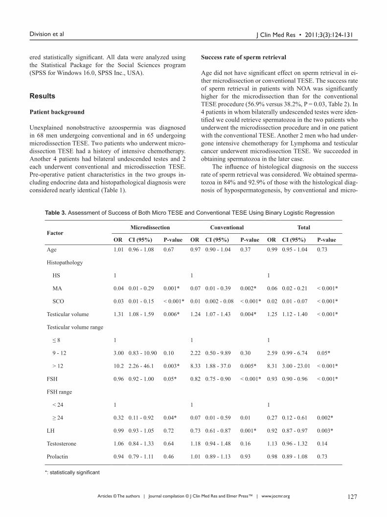
Table 3. Assessment of Success of Both Micro TESE and Conventional TESE Using Binary Logistic Regression
*: statistically significant
FactorMicrodissection Conventional Total
OR CI (95%) P-value OR CI (95%) P-value OR CI (95%) P-value
Age 1.01 0.96 - 1.08 0.67 0.97 0.90 - 1.04 0.37 0.99 0.95 - 1.04 0.73
Histopathology
HS 1 1 1
MA 0.04 0.01 - 0.29 0.001* 0.07 0.01 - 0.39 0.002* 0.06 0.02 - 0.21 < 0.001*
SCO 0.03 0.01 - 0.15 < 0.001* 0.01 0.002 - 0.08 < 0.001* 0.02 0.01 - 0.07 < 0.001*
Testicular volume 1.31 1.08 - 1.59 0.006* 1.24 1.07 - 1.43 0.004* 1.25 1.12 - 1.40 < 0.001*
Testicular volume range
≤ 8 1 1 1
9 - 12 3.00 0.83 - 10.90 0.10 2.22 0.50 - 9.89 0.30 2.59 0.99 - 6.74 0.05*
> 12 10.2 2.26 - 46.1 0.003* 8.33 1.88 - 37.0 0.005* 8.31 3.00 - 23.01 < 0.001*
FSH 0.96 0.92 - 1.00 0.05* 0.82 0.75 - 0.90 < 0.001* 0.93 0.90 - 0.96 < 0.001*
FSH range
< 24 1 1 1
≥ 24 0.32 0.11 - 0.92 0.04* 0.07 0.01 - 0.59 0.01 0.27 0.12 - 0.61 0.002*
LH 0.99 0.93 - 1.05 0.72 0.73 0.61 - 0.87 0.001* 0.92 0.87 - 0.97 0.003*
Testosterone 1.06 0.84 - 1.33 0.64 1.18 0.94 - 1.48 0.16 1.13 0.96 - 1.32 0.14
Prolactin 0.94 0.79 - 1.11 0.46 1.01 0.89 - 1.13 0.93 0.98 0.89 - 1.08 0.73
126 127

J Clin Med Res • 2011;3(3):124-131 Conventional and Microdissection Testicular Sperm Extraction
Articles © The authors | Journal compilation © J Clin Med Res and Elmer Press™ | www.jocmr.org
dissection TESE, respectively. We retrieved sperm in 27.3% and 36.4% of men with maturation arrest, respectively. For those with SCOS, SRR was significantly higher by microdis-section TESE (26.9% versus 6.2%, P = 0.03, Table 3).
Sperm retrieval rate was positive in 10/39 (25.6%) pa-tients with FSH ≥ 2N (24 mIU/mL) (9/23 with microdis-section, 1/16 with conventional TESE); in 53/94 (56.4%) patients with < 2N (< 24 mIU/mL) (28/42 with microdissec-tion, 25/52 with conventional TESE). SRR was significantly higher by microdissection TESE for those with FSH ≥ 2N (P = 0.02, Table 2). Increase in FSH levels showed significant failure of sperm retrieval in general (OR = 0.93, P < 0.001, Table 3). This failure was significant in conventional TESE by odds of 0.82 (P < 0.001) versus the odds of 0.96 by Mi-crodissection TESE (P = 0.05). Also, increase in LH levels showed significant failure of sperm retrieval in general (OR = 0.93, P = 0.003, Table 3). This failure was also significant in conventional TESE (OR = 0.73, P < 0.001). However, LH levels had no significant influence on SRR by microdissec-tion TESE (P = 0.72). There was no effect of Testosterone or Prolactin levels on SRR by using either technique.
Increase in testicular volume significantly increased the success rate of sperm retrieval by 1.25 folds in both methods (P < 0.001, Table 3). This increase was significantly higher with the microdissection by 1.31 folds (P = 0.006) versus 1.24 folds with conventional TESE (P = 0.004). In Table 4 we assessed the success of microdissection compared to conventional TESE using binary logistic regression. The in-volvement of other factors in microdissection (FSH, testicu-lar volume, histopathology, or any combination of them) sig-nificantly increases the success rate by a factor of 0.4 - 1.5.
According to the statistical analysis, FSH value and the surgical procedure were the two variables that could signifi-cantly predict positive sperm retrieval (P < 0.05). The testis
volume and histology were shown to play a significant but less important role.
In addition, major postoperative complications, such as acute epididymitis, scrotal hematoma, and testicular hydro-cele, were not seen significantly in this study. Only 5 (7.4%) patients from the conventional group and 3 (4.6%) patients from the microdissection group developed scrotal wall he-matoma that resolved shortly during follow-up. In addition, no patient required hormone replacement therapy for treat-ment of post-operative hypogonadism.
Discussion The identification of areas in which spermatogenesis still occurs represents the background for the addition of mag-nification to TESE. Individual seminiferous tubules can be seen under the microscope allowing the identification of larger, whitish and opaque tubules in which spermatogenesis is active in opposition to tubules where no sperm produc-tion occurs [9]. This strategy could facilitate the removal of smaller amounts of testicular tissue, which becomes crucial in the presence of testicular atrophy. In addition, the iden-tification of avascular regions for the opening of the tunica albuginea could minimize the chances of vascular injury. Multiple biopsy samples from different regions of the testis may increase the possibility of detecting spermatozoa with conventional TESE.
Some authors compared the results obtained by micro-dissection and conventional TESE in NOA patients [9-13] and reported a higher efficacy by microdissection in yield-ing positive sperm recovery, even when multiple TESE is performed [12, 13].
Several studies have compared the two techniques. Oka-
Table 4. Assessment of Success of Microdissection TESE Compared to Conventional TESE Using Binary Logistic Regression
*: statistically significant
Microdissection TESE Combined With Other Factors OR CI (95%) P-value
Alone 2.14 1.07 - 4.27 0.03*
Adjusted to FSH 3.54 1.55 - 8.08 0.003*
Adjusted to Testicular Volume 2.50 1.17 - 5.35 0.02*
Adjusted to Histopathology 2.96 1.10 - 7.99 0.03*
Adjusted to FSH and Testicular Volume 3.68 1.53 - 8.86 0.004*
Adjusted to FSH and Histopathology 3.47 1.25 - 9.60 0.01*
Adjusted to Testicular Volume and Histopathology 3.00 1.11 - 8.10 0.03*
Adjusted to FSH, Testicular Volume and Histopathology 3.56 1.28 - 9.92 0.02*
128 129

J Clin Med Res • 2011;3(3):124-131Division et al
Articles © The authors | Journal compilation © J Clin Med Res and Elmer Press™ | www.jocmr.org
da et al. [12] in their retrospective study including different patient groups, the SRR was 16.7% in the conventional TESE group and 44.6% in the microdissection group. Consistent with the results of most of the studies, the SRR in our study was significantly higher by microdissection TESE (56.9%) than conventional TESE (38.2%) (P = 0.03). Ramasamy et al. reported the outcomes of 460 patients with NOA treated with microdissection TESE [14]. In their report, the SRR by microdissection TESE was 57%, whereas that by conven-tional TESE was 32% which were approximately similar to our results. In a prospective comparative study of patients with NOA and bilaterally identical testicular histology who underwent conventional TESE on one testis and microdis-section TESE on the other [11], the SRR by microdissection TESE was higher (47%) than by conventional TESE (30%) indicating the efficacy of microdissection TESE for sperm retrieval. In addition, postoperative acute and chronic com-plications were significantly lower in the microsurgical side compared with the conventional side [11].
We also investigated the SRR by microdissection TESE compared to that by conventional TESE of each type of tes-ticular histology in patients with NOA. Furthermore, SRR by microdissection TESE for patients with hypospermato-genesis was 92.9%, whereas that by conventional TESE was 84%. We also found SRRs by microdissection TESE for maturation arrest and SCOS were 36.4% and 27.3%, respec-tively, whereas those by conventional TESE were 26.9%, and 6.2%. Only those of SCOS showed significantly higher SRR by microdissection TESE. Recently, Ramasamy et al. [14] reported excellent SRRs of 81%, 44%, and 41% for hypospermatogenesis, maturation arrest, and SCOS, respec-tively. The outcome of TESE may depend on factors other than urological technique, such as embryonic factors and human skills especially for those in the biology lab. In our study spermatozoa were detected in 61.7% of all cases dur-ing histopathogical examination of very small samples sent to the pathology lab compared to 45% detected by the people in the IVF lab. Thus, it is well accepted that microdissection TESE offers a great advantage for patients with all types of testicular histology. Tsujimura et al. [15] performed salvage microdissection TESE on the patients when conventional TESE failed to show spermatozoa and reported that the SRR increased with microdissection TESE.
Unlike these studies, there have been other studies showing no difference in the SRR between the two tech-niques [16]. There was no significant difference in the fer-tilization rates, between conventional TESE and microdis-section TESE. Mulhall and co-workers [16] reported that the SRR was significantly higher with microdissection TESE than with conventional TESE in the patients with NOA and atrophic testis.
Microdissection TESE also avoided such complications as hematoma, fibrosis, and androgen decline, which other-wise might have been caused by conventional TESE in the
patients with atrophic testes. We performed microdissection TESE on 17 patients whose testes were atrophic (testis vol-umes were ≤ 8 mL). Despite this effort, the SRR by micro-dissection was only 29.4% in this group which was still non-significantly higher than the conventional group (16.7%, P = 0.4). The small number of the cases may influence our sta-tistics workup.
Clinically, testicular volume is correlated with sper-matogenesis. Testicular volume had been found to have poor predictive value for successful TESE, however, because topographical variations in testicular pathology, independent of testicular volume, can occur [17]. Indeed, it had been re-ported that there is no statistically significant difference in testicular volume between patients with retrievable sper-matozoa and those without [17, 18]. Furthermore, no lower limit of testicular volume for the absence of spermatozoa has been identified. Spermatozoa are often retrieved from tes-tes with volumes less than 5 mL by microdissection TESE. Thus, small testicular volume itself does not preclude suc-cessful microdissection TESE [17]. Similar to others, we found a positive relation between the SRR and testis volume [19, 20].
Therefore, it can be suggested that the patients with NOA whose testis volumes are lower should be informed about the low SRR with conventional or microdissection TESE. The high amounts of removed testicular tissue may cause testicu-lar insufficiency with a decrease in testosterone levels, espe-cially in the hypoplastic/atrophic testicles. Schlegel [9] and Amer et al. [11] reported that the amount of removed testicu-lar tissue in microdissectional TESE was significantly lower than with the conventional method. We could not measure the amount of testicular tissue removed in patients during the TESE operation. The missing information is a possible limitation of our study.
In our study, increase in FSH levels showed significant failure of sperm retrieval in general which was more signifi-cant in conventional TESE by odds of 0.82 (P < 0.001) ver-sus the odds of 0.96 for microdissection TESE (P = 0.05). Although previous studies revealed a negative correlation between increased FSH levels and the SRR, recent studies showed no significant relation between FSH levels and the SRR [19]. Even in their study, Ramasamy and co-workers [21] reported lower SRR in the group of patients with FSH levels less than 15 IU/mL. Consistent with the literature, a significant relation between FSH levels and the SRR was detected in our study. In addition, testicular volume plays an important significant role in the SRR especially when the volume was greater than 12 cm (Table 3).
Regarding histopathology, only SCOS significantly af-fects the SRR of the two procedures. We and other authors found FSH value and the surgical procedure were the two variables significantly predicting positive sperm retrieval [20]. Increase in LH levels showed significant failure of sperm retrieval in general. Similar to others no difference
128 129

J Clin Med Res • 2011;3(3):124-131 Conventional and Microdissection Testicular Sperm Extraction
Articles © The authors | Journal compilation © J Clin Med Res and Elmer Press™ | www.jocmr.org
was found in the LH and FSH levels between patients in whom sperm were retrieved successfully and those in whom no sperm were found [14].
The combination of different factors in microdissection TESE (FSH, testicular volume, histopathology, or any com-bination of them) significantly increased the success of SRR.
Microdissection TESE was reported to cause significant-ly fewer acute and chronic complications than conventional procedures based on post-operative ultrasonography [11]. However, major complications, such as acute epididymitis, big scrotal hematoma and testicular hydrocele were not seen in any patient in either the microdissection or the multiple TESE group in the present study. In this study, scrotal wall hematoma was non-significantly lower in the microdissec-tion group compared to conventional TESE. In addition, no patient required hormone replacement therapy for treatment of post-operative hypogonadism. These findings suggest that microdissection TESE is safe in terms of both surgical and endocrinological complications.
Conclusion
Microdissection TESE significantly had twice better prob-ability of success rate for sperm retrieval when compared with conventional TESE. All things considered, performing microdissection instead of conventional TESE is still the most effective treatment alternative in terms of high SRR and fewer complications. There was a relation between the SRR and testicular volume and FSH levels. Levels of testos-terone or prolactin had no effect on the success rate of sperm retrieval using either method. Microdissection TESE ap-pears to be recommendable especially in cases of atrophied testicles, high FSH concentration, or when SCOS with high FSH concentration can be predicted on the basis of the pre-operative prognostic data.
References
1. Donoso P, Tournaye H, Devroey P. Which is the best sperm retrieval technique for non-obstructive azo-ospermia? A systematic review. Hum Reprod Update 2007;13(6):539-549.
2. Palermo GD, Schlegel PN, Hariprashad JJ, Ergun B, Mielnik A, Zaninovic N, Veeck LL, et al. Fertiliza-tion and pregnancy outcome with intracytoplasmic sperm injection for azoospermic men. Hum Reprod 1999;14(3):741-748.
3. Ezeh UI. Beyond the clinical classification of azoosper-mia: opinion. Hum Reprod 2000;15(11):2356-2359.
4. Raman JD, Schlegel PN. Testicular sperm extraction with intracytoplasmic sperm injection is successful for the treatment of nonobstructive azoospermia associated
with cryptorchidism. J Urol 2003;170(4 Pt 1):1287-1290.
5. Su LM, Palermo GD, Goldstein M, Veeck LL, Rosen-waks Z, Schlegel PN. Testicular sperm extraction with intracytoplasmic sperm injection for nonobstructive azoospermia: testicular histology can predict success of sperm retrieval. J Urol 1999;161(1):112-116.
6. Devroey P, Liu J, Nagy Z, Goossens A, Tournaye H, Camus M, Van Steirteghem A, et al. Pregnancies after testicular sperm extraction and intracytoplasmic sperm injection in non-obstructive azoospermia. Hum Reprod 1995;10(6):1457-1460.
7. Tournaye H, Verheyen G, Nagy P, Ubaldi F, Goossens A, Silber S, Van Steirteghem AC, et al. Are there any pre-dictive factors for successful testicular sperm recovery in azoospermic patients? Hum Reprod 1997;12(1):80-86.
8. Seo JT, Ko WJ. Predictive factors of successful testicu-lar sperm recovery in non-obstructive azoospermia pa-tients. Int J Androl 2001;24(5):306-310.
9. Schlegel PN. Testicular sperm extraction: microdissec-tion improves sperm yield with minimal tissue excision. Hum Reprod 1999;14(1):131-135.
10. Silber SJ. Microsurgical TESE and the distribution of spermatogenesis in non-obstructive azoospermia. Hum Reprod 2000;15(11):2278-2284.
11. Amer M, Ateyah A, Hany R, Zohdy W. Prospective comparative study between microsurgical and conven-tional testicular sperm extraction in non-obstructive azo-ospermia: follow-up by serial ultrasound examinations. Hum Reprod 2000;15(3):653-656.
12. Okada H, Dobashi M, Yamazaki T, Hara I, Fujisawa M, Arakawa S, Kamidono S. Conventional versus micro-dissection testicular sperm extraction for nonobstructive azoospermia. J Urol 2002;168(3):1063-1067.
13. Tsujimura A, Matsumiya K, Miyagawa Y, Tohda A, Miura H, Nishimura K, Koga M, et al. Conventional multiple or microdissection testicular sperm extraction: a comparative study. Hum Reprod 2002;17(11):2924-2929.
14. Ramasamy R, Yagan N, Schlegel PN. Structural and functional changes to the testis after conventional ver-sus microdissection testicular sperm extraction. Urology 2005;65(6):1190-1194.
15. Tsujimura A, Miyagawa Y, Takao T, Takada S, Koga M, Takeyama M, Matsumiya K, et al. Salvage microdissec-tion testicular sperm extraction after failed conventional testicular sperm extraction in patients with nonobstruc-tive azoospermia. J Urol 2006;175(4):1446-1449; dis-cussion 1449.
16. Mulhall JP, Ghaly SW, Aviv N, Ahmed A. The utility of optical loupe magnification for testis sperm extrac-tion in men with nonobstructive azoospermia. J Androl 2005;26(2):178-181.
130 131
J Clin Med Res • 2011;3(3):124-131Division et al
Articles © The authors | Journal compilation © J Clin Med Res and Elmer Press™ | www.jocmr.org
17. Tsujimura A. Microdissection testicular sperm extrac-tion: prediction, outcome, and complications. Int J Urol 2007;14(10):883-889.
18. Hibi H, Ohori T, Yamada Y, Honda N, Asada Y. Prob-ability of sperm recovery in non-obstructive azoosper-mic patients presenting with testes volume less than 10 ml/FSH level exceeding 20 mIU/ml. Arch Androl 2005;51(3):225-231.
19. Turunc T, Gul U, Haydardedeoglu B, Bal N, Kuzgun-bay B, Peskircioglu L, Ozkardes H. Conventional testicular sperm extraction combined with the micro-dissection technique in nonobstructive azoospermic
patients: a prospective comparative study. Fertil Steril 2010;94(6):2157-2160.
20. Colpi GM, Colpi EM, Piediferro G, Giacchetta D, Gaz-zano G, Castiglioni FM, Magli MC, et al. Microsurgi-cal TESE versus conventional TESE for ICSI in non-obstructive azoospermia: a randomized controlled study. Reprod Biomed Online 2009;18(3):315-319.
21. Ramasamy R, Lin K, Gosden LV, Rosenwaks Z, Palermo GD, Schlegel PN. High serum FSH levels in men with nonobstructive azoospermia does not affect success of microdissection testicular sperm extraction. Fertil Steril 2009;92(2):590-593.
130 131
Related Documents