RESEARCH Open Access Clinical application for the preservation of phospho- proteins through in-situ tissue stabilization C Bart Rountree 1,2*† , Colleen A Van Kirk 2† , Hanning You 1,2 , Wei Ding 1,2 , Hien Dang 1,2 , Heather D VanGuilder 2 , Willard M Freeman 2 Abstract Background: Protein biomarkers will play a pivotal role in the future of personalized medicine for both diagnosis and treatment decision-making. While the results of several pre-clinical and small-scale clinical studies have demonstrated the value of protein biomarkers, there have been significant challenges to translating these findings into routine clinical care. Challenges to the use of protein biomarkers include inter-sample variability introduced by differences in post-collection handling and ex vivo degradation of proteins and protein modifications. Results: In this report, we re-create laboratory and clinical scenarios for sample collection and test the utility of a new tissue stabilization technique in preserving proteins and protein modifications. In the laboratory setting, tissue stabilization with the Denator Stabilizor T1 resulted in a significantly higher yield of phospho-protein when compared to standard snap freeze preservation. Furthermore, in a clinical scenario, tissue stabilization at collection resulted in a higher yield of total phospho-protein, total phospho-tyrosine, pErkT202/Y204 and pAktS473 when compared to standard methods. Tissue stabilization did not have a significant effect on other post-translational modifications such as acetylation and glycosylation, which are more stable ex-vivo. Tissue stabilization did decrease total RNA quantity and quality. Conclusion: Stabilization at the time of collection offers the potential to better preserve tissue protein and protein modification levels, as well as reduce the variability related to tissue processing delays that are often associated with clinical samples. Background Recent advances in proteomic technologies have spurred a number of reports examining distinct alterations in protein expression [1,2] or modification [3-6] that are associated with, or can classify, disease states in human patients. Although these biomarker studies provide important analytical and diagnostic tools, a challenge for translational research is the transition of findings from the controlled laboratory environment to the clinical setting, where variation in tissue acquisition and hand- ling practices can introduce significant data variability. This variation can confound data analysis and interpre- tation, and in turn, impact patient diagnosis and prog- nosis [7]. Combined with clinical heterogeneity resulting from genetic, physiological, and environmental factors, which are typically controlled for in animal models implemented in the laboratory setting, technical variance introduced during tissue collection in the clinical research setting reduces the statistical and classification power of translational studies. This is especially true regarding measurements of protein abundance and modification (e.g. phosphorylation). Standardization pro- cedures have been proposed for plasma and serum col- lection in biomarker studies [8] and technologies for sample preservation of plasma and serum have been developed [9]. While no standards currently exist for tis- sue collection, technical approaches to preserve proteins and reduce technical variance have been proposed [10]. Whether in the laboratory or clinical setting, varia- tions in tissue retrieval and processing, and any delay in sample stabilization (e.g., cryopreservation, fixation) can dramatically alter the quantitative characteristics of the tissue proteome. As tissue protein biomarkers seek to * Correspondence: [email protected] † Contributed equally 1 Department of Pediatrics, Penn State Hershey Medical Center, 500 University Drive, Hershey, PA 17033 Full list of author information is available at the end of the article Rountree et al. Proteome Science 2010, 8:61 http://www.proteomesci.com/content/8/1/61 © 2010 Rountree et al; licensee BioMed Central Ltd. This is an Open Access article distributed under the terms of the Creative Commons Attribution License (http://creativecommons.org/licenses/by/2.0), which permits unrestricted use, distribution, and reproduction in any medium, provided the original work is properly cited.

Welcome message from author
This document is posted to help you gain knowledge. Please leave a comment to let me know what you think about it! Share it to your friends and learn new things together.
Transcript
RESEARCH Open Access
Clinical application for the preservation of phospho-proteins through in-situ tissue stabilizationC Bart Rountree1,2*†, Colleen A Van Kirk2†, Hanning You1,2, Wei Ding1,2, Hien Dang1,2,Heather D VanGuilder2, Willard M Freeman2
Abstract
Background: Protein biomarkers will play a pivotal role in the future of personalized medicine for both diagnosisand treatment decision-making. While the results of several pre-clinical and small-scale clinical studies havedemonstrated the value of protein biomarkers, there have been significant challenges to translating these findingsinto routine clinical care. Challenges to the use of protein biomarkers include inter-sample variability introduced bydifferences in post-collection handling and ex vivo degradation of proteins and protein modifications.
Results: In this report, we re-create laboratory and clinical scenarios for sample collection and test the utility of anew tissue stabilization technique in preserving proteins and protein modifications. In the laboratory setting, tissuestabilization with the Denator Stabilizor T1 resulted in a significantly higher yield of phospho-protein whencompared to standard snap freeze preservation. Furthermore, in a clinical scenario, tissue stabilization at collectionresulted in a higher yield of total phospho-protein, total phospho-tyrosine, pErkT202/Y204 and pAktS473 whencompared to standard methods. Tissue stabilization did not have a significant effect on other post-translationalmodifications such as acetylation and glycosylation, which are more stable ex-vivo. Tissue stabilization did decreasetotal RNA quantity and quality.
Conclusion: Stabilization at the time of collection offers the potential to better preserve tissue protein and proteinmodification levels, as well as reduce the variability related to tissue processing delays that are often associatedwith clinical samples.
BackgroundRecent advances in proteomic technologies have spurreda number of reports examining distinct alterations inprotein expression [1,2] or modification [3-6] that areassociated with, or can classify, disease states in humanpatients. Although these biomarker studies provideimportant analytical and diagnostic tools, a challenge fortranslational research is the transition of findings fromthe controlled laboratory environment to the clinicalsetting, where variation in tissue acquisition and hand-ling practices can introduce significant data variability.This variation can confound data analysis and interpre-tation, and in turn, impact patient diagnosis and prog-nosis [7]. Combined with clinical heterogeneity resulting
from genetic, physiological, and environmental factors,which are typically controlled for in animal modelsimplemented in the laboratory setting, technical varianceintroduced during tissue collection in the clinicalresearch setting reduces the statistical and classificationpower of translational studies. This is especially trueregarding measurements of protein abundance andmodification (e.g. phosphorylation). Standardization pro-cedures have been proposed for plasma and serum col-lection in biomarker studies [8] and technologies forsample preservation of plasma and serum have beendeveloped [9]. While no standards currently exist for tis-sue collection, technical approaches to preserve proteinsand reduce technical variance have been proposed [10].Whether in the laboratory or clinical setting, varia-
tions in tissue retrieval and processing, and any delay insample stabilization (e.g., cryopreservation, fixation) candramatically alter the quantitative characteristics of thetissue proteome. As tissue protein biomarkers seek to
* Correspondence: [email protected]† Contributed equally1Department of Pediatrics, Penn State Hershey Medical Center, 500University Drive, Hershey, PA 17033Full list of author information is available at the end of the article
Rountree et al. Proteome Science 2010, 8:61http://www.proteomesci.com/content/8/1/61
© 2010 Rountree et al; licensee BioMed Central Ltd. This is an Open Access article distributed under the terms of the CreativeCommons Attribution License (http://creativecommons.org/licenses/by/2.0), which permits unrestricted use, distribution, andreproduction in any medium, provided the original work is properly cited.
make the transition from the laboratory to the clinic, areal obstacle is standardizing tissue sample collectionand processing in and around the operating suite, wheremost clinical samples are obtained. Total proteinamounts and post-translational modifications are rapidlyimpacted by post-collection enzymatic activity. Forexample, ex vivo protease and phosphatase activity isretained but does not reflect true physiological condi-tions. Artifacts resulting from this residual activity notonly increase inter-sample variability but also contributeto quantitation inaccuracies, particularly in measures ofdynamic modification states of a given protein (e.g.,phosphorylation) [11-13]. Traditional approaches to pre-serving tissues, including freezing and chemical fixation,require the availability of dry ice and chemicals in theoperating suite. In the clinical environment, the primaryfocus of the surgical team is on the patient. In this set-ting, several hours may elapse from the time of tissuecollection to preservation, depending on the time of col-lection and the availability of personnel [7].A recent report by Svensson et al. demonstrates the suc-
cess of rapid tissue stabilization in improving proteomicanalyses. Using an approach combining heat and pressureinactivation of enzymes ex vivo, samples can be rapidlystabilized (< 1 minute) to prevent protein degradation andloss of post-translational modifications in tissue samples[10]. This technique does not utilize dry ice or chemicalsand reduces sample complexity by preventing the forma-tion of abundant protein degradation fragments and main-tains modified species for up to two hours at roomtemperature. More recently, several papers have high-lighted this technique for proteomic and peptidomic stabi-lization [14-17]. In this report, we seek to addressunanswered questions related to this technique such as:1) the stabilization of nucleic acid in combination with thetissue proteome, 2) an analysis of the entire phospho-proteome in addition to specific phospho-proteins such asErk and Akt, 3) a detailed analysis of multiple clinical sce-narios including a two hour room-temperature and twohour 4°C incubation, and 4) an analysis of non-phosphory-lation protein modification such as glycosylation. Theadoption of this preservation technique in clinical tissuesample collection has the potential to mitigate the deleter-ious effects of unavoidable delays in sample preservation,which would allow for improved biomarker screening.The purpose of the present study was to determinewhether this commercially available method of rapid tissuestabilization increases phospho-protein stability andimproves quantitation in tissue samples reflecting labora-tory and clinically-relevant scenarios of sample collection.
ResultsIn order to capture the different potential laboratory andclinical scenarios, we organized animals into groups as
depicted in Figure 1A. For the laboratory setting, follow-ing tissue dissection, samples were: 1) snap frozenimmediately for storage at -80°C, 2) stabilized immedi-ately prior to storage at -80°C, or 3) snap frozen imme-diately prior to storage at -80°C and stabilized afterremoval from storage (Figure 1A). To reflect the broaderarray of clinically-relevant tissue handling scenarios,liver samples were dissected into six pieces as follows:1) maintained at room temperature for two hours, and
B
Brain, Lungn=6 n=6
stabilize, freezesnap-freeze
snap-freezeimmediate treatment storage post-treatment
-80°C n/an/a
stabilize-80°C-80°C
Labo
rato
ry S
cena
rioC
linic
al S
cena
rio
Livern=6
immediate treatment delay-to-storageRT RT, 2hr -80°C
wet ice 4°C, 2hr -80°Cstabilize RT, 2hr -80°C
stabilize 4°C, 2hr -80°C
stabilize, freeze n/a -80°Csnap-freeze n/a -80°C
storage
A
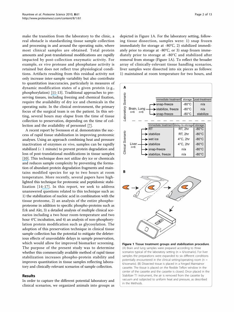
Figure 1 Tissue treatment groups and stabilization procedure.(A) Brain and lung samples were prepared according to threescenarios typical of the laboratory setting (n = 6/scenario). For liversamples the preparations were expanded to six different conditionspotentially encountered in the clinical setting/operating room (n =6/scenario). (B) Dissected tissue is placed in a hinged Maintainorcassette. The tissue is placed on the flexible Teflon window in thecenter of the cassette and the cassette is closed. Once placed in theStabilizer T1 instrument, the air is removed from the cassette byvacuum and subjected to uniform heat and pressure, as describedin the Methods.
Rountree et al. Proteome Science 2010, 8:61http://www.proteomesci.com/content/8/1/61
Page 2 of 13

stored at -80°C; 2) stabilized immediately, maintained atroom temperature for two hours, and stored at -80°C; 3)maintained at 4°C for two hours, and stored at -80°C; 4)stabilized immediately, maintained at 4°C for two hours,and stored at -80°C; 5) snap frozen immediately ("best-case scenario” control) and stored at -80°C; or 6) stabi-lized immediately, snap frozen, and stored at -80°C(Figure 1A).In the “laboratory setting” model, immediate stabiliza-
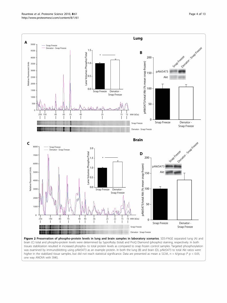
tion of brain and lung tissue following dissection resultedin higher phospho-protein levels compared to snap fro-zen controls. Using automated densitometry methods,total lane volumes were determined for both the phos-pho-protein and total protein stains, and ratios of phos-pho- to total protein were calculated. In lung tissue,phospho-protein to total protein ratios were significantlygreater in stabilized samples compared to snap frozencontrols and snap frozen then stabilized samples (1.13 ±0.03 vs. 1.0 ± 0.03 and 0.98 ± 0.03 respectively, p = 0.011,n = 6/group) (Figure 2A). No statistically significant dif-ference was observed between snap frozen controls andsnap frozen then stabilized samples (data not shown). Inbrain, the average ratio of phospho-protein to total pro-tein was significantly greater in stabilized samples com-pared to snap frozen controls and snap frozen thenstabilized samples (1.31 ± 0.05 vs. 1.0 ± 0.03 and 1.0 ±0.04, respectively; p < 0.001, n = 6/group) (Figure 2C).Once again, no statistically significant difference wasobserved between snap frozen controls and snap frozenthen stabilized samples (data not shown). These datademonstrate that significantly higher levels of total phos-phorylated proteins are detected in stabilized samplesisolated from both brain (Figure 2C) and lung(Figure 2A) than in snap frozen tissue samples. This istrue for both small and large molecular weight phospho-proteins. Graph insets illustrate the average phospho-protein to total protein ratios scaled to the respectivecontrol means. To further analyze the phospho-proteinlevels, pAktS473 was examined by immunoblotting inboth the lung and brain. While higher pAktS473/totalAkt ratios were observed in stabilized tissue versus snapfrozen controls, this difference did not reach statisticalsignificance (Figures 2B and 2D respectively).Although the laboratory setting provides a well con-
trolled environment for tissue collection, the clinical set-ting is a more challenging venue. To create an accuraterecapitulation of the clinical setting, we first determinedtypical tissue collection times in the clinic. In ourresearch hospital setting, laboratory staff (CBR) acted totransport liver tissue resected from patients directlyfrom the OR to pathology for initial processing, andthen to the laboratory for cryopreservation as part of anIRB approved protocol. The average time from tissuecollection to cryopreservation, with staff immediately
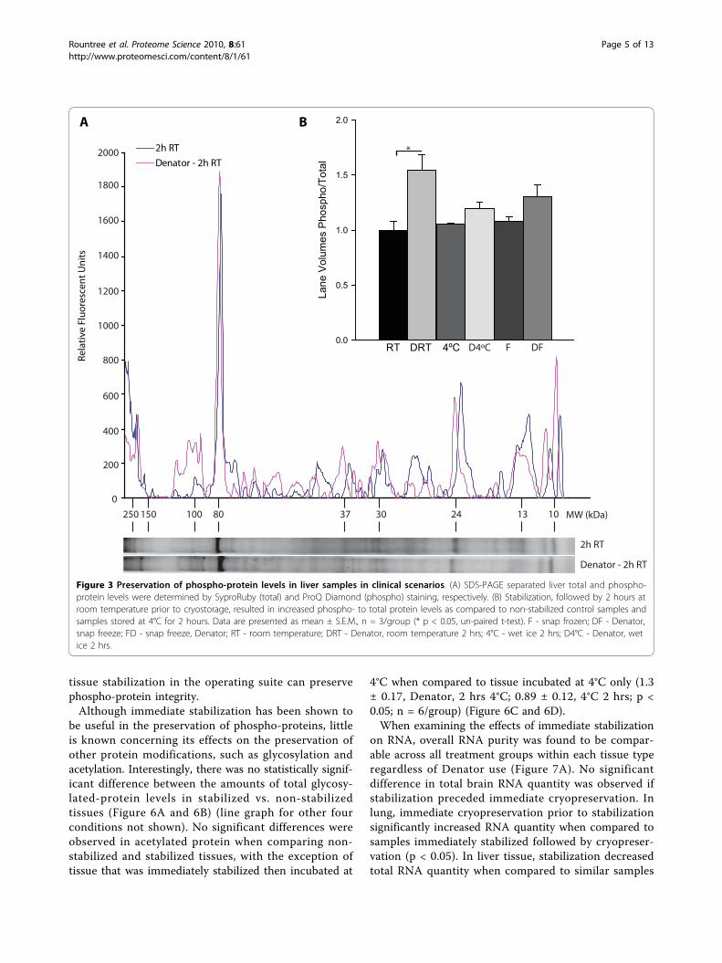
available for tissue transport in the OR at the time ofinitial retrieval, was 66 ± 14 minutes (n = 4). Even inthis ideal setting, there is an increased probability thatspecific disease biomarkers, especially phospho-proteins,may be significantly altered, thereby confounding theanalysis and interpretation of any biochemical experi-ments. To model the clinical environment, “clinical con-trol” liver samples were left at room temperature or onwet ice (4°C) for two hours prior to cryopreservation.These samples were compared to tissue stabilized imme-diately and left at room temperature for two hours.Total phospho-protein levels in stabilized samples wereapproximately 50% higher than in samples incubated atroom temperature or 4°C (1.5 ± 0.14, Denator, 2 hrsRT; 1.0 ± 0.08, RT 2 hrs, p < 0.02, n = 3/group) (Figure3A and 3B).To more specifically examine preservation of phos-
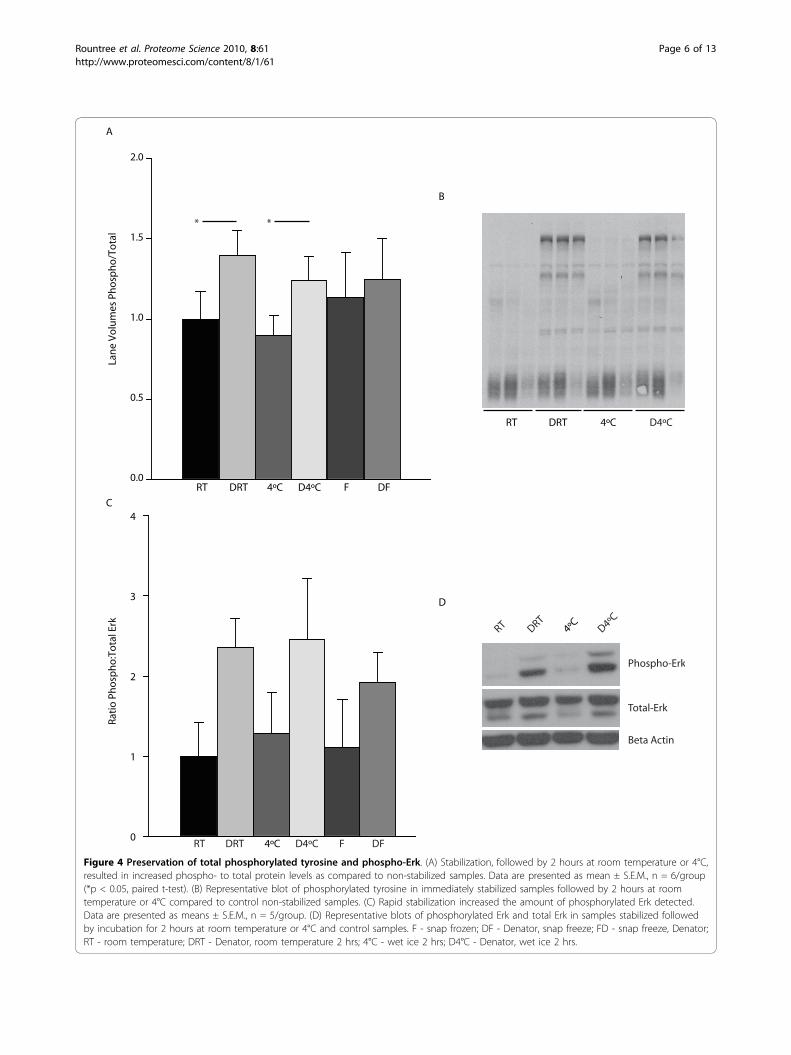
phorylation, pan phospho-tyrosine levels and specificphosphorylated proteins were examined. Total phospho-tyrosine levels were found to be significantly higher intissue that was immediately stabilized when comparedto tissue incubated at room temperature (1.4 ± 0.15,Denator, 2 hrs RT vs. 1.0 ± 0.17, RT 2 hrs; p < 0.05; n =6/group) (Figure 4A and 4B). Similarly, immediate stabi-lization followed by two hours at 4°C preserved a signifi-cantly higher amount of phospho-tyrosine incomparison to samples incubated at 4°C (1.2 ± 0.15,Denator, 2 hrs 4°C vs. 0.9 ± 0.13, 4°C 2 hrs; p < 0.05,n = 6/group). Erk is a major cell signaling protein in theMAPKinase pathway that is activated by phosphoryla-tion on threonine 202 and tyrosine 204. Although therewas a marked increase in total phospho-Erk followingstabilization in both room temperature and 4°C pairedgroups, the overall effect was not statistically significant(Figure 4C and 4D).Akt is a second intracellular signal mediator activated
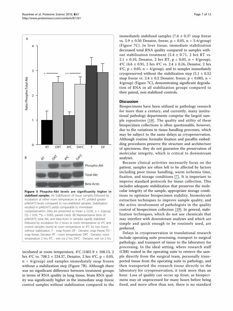
by the PI3Kinase pathway and is phosphorylated on ser-ine (S473) residues rather than on tyrosine. To examinethe effects of immediate stabilization on the preservationof serine/threonine phosphorylation, relative amounts ofphospho-Akt were determined. Immediate stabilizationfollowed by two hours at room temperature preservedsignificantly higher levels of pAktS473 as compared tonon-stabilized samples (2.5 ± 0.24, Denator, 2 hrs RT;1.0 ± 0.36, RT 2 hrs, p < 0.05, n = 5/group) (Figure 5Aand 5B). Similarly, immediate stabilization followed bytwo hours at 4°C yielded higher levels of pAktS473 ascompared to non-stabilized samples incubated at 4°Cfor two hours alone (2.45 ± 0.30, Denator, 2 hrs 4°C;1.19 ± 0.15, 4°C 2 hrs, p < 0.005). This indicates thatimmediate stabilization preserves phospho-Akt compar-able to immediate cryopreservation regardless of delaysof 2 hrs at room temperature and 4°C (Figure 5). Theseresults further demonstrate that integration of rapid
Rountree et al. Proteome Science 2010, 8:61http://www.proteomesci.com/content/8/1/61
Page 3 of 13
0
8000
1000
2000
3000
4000
5000
6000
7000
250 101522253640487080140
Snap FreezeDenator - Snap Freeze
4000
3500
5000
0
500
4500
3000
1500
2000
1000
2500
250 12152540556585150
Lan
e V
olu
mes
Ph
osp
ho
/To
tal
Snap Freeze Denator -Snap Freeze
0.0
0.5
1.0
1.5
0.0
0.5
1.0
1.5
2.0
*
*
Lan
e V
olu
mes
Ph
osp
ho
/To
tal
Snap Freeze Denator -Snap Freeze
Snap Freeze
Denator - Snap Freeze
Snap Freeze
Denator - Snap Freeze
Snap FreezeDenator - Snap Freeze
A
C
Lung
Brain
Snap Freeze Denator - Snap Freeze
pAktS473
Akt
Snap Freeze
Denator -
Snap Freeze
B
0
50
100
150
200
pA
ktS4
73/t
ota
l Akt
(% m
ean
sn
ap fr
oze
n)
pAktS473
Akt
Snap Freeze
Denator -
Snap Freeze
pA
ktS4
73/t
ota
l Akt
(% m
ean
sn
ap fr
oze
n)
D
0
50
100
150
200
Snap Freeze Denator -Snap Freeze
Rela
tive
Fluo
resc
ent U
nits
Rela
tive
Fluo
resc
ent U
nits
MW (kDa)
MW (kDa)
Figure 2 Preservation of phospho-protein levels in lung and brain samples in laboratory scenarios. SDS-PAGE separated lung (A) andbrain (C) total and phospho-protein levels were determined by SyproRuby (total) and ProQ Diamond (phospho) staining, respectively. In bothtissues stabilization resulted in increased phospho- to total protein levels as compared to snap frozen control samples. Targeted phosphorylationwas examined by immunoblotting using pAktS473 as an example protein. In both the lung (B) and brain (D), pAktS473 to total Akt ratios werehigher in the stabilized tissue samples, but did not reach statistical significance. Data are presented as mean ± S.E.M., n = 6/group (* p < 0.05,one way ANOVA with SNK).
Rountree et al. Proteome Science 2010, 8:61http://www.proteomesci.com/content/8/1/61
Page 4 of 13
tissue stabilization in the operating suite can preservephospho-protein integrity.Although immediate stabilization has been shown to
be useful in the preservation of phospho-proteins, littleis known concerning its effects on the preservation ofother protein modifications, such as glycosylation andacetylation. Interestingly, there was no statistically signif-icant difference between the amounts of total glycosy-lated-protein levels in stabilized vs. non-stabilizedtissues (Figure 6A and 6B) (line graph for other fourconditions not shown). No significant differences wereobserved in acetylated protein when comparing non-stabilized and stabilized tissues, with the exception oftissue that was immediately stabilized then incubated at
4°C when compared to tissue incubated at 4°C only (1.3± 0.17, Denator, 2 hrs 4°C; 0.89 ± 0.12, 4°C 2 hrs; p <0.05; n = 6/group) (Figure 6C and 6D).When examining the effects of immediate stabilization
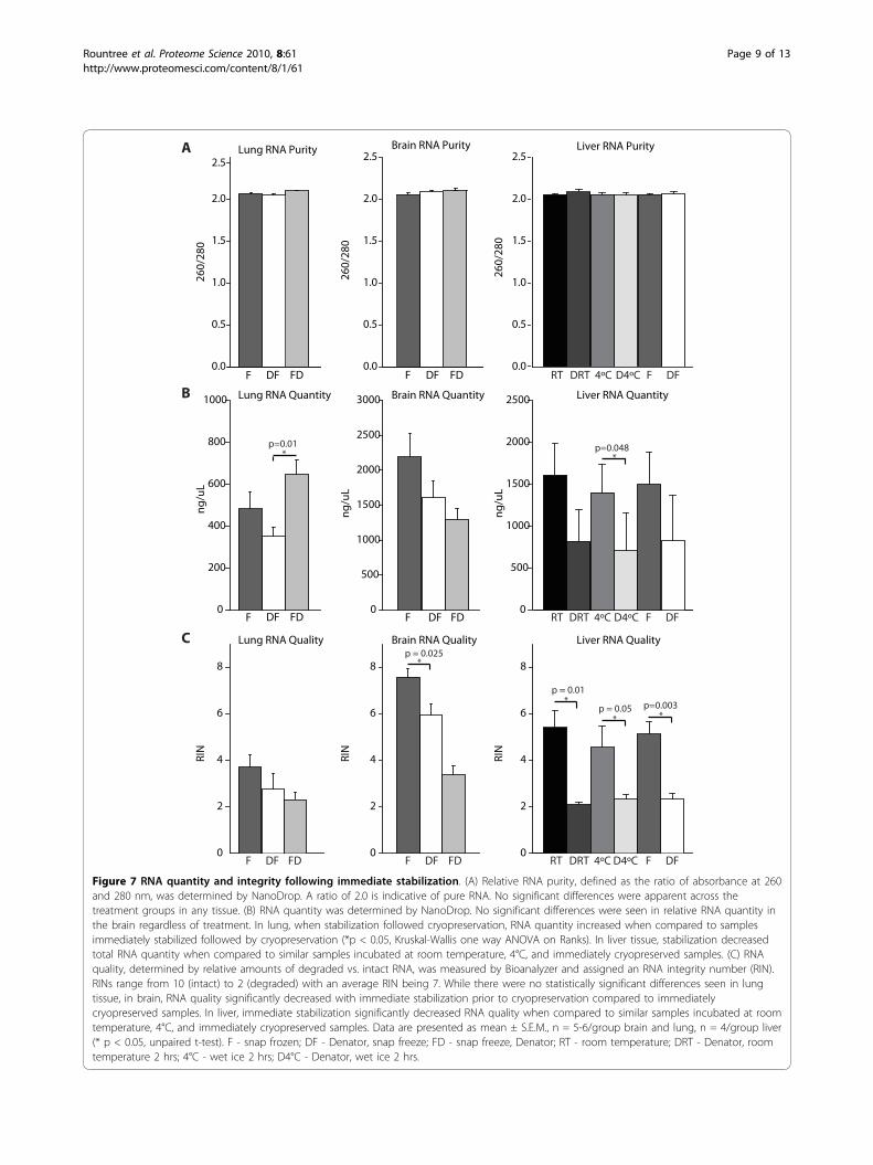
on RNA, overall RNA purity was found to be compar-able across all treatment groups within each tissue typeregardless of Denator use (Figure 7A). No significantdifference in total brain RNA quantity was observed ifstabilization preceded immediate cryopreservation. Inlung, immediate cryopreservation prior to stabilizationsignificantly increased RNA quantity when compared tosamples immediately stabilized followed by cryopreser-vation (p < 0.05). In liver tissue, stabilization decreasedtotal RNA quantity when compared to similar samples
2h RT
Denator - 2h RT
2h RT
Denator - 2h RT
2000
600
200
400
800
1000
1200
1400
1600
1800
0
250 101324303780100150
A B
MW (kDa)
Rela
tive
Fluo
resc
ent U
nits
Lane
Vol
umes
Pho
spho
/Tot
al
0.0
0.5
1.0
1.5
2.0
RT DRT 4ºC D4ºC F DF
*
Figure 3 Preservation of phospho-protein levels in liver samples in clinical scenarios. (A) SDS-PAGE separated liver total and phospho-protein levels were determined by SyproRuby (total) and ProQ Diamond (phospho) staining, respectively. (B) Stabilization, followed by 2 hours atroom temperature prior to cryostorage, resulted in increased phospho- to total protein levels as compared to non-stabilized control samples andsamples stored at 4°C for 2 hours. Data are presented as mean ± S.E.M., n = 3/group (* p < 0.05, un-paired t-test). F - snap frozen; DF - Denator,snap freeze; FD - snap freeze, Denator; RT - room temperature; DRT - Denator, room temperature 2 hrs; 4°C - wet ice 2 hrs; D4°C - Denator, wetice 2 hrs.
Rountree et al. Proteome Science 2010, 8:61http://www.proteomesci.com/content/8/1/61
Page 5 of 13
B
RT DRT 4ºC D4ºC
A
Lan
e V
olu
mes
Ph
osp
ho
/To
tal
0.0
0.5
1.0
1.5
2.0
RT DRT 4ºC D4ºC F DF
* *
C
Rati
o P
ho
sph
o:T
ota
l Erk
0
1
2
3
4
RT DRT F DF4ºC D4ºC
D
D
RT DRT4ºC D4ºC
Phospho-Erk
Total-Erk
Beta Actin
Figure 4 Preservation of total phosphorylated tyrosine and phospho-Erk. (A) Stabilization, followed by 2 hours at room temperature or 4°C,resulted in increased phospho- to total protein levels as compared to non-stabilized samples. Data are presented as mean ± S.E.M., n = 6/group(*p < 0.05, paired t-test). (B) Representative blot of phosphorylated tyrosine in immediately stabilized samples followed by 2 hours at roomtemperature or 4°C compared to control non-stabilized samples. (C) Rapid stabilization increased the amount of phosphorylated Erk detected.Data are presented as means ± S.E.M., n = 5/group. (D) Representative blots of phosphorylated Erk and total Erk in samples stabilized followedby incubation for 2 hours at room temperature or 4°C and control samples. F - snap frozen; DF - Denator, snap freeze; FD - snap freeze, Denator;RT - room temperature; DRT - Denator, room temperature 2 hrs; 4°C - wet ice 2 hrs; D4°C - Denator, wet ice 2 hrs.
Rountree et al. Proteome Science 2010, 8:61http://www.proteomesci.com/content/8/1/61
Page 6 of 13
incubated at room temperature, 4°C (1401.9 ± 168.13, 2hrs 4°C vs. 708.2 ± 224.37, Denator, 2 hrs 4°C, p < 0.05,n = 4/group) and samples immediately snap frozenwithout a stabilization step (Figure 7B). Although therewas no significant difference between treatment groupsin terms of RNA quality in lung tissue, brain RNA qual-ity was significantly higher in the immediate snap freezecontrol samples without stabilization compared to the
immediately stabilized samples (7.6 ± 0.37 snap freezevs. 5.9 ± 0.50 Denator, freeze, p < 0.05, n = 5-6/group)(Figure 7C). In liver tissue, immediate stabilizationdecreased total RNA quality compared to samples with-out stabilization treatment (5.4 ± 0.71, 2 hrs RT vs.2.1 ± 0.10, Denator, 2 hrs RT, p < 0,05, n = 4/group),4°C (4.6 ± 0.91, 2 hrs 4°C vs. 2.4 ± 0.16, Denator, 2 hrs4°C, p < 0.05, n = 4/group), and to samples immediatelycryopreserved without the stabilization step (5.1 ± 0.52snap freeze vs. 2.4 ± 0.2 Denator, freeze, p < 0.005, n =4/group) (Figure 7C), demonstrating significant degrada-tion of RNA in all stabilization groups compared totheir paired, non-stabilized controls.
DiscussionBiospecimens have been utilized in pathology researchfor more than a century, and currently, many institu-tional pathology departments comprise the largest sam-ple repositories [18]. The quality and utility of thesebiospecimen collections is often questionable, however,due to the variations in tissue handling processes, whichmay be subject to the same delays as cryopreservation.Although routine formalin fixation and paraffin embed-ding procedures preserve the structure and architectureof specimens, they do not guarantee the preservation ofmolecular integrity, which is critical to downstreamanalyses.Because clinical activities necessarily focus on the
patient, samples are often left to be affected by factorsincluding poor tissue handling, warm ischemia time,fixation, and storage conditions [7]. It is important toimprove standard protocols for tissue collection. Thisincludes adequate stabilization that preserves the mole-cular integrity of the sample, appropriate storage condi-tions to optimize biospecimen stability, biomoleculeextraction techniques to improve sample quality, andthe active involvement of pathologists in the qualitycontrol of biospecimen collection [19]. In general, stabi-lization techniques, which do not use chemicals thatmay interfere with downstream analyses and which aresimple and quick enough to be routinely used, arepreferred.Delays in cryopreservation in translational research
include operating suite processing, transport to surgicalpathology, and transport of tissue to the laboratory forprocessing. In the ideal setting, where research staff(CBR) waited in the operating suite to retrieve the sam-ple directly from the surgical team, personally trans-ported tissue from the operating suite to pathology, andthen transported the research tissue directly to thelaboratory for cryopreservation, it took more than anhour. Loss of quality can occur up front, as biospeci-mens may sit unprocessed for many hours before beingfixed, and more often than not, there is no standard
A
Rati
o P
ho
sph
o:T
ota
l Akt
0
1
2
3
4
RT DRT F DF4ºC D4ºC
* * *
B RT DRT4ºC D4ºC
Phospho-Akt
Total-Akt
Beta Actin
Figure 5 Phopsho-Akt levels are significantly higher instabilized samples. (A) Stabilization of tissue samples followed byincubation at either room temperature or at 4°C yielded greaterpAktS473 levels compared to non-stabilized samples. Stabilizationresulted in pAktS473 yields comparable to immediatecryopreservation. Data are presented as mean ± S.E.M., n = 5/group(*p < 0.05, **p < 0.005, paired t-test). (B) Representative blots ofpAktS473, total Akt, and beta-Actin in samples rapidly stabilizedfollowed by incubation for 2 hours at room temperature or 4°C andcontrol samples stored at room temperature or 4°C for two hourswithout stabilization. F - snap frozen; DF - Denator, snap freeze; FD -snap freeze, Denator; RT - room temperature; DRT - Denator, roomtemperature 2 hrs; 4°C - wet ice 2 hrs; D4°C - Denator, wet ice 2 hrs.
Rountree et al. Proteome Science 2010, 8:61http://www.proteomesci.com/content/8/1/61
Page 7 of 13

0
20
40
60
80
100
120
140
160 2h RTDenator - 2h RT
2h RT
Denator - 2h RT
MW (kDa)
A
0.0
Lan
e V
olu
mes
Gly
co/T
ota
l
0.2
0.4
0.6
0.8
1.0
1.2
RT DRT 4ºC D4ºC F DF
B
180 8297 2942 18 14
D
RT DRT 4ºC D4ºC
C
Lan
e V
olu
mes
Ace
tyla
ted
/To
tal
0.0
0.5
1.0
1.5
2.0
RT DRT 4ºC D4ºC F DF
*
Rela
tive
Flu
ore
scen
t U
nit
s
Figure 6 Effects of rapid stabilization on protein glycosylation and acetylation. (A) SDS-PAGE separated liver total protein and glyco-protein levels were determined by SyproRuby (total) and ProQ Emerald (glyco) staining, respectively. (B) Stabilization had no effect, eitherpositive or negative, on the ratio of glyco:total protein. Data are presented as mean ± S.E.M., n = 3/group. (C) Total acetylated lysine wasassessed, and stabilization followed by 2 hours at 4°C resulted in increased acetylated protein to total protein levels as compared to non-stabilized samples stored at 4°C for 2 hours. Data are presented as mean ± S.E.M., n = 6/group (*p < 0.05, paired t-test). (D) Representative blotof total acetylated lysine in immediately stabilized samples followed by 2 hours at room temperature or 4°C prior to cryostorage and controlsamples stored at room temperature or 4°C for 2 hours. F - snap frozen; DF - Denator, snap freeze; FD - snap freeze, Denator; RT - roomtemperature; DRT - Denator, room temperature 2 hrs; 4°C - wet ice 2 hrs; D4°C - Denator, wet ice 2 hrs.
Rountree et al. Proteome Science 2010, 8:61http://www.proteomesci.com/content/8/1/61
Page 8 of 13
FDF DF
Brain RNA Quality
RIN
0
2
4
6
8 *p = 0.025
DF FD F
Brain RNA Purity
260/
280
0.0
0.5
1.0
1.5
2.0
2.5
Brain RNA Quantity
ng/u
L
0
500
1000
1500
2000
2500
3000
DF FD F
Lung RNA Quality
RIN
0
2
4
6
8
DF FDF
C
DF FD F
Lung RNA Purity
260/
280
2.5
0.0
0.5
1.0
1.5
2.0
A
Lung RNA Quantity
ng/u
L
0
200
400
600
800
1000
DF FD F
*p=0.01
B
D4ºCDRT 4ºCRT DFF
Liver RNA Quality
0
2
4
6
8
RIN
*p = 0.05
p = 0.01
*p=0.003
Liver RNA Purity
260/
280
0.0
0.5
1.0
1.5
2.0
2.5
D4ºCDRT 4ºCRT DFF
D4ºCDRT 4ºCRT DFF0
500
1000
1500
2000
2500
ng/u
L
Liver RNA Quantity
*p=0.048
*
Figure 7 RNA quantity and integrity following immediate stabilization. (A) Relative RNA purity, defined as the ratio of absorbance at 260and 280 nm, was determined by NanoDrop. A ratio of 2.0 is indicative of pure RNA. No significant differences were apparent across thetreatment groups in any tissue. (B) RNA quantity was determined by NanoDrop. No significant differences were seen in relative RNA quantity inthe brain regardless of treatment. In lung, when stabilization followed cryopreservation, RNA quantity increased when compared to samplesimmediately stabilized followed by cryopreservation (*p < 0.05, Kruskal-Wallis one way ANOVA on Ranks). In liver tissue, stabilization decreasedtotal RNA quantity when compared to similar samples incubated at room temperature, 4°C, and immediately cryopreserved samples. (C) RNAquality, determined by relative amounts of degraded vs. intact RNA, was measured by Bioanalyzer and assigned an RNA integrity number (RIN).RINs range from 10 (intact) to 2 (degraded) with an average RIN being 7. While there were no statistically significant differences seen in lungtissue, in brain, RNA quality significantly decreased with immediate stabilization prior to cryopreservation compared to immediatelycryopreserved samples. In liver, immediate stabilization significantly decreased RNA quality when compared to similar samples incubated at roomtemperature, 4°C, and immediately cryopreserved samples. Data are presented as mean ± S.E.M., n = 5-6/group brain and lung, n = 4/group liver(* p < 0.05, unpaired t-test). F - snap frozen; DF - Denator, snap freeze; FD - snap freeze, Denator; RT - room temperature; DRT - Denator, roomtemperature 2 hrs; 4°C - wet ice 2 hrs; D4°C - Denator, wet ice 2 hrs.
Rountree et al. Proteome Science 2010, 8:61http://www.proteomesci.com/content/8/1/61
Page 9 of 13
protocol or requirement to document how the tissue ishandled. Without this knowledge, a researcher has noway of determining whether the results of downstreammolecular experimentation reflect the effects of tissuehandling or the in vivo state. The tissue stabilizationprocess presented here takes less than 30 seconds andprovides a simple and effective mechanism for rapid sta-bilization of clinical tissues that more easily preservesprotein biomarkers over alternative preservation meth-ods such as snap freezing. Additionally, using thismethod, stabilized samples can be kept at room tem-perature for several hours prior to cryopreservation ortissue analysis without decreases in phospho-proteincontent. This provides investigators with a tool thatincreases consistency of samples collected in the operat-ing suite for proteomic analysis. Immediate stabilizationalso did not alter the levels of protein modificationssuch as glycosylation and acetylation, which are moreresistant to ex-vivo degradation [20,21]. This findingserves as a negative control, suggesting that stabilizationdoes not introduce any variability or alter such modifi-cations, making examination of a wide variety of proteinmodifications possible. However, RNA quantity andquality is significantly reduced as a result of this stabili-zation technique, indicating a potential limitation foruse when examining the tissue proteome only. By usingthe Stabilizor T1 in the operating room or pathologysuite, the clinical team may continue to focus on patientcare and rapidly stabilize a part of biopsy samples to sig-nificantly decrease phospho-protein degradation.A number of diagnostic biomarker development effortshave focused on protein expression [1,2]. Recent studieshave demonstrated that protein modification levels canbe used for diagnostic and prognostic purposes[3,5,22,6]. Usage of the Stabilizor T1 would potentiallyallow for more accurate biochemical diagnostic tests,especially those designed to monitor phospho-proteinlevels. This process requires one piece of instrumenta-tion, is rapid (< 1 min), and prevents decreases in phos-pho-protein content with short-term storage at roomtemperature. Future studies will test implementation ofthis stabilization process in the operating suite toimprove the quality of tissue specimen collection.
ConclusionsTissue stabilization at collection offers the potential tomore accurately preserve tissue protein and proteinmodification levels, such as phosphorylation, and reducevariability related to tissue processing delays.
Materials and methodsAnimalsThree month old male C57BL6/J mice acquired fromJackson Laboratory (Bar Harbor, ME) were housed four
per cage in solid-bottom cages in the Hershey Centerfor Applied Research animal facility, and maintained ina temperature-controlled environment on a 12/12 hourlight/dark cycle with free access to water and food (Har-lan Teklad irradiated mouse diet 7912, Madison, WI).All procedures were conducted in compliance withPenn State University guidelines for the use of labora-tory animals and approved by the Institutional AnimalCare and Use Committee.
Tissue ProcessingImmediately following sacrifice, brain, lung and liver tis-sue was rapidly dissected. Brain and lung tissue weretreated in three ways to model the standard controlledlaboratory research setting (n = 6/group): 1) snap frozenimmediately on dry ice prior to storage at -80°C andprocessed for biochemical analyses after thawing nor-mally, 2) stabilized immediately prior to storage at -80°Cand processed after thawing, or 3) snap frozen immedi-ately on dry ice prior to storage at -80°C and then stabi-lized after removal from storage and before biochemicalanalyses (Figure 1A). Liver samples (n = 6/group) weredissected into six pieces of roughly equal size that weretreated as follows: 1) snap frozen immediately on dry iceand stored at -80°C; 2) stabilized immediately, frozen ondry ice, and stored at -80°C; 3) stabilized immediately,maintained on wet ice (4°C) for two hours, and storedat -80°C; 4) stabilized immediately, maintained at roomtemperature for two hours, and stored at -80°C; 5)maintained on wet ice (4°C) for two hours, and storedat -80°C; or 6) maintained at room temperature for twohours, and stored at -80°C (Figure 1A).
Tissue StabilizationTissue stabilization was conducted using Stabilizor T1instrumentation (Denator AB, Gothenburg, Sweden).Tissue samples were placed in inert polycarbonate/ther-moplastic (Teflon-fluorinated ethylene propylene) Main-tainor Cards (Denator AB), and air was removed byautomated vacuum to minimize potential protein oxida-tion and maximize efficient heat transfer. Samples werethen subjected to 5 mbar of pressure and heated to 95°C for 20 seconds to eliminate residual ex vivo biologicalactivity (Figure 1B).
Protein isolation and quantitationProtein was extracted according to Denator AB-recommended procedures. Using an automated RetschTissueLyser II bead mill (Qiagen, Inc., Germantown,MD) and stainless steel beads pre-chilled on dry ice, fro-zen tissue samples were homogenized at 15 Hz for oneminute. A volume of 1% SDS, equal to 10 times thesample mass, was then added to each sample prior totissue disruption at 15 Hz for one minute. The
Rountree et al. Proteome Science 2010, 8:61http://www.proteomesci.com/content/8/1/61
Page 10 of 13
homogenization beads were then removed, and samplehomogenates were incubated at 95°C for 10 minuteswith shaking. During this incubation period, each sam-ple was briefly sonicated (40 W, two seconds) at fiveminute intervals. Soluble protein was recovered by cen-trifuging tissue homogenates (10,000 × g, 4°C, 10 min-utes) to pellet insoluble protein. Soluble proteinconcentrations were determined by BCA quantitationassay (Pierce, Rockford, IL).
Total and phospho-protein analysisRelative abundance of total and phospho-protein wasdetermined by SDS-PAGE followed by SyproRuby (total)and ProQ Diamond (phospho) gel staining (MolecularProbes, Eugene, OR). For brain, lung, and liver samples,equal protein in equal volumes (30 μg in 12 μL) wasseparated by molecular weight using precast CriterionTris-HCl 10.5%-14% acrylamide gradient gels (BioRad,Hercules, CA). Upon completion of electrophoresis, gelswere fixed in 50% methanol/10% acetic acid and sequen-tially post-stained first with ProQ Diamond and SyproR-uby according to manufacturer’s instructions. Briefly,fixed gels were incubated with ProQ Diamond phospho-protein stain for 90 minutes at room temperature withgentle shaking. Following destaining with 20% acetoni-trile/50 mM sodium acetate (pH 4.0), gels were imagedwith a Typhoon 9410 fluorescent imager (GE Health-care, Piscataway, NJ) with the following settings: greenlaser, 532 nm excitation, 555 nm (20 nm bandpass)emission. To image total protein abundance, gels werethen co-stained with SyproRuby by overnight incubationat room temperature with gentle shaking. After destain-ing with 10% methanol/7% acetic acid, gels were imagedwith the following settings: green laser, 532 nm excita-tion, 610 nm (30 nm bandpass) emission. Relative abun-dance of total protein and phospho-protein wasquantitated by automated digital densitometry (1D gelanalysis, ImageQuant TL software; Molecular Dynamics,Sunnyvale, CA).
Immunoblot Analysis - LiverTo determine the relative abundance of phosphorylatedtyrosine and acetylated lysine, 20 μg of soluble proteinisolated from liver tissue was separated by molecularweight using precast Criterion Tris-HCl 10.5%-14%acrylamide gradient gels (BioRad, Hercules, CA) andtransferred to polyvinylidene difluoride (PVDF) mem-branes (GE Healthcare). Following transfer, membraneswere incubated in 0.1% w/v Ponseau S for five minutes,rinsed with ddH2O, and then imaged using a reflectivescanner. Membranes were then blocked for one hour in5% BSA in PBST (PBS and 0.1% Tween). Blotswere incubated overnight at 4°C with primary antibodies(phospho-tyrosine and acetylated lysine mouse
monoclonal antibodies, Cell Signaling, Danvers, MA)diluted in blocking solution. After washing with PBST,each blot was incubated with horseradish peroxidase(HRP)-conjugated mouse secondary antibody for twohours at room temperature. Signals were detected withECL substrate (ThermoScientific, Rockford, IL), devel-oped on film, and quantitated using automated digitaldensitometry (ImageQuant TL software, GE Healthcare).To quantitate total and phosphorylated target proteins
in the liver, 40 μg of soluble protein isolated from liversamples were separated by molecular weight usingNuPage Bis-Tris 4-12% acrylamide precast gels (Invitro-gen, Carlsbad, CA) and transferred to PVDF membranes(Invitrogen). After blocking with 5% nonfat milk in TBSTbuffer (20 mM Tris-HCl, pH 7.6, 136 mM NaCl, and0.1% Tween-20) at room temperature (RT) for one hour,blots were incubated overnight at 4°C with primary anti-bodies (total Akt, phospho-Akt, total Erk, and phospho-Erk, all rabbit monoclonal antibodies, Cell Signaling,Danvers, MA; b-actin mouse monoclonal antibody,Sigma, St. Louis, MO) diluted in blocking solution. Afterwashing with TBST, blots were incubated with horserad-ish peroxidase (HRP)-conjugated species appropriate sec-ondary antibodies (Amersham Biosciences, Pittsburgh,PA) for one hour at room temperature. Signals weredetected with ECL substrate (Amersham Pharmacia Bio-tech, Piscataway, NJ), developed on film and quantitatedusing automated digital densitometry.
Immunoblot Analysis - Brain and LungTo quantitate total and phosphorylated target proteinsin the brain and lung, immunoblotting was performedas described above, using 30 μg of soluble lung proteinand 15 μg of soluble brain protein.
Total and glyco-protein analysisRelative abundance of total and glyco-protein was deter-mined by SDS-PAGE followed by SyproRuby (total) andProQ Emerald (glyco) gel staining (Molecular Probes,Eugene, OR). Equal protein in equal volumes (30 μg in12 μL) was separated by molecular weight using precastCriterion Tris-HCl 10.5%-14% acrylamide gradient gels.Subsequently, gels were fixed in 50% methanol and 5%acetic acid in ddH2O for 45 minutes two times withgentle agitation and then washed twice in 3% glacialacetic acid for 15 minutes. Following this, gels wereincubated in an oxidizing solution (periodic acid dis-solved in 3% acetic acid) for 30 minutes, and thenwashed three times in 3% glacial acetic acid. Gels werethen placed into a light protected box and incubatedwith the ProQ Emerald staining solution for one hourwith gentle agitation and imaged using a UV imager,EpiChemi Darkroom (UVP Bioimaging Systems, Upland,CA), at 302 nm. Following imaging, gels were rinsed
Rountree et al. Proteome Science 2010, 8:61http://www.proteomesci.com/content/8/1/61
Page 11 of 13

with ddH2O and stained overnight with Sypro Ruby.Gels were then washed in 10% methanol and 7% aceticacid two times for 15 minutes, rinsed twice for five min-utes in ddH2O, and scanned using a Typhoon 9410fluorescent imager as described above.
RNA isolationTotal RNA was isolated from each tissue using standardisolation methods [23-25]. Briefly, each tissue samplewas homogenized in 500 μL cold Tri-Reagent (Sigma-Aldrich, St. Louis, MO) using an automated Retsch Tis-sueLyser II bead mill (Qiagen, Inc., Germantown, MD)and stainless steel beads at 15 Hz for one minute. Sam-ple volume was brought to 1 mL with additional Tri-Reagent and 0.1 mL BCP (Molecular Research Center,Inc., Cincinnati, OH) was added to separate phases.RNA was precipitated by adding 0.1 mL isopropanol tothe isolated aqueous phase and incubating overnight.Following precipitation, RNA was purified using QiagenRNeasy spin columns (Qiagen, Inc., Valencia, CA) andresuspended in RNase-free water.
RNA quantitation and analysisBoth quality and quantity were evaluated using the RNA6000 Nano LabChip with an Agilent 2100 Expert Bioanaly-zer (Agilent, Palo Alto, CA) and NanoDrop ND100 (Nano-drop, Wilmington, DE) respectively. RNA integritynumbers (RINs), as measured by Bioanalyzer, are an accu-rate measure of RNA degradation with a range from 10(intact) to 2 (degraded), and were therefore used to demon-strate RNA quality. Ratios of absorbance at 260 and 280nm, as measured by Nanodrop, were used to measure RNApurity. Typically, a ratio of 2.0 is indicative of pure RNA.
Statistical AnalysisAll data was scaled to respective control means. Statisti-cal analyses were performed using two-tailed t-tests, aone way ANOVA with a Student-Newman-Keuls (SNK)post hoc analysis, or a Kruskal-Wallis one way ANOVAfor any data that failed normality testing, SigmaStat 3.5(Systat Software, San Jose, CA).
AcknowledgementsThis publication was made possible by generous support from the NationalInstitutes of Health, NIDD, K08DK080928 (CBR); the American Cancer Society,Research Scholar Award, RSG-10-073-01-TBG (CBR); American NationalInstitute on Aging Grant R01AG026607 (WMF).
Author details1Department of Pediatrics, Penn State Hershey Medical Center, 500University Drive, Hershey, PA 17033. 2Department of Pharmacology, PennState College of Medicine, 500 University Drive, Hershey, PA 17033.
Authors’ contributionsCBR performed initial conceptual design, designed all experiments,performed necroscopy for tissue dissection, performed data analysis, andwrote the manuscript. CAVK designed all experiments, executed
experiments in brain and lung protein analysis and RNA analysis, preformeddata analysis, and co-wrote the manuscript. HY executed experiments inliver sample preparation, protein analysis, and preformed densitrometryanalysis. WD executed experiments in liver sample preparation and proteinanalysis, and preformed densitrometry and data analysis. HD executedexperiments in liver protein analysis and RNA analysis and performed dataanalysis. HDVG designed experiments, executed experiments in brain andlung tissue processing and protein analysis, preformed data analysis, andassisted in initial manuscript draft. WMF Performed initial conceptual design,designed all experiments, assisted in necroscopy for tissue dissection,performed data analysis, and critical revisions of the manuscript. All authorshave read and approved of the final manuscript.
Competing interestsThe authors declare that they have no competing interests.
Received: 12 August 2010 Accepted: 22 November 2010Published: 22 November 2010
References1. Esuvaranathan K, Chiong E, Thamboo TP, Chan YH, Kamaraj R,
Mahendran R, Teh M: Predictive value of p53 and pRb expression insuperficial bladder cancer patients treated with BCG and interferon-alpha. Cancer 2007, 109:1097-1105.
2. Shariat SF, Chade DC, Karakiewicz PI, Ashfaq R, Isbarn H, Fradet Y,Bastian PJ, Nielsen ME, Capitanio U, Jeldres C, Montorsi F, Lerner SP,Sagalowsky AI, Cote RJ, Lotan Y: Combination of multiple molecularmarkers can improve prognostication in patients with locally advancedand lymph node positive bladder cancer. J Urol 2010, 183:68-75.
3. Andersen JN, Sathyanarayanan S, Di BA, Chi A, Zhang T, Chen AH,Dolinski B, Kraus M, Roberts B, Arthur W, Klinghoffer RA, Gargano D, Li L,Feldman I, Lynch B, Rush J, Hendrickson RC, Blume-Jensen P, Paweletz CP:Pathway-Based Identification of Biomarkers for Targeted Therapeutics:Personalized Oncology with PI3K Pathway Inhibitors. Sci Transl Med 2010,2:43ra55-.
4. Sun W, Xing B, Sun Y, Du X, Lu M, Hao C, Lu Z, Mi W, Wu S, Wei H, Gao X,Zhu Y, Jiang Y, Qian X, He F: Proteome analysis of hepatocellularcarcinoma by two-dimensional difference gel electrophoresis: novelprotein markers in hepatocellular carcinoma tissues. Mol Cell Proteomics2007, 6:1798-1808.
5. Arun P, Brown MS, Ehsanian R, Chen Z, Van Waes C: Nuclear NF-kappaBp65 phosphorylation at serine 276 by protein kinase A contributes tothe malignant phenotype of head and neck cancer. Clin Cancer Res 2009,15:5974-5984.
6. Kreisberg JI, Malik SN, Prihoda TJ, Bedolla RG, Troyer DA, Kreisberg S,Ghosh PM: Phosphorylation of Akt (Ser473) is an excellent predictor ofpoor clinical outcome in prostate cancer. Cancer Res 2004, 64:5232-5236.
7. Riegman PH, Morente MM, Betsou F, de BP, Geary P: Biobanking for betterhealthcare. Mol Oncol 2008, 2:213-222.
8. Tuck MK, Chan DW, Chia D, Godwin AK, Grizzle WE, Krueger KE, Rom W,Sanda M, Sorbara L, Stass S, Wang W, Brenner DE: Standard operatingprocedures for serum and plasma collection: early detection researchnetwork consensus statement standard operating procedure integrationworking group. J Proteome Res 2009, 8:113-117.
9. Yi J, Kim C, Gelfand CA: Inhibition of intrinsic proteolytic activitiesmoderates preanalytical variability and instability of human plasma. JProteome Res 2007, 6:1768-1781.
10. Svensson M, Boren M, Skold K, Falth M, Sjogren B, Andersson M,Svenningsson P, Andren PE: Heat stabilization of the tissue proteome: anew technology for improved proteomics. J Proteome Res 2009,8:974-981.
11. Ferrer I, Santpere G, Arzberger T, Bell J, Blanco R, Boluda S, Budka H,Carmona M, Giaccone G, Krebs B, Limido L, Parchi P, Puig B, Strammiello R,Strobel T, Kretzschmar H: Brain protein preservation largely depends onthe postmortem storage temperature: implications for study of proteinsin human neurologic diseases and management of brain banks: aBrainNet Europe Study. J Neuropathol Exp Neurol 2007, 66:35-46.
12. Fountoulakis M, Hardmeier R, Hoger H, Lubec G: Postmortem changes inthe level of brain proteins. Exp Neurol 2001, 167:86-94.
13. Franzen B, Yang Y, Sunnemark D, Wickman M, Ottervald J, Oppermann M,Sandberg K: Dihydropyrimidinase related protein-2 as a biomarker for
Rountree et al. Proteome Science 2010, 8:61http://www.proteomesci.com/content/8/1/61
Page 12 of 13

temperature and time dependent post mortem changes in the mousebrain proteome. Proteomics 2003, 3:1920-1929.
14. Goodwin RJ, Lang AM, Allingham H, Boren M, Pitt AR: Stopping the clockon proteomic degradation by heat treatment at the point of tissueexcision. Proteomics 2010, 10:1751-1761.
15. Robinson AA, Westbrook JA, English JA, Boren M, Dunn MJ: Assessing theuse of thermal treatment to preserve the intact proteomes of post-mortem heart and brain tissue. Proteomics 2009, 9:4433-4444.
16. Rossbach U, Nilsson A, Falth M, Kultima K, Zhou Q, Hallberg M, Gordh T,Andren PE, Nyberg F: A quantitative peptidomic analysis of peptidesrelated to the endogenous opioid and tachykinin systems in nucleusaccumbens of rats following naloxone-precipitated morphinewithdrawal. J Proteome Res 2009, 8:1091-1098.
17. Scholz B, Skold K, Kultima K, Fernandez C, Waldemarson S, Savitski MM,Svensson M, Boren M, Stella R, Andren PE, Zubarev R, James P: Impact oftemperature dependent sampling procedures in proteomics andpeptidomics - A characterization of the liver and pancreas post mortemdegradome. Mol Cell Proteomics 2010.
18. Riegman PH, Jong de BW, Llombart-Bosch A: The Organization ofEuropean Cancer Institute Pathobiology Working Group and its supportof European biobanking infrastructures for translational cancer research.Cancer Epidemiol Biomarkers Prev 2010, 19:923-926.
19. Bevilacqua G, Bosman F, Dassesse T, Hofler H, Janin A, Langer R,Larsimont D, Morente MM, Riegman P, Schirmacher P, Stanta G, Zatloukal K,Caboux E, Hainaut P: The role of the pathologist in tissue banking:European Consensus Expert Group Report. Virchows Arch 2010,456:449-454.
20. Kanninen K, Goldsteins G, Auriola S, Alafuzoff I, Koistinaho J: Glycosylationchanges in Alzheimer’s disease as revealed by a proteomic approach.Neurosci Lett 2004, 367:235-240.
21. Pidsley R, Mill J: Epigenetic Studies of Psychosis: Current Findings,Methodological Approaches, and Implications for Postmortem Research.Biol Psychiatry 2010.
22. Irish JM, Kotecha N, Nolan GP: Mapping normal and cancer cell signallingnetworks: towards single-cell proteomics. Nat Rev Cancer 2006, 6:146-155.
23. Brucklacher RM, Patel KM, VanGuilder HD, Bixler GV, Barber AJ,Antonetti DA, Lin CM, LaNoue KF, Gardner TW, Bronson SK, Freeman WM:Whole genome assessment of the retinal response to diabetes reveals aprogressive neurovascular inflammatory response. BMC Med Genomics2008, 1:26-.
24. Freeman WM, Nader MA, Nader SH, Robertson DJ, Gioia L, Mitchell SM,Daunais JB, Porrino LJ, Friedman DP, Vrana KE: Chronic cocaine-mediatedchanges in non-human primate nucleus accumbens gene expression. JNeurochem 2001, 77:542-549.
25. Freeman WM, Bixler GV, Brucklacher RM, Lin CM, Patel KM, VanGuilder HD,LaNoue KF, Kimball SR, Barber AJ, Antonetti DA, Gardner TW, Bronson SK: Amultistep validation process of biomarkers for preclinical drugdevelopment. Pharmacogenomics J 2010, 10:385-395.
doi:10.1186/1477-5956-8-61Cite this article as: Rountree et al.: Clinical application for the preservationof phospho-proteins through in-situ tissue stabilization. Proteome Science2010 8:61.
Submit your next manuscript to BioMed Centraland take full advantage of:
• Convenient online submission
• Thorough peer review
• No space constraints or color figure charges
• Immediate publication on acceptance
• Inclusion in PubMed, CAS, Scopus and Google Scholar
• Research which is freely available for redistribution
Submit your manuscript at www.biomedcentral.com/submit
Rountree et al. Proteome Science 2010, 8:61http://www.proteomesci.com/content/8/1/61
Page 13 of 13
Related Documents