Class I Ribonucleotide Reductases: overall activity regulation, oligomerization, and drug targeting. Venkateswara Rao Jonna Department of Medical Biochemistry and Biophysics Umeå 2017

Welcome message from author
This document is posted to help you gain knowledge. Please leave a comment to let me know what you think about it! Share it to your friends and learn new things together.
Transcript

Class I Ribonucleotide Reductases: overall activity regulation,
oligomerization, and drug targeting.
Venkateswara Rao Jonna
Department of Medical Biochemistry and Biophysics
Umeå 2017

Responsible publisher under swedish law: the Dean of the Medical Faculty
This work is protected by the Swedish Copyright Legislation (Act 1960:729)
ISBN: 978-91-7601-703-6
ISSN: 0346-6612, New Series Number 1894
Cover: © Venkateswara Rao Jonna
Electronic version is available at http://umu.diva-portal.org/
Printed by: VMC-KBC, Umeå University
Umeå, Sweden, 2017


ii

iii
Table of Contents List of publications: v Abbreviations: vi Abstract: 1 Aim: 3 Introduction 3
Mechanism: 4 Radical generation and transfer: 4 Substrate reduction: 5
RNR classification: 6 Class I: 6 Class Ia: 8 Class Ib: 8 Class Ic: 8 Class II: 9 Class III: 9 Coexistence of different RNR classes and different variants: 9
Evolution: 10 Introduction to Class I RNR: 11
Multilevel regulation: 12 Allosteric regulation: 13 Specificity regulation and oligomerization: 15 Overall activity regulation and oligomerization: 17 Multiple mechanisms of overall activity regulation: 17 Eukaryotic model of overall activity regulation 18 Prokaryotic model of overall activity regulation: 20
Medicine: 22 Methodology: 25
Gas-phase electrophoretic macromolecule analysis (GEMMA): 26 Advantages and disadvantages: 28
Results and Summary 30 Paper I: 30 Paper II: 34 Paper III: 36
Future plans: 38 Targeting P. aeruginosa RNR. 38 Multiple mechanisms of allosteric overall activity regulation 39
Acknowledgements: 41 References 45

iv

v
List of publications:
I. Jonna, V.R., Crona, M., Rofougaran, R., Lundin, D., Johansson, S., Brännstrom, K., Sjöberg, B.-M., and Hofer, A. (2015). Diversity in Overall Activity Regulation of Ribonucleotide Reductase. J Biol Chem 290, 17339-17348.
II. Johansson, R., Jonna, V.R., Kumar, R., Nayeri, N., Lundin, D., Sjoberg, B.M., Hofer, A., and Logan, D.T. (2016). Structural Mechanism of Allosteric Activity Regulation in a Ribonucleotide Reductase with Double ATP Cones. Structure 24, 906-917.
III. Crona, M., Codo, P., Jonna, V.R., Hofer, A., Fernandes, A.P.,
and Tholander, F. (2016). A ribonucleotide reductase inhibitor with deoxyribonucleoside-reversible cytotoxicity. Mol Oncol.
IV. Chiruvella, K.K., Rajaei, N., Jonna, V.R., Hofer, A., and
Astrom, S.U. (2016). Biochemical Characterization of Kat1: a Domesticated hAT-Transposase that Induces DNA Hairpin Formation and MAT-Switching. Sci Rep 6, 21671.
V. Sadanandan, S.A., Ekstrom, J.O., Jonna, V.R., Hofer, A., and
Hultmark, D. (2016). VP3 is crucial for the stability of Nora virus virions. Virus Res 223, 20-27.
Note: Publications IV and V are not part of the thesis.

vi
Abbreviations:
RNR
Ribonucleotide reductase
NTP
Nucleoside triphosphate
ATP
Adenosine triphosphate
UDP
Uridine diphosphate
ADP
Adenosine diphosphate
CDP
Cytidine diphosphate
GDP
Guanosine diphosphate
dNTP
Deoxyribonucleoside triphosphate
dATP
Deoxyadenosine triphosphate
dGTP
Deoxyguanosine triphosphate
dCTP
Deoxycytidine triphosphate
Gemcitabine
Gemcitabine-5′-diphosphate
NSC37375
(redoxal, 2-[(4-{4-[(2-carboxyphenyl)amino]-3- methoxyphenyl}-2-methoxyphenyl)amino]benzoic acid)
GEMMA
Gas-phase electrophoretic macromolecule analysis
1
Abstract:
Ribonucleotide reductase (RNR) is a key enzyme in the de novo
biosynthesis and homeostatic maintenance of all four DNA building blocks
by being able to make deoxyribonucleotides from the corresponding
ribonucleotides. It is important for the cell to control the production of a
balanced supply of the dNTPs to minimize misincorporations in DNA.
Because RNR is the rate-limiting enzyme in DNA synthesis, it is an
important target for antimicrobial and antiproliferative molecules. The
enzyme RNR has one of the most sophisticated allosteric regulations
known in Nature with four allosteric effectors (ATP, dATP, dGTP, and
dTTP) and two allosteric sites. One of the sites (s-site) controls the
substrate specificity of the enzyme, whereas the other one (a-site)
regulates the overall activity. The a-site binds either dATP, which inhibits
the enzyme or ATP that activates the enzyme. In eukaryotes, ATP
activation is directly through the a-site and in E. coli it is a cross-talk effect
between the a and s-sites. It is important to study and get more knowledge
about the overall activity regulation of RNR, both because it has an
important physiological function, but also because it may provide
important clues to the design of antibacterial and antiproliferative drugs,
which can target RNR.
Previous studies of class I RNRs, the class found in nearly all eukaryotes
and many prokaryotes have revealed that the overall activity regulation is
dependent on the formation of oligomeric complexes. The class I RNR
consists of two subunits, a large α subunit, and a small β subunit. The
oligomeric complexes vary between different species with the mammalian
and yeast enzymes cycle between structurally different active and inactive
α6β2 complexes, and the E. coli enzyme cycles between active α2β2 and
inactive α4β4 complexes. Because RNR equilibrates between many
different oligomeric forms that are not resolved by most conventional

2
methods, we have used a technique termed gas-phase electrophoretic
macromolecule analysis (GEMMA). In the present studies, our focus is on
characterizing both prokaryotic and mammalian class I RNRs. In one of
our projects, we have studied the class I RNR from Pseudomonas
aeruginosa and found that it represents a novel mechanism of overall
activity allosteric regulation, which is different from the two known overall
activity allosteric regulation found in E. coli and eukaryotic RNRs,
respectively. The structural differences between the bacterial and the
eukaryote class I RNRs are interesting from a drug developmental
viewpoint because they open up the possibility of finding inhibitors that
selectively target the pathogens. The biochemical data that we have
published in the above project was later supported by crystal structure and
solution X-ray scattering data that we published together with Derek T.
Logan`s research group.
We have also studied the effect of a novel antiproliferative molecule,
NSC73735, on the oligomerization of the human RNR large subunit. This
collaborative research results showed that the molecule NSC73735 is the
first reported non-nucleoside molecule which alters the oligomerization to
inhibit human RNR and the molecule disrupts the cell cycle distribution
in human leukemia cells.

3
Aim:
The specificity regulation of RNR is well studied and documented, whereas
the mechanism behind the overall activity regulation is only partially
known. Our primary goal is to understand more about the oligomerization
and allosteric overall activity regulation mechanisms of class 1 RNRs in
different organisms. These studies can be an important step towards the
identification of novel drugs to target RNRs of pathogenic organisms and
give more knowledge about the evolution of different overall activity
mechanisms.
Introduction
It is amazing that the life starts with only four letters A, T, G, and C that
are the bases in DNA. These DNA bases are the key elements in
deoxyribonucleotides (dNTPs): dATP, dTTP, dGTP, and dCTP, which are
building blocks to construct the DNA. The DNA is a genetic material,
which carries the phenotypic information from one generation to the next.
Every free-living organism, and many viruses as well, need DNA as a
source to carry the genetic information. All four dNTPs are synthesized by
salvage and de novo pathways. Ribonucleotide reductase (RNR) is a key
enzyme in the de novo pathway, to reduce the RNA building blocks,
ribonucleotides (NDPs/NTPs), to DNA building blocks,
deoxyribonucleotides (dNDPs/dNTPs) (Reichard, 1993).
The first RNR activity was observed in the year 1950 by a Swedish
researcher Peter Reichard and coworkers, where they observed the
conversion of ribonucleotides to deoxyribonucleotides (Hammarsten et
al., 1950; Reichard and Estborn, 1951). Later they found the enzyme
involved in the conversion which is ribonucleotide reductase. Because
RNR is important in DNA synthesis, soon after its discovery the field of
RNR become well known and the study of RNR is extended in various

4
areas of science like biochemistry, biophysics, evolution and more
significantly in the field of biomedicine. Seven decades after its discovery,
RNR is still a popular field to study in the scientific community. Perhaps,
it is no exaggeration to say that RNR is the most interesting enzyme to
study as described in a recent publication (Mathews, 2016).
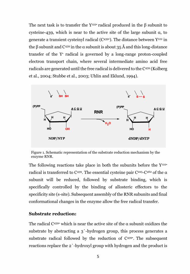
Mechanism:
The enzyme RNR reduces RNA precursors NDPs/NTPs to the
corresponding DNA precursors dNDPs/dNTPs by replacing the 2`-
hydroxyl group in the ribose sugar of the nucleotide with a hydrogen atom
(Figure 1). The mechanism is dependent on a free radical generation and
transfer (Thelander, 1974). The reduction mechanism of the substrate is
initiated by the oxidation of the active site-cysteinyl radical (S•), which is
in the large subunit (α) of the enzyme. The free radical required for the
substrate reduction is generated by a di-mental-oxygen center present in
the subunit β.
Radical generation and transfer:
The small subunit β harbors a di-iron (FeII/FeII) center required for the
free radical generation. The reaction is initiated by the oxidation of
FeII/FeII to FeIII-O- FeIII with O2. One of the oxygen atom together with H+
leaves as an H2O molecule and the other oxygen atom becomes part of the
FeIII-O- FeIII center. Four electrons are needed for the above process. The
first two electrons come from the oxidation of the two FeII ions, and one
electron comes from an external source, which is believed to come from
tryptophan-48 in the β subunit (Baldwin et al., 2000). The fourth electron
comes from the oxidation of tyrosine- 122 of the β subunit, which
generates an unusual stable tyrosyl radical (Y122•) (Eklund et al., 2001;
Kolberg et al., 2004; Nordlund and Eklund, 1993; Nordlund et al., 1990).
5
The next task is to transfer the Y122• radical produced in the β subunit to
cysteine-439, which is near to the active site of the large subunit α, to
generate a transient cysteinyl radical (C439•). The distance between Y122 in
the β subunit and C439 in the α subunit is about 35 Å and this long-distance
transfer of the Y• radical is governed by a long-range proton-coupled
electron transport chain, where several intermediate amino acid free
radicals are generated until the free radical is delivered to the C439 (Kolberg
et al., 2004; Stubbe et al., 2003; Uhlin and Eklund, 1994).
The following reactions take place in both the subunits before the Y122•
radical is transferred to C439. The essential cysteine pair C225-C462 of the α
subunit will be reduced, followed by substrate binding, which is
specifically controlled by the binding of allosteric effectors to the
specificity site (s-site). Subsequent assembly of the RNR subunits and final
conformational changes in the enzyme allow the free radical transfer.
Substrate reduction: The radical C439• which is near the active site of the α subunit oxidizes the
substrate by abstracting a 3`-hydrogen group, this process generates a
substrate radical followed by the reduction of C439•. The subsequent
reactions replace the 2`-hydroxyl group with hydrogen and the product is
Figure 1. Schematic representation of the substrate reduction mechanism by the enzyme RNR.

6
formed. The final product is released by making the reduced C439 to its
oxidized form C439• so the free radical is reintroduced (Figure 1) (Nordlund
and Reichard, 2006).
RNR classification:
The primary mechanism to reduce RNA building blocks to its DNA
building blocks by radical chemistry is well conserved throughout the RNR
evolution. The tertiary structure of the active site, where all RNRs share a
common 10-stranded αβ-barrel and the specificity regulation are also
evolutionarily well conserved (Nordlund and Reichard, 2006). Other than
these similarities, the RNR family differ in the type of substrate used for
the reduction and oxygen requirement during the reaction. The enzyme is
grouped into three broad classes and several subclasses mainly depending
on how the free radical is generated in each catalytic cycle (Table 1)
(Nordlund and Reichard, 2006). All three major classes of RNR; class I,
II, and III are grouped to several subclasses based on the RNR`s catalytic
subunit amino acid sequences. The subclassification includes NrdAe, g, h,
i, k, n and z of the class Ia and Ic, NrdE of the class Ib, NrdJd, f, and m of
class II, and NrdDa, b, c, d, f, h, and I of class III. This type of extensive
subclassification of the enzyme is important to predict the mechanistic
differences in RNR`s family.
Class I:
The class I RNRs are aerobic often referred as to the eukaryotic RNRs
(Cotruvo and Stubbe, 2011a; Nordlund and Reichard, 2006), and this class
is also present in bacteria, viruses and some archaea (Lundin et al., 2010;
Torrents, 2014). Depending on the metal cluster needed for the free radical
generation, the Class I RNR has been further subdivided into three
subclasses; a, b, and c. All three subclasses need oxygen to form a metal-
oxygen center for free radical formation.

7
Table 1. Different classes and subclasses of RNR and their characteristics.
Class Ia Class Ib Class Ic Class II Class III
Distribution
Eukaryotes, several bacteria, viruses, and few archaea
Bacteria and bacterio-phages
Eukaryotes, several bacteria, viruses, and few archaea
Mostly in Bacteria and in a few viruses and archaea.
Mostly in archaea, bacteria, and bacterio-phages
Oxygen requirement
Aerobic Aerobic Aerobic Independent Anaerobic
Catalytic subunit
NrdA (α) NrdE (α) NrdA (α) NrdJ (α) NrdD (α)
Radical generating factors
NrdB (β) NrdF (β) NrdB (β) AdoCbl NrdG
Metal FeIII-O-FeIII
MnIII-O-MnIII or
FeIII-O-FeIII
MnIV-O-FeIII Co 4Fe-4S
Radical Tyr•
…Cys•
Tyr•
…Cys•
Phe•/Leu•/Val•
…Cys•
dAdo•
…Cys• Gly•…Cys•
Additional factors
Thioredoxin and glutaredoxin
NrdI to generate Mn-o-Mn center
Thioredoxin and glutaredoxin
Thioredoxin NrdG to generate Gly• radical
Substrate NDP NDP NDP NDP/NTP NTP
ATP cone Present None Present Rarely present
Present
Active complex
α2β2 and
α6β2-6 α2β2
Similar to class Ia
α, and α2 α2
Inactive complex
α6β2 (eukar.) α4β4 (E. coli) α4β2 (P. aerug.)
None Similar to class Ia
? α2
8
Class Ia: This subclass is also referred to as canonical class 1 RNRs.
Similar to other class I RNRs, it consists of two protein subunits needed
for the enzyme to be active. The gene NrdA encodes for the large subunit
R1 (α), and the gene NrdB encodes for the small subunit R2 (β). The α
subunit harbors a catalytic site and in most of the cases two types of
allosteric sites. The β subunit harbors a free radical generating machinery,
where it generates the Y122• radical by creating a FeIII-O-FeIII center near
the catalytic site. The radical is transferred to the catalytic site of the α
subunit via a long-range electron transport chain (~35 Å) consisting of
aromatic amino acid residues to generate the C439• radical, which
initiates the reduction process. This is the most studied class of the RNR
family and the E. coli RNR is often considered as a prototype.
Class Ib: In the class Ib enzymes, the large subunit R1 (α) is encoded by
the NrdE gene and the small subunit R2 (β) by the NrdF gene. The subunit
α harbors a catalytic site and in contrast to the class Ia enzymes, it harbors
only one allosteric site (s-site). The most significant difference compared
to the canonical class 1 RNRs is the type of metal oxygen center (FeIII-O-
FeIII or MnIII-O-MnIII) required to generate the Y122• radical. This class of
enzymes needs an additional protein, NrdI, for the MnIII-O-MnIII center to
generate the Y122• radical but there is no requirement of this extra protein
for the FeIII-O-FeIII center. Recent studies indicate that the MnIII-O-MnIII
center is the physiological form for the generation of Y122• radical (Cotruvo
and Stubbe, 2011b; Cox et al., 2010; Crona et al., 2011b; Martin and Imlay,
2011).
Class Ic: Like the canonical subclass, the two essential α and β subunits
are encoded by the NrdA and NrdB genes, respectively. In class Ic the
subunit α shares similar features as in canonical class. This subclass has
been classified entirely based on the subunit β (Hogbom et al., 2004).
Unlike the other class I RNRs, the Y122• radical is replaced with a
phenylalanine, leucine or valine, and the subunits are called NrdBPhe,

9
NrdBLeu or NrdBVal respectively. The metal center needed for the
generation of the free radical is the MnIV-O-FeIII center.
Class II: The enzyme activity is independent of oxygen presence in class II RNRs
and present in all three domains of life and in some viruses (Lundin et al.,
2015). The choice of substrates for this class of enzymes is either NDPs or
NTPs. The enzyme has a single protein subunit (α), which is encoded by
the gene NrdJ and the class has been divided into two subgroups:
monomeric and dimeric class II RNRs. The enzymes from this class use
the vitamin B12-derived coenzyme 5′-deoxyadenosylcobalamin (AdoCbl)
to generate the cysteinyl radical.
Class III: The class III RNRs are anaerobic and present in bacteria, bacteriophages,
archaea and some eukaryotes. The enzyme has one subunit, which is α,
and the NrdD gene encodes the protein. The class III proteins use a glycyl
radical for catalysis, which is generated by an additional protein called
activase (NrdG). The activase is not a real subunit because the enzyme will
remain active also after the activase has left the complex. The activity of
the α subunit can remain for several reaction cycles. The subunit α harbors
both the allosteric regulatory sites like in canonical class I RNR and uses
NTPs as substrates.
Coexistence of different RNR classes and different variants: Coexistence of different classes is often an advantage for the organisms
that live in a rapidly changing environment. Coexistence of all three classes
of RNRs has been reported in more than five percent of fully sequenced
bacterial genomes, one archeon, and in a few unicellular eukaryotes
(Hofer et al., 2012; Lundin et al., 2010). One such example is P.

10
aeruginosa, which genome encodes all three classes of RNR, and each
class RNR is differentially expressed depending on the external
environment (Jordan et al., 1999; Sjöberg and Torrents, 2011; Torrents et
al., 2005). Few eukaryotic genomes harbor alternative class Ia RNR
isoforms. The mammalian genome encodes an alternative small subunit
p53R2. The p53R2 protein was originally reported to be crucial for DNA
repair (Tanaka et al., 2000), but later findings showed that the protein`s
primary function is to provide building blocks for mitochondrial DNA
synthesis (Håkansson et al., 2006). The genome of Saccharomyces
cerevisiae encodes two isoforms of each RNR subunit R1 and R3 are
isoforms of subunit α and R2 and R4 are isoforms of subunit β. The R4
subunit is inactive because it lacks some amino acids to form an iron
center but the subunit is still needed for correct folding of the R2 subunit.
Therefore, the active form of the β dimer consists of a R2-R4 heterodimer
(Chabes et al., 2000; Wang et al., 1997). In contrast to the budding yeast,
the class Ia RNR of the Aeromonas hydrophilia bacteriophage (Aeh1) has
an α subunit that consists of two polypeptides (αa + αb) and the
holoenzyme is a (αa + αb)2β2 heterotetramer (Crona et al., 2011a).
Evolution: Perhaps life would not exist without the introduction of RNR to be able to
replace the genetic material from RNA to DNA. The enzyme might have
played a significant role in the transition from the RNA to DNA world. The
common structural features in the active site, the mechanism governed by
the free radical chemistry, and the similar allosteric specificity regulation
in almost all classes of RNRs leads to the conclusion that the RNRs have
evolved by a divergent evolutionary pathway with either class II or class
III RNRs as a common ancestor for the present day RNRs (Reichard, 1993,
1997; Stubbe, 1990, 1998). The recent structural and bioinformatic studies
of the ATP cone domain proposes that this domain is a jumping element

11
that has been lost and gained several times during the evolution, probably
by recombination between the RNR genes (Jonna et al., 2015).
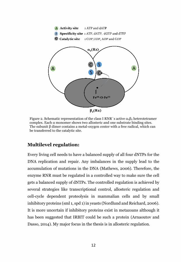
Introduction to Class I RNR: All class I RNR proteins have two subunits, subunit α which is a large
subunit and subunit β which is a small subunit. The α subunit has two
allosteric sites; the specificity site (s-site) and activity site (a-site) (Figure
2). In addition, it has one catalytic site, where the substrate binds. The
specificity regulation via the s-site decides which of the four substrates to
be reduced in the catalytic site to maintain the relative ratios between the
four dNTPs in the cell. The allosteric specificity regulation is well
conserved in all classes of RNRs, and it has been well studied (more details
about the specificity regulation is discussed below). The a-site regulates
the overall activity of the enzyme by switching it on and off. Binding of the
effector ATP makes the eukaryotic RNR hyperactive, and the effector
dATP inhibit the enzyme. The overall activity regulation from the a-site
controls the absolute concentration of the dNTP pool in the cell. The a-site
is located in the N-terminus of the α subunit, which is called the ATP cone
(Aravind et al., 2000), where the effectors can bind. More details about
overall activity regulation are discussed in the overall activity regulation
section.
The subunit β harbors the metal oxygen center (FeIII-O-FeIII), which is
required to generate and stabilize the Y122• free radical (Figure 2). The free
radical is further transferred to generate a transient C439• radical in the
active site of the α subunit. The free radical transfer from subunit β to the
active site of the α subunit is governed by a long-range proton-coupled
electron transport chain. More details about the free radical types are
discussed in the above RNR classification section.
12
Figure 2. Schematic representation of the class I RNR`s active α2β2 heterotetramer complex. Each α monomer shows two allosteric and one substrate binding sites. The subunit β dimer contains a metal-oxygen center with a free radical, which can be transferred to the catalytic site.
Multilevel regulation: Every living cell needs to have a balanced supply of all four dNTPs for the
DNA replication and repair. Any imbalances in the supply lead to the
accumulation of mutations in the DNA (Mathews, 2006). Therefore, the
enzyme RNR must be regulated in a controlled way to make sure the cell
gets a balanced supply of dNTPs. The controlled regulation is achieved by
several strategies like transcriptional control, allosteric regulation and
cell-cycle dependent proteolysis in mammalian cells and by small
inhibitory proteins (sml 1, spd 1) in yeasts (Nordlund and Reichard, 2006).
It is more uncertain if inhibitory proteins exist in metazoans although it
has been suggested that IRBIT could be such a protein (Arnaoutov and
Dasso, 2014). My major focus in the thesis is in allosteric regulation.

13
Allosteric regulation: As mentioned above, RNR is perhaps the most interesting enzyme in the
world and its sophisticated allosteric regulation is one of the reasons why
the RNR is interesting. The subunit α has two allosteric sites; the
specificity site (s-site) and activity site (a-site). Over the past few years,
several RNR structures from different organisms including several
bacteria and few eukaryotes have been resolved and given new insights
into the mechanism of allosteric regulation (Ando et al., 2011; Fairman et
al., 2011; Logan, 2011; Wang et al., 2007).
Figure 3. Allosteric regulation of class I RNR. Allosteric effectors are representd in the circles. a and s letters represent the a and s-sites respectively. The plus and minus circle signs represent the effector's activation and inhibition effects on the enzyme. The enzyme NDP kinase (1) is involved in the final reaction step of dNTP synthesis. The concentration ratio between dCTP and dTTP is controlled by an additional enzyme dCMP deaminase, or dCTP deaminase in some prokaryotes (2). Dotted arrows represents the regulation of dCMP or dCTP deaminase (2) by dCTP and dTTP.

14
Specificity regulation:
In contrast to PCR reaction mixtures, the four dNTPs are not in equimolar
concentration in the cell. The relative ratios and the overall concentration
of each of the four dNTPs differ from species to species. The physiological
dNTP pools in mammalian cells are approximately 37 µM dTTP, 29 µM
dCTP, 24 µM dATP, and 5 µM dGTP (Traut, 1994). The relative dNTP pool
levels in E. coli follow the same trend as in mammals, except that the total
dNTP pool is five to ten times higher (Buckstein et al., 2008). To maintain
the relative dNTP pools, RNR has developed a strategy called allosteric
specificity regulation. The s-site can bind to either of the effectors dATP,
ATP, dTTP, or dGTP and decides what substrate to reduce in the catalytic
site by a conformational change in loop 2, which is located between the s-
site and the catalytic site. The effectors dATP or ATP promotes the
reduction of CDP or UDP, dGTP stimulates ADP reduction, and dTTP
stimulates the reduction of GDP (Figure 3) (Nordlund and Reichard,
2006).
The mechanism of allosteric specificity regulation is well conserved in all
classes of RNRs except in a Herpesviridae class I RNRs, where the enzyme
lacks specificity regulation altogether (Averett et al., 1983). The
mechanism of specificity regulation has been well-studied by using
biochemical, biophysical and structural studies and the regulation follows
the scheme illustrated in Figure 3 (Hofer et al., 2012). When the cell is in
the beginning of s-phase, the effector ATP binds to the s-site and promote
the formation of dCTP/dTTP. The effector dTTP then promotes the
production of dGTP, which further promotes the production of dATP and
the dATP/ATP will start the cycle all over again by binding to the s-site. In
mammalian cells, ATP or dATP hyperactivates or inhibits the enzyme
respectively by binding to the a-site (Figure 3). Thus the dNTP levels are
maintained in the cell. Since dCTP is not an s-site effector, an additional
regulation by dCMP deaminase (dCTP deaminase in some prokaryotes) is

15
needed to maintain the ratio between dCTP and dTTP. The enzyme dCMP
deaminase is allosterically regulated by dCTP (activator) and dTTP
(inhibitor) as shown in Figure 3.
Specificity regulation and oligomerization: The catalytic subunit α is a monomer with no effector bound to the s-site.
The binding of any of the specificity effectors to the s-site promotes the
dimerization of the subunit α with the s-sites and active sites being at the
dimer interface. The subunit α dimerization subsequently promotes the
binding of the β dimer to form an active α2β2 tetramer.
Overall activity regulation: Like the specificity regulation to maintain the relative ratios between the
four dNTPs, the enzyme has developed a strategy called overall activity
regulation to keep the total concentration of dNTPs in the cell. The ATP
cone domain of the a-site regulates the overall activity regulation by
turning the enzyme on and off. In mammalian RNR, the allosteric effectors
ATP and dATP control the overall activity regulation. Binding of the
effector ATP to the ATP cone domain makes the enzyme hyperactive, and
the effector dATP inhibits the enzyme. Around 100 amino acids in the N-
terminus of the α subunit makes up the ATP cone, which is also called a-
site (Aravind et al., 2000). More about the ATP cone is discussed in the
ATP cone section. Both the effectors ATP and dATP can bind to either of
the allosteric sites and regulates specificity and activity regulation. The
effector ATP has a low affinity towards both the sites but can still compete
with the other effectors because the physiological concentration of ATP is
very high (3 mM) in mammalian and E. coli cells (Bochner and Ames,
1982; Buckstein et al., 2008). The effector dATP has 10 to 20 times lower
affinity to the a-site than the s-site (Brown and Reichard, 1969b; Ormö
and Sjöberg, 1990; Reichard et al., 2000). At low concentrations, dATP
acts as a specificity regulator, and at high concentration, it acts as an

16
overall activator by binding to both s- and a-sites. The cellular
concentration of dATP serves as a sensor to turn off the enzyme when it is
enough dNTP pools for DNA synthesis. At high cellular dATP
concentration, RNR senses the proper homeostasis between the four
dNTPs and the dATP further binds to both allosteric sites where it will
inhibit the enzyme via a-site regulation.
ATP-cone:
Bioinformatics studies reveal that the ATP cone distribution is common in
all different classes of RNRs except class Ib which lacks the ATP cone and
class II where it is only rarely present (Nordlund and Reichard, 2006).
Surprisingly multiple ATP cones are commonly observed in all classes of
RNR. Two such examples having multiples ATP cones are P. aeruginosa
and Chlamydia trachomatis class Ia RNRs (Roshick et al., 2000; Torrents
et al., 2006). Among the two ATP cones of the α subunit of P. aeruginosa
class Ia RNR, only the N-terminal ATP cone is functional (Torrents et al.,
2006). The class Ia RNR of the bacteria C. trachomatis have instead three
ATP cones, among that the first two N-terminal cones are functional
(Jonna et al., 2015). Structural and bioinformatic studies show that the
conserved KR(D/N) motif is crucial for the functionality of the ATP cone
domain. Amino acid sequence studies revealed that the arginine (R) in the
conserved motif is replaced by an alanine (A) residue in the nonfunctional
ATP-cone domain. The amino acid arginine is critical for binding of the
phosphate moiety of the overall activity effectors ATP and dATP to the ATP
cone (Jonna et al., 2015). The structural studies of human RNR have
revealed how the ATP cone discriminates between ATP and dATP binding
(Fairman et al., 2011). The ATP cone is formed by four helices which are
covered by a β hairpin at one end forming a pocket. The absence of 2`-
hydroxyl group of dATP makes it bind deeper in the pocket and the residue
Ile 18 seems to be important in the prevention of deeper binding of ATP
by acting as a stereochemical barrier (Fairman et al., 2011).
17
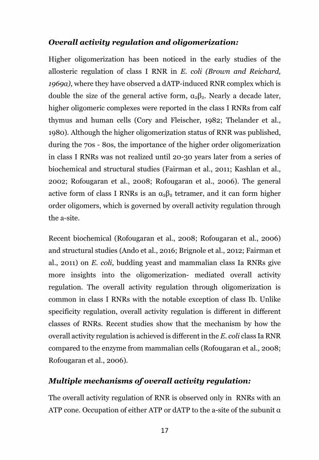
Overall activity regulation and oligomerization: Higher oligomerization has been noticed in the early studies of the
allosteric regulation of class I RNR in E. coli (Brown and Reichard,
1969a), where they have observed a dATP-induced RNR complex which is
double the size of the general active form, α2β2. Nearly a decade later,
higher oligomeric complexes were reported in the class I RNRs from calf
thymus and human cells (Cory and Fleischer, 1982; Thelander et al.,
1980). Although the higher oligomerization status of RNR was published,
during the 70s - 80s, the importance of the higher order oligomerization
in class I RNRs was not realized until 20-30 years later from a series of
biochemical and structural studies (Fairman et al., 2011; Kashlan et al.,
2002; Rofougaran et al., 2008; Rofougaran et al., 2006). The general
active form of class I RNRs is an α2β2 tetramer, and it can form higher
order oligomers, which is governed by overall activity regulation through
the a-site.
Recent biochemical (Rofougaran et al., 2008; Rofougaran et al., 2006)
and structural studies (Ando et al., 2016; Brignole et al., 2012; Fairman et
al., 2011) on E. coli, budding yeast and mammalian class Ia RNRs give
more insights into the oligomerization- mediated overall activity
regulation. The overall activity regulation through oligomerization is
common in class I RNRs with the notable exception of class Ib. Unlike
specificity regulation, overall activity regulation is different in different
classes of RNRs. Recent studies show that the mechanism by how the
overall activity regulation is achieved is different in the E. coli class Ia RNR
compared to the enzyme from mammalian cells (Rofougaran et al., 2008;
Rofougaran et al., 2006).
Multiple mechanisms of overall activity regulation: The overall activity regulation of RNR is observed only in RNRs with an
ATP cone. Occupation of either ATP or dATP to the a-site of the subunit α
18
at physiological conditions (where the s-site is also occupied) initiates the
overall activity regulation, by altering the RNR oligomeric status.
Interestingly, Trypanosoma brucei and bacteriophage T4 class Ia RNRs
do not have dATP-induced overall activity inhibition though they have an
ATP cone which can bind dATP (Hofer et al., 2012). Recent structural,
biochemical and bioinformatic data reveal that there are two different
mechanisms (the E. coli and eukaryotic models) to achieve overall activity
regulation. In my first publication, we have discovered a third way of
overall activity regulation, which I will discuss in the section of results and
summary.
Eukaryotic model of overall activity regulation-S. cerevisiae, slime mold, and mammalian (mouse and human): Most of the details about the molecular and structural mechanisms behind
the overall activity regulation of eukaryotic class Ia RNR come from the
following published biochemical and structural data on yeast and
mammalian RNRs (Ando et al., 2016; Crona et al., 2013; Fairman et al.,
2011; Hofer et al., 2012; Logan, 2011; Rofougaran et al., 2006). The
general active RNR complex governed by s-site effectors is an α2β2
tetramer. Overall activity regulation in eukaryotic class Ia RNRs is
regulated by binding of either ATP (hyperactivation) or dATP (inhibition)
to the a-site of the ATP-cone. Oligomerization-driven overall activity
regulation was under debate until the studies of the mouse class Ia RNR,
where our lab has used a novel method for oligomerization status called
GEMMA. This study revealed that the effectors ATP and dATP induce
higher complexes, which are active α6 and inactive α6 complexes
respectively (Figure 4). Because the technique GEMMA is relatively new,
the results were reconfirmed by using size exclusion chromatography and
to get more exact results of the mass electrospray ionization mass
spectrometry was used. The active and inactive α6 complexes further bind
to the β2 dimer and generate an ATP-induced active α6β2 complex and
19
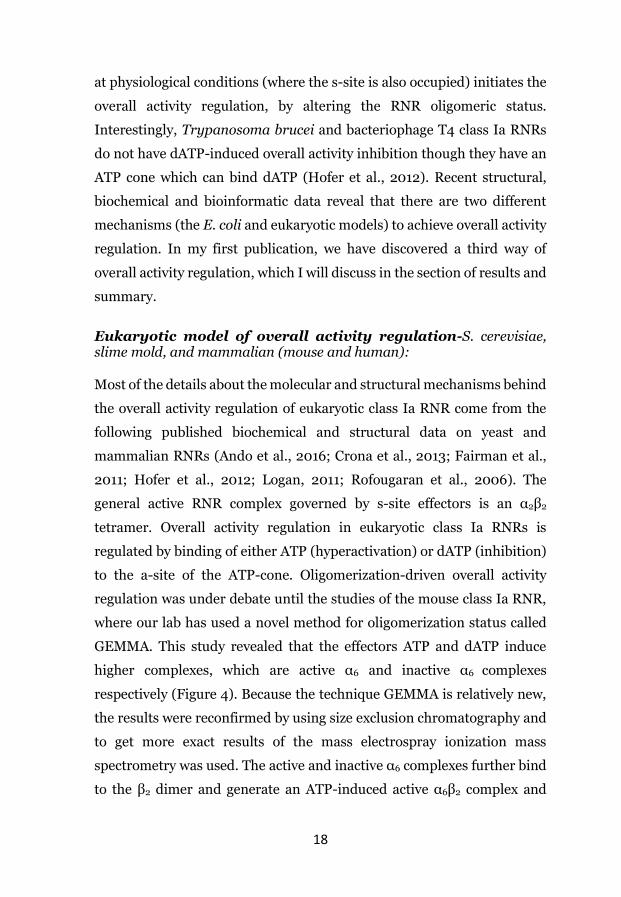
dATP-induced inactive α6β2 complex (Figure 4). Thus both the a-site
effectors induce the same quaternary complex with functionally different
oligomers. In contrast to the published ATP-induced active α6β2 complex
(Rofougaran et al., 2006), later gel filtration studies show that ATP
induces an α6β6 complex in the presence of the substrate CDP analog
gemcitabine-5′-diphosphate (Wang et al., 2007). Though this is not a
natural substrate for RNR, the chances of the existence of an active α6β6
complex cannot be ruled out.
Figure 4. The eukaryotic model of overall activity regulation. The general active form is α2β2 complex induced by specificity site effectors binding to the s-site. Binding of either ATP or dATP to the a-site induces the active α6a or inactive α6i complexes respectively, which further binds up to one β dimer in the case of the inhibited α6 complex and one to three β dimers in case of the activated α6 complex.
The molecular mechanisms behind the induction of similar quaternary
structures with functional differences by the effectors ATP and dATP
remained a question until the advancements in solving the structure of S.
cerevisiae class Ia RNR (Fairman et al., 2011). The x-ray crystal structure
revealed that the dATP-induced α6 complex is a ring-like structure formed
by three α dimers. dATP losts its ability to produce an α6 ring formation
when a point mutation (D16R) was introduced on each dimer surface. On
the other hand, ATP could still induce the hexamerization even in the
D16R mutant, which proves that α hexamers induced by ATP and dATP

20
are structurally different. Further studies with cryo-electron microscopy
(cryo-EM) revealed the details about the binding of the β dimer to the
inactive α6 ring, where the β dimer sits inside the ring in a way that the
electron transport chain is interrupted between the β and α subunits.
Whereas it is known for sure that dATP induces α6β2 complexes, it is more
uncertain how many β dimers can bind to the ATP-induced hexamer and
the complex is therefore described as an α6βn complex. The structural and
molecular mechanisms behind why the complex is active are yet to be
discovered (Fairman et al., 2011; Rofougaran et al., 2006).
Prokaryotic model of overall activity regulation: E. coli class Ia RNR is the most studied and understood enzyme among all
the classes, and it is often mentioned as a prototype of class Ia RNRs.
During early years of RNR research, Reichard and coworkers investigated
the oligomerization status of the E. coli class Ia RNR by using
ultracentrifugation and found that ATP induces an active α2β2 complex
and dATP induces an inhibited complex which is twice the size of the active
complex (Brown and Reichard, 1969a). Recent biochemical and structural
studies of the mammalian and S. cerevisiae proteins provided much more
information on overall activity regulation (Fairman et al., 2011;
Rofougaran et al., 2006). Further studies from our lab have started to
unveil the overall activity regulation of the prototype class Ia enzyme from
E. coli, and then proceeding studies from Drennan`s group completely
revealed the molecular mechanism of the E. coli class Ia enzyme overall
activity inhibition by dATP by being able to crystalize the complex.
Though the outcome (to maintain the absolute concentration of dNTP
pools) of the overall activity regulation is similar, the E. coli enzyme has
many differences when compared to the yeast and mammalian enzymes
(Figure 5 and 10). The differences are mostly about how the overall activity
regulation is performed. Unlike the eukaryotic proteins, the E. coli enzyme

21
does not make ATP-induced active complexes which are larger than α2β2.
The enzyme uses a cross-talk strategy to become inhibited. This inhibition
can occur by dATP alone or by a combination of ATP and dNTPs. dATP at
high concentration binds to both the sites and inhibits the enzyme by α4β4
formation. A second way is that the effector ATP from the a-site can inhibit
the enzyme by sensing the concentrations of dNTPs by their occupation of
the s-site. Thus the E. coli class Ia RNR is cycling between active α2β2 and
inactive α4β4 complexes by a cross-talk between the allosteric sites (Figure
5). In contrast, ATP has always an activating effect from the a-site in
eukaryotic RNRs. The enzyme from E. coli also differs from the eukaryotic
enzymes based on the requirements of the β subunit to form the inactive
α4β4 complex. The E. coli enzyme cannot form an inactive α4 complex.
Instead, it forms an inactive α4β4 complex by dimerization of the α2β2
tetramer. However, the eukaryotic enzyme can form ATP-induced α6a
complexes (active form) and dATP-induced α6i complexes (inactive form).
Further binding of the β dimers leads to the formation of active α6βn and
inactive α6β2 complexes (Figure 4 and 10).
The above studies show the significant differences of how the RNR is
inhibited by dATP-induced overall activity regulation in correlation with
the oligomerization of its subunits (Figure 10). They clearly show that
there are two different overall activity regulations to achieve the inhibitory
effect. The eukaryotic model is well established and is studied in several
organisms like baker`s yeast, several mammal species, and Dictyostelium
discoideum. Because of the limited information available about the overall
activity regulation of other bacteria than E. coli, it has not been clear if the
overall activity mechanism that the E. coli class Ia RNR uses is common
to all the bacterial class Ia RNRs or if there are other types of mechanisms
yet to be discovered in other types of bacteria. Therefore, it is important to
study the overall activity regulation from different bacterial class Ia RNRs.
It is clear that the overall activity regulation coupled with higher order
oligomerization is present in the organisms with a functional ATP cone
22
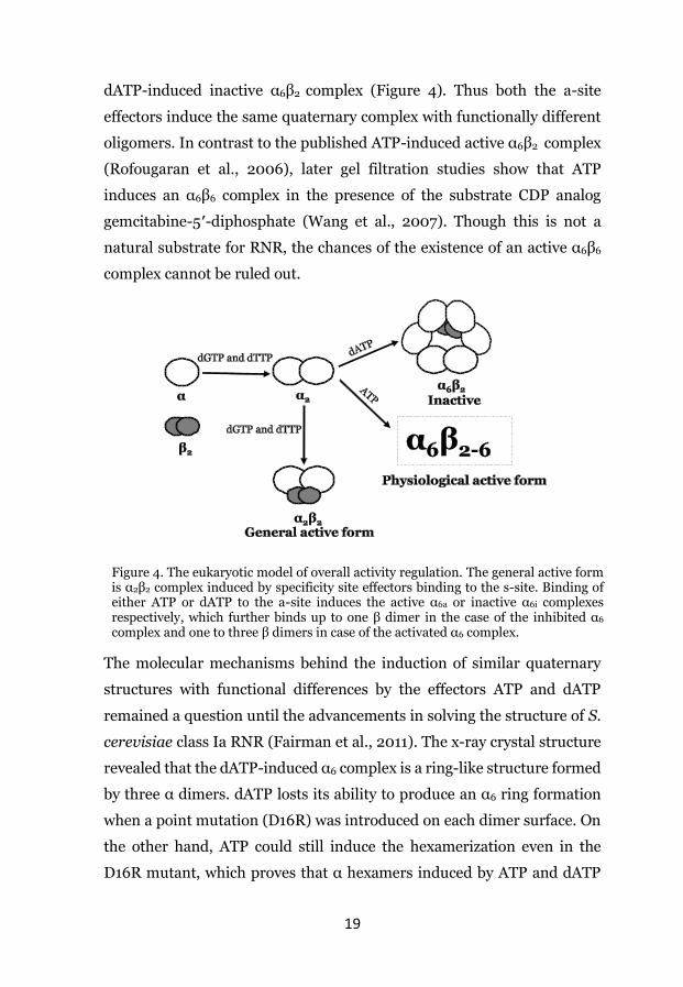
and the ATP cone is believed to be lost and gained several times during the
evolution. Therefore it is likely that there are several mechanisms of
overall activity regulation, where each mechanism develops when the
ATP cone is lost and gained.
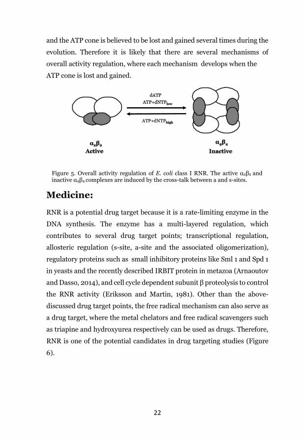
Medicine: RNR is a potential drug target because it is a rate-limiting enzyme in the
DNA synthesis. The enzyme has a multi-layered regulation, which
contributes to several drug target points; transcriptional regulation,
allosteric regulation (s-site, a-site and the associated oligomerization),
regulatory proteins such as small inhibitory proteins like Sml 1 and Spd 1
in yeasts and the recently described IRBIT protein in metazoa (Arnaoutov
and Dasso, 2014), and cell cycle dependent subunit β proteolysis to control
the RNR activity (Eriksson and Martin, 1981). Other than the above-
discussed drug target points, the free radical mechanism can also serve as
a drug target, where the metal chelators and free radical scavengers such
as triapine and hydroxyurea respectively can be used as drugs. Therefore,
RNR is one of the potential candidates in drug targeting studies (Figure
6).
Figure 5. Overall activity regulation of E. coli class I RNR. The active α2β2 and inactive α4β4 complexes are induced by the cross-talk between a and s-sites.

23
Although RNR is an obvious target for antimicrobial and antiproliferative
drugs, there are only a few novel drugs that have been characterized so far
(Table 2) because of the lack of a high throughput screening method for
RNR activity until recently (Tholander and Sjöberg, 2012). The newly
developed high-throughput PCR-based RNR activity study in microwell
format allows the screening of several drugs at a time (Tholander and
Sjöberg, 2012). With this technique, they screened several drugs and
found 27 drugs which target the P. aeruginosa class Ia RNR (Tholander
and Sjöberg, 2012).
Figure 6. Different strategies to target the enzyme RNR

24
Table 2. Established drugs in cancer therapy
Inhibitor Target type Cancer type
Hydroxyurea
Radical scavenger and metal chelator
Myeloid leukemia, melanoma, and head and neck cancer
Triapine Metal chelator In clinical trials
Gemcitabine Alters the α subunit oligomerization
Lung, bladder, breast, ovarian and pancreatic cancers.
Clofarabine Oligomerization Refractory pediatric leukemia.
Cladribine Oligomerization Hematological cancers
Fludarabine Oligomerization Hematological cancers
Nucleoside analogues

25
Methodology:

26
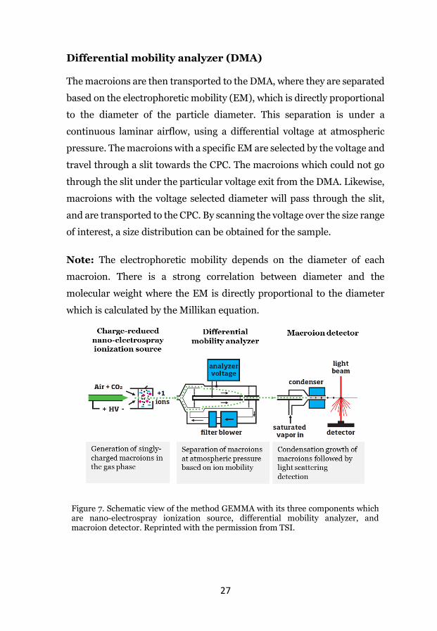
Gas-phase electrophoretic macromolecule analysis (GEMMA): The technique GEMMA (Bacher et al., 2001; Kemptner et al., 2010) is used
to determine the molecular mass of globular proteins and viruses and is
relatively new in the field of biological science. The updated version of
GEMMA is called MacroIMS-Macroion Mobility Spectrometer and the
provider is TSI. The TSI company`s expertise is in aerosol and particle
research, and recently they have expanded their products to biological
studies. The three units ESI, DMA, and CPC put together is called
GEMMA (Figure 7). The underlying principle mechanism of GEMMA is
the detection of the electrophoretic mobility of the charge-reduced ionized
biopolymers.
Electrospray aerosol generator (Nano-electrospray ionizer) ESI:
This unit contains a sample chamber, an electrode and a silica capillary.
The aqueous solution with the biopolymer in question is transported
through the capillary by atmospheric pressure and high voltage. When the
sample reaches the capillary tip, it forms a cone jet of differentially charged
monodisperse aerosol droplets with an average diameter of 160 nM. In
principle, each droplet analyzed contains only one biomolecule. Then the
monodisperse aerosol droplets are transported to the charge reducing
chamber where each droplet is dried and charge reduced to single positive
or negative charged aerosols (macroions) by means of a polonium source,
which introduce neutralizing gas ions in the air/CO2 mixture that enters
the chamber.
27
Differential mobility analyzer (DMA) The macroions are then transported to the DMA, where they are separated
based on the electrophoretic mobility (EM), which is directly proportional
to the diameter of the particle diameter. This separation is under a
continuous laminar airflow, using a differential voltage at atmospheric
pressure. The macroions with a specific EM are selected by the voltage and
travel through a slit towards the CPC. The macroions which could not go
through the slit under the particular voltage exit from the DMA. Likewise,
macroions with the voltage selected diameter will pass through the slit,
and are transported to the CPC. By scanning the voltage over the size range
of interest, a size distribution can be obtained for the sample.
Note: The electrophoretic mobility depends on the diameter of each
macroion. There is a strong correlation between diameter and the
molecular weight where the EM is directly proportional to the diameter
which is calculated by the Millikan equation.
Figure 7. Schematic view of the method GEMMA with its three components which are nano-electrospray ionization source, differential mobility analyzer, and macroion detector. Reprinted with the permission from TSI.

28
Condensation particle counter (CPC) After the DMA, that the macroions reach the CPC for quantification. The
macroions that reach the CPC are too small to be detected by the laser
optical device. The n-butanol vapor condensation process makes the
macroions large enough (10 µm) to be detected by the instrument for the
quantification.
Advantages and disadvantages: GEMMA is a relatively new mass spectrometry-related method to quantify
biomolecules, and it certainly has many advantages compared to the
methods that are already being used. The most important benefits are the
small required sample quantity (0.01 mg/ml of protein in 20 µl), and the
short time of analysis (2 min). Most mass spectrometry-based techniques
like matrix-assisted laser desorption/ionization (MALDI), and
electrospray ionization are suitable methods to quantify accurate
molecular masses of non-covalent high molecular weight biomolecules.
But these two approaches have some limitations (Bacher et al., 2001); (i)
method optimization for each sample, (ii) difficulty in charge
determination of multiply charged ions at high mass range, (iii) complexes
with hydrophobic interactions are very sensitive to decomposition under
the high vacuum, and (iv) extremely low sensitivity for large complexes.
The latter factor makes the method not suitable for quantification because
of a strong underestimation of oligomerization. Size-exclusion
chromatography has also limitations and we found that it was difficult to
separate and quantify mixtures of different protein complexes which are
in fast equilibrium between the complexes. On the other hand, GEMMA is
an advantageous method to detect and quantify different oligomeric
protein complexes which are in rapid equilibrium with noncovalent
interactions.

29
The main limitation for GEMMA is a requirement of only using volatile
salts and buffers such as ammonium acetate. In RNR research, GEMMA
is proven to be a suitable method to study the oligomerization status of the
subunits (Rofougaran et al., 2006), but the method has limitations
dependent on the protein, where some proteins clog the capillary by
binding. The problem with the clogging can be overcome by using soft
detergents like Tween-20, or by using the updated version of GEMMA
termed Macro-IMS, which seems to have fewer problems with the clogging
due to that the capillary length is minimized.

30
Results and Summary Paper I: Diversity in Overall Activity Regulation of Ribonucleotide Reductase
The mechanism behind the overall activity regulation of RNRs is only
partially known. Previous studies of class I RNRs have revealed that the
overall activity regulation is dependent on the formation of oligomeric
complexes. Until now, there are two types of mechanisms to achieve
overall activity regulations that have been reported, a eukaryotic model
where the mammalian and yeast enzymes cycle between structurally
different active and inactive α6β2 complexes (Fairman et al., 2011; Hofer et
al., 2012; Rofougaran et al., 2006) and a prokaryotic model where
Escherichia coli enzyme cycling between active α2β2 and inactive α4β4
complexes (Rofougaran et al., 2008; Uhlin and Eklund, 1994). These two
mechanisms are here presented to be the outcome of the loss and gain of
ATP cones during the evolution. One such example where an extra ATP
cone is gained is the class Ia RNR of P. aeruginosa. From previous studies,
it is known that P. aeruginosa has all three classes of RNRs and that the α
subunit of its class Ia RNR has two ATP cones, of which only the N-
terminal ATP cone is functional (Torrents et al., 2006).
The quaternary structure of ATP-induced P. aeruginosa class I RNR
analysis from the previous studies (Torrents et al., 2006) is ambiguous,
which is a common limitation of gel filtration studies with rapidly
equilibrating protein complexes. Therefore in this study, we have used a
technique termed Gas-phase electrophoretic macromolecule analysis
(GEMMA) to investigate the oligomerization status of the P. aeruginosa
class I RNR. This method is an alternative method which is suitable to
study different oligomeric complexes in fast equilibrium. In this project,
the allosteric regulation of P. aeruginosa class I RNR is analyzed by using
the following methods; GEMMA, ITC, enzyme activity assays, and
nucleotide filter binding assays.

31
We have started with four substrate enzyme assays to see if this enzyme
follows the same specificity regulation as other enzymes and we found it
follows the same trend except for UDP, which is a very poor substrate for
the RNR in this organism. It seems that most of the dTTP comes from an
alternative pathway where CDP reduction is followed by deamination
(explained in the specificity regulation section). This pathway is common
in many organisms but likely to be especially crucial in P. aeruginosa.
We have examined the oligomerization of the subunits in the presence of
different combinations of the allosteric effectors. The subunit β is mostly
a dimer irrespectively of any effector presence. The subunit α is mostly
monomer with no effectors, but we could see minor populations of dimers
and tetramers. The small amount of dimerization of the α subunit with no
effector is common among other characterized class Ia RNRs, but
tetramerization is something new to this organism. Later, we repeated the
experiments in the ~∆147 N-terminal ATP cone deleted mutant and the
results show that the mutant cannot form any tetramers. Therefore, it is
clear that the tetramers that we have observed in the wild-type protein
with no effectors are real. Subsequently, we have analyzed the effect of
allosteric effectors on the oligomerization status of the subunit α alone and
in combination with the subunit β. The results show that all the effectors
including s-site effectors induce α tetramerization. The tetramers induced
by s-site effectors dissociate into dimers in the presence of Mg2+ ions
(physiologically relevant) and forms an active α2β2 tetramer by binding to
the β dimer. However, the dATP-induced tetramers are stable also in the
presence of Mg2+ and readily bind to the β2 dimer forming an α4β2 hexamer
(Figure 8), which is inactive (enzyme activity assay data, Figure 7D in
paper I).
The nucleotide filter binding and enzyme activity assay experiments gave
us further details about how the allosteric effectors are binding to the
allosteric sites and make the enzyme active or inactive. Perhaps the most

32
striking observation in this publication is that there are two dATPs binding
to the functional N-terminal ATP cone. This is the first ever reported case
where two dATPs bind to the a-site. Surprisingly, dGTP which is generally
known as a s-site effector can also bind to the a-site in this organism.
However, this binding has no physiological effect because ATP readily
competes out the dGTP from the a-site and the dGTP-induced tetramers
are dissociated to dimers.
To study the role of the ATP on overall activity regulation, we have
performed enzyme activity assays. In a comparative study, we have
checked the ATP function from the a-site in mouse and P. aeruginosa class
Ia RNRs. We have performed GDP reduction with saturated amounts of
dTTP (2 mM dTTP completely saturates the s-site) and analyzed the
enzyme activity by adding increasing amounts of ATP (in this case ATP
only binds to the a-site). As previously described (Eriksson et al., 1979;
Rofougaran et al., 2006), the GDP reduction activity of the mouse enzyme
raises 2-3 fold by increasing the ATP concentration with saturated
amounts of dTTP, whereas ATP has no such effect on the the P. aeruginosa
enzyme. This shows that ATP has an activating effect through the a-site on
the mouse enzyme and no effect on the P. aeruginosa enzyme. Further
experiments on the P. aeruginosa enzyme showed that although the ATP
does not have any direct activation effect it will passively activate the
enzyme by competing out the binding of dGTP and dATP to the a-site.

33
Figure 8. Overall activity regulation of P. aeruginosa class I RNR. The active α2β2
complex is induced by all the effectors except dATP. The inactive α4β2 complex is induced by dATP through a-site.
Conclusions As indicated before (Torrents et al., 2006), we confirm that there is a third
kind of allosteric activity regulation in the P. aeruginosa class Ia RNR
when compared to the E. coli and mammalian enzymes (Figure 10). These
many mechanisms behind the overall activity regulation are interesting
from an evolutionary viewpoint and from a drug development view, where
the specific features of each pathogen can be targeted (Figure 9).

34
Paper II: Structural Mechanism of Allosteric Activity
Regulation in a Ribonucleotide Reductase with Double
ATP Cones
In paper 1, we have used different biochemical methods to characterize the
P. aeruginosa class I RNR overall activity regulation in correlation with
the oligomerization. The second article is a natural continuation of my first
article. Here we have studied the molecular mechanism behind the overall
activity regulation of P. aeruginosa class I RNR with structural and
biochemical techniques. Our coauthors have done the structural analysis
of the enzyme by using crystallography and small-angle X-ray scattering
(SAXS) and we have provided the biochemical evidence to strengthen their
structural data.
The 2.3 Å resolution dATP-induced structure of subunit α is a tetramer
which is in agreement to our paper 1 results. The structure clearly shows
the occupation of two dATP molecules neutralized by Mg2+ on the ATP
cone 1 domain. We did biochemical studies on three mutants where the
structurally defined interaction surfaces were altered to confirm the
structural data with biochemical evidence. With site-directed
mutagenesis, we have created three different point mutations on subunit
α, which are R119D, E106A/E126A, and H72A/D73A/Y830A. The
biochemical studies include GEMMA, enzyme activity measurements, and
nucleotide binding experiments to study the overall activity behaviors of
the three mutant enzymes and compare with the wildtype. The Scatchard
plot results show that each of all three mutants can bind up to three dATP
molecules (Figure 5 in paper II). The GEMMA analysis shows that the
R119D, and E106A/E126A mutants have lost their ability to tetramerize.
Interestingly, the mutant H72A/D73A/Y830A has only a partial
tetramerization ability. To check the oligomerization status of the mutant
α subunit in relations to the activity, we have performed enzyme activity

35
assays. These results show that the mutants R119D, and E106A/E126A
have lost the ability to be inhibited by dATP which agrees with the
GEMMA results where these two mutants cannot tetramerize (Figure 5 in
paper II). The mutant H72A/D73A/Y830A could only partially be
inhibited by dATP, which is in agreement with the GEMMA data.
The above results provide the structural evidence of a third kind of overall
activity regulation in P. aeruginosa class I RNR, which is quite different
from the previously two known eukayotic and E. coli models (Figure 10).
The sequence analysis data shows that the occurrence of multiple ATP
cones is widely spread in different groups and subgroups of RNRs. The
structural and sequence data helps us to predict the presence and
functionality of multiple ATP cones in different organisms and at the same
time, it gives us the information to predict the binding of more than one
dATPs to the predicted functional ATP cone. Since we know the details
about the molecular mechanisms behind the unusual binding of two
dATPs to the functional N-terminal ATP cone, this information can lead
us to develop new dATP analogues which target the P. aeruginosa class I
RNR with minimized side effects on the host system (see the future plans
and figure 9). One possibility could then be to connect two dATP analogues
into one molecule.

36
Paper III: A ribonucleotide reductase inhibitor with deoxyribonucleoside-reversible cytotoxicity RNR controlled dNTP synthesis is crucial for DNA replication fidelity and
repair. The characteristic of most cancer types is uncontrolled DNA
replication and repair, which can be the consequence of RNR
misregulation. Therefore the enzyme is undoubtedly a good target for
anticancer drugs. There are several established drugs like gemcitabine and
hydroxyurea, which are already being used in cancer therapy. The drug
hydroxyurea is a metal chelator and radical scavenger. It removes the
radical and radical generating di-iron center of the β subunit which makes
the enzyme inactive (Aye et al., 2015; Gräslund et al., 1982). It has been
reported that cells treated with the drug hydroxyurea become resistant by
upregulating the β subunit (Aye et al., 2015; McClarty et al., 1987;
Åkerblom et al., 1981) and its metal chelating property can target other
metalloproteins like carbonic anhydrase (Temperini et al., 2006).
In a recent PCR-based drug screening study, novel inhibitors that target
and inhibit the P. aeruginosa class I RNR have been found (Tholander and
Sjöberg, 2012). These drugs are also inhibitors of the human RNR. Our
paper III deals with the effect of non-nucleoside RNR inhibitors on human
RNR and cellular proliferation. Out of several human inhibitors studied,
the drug NSC73735 possesses many characteristics to be a potential lead
drug candidate to target RNR.
Our coauthors initiated this study. The biological activity, RNR inhibition,
binding properties to the RNR subunits, cytotoxicity, and effect on
deoxynucleotide pools of the inhibitors were tested by using enzyme
activity assays, thermal shift assays, FACS, and HPLC respectively. All the
above analyses provided the drug candidate NSC37375 as the best hit
among the 12 drugs analyzed. In this article, I was involved in the

37
oligomerization experiments by using GEMMA (Figure 1 and
supplemental figure S2 in paper III). We have studied the mechanism of
how NSC37375 inhibits the enzyme. From the thermal shift assays, it is
evident that the drug binds to the α subunit of the enzyme. Here, we have
analyzed the effect of the drug on the oligomerization status of the enzyme.
The experiments were performed either by incubating the α subunit with
ATP+NSC73735 or dATP+NSC73735. The results clearly show the
destabilization of the ATP-induced active α hexamers and dATP-induced
inactive α hexamer to dimers in the presence of the inhibitor NSC73735.
The nucleotide pool studies show that the drug-driven RNR inhibition at
physiological conditions is by destabilization of the ATP-induced hexamer
complex.
Conclusion: The antiproliferative molecule NSC37375 is the first reported molecule
that interferes the oligomerization of the large subunit of RNR to inhibit
human RNR. The molecule NSC37375 is reported to have more specificity
to human RNR than the currently used antiproliferative drug
hydroxyurea.

38
Future plans: P. aeruginosa is a gram-negative opportunistic pathogen in
immunocompromised patients and causes severe nosocomial infections
like pneumonia and septic shock (Torrents et al., 2006). It develops
resistance to most of the established antibiotics, and targeting nucleotide
metabolism can be a new choice of making drugs. The studies of our first
paper (Jonna et al., 2015) combined with the drug targeting studies
(Tholander and Sjöberg, 2012) could be an important step towards the
establishment of novel drugs to target RNRs of pathogenic organisms.
Targeting P. aeruginosa RNR. We have observed several differences in the allosteric regulation of P.
aeruginosa RNR when compared to the mammalian RNR (paper I and II).
In this project, we are planning to specifically target the a-site binding
properties of the P. aeruginosa RNR since we know that it has broad
specificity. In particular, the a-site of the α subunit can bind dGTP, which
is unique to this parasite and dGTP induces tetramer formation, which is
the inactive RNR complex. A possibility could be that novel
deoxyguanosine analogues can be developed, which specifically target the
protein (Figure 9).
So far, we have only done studies on the isolated enzyme but our idea for
the future is that the effect of the corresponding target studies will be
evaluated on the growth and dNTP pools of P. aeruginosa cells with
mammalian cells as a reference.
We are also planning to use GEMMA as a screening tool to check the
oligomerization status of the P. aeruginosa RNR in the presence of
inhibitors, which we know inhibit the P. aeruginosa RNR but the
mechanism is not known. Here, we will study the ability of the inhibitors
to inactivate the enzyme by altering the oligomerization status from an

39
active α dimer to its inactive monomer or tetramer (Figure 9). Once we
find a potential inhibitor that alters the oligomerization state of the
enzyme, we will study the binding properties of the inhibitor to the
enzyme.
Multiple mechanisms of allosteric overall activity regulation In paper 1, we have observed several differences in the allosteric regulation
and a novel kind of allosteric overall activity mechanism of P. aeruginosa
RNR when compared to the two known regulation models primarily
characterized in E. coli and mammalian RNRs (Figure 10). In future
studies, our idea is to identify the possible occurrence of even more
allosteric overall activity regulation mechanisms in the class I RNRs of
different organisms. We are especially interested in RNRs from different
pathogens to exploit the differences in overall activity mechanism to
specifically target the pathogenic RNRs. We then start with bioinformatic
analysis to find organisms, which are distantly related, based on the RNR
protein sequence similarities and therefore more likely to contain new
Figure 9. Three different strategies to target the P. aeruginosa class I RNR includes either by stabilizing the inactive complex α4 or by dissociation of the active complex α2 to its inactive monomers.

40
allosteric mechanisms of overall activity regulation. Once we have selected
RNR candidates from the bioinformatic analysis, each RNR candidate will
be cloned for protein expression and purification. Allosteric overall
activity regulation is studied by GEMMA, filter binding studies, enzyme
activity assays, and other methods that our group has been using for to
study oligomerization and allosteric regulation. This project will
subsequently lead to several subprojects.
Figure 10. Three known mechanisms of overall activity regulation in class I RNRs

41
Acknowledgements:
Umeå is my second home after all these years. I have been living here for about 11 years and of course, I have got several friends, whose support made my journey smooth and happy. I start with Anders Hofer, he is my mentor. Without his continuous guidance and support, I would not imagine myself to this moment of my life. He is the best teacher that I have come across in my life. I feel gratitude to Sven Carlsson and Andrei Chabes for giving me feedback on my work during seminars. I would also like to thank my labmate Farahnaz Ranjbarian. It has been almost six years that we know each other and you are the best labmate that I ever had. Thank you for your concern about myself and now I promise you that I will do what I have been telling you all these years. I also want to thank my wonderful officemates, Saima and Andreas for helping me in structure related questions and Khallil for having fun discussions. I would like to thank, Ikenna, Marcus, Phong for having good lunch talks. Jeanette and Sonja for helping me to translate letters from Swedish to English. Ingrid Råberg, Jenny Fossen, Anna Sjöström and Clas Wikström for helping me in solving administrative problems and my visa process. I am thankful to Kristoffer for ITC work, Ulf persson, and Sushma, for helping me in the yeast work, and Vladimir for discussing RNR related questions, Elisabeth for helping us in autoclaving. Stefanie Mangold and Joanna Potrykus: You both are my first mentors and I have learned many things from you. Though I am not in regular contact with you, I always remind you. Marios and Melis: We know each other from almost eleven years. I always remember the amazing lunch meetings that we had together. I hope we will have continuous friendship forever. I still remember Katerina, Julia, and Bindu from our Master`s program. Suman; Thank you for accompanying me to Umeå for the first time. Most of the best times in Umeå that I had is with you. For sure we will be in touch rest of the life. Murali (computer expert), Brahmaiah

42
(job expert), Mahesh (dal specialist) Karunakar (my bank) Srinu Lingala (party animal), Prasanth (neuroscience), Srinu Oruganti (big brother), Sridhar (share market), Ravi sha (tagore), Vivek, Sarvana, Dinakar and many others made my journey smooth and happy. All these years we shared all the emotions (mostly happiness and little fightings). Ramesh Tati: I never forget the days when we were teaching in Satavahana college. You are the first guy who introduced me to Umeå and Lund. I think you are among one of the few guys that I must say thank you. I wish all the best for you and your family. Thanks again for your`s, Praveen Papareddy`s, and Rjender Baddam`s support during my stay in Lund. Ravi, Vidhya, and Kanasu: Ravi, Thank you for your guidance all the time when I need it. Thank you, Vidya for giving suggestions and support to Bindu. I am sure Kanasu and Eeshu having the best times in the school. Munender, Sharvani and Adhya: You are definitely as part of my family. Munender you are my brother, well-wisher, advisor and what not. Thank you very much for your support in the lab when you were in Anders lab. Thank you Sharvani for helping and supporting Bindu. I have seen Adhya from his day one and he is amazingly grown up. You became, even more, close when you were here for the last few months and it was not easy for us when you three were leaving Umeå. Sisir, Santhi and Gamana: If I start writing about you Sisir (bava garu), I am sure that I don't have enough space in the thesis. It has been eleven years that we have met in Stipendiegränd 10D. I still remember the long nights and never ending happiness that we have shared among our friends in olden days. Those were the best times when all our friends were here in Umeå. Santhi (chelli), I always remember your first experience of snow in Umeå and amazing times in Örebro. Gamana (mena kodalu), though we have not met you yet, I am sure Eeshu and you will have best times when you meet each other. Karthik and Gowthami: Since Gowthami is here in Umeå I think we meet almost every weekend for dinners. You both are very nearby heart, not only for me but also for Bindu and Eeshu. Thank you Karthik once again for finishing and releasing our short film as per the schedule, you know what I mean right.

43
Pramod, Neetha and Praneetha: Pintu, the first thing that reminds you is your Bismilla bath (dude it is not Bismilla bath, it is Bisibelebath). I admire your traveling spirit and you are the first guy that I would contact about traveling tips. You have a great family and I wish you three will have a wonderful life ahead. Sujith and Ramya: Though we met recently, both of our families became very close. I thank you Ramya for your support to Bindu and I think Eeshu had best times with you and Sujith. Soumaya and Hareesha: You both are amazing friends to me. I am missing all the good times that we had. I still remember the badminton that we have played the whole night. I wish all the best for your bright future. Chaitanya (my beloved brother), Harsha (electronics expert). I remember all the movies and TV series that we have watched together and you are my best roommates ever. I cannot forget the delicious ICA pizzas that we had every Friday. Edvin, Maria: I think we both are among few of our friends who survived in Umeå long time. You will be the first person that I would contact about movies. I wish you and your family will have a great future. Mohan and Madhavi: Thank you very much for both of you for the dinners and good times that we have spent. When it comes to the food, no doubt Jani is the best cook, especially biryani and katta. I remember, at one point we used to meet every weekend for Jani`s biryani. Lalitha and Surya, I am very happy for the dinner times that we have spent and thank you Surya for talks during my visa process. Bharat, Yugi, and Sarath: Thank you very much for your support all the time. I always remember the good times and sharings that we have gone through. Sai Madhav: It has been quite a while since you have moved to the US, but I still appreciate your help, sharing a house with me. I always admire your commitment to physical fitness. Hanuma Kumar and Vishnu: Kumar, you are more than my brother and I appreciate your never ending kindness to others. Now

44
you have a great family and I wish that you will have a successful life ahead. Vishnu, thank you very much for sharing your robotic ideas and the chocolates that you bring every time we meet. Durga prasad, you are one of my best friends. I feel very happy when I remember all the things that we have done together in the college days. Thank you very much for introducing me to the teaching field. Balu (more than a friend), Lenin (closest friend), Surya Gaya (closest friend), Vidyadhar (playful and amusing friend), Sriram and Sunil are the friends that I always wanted to be with. Rajesh: Thank you very much for your coffee and sharing your political ideas with me. I wish you will have a great future ahead. I am sure that we will definitely be in contact with each other. Madala Raghavarao: You always supports me when I need. Anubhav: Thank you very much for your delicious burger and very nice talks during our meetings. Rathi and Deepak: Thank you very much for the amazing dinner and the movie. I wish you will have an amazing future. Syam, Reshma, and Amaya: Thank you very much for the good times that we have spent with our friends and family. I wish you all the best ahead for your amazing future. Reza Rofougaran, Ava Hosseinzadeh, and Dana: Reza, you are also like my brother and thank you very much for your help in the lab. I have already told you that you are a good teacher. I never forget the amazing sabzi and rice that Ava makes. I wish your family have a great future and I am sure that we will be in contact in the future also. Mridula: You are the most independent girl that I have met. Thank you very much for sharing your ideas and helping me in writing my thesis. To my family: I am so glad that I have such a wonderful family. I am very much grateful to my parents and Bindu`s parents for their continuous support. I feel very happy for my wife Bindu`s incredible support during my thesis writing. I have been missing my wonderful son Eeshu for last few months and here I am, coming again to enjoy the time with you, my son.

45
References
Ando, N., Brignole, E.J., Zimanyi, C.M., Funk, M.A., Yokoyama, K., Asturias, F.J., Stubbe, J., and Drennan, C.L. (2011). Structural interconversions modulate activity of Escherichia coli ribonucleotide reductase. Proc Natl Acad Sci U S A 108, 21046-21051.
Ando, N., Li, H., Brignole, E.J., Thompson, S., McLaughlin, M.I., Page, J.E., Asturias, F.J., Stubbe, J., and Drennan, C.L. (2016). Allosteric Inhibition of Human Ribonucleotide Reductase by dATP Entails the Stabilization of a Hexamer. Biochemistry 55, 373-381.
Aravind, L., Wolf, Y.I., and Koonin, E.V. (2000). The ATP-cone: an evolutionarily mobile, ATP-binding regulatory domain. J Mol Microbiol Biotechnol 2, 191-194.
Arnaoutov, A., and Dasso, M. (2014). Enzyme regulation. IRBIT is a novel regulator of ribonucleotide reductase in higher eukaryotes. Science (New York, NY) 345, 1512-1515.
Averett, D.R., Lubbers, C., Elion, G.B., and Spector, T. (1983). Ribonucleotide reductase induced by herpes simplex type 1 virus. Characterization of a distinct enzyme. J Biol Chem 258, 9831-9838.
Aye, Y., Li, M., Long, M.J., and Weiss, R.S. (2015). Ribonucleotide reductase and cancer: biological mechanisms and targeted therapies. Oncogene 34, 2011-2021.
Bacher, G., Szymanski, W.W., Kaufman, S.L., Zollner, P., Blaas, D., and Allmaier, G. (2001). Charge-reduced nano electrospray ionization combined with differential mobility analysis of peptides, proteins, glycoproteins, noncovalent protein complexes and viruses. J Mass Spectrom 36, 1038-1052.
Baldwin, J., Krebs, C., Ley, B.A., Edmondson, D.E., Huynh, B.H., and Bollinger, J.H. (2000). Mechanism of rapid electron transfer during oxygen activation in the R2 subunit of Escherichia coli ribonucleotide reductase. 1. Evidence for a transient tryptophan radical. Journal of the American Chemical Society 122, 12195-12206.
Bochner, B.R., and Ames, B.N. (1982). Complete analysis of cellular nucleotides by two-dimensional thin layer chromatography. J Biol Chem 257, 9759-9769.

46
Brignole, E.J., Ando, N., Zimanyi, C.M., and Drennan, C.L. (2012). The prototypic class Ia ribonucleotide reductase from Escherichia coli: still surprising after all these years. Biochem Soc Trans 40, 523-530.
Brown, N.C., and Reichard, P. (1969a). Ribonucleoside diphosphate reductase. Formation of active and inactive complexes of proteins B1 and B2. J Mol Biol 46, 25-38.
Brown, N.C., and Reichard, P. (1969b). Role of effector binding in allosteric control of ribonucleoside diphosphate reductase. J Mol Biol 46, 39-55.
Buckstein, M.H., He, J., and Rubin, H. (2008). Characterization of nucleotide pools as a function of physiological state in Escherichia coli. J Bacteriol 190, 718-726.
Chabes, A., Domkin, V., Larsson, G., Liu, A., Gräslund, A., Wijmenga, S., and Thelander, L. (2000). Yeast ribonucleotide reductase has a heterodimeric iron-radical-containing subunit. Proc Natl Acad Sci U S A 97, 2474-2479.
Cory, J.G., and Fleischer, A.E. (1982). The molecular weight of Ehrlich tumor cell ribonucleotide reductase and its subunits: effector-induced changes. Arch Biochem Biophys 217, 546-551.
Cotruvo, J.A., and Stubbe, J. (2011a). Class I ribonucleotide reductases: metallocofactor assembly and repair in vitro and in vivo. Annu Rev Biochem 80, 733-767.
Cotruvo, J.A., and Stubbe, J. (2011b). Escherichia coli class Ib ribonucleotide reductase contains a dimanganese(III)-tyrosyl radical cofactor in vivo. Biochemistry 50, 1672-1681.
Cox, N., Ogata, H., Stolle, P., Reijerse, E., Auling, G., and Lubitz, W. (2010). A tyrosyl-dimanganese coupled spin system is the native metalloradical cofactor of the R2F subunit of the ribonucleotide reductase of Corynebacterium ammoniagenes. J Am Chem Soc 132, 11197-11213.
Crona, M., Avesson, L., Sahlin, M., Lundin, D., Hinas, A., Klose, R., Soderbom, F., and Sjöberg, B.-M. (2013). A rare combination of ribonucleotide reductases in the social amoeba Dictyostelium discoideum. J Biol Chem 288, 8198-8208.
47
Crona, M., Moffatt, C., Friedrich, N.C., Hofer, A., Sjöberg, B.-M., and Edgell, D.R. (2011a). Assembly of a fragmented ribonucleotide reductase by protein interaction domains derived from a mobile genetic element. Nucleic Acids Res 39, 1381-1389.
Crona, M., Torrents, E., Rohr, A.K., Hofer, A., Furrer, E., Tomter, A.B., Andersson, K.K., Sahlin, M., and Sjöberg, B.-M. (2011b). NrdH-redoxin protein mediates high enzyme activity in manganese-reconstituted ribonucleotide reductase from Bacillus anthracis. J Biol Chem 286, 33053-33060.
Eklund, H., Uhlin, U., Farnegardh, M., Logan, D.T., and Nordlund, P. (2001). Structure and function of the radical enzyme ribonucleotide reductase. Prog Biophys Mol Biol 77, 177-268.
Eriksson, S., and Martin, D.W., Jr. (1981). Ribonucleotide reductase in cultured mouse lymphoma cells. Cell cycle-dependent variation in the activity of subunit protein M2. J Biol Chem 256, 9436-9440.
Eriksson, S., Thelander, L., and Akerman, M. (1979). Allosteric regulation of calf thymus ribonucleoside diphosphate reductase. Biochemistry 18, 2948-2952.
Fairman, J.W., Wijerathna, S.R., Ahmad, M.F., Xu, H., Nakano, R., Jha, S., Prendergast, J., Welin, R.M., Flodin, S., Roos, A., et al. (2011). Structural basis for allosteric regulation of human ribonucleotide reductase by nucleotide-induced oligomerization. Nat Struct Mol Biol 18, 316-322.
Gräslund, A., Ehrenberg, A., and Thelander, L. (1982). Characterization of the free radical of mammalian ribonucleotide reductase. J Biol Chem 257, 5711-5715.
Hammarsten, E., Reichard, P., and Saluste, E. (1950). Pyrimidine Nucleosides as Precursors of Pyrimidines in Polynucleotides. Journal of Biological Chemistry 183, 105-109.
Hofer, A., Crona, M., Logan, D.T., and Sjöberg, B.-M. (2012). DNA building blocks: keeping control of manufacture. Crit Rev Biochem Mol Biol 47, 50-63.
Hogbom, M., Stenmark, P., Voevodskaya, N., McClarty, G., Graslund, A., and Nordlund, P. (2004). The radical site in chlamydial ribonucleotide reductase defines a new R2 subclass. Science (New York, NY) 305, 245-248.
48
Håkansson, P., Hofer, A., and Thelander, L. (2006). Regulation of mammalian ribonucleotide reduction and dNTP pools after DNA damage and in resting cells. J Biol Chem 281, 7834-7841.
Jonna, V.R., Crona, M., Rofougaran, R., Lundin, D., Johansson, S., Brännstrom, K., Sjöberg, B.-M., and Hofer, A. (2015). Diversity in Overall Activity Regulation of Ribonucleotide Reductase. J Biol Chem 290, 17339-17348.
Jordan, A., Torrents, E., Sala, I., Hellman, U., Gibert, I., and Reichard, P. (1999). Ribonucleotide reduction in Pseudomonas species: simultaneous presence of active enzymes from different classes. J Bacteriol 181, 3974-3980.
Kashlan, O.B., Scott, C.P., Lear, J.D., and Cooperman, B.S. (2002). A comprehensive model for the allosteric regulation of mammalian ribonucleotide reductase. Functional consequences of ATP- and dATP-induced oligomerization of the large subunit. Biochemistry 41, 462-474.
Kemptner, J., Marchetti-Deschmann, M., Siekmann, J., Turecek, P.L., Schwarz, H.P., and Allmaier, G. (2010). GEMMA and MALDI-TOF MS of reactive PEGs for pharmaceutical applications. J Pharm Biomed Anal 52, 432-437.
Kolberg, M., Strand, K.R., Graff, P., and Andersson, K.K. (2004). Structure, function, and mechanism of ribonucleotide reductases. Biochim Biophys Acta 1699, 1-34.
Logan, D.T. (2011). Closing the circle on ribonucleotide reductases. Nat Struct Mol Biol 18, 251-253.
Lundin, D., Berggren, G., Logan, D.T., and Sjöberg, B.-M. (2015). The origin and evolution of ribonucleotide reduction. Life (Basel) 5, 604-636.
Lundin, D., Gribaldo, S., Torrents, E., Sjöberg, B.-M., and Poole, A.M. (2010). Ribonucleotide reduction - horizontal transfer of a required function spans all three domains. BMC evolutionary biology 10, 383.
Martin, J.E., and Imlay, J.A. (2011). The alternative aerobic ribonucleotide reductase of Escherichia coli, NrdEF, is a manganese-dependent enzyme that enables cell replication during periods of iron starvation. Mol Microbiol 80, 319-334.

49
Mathews, C.K. (2006). DNA precursor metabolism and genomic stability. FASEB J 20, 1300-1314.
Mathews, C.K. (2016). The Most Interesting Enzyme in the World. Structure 24, 843-844.
McClarty, G.A., Chan, A.K., Engstrom, Y., Wright, J.A., and Thelander, L. (1987). Elevated expression of M1 and M2 components and drug-induced posttranscriptional modulation of ribonucleotide reductase in a hydroxyurea-resistant mouse cell line. Biochemistry 26, 8004-8011.
Nordlund, P., and Eklund, H. (1993). Structure and function of the Escherichia coli ribonucleotide reductase protein R2. J Mol Biol 232, 123-164.
Nordlund, P., and Reichard, P. (2006). Ribonucleotide reductases. Annu Rev Biochem 75, 681-706.
Nordlund, P., Sjoberg, B.M., and Eklund, H. (1990). Three-dimensional structure of the free radical protein of ribonucleotide reductase. Nature 345, 593-598.
Ormö, M., and Sjöberg, B.-M. (1990). An ultrafiltration assay for nucleotide binding to ribonucleotide reductase. Anal Biochem 189, 138-141.
Reichard, P. (1993). From RNA to DNA, why so many ribonucleotide reductases? Science (New York, NY) 260, 1773-1777.
Reichard, P. (1997). The evolution of ribonucleotide reduction. Trends Biochem Sci 22, 81-85.
Reichard, P., Eliasson, R., Ingemarson, R., and Thelander, L. (2000). Cross-talk between the allosteric effector-binding sites in mouse ribonucleotide reductase. J Biol Chem 275, 33021-33026.
Reichard, P., and Estborn, B. (1951). Utilization of desoxyribosides in the synthesis of polynucleotides. J Biol Chem 188, 839-846.
Rofougaran, R., Crona, M., Vodnala, M., Sjöberg, B.-M., and Hofer, A. (2008). Oligomerization status directs overall activity regulation of the Escherichia coli class Ia ribonucleotide reductase. J Biol Chem 283, 35310-35318.

50
Rofougaran, R., Vodnala, M., and Hofer, A. (2006). Enzymatically active mammalian ribonucleotide reductase exists primarily as an alpha6beta2 octamer. J Biol Chem 281, 27705-27711.
Roshick, C., Iliffe-Lee, E.R., and McClarty, G. (2000). Cloning and characterization of ribonucleotide reductase from Chlamydia trachomatis. J Biol Chem 275, 38111-38119.
Sjöberg, B.-M., and Torrents, E. (2011). Shift in ribonucleotide reductase gene expression in Pseudomonas aeruginosa during infection. Infect Immun 79, 2663-2669.
Stubbe, J. (1990). Ribonucleotide reductases: amazing and confusing. J Biol Chem 265, 5329-5332.
Stubbe, J. (1998). Ribonucleotide reductases in the twenty-first century. Proc Natl Acad Sci U S A 95, 2723-2724.
Stubbe, J., Nocera, D.G., Yee, C.S., and Chang, M.C. (2003). Radical initiation in the class I ribonucleotide reductase: long-range proton-coupled electron transfer? Chem Rev 103, 2167-2201.
Tanaka, H., Arakawa, H., Yamaguchi, T., Shiraishi, K., Fukuda, S., Matsui, K., Takei, Y., and Nakamura, Y. (2000). A ribonucleotide reductase gene involved in a p53-dependent cell-cycle checkpoint for DNA damage. Nature 404, 42-49.
Temperini, C., Innocenti, A., Scozzafava, A., and Supuran, C.T. (2006). N-hydroxyurea--a versatile zinc binding function in the design of metalloenzyme inhibitors. Bioorg Med Chem Lett 16, 4316-4320.
Thelander, L. (1974). Reaction mechanism of ribonucleoside diphosphate reductase from Escherichia coli. Oxidation-reduction-active disulfides in the B1 subunit. J Biol Chem 249, 4858-4862.
Thelander, L., Eriksson, S., and Åkerman, M. (1980). Ribonucleotide reductase from calf thymus. Separation of the enzyme into two nonidentical subunits, proteins M1 and M2. J Biol Chem 255, 7426-7432.
Tholander, F., and Sjöberg, B.-M. (2012). Discovery of antimicrobial ribonucleotide reductase inhibitors by screening in microwell format. Proc Natl Acad Sci U S A 109, 9798-9803.

51
Torrents, E. (2014). Ribonucleotide reductases: essential enzymes for bacterial life. Front Cell Infect Microbiol 4, 52.
Torrents, E., Poplawski, A., and Sjöberg, B.-M. (2005). Two proteins mediate class II ribonucleotide reductase activity in Pseudomonas aeruginosa: expression and transcriptional analysis of the aerobic enzymes. J Biol Chem 280, 16571-16578.
Torrents, E., Westman, M., Sahlin, M., and Sjöberg, B.-M. (2006). Ribonucleotide reductase modularity: Atypical duplication of the ATP-cone domain in Pseudomonas aeruginosa. J Biol Chem 281, 25287-25296.
Traut, T.W. (1994). Physiological concentrations of purines and pyrimidines. Mol Cell Biochem 140, 1-22.
Uhlin, U., and Eklund, H. (1994). Structure of ribonucleotide reductase protein R1. Nature 370, 533-539.
Wang, J., Lohman, G.J., and Stubbe, J. (2007). Enhanced subunit interactions with gemcitabine-5'-diphosphate inhibit ribonucleotide reductases. Proc Natl Acad Sci U S A 104, 14324-14329.
Wang, P.J., Chabes, A., Casagrande, R., Tian, X.C., Thelander, L., and Huffaker, T.C. (1997). Rnr4p, a novel ribonucleotide reductase small-subunit protein. Mol Cell Biol 17, 6114-6121.
Åkerblom, L., Ehrenberg, A., Gräslund, A., Lankinen, H., Reichard, P., and Thelander, L. (1981). Overproduction of the free radical of ribonucleotide reductase in hydroxyurea-resistant mouse fibroblast 3T6 cells. Proc Natl Acad Sci U S A 78, 2159-2163.
Related Documents


