Chronic elevation of extracellular glutamate due to transport blockade is innocuous for spinal motoneurons in vivo Luis B. Tovar-y-Romo, Luz Diana Santa-Cruz, Ange ´ lica Zepeda 1 , Ricardo Tapia * Departamento de Neurociencias, Instituto de Fisiologı´a Celular, Universidad Nacional Auto ´noma de Me ´xico, AP 70-253, 04510 Me ´xico, D.F., Mexico 1. Introduction Amyotrophic lateral sclerosis (ALS) is a neurodegenerative disease characterized by the selective and progressive loss of motoneurons in the spinal cord, brainstem and motor cortex. ALS occurs in two main forms, the familial (FALS) that accounts for about 10% of cases and the sporadic (SALS) that comprises the remainder 90%. To date, the precise mechanisms that cause the selective death of motoneurons are not understood, but there is evidence suggesting that motoneuron degeneration in ALS may be related to disturbances of glutamatergic neurotransmission leading to excitotoxicity. Abnormalities in glutamate metabolism (Plaitakis, 1990; Rothstein et al., 1990), and elevated glutamate levels in the cerebrospinal fluid of SALS patients (Shaw et al., 1995; Spreux-Varoquaux et al., 2002) have been found by some groups, but not by others (Perry et al., 1990). Furthermore, there is evidence for a reduction of the glutamate transporter EAAT2 in motor cortex and spinal cord of SALS patients (Rothstein et al., 1992, 1995). Thus, it has been suggested that deficiencies in the expression and function of the glutamate transporter system may result in elevated levels of extracellular glutamate, which would lead to neuronal death through excitotoxicity. However, we have previously demonstrated that the infusion by means of reverse microdialysis of the glutamate transport blocker L-2,4-trans-pyrrolidine-dicarboxylate (PDC) in the rat lumbar spinal cord in vivo results in a remarkable increase in the endogenous extracellular glutamate concentration (about 5-fold) and that this increase elicited no damage to motoneurons and no motor alterations (Corona and Tapia, 2004). As the accumulation of Neurochemistry International 54 (2009) 186–191 ARTICLE INFO Article history: Received 14 July 2008 Received in revised form 24 September 2008 Accepted 30 September 2008 Available online 3 December 2008 Keywords: Spinal cord Glutamate transport Amyotrophic lateral sclerosis Motoneurons ABSTRACT Glutamate-mediated excitotoxicity has been considered to play an important role in the mechanism of spinal motoneuron death in amyotrophic lateral sclerosis (ALS), and some reports suggest that this excitotoxicity may be due to a decreased glutamate transport and the consequent elevation of its extracellular level. We have previously shown that short lasting increments in extracellular glutamate due to administration of the non-selective glutamate transport blocker L-2,4-trans-pyrrolidine- dicarboxylate (PDC) by microdialysis in the rat spinal cord do not induce motoneuron damage. In the present work we examined the potential involvement of chronic glutamate transport blockade as a causative factor of spinal motoneuron death and paralysis in vivo. Using osmotic minipumps, we infused directly in the spinal cord for up to 10 days PDC and another glutamate transport blocker, DL-threo-b- benzyloxyaspartate (TBOA), and we measured by means of microdialysis and HPLC the extracellular concentration of glutamate and other amino acids. We found that after the infusion of both PDC and TBOA the concentration of endogenous extracellular glutamate was 3–4-fold higher than that of the controls. Nevertheless, in spite of this elevation no motoneuron degeneration or gliosis were observed, assessed by histological examination and choline acetyltransferase and glial fibrillary acidic protein immunocytochemistry. In accord with this lack of toxic effect, no motor deficits, assessed by three motor activity tests, were observed. Because we had previously shown that under identical experimental conditions the infusion of a- amino-3-hydroxy-5-methyl-4-isoxazole propionate (AMPA) induced progressive motoneuron death and paralysis, we conclude that prolonged elevation of extracellular glutamate due to its transport blockade in vivo is innocuous for spinal motoneurons and therefore that these results do not support the hypothesis that glutamate transport deficiency plays a crucial role as a causal factor of spinal motoneuron degeneration in ALS. ß 2008 Elsevier Ltd. All rights reserved. * Corresponding author. Tel.: +52 55 56225642; fax: +52 55 56225607. E-mail address: [email protected] (R. Tapia). 1 Present address: Departamento de Medicina Geno ´ mica y Toxicologı ´a, Instituto de Investigaciones Biome ´ dicas, Universidad Nacional Auto ´ noma de Me ´ xico, 04510 Me ´ xico, D.F., Me ´ xico. Contents lists available at ScienceDirect Neurochemistry International journal homepage: www.elsevier.com/locate/neuint 0197-0186/$ – see front matter ß 2008 Elsevier Ltd. All rights reserved. doi:10.1016/j.neuint.2008.09.015

Welcome message from author
This document is posted to help you gain knowledge. Please leave a comment to let me know what you think about it! Share it to your friends and learn new things together.
Transcript
Chronic elevation of extracellular glutamate due to transport blockade isinnocuous for spinal motoneurons in vivo
Luis B. Tovar-y-Romo, Luz Diana Santa-Cruz, Angelica Zepeda 1, Ricardo Tapia *
Departamento de Neurociencias, Instituto de Fisiologıa Celular, Universidad Nacional Autonoma de Mexico, AP 70-253, 04510 Mexico, D.F., Mexico
Neurochemistry International 54 (2009) 186–191
A R T I C L E I N F O
Article history:
Received 14 July 2008
Received in revised form 24 September 2008
Accepted 30 September 2008
Available online 3 December 2008
Keywords:
Spinal cord
Glutamate transport
Amyotrophic lateral sclerosis
Motoneurons
A B S T R A C T
Glutamate-mediated excitotoxicity has been considered to play an important role in the mechanism of
spinal motoneuron death in amyotrophic lateral sclerosis (ALS), and some reports suggest that this
excitotoxicity may be due to a decreased glutamate transport and the consequent elevation of its
extracellular level. We have previously shown that short lasting increments in extracellular glutamate
due to administration of the non-selective glutamate transport blocker L-2,4-trans-pyrrolidine-
dicarboxylate (PDC) by microdialysis in the rat spinal cord do not induce motoneuron damage. In
the present work we examined the potential involvement of chronic glutamate transport blockade as a
causative factor of spinal motoneuron death and paralysis in vivo. Using osmotic minipumps, we infused
directly in the spinal cord for up to 10 days PDC and another glutamate transport blocker, DL-threo-b-
benzyloxyaspartate (TBOA), and we measured by means of microdialysis and HPLC the extracellular
concentration of glutamate and other amino acids. We found that after the infusion of both PDC and
TBOA the concentration of endogenous extracellular glutamate was 3–4-fold higher than that of the
controls. Nevertheless, in spite of this elevation no motoneuron degeneration or gliosis were observed,
assessed by histological examination and choline acetyltransferase and glial fibrillary acidic protein
immunocytochemistry. In accord with this lack of toxic effect, no motor deficits, assessed by three motor
activity tests, were observed.
Because we had previously shown that under identical experimental conditions the infusion of a-
amino-3-hydroxy-5-methyl-4-isoxazole propionate (AMPA) induced progressive motoneuron death
and paralysis, we conclude that prolonged elevation of extracellular glutamate due to its transport
blockade in vivo is innocuous for spinal motoneurons and therefore that these results do not support the
hypothesis that glutamate transport deficiency plays a crucial role as a causal factor of spinal
motoneuron degeneration in ALS.
� 2008 Elsevier Ltd. All rights reserved.
Contents lists available at ScienceDirect
Neurochemistry International
journa l homepage: www.e lsev ier .com/ locate /neuint
1. Introduction
Amyotrophic lateral sclerosis (ALS) is a neurodegenerativedisease characterized by the selective and progressive loss ofmotoneurons in the spinal cord, brainstem and motor cortex. ALSoccurs in two main forms, the familial (FALS) that accounts forabout 10% of cases and the sporadic (SALS) that comprises theremainder 90%. To date, the precise mechanisms that cause theselective death of motoneurons are not understood, but there isevidence suggesting that motoneuron degeneration in ALS may berelated to disturbances of glutamatergic neurotransmission
* Corresponding author. Tel.: +52 55 56225642; fax: +52 55 56225607.
E-mail address: [email protected] (R. Tapia).1 Present address: Departamento de Medicina Genomica y Toxicologıa, Instituto
de Investigaciones Biomedicas, Universidad Nacional Autonoma de Mexico, 04510
Mexico, D.F., Mexico.
0197-0186/$ – see front matter � 2008 Elsevier Ltd. All rights reserved.
doi:10.1016/j.neuint.2008.09.015
leading to excitotoxicity. Abnormalities in glutamate metabolism(Plaitakis, 1990; Rothstein et al., 1990), and elevated glutamatelevels in the cerebrospinal fluid of SALS patients (Shaw et al., 1995;Spreux-Varoquaux et al., 2002) have been found by some groups, butnot by others (Perry et al., 1990). Furthermore, there is evidence for areduction of the glutamate transporter EAAT2 in motor cortex andspinal cord of SALS patients (Rothstein et al., 1992, 1995). Thus, it hasbeen suggested that deficiencies in the expression and function ofthe glutamate transporter system may result in elevated levels ofextracellular glutamate, which would lead to neuronal deaththrough excitotoxicity. However, we have previously demonstratedthat the infusion by means of reverse microdialysis of the glutamatetransport blocker L-2,4-trans-pyrrolidine-dicarboxylate (PDC) in therat lumbar spinal cord in vivo results in a remarkable increase in theendogenous extracellular glutamate concentration (about 5-fold)and that this increase elicited no damage to motoneurons and nomotor alterations (Corona and Tapia, 2004). As the accumulation of

L.B. Tovar-y-Romo et al. / Neurochemistry International 54 (2009) 186–191 187
glutamate in these acute experiments lasted for only 1 h, which wasthe period of PDC infusion, it is possible that a neurotoxicity didnot occur because of the short duration of the exposure to increasedglutamate, in spite of the fact that an even shorter exposure toa-amino-3-hydroxy-5-methyl-4-isoxazole propionate (AMPA)induced paralysis and motoneuron death by overactivation ofAMPA-type glutamate receptors permeable to Ca2+ (Corona andTapia, 2004, 2007). Therefore, we have now studied the effects ofprolonged blockade of glutamate transport on the levels ofextracellular glutamate and neurodegeneration in the rat spinalcord, and on the rat motor behavior.
For this purpose, we used two inhibitors of glutamate uptake,the transportable blocker PDC, which may exchange withintracellular glutamate, and the non-transportable blocker DL-threo-b-benzyloxyaspartate (TBOA). These compounds werecontinuously infused in the rat lumbar spinal cord with the helpof osmotic minipumps, and the motor behavior of the rats wasstudied and correlated with spinal motoneuron histology. Usingthis system of continuous delivery through osmotic minipumps,we have previously shown that the chronic infusion of AMPAinduces a progressive and permanent bilateral motor paralysis dueto the death of spinal motoneurons (Tovar-y-Romo et al., 2007),thus validating this procedure.
2. Materials and methods
2.1. Animals and surgical procedures
Adult male Wistar rats (290–310 g) were used in all the experiments. They were
housed in a laboratory environment with a 12 h artificial light/dark cycle and with
food and water ad libitum. Procedures were performed in accordance with the Rules
for Research in Health Matters (Mexico), with approval of the local Animal Care
Committee, and all efforts were made to minimize animal suffering and reduce the
number of animals used.
Implantation of osmotic minipumps was performed as described (Tovar-y-Romo
et al., 2007). Briefly, rats were anesthetized with halothane in a 95% O2/5% CO2
mixture and placed in a stereotaxic spinal unit. Lumbar vertebrae were exposed and
the spinous process of the second lumbar vertebra was removed. Two�1 mm holes
were drilled on the left (contralateral) side of the lamina, and stainless-steel screws
(1 mm diameter; 3.7 mm long) were inserted without reaching the surface of the
spinal cord in order to anchor the implant. A laminectomy of the right side of the
same vertebra was made and after cutting the meninges the tip of the inner cannula
(2 mm long and 0.24 mm diameter) of a microdialysis probe (described below),
from which the dialysis membrane and the outer cannula were removed, was
inserted. A cannula guide was placed rostrally to the infusion cannula and was
lowered 0.3 mm down the surface of the spinal tissue. Dental cement was poured
over the screws, the plastic support of the infusion cannula, and the base of the
cannula guide. The osmotic minipump (Alzet, Model 2004, approximate capacity
200 mL, flow rate 0.25 mL/h; Durec, Cupertino, CA, USA) was placed subcutaneously
in the back of the rat, caudally to the skin incision, and its tubing was attached to the
cannula. Finally, the skin incision was closed with surgical clips and rats received an
i.m. dose of penicillin and were returned to their cages.
The minipumps were filled with Ringer–Krebs medium of the following
composition (in mM): 118 NaCl, 4.5 KCl, 2.5 MgSO4, 4.0 NaH2PO4, 2.5 CaCl2, 25
NaHCO3, and 10 glucose, pH 7.4. In control animals the pump contained only
this medium; in the experimental groups 50 mM PDC (Tocris, Ellisville, MO,
USA) or 50 mM TBOA (Tocris) was added to the medium and NaCl concentration
was reduced to 93 mM to maintain iso-osmolarity. Pumps were filled 24 h
before the surgery and immersed in a sterile saline solution at 37 8C for
stabilization.
2.2. Assessment of motor function
Motor assessment was performed as previously described (Tovar-y-Romo et al.,
2007). Rats were trained for 2 days prior to the surgery on two motor tests: a
variation of the paw grip endurance (PGE) task (Weydt et al., 2003) and a rotarod
test (Columbus Instruments, Columbus, OH, USA). After implantation animals were
evaluated in each test routinely until the time of fixation. For the PGE test, rats were
placed on a horizontally oriented grid (30 cm � 19 cm) attached to a mechanical
rotator that turned the grid to a vertical position. The time taken by the rats for
climbing to the top of the grid, or the latency to fall from the grid if they were unable
to climb was scored with a cutoff time of 40 s. Data are presented in the figure as
time to climb because none of the animals developed motor deficits (see Section 3).
For the rotarod test, rats walked on an accelerating (0.2 rpm/s) rod, starting from
10 rpm for three trials, the time before falling was scored with a cutoff of 120 s. In
addition to the motor tests described, a qualitative evaluation was performed on the
overall stride pattern analyzing the rear footprints obtained after staining with
black ink the hindpaws and making the rats walk along a paper runway.
2.3. Microdialysis and amino acid analysis
To assess the concentration of extracellular glutamate, rats were subjected to
spinal cord microdialysis 7–10 days after the beginning of infusion of PDC, TBOA or
Krebs medium (control). This time period was chosen because infusion with AMPA
by the same osmotic minipump procedure resulted in a dose-dependent severe
progressive motor deficit starting 2–5 days after the beginning of the infusion, and
by days 6–12 all rats showed partial or complete hindlimb paralysis (Tovar-y-Romo
et al., 2007). Animals were anesthetized with 5% halothane in a 95% O2/5% CO2
mixture and placed in the stereotaxic spinal unit. They were maintained under low
anesthesia (1–2% halothane) throughout the experiment. The surgical clips were
removed and the skin incision was pulled apart to expose the implant; then a
microdialysis probe (dialysis membrane 2 mm long and 0.24 mm diameter, CMA/7,
Stockholm, Sweden) was inserted through the previously implanted cannula guide.
The probes were perfused continuously with Ringer–Krebs, at a flux rate of 2 mL/
min, using a microsyringe mounted on a microinjection pump (model CMA/100,
Carnegie, Sweden). Ten consecutive fractions of 25 mL (12.5 min) of perfusate were
collected. At the end of the experiment, rats were transcardially fixed for histology
and immunohistochemistry, as described below.
Glutamate was measured in the dialysates by HPLC, as previously described
(Massieu et al., 1995; Salazar et al., 1994). Briefly, the 25 mL collected fractions were
derivatized with the same volume of o-phthaldialdehyde and injected into a
Beckman liquid chromatograph system. An ODS column (25 cm � 4 mm internal
diameter) was used, and the column effluent was monitored with a fluorescence
detector. The mobile phase was methanol/potassium acetate (0.1 M, pH 5.5) and
was run at a rate of 1.5 mL/min in a 25–75% methanol linear gradient (15 min
duration). Besides glutamate this procedure allows the measurement of aspartate,
glutamine, glycine, taurine and alanine.
2.4. Histology and immunohistochemistry
For histological and immunohistochemical analyses, immediately after the
microdialysis rats were perfused transcardially with 250 mL of ice-cold 0.9% saline,
followed by 250 mL of ice-cold 4% paraformaldehyde in phosphate buffer pH 7.4.
Spinal cords were removed, postfixed at 4 8C, and successively transferred to
sucrose solutions (up to 30%). Transverse 40 mm sections of the lumbar region,
where the infusion was made, were obtained in a cryostat. Alternate sections were
mounted on gelatin-covered slides and processed for histology (cresyl violet
staining), or left free-floating on pH 7.4 phosphate-buffered saline 0.9% (PBS) for
double immunohistochemistry for choline acetyltransferase (ChAT) and glial
fibrillary acidic protein (GFAP). The latter sections were incubated in 5% bovine
serum albumin and normal rabbit serum (1:25) in PBS–Triton X-100 (0.3%) for 2 h,
and then incubated with a goat polyclonal anti-ChAT antibody (1:100, Chemicon,
Temecula, CA, USA) and a mouse anti-GFAP antibody (1:1000 Sigma) for 48 h with
shaking at 4 8C. Sections were then washed 3 times for 10 min each in PBS–Triton X-
100 (0.3%) and incubated with a biotynil-conjugated anti-goat antibody (1:200,
Vector, Burlingame, CA) for 1 h. After 3 washes in PBS–Triton X-100, sections were
incubated for 2 h with avidine–Texas Red conjugate (1:200, pH 8.2, Vector) and a
fluorescein-conjugated anti-rabbit antibody (1:250 Zymed, San Francisco, CA) for
2 h. Finally, sections were washed 3 times for 10 min in PBS and mounted on silane
(g-methacryloxypropyltrimethoxysilane; Sigma)-covered slides and coverslipped
with fluorescent mounting medium (DAKO, Carpinteria, CA). Cross-reactivity was
excluded by appropriate controls incubated in the absence of primary antibodies;
these control sections showed no immunostaining.
Sections were visualized under confocal microscopy (Olympus IX81) and merged
images are the overlay of 2 laser sections in the Z plane merged for the FITC and
Texas Red channels with the Olympus Fluoview 1.6 Viewer. Morphologically
undamaged motoneurons (i.e. >25 mm diameter with a distinguishable nucleus)
stained with cresyl violet were counted in a 10�microscopic field. The number of
cells was determined in sections where the trace of the infusion cannula was
evident; five sections per rat were analyzed and the values were averaged.
2.5. Statistical analysis
Comparisons regarding changes in amino acid levels in dialysates and number of
motoneurons were made with ANOVA followed by a Fisher’s post hoc test. A value
of p < 0.05 was considered statistically different.
3. Results
3.1. Chronic blockade of glutamate transport does not induce motor
alterations
As previously described (Tovar-y-Romo et al., 2007), none of thecontrol rats infused with Ringer–Krebs medium (n = 4) showed
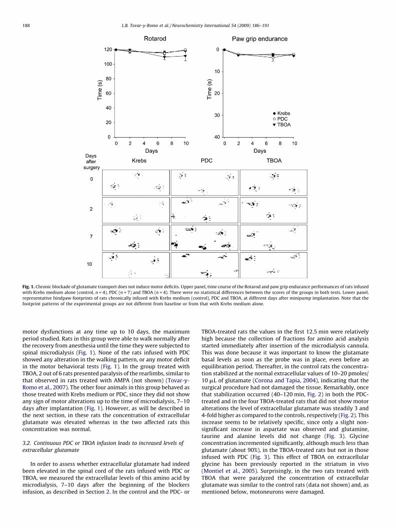
Fig. 1. Chronic blockade of glutamate transport does not induce motor deficits. Upper panel, time course of the Rotarod and paw grip endurance performances of rats infused
with Krebs medium alone (control, n = 4), PDC (n = 7) and TBOA (n = 4). There were no statistical differences between the scores of the groups in both tests. Lower panel,
representative hindpaw footprints of rats chronically infused with Krebs medium (control), PDC and TBOA, at different days after minipump implantation. Note that the
footprint patterns of the experimental groups are not different from baseline or from that with Krebs medium alone.
L.B. Tovar-y-Romo et al. / Neurochemistry International 54 (2009) 186–191188
motor dysfunctions at any time up to 10 days, the maximumperiod studied. Rats in this group were able to walk normally afterthe recovery from anesthesia until the time they were subjected tospinal microdialysis (Fig. 1). None of the rats infused with PDCshowed any alteration in the walking pattern, or any motor deficitin the motor behavioral tests (Fig. 1). In the group treated withTBOA, 2 out of 6 rats presented paralysis of the rearlimbs, similar tothat observed in rats treated with AMPA (not shown) (Tovar-y-Romo et al., 2007). The other four animals in this group behaved asthose treated with Krebs medium or PDC, since they did not showany sign of motor alterations up to the time of microdialysis, 7–10days after implantation (Fig. 1). However, as will be described inthe next section, in these rats the concentration of extracellularglutamate was elevated whereas in the two affected rats thisconcentration was normal.
3.2. Continuous PDC or TBOA infusion leads to increased levels of
extracellular glutamate
In order to assess whether extracellular glutamate had indeedbeen elevated in the spinal cord of the rats infused with PDC orTBOA, we measured the extracellular levels of this amino acid bymicrodialysis, 7–10 days after the beginning of the blockersinfusion, as described in Section 2. In the control and the PDC- or
TBOA-treated rats the values in the first 12.5 min were relativelyhigh because the collection of fractions for amino acid analysisstarted immediately after insertion of the microdialysis cannula.This was done because it was important to know the glutamatebasal levels as soon as the probe was in place, even before anequilibration period. Thereafter, in the control rats the concentra-tion stabilized at the normal extracellular values of 10–20 pmoles/10 mL of glutamate (Corona and Tapia, 2004), indicating that thesurgical procedure had not damaged the tissue. Remarkably, oncethat stabilization occurred (40–120 min, Fig. 2) in both the PDC-treated and in the four TBOA-treated rats that did not show motoralterations the level of extracellular glutamate was steadily 3 and4-fold higher as compared to the controls, respectively (Fig. 2). Thisincrease seems to be relatively specific, since only a slight non-significant increase in aspartate was observed and glutamine,taurine and alanine levels did not change (Fig. 3). Glycineconcentration incremented significantly, although much less thanglutamate (about 90%), in the TBOA-treated rats but not in thoseinfused with PDC (Fig. 3). This effect of TBOA on extracellularglycine has been previously reported in the striatum in vivo(Montiel et al., 2005). Surprisingly, in the two rats treated withTBOA that were paralyzed the concentration of extracellularglutamate was similar to the control rats (data not shown) and, asmentioned below, motoneurons were damaged.

Fig. 2. Chronic blockade of glutamate transport augments the extracellular
concentration of glutamate. Extracellular glutamate concentration was measured
by microdialysis and HPLC as described in Section 2, 7–10 days after osmotic pump
implantation and continuous Ringer–Krebs medium (control, n = 4), PDC (n = 7) and
TBOA (n = 4; in the other two rats of this group the amino acid changes were not
significant) infusion. Each point is the value determined in the corresponding
microdialysis fraction obtained every 12.5 min (mean � S.E.M.). Note that the steady
state concentration of glutamate was about 3 and 4-fold higher in the PDC and TBOA-
treated rats, respectively, than in the control animals. *p < 0.05 for PDC and #p < 0.05
for TBOA, as compared to the corresponding control value (ANOVA).
L.B. Tovar-y-Romo et al. / Neurochemistry International 54 (2009) 186–191 189
3.3. Elevated extracellular glutamate due to transport blockade does
not induce spinal motoneuron damage
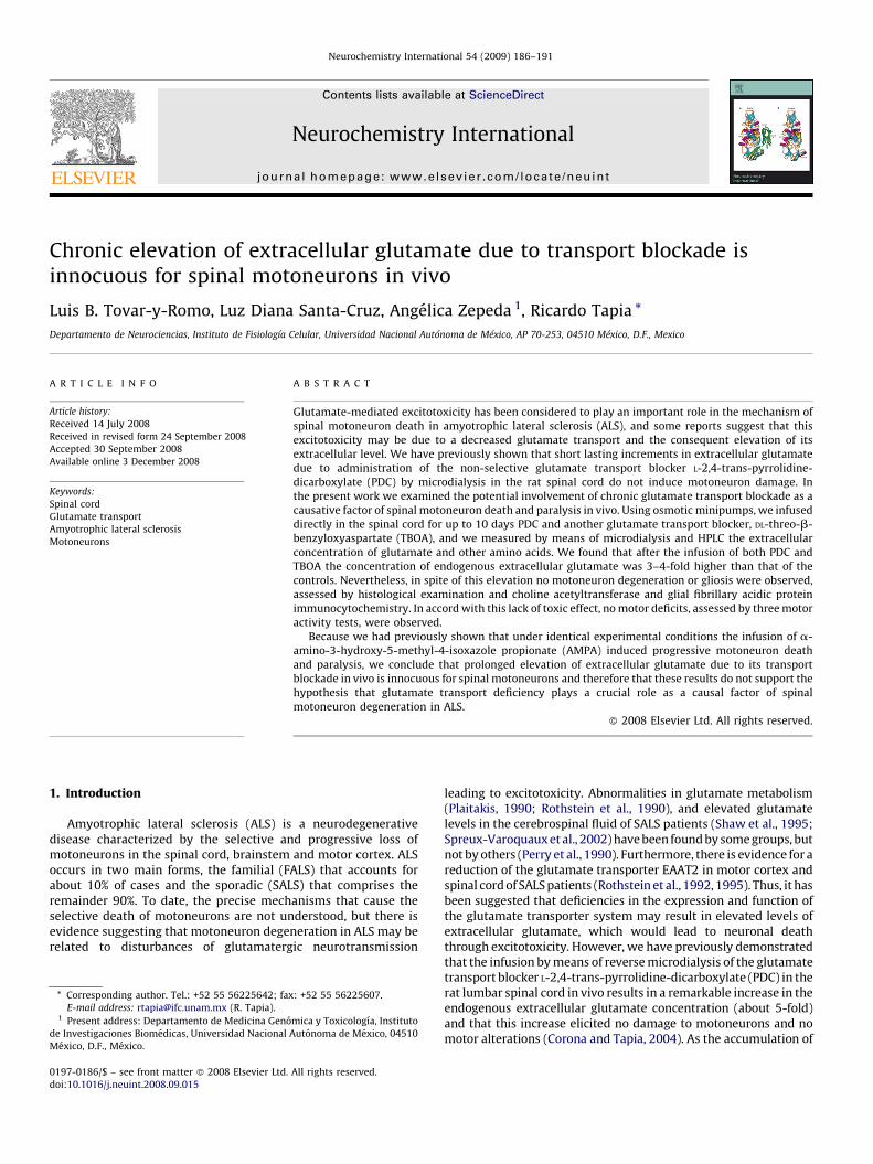
The lack of behavioral effects of PDC infusion was in agreementwith the histological and immunohistological observations, whichshowed that the number of motoneurons in the area of infusionwas normal, that they appeared as healthy as those of the controlrats, and that no glial reaction was developed (Fig. 4; quantitativedata is shown in Fig. 5). In addition, no neuronal damage wasobserved in the dorsal horn (not shown). These observationsindicate that no cell damage occurred as a consequence ofglutamate transport blockade by PDC infusion.
Similarly to the PDC-treated animals, in the four rats treatedwith TBOA that did not present motor alterations and had a 4-foldincrement in extracellular glutamate, no glial reaction and no
Fig. 3. Extracellular levels of different amino acids after chronic treatment with
glutamate transport blockers. The extracellular concentration of aspartate,
glutamate, glutamine, glycine, taurine and alanine were measured as described
in Fig. 2. Each bar represents the mean � S.E.M. of the averaged values in the
microdialysis fractions 2–10 (minutes 25–120, Fig. 2) of extracellular levels of amino
acids. *p < 0.05 and #p < 0.01 as compared with controls (ANOVA). No significant
changes were observed in the other amino acids.
histological or immunocytological alterations were observed(Fig. 4; quantitative data is shown in Fig. 5). In the two ratstreated with TBOA that showed motor deficits a notable loss ofmotoneurons was observed (not shown), similar to that producedby chronic AMPA infusion (Tovar-y-Romo et al., 2007).
4. Discussion
In this study we demonstrate that the chronic infusion of theglutamate transport inhibitors PDC and TBOA directly in thelumbar spinal cord elevates the extracellular concentration ofglutamate but does not result in neuronal damage. This lack ofdeleterious effect cannot be ascribed to an insufficient diffusion ofthe inhibitors into the ventral horn, because we have previouslydemonstrated that under identical experimental conditions theinfusion of AMPA induces in a dose-dependent manner aprogressive degeneration of the spinal motoneurons, leading toparalysis (Tovar-y-Romo et al., 2007).
Although in spinal cord cultures the blockade of glutamatetransport by PDC produces neuronal damage after long exposureperiods (Carriedo et al., 1996; Matyja et al., 2005; Rothstein et al.,1993; Velasco et al., 1996), we have previously shown that in vivothe acute infusion of PDC by reverse microdialysis causes a severalfold increase of the neurotransmitter in the extracellular fluidwithout generating neuronal death, neither in the spinal cord(Corona and Tapia, 2004) nor in the hippocampus or striatum(Massieu et al., 1995; Massieu and Tapia, 1997). Furthermore, PDCinduced also a large elevation of glutamate and was innocuouswhen infused in the motor cortex and hippocampus of a transgenicALS mouse in vivo (Tovar-y-Romo and Tapia, 2006). These findings,however, did not rule out the possibility that an extendedinhibition of glutamate transport may have deleterious effectson neurons (Rattray and Bendotti, 2006).
In our present experiments with PDC and TBOA we detected a3 and 4-fold increase in extracellular glutamate respectively, upto 10 days after its continuous infusion in the spinal cord.Because PDC administered by microdialysis induced a notableelevation of glutamate within 20 min, that persisted for as longas PDC was perfused (Corona and Tapia, 2004), it is reasonable toconclude that in the present experiments the concentration ofglutamate increased promptly from the beginning of theinfusion and remained constant thereafter, up to the time ofthe microdialysis procedure. The increase in glutamate con-centration after infusion of PDC may be due not only to theblockade of glutamate transport, but also to heteroexchange(Volterra et al., 1996; Waagepetersen et al., 2001). Nonetheless,it is remarkable that in spite of this significant increase inendogenous glutamate, none of the animals receiving PDCshowed any behavioral alteration in motor performance at anytime, and the histological and immunocytochemical analysesrevealed no cellular abnormalities.
TBOA is a potent non-transportable competitive glutamateuptake blocker that impedes the function of the transporters aswell as heteroexchange (Shimamoto et al., 1998, 2000). In thepresent experiments, in four of six rats the infusion of TBOAinduced an even larger increase in extracellular glutamate thanthat produced by PDC, and similarly to PDC this increase did notcause any motor behavioral alterations or motoneuron loss. Wecannot offer an explanation for the fact that two rats treated withTBOA showed paralysis associated to motoneuron loss, whereasfour rats behaved exactly as the PDC-treated animals. However,because in the latter four animals glutamate levels were elevated,whereas in the former two rats glutamate concentration did notincrease, it seems possible that TBOA might produce a directexcitotoxic effect on motoneurons. In support of this possibility, itwas recently found that the intrahippocampal injection of TBOA

Fig. 4. Chronic blockade of glutamate transport does not induce motoneuron death. Representative micrographs of ChAT (red) and GFAP (green) immunohistochemistry in
sections of the lumbar spinal cord, 7–10 days after pump implantation and continuous infusion with Krebs medium (control), PDC and TBOA. Motoneurons look healthy and
gliosis is absent in all groups. Scale bar = 100 mm.
L.B. Tovar-y-Romo et al. / Neurochemistry International 54 (2009) 186–191190
induced epilepsy and neurodegeneration in rat hippocampus,while PDC was innocuous, and that this effect was blocked by MK-801 and NBQX (Montiel et al., 2005), suggesting that TBOA mightactivate glutamate receptors. In rat brain membranes TBOA bindswith high affinity to NMDA receptors and with less affinity to non-NMDA receptors (Shimamoto et al., 1998, 2000), but no data areavailable in the spinal cord. So, the lack of neurotoxicity observedin the TBOA-infused rats showing elevated extracellular glutamateconcentrations, together with the results in the PDC experiments,leads us to conclude that even a prolonged accumulation ofextracellular endogenous glutamate arising from its transportblockade is not capable of overactivating glutamate receptors and
therefore do not cause spinal motoneuron death in vivo. Thisconclusion is also supported by previous work showing that theinfusion of PDC in the striatum and the hippocampus induces asubstantial elevation of extracellular glutamate but does notinduce neuronal damage, in rats and mice, including ALStransgenic mice (Corona and Tapia, 2004; Massieu et al., 1995;Massieu and Tapia, 1997; Tovar-y-Romo and Tapia, 2006; Tapiaet al., 1999). It might be argued that a 7–10 days period is notlonger enough to discard the possibility that a more extendedinhibition of glutamate transport could be detrimental formotoneurons. However, our experiments with the chronic infusionof AMPA using osmotic minipumps show that the motor

Fig. 5. Motoneuron number does not change after PDC or TBOA chronic
administration. Number of healthy motoneurons in the ipsilateral and
contralateral ventral horns of rats infused with Ringer–Krebs medium (control,
n = 4), PDC (n = 7) and TBOA (n = 4). Five 40 mm sections from each rat were
analyzed. Values are means � S.E.M.
L.B. Tovar-y-Romo et al. / Neurochemistry International 54 (2009) 186–191 191
performance deficiencies occur within the first 2–6 days ofinfusion (Tovar-y-Romo et al., 2007).
In this respect, it is noteworthy that, although reductions inglutamate transporter EAAT2 have been reported in ALS transgenicrodents and in the spinal cord of ALS patients, the majority of thesedata are more consistent with the possibility that these changesare a consequence rather than a cause of ALS (see Rattray andBendotti, 2006, for a review on this point). For example, in SOD1/G93A ALS mice, when GLT1 is decreased in heterozygous GLT1�animals the onset of paralysis is not modified in comparison withSOD1/G93A mice that have the normal amount of the transporterprotein, implying that loss of GLT1 is not an initiator of the disease(Pardo et al., 2006).
It is also worth mentioning that the 3–4-fold elevation ofextracellular concentration of glutamate that we found in ourexperiments is higher than that observed in the CSF of a subset ofSALS patients, which in most cases was less than twice the upperlimit parameter (Spreux-Varoquaux et al., 2002).
In conclusion, our results show that the persistent increasedlevels of extracellular glutamate in the spinal cord during severaldays due to transport deficit are not capable of elicitingexcitotoxicity and therefore these data do not support thehypothesis that a deficiency in glutamate transport is causallyinvolved in motoneuron degeneration in ALS.
Acknowledgements
This work was supported by DGAPA-UNAM (project IN209807)and CONACYT (project 60322). LBTR and LDSC are recipients of afellowship from CONACYT.
References
Carriedo, S.G., Yin, H.Z., Weiss, J.H., 1996. Motor neurons are selectively vulnerableto AMPA/kainate receptor-mediated injury in vitro. J. Neurosci. 16, 4069–4079.
Corona, J.C., Tapia, R., 2004. AMPA receptor activation, but not the accumulation ofendogenous extracellular glutamate, induces paralysis and motor neuron deathin rat spinal cord in vivo. J. Neurochem. 89, 988–997.
Corona, J.C., Tapia, R., 2007. Ca2+-permeable AMPA receptors and intracellular Ca2+
determine motoneuron vulnerability in rat spinal cord in vivo. Neuropharma-cology 52, 1219–1228.
Massieu, L., Morales-Villagran, A., Tapia, R., 1995. Accumulation of extracellularglutamate by inhibition of its uptake is not sufficient for inducing neuronaldamage: an in vivo microdialysis study. J. Neurochem. 64, 2262–2272.
Massieu, L., Tapia, R., 1997. Glutamate uptake impairment and neuronal damage inyoung and aged rats in vivo. J. Neurochem. 69, 1151–1160.
Matyja, E., Naganska, E., Taraszewska, A., Rafalowska, J., 2005. The mode of spinalmotor neurons degeneration in a model of slow glutamate excitotoxicity invitro. Folia Neuropathol. 43, 7–13.
Montiel, T., Camacho, A., Estrada-Sanchez, A.M., Massieu, L., 2005. Differentialeffects of the substrate inhibitor PDC and the non-substrate inhibitor DL-threo-beta-benzyloxyaspartate (DL-TBOA) of glutamate transporters on neu-ronal damage and extracellular amino acid levels in rat brain in vivo. Neu-roscience 133, 667–678.
Pardo, A.C., Wong, V., Benson, L.M., Dykes, M., Tanaka, K., Rothstein, J.D., Maragakis,N.J., 2006. Loss of the astrocyte glutamate transporter GLT1 modifies disease inSOD1(G93A) mice. Exp. Neurol. 201, 120–130.
Perry, T.L., Krieger, C., Hansen, S., Eisen, A., 1990. Amyotrophic lateral sclerosis:amino acid levels in plasma and cerebrospinal fluid. Ann. Neurol. 28, 12–17.
Plaitakis, A., 1990. Glutamate dysfunction and selective motor neuron degenerationin amyotrophic lateral sclerosis: a hypothesis. Ann. Neurol. 28, 3–8.
Rattray, M., Bendotti, C., 2006. Does excitotoxic cell death of motor neurons in ALSarise from glutamate transporter and glutamate receptor abnormalities? Exp.Neurol. 201, 15–23.
Rothstein, J.D., Tsai, G., Kuncl, R.W., Clawson, L., Cornblath, D.R., Drachman, D.B.,Pestronk, A., Stauch, B.L., Coyle, J.T., 1990. Abnormal excitatory amino acidmetabolism in amyotrophic lateral sclerosis. Ann. Neurol. 28, 18–25.
Rothstein, J.D., Martin, L.J., Kuncl, R.W., 1992. Decreased glutamate transport by thebrain and spinal cord in amyotrophic lateral sclerosis. New Engl. J. Med. 326,1464–1468.
Rothstein, J.D., Jin, L., Dykes-Hoberg, M., Kuncl, R.W., 1993. Chronic inhibition ofglutamate uptake produces a model of slow neurotoxicity. Proc. Natl. Acad. Sci.U.S.A. 90, 6591–6595.
Rothstein, J.D., Van Kammen, M., Levey, A.I., Martin, L.J., Kuncl, R.W., 1995. Selectiveloss of glial glutamate transporter GLT-1 in amyotrophic lateral sclerosis. Ann.Neurol. 38, 73–84.
Salazar, P., Montiel, T., Brailowsky, S., Tapia, R., 1994. Decrease of glutamatedecarboxylase activity after in vivo cortical infusion of gamma-aminobutyricacid. Neurochem. Int. 24, 363–368.
Shaw, P.J., Forrest, V., Ince, P.G., Richardson, J.P., Wastell, H.J., 1995. CSF and plasmaamino acid levels in motor neuron disease: elevation of CSF glutamate in asubset of patients. Neurodegeneration 4, 209–216.
Shimamoto, K., Lebrun, B., Yasuda-Kamatani, Y., Sakaitani, M., Shigeri, Y., Yumoto,N., Nakajima, T., 1998. DL-Threo-beta-benzyloxyaspartate, a potent blocker ofexcitatory amino acid transporters. Mol. Pharmacol. 53, 195–201.
Shimamoto, K., Shigeri, Y., Yasuda-Kamatani, Y., Lebrun, B., Yumoto, N., Nakajima, T.,2000. Syntheses of optically pure beta-hydroxyaspartate derivatives as gluta-mate transporter blockers. Bioorg. Med. Chem. Lett. 10, 2407–2410.
Spreux-Varoquaux, O., Bensimon, G., Lacomblez, L., Salachas, F., Pradat, P.F., LeForestier, N., Marouan, A., Dib, M., Meininger, V., 2002. Glutamate levels incerebrospinal fluid in amyotrophic lateral sclerosis: a reappraisal using a newHPLC method with coulometric detection in a large cohort of patients. J. Neurol.Sci. 193, 73–78.
Tapia, R., Medina-Ceja, L., Pena, F., 1999. On the relationship between extracellularglutamate, hyperexcitation and neurodegeneration, in vivo. Neurochem. Int. 34,23–31.
Tovar-y-Romo, L.B., Tapia, R., 2006. Cerebral neurons of transgenic ALS mice arevulnerable to glutamate release stimulation but not to increased extracellularglutamate due to transport blockade. Exp. Neurol. 199, 281–290.
Tovar-y-Romo, L.B., Zepeda, A., Tapia, R., 2007. Vascular endothelial growth factorprevents paralysis and motoneuron death in a rat model of excitotoxic spinalcord neurodegeneration. J. Neuropathol. Exp. Neurol. 66, 913–922.
Velasco, I., Tapia, R., Massieu, L., 1996. Inhibition of glutamate uptake inducesprogressive accumulation of extracellular glutamate and neuronal damage inrat cortical cultures. J. Neurosci. Res. 44, 551–561.
Volterra, A., Bezzi, P., Rizzini, B.L., Trotti, D., Ullensvang, K., Danbolt, N.C., Racagni,G., 1996. The competitive transport inhibitor L-trans-pyrrolidine-2,4-dicar-boxylate triggers excitotoxicity in rat cortical neuron-astrocyte co-culturesvia glutamate release rather than uptake inhibition. Eur. J. Neurosci. 8, 2019–2028.
Waagepetersen, H.S., Shimamoto, K., Schousboe, A., 2001. Comparison of effects ofDL-threo-beta-benzyloxyaspartate (DL-TBOA) and L-trans-pyrrolidine-2,4-dicar-boxylate (t-2,4-PDC) on uptake and release of [3h]D-aspartate in astrocytes andglutamatergic neurons. Neurochem. Res. 26, 661–666.
Weydt, P., Hong, S.Y., Kliot, M., Moller, T., 2003. Assessing disease onset andprogression in the SOD1 mouse model of ALS. Neuroreport 14, 1051–1054.
Related Documents