1 Chronic alcohol ingestion in rats alters lung metabolism, promotes lipid accumulation, and impairs alveolar macrophage functions Freddy Romero 1 , Dilip Shah 1 , Michelle Duong 1 , William Stafstrom 1 , Jan B. Hoek 2 , Caleb B. Kallen 4 , Charles H. Lang 3 and Ross Summer 1 *. 1 Center for Translational Medicine, Thomas Jefferson University, Philadelphia, PA 19107 2 Department of Pathology, Anatomy, and Cell Biology, Thomas Jefferson University, Philadelphia, Pennsylvania 19107. 3 Department of Cellular and Molecular Physiology, Penn State College of Medicine, Hershey, Pennsylvania 17033 4 Department of Obstetrics and Gynecology, Thomas Jefferson University, Philadelphia, Pennsylvania 19107. Running title: Alcohol induces lipid accumulation and impairs macrophage function in lung Key words: chronic alcohol ingestion, adenosine monophosphate-activated protein kinase, surfactant lipids, macrophage *To whom correspondence should be addressed: email: [email protected] Acknowledgement: Research was supported by funding from the National Institutes of Health (NIH) R01HL105490 and R37 AA11290. Page 1 of 36 AJRCMB Articles in Press. Published on 18-June-2014 as 10.1165/rcmb.2014-0127OC Copyright © 2014 by the American Thoracic Society

Welcome message from author
This document is posted to help you gain knowledge. Please leave a comment to let me know what you think about it! Share it to your friends and learn new things together.
Transcript
1
Chronic alcohol ingestion in rats alters lung metabolism, promotes lipid accumulation, and
impairs alveolar macrophage functions
Freddy Romero1, Dilip Shah
1, Michelle Duong
1, William Stafstrom
1, Jan B. Hoek
2, Caleb B.
Kallen4, Charles H. Lang
3 and Ross Summer
1*.
1 Center for Translational Medicine, Thomas Jefferson University, Philadelphia, PA 19107
2 Department of Pathology, Anatomy, and Cell Biology, Thomas Jefferson University,
Philadelphia, Pennsylvania 19107.
3 Department of Cellular and Molecular Physiology, Penn State College of Medicine, Hershey,
Pennsylvania 17033
4 Department of Obstetrics and Gynecology, Thomas Jefferson University, Philadelphia,
Pennsylvania 19107.
Running title: Alcohol induces lipid accumulation and impairs macrophage function in lung
Key words: chronic alcohol ingestion, adenosine monophosphate-activated protein kinase,
surfactant lipids, macrophage
*To whom correspondence should be addressed:
email: [email protected]
Acknowledgement: Research was supported by funding from the National Institutes of Health
(NIH) R01HL105490 and R37 AA11290.
Page 1 of 36 AJRCMB Articles in Press. Published on 18-June-2014 as 10.1165/rcmb.2014-0127OC
Copyright © 2014 by the American Thoracic Society
2
Abstract
Chronic alcoholism impairs pulmonary immune homeostasis and predisposes to inflammatory
lung diseases, including infectious pneumonia and acute respiratory distress syndrome. While
alcoholism has been shown to alter hepatic metabolism leading to lipid accumulation, hepatitis,
and eventually cirrhosis, the effects of alcohol on pulmonary metabolism remain largely
unknown. Because both the lung and the liver actively engage in lipid synthesis, we
hypothesized that chronic alcoholism would impair pulmonary metabolic homeostasis in ways
similar to its effects in the liver. We reasoned that perturbations in lipid metabolism might
contribute to the impaired pulmonary immunity observed in people who chronically consume
alcohol. We studied the metabolic consequences of chronic alcohol consumption in rat lungs in
vivo and in alveolar epithelial type II (AEII) cells and alveolar macrophages in vitro. We found
that chronic alcohol ingestion significantly alters lung metabolic homeostasis, inhibiting AMP-
activated protein kinase, increasing lipid synthesis, and suppressing the expression of genes
essential to metabolizing fatty acids. Further, we show that these metabolic alterations promoted
a lung phenotype that is reminiscent of alcoholic fatty liver and is characterized by marked
accumulation of triacylglycerides and free fatty acids within distal airspaces, alveolar
macrophages, and to a lesser extent, AEII cells. We provide evidence that the metabolic
alterations in alcohol-exposed rats are mechanistically linked to immune impairments in the
alcoholic lung: the elevations in fatty acids alter alveolar macrophage phenotypes and suppress
both phagocytic functions and agonist-induced inflammatory responses. In summary, our work
demonstrates that chronic alcohol ingestion impairs lung metabolic homeostasis and promotes
pulmonary immune dysfunction. These findings suggest that therapies aimed at reversing
alcohol-related metabolic alterations might be effective for preventing and/or treating alcohol-
related pulmonary disorders.
Page 2 of 36 AJRCMB Articles in Press. Published on 18-June-2014 as 10.1165/rcmb.2014-0127OC
Copyright © 2014 by the American Thoracic Society
3
Clinical Relevance: Chronic alcohol abuse is a risk factor for bacterial pneumonia and ARDS;
the molecular mechanisms underlying this association are not understood. Our work
demonstrates that chronic alcohol exposure induces significant metabolic changes in the lung
including marked accumulation of triacylglycerides and free fatty acids within distal airspaces
and alveolar macrophages. Furthermore, we provide evidence linking these lipid abnormalities
to phenotypic and functional impairments in alveolar macrophages, suggesting that these
metabolic disturbances may contribute to the pathogenesis of alcohol-related inflammatory lung
diseases. Together, these observations have broad implications for studying the effects of alcohol
on lung homeostasis and immunity and may be relevant to the pathogenesis of other
inflammatory pulmonary disorders.
Page 3 of 36 AJRCMB Articles in Press. Published on 18-June-2014 as 10.1165/rcmb.2014-0127OC
Copyright © 2014 by the American Thoracic Society
4
Introduction
Chronic ethanol (EtOH) consumption injures all tissues but certain organs, such as the liver,
heart and brain, are particularly susceptible. Although the lung is not considered to be the most
important target of EtOH-mediated tissue injury, pulmonary toxicity is well-documented (1, 2).
Heavy EtOH consumption disrupts various biological processes in the lung including
mucociliary clearance, oxidant-antioxidant balance, and alveolar macrophage (AM) function (3-
5). Moreover, chronic EtOH abuse predisposes to the development of various inflammatory lung
disorders, including infectious pneumonia and acute respiratory distress syndrome (ARDS), and
clinical outcomes for these conditions are worse in patients that chronically consume EtOH (6-
8).
Research over the past several decades has focused on the inflammatory nature of EtOH-induced
lung disorders and has explored the immune mechanisms underlying susceptibility to these
diseases (9-11). This mechanistic focus in the lung contrasts the intense study of the effects of
EtOH on metabolic homeostatic processes in other organs such as the liver. It has been shown
that EtOH induces significant metabolic disturbances in the liver, and these perturbations are
thought to contribute to the pathogenesis of EtOH-related liver diseases such as alcoholic fatty
liver disease (AFLD), steatohepatitis, and cirrhosis (12-14).
To date, little is known regarding the effects of EtOH on lung metabolism. This is particularly
surprising when one considers that the lung, like the liver, synthesizes lipids de novo. In the lung,
these lipids are synthesized by specialized cells that reside within distal airspaces called type II
alveolar epithelial (AEII) cells (15, 16) and their production is required for generating the
surfactant monolayer that is critical for reducing surface tension and protecting the underlying
respiratory epithelium. Surfactant lipids are comprised principally (~85%) of phospholipids
Page 4 of 36 AJRCMB Articles in Press. Published on 18-June-2014 as 10.1165/rcmb.2014-0127OC
Copyright © 2014 by the American Thoracic Society
5
(PLs), whereas cholesterol, triacylglycerides (TG) and free fatty acids (FA) collectively represent
only 10-15% of the total surfactant lipid pool (15, 17).
Studies investigating the effects of EtOH on surfactant lipid homeostasis have focused
principally on phospholipid (PL) production. For example, Guidot et al. showed that EtOH-fed
rats have reduced incorporation of [3H] choline into PLs in AEII cells (18). Similarly, Wagner et
al. found that pre-feeding rats for 3 days with low concentrations of EtOH significantly
decreased precursor incorporation into PLs (19). Although an anticipated reduction in overall PL
concentrations would be expected based upon these studies, total and fractionated forms of PLs
have not been shown to be significantly decreased in chronic EtOH exposed lungs, suggesting
that the lung posseses mechanisms for limiting excursions in PL levels in response to chronic
EtOH ingestion. To date, studies examing the effects of EtOH on other lipid species in the lung
are limited, though existing evidence suggests that cholesterol and cholesterol esters are not
significantly affected while TGs appear to be markedly increased in response to chronic EtOH
exposure (17, 19). The molecular mechanisms mediating TG accumulation in EtOH-exposed
lungs, and the functional significance of these biochemical changes, remain unknown.
Because chronic EtOH consumption is known to alter lipid homeostasis in the liver, we
hypothesized that similar metabolic disturbances would be observed in the lung and that these
changes might, at least in part, contribute to development of the immune impairments observed
in response to chronic alcohol consumption. Consistent with this hypothesis, we observed
decreased AMPK activation and increased lipid synthesis in EtOH exposed lungs and in cultured
AEII cells. These metabolic changes were associated with marked lipid accumulation in EtOH
Page 5 of 36 AJRCMB Articles in Press. Published on 18-June-2014 as 10.1165/rcmb.2014-0127OC
Copyright © 2014 by the American Thoracic Society
6
affected lungs, a phenotype that we have now coined “the alcoholic fatty lung”. Quantitative
lipid analyses further demonstrated that lipid accumulation is largely attributable to increases in
TGs and FAs, while phospholipids and cholesterol fractions are not significantly changed in
response to chronic EtOH consumption. Further, we provide evidence suggesting that lipid
accumulation is causally related to immune impairments in the alcoholic lung by altering
alveolar macrophage (AM) phenotype and function.
Materials and Methods
Rat Model of Chronic Alcohol Ingestion
Male Sprague-Dawley rats (Charles River Laboratory, Wilmington, MA) were fed with Lieber-
DeCarli liquid (36% calories from EtOH) or nonalcoholic isocaloric control liquid diet (Research
Diets, New Brunswick, NJ) for 4 months (20). Information on body weight and plasma EtOH
levels are provided in Supplemental Table 1. Animal protocols were reviewed and approved by
the Institutional Animal Care and Use Committees at Penn State College of Medicine and
Thomas Jefferson University, and adhered to the National Institutes of Health (NIH) guidelines
for the use of experimental animals.
Bronchoalveolar lavage recovery and fractionation
Bronchoalveolar lavage (BAL), total cell counts and differential cell counts were performed as
previously described (21).
Alveolar Epithelial Type II Cell Culture and Treatment
Rat alveolar type II epithelial cell line (L2 cells) was obtained from ATCC (Manassas, VA) and
cultured according to company protocol. See supplemental methods for details.
Page 6 of 36 AJRCMB Articles in Press. Published on 18-June-2014 as 10.1165/rcmb.2014-0127OC
Copyright © 2014 by the American Thoracic Society
7
Lipid Droplet Determination
See supplemental methods section for details.
Lipid Extraction and Analysis
Total lipids were extracted from BAL fluid, L2 cells, and lung tissue using a modified method of
Bligh and Dyer, using chloroform/methanol (2:1) (22). See supplemental methods for further
details regarding the extraction methods and separation protocols used in our studies.
NR8383 Cell Culture and Lipopolysaccharide (LPS) Stimulation
Rat alveolar macrophages NR8383 cells were purchased from ATCC (Manassas, VA). Cells
were cultured in RPMI 1640 media containing 15% FBS and 1% penicillin/streptomycin. After
reaching 80% confluence, NR8383 were cultured with BSA conjugated palmitic acid (125 or 250
µM) or BSA alone for 24 h followed by exposure to vehicle or lipopolysaccharide (Escherichia
coli 01111:B4; 1µg/ml Sigma-Aldrich, St. Louis, MO) for 6 h.
Cytokine Analysis
Interleukin-6 (IL6), tumor necrosis factor-alpha (TNF-α), transforming growth factor beta (TGF-
β1) and monocyte chemoattractant protein-1 (MCP1) were measured using commercially
available ELISA kits (R&D System Inc., Minneapolis, MN) per published protocols (21).
RNA Isolation and Quantitative Real-Time PCR
Page 7 of 36 AJRCMB Articles in Press. Published on 18-June-2014 as 10.1165/rcmb.2014-0127OC
Copyright © 2014 by the American Thoracic Society
8
The following gene transcripts were evaluated in this study: Srebf1, Acaca, Fasn, Ppara, Cpt1a,
Abcg1, Abca1, Cd36, Msr1, Tgf-β1, Arg1 and Chi3l3. The housekeeping genes Gapdh and Hprt1
were used for normalization. See supplemental materials for specific details regarding
quantitative RT-PCR and for the olignucleotide sequences used in these studies.
Western Blot Analysis
Western blot analysis was performed for AMPK, pAMPK, ACACA, Acetyl-CoA synthase,
FASN, GAPDH, ADRP, SREBF1, DGAT1 AND CYP2E1; detailed protocols are described in
the supplemental methods section.
Macrophage Phagocytosis Assay
Details are described in the supplemental methods section.
Alcohol Dehydrogenase (ADH) Activity
ADH activity in lung tissue and cultured cells was quantified using a commercially available kit
(Bio Vision, Montain View, CA) according to the manufacturer’s instructions.
Plasma measurements
See the supplemental methods section for details regarding measurments of plasma alanine and
aspartate aminotransferases and EtOH concentration.
Statistical Analysis
Page 8 of 36 AJRCMB Articles in Press. Published on 18-June-2014 as 10.1165/rcmb.2014-0127OC
Copyright © 2014 by the American Thoracic Society
9
Statistics were performed using GraphPad Prism 5.0 software. Two-group comparisons were
analyzed by unpaired Student’s t-test, and multiple-group comparisons were performed by one-
way analysis of variance followed by Tukey post hoc analysis. Statistical significance was
achieved when P < 0.05 at 95% confidence interval.
Results
Chronic alcohol ingestion promotes lipid accumulation in the lung
To determine the effects of chronic EtOH ingestion on lipid homeostasis we first assessed
whether hepatic steatosis developed in our model. As shown in figure 1A, we detected a
significant increase in TG and cholesterol levels in livers from chronic EtOH exposed rats.
Moreover, these metabolic changes were associated with an increase in plasma alanine and
aspartate aminotransferases (fig 1B), indicating that chronic EtOH consumption induced
hepatocellular injury in our model.
Next, we studied the effects of chronic EtOH exposure on lung lipid homeostasis. When
comparing control and EtOH-exposed whole lungs we did not detect differences in total PLs,
phosphotidylcholine (PC) or phosphotidylserine (PS) (fig 1C). Similarly, total cholesterol levels
in whole lung were not significantly affected by chronic EtOH ingestion (fig 1D), as previously
described (17). We did, however, observe a marked increase in TGs and FAs in whole lung (fig
1E and fig 1F) and in BAL fluid (fig 1G and fig 1H) after chronic EtOH ingestion. These
findings were associated with increased ADRP (a.k.a. Plin2) protein content (fig 2A), a marker
of increased intracellular TG mobilization and storage. Moreover, Oil Red O staining of the lung
detected massive accumulation of neutral lipids (red color) in AMs while more subtle increases
in lipid droplets were observed in other parenchymal cells including AEII cells (fig 2B)
Page 9 of 36 AJRCMB Articles in Press. Published on 18-June-2014 as 10.1165/rcmb.2014-0127OC
Copyright © 2014 by the American Thoracic Society
10
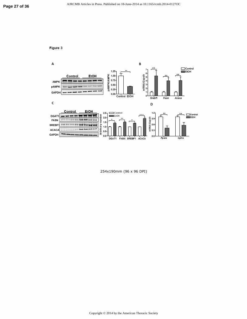
Chronic alcohol ingestion promotes lipid synthesis in the lung
To assess whether TG and FA accumulation resulted from metabolic disturbances that promote
lipid synthesis we evaluated the activation state of AMPK in the lungs of control and EtOH-fed
rats. AMPK is a serine-threonine kinase that regulates substrate utilization in cells. Inhibition of
AMPK by de-phosphorylation at Thr172 is associated with activation of anabolic pathways (such
as lipid synthesis) and suppression of catabolic processes (such as the breakdown of fatty acids)
(23). Consistent with this functional paradigm, AMPK phosphorylation was significantly
decreased in the lung after chronic EtOH consumption and this was associated with up-
regulation of transcripts and proteins for several key factors involved in lipid synthesis including
sterol regulatory-element binding protein1 (Srebf1), fatty acid synthase (Fasn), acetyl CoA
carboxylase (Acaca) and diacylglycerol O-acyltransferase 1 (Dgat1) (fig 3A-C). In addition, we
observed decreased mRNA expression of peroxisome proliferator-activated receptor alpha
(Ppara) and carnitine palmitoyltransferase I (Cpt1a) in lungs from chronic EtOH-fed rats(fig
3D), suggesting that impaired breakdown of FAs may also contribute to TG and FA
accumulation.
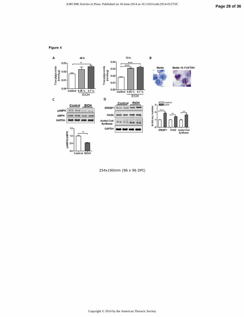
Ethanol modifies AEII cell metabolism by inhibiting AMPK activation and promoting lipid
synthesis
Our data suggested that alcohol induces lipid synthesis in the lung. To determine whether this
might be a direct effect of alcohol on AEII cells, we tested whether EtOH directly modifies
metabolism in L2 cells, a model cell system that demonstrates many features of alveolar
epithelial cells. We detected marked intracellular TG accumulation in response to both low
(0.05%) and high (0.1% v/v) concentrations of EtOH after 48 or 72 h exposure (fig 4A).
Similarly, Oil Red O staining detected increased lipid droplet formation in L2 cells after EtOH
exposure (fig 4B), consistent with our histological observations of increased intracellular lipids
Page 10 of 36 AJRCMB Articles in Press. Published on 18-June-2014 as 10.1165/rcmb.2014-0127OC
Copyright © 2014 by the American Thoracic Society
11
in AEII cells from chronic EtOH-fed rats. In contrast, intracellular lipid accumulation was not
detected in rat NR8383 AMs after culture in EtOH, suggesting that in vivo lipid accumulation in
macrophages might result from enhanced lipid uptake rather than from increased production
(data not shown). As observed in the alcohol-exposed lung, we found that increased TG
accumulation in L2 cells was associated with broad metabolic changes including decreased
activation of AMPK, increased expression of SREBF1, and increased expression of the lipid
synthesizing enzyme Acetyl-CoA synthase (fig 4 C, D). However, levels of Ppara and Cpt1a
transcripts were not significantly altered in response to EtOH (data not shown), suggesting that
TG accumulation in L2 cells occurs primarily from increased lipid synthesis. Together, these
findings indicate that EtOH significantly modifies AEII cell lipid metabolism leading to lipid
accumulation in macrophages, AEII cells and the extracellular air spaces of the chronic EtOH-
exposed lung.
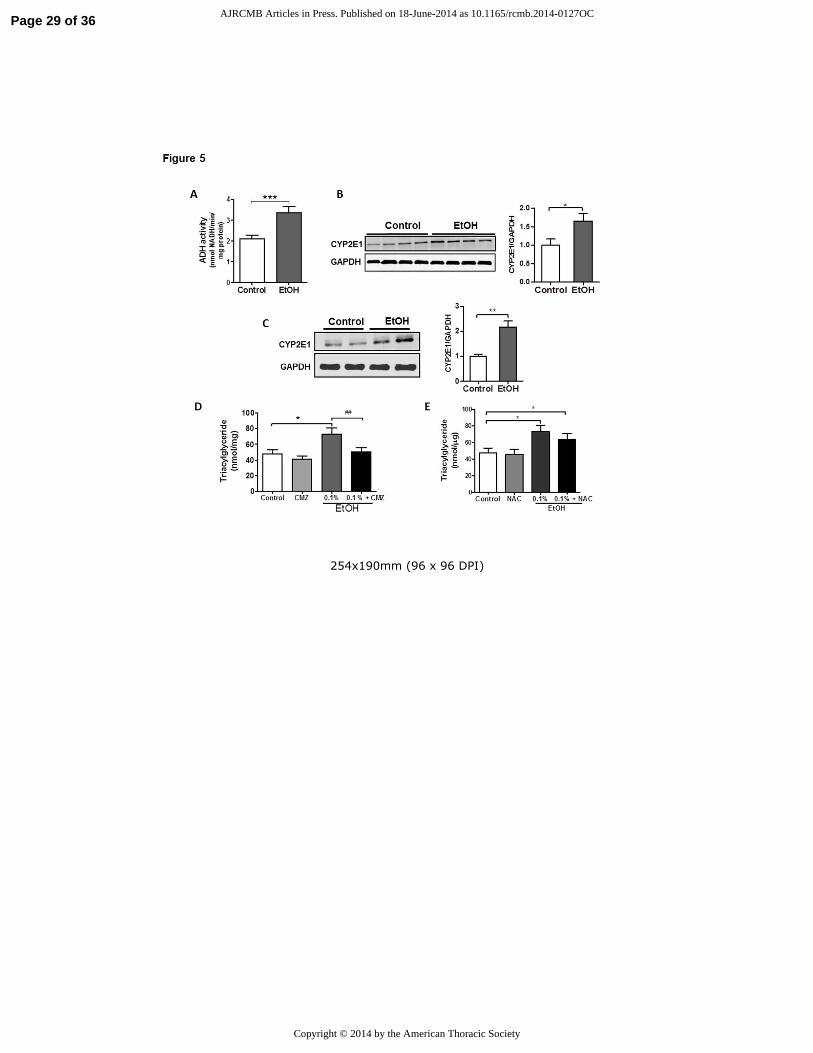
Blocking CYP2E1 activity in AEII cells limits triacylglyceride accumulation
Although the liver is predominantly responsible for metabolizing EtOH, the lung possesses
similar enzymatic capacity. As shown in figure 5A&B, alcohol dehydrogenase (ADH) activity
and the CYP2E1 enzyme are present in the lung and enhanced in response to EtOH. Because
byproducts of EtOH metabolism are largely responsible for mediating lipid accumulation in the
liver, we sought to determine whether EtOH or one of its metabolites is most important in
promoting TG synthesis in L2 cells.
In contrast to whole lung, ADH activity was not detected in L2 cells cultured in media alone or
in media supplemented with EtOH (data not shown). However, expression of the CYP2E1
enzyme was readily identified in L2 cells and levels were significantly increased in response to
Page 11 of 36 AJRCMB Articles in Press. Published on 18-June-2014 as 10.1165/rcmb.2014-0127OC
Copyright © 2014 by the American Thoracic Society
12
EtOH (fig 5C). To assess whether this enzymatic activity is important in promoting metabolic
alterations in L2 cells, we examined the effects of EtOH on TG levels in cells cultured in the
presence or absence of chlormethiazole (CMZ), a CYP2E1 inhibitor. Pre-treatment with CMZ
(100 µM) for 24 h completely prevented TG accumulation in EtOH-treated L2 cells (fig 5D)
indicating that products of EtOH metabolism are involved in TG accumulation after EtOH
exposure. Interestingly, treating L2 cells with the antioxidant NAC (5 mM) for 24 h failed to
attenuate EtOH-induced TG accumulation suggesting that, at least under these experimental
conditions, lipid accumulation may not depend upon the generation of reactive oxygen species
(ROS) (fig 5E). However, we recognized that antioxidant effects of NAC were not directly
monitored in these studies.
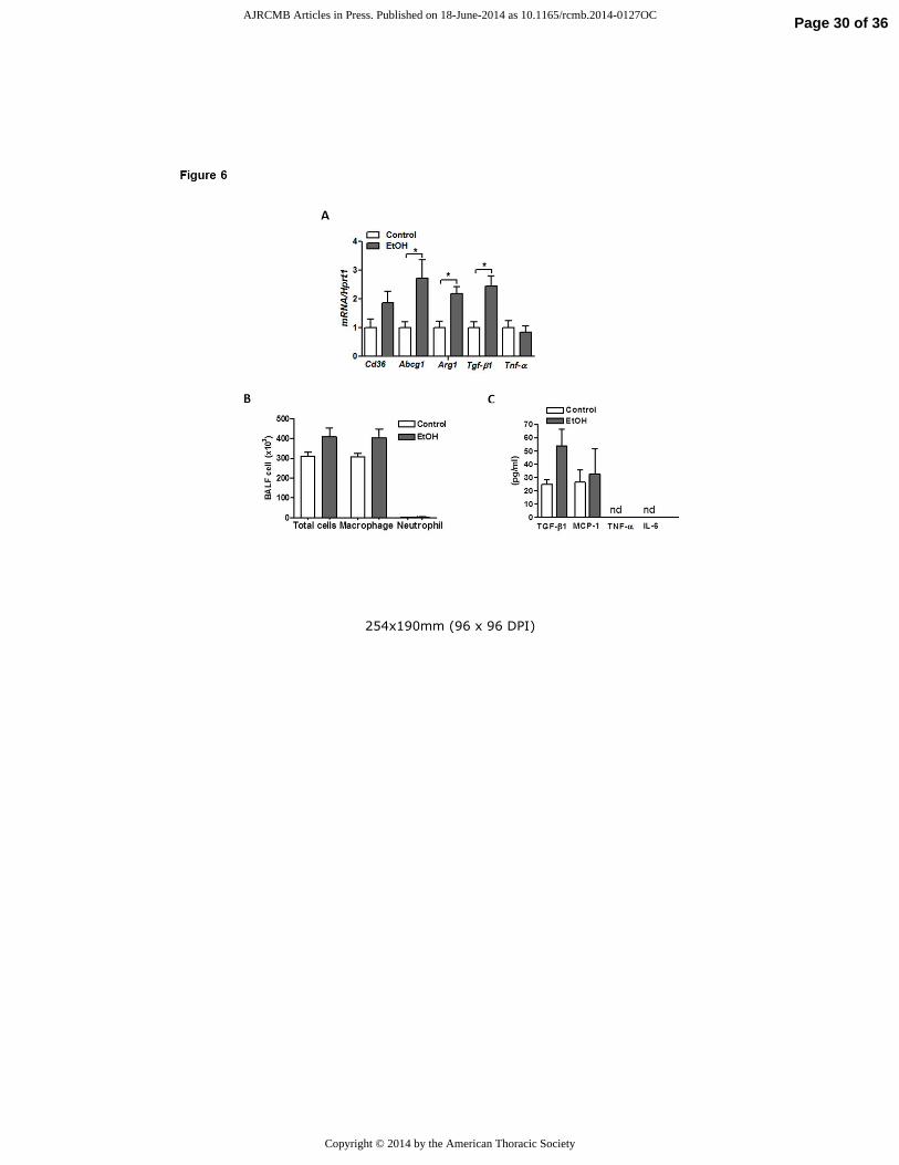
Chronic alcohol exposure alters macrophage phenotype in the lung.
Since EtOH is known to alter AM behavior, we sought to evaluate the effects of chronic EtOH
consumption on AM phenotypes in our model system. Consistent with prior reports, we detected
a two-fold increase in mRNA content of Tgf-b1 in freshly isolated primary AMs from lungs of
EtOH-fed rats (fig 6A) and this was associated with a non-significant increase in TGF-β1 protein
in BAL fluid (fig 6B). The increased Tgf-b1 expression was associated with enhanced expression
of Arg1 (fig 6A) but not Tnf-α transcripts after chronic EtOH feeding. Together, this gene
expression profile suggested that chronic EtOH intake polarizes AMs toward an M2 reparative
phenotype. Interestingly, AMs isolated from lungs of EtOH-fed rats also displayed higher
mRNA expression of the lipid receptor Cd36 as well as the lipid transporter Abcg1, which
presumably serves as an adaptive response to the increased extracellular lipids (fig 6A). Despite
these changes in AM gene expression, we did not observe a significant increase in the total
Page 12 of 36 AJRCMB Articles in Press. Published on 18-June-2014 as 10.1165/rcmb.2014-0127OC
Copyright © 2014 by the American Thoracic Society
13
number of AMs recovered from BAL fluid or in levels of the inflammatory markers MCP1,
TNF-α or IL-6 in BAL fluid from EtOH fed rats. The latter observation may reflect low
expression levels of some of these factors (i.e. TNF-α and IL-6) as well as a dilutional effect of
lavage, rendering some factors undetectable in our experiments (fig 6B).
Fatty acids promote M2 polarization and exacerbate the suppressive effects of alcohol on
alveolar macrophage (AM) function.
Because chronic EtOH consumption was associated with changes in AM gene expression we
hypothesized that lipid accumulation within distal airspaces might contribute to these findings.
To test this hypothesis, we examined the effect of the fatty acid palmitate on AM phenotype in
culture. Consistent with findings AMs isolated from EtOH-exposed lungs, rat AMs cultured in
media containing 250 µM of palmitic acid displayed increased mRNA expression of lipid
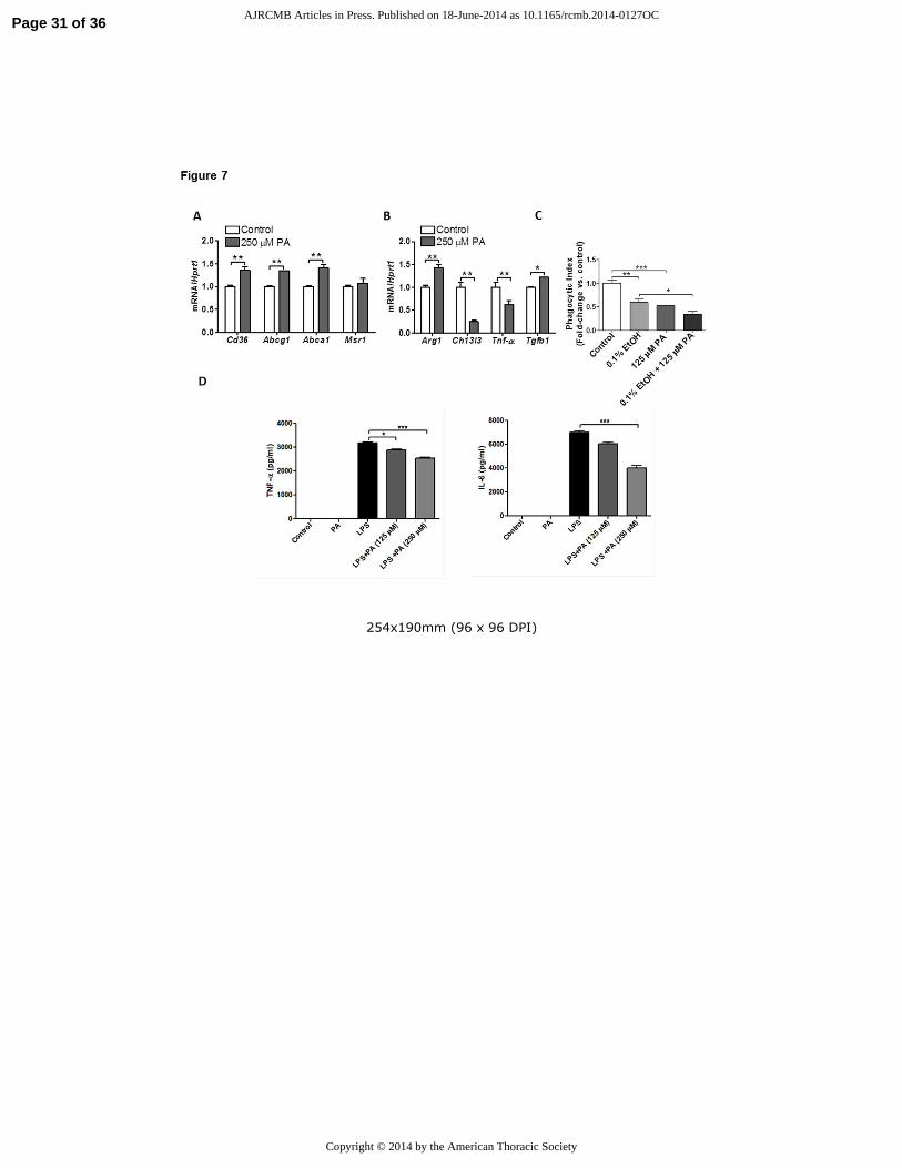
receptor Cd36 and lipid efflux transporters Abca1 and Abcg1 (fig 7A). Moreover, palmitic acid
promoted a shift in macrophage phenotype to an M2 "reparative" subtype characterized by
increased mRNA expression of Arg1 and Tgfb1 and decreased expression of the M1 marker Tnf-
α (fig 7B).
To determine whether these phenotypic changes were associated with functional impairments we
assessed the phagocytic capacity of AMs cultured in the presence or absence of palmitic acid. As
shown in figure 7C, palmitic acid significantly reduced uptake of heat-killed E. coli by AMs.
Moreover, this functional deficit was equivalent to that observed with alcohol treatments, and
was additive when cells were cultured with alcohol plus palmitate. Furthermore, palmitic acid
blunted agonist-induced inflammatory responses in AM, decreasing both TNFα and IL-6
production in response to LPS (fig 7 D, E). Taken together, these findings indicate that
Page 13 of 36 AJRCMB Articles in Press. Published on 18-June-2014 as 10.1165/rcmb.2014-0127OC
Copyright © 2014 by the American Thoracic Society
14
extracellular FA accumulation alters AM phenotype and function and suggests that metabolic
abnormalities may be mechanistically linked to immune impairments in the alcoholic lung.
Discussion
Chronic EtOH abuse predisposes to the development of inflammatory lung diseases and several
mechanisms have been proposed to explain these clinical observations. These include altered
oxidant-antioxidant balance (24, 25) and direct suppressive effects on alveolar macrophages (26,
27). In this study, we describe a mechanism in which EtOH impairs lung lipid homeostasis,
associated with altered activation of AMPK, with the net effects of enhanced lipid synthesis and
impaired immune function. We show that EtOH induces marked lipid accumulation in the lung, a
pathological phenotype reminiscent of the alcoholic fatty liver, and we provide data implicating
these lipids in the functional impairment of alveolar macrophages.
The major lipid species that were increased in lungs from chronic EtOH-fed ratswere TGs and
FAs. The functional importance of these lipids in the adult lung has not been fully determined
although it is believed that TGs and FAs contribute to the fluidity of the surfactant monolayer
and provide fatty acid acyl chains for PL production (15, 28). For the latter reason, and because
we observed enhanced expression of the lipid synthesizing machinery (e.g. Fasn) in response to
EtOH, we were surprised to find that PL levels were not significantly altered in response to
chronic EtOH consumption. However, we postulate that the lung has evolved protective
mechanisms to limit fluctuations in PL levels which might otherwise imperil pulmonary
function. This hypothesis is consistent with reports showing decreased PL synthesis in AEII
cells after an acute EtOH exposure but stable PL levels after chronic EtOH ingestion (18, 19).
Page 14 of 36 AJRCMB Articles in Press. Published on 18-June-2014 as 10.1165/rcmb.2014-0127OC
Copyright © 2014 by the American Thoracic Society
15
While we did not observe significant changes in total PL levels in response to chronic EtOH
feeding, we recognize that our findings do not exclude the possibility that EtOH might have
altered the type of PLs produced. In the liver, chronic EtOH ingestion significantly disrupts
hepatic desaturase activity, leading to marked changes in the intrahepatic fatty acyl-chain
composition (29). Similar biochemical changes in the lung might have important consequences;
recent studies have demonstrated that genetic mutations leading to deranged fatty acid
composition in the lung result in enhanced fibroproliferative responses as well as altered
pulmonary compliance (30, 31).
Our findings are consistent with a model in which EtOH-induced accumulation of pulmonary TG
and FA results from increased lipid synthesis in AEII cells. This is based on our findings
showing that several key factors (e.g. SREBF1, DGAT1) involved in TG synthesis are
upregulated in the alcohol exposed lung (32). Most notably, we detected a marked increase in the
transcription factor Srebf1 in both whole lung and in cultured AEII cells. Recent evidence
indicates that Srebf1 plays an important role in regulating TG levels in the lung (33). Mice with
deletions of Insig1 and Insig2, both of which encode for proteins that inhibit Srebf, displayed
enhanced Srebf1 function associated with marked accumulation of TGs in both AEII cells and
AMs (32, 34, 35). Conversely, deletion of the Srefp cleavage-associated protein, which is
required for Srebf activation, was associated with a non-significant decrease in TG levels in AEII
cells. These findings suggest that the effects of EtOH on Srebf1 might contribute to lipid
accumulation in affected lungs (36).
Another mechanism by which EtOH might contribute to lipid accumulation in the lung is by
inhibiting the breakdown of fatty acids (37). Importantly, this mechanism has been shown to play
a critical role in lipid accumulation in the alcoholic liver (38). Consistent with this possibility, we
Page 15 of 36 AJRCMB Articles in Press. Published on 18-June-2014 as 10.1165/rcmb.2014-0127OC
Copyright © 2014 by the American Thoracic Society
16
found that Ppara and Cpt1a mRNA expression were significantly decreased in the EtOH-
exposed rat lung. Our observation that similar decreases in Ppara and Cpt1a expression were not
seen in cultured AEII cells in response to EtOH suggests that TG accumulation in our model
systems may depend more upon enhanced production rather than reduced elimination of excess
lipids.
We observed that chronic EtOH ingestion inhibits AMPK activation in the lung. Decreased
AMPK phosphorylation was demonstrated in both whole lung tissue and in cultured AEII cells
after exposure to EtOH. AMPK is a central regulator of metabolism and might reasonably be
anticipated to participate in the metabolic defects seen in the EtOH-exposed lung (39, 40).
Consistent with this hypothesis, decreased AMPK has been shown to promote alcoholic fatty
liver disease and pharmacological activation of AMPK has been shown to limit alcohol-induced
steatosis in mice (41, 42). Such observations provide a rationale for testing whether activators of
AMPK have similar effects on lipid homeostasis in the lung.
The ability of the lung to metabolize EtOH indicates that deleterious effects can be induced by
either EtOH or one its metabolites. In this study, we demonstrated that TG accumulation in AEII
cells is largely dependent on the ability of AEII cells to metabolize EtOH; inhibition of the
CYP2E1 enzyme with CMZ completely abolished TG accumulation (43). We believe that these
findings may have important clinical implications because drugs that inhibit the cytochrome
enzymes are clinically available. Whether inhibition of CYP2E1 can prevent the toxic effects of
EtOH on pulmonary cells in our animal models or in humans remains to be studied.
One important complication of alcoholic liver disease is the development of respiratory
insufficiency. This is explained by various factors including ascites, hepatic hydrothorax, as well
Page 16 of 36 AJRCMB Articles in Press. Published on 18-June-2014 as 10.1165/rcmb.2014-0127OC
Copyright © 2014 by the American Thoracic Society
17
as the enigmatic condition known as hepato-pulmonary syndrome (HPS). While arterial
hypoxemia in patients with liver disease is often attributed to diverse mechanisms (i.e. lung
restriction, vascular shunts), we hypothesize that lipid accumulation within the distal airspaces
might also play a role. This hypothesis is supported by the observation that TG accumulation in
the lungs of male Zucker diabetic fatty (ZDF) rats is associated with capillary basement
membrane thickening and impairments in gas exchange (44, 45). Future studies examining the
relationship between lung lipid abnormalities and the development of respiratory insufficiency in
alcoholic patients are warranted.
Our observations also suggest a mechanistic link between metabolic abnormalities and immune
impairments in the lung. To date, the majority of studies focusing on immune dysregulation have
investigated the direct effects of EtOH on immune cell function (46-48). In this study, we
propose an alternative hypothesis, namely that chronic EtOH exposure of AEII cells stimulates a
paracrine lipid excess wherein FAs released into the distal air spaces are concentrated by
macrophages, promoting an alternative macrophage phenotype and impairing macrophage
function. Ongoing studies will test whether anti-lipid therapies might prevent and/or treat EtOH-
related inflammatory lung disorders.
In summary, we found that chronic alcohol exposure alters lung metabolic homeostasis and our
data suggest that these abnormalities might play a role in the development of inflammatory lung
diseases. We anticipate that these findings will open new avenues of research regarding the
effects of alcohol on lung biology and will provide a foundation for future clinical investigations
examining the role of metabolic changes in the development of diverse lung diseases.
Page 17 of 36 AJRCMB Articles in Press. Published on 18-June-2014 as 10.1165/rcmb.2014-0127OC
Copyright © 2014 by the American Thoracic Society
18
References:
1. Perlino CA, Rimland D. Alcoholism, leukopenia, and pneumococcal sepsis. Am Rev Respir Dis
1985;132(4):757-760.
2. Cook RT. Alcohol abuse, alcoholism, and damage to the immune system--a review. Alcohol Clin
Exp Res 1998;22(9):1927-1942.
3. Zhang P, Bagby GJ, Happel KI, Summer WR, Nelson S. Pulmonary host defenses and alcohol.
Front Biosci 2002;7:d1314-1330.
4. Polikandriotis JA, Rupnow HL, Elms SC, Clempus RE, Campbell DJ, Sutliff RL, Brown LA, Guidot
DM, Hart CM. Chronic ethanol ingestion increases superoxide production and nadph oxidase expression
in the lung. Am J Respir Cell Mol Biol 2006;34(3):314-319.
5. Brown SD, Brown LA. Ethanol (etoh)-induced tgf-beta1 and reactive oxygen species production
are necessary for etoh-induced alveolar macrophage dysfunction and induction of alternative activation.
Alcohol Clin Exp Res 2012;36(11):1952-1962.
6. Moss M, Bucher B, Moore FA, Moore EE, Parsons PE. The role of chronic alcohol abuse in the
development of acute respiratory distress syndrome in adults. JAMA 1996;275(1):50-54.
7. Berkowitz DM, Danai PA, Eaton S, Moss M, Martin GS. Alcohol abuse enhances pulmonary
edema in acute respiratory distress syndrome. Alcohol Clin Exp Res 2009;33(10):1690-1696.
8. Moss M, Burnham EL. Chronic alcohol abuse, acute respiratory distress syndrome, and multiple
organ dysfunction. Crit Care Med 2003;31(4 Suppl):S207-212.
9. Joshi PC, Guidot DM. The alcoholic lung: Epidemiology, pathophysiology, and potential
therapies. Am J Physiol Lung Cell Mol Physiol 2007;292(4):L813-823.
10. Mitchell PO, Jensen JS, Ritzenthaler JD, Roman J, Pelaez A, Guidot DM. Alcohol primes the
airway for increased interleukin-13 signaling. Alcohol Clin Exp Res 2009;33(3):505-513.
11. Zhong W, Cui Y, Yu Q, Xie X, Liu Y, Wei M, Ci X, Peng L. Modulation of lps-stimulated pulmonary
inflammation by borneol in murine acute lung injury model. Inflammation 2014.
12. O'Shea RS, Dasarathy S, McCullough AJ. Alcoholic liver disease. Hepatology 2009;51(1):307-328.
13. Menon KV, Gores GJ, Shah VH. Pathogenesis, diagnosis, and treatment of alcoholic liver disease.
Mayo Clin Proc 2001;76(10):1021-1029.
14. Lee M, Kowdley KV. Alcohol's effect on other chronic liver diseases. Clin Liver Dis
2012;16(4):827-837.
15. Whitsett JA, Wert SE, Weaver TE. Alveolar surfactant homeostasis and the pathogenesis of
pulmonary disease. Annu Rev Med 2009;61:105-119.
16. Mason RJ. Surfactant synthesis, secretion, and function in alveoli and small airways. Review of
the physiologic basis for pharmacologic intervention. Respiration 1987;51 Suppl 1:3-9.
17. Liau DF, Hashim SA, Pierson RN, 3rd, Ryan SF. Alcohol-induced lipid change in th lung. J Lipid Res
1981;22(4):680-686.
18. Holguin F, Moss I, Brown LA, Guidot DM. Chronic ethanol ingestion impairs alveolar type ii cell
glutathione homeostasis and function and predisposes to endotoxin-mediated acute edematous lung
injury in rats. J Clin Invest 1998;101(4):761-768.
19. Wagner M, Heinemann HO. Effect of ethanol on phospholipid metabolism by the rat lung. Am J
Physiol 1975;229(5):1316-1320.
20. Lang CH, Derdak Z, Wands JR. Strain-dependent differences for suppression of insulin-stimulated
glucose uptake in skeletal and cardiac muscle by ethanol. Alcohol Clin Exp Res 2014.
21. Shah D, Romero F, Stafstrom W, Duong M, Summer R. Extracellular atp mediates the late phase
of neutrophil recruitment to the lung in murine models of acute lung injury. Am J Physiol Lung Cell Mol
Physiol 2013;306(2):L152-161.
Page 18 of 36 AJRCMB Articles in Press. Published on 18-June-2014 as 10.1165/rcmb.2014-0127OC
Copyright © 2014 by the American Thoracic Society
19
22. Iverson SJ, Lang SL, Cooper MH. Comparison of the bligh and dyer and folch methods for total
lipid determination in a broad range of marine tissue. Lipids 2001;36(11):1283-1287.
23. Viollet B, Mounier R, Leclerc J, Yazigi A, Foretz M, Andreelli F. Targeting amp-activated protein
kinase as a novel therapeutic approach for the treatment of metabolic disorders. Diabetes Metab
2007;33(6):395-402.
24. Liang Y, Harris FL, Jones DP, Brown LA. Alcohol induces mitochondrial redox imbalance in
alveolar macrophages. Free Radic Biol Med 2013;65:1427-1434.
25. Jensen JS, Fan X, Guidot DM. Alcohol causes alveolar epithelial oxidative stress by inhibiting the
nuclear factor (erythroid-derived 2)-like 2-antioxidant response element signaling pathway. Am J Respir
Cell Mol Biol 2013;48(4):511-517.
26. Thevenot P, Saravia J, Giaimo J, Happel KI, Dugas TR, Cormier SA. Chronic alcohol induces m2
polarization enhancing pulmonary disease caused by exposure to particulate air pollution. Alcohol Clin
Exp Res 2013;37(11):1910-1919.
27. Curry-McCoy TV, Venado A, Guidot DM, Joshi PC. Alcohol ingestion disrupts alveolar epithelial
barrier function by activation of macrophage-derived transforming growth factor beta1. Respir Res
2013;14:39.
28. Agassandian M, Mallampalli RK. Surfactant phospholipid metabolism. Biochim Biophys Acta
2013;1831(3):612-625.
29. Umeki S, Shiojiri H, Nozawa Y. Chronic ethanol administration decreases fatty acyl-coa
desaturase activities in rat liver microsomes. FEBS Lett 1984;169(2):274-278.
30. Sunaga H, Matsui H, Ueno M, Maeno T, Iso T, Syamsunarno MR, Anjo S, Matsuzaka T, Shimano
H, Yokoyama T, et al. Deranged fatty acid composition causes pulmonary fibrosis in elovl6-deficient
mice. Nat Commun 2013;4:2563.
31. Goetzman ES, Alcorn JF, Bharathi SS, Uppala R, McHugh KJ, Kosmider B, Chen R, Zuo YY, Beck
ME, McKinney RW, et al. Long-chain acyl-coa dehydrogenase deficiency as a cause of pulmonary
surfactant dysfunction. J Biol Chem 2014;289(15):10668-10679.
32. Plantier L, Besnard V, Xu Y, Ikegami M, Wert SE, Hunt AN, Postle AD, Whitsett JA. Activation of
sterol-response element-binding proteins (srebp) in alveolar type ii cells enhances lipogenesis causing
pulmonary lipotoxicity. J Biol Chem 2012;287(13):10099-10114.
33. Zhang F, Pan T, Nielsen LD, Mason RJ. Lipogenesis in fetal rat lung: Importance of c/ebpalpha,
srebp-1c, and stearoyl-coa desaturase. Am J Respir Cell Mol Biol 2004;30(2):174-183.
34. Shimano H. Sterol regulatory element-binding proteins (srebps): Transcriptional regulators of
lipid synthetic genes. Prog Lipid Res 2001;40(6):439-452.
35. Besnard V, Wert SE, Stahlman MT, Postle AD, Xu Y, Ikegami M, Whitsett JA. Deletion of scap in
alveolar type ii cells influences lung lipid homeostasis and identifies a compensatory role for pulmonary
lipofibroblasts. J Biol Chem 2009;284(6):4018-4030.
36. You M, Fischer M, Deeg MA, Crabb DW. Ethanol induces fatty acid synthesis pathways by
activation of sterol regulatory element-binding protein (srebp). J Biol Chem 2002;277(32):29342-29347.
37. Sozio M, Crabb DW. Alcohol and lipid metabolism. Am J Physiol Endocrinol Metab
2008;295(1):E10-16.
38. Crabb DW. Alcohol deranges hepatic lipid metabolism via altered transcriptional regulation.
Trans Am Clin Climatol Assoc 2004;115:273-287.
39. Jian MY, Alexeyev MF, Wolkowicz PE, Zmijewski JW, Creighton JR. Metformin-stimulated ampk-
alpha1 promotes microvascular repair in acute lung injury. Am J Physiol Lung Cell Mol Physiol
2013;305(11):L844-855.
40. Zhao X, Zmijewski JW, Lorne E, Liu G, Park YJ, Tsuruta Y, Abraham E. Activation of ampk
attenuates neutrophil proinflammatory activity and decreases the severity of acute lung injury. Am J
Physiol Lung Cell Mol Physiol 2008;295(3):L497-504.
Page 19 of 36 AJRCMB Articles in Press. Published on 18-June-2014 as 10.1165/rcmb.2014-0127OC
Copyright © 2014 by the American Thoracic Society
20
41. Garcia-Villafranca J, Guillen A, Castro J. Ethanol consumption impairs regulation of fatty acid
metabolism by decreasing the activity of amp-activated protein kinase in rat liver. Biochimie
2008;90(3):460-466.
42. Ben Mosbah I, Massip-Salcedo M, Fernandez-Monteiro I, Xaus C, Bartrons R, Boillot O, Rosello-
Catafau J, Peralta C. Addition of adenosine monophosphate-activated protein kinase activators to
university of wisconsin solution: A way of protecting rat steatotic livers. Liver Transpl 2007;13(3):410-
425.
43. Correa M, Viaggi C, Escrig MA, Pascual M, Guerri C, Vaglini F, Aragon CM, Corsini GU. Ethanol
intake and ethanol-induced locomotion and locomotor sensitization in cyp2e1 knockout mice.
Pharmacogenet Genomics 2009;19(3):217-225.
44. Foster DJ, Ravikumar P, Bellotto DJ, Unger RH, Hsia CC. Fatty diabetic lung: Altered alveolar
structure and surfactant protein expression. Am J Physiol Lung Cell Mol Physiol 2010;298(3):L392-403.
45. Farkas GA, Schlenker EH. Pulmonary ventilation and mechanics in morbidly obese zucker rats.
Am J Respir Crit Care Med 1994;150(2):356-362.
46. Chen MM, Bird MD, Zahs A, Deburghgraeve C, Posnik B, Davis CS, Kovacs EJ. Pulmonary
inflammation after ethanol exposure and burn injury is attenuated in the absence of il-6. Alcohol
2013;47(3):223-229.
47. Bird MD, Zahs A, Deburghgraeve C, Ramirez L, Choudhry MA, Kovacs EJ. Decreased pulmonary
inflammation following ethanol and burn injury in mice deficient in tlr4 but not tlr2 signaling. Alcohol
Clin Exp Res 2010;34(10):1733-1741.
48. Standiford TJ, Danforth JM. Ethanol feeding inhibits proinflammatory cytokine expression from
murine alveolar macrophages ex vivo. Alcohol Clin Exp Res 1997;21(7):1212-1217.
Figure Legends:
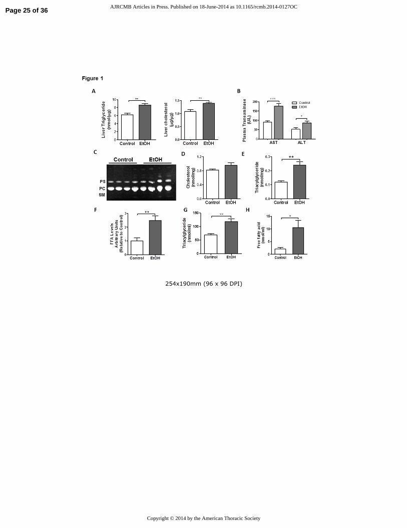
Figure 1. Chronic EtOH ingestion induces lipid accumulation in the liver and lung. A) Chronic
EtOH ingestion increased TGs and cholesterol levels in the liver (n=4 each group, **p<0.01 vs
control group). B) Plasma aspartate and alanine aminotransferase levels are elevated in chronic
EtOH fed rats (n=10 each group, **p<0.01 vs control group). C) Chronic EtOH ingestion did
not significantly increase total PLs, PC or PS in whole lung (n=10). The image is representative
of two different TLC. D) Chronic EtOH ingestion did not significantly increase total cholesterol
levels in whole lung (n=10). E-F) Chronic EtOH ingestion enhanced TG and FA levels in whole
lung (n=10, **p<0.01 vs control group). G-H) Chronic EtOH ingestion enhanced TG (n=10,
**p<0.01 vs control group) and FA levels in BAL (n=10, *p<0.05 vs control group). All data are
expressed as mean ± SE. The statistical significance was assessed using a Student’s unpaired t
test.
Page 20 of 36 AJRCMB Articles in Press. Published on 18-June-2014 as 10.1165/rcmb.2014-0127OC
Copyright © 2014 by the American Thoracic Society
21
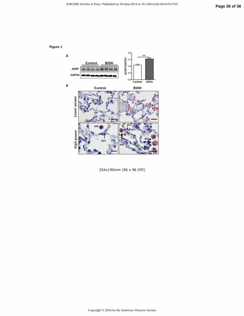
Figure 2. Chronic EtOH ingestion induces alveolar macrophage lipid accumulation. A) Western
blot analysis for ADRP in lungs from chronic EtOH-fed rats (n=8). The image is representative
of at least two different Western blots. Densitometry measurements showing increased ADRP
expression in the lung from chronic EtOH-fed rats (**p<0.01 vs Control group). All data are
expressed as mean ± SE. The statistical significance was assessed using a Student’s unpaired t
test. B) Oil Red O staining of the lung demonstrates massive neutral accumulation in alveolar
macrophages (AM) from EtOH fed rats while a more modest accumulation of lipid droplets was
observed in AEII cells (thin arrow).
Figure 3. Chronic EtOH ingestion promotes metabolic changes in the lung. A) Chronic EtOH
ingestion decreased AMPK activation but had no effect on total protein levels in the lung (n=8,
**p<0.01 vs Control group). The image is representative of at least two different blots. B)
Transcript levels for Srebf1, Fasn, Acaca are increased in the lung after chronic EtOH
consumption (n=8, **p<0.01 vs Control group). C) Protein expression for diacylglycerol O-
acyltransferase 1 (DGAT1), sterol regulatory-element binding protein1 (SREBF1), fatty acid
synthase (FASN) and Acetyl CoA carboxylase (ACACA) levels are increased in the lung after
chronic EtOH ingestion (n=8, *p<0.05, **p<0.01, ***p<0.001 vs Control group). The image is
representative of two different western blots. D) Peroxisome proliferator-activated receptor alpha
(Ppara) and carnitine palmitoyltransferase I (Cpt1a) transcript levels are decreased in the lung
after chronic EtOH intake (n=8, **p<0.01 vs Control group). Data are expressed as mean ± SE.
The statistical significance was assessed using a Student’s unpaired t test.
Figure 4. EtOH alters metabolic homeostasis in alveolar epithelial type II (AEII) cells leading to
intracellular triacylglyceride (TG) accumulation. A) Rat L2 AEII cells accumulate TGs when
cultured in 0.05% or 0.1% EtOH for either 48 or 72 h (n=6, *p<0.05, ***p<0.001 vs Control
Page 21 of 36 AJRCMB Articles in Press. Published on 18-June-2014 as 10.1165/rcmb.2014-0127OC
Copyright © 2014 by the American Thoracic Society
22
group) B) Oil Red O staining of L2 cells demonstrated increased lipid droplet formation in
response to EtOH. C) EtOH decreases AMPK activation (i.e., decreased phosphorylation) in L2
cells (n=6, **p<0.01 vs Control group). The image is representative of three different blots (D)
EtOH enhances protein content of SREBF1 and acetyl-CoA synthase in L2 cells (n=6, **p<0.01,
***p<0.001). Data are expressed as mean ± SE. In (A), the statistical significance was assessed
with a one-way ANOVA test, whereas, in (C-D) we used a Student’s unpaired t test.
Figure 5. Inhibition of CYP2E1 activity blocks triacylglyceride (TG) accumulation in alveolar
epithelial type II cells. A) Alcohol dehydrogenase (ADH) activity is detected in the lung and
activity increases in response to chronic alcohol consumption (n=6, ***p<0.001 vs Control
group). B) EtOH induces protein expression of CYP2E1 in the rat lung (n=6, *p<0.05 vs Control
group). The image is representative of three different blots. C) EtOH induces protein expression
of CYP2E1 in rat L2 AEII cells (n=6, **p<0.01 vs Control group). D) Treatment of L2 cells with
the CYP2E1 inhibitor chlormethiazole (CMZ) for 24 h blocked EtOH induced TG accumulation
(n=6, *p<0.05 vs Control group, ##
p<0.01 vs EtOH group). E) The anti-oxidant N-acetyl cysteine
did not attenuate EtOH-induced TG accumulation in L2 cells (n=6, *p<0.05 vs Control group).
Data are expressed as mean ± SE. In (A-C), the statistical significance was assessed using a
Student’s unpaired t test, whereas, in (D-E) we used a one-way ANOVA test.
Figure 6. Chronic EtOH exposure alters primary alveolar macrophage phenotype. A) Transcript
levels for Cd36, Abcg1 and Tgfb1 in freshly isolated AMs from lungs of EtOH-fed rats. B)
Enzyme-linked immunosorbent assay for IL6, TNF-α, TGFβ1 and MCP1 in BAL fluid from
control and EtOH-fed rats (n=6, **p<0.01 vs Control group). C) Total and differential cell
counts in BAL fluid from control and EtOH fed rats. Data are expressed as mean ± SE. The
statistical significance was assessed using a Student’s unpaired t test.
Page 22 of 36 AJRCMB Articles in Press. Published on 18-June-2014 as 10.1165/rcmb.2014-0127OC
Copyright © 2014 by the American Thoracic Society
23
Figure 7. Increases in fatty acids (FA) alter alveolar macrophage (AM) phenotype and function.
A) Rat AMs cultured in media containing 250 µM of palmitic acid (PA) displayed an increased
mRNA expression of lipid receptor Cd36 and the lipid efflux transporters Abca1 and Abcg1
(n=6, **p<0.01 vs Control group). B) PA promotes a shift toward an M2 macrophage phenotype
in rat AMs, characterized by enhanced mRNA expression or Arg1 and Tgfb1 and suppression of
Tnf-α transcripts (n=6, *p<0.05, **p<0.01 vs Control group). C) PA significantly reduced uptake
of heat-killed E. coli by AMs and this effect was enhanced with EtOH treatment (n=6, **p<0.01,
***p<0.001 vs Control group and *p<0.05 vs EtOH group). D) PA blunts LPS-induced pro-
inflammatory responses in rat AMs (n=6, *p<0.05, ***p<0.001 vs Control group). Data are
expressed as mean ± SE. The statistical significance was assessed using a one-way ANOVA test.
Page 23 of 36 AJRCMB Articles in Press. Published on 18-June-2014 as 10.1165/rcmb.2014-0127OC
Copyright © 2014 by the American Thoracic Society
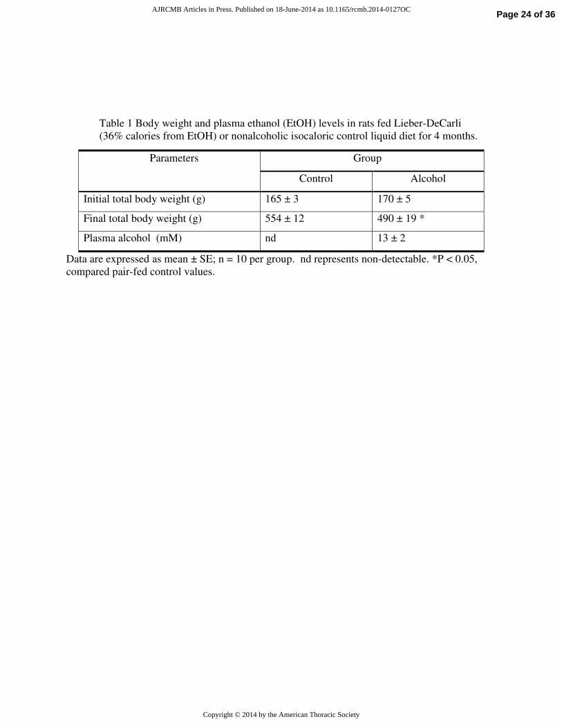
Table 1 Body weight and plasma ethanol (EtOH) levels in rats fed Lieber-DeCarli
(36% calories from EtOH) or nonalcoholic isocaloric control liquid diet for 4 months.
Data are expressed as mean ± SE; n = 10 per group. nd represents non-detectable. *P < 0.05,
compared pair-fed control values.
Parameters Group
Control Alcohol
Initial total body weight (g) 165 ± 3 170 ± 5
Final total body weight (g) 554 ± 12 490 ± 19 *
Plasma alcohol (mM) nd 13 ± 2
Page 24 of 36 AJRCMB Articles in Press. Published on 18-June-2014 as 10.1165/rcmb.2014-0127OC
Copyright © 2014 by the American Thoracic Society
254x190mm (96 x 96 DPI)
Page 25 of 36 AJRCMB Articles in Press. Published on 18-June-2014 as 10.1165/rcmb.2014-0127OC
Copyright © 2014 by the American Thoracic Society
254x190mm (96 x 96 DPI)
Page 26 of 36 AJRCMB Articles in Press. Published on 18-June-2014 as 10.1165/rcmb.2014-0127OC
Copyright © 2014 by the American Thoracic Society
254x190mm (96 x 96 DPI)
Page 27 of 36 AJRCMB Articles in Press. Published on 18-June-2014 as 10.1165/rcmb.2014-0127OC
Copyright © 2014 by the American Thoracic Society
254x190mm (96 x 96 DPI)
Page 28 of 36 AJRCMB Articles in Press. Published on 18-June-2014 as 10.1165/rcmb.2014-0127OC
Copyright © 2014 by the American Thoracic Society
254x190mm (96 x 96 DPI)
Page 29 of 36 AJRCMB Articles in Press. Published on 18-June-2014 as 10.1165/rcmb.2014-0127OC
Copyright © 2014 by the American Thoracic Society
254x190mm (96 x 96 DPI)
Page 30 of 36 AJRCMB Articles in Press. Published on 18-June-2014 as 10.1165/rcmb.2014-0127OC
Copyright © 2014 by the American Thoracic Society
254x190mm (96 x 96 DPI)
Page 31 of 36 AJRCMB Articles in Press. Published on 18-June-2014 as 10.1165/rcmb.2014-0127OC
Copyright © 2014 by the American Thoracic Society
Supplemental Methods
Plasma measurments. Plasma ALT and AST were determined by standard enzymatic
procedures (Sigma-Aldrich; St. Louis, MO) and blood alcohol levels were determined by a rapid
analyzer (Analox Instruments, Lunenburg, MA). Measurements were performed on blood
samples collected from rats between 8-9am.
Alveolar Epithelial Type II Cell Culture and Treatment
Rat alveolar type II epithelial cell line (L2 cells) was cultured with media supplemented with
EtOH at final concentrations of 0.05% or 0.1 % v/v based on published protocols (2). In select
studies, L2 cells were cultured in the presence or absence of the cytochrome P450 inhibitor CMZ
(100 µM) or with N-acetyl cysteine (NAC; 5 mM; both from Sigma Chemical Co; St. Louis,
MO). To minimize EtOH evaporation media was replaced every 4-6 h during the daytime.
Lipid Droplet Determination
Lipid droplets were evaluated in L2 cells and NR8383 cells cultured in chamber slides
containing media with or without EtOH. At specified time points, cells were fixed for 10 min in
10% buffered formalin followed by treatment with propylene glycol. Cells were then stained
with Oil Red O solution for 15 min at 60°C followed by counterstaining with Gill’s Hematoxylin
solution for 30 sec. After mounting with glycerol-PBS medium, red stained lipid droplets were
visualized by light microscopy.
Lipid Extraction and Analysis
Page 32 of 36 AJRCMB Articles in Press. Published on 18-June-2014 as 10.1165/rcmb.2014-0127OC
Copyright © 2014 by the American Thoracic Society
Total lipids were extracted from BAL fluid, L2 cells, and lung tissue using a modified method of
Bligh and Dyer,using chloroform/methanol (2:1) (3). The organic phase was obtained by
centrifugation at 200g. Lipids were dried under a gentle stream of nitrogen gas. The lipid extracts
were assayed for triacylglyceride, free fatty acid and total cholesterol content according to the
manufacturer’s protocols (BioVision, Mountain View, CA). In other experiments, extracts were
dissolved in chloroform (200 µl) and aliquots, together with standards, were separated by TLC
on silica gel G-60 plates (Sigma-Aldrich) as previously described (4). The neutral lipid
components were separated using a mixture of n-hexane: diethyl ether: acetic acid (70:30:1)
while the polar lipid components were separated using a mixture of chloroform: methanol: acetic
acid: water (85:15:10:3.5). After TLC separation, plates were removed, air dried and sprayed
with primuline (Sigma-Aldrich, St. Louis, MO) to label lipid spots prior to visualizing under UV
light. Spots were identified by comparison with lipid standards.
RNA Isolation and Quantitative Real-Time PCR
Total RNA was extracted from cells or lung tissue using the RNeasy Mini-Kit (QIAGEN,
Valencia, CA) according to the manufacturer’s instructions. Quality and quantity of RNA was
asssessed using a Nano-Drop spectrophotometer. The first strand of cDNA was synthesized with
1 µg of RNA using the GoScript™ Reverse Transcription System (Promega, Madison, WI).
SYBR Green Real-Time PCR was performed with the IQ5 Multicolor Real-Time PCR Detection
System (Bio-Rad, Hercules, CA.), with the following cycle conditions: initial denaturation (95
ºC for 3 min), followed by 40 cycles of amplification (95 ºC for 15 sec) and annealing (60ºC for
45 sec). All of the assays were done in triplicate. The following gene specific primers (sense and
anti-sense) were used in the study (IDT, USA): Srebf1 (forward) 5'- GGA GCC ATG GAT TGC
Page 33 of 36 AJRCMB Articles in Press. Published on 18-June-2014 as 10.1165/rcmb.2014-0127OC
Copyright © 2014 by the American Thoracic Society
ACA TT -3' and (reverse) 5'- AGG AAG GCT TCC AGA GAG GA -3'; Acaca (forward) 5'-
AGG AAG ATG GTG TCC CGC TCT G -3' and (reverse) 5'- GGG GAG ATG TGC TGG GTC
AT -3'; Fasn(forward) 5'- AGG TGC TAG AGG CCC TGC TA -3' and (reverse) 5'- GTG CAC
AGA CAC CTT CCC AT -3'; Ppara (forward) 5'- AGG CTA TCC CAG GCT TTG C -3' and
(reverse) 5'- CGT CTG ACT CGG TCT TTT G -3'; Cpt1a (forward) 5'- CTC CTG AGC AGT
TAC CAA TGC -3' and (reverse) 5'- GAA CCT TGG CTG CGG TAA GAC -3'; Abcg1
(forward) 5’- GAA GGT TGC CAC AGC TTC TC- 3’, (reverse) 5’CAT GGT CTT GGC CAG
GTA GT ’3, Abca1 (forward) 5’- AAC AGT TTG TGG CCC TTT TG 3’, (reverse) 5’- AGT
TCC AGG CTG GGC TAC TT - 3’; Cd36 (forward) 5’-GAA GCA CTG AAG AAT CTG AAG
AG-3’, (reverse) 5’-TCC AAC ACC AAG TAA GAC CAT C -3’; Msr1 (forward) 5’- ATG GCA
CAG TGG GAT GAC TTT-3’, (reverse) 5’- TTT ATA AGA CTT CAT CCT CTC’; Tgf-β1
(forward) 5’-TCC CAA ACG TCG AGG TGA C-3’, (reverse) 5’- CAG GTG TTG AGC CCT TTT
CCA-3’; Tnf (forward) 5’- CCC AGA CCC TCA CAC TCA GAT-3’, (reverse) 5’-TTG TCC CTT
GAA GAG AAC CTG-3’; Arg1 (forward) 5’-GCT GTC TTC CCA AGA GTT GGG -3’, (reverse)
5’-ATG GAA GAG ACC TTC AGC TAC -3’; Chi3l3 (forward) 5’-GAC TTG CGT GAC TAT
GAA GC -3’ (reverse) 5’-TGA CGG TTC TGA GGA GTA GA-3’;Gapdh (forward) 5'- GAA
CGG GAA GCT CAC TGG C -3' and (reverse) 5'- GCA TGT CAG ATC CAC AAC GG -3';
Hprt1 (forward) 5’-GCT CGA GAT GTC ATG AAC GAG A-3’, (reverse) 5’-TCA GCG CTT
TAA TGT AAT CCA AGC-3’. Quantitative values were obtained from the threshold cycle (Ct)
number that indicates an exponential amplification of the PCR product and calculated as the
change (n-fold) in value of the treatment group according with the 2(-Delta Delta Ct) method (5).
PCR products were confirmed by melting-curve analysis. The housekeeping genes Gapdh and
Hprt1 were used for normalization.
Page 34 of 36 AJRCMB Articles in Press. Published on 18-June-2014 as 10.1165/rcmb.2014-0127OC
Copyright © 2014 by the American Thoracic Society
Western Blot Analysis
Cell lysates or whole lung tissue were homogenized in ice cold lysis buffer (PBS, 0.05% Triton
X-100, pH 7.4) containing protease inhibitors (Roche Complete mini) and phosphatase
inhibitors. Lung nuclear fractions were extracted using a commercially available kit (Active
motif, Carlsbad, CA) according to the manufacturer’s instructions. Protein concentrationswere
determined by PierceTM
BCA assay kit (Thermo Scientific, Rockford, IL). Protein samples (20
µg) were solubilized in 4 × Laemmli sample buffer, heated at 95°C for 10 min, centrifuged at
3,000 g for 1 min, loaded on a 10% Tris-HCl-SDS-polyacrylamide gel and run for 1 h at 120 V.
Protein was transferred to a nitrocellulose membrane (Bio-Rad) and then blocked with Odyssey
Blocking Buffer (Li-Cor Biosciences, Lincoln, NE) for 1 h at room temperature. After blocking
step, the membrane was incubated overnight at 4°C with a specific polyclonal rabbit primary
antibody to AMPK, pAMPK, ACACA, FASN, ADRP, GAPDH (Cell Signaling, Danvers, MA),
ADRP, SREBF1, DGAT1, CYP2E1 (Abcam, Cambridge, MA) at a dilution of 1:1.000 in
blocking buffer with 0.1% Tween-20 followed by incubation with donkey anti-rabbit or anti-
mouse secondary antibody (Li-Cor Biosciences, Lincoln, NE) at a dilution of 1:5,000 in blocking
buffer. After three washes with PBS, all immunoblots were visualized using the Odyssey
infrared imaging system (Li-Cor Biosciences). Densitometry analysis was performed using
Image J image processing software (Wayne Rasband, NIH, Bethesda, MD, USA). Pixel intensity
was normalized to the GAPDH loading control for each sample.
Macrophage Phagocytosis Assay
Page 35 of 36 AJRCMB Articles in Press. Published on 18-June-2014 as 10.1165/rcmb.2014-0127OC
Copyright © 2014 by the American Thoracic Society
Fluorescein conjugated E. coli K-12 particles (Invitrogen) were used to measure the rate of
phagocytosis in NR8383 cells. The ratio of bioparticles to macrophage cells was 100:1. Cells
were grown in 6-well plate and cultured for 24 h prior to the addition of either BSA conjugated
palmitic acid (125 µM) or BSA alone in the presence or absence of 0.1 % EtOH. Following
incubation with or without free fatty acids for 24 h, NR8383 cells were seeded in white/black 96-
well plate at 50,000 cells/well and incubated for 2 h NR8383 culture media at 37°C in 5% CO2.
The bioparticles were added and incubated for an additional 2 h at 37°C. This step was followed
by removal of the media and exposure of cells to 0.4% trypan blue (Sigma) for 1 min in order to
reduce extracellular fluorescence. Finally, after multiple washes quantitative measurements of
particle uptake were determined by measuring cellular excitation wavelength of 485/20 nm and
emission wavelength at 528/20 nm. All samples were run in quadruplicate.
References:
1. Lang CH, Derdak Z, Wands JR. Strain-dependent differences for suppression of insulin-
stimulated glucose uptake in skeletal and cardiac muscle by ethanol. Alcohol Clin Exp Res 2014.
2. Yeligar SM, Harris FL, Hart CM, Brown LA. Ethanol induces oxidative stress in alveolar
macrophages via upregulation of nadph oxidases. J Immunol 2012;188(8):3648-3657.
3. Iverson SJ, Lang SL, Cooper MH. Comparison of the bligh and dyer and folch methods
for total lipid determination in a broad range of marine tissue. Lipids 2001;36(11):1283-1287.
4. Matyash V, Liebisch G, Kurzchalia TV, Shevchenko A, Schwudke D. Lipid extraction by
methyl-tert-butyl ether for high-throughput lipidomics. J Lipid Res 2008;49(5):1137-1146.
5. Livak KJ, Schmittgen TD. Analysis of relative gene expression data using real-time
quantitative pcr and the 2(-delta delta c(t)) method. Methods 2001;25(4):402-408.
Page 36 of 36 AJRCMB Articles in Press. Published on 18-June-2014 as 10.1165/rcmb.2014-0127OC
Copyright © 2014 by the American Thoracic Society
Related Documents