This journal is © The Royal Society of Chemistry 2014 Chem. Soc. Rev. Cite this: DOI: 10.1039/c3cs60269a Ultrafast dynamics of single molecules Daan Brinks,† ab Richard Hildner,† ac Erik M. H. P. van Dijk, d Fernando D. Stefani, ae Jana B. Nieder, a Jordi Hernando f and Niek F. van Hulst* ag The detection of individual molecules has found widespread application in molecular biology, photo- chemistry, polymer chemistry, quantum optics and super-resolution microscopy. Tracking of an individual molecule in time has allowed identifying discrete molecular photodynamic steps, action of molecular motors, protein folding, diffusion, etc. down to the picosecond level. However, methods to study the ultra- fast electronic and vibrational molecular dynamics at the level of individual molecules have emerged only recently. In this review we present several examples of femtosecond single molecule spectroscopy. Starting with basic pump–probe spectroscopy in a confocal detection scheme, we move towards deterministic coherent control approaches using pulse shapers and ultra-broad band laser systems. We present the detection of both electronic and vibrational femtosecond dynamics of individual fluorophores at room temperature, showing electronic (de)coherence, vibrational wavepacket interference and quantum control. Finally, two colour phase shaping applied to photosynthetic light-harvesting complexes is presented, which allows investigation of the persistent coherence in photosynthetic complexes under physiological conditions at the level of individual complexes. Key learning points (1) Strategies to acquire the femtosecond dynamic response of single molecules. (2) Intramolecular vibrational relaxation of a single molecule. (3) Electronic coherence and quantum control of individual molecules at room temperature. (4) Vibrational wavepacket interference in a single molecule. (5) Coherent electronic energy transfer in individual photosynthetic complexes at room temperature. 1. Introduction: motivation for femtosecond single-molecule detection Over the last 25 years the optical detection of single molecules has developed tremendously. 1–4 Observing individual mole- cules one by one removes the usual ensemble averaging and enables the detection of the different subpopulations present in complex and heterogeneous systems (Fig. 1). Furthermore, following the behaviour of individual molecules in time, with- out the need to synchronize the ensemble, provides extensive insights into the dynamic behaviour of (bio-)molecules. 5 Indi- vidual molecules are influenced by their local environment, resulting in variations from molecule to molecule in their steady state absorption/emission spectra, transition dipole orientation, triplet and fluorescence lifetimes, etc. 6–9 Single molecule experiments consistently show that chemically iden- tical molecules exhibit large spatial and temporal heterogeneity for all parameters studied. The dynamic range of the processes that can be studied in conventional single molecule experiments is typically limited. Detecting the Stokes shifted fluorescence arising from an individual molecular system has proven to be the most straight- forward way to enable background free detection and discriminate the weak response of a single molecule from both the scattered irradiation field and the signal of the surrounding medium. 10 a ICFO - Institut de Ciencies Fotoniques, Mediterranean Technology Park, 08860 Castelldefels, Barcelona, Spain. E-mail: [email protected]; Web: www.ICFO.eu b Department of Chemistry and Chemical Biology, Harvard University, Cambridge, MA 02138, USA c Experimentalphysik IV, Universita ¨t Bayreuth, 95440 Bayreuth, Germany d Philips Healthcare, 5680DA Best, The Netherlands e CIBION - Center for Bionanoscience Research, CONICET, Buenos Aires, Argentina f Dept. de Quı ´mica, Universitat Auto `noma de Barcelona, 08193 Cerdanyola del Valle `s, Barcelona, Spain g ICREA - Institucio´ Catalana de Recerca i Estudis Avançats, 08015 Barcelona, Spain † Equal contribution. Received 22nd July 2013 DOI: 10.1039/c3cs60269a www.rsc.org/csr Chem Soc Rev TUTORIAL REVIEW Published on 28 January 2014. Downloaded by Max Planck Institut fuer on 28/01/2014 15:13:46. View Article Online View Journal

Welcome message from author
This document is posted to help you gain knowledge. Please leave a comment to let me know what you think about it! Share it to your friends and learn new things together.
Transcript

This journal is©The Royal Society of Chemistry 2014 Chem. Soc. Rev.
Cite this:DOI: 10.1039/c3cs60269a
Ultrafast dynamics of single molecules
Daan Brinks,†ab Richard Hildner,†ac Erik M. H. P. van Dijk,d Fernando D. Stefani,ae
Jana B. Nieder,a Jordi Hernandof and Niek F. van Hulst*ag
The detection of individual molecules has found widespread application in molecular biology, photo-
chemistry, polymer chemistry, quantum optics and super-resolution microscopy. Tracking of an individual
molecule in time has allowed identifying discrete molecular photodynamic steps, action of molecular
motors, protein folding, diffusion, etc. down to the picosecond level. However, methods to study the ultra-
fast electronic and vibrational molecular dynamics at the level of individual molecules have emerged only
recently. In this review we present several examples of femtosecond single molecule spectroscopy. Starting
with basic pump–probe spectroscopy in a confocal detection scheme, we move towards deterministic
coherent control approaches using pulse shapers and ultra-broad band laser systems. We present the
detection of both electronic and vibrational femtosecond dynamics of individual fluorophores at room
temperature, showing electronic (de)coherence, vibrational wavepacket interference and quantum control.
Finally, two colour phase shaping applied to photosynthetic light-harvesting complexes is presented, which
allows investigation of the persistent coherence in photosynthetic complexes under physiological conditions
at the level of individual complexes.
Key learning points(1) Strategies to acquire the femtosecond dynamic response of single molecules.(2) Intramolecular vibrational relaxation of a single molecule.(3) Electronic coherence and quantum control of individual molecules at room temperature.(4) Vibrational wavepacket interference in a single molecule.(5) Coherent electronic energy transfer in individual photosynthetic complexes at room temperature.
1. Introduction: motivation forfemtosecond single-moleculedetection
Over the last 25 years the optical detection of single moleculeshas developed tremendously.1–4 Observing individual mole-cules one by one removes the usual ensemble averaging and
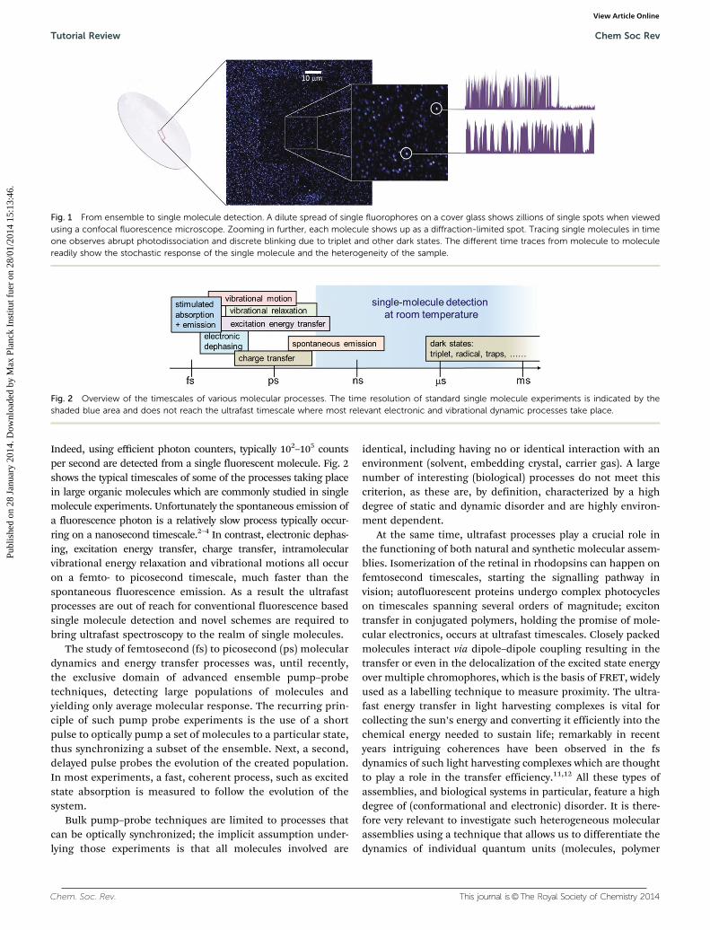
enables the detection of the different subpopulations presentin complex and heterogeneous systems (Fig. 1). Furthermore,following the behaviour of individual molecules in time, with-out the need to synchronize the ensemble, provides extensiveinsights into the dynamic behaviour of (bio-)molecules.5 Indi-vidual molecules are influenced by their local environment,resulting in variations from molecule to molecule in theirsteady state absorption/emission spectra, transition dipoleorientation, triplet and fluorescence lifetimes, etc.6–9 Singlemolecule experiments consistently show that chemically iden-tical molecules exhibit large spatial and temporal heterogeneityfor all parameters studied.
The dynamic range of the processes that can be studied inconventional single molecule experiments is typically limited.Detecting the Stokes shifted fluorescence arising from anindividual molecular system has proven to be the most straight-forward way to enable background free detection and discriminatethe weak response of a single molecule from both the scatteredirradiation field and the signal of the surrounding medium.10
a ICFO - Institut de Ciencies Fotoniques, Mediterranean Technology Park,
08860 Castelldefels, Barcelona, Spain. E-mail: [email protected];
Web: www.ICFO.eub Department of Chemistry and Chemical Biology, Harvard University, Cambridge,
MA 02138, USAc Experimentalphysik IV, Universitat Bayreuth, 95440 Bayreuth, Germanyd Philips Healthcare, 5680DA Best, The Netherlandse CIBION - Center for Bionanoscience Research, CONICET, Buenos Aires, Argentinaf Dept. de Quımica, Universitat Autonoma de Barcelona, 08193 Cerdanyola del Valles,
Barcelona, Spaing ICREA - Institucio Catalana de Recerca i Estudis Avançats, 08015 Barcelona, Spain
† Equal contribution.
Received 22nd July 2013
DOI: 10.1039/c3cs60269a
www.rsc.org/csr
Chem Soc Rev
TUTORIAL REVIEW
Publ
ishe
d on
28
Janu
ary
2014
. Dow
nloa
ded
by M
ax P
lanc
k In
stitu
t fue
r on
28/
01/2
014
15:1
3:46
.
View Article OnlineView Journal
Chem. Soc. Rev. This journal is©The Royal Society of Chemistry 2014
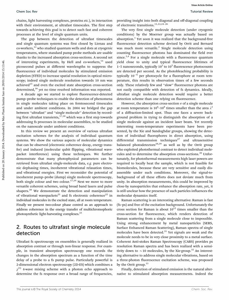
Indeed, using efficient photon counters, typically 102–105 countsper second are detected from a single fluorescent molecule. Fig. 2shows the typical timescales of some of the processes taking placein large organic molecules which are commonly studied in singlemolecule experiments. Unfortunately the spontaneous emission ofa fluorescence photon is a relatively slow process typically occur-ring on a nanosecond timescale.2–4 In contrast, electronic dephas-ing, excitation energy transfer, charge transfer, intramolecularvibrational energy relaxation and vibrational motions all occuron a femto- to picosecond timescale, much faster than thespontaneous fluorescence emission. As a result the ultrafastprocesses are out of reach for conventional fluorescence basedsingle molecule detection and novel schemes are required tobring ultrafast spectroscopy to the realm of single molecules.
The study of femtosecond (fs) to picosecond (ps) moleculardynamics and energy transfer processes was, until recently,the exclusive domain of advanced ensemble pump–probetechniques, detecting large populations of molecules andyielding only average molecular response. The recurring prin-ciple of such pump probe experiments is the use of a shortpulse to optically pump a set of molecules to a particular state,thus synchronizing a subset of the ensemble. Next, a second,delayed pulse probes the evolution of the created population.In most experiments, a fast, coherent process, such as excitedstate absorption is measured to follow the evolution of thesystem.
Bulk pump–probe techniques are limited to processes thatcan be optically synchronized; the implicit assumption under-lying those experiments is that all molecules involved are
identical, including having no or identical interaction with anenvironment (solvent, embedding crystal, carrier gas). A largenumber of interesting (biological) processes do not meet thiscriterion, as these are, by definition, characterized by a highdegree of static and dynamic disorder and are highly environ-ment dependent.
At the same time, ultrafast processes play a crucial role inthe functioning of both natural and synthetic molecular assem-blies. Isomerization of the retinal in rhodopsins can happen onfemtosecond timescales, starting the signalling pathway invision; autofluorescent proteins undergo complex photocycleson timescales spanning several orders of magnitude; excitontransfer in conjugated polymers, holding the promise of mole-cular electronics, occurs at ultrafast timescales. Closely packedmolecules interact via dipole–dipole coupling resulting in thetransfer or even in the delocalization of the excited state energyover multiple chromophores, which is the basis of FRET, widelyused as a labelling technique to measure proximity. The ultra-fast energy transfer in light harvesting complexes is vital forcollecting the sun’s energy and converting it efficiently into thechemical energy needed to sustain life; remarkably in recentyears intriguing coherences have been observed in the fsdynamics of such light harvesting complexes which are thoughtto play a role in the transfer efficiency.11,12 All these types ofassemblies, and biological systems in particular, feature a highdegree of (conformational and electronic) disorder. It is there-fore very relevant to investigate such heterogeneous molecularassemblies using a technique that allows us to differentiate thedynamics of individual quantum units (molecules, polymer
Fig. 1 From ensemble to single molecule detection. A dilute spread of single fluorophores on a cover glass shows zillions of single spots when viewedusing a confocal fluorescence microscope. Zooming in further, each molecule shows up as a diffraction-limited spot. Tracing single molecules in timeone observes abrupt photodissociation and discrete blinking due to triplet and other dark states. The different time traces from molecule to moleculereadily show the stochastic response of the single molecule and the heterogeneity of the sample.
Fig. 2 Overview of the timescales of various molecular processes. The time resolution of standard single molecule experiments is indicated by theshaded blue area and does not reach the ultrafast timescale where most relevant electronic and vibrational dynamic processes take place.
Tutorial Review Chem Soc Rev
Publ
ishe
d on
28
Janu
ary
2014
. Dow
nloa
ded
by M
ax P
lanc
k In
stitu
t fue
r on
28/
01/2
014
15:1
3:46
. View Article Online
This journal is©The Royal Society of Chemistry 2014 Chem. Soc. Rev.
chains, light harvesting complexes, proteins etc.), in interactionwith their environment, at ultrafast timescales. The first steptowards achieving this goal is to detect such fast and coherentprocesses at the level of single quantum units.
The gap between the detection of ultrafast timescalesand single quantum systems was first closed by Lienau andco-workers,13 who studied quantum wells and dots at cryogenictemperatures, where standard pump probe methods are usablethanks to the increased absorption cross-sections. A second setof interesting experiments, by Hell and co-workers,14 usedpicosecond pulses at different wavelengths to suppress thefluorescence of individual molecules by stimulated emissiondepletion (STED) to increase spatial resolution in optical micro-scopy; indeed single molecule resolution towards 10 nm wasachieved15 and even the excited state absorption cross-sectiondetermined,16 yet no time resolved information was reported.
A decade ago we started to explore fluorescence-detectedpump–probe techniques to enable the detection of phenomenain single molecules taking place on femtosecond timescalesand under ambient conditions. In 2004 we bridged the gapbetween ‘‘ultrafast’’ and ‘‘single molecule’’ detection by record-ing first ultrafast transients,17,18 which was a first step towardsaddressing fs processes in molecular assemblies, to be studiedon the nanoscale under ambient conditions.
In this review we present an overview of various ultrafastexcitation schemes for the analysis of individual quantumsystems. We show the various aspects of molecular dynamicsthat can be observed (electronic coherence decay, energy trans-fer) and induced (molecular qubit flipping, vibrational wave-packet interference) using these techniques. We furtherdemonstrate that many photophysical parameters can beretrieved from ultrafast single-molecule data, e.g. pure electro-nic dephasing times, incoherent vibrational relaxation times,and vibrational energies. First we reconsider the potential ofincoherent pump–probe (dump) single molecule spectroscopy,both single colour and two colour.17,19 Next we move to moreversatile coherent schemes, using broad band lasers and pulseshapers.20 We demonstrate the detection and manipulationof vibrational wavepackets21 and fs electronic coherence22 inindividual molecules in the excited state, all at room temperature.Finally we present two-colour phase control as an approach toaddress coherence in the energy transfer of multichromophoricphotosynthetic light-harvesting complexes.23
2. Routes to ultrafast single moleculedetection
Ultrafast fs spectroscopy on ensembles is generally realized inabsorption contrast or through non-linear response. For exam-ple, in transient absorption spectroscopy one records thechanges in the absorption spectrum as a function of the timedelay of a probe to a fs pump pulse. Particularly powerful is2-dimensional electron spectroscopy (2D-ES) which combines aw(3) 4-wave mixing scheme with a photon echo approach todetermine the fs response over a broad range of frequencies,
providing insight into both diagonal and off-diagonal couplingof electronic transitions.11,12,24–26
The very first single molecule detection (under cryogenicconditions) by the Moerner group was actually based onabsorption.1 Yet soon it was realized that the background-freefluorescence detection scheme devised by Orrit and Bernardwas much more versatile.2 Single molecule detection usingcounting fluorescence photons has dominated the field eversince.3,4 For a single molecule with a fluorescence quantumyield close to unity and typical fluorescence lifetimes of1–5 nanoseconds, typically 103 to 105 fluorescence photocountsare detected per second. As the photobleaching probability istypically 10�6 per photocycle for a fluorophore at room tem-perature, this results in observation times of a few secondsonly. These relatively few and ‘‘slow’’ fluorescence photons arenot easily compatible with detection of fs dynamics. Ideally,ultrafast single molecule detection would require a betterdetection scheme than one relying on fluorescence.
However, the absorption cross-section s of a single moleculeat room temperature is 106–108 times smaller than the area l2
of a diffraction-limited spot. Thus one faces a severe back-ground problem in trying to distinguish the absorption of asingle molecule against an incident laser beam. Yet recentlyinteresting room-temperature experiments have been pre-sented, by the Xie and Sandoghdar groups, showing the detec-tion of individual fluorophores in direct absorption, usingdifferential transmission techniques27 and ultrasensitivebalanced photodetectors28,29 as well as by the Orrit groupwho exploited photothermal contrast to detect individual mole-cules and to determine the absorption cross-sections.30 Unfor-tunately, for photothermal measurements high laser powers arerequired to locally heat the sample, which is not feasible forbiomolecules, because these are very sensitive and easily dis-assemble under such conditions. Moreover, the signal-to-background of all these efforts does not deviate much fromunity. In absorption measurements, this could be improved byclose-by nanoparticles that enhance the absorption rate, yet, itis still unclear how the presence of such particles influences themolecular dynamics itself.
Raman scattering is an interesting alternative: Raman is fast(fs–ps) and free of the excitation background. Unfortunately thecross section for Raman is about 1012 times smaller than thecross-section for fluorescence, which renders detection ofRaman scattering from a single molecule close to impossible.Using strong enhancement by metal nanoparticles (SERS,Surface Enhanced Raman Scattering), Raman spectra of singlemolecules have been detected.31 Yet signals are weak and themolecule needs to be in very close proximity to a metal surface.Coherent Anti-stokes Raman Spectroscopy (CARS) provides psresolution Raman spectra and has been realized with a sensi-tivity down to B10 molecules, by the Xie-group.32 An interest-ing alternative to address single molecular vibrations, based ona three-photon fluorescence excitation scheme, was proposedby the Orrit group.33
Finally, detection of stimulated emission is the natural alter-native to stimulated absorption measurements. Indeed the
Chem Soc Rev Tutorial Review
Publ
ishe
d on
28
Janu
ary
2014
. Dow
nloa
ded
by M
ax P
lanc
k In
stitu
t fue
r on
28/
01/2
014
15:1
3:46
. View Article Online
Chem. Soc. Rev. This journal is©The Royal Society of Chemistry 2014
detection of a very small number of (non-fluorescent) mole-cules by stimulated emission has been shown.34 The Hellgroup16 used STED of fluorescence to measure the stimulatedemission cross-section for the STED-transition of single mole-cules at room temperature. Yet the detection of stimulatedemission of a single molecule at room temperature is still to beshown. Moreover, the stimulated emission signal is very sensi-tive to the wavelength, duration, and timing of the stimulationpulse. In combination with the complex pulse sequencesneeded to address ultrafast dynamics in molecular systems,this poses a big challenge at the single-molecule level.
Thus, despite progress on various alternatives, for singlefluorescent molecules the best signal-to-noise and signal-to-background ratios are still achieved by recording the incoherentspontaneous fluorescence emission. All experiments presentedin this review rely on fluorescence detection. This is done on anepi-confocal microscope. Single molecules are embedded in athin (20–100 nm) polymer (PMMA, PVA) layer at a concentrationof around 10�9 molar. Confocal microscopy consists of focusinga laser beam down to its diffraction limit and raster scanningeither the sample or the beam to build up an image of intensityas a function of position. It relies on rejection of out-of-focusfluorescence by imaging the laser focus on a pinhole. In the caseof the experiments described here, avalanche photodiodes withsufficiently small detection areas were used such that the detec-tion area itself effectively functioned as a pinhole. While thepulse trains described in the various paragraphs are created indifferent fashions and with different lasers, the excitation–detection setup is identical. A high numerical aperture (1.3NA)objective is used to focus the light on the sample and collect thefluorescence emitted by the molecule. The fluorescence is sepa-rated from the excitation light by an appropriate dichroic mirror,passes suitable long-pass filters to reject residual laser light, andis finally detected using an avalanche photodiode. The samplescanning, delay-line positioning, pulse shapers, shutters etc. areall computer controlled.
The main challenge in ultrafast single molecule experimentsis therefore to translate information about the ultrafast dynamicsinto changes in the probability for spontaneous emission, whichis directly related to the excited-state population probability.
3. Incoherent single moleculepump–dump spectroscopy
In a typical fluorescence experiment, an electronic moleculartransition S0–S1 is excited by irradiation with a light source, e.g.a short (femtosecond) laser pulse. If the fluorescence quantumyield (Ff) of the system is close to unity, the resulting photo-generated excited state will decay to vibrationally lower lyingstates via Intramolecular Vibrational Relaxation (IVR) on apicosecond timescale and, after some nanoseconds, the mole-cule will finally return to the electronic ground state by sponta-neous emission of a photon. In an initial basic approximationwe ignore coherent effects for laser pulses longer than thecoherence time of the transition (B20–80 fs for large organicmolecules at room temperature), such that population rates aresufficient to describe the dynamics. Then the competitionbetween absorption and stimulated emission restricts themaximum population probability of photo-exciting a moleculeto fifty percent, a limit situation referred to as saturation of theoptical transition.
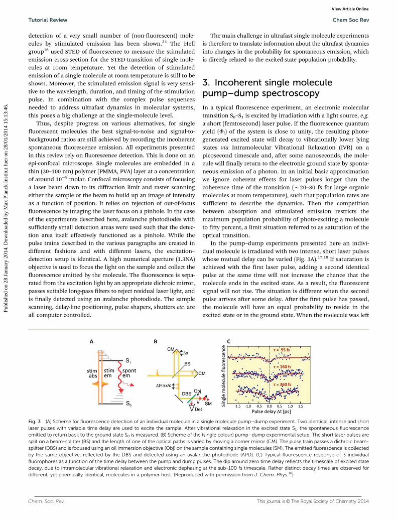
In the pump–dump experiments presented here an indivi-dual molecule is irradiated with two intense, short laser pulseswhose mutual delay can be varied (Fig. 3A).17,18 If saturation isachieved with the first laser pulse, adding a second identicalpulse at the same time will not increase the chance that themolecule ends in the excited state. As a result, the fluorescentsignal will not rise. The situation is different when the secondpulse arrives after some delay. After the first pulse has passed,the molecule will have an equal probability to reside in theexcited state or in the ground state. When the molecule was left
Fig. 3 (A) Scheme for fluorescence detection of an individual molecule in a single molecule pump–dump experiment. Two identical, intense and shortlaser pulses with variable time delay are used to excite the sample. After vibrational relaxation in the excited state S1, the spontaneous fluorescenceemitted to return back to the ground state S0 is measured. (B) Scheme of the (single colour) pump–dump experimental setup. The short laser pulses aresplit on a beam-splitter (BS) and the length of one of the optical paths is varied by moving a corner mirror (CM). The pulse train passes a dichroic beam-splitter (DBS) and is focused using an oil immersion objective (Obj) on the sample containing single molecules (SM). The emitted fluorescence is collectedby the same objective, reflected by the DBS and detected using an avalanche photodiode (APD). (C) Typical fluorescence response of 3 individualfluorophores as a function of the time delay between the pump and dump pulses. The dip around zero time delay reflects the timescale of excited statedecay, due to intramolecular vibrational relaxation and electronic dephasing at the sub-100 fs timescale. Rather distinct decay times are observed fordifferent, yet chemically identical, molecules in a polymer host. (Reproduced with permission from J. Chem. Phys.18)
Tutorial Review Chem Soc Rev
Publ
ishe
d on
28
Janu
ary
2014
. Dow
nloa
ded
by M
ax P
lanc
k In
stitu
t fue
r on
28/
01/2
014
15:1
3:46
. View Article Online
This journal is©The Royal Society of Chemistry 2014 Chem. Soc. Rev.
in the excited state, it will have redistributed some of its energyby the time the second pulse arrives. The molecule will be in astate from where it can no longer be stimulated back to theground state by the second pulse and it will ultimately decay byemitting a fluorescence photon. If the molecule remained inthe ground state after the first pulse passed, the second pulsewill have a chance to excite the molecule, thus leading to anincrease in the total fluorescence signal. Consequently, varyingthe time delay between the pulses will result in modulation ofthe fluorescence emission intensity, with a minimum in thedetected signal for zero time delay. The rise time from theminimum to the plateau at long delays is a measure of the timethat the molecule remains in the initial excited state. Moleculesthat redistribute their excited state energy quickly will result inmore narrow minima. Using a three level model of the energylevels in the molecule and taking into account the length of thelaser pulses it is relatively straightforward to fit the data andrecover the energy redistribution time (ter). However, thismodel is too simplistic at timescales below 100 fs, for whichcoherence does play a role and the phase between the pulsesneeds to be taken into account, as we will see later.
Pump–dump measurements on single molecules are per-formed in an experimental setup schematically presented inFig. 3B. Typically a mode-locked laser or (frequency doubled)optical parametric oscillator provides the visible laser pulses(here B280 fs, repetition rate 1 MHz) to irradiate the sample,while maintaining saturating peak powers. The laser pulses aresplit on a beam splitter and recombined on the same beamsplitter after reflection on corner mirrors. One of the mirrors isplaced on a translation stage allowing the delay between thepulses to be varied from �3 to +3 ps in a few seconds. The delayline is not phase-stabilized, such that the phase is averagedduring the photon counting integration time. A shutter isplaced in one of the branches such that one of the pulses canbe temporally blocked and therefore the behaviour of thesystem followed under single pulse excitation. Further experi-mental details are presented elsewhere.18 A typical fluorescenceresponse of single molecules as a function of time delay is
shown in Fig. 3C. The fluorescence decay around zero timedelay is readily appreciated.
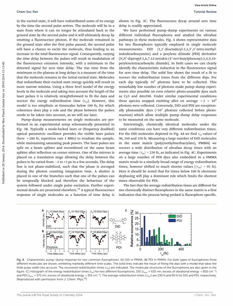
We have performed pump–dump experiments on variousdifferent individual fluorophores and studied the ultrafastpathways in these molecules. Fig. 4 shows representative datafor two fluorophores typically employed in single moleculemeasurements: DiD (1,10-dioctadecyl-3,3,3 0,30-tetra-methyl-indodicarbocyanine) and a perylene diimide (PDI) derivative(N,N 0-dipropyl-1,6,7,12-tetrakis-(4 0-tert-butylphenoxy)-3,4,9,10-perylenetetracarboxylic diimide). In both cases we can clearlyidentify the characteristic reduction in the fluorescence signalfor zero time delay. The solid line shows the result of a fit torecover the redistribution times from the different dips. Foreach dip typically 104 photons have to be collected. Thisremarkably low number of photons made pump–dump experi-ments also possible on even relative photo-unstable dyes suchas Cy5 and Atto590. Under similar experimental conditionsthese species stopped emitting after on average B3 � 104
photons were collected. Conversely, DiD and PDI are exception-ally photostable dyes (>105 photons collected before photo-reaction) which allow multiple pump–dump delay responsesto be measured on the same molecule.
Interestingly, chemically identical molecules under thesame conditions can have very different redistribution times.For the DiD molecules depicted in Fig. 4A we find ter values of90, 160 and 350 fs. Measuring a large number of DiD moleculesin the same matrix (poly(methylmethacrylate), PMMA) werecover a wide distribution of ultrafast decay times with anaverage time hteri = 230 fs, as indicated in Fig. 4C. Experimentson a large number of PDI dyes also embedded in a PMMAmatrix result in a similarly broad range of energy redistributiontimes, however shifted to much shorter values (hteri = 95 fs).Here it should be noted that for times below 100 fs electronicdephasing will play a dominant role which limits the shortesttimes observable for PDI.
The fact that the average redistribution times are different fortwo chemically distinct fluorophores in the same matrix is a firstindication that the process being probed is fluorophore specific.
Fig. 4 Characteristic pump–dump response for two common fluorophores: (A) DiD in PMMA, (B) PDI in PMMA. For both types of fluorophores threedifferent molecules are shown, exhibiting markedly different time scales. The solid lines indicate the result of fitting the dips with a model that takes thefinite pulse width into account. The recovered redistribution times (ter) are indicated. The molecular structures of the fluorophores are also given in thefigure. (C) Histogram of the energy redistribution times (ter) for two different fluorophores, DiD (lexc = 635 nm, excess of vibrational energy B850 cm�1)and PDI (lexc = 575 nm, excess of vibrational energy B355 cm�1). The average redistribution times (hteri) are 230 fs and 95 fs for DiD and PDI, respectively.(Reproduced with permission from J. Chem. Phys.18)
Chem Soc Rev Tutorial Review
Publ
ishe
d on
28
Janu
ary
2014
. Dow
nloa
ded
by M
ax P
lanc
k In
stitu
t fue
r on
28/
01/2
014
15:1
3:46
. View Article Online
Chem. Soc. Rev. This journal is©The Royal Society of Chemistry 2014
We have further investigated this issue by performing pump–dump experiments on the same dye (DiD) embedded in twodifferent polymer matrices (PMMA and Zeonex). A very minoreffect of the surrounding matrix on ter has been encountered,the average redistribution times measured for both samplesunder equivalent excitation conditions taking similar values(hteri = 230 fs for PMMA and hteri = 180 fs for Zeonex). Therefore,we conclude that the pump–dump experiments mainly reporton an intramolecular process. Indeed, solvent-independent(sub-)picosecond relaxation times in optically excited fluoro-phores are commonly assigned to intramolecular redistributionof the excited state energy over different vibrational states justafter excitation: intramolecular vibrational energy relaxation(IVR).35 When a molecule is brought into an electronicallyexcited state, IVR ensures a quick redistribution of the vibra-tional energy of the system over different vibrational modes,while the transfer of its excess of vibrational energy to theenvironment takes place on a longer (picoseconds) timescale.Noticeably, the initial redistribution of the energy already leavesthe molecule in a state from where it can no longer be stimulatedback to the ground state. The time measured therefore reportson such an initial IVR process, which results from the couplingbetween the electronic and vibrational modes of the molecule. Astrong coupling will result in a fast redistribution of the energyand thus narrow minimum. Wider minima on the other handare an indication of a reduced coupling between optically activeand inactive modes, resulting in the molecule remaining longerin the initially excited state. The intramolecular coupling deter-mining the IVR dynamics actually depends on several variables,such as the nature of the initial Franck–Condon state preparedby optical excitation, the density of vibrational states it cancouple with and the coupling matrix elements between thosestates.36 Together with the influence of electronic dephasing, theter differences encountered for distinct fluorophores in oursingle molecule experiments are hard to rationalize, whichcomplicates direct comparison with IVR times reported in bulkfor similar molecules.37
The broad distribution of times indicates that in a polymerhost at room temperature chemically identical molecules dodisplay large variations in the ultrafast redistribution of their
excited state energy. Single molecule experiments have shownalready large variations of several spectroscopic parameters for‘‘identical’’ molecules. For instance the triplet state and singletexcited state lifetime and fluorescence spectra have been shownto vary from molecule to molecule.7–9 Here, however we findthat these variations even extend to intramolecular processeson femtosecond timescale. These variations are indicative forthe different conformational states of the individual moleculesthat are induced by the nano-environment. Different conforma-tions result in variations in the coupling between the electronicand vibrational modes, an effect that was observed before insteady state single molecule fluorescence emission spectra.38
These previous studies reported on the coupling between theelectronic and ground state vibronic modes, the single mole-cule pump–dump data now report on the processes takingplace in the electronically excited states.
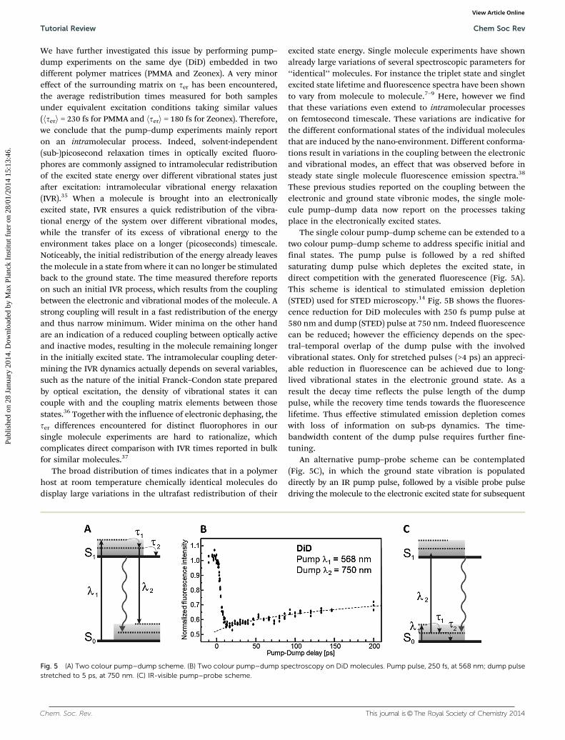
The single colour pump–dump scheme can be extended to atwo colour pump–dump scheme to address specific initial andfinal states. The pump pulse is followed by a red shiftedsaturating dump pulse which depletes the excited state, indirect competition with the generated fluorescence (Fig. 5A).This scheme is identical to stimulated emission depletion(STED) used for STED microscopy.14 Fig. 5B shows the fluores-cence reduction for DiD molecules with 250 fs pump pulse at580 nm and dump (STED) pulse at 750 nm. Indeed fluorescencecan be reduced; however the efficiency depends on the spec-tral–temporal overlap of the dump pulse with the involvedvibrational states. Only for stretched pulses (>4 ps) an appreci-able reduction in fluorescence can be achieved due to long-lived vibrational states in the electronic ground state. As aresult the decay time reflects the pulse length of the dumppulse, while the recovery time tends towards the fluorescencelifetime. Thus effective stimulated emission depletion comeswith loss of information on sub-ps dynamics. The time-bandwidth content of the dump pulse requires further fine-tuning.
An alternative pump–probe scheme can be contemplated(Fig. 5C), in which the ground state vibration is populateddirectly by an IR pump pulse, followed by a visible probe pulsedriving the molecule to the electronic excited state for subsequent
Fig. 5 (A) Two colour pump–dump scheme. (B) Two colour pump–dump spectroscopy on DiD molecules. Pump pulse, 250 fs, at 568 nm; dump pulsestretched to 5 ps, at 750 nm. (C) IR-visible pump–probe scheme.
Tutorial Review Chem Soc Rev
Publ
ishe
d on
28
Janu
ary
2014
. Dow
nloa
ded
by M
ax P
lanc
k In
stitu
t fue
r on
28/
01/2
014
15:1
3:46
. View Article Online
This journal is©The Royal Society of Chemistry 2014 Chem. Soc. Rev.
fluorescence read-out. A similar scheme was also proposed byOrrit and colleagues.33 So far this scheme has not yet beenrealized for single molecules, as the transitions need to besaturated and direct single and two photon excitations of themolecule result in a fluorescence background which complicatesdetection of the effect of the IR pump.
Despite the results shown the incoherent pump–dumpscheme has a few severe drawbacks. First, for both pulses tointeract with a single molecule the transitions need to besaturated. Obviously single molecule excitation at saturationpower density results in fast photobleaching. Even for photo-stable fluorophores (106 photo cycles) and a reduced repetitionrate (1 MHz) the typical observation time is only seconds.Second, the lack of a phase relation between pump and dumppulses prevents disentangling the role of coherence anddephasing effects in the recorded time traces. Clearly a coher-ent excitation scheme operating in the non-saturating regime islargely preferred, which is the topic of the next sections.
4. Control of single moleculeelectronic coherence
The next step in fs excitation of single molecules is to gaincontrol over both the time delay and the relative phase betweenboth pulses. An interesting extra dimension then opens up inthe experiments. The experiment can now be described as thefirst pulse creating a coherent superposition state in themolecule, and the second pulse probing the phase memory inthat coherent superposition created by the first pulse. Afterinteraction of the molecule with such pulse sequences, theexcited state population probability, and with it the incoherentspontaneous emission, becomes a function of both inter-pulsedelay time and phase difference. Notably, the time-phase depen-dence of the fluorescence can be related to molecular propertiesinfluencing excited state dynamics, for example through pure
electronic dephasing and incoherent vibrational relaxation.Equally important, the effect of phase control is detected throughinterference between the molecular polarization induced by thefield of the first pulse, and the field of the second pulse. In thispicture, the concept of saturation does not hold anymore andmeaningful experiments can already be done at low excitationpowers.
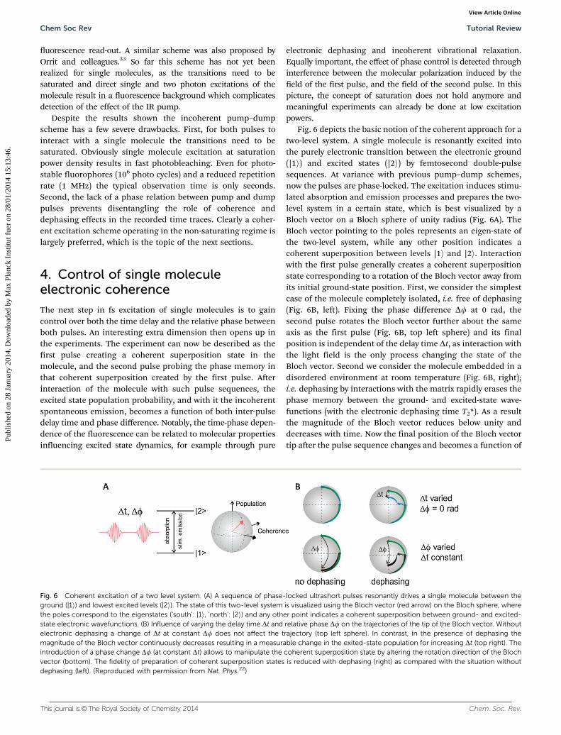
Fig. 6 depicts the basic notion of the coherent approach for atwo-level system. A single molecule is resonantly excited intothe purely electronic transition between the electronic ground(|1i) and excited states (|2i) by femtosecond double-pulsesequences. At variance with previous pump–dump schemes,now the pulses are phase-locked. The excitation induces stimu-lated absorption and emission processes and prepares the two-level system in a certain state, which is best visualized by aBloch vector on a Bloch sphere of unity radius (Fig. 6A). TheBloch vector pointing to the poles represents an eigen-state ofthe two-level system, while any other position indicates acoherent superposition between levels |1i and |2i. Interactionwith the first pulse generally creates a coherent superpositionstate corresponding to a rotation of the Bloch vector away fromits initial ground-state position. First, we consider the simplestcase of the molecule completely isolated, i.e. free of dephasing(Fig. 6B, left). Fixing the phase difference Df at 0 rad, thesecond pulse rotates the Bloch vector further about the sameaxis as the first pulse (Fig. 6B, top left sphere) and its finalposition is independent of the delay time Dt, as interaction withthe light field is the only process changing the state of theBloch vector. Second we consider the molecule embedded in adisordered environment at room temperature (Fig. 6B, right);i.e. dephasing by interactions with the matrix rapidly erases thephase memory between the ground- and excited-state wave-functions (with the electronic dephasing time T2*). As a resultthe magnitude of the Bloch vector reduces below unity anddecreases with time. Now the final position of the Bloch vectortip after the pulse sequence changes and becomes a function of
Fig. 6 Coherent excitation of a two level system. (A) A sequence of phase-locked ultrashort pulses resonantly drives a single molecule between theground (|1i) and lowest excited levels (|2i). The state of this two-level system is visualized using the Bloch vector (red arrow) on the Bloch sphere, wherethe poles correspond to the eigenstates (‘south’: |1i, ‘north’: |2i) and any other point indicates a coherent superposition between ground- and excited-state electronic wavefunctions. (B) Influence of varying the delay time Dt and relative phase Df on the trajectories of the tip of the Bloch vector. Withoutelectronic dephasing a change of Dt at constant Df does not affect the trajectory (top left sphere). In contrast, in the presence of dephasing themagnitude of the Bloch vector continuously decreases resulting in a measurable change in the exited-state population for increasing Dt (top right). Theintroduction of a phase change Df (at constant Dt) allows to manipulate the coherent superposition state by altering the rotation direction of the Blochvector (bottom). The fidelity of preparation of coherent superposition states is reduced with dephasing (right) as compared with the situation withoutdephasing (left). (Reproduced with permission from Nat. Phys.22)
Chem Soc Rev Tutorial Review
Publ
ishe
d on
28
Janu
ary
2014
. Dow
nloa
ded
by M
ax P
lanc
k In
stitu
t fue
r on
28/
01/2
014
15:1
3:46
. View Article Online
Chem. Soc. Rev. This journal is©The Royal Society of Chemistry 2014
Dt, with decreasing excited state population (Fig. 6B, top rightsphere, green and blue arrows). Recording the decay of thefluorescence with Dt provides information about the residualcoherent superposition state of the molecule. More specificallythe degree of (de)coherence can be probed by exploiting therelative phase Df between the electric fields of the laser pulses,because Df determines the rotation direction of the Blochvector induced by the second pulse. A p phase shift fully invertsthe rotation direction and moves the Bloch vector back towardsits ground-state position (Fig. 6B, bottom, green arrows: Df = 0,black arrows: Df = p). The change of Df therefore allowsmanipulating the motion of the Bloch vector at a discrete timeduring loss of phase memory, giving direct insight into thedephasing dynamics of single molecules.
Pulse pair generation by a simple delay line is not sufficientto independently control both time delay and phase differencebetween pulse pairs; for this, one needs a pulse shaper. Anacousto-optic programmable dispersive filter (AOPDF, Dazzler,Fastlite) is a versatile pulse shaper, exploiting acoustic wavestravelling in a birefringent acousto-optic crystal to achieveamplitude and phase shaping of an incident fs-pulse. Unfortu-nately an AOPDF only allows limited bandwidth and low pulserepetition rates. Here we used such an AOPDF to shape theoutput of an optical parametric oscillator with 18–21 nmFWHM spectral bandwidth, and compress the pulses to thetransform-limit of 70–75 fs (FWHM). Moreover a pulse pickerreduced the 76 MHz repetition rate to effectively 500 kHz(bunches of pulses with a repetition rate of 25 kHz, repetitionrate within bunches: 4 MHz) to match the input of the AOPDF.With the operation conditions for an AOPDF met, the shaperwas used to control both delay time Dt and phase difference Dfbetween the electric fields of the output pulses. The output ofthe AOPDF was spatially filtered in a lens–pinhole-lens combi-nation, and directed into a confocal microscope.
As a test case to influence single molecule electronic coher-ences, we excited single TDI (terrylenediimide) molecules in aPMMA film with the phase-locked double-pulse sequences.22
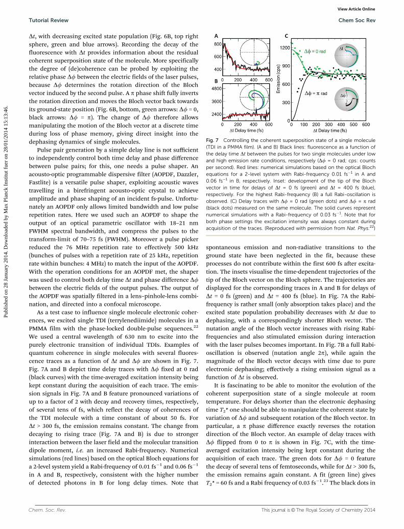
We used a central wavelength of 630 nm to excite into thepurely electronic transition of individual TDIs. Examples ofquantum coherence in single molecules with several fluores-cence traces as a function of Dt and Df are shown in Fig. 7.Fig. 7A and B depict time delay traces with Df fixed at 0 rad(black curves) with the time-averaged excitation intensity beingkept constant during the acquisition of each trace. The emis-sion signals in Fig. 7A and B feature pronounced variations ofup to a factor of 2 with decay and recovery times, respectively,of several tens of fs, which reflect the decay of coherences ofthe TDI molecule with a time constant of about 50 fs. ForDt > 300 fs, the emission remains constant. The change fromdecaying to rising trace (Fig. 7A and B) is due to strongerinteraction between the laser field and the molecular transitiondipole moment, i.e. an increased Rabi-frequency. Numericalsimulations (red lines) based on the optical Bloch equations fora 2-level system yield a Rabi-frequency of 0.01 fs�1 and 0.06 fs�1
in A and B, respectively, consistent with the higher numberof detected photons in B for long delay times. Note that
spontaneous emission and non-radiative transitions to theground state have been neglected in the fit, because theseprocesses do not contribute within the first 600 fs after excita-tion. The insets visualise the time-dependent trajectories of thetip of the Bloch vector on the Bloch sphere. The trajectories aredisplayed for the corresponding traces in A and B for delays ofDt = 0 fs (green) and Dt = 400 fs (blue). In Fig. 7A the Rabi-frequency is rather small (only absorption takes place) and theexcited state population probability decreases with Dt due todephasing, with a correspondingly shorter Bloch vector. Thenutation angle of the Bloch vector increases with rising Rabi-frequencies and also stimulated emission during interactionwith the laser pulses becomes important. In Fig. 7B a full Rabi-oscillation is observed (nutation angle 2p), while again themagnitude of the Bloch vector decays with time due to pureelectronic dephasing; effectively a rising emission signal as afunction of Dt is observed.
It is fascinating to be able to monitor the evolution of thecoherent superposition state of a single molecule at roomtemperature. For delays shorter than the electronic dephasingtime T2* one should be able to manipulate the coherent state byvariation of Df and subsequent rotation of the Bloch vector. Inparticular, a p phase difference exactly reverses the rotationdirection of the Bloch vector. An example of delay traces withDf flipped from 0 to p is shown in Fig. 7C, with the time-averaged excitation intensity being kept constant during theacquisition of each trace. The green dots for Df = 0 featurethe decay of several tens of femtoseconds, while for Dt > 300 fs,the emission remains again constant. A fit (green line) givesT2* = 60 fs and a Rabi frequency of 0.03 fs�1.23 The black dots in
Fig. 7 Controlling the coherent superposition state of a single molecule(TDI in a PMMA film). (A and B) Black lines: fluorescence as a function ofthe delay time Dt between the pulses for two single molecules under lowand high emission rate conditions, respectively (Df = 0 rad; cps: countsper second). Red lines: numerical simulations based on the optical Blochequations for a 2-level system with Rabi-frequency 0.01 fs�1 in A and0.06 fs�1 in B, respectively. Inset: development of the tip of the Blochvector in time for delays of Dt = 0 fs (green) and Dt = 400 fs (blue),respectively. For the highest Rabi-frequency (B) a full Rabi-oscillation isobserved. (C) Delay traces with Df = 0 rad (green dots) and Df = p rad(black dots) measured on the same molecule. The solid curves representnumerical simulations with a Rabi-frequency of 0.03 fs�1. Note that forboth phase settings the excitation intensity was always constant duringacquisition of the traces. (Reproduced with permission from Nat. Phys.22)
Tutorial Review Chem Soc Rev
Publ
ishe
d on
28
Janu
ary
2014
. Dow
nloa
ded
by M
ax P
lanc
k In
stitu
t fue
r on
28/
01/2
014
15:1
3:46
. View Article Online

This journal is©The Royal Society of Chemistry 2014 Chem. Soc. Rev.
Fig. 7C show a time delay measurement on the same moleculewith the pulses out-of-phase (Df = p). Clearly the coherenceresults in destructive interference and reduced probability toreach the excited state. It should be noted that the time-averaged excitation intensity was kept constant for all timedelay; thus the fluorescence reduction reflects purely the molec-ular coherence. These data demonstrate convincingly thepotential to manipulate the Bloch vector of single moleculesat room temperature despite femtosecond dephasing times; forDt o T2*, the Bloch vector can be rotated to any arbitraryposition and any coherent superposition state, by an appro-priate choice of Dt and Df.
Comparing the coherent time delay response in Fig. 7 to theincoherent pump–dump response in Fig. 4,18 it becomes clear thatthe incoherent experiments contain a phase averaged contributionof the electronic coherence at time scales below 100 fs, while forlonger times incoherent vibrational relaxation comes into play. Infact, interplay between electronic dephasing and vibrational relaxa-tion was also observed in the coherent time delay experiments,showing distinctly different time constants which allows disentan-gling both effects; for details see ref. 20.
5. Single molecule vibrationalwavepacket interference
Having established basic phase control of individual mole-cules, now let’s extend the method to excitation by broadbandpulses, covering all vibrational sidebands of the electronictransition. With this, one enters the realm of coherent control:the targeted steering of a quantum system through phaseshaped excitation pulses into one state from a multitude ofaccessible ones. Obviously we aim to maintain the potential of
single molecule detection. The coherent control of dynamicprocesses in molecular ensembles has been implemented in verysophisticated experiments in the last two decades.39 Using cleverlytailored ultrashort femtosecond pulses, steering of reaction path-ways, optimizing energy conversion40–42 and control of spatialconfinement of optical fields43,44 has been shown. Particularly forcomplex systems such as large (bio)molecules with many nuclearand electronic degrees of freedom, for which ab initio quantummechanical calculations fail, the elegant approach of genetic self-learning algorithms has led to the coherent control of a widevariety of photo-induced processes.41,42 The resulting optimizedpulse shape reflects the dynamics of the underlying processes.The closed-loop experiments require a large number of iterations;as a result such approaches are out of reach for fluorescent singlemolecules, which bleach on a time scale of seconds and provideonly a limited number of photocounts. Fortunately optimal con-trol theory and experiments on large molecules have demon-strated that complex pulse shapes can often be simplified tophysically more intuitive shapes based on trains of pulses withcontrolled width, delay and phase relations. Therefore we addresssingle molecules with pairs of broad-band pulses, again varyingtime delay between the envelop Dt and relative carrier phasedifference Df.20 We demonstrate the observation of vibrationalwavepacket interference and phase control of the wavepacket foran individual fluorophore at room temperature.21
As a molecule of study we use an even higher rylene homo-logue than the TDI in the previous section: dinaphtho-quaterrylenebis(dicarbox-imide), in short DN-QDI.45 DN-QDIis a photostable fluorophore with high quantum efficiency.With its 4 naphthalene units the absorption spectrum is shiftedto the near-infrared with a maximum at 700 nm (in toluenesolution), see Fig. 8A. DN-QDI exhibits a prominent vibrationalprogression, at around 1350 cm�1.
Fig. 8 (A) Absorption and emission spectra of the fluorophore (DN-QDI, dinaphto-quaterrylenebis(dicarboximide)42) and the broad-band excitation ofthe laser used. (B) 4f pulse shaper based on a spatial light modulator (SLM), operating in double-pass by passing the beam through a beam splitter (BS) andputting a mirror at the output of the shaper. (C) Single fluorescent molecules are excited and detected in an epi-confocal microscope. Each individualmolecule is excited with shaped sequences of pulses, with an inter-pulse time delay Dt and phase shift Df as controlled by the 4f shaper. (Reproducedwith permission from Nature,21 Opt. Express.47)
Chem Soc Rev Tutorial Review
Publ
ishe
d on
28
Janu
ary
2014
. Dow
nloa
ded
by M
ax P
lanc
k In
stitu
t fue
r on
28/
01/2
014
15:1
3:46
. View Article Online
Chem. Soc. Rev. This journal is©The Royal Society of Chemistry 2014
The coherent broad band excitation is provided by theoutput of a 85 MHz repetition rate mode-locked OctaviusTitanium:Sapphire-laser (Octavius-85M, Menlo Systems, Thor-labs). The Octavius spectrum stretches from B550 nm wave-length to deep into the infrared, spanning an ‘‘octave’’ infrequency, providing a 7 fs pulse when Fourier limited. Herewe selected a spectral bandwidth of 120 nm (15 fs) around thecentral wavelength of 676 nm (green band in Fig. 8A), thuscovering nearly the entire DN-QDI absorption spectrum, andinteracting with a manifold of vibrational levels in the electro-nically excited state. We employed a 4f-pulse shaper based on aSpatial Light Modulator (SLM) for dispersion control and pulseshaping (Fig. 8B). The 4f-pulse shaper was in-house adaptedfrom a commercial shaper (MIIPS-box, Biophotonics SolutionsInc.).46 Pulse calibration was performed using MultiphotonIntrapulse Interference PhaseScan (MIIPS) with the secondharmonic spectrum being detected in the sample plane. Theshaper is designed in a double-pass configuration47 with amirror at the end of the beam path reflecting the light backfor a second pass through the shaper (Fig. 8B). This double-pass configuration minimises spatio-temporal coupling andallows larger phase distortions to be compensated.47 In allexperiments pulses were first compressed to their transformlimit of 15 fs (about �0.1 rad residual spectral phase variation)in the sample plane, using a MIIPS control loop.46 Finally, as inthe previous section, single molecules are excited and detectedusing a confocal microscope with a high 1.3NA objective andsingle photon counting avalanche photodiodes (Fig. 8C).
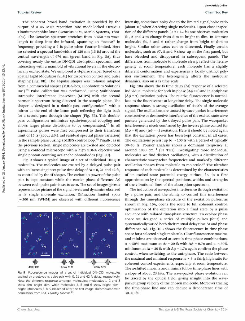
Fig. 9 shows a typical image of a set of individual DN-QDImolecules. The molecules are excited by a delayed pulse pairwith an increasing inter-pulse time delay of Dt = 0, 21 and 42 fs,as controlled by the 4f shaper. The excitation power of the pulsepair is kept constant while the carrier phase difference Dfbetween each pulse pair is set to zero. The set of images gives arepresentative picture of the signal levels and dynamics observedin fs single molecule excitation. Diffraction limited spots(B300 nm FWHM) are observed with different fluorescence
intensity, sometimes noisy due to the limited signal/noise ratio(about 10) when detecting single molecules. Upon close inspec-tion of the different panels (0–21–42 fs) one observes molecules#1, 2 and 3 to change from dim to bright to dim. In contrastmolecules #4, 5 and 6 rather change from bright to dim tobright. Similar other cases can be discerned. Finally certainmolecules, such as #7, 8 and 9 show up in the first panel, buthave bleached and disappeared in subsequent panels. Thedifferences from molecule to molecule clearly reflect the hetero-geneity at room temperature; each molecule has a slightlydifferent conformation and experiences a locally distinct poly-mer environment. The heterogeneity affects the moleculardynamics, also on a fs time scale.
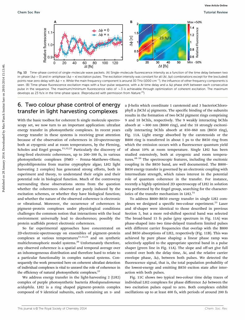
Fig. 10A shows the fs time delay (Dt) response of a selectedindividual molecule for both in-phase (Df = 0) and in-antiphase(Df = p) excitation pulses. The relative fluorescence is normal-ized to the fluorescence at long time delay. The single moleculeresponse shows a strong oscillation of �10% of the averagesignal. The oscillations are caused by wave-packet interference:constructive or destructive interference of the excited state wavepackets generated by the delayed pulse pair. The wavepacketinterference is nicely confirmed by the inverse phase control for(Df = 0) and (Df = p) excitation. Here it should be noted againthat the excitation power has been kept constant in all cases.The oscillations persist up to B100 fs with a period of typically30–40 fs. Fourier analysis shows a dominant frequency ataround 1000 cm�1 (33 THz). Investigating more individualmolecules we find distinct oscillations, with a distribution ofcharacteristic wavepacket frequencies and markedly differentoscillation phases from molecule to molecule.21 The ultrafastresponse of each molecule is determined by the characteristicsof its excited state potential energy surface; i.e. in a firstapproximation by the spectral positions, widths and strengthsof the vibrational lines of the absorption spectrum.
The induction of wavepacket interference through excitationby a pulse pair, and the ability to control this interferencethrough the time-phase structure of the excitation pulses, asshown in Fig. 10A, opens the route to full coherent control:optimisation of the excitation into a final state by a pulsesequence with tailored time-phase structure. To explore phasespace we designed a series of multiple pulses (four) andsystematically varied both their mutual delay time Dt and phasedifference Df. Fig. 10B shows the fluorescence in time-phasespace for a selected single molecule. Clear fluorescence maximaand minima are observed at certain time-phase combinations.A B50% maximum at Dt = 20 fs with Df = 0.7p and a B50%minimum at Dt = 20 fs with Df = 1.7p again confirm the phasecontrol, when switching to the anti-phase. The ratio betweenthe maximal and minimal response is B3: a fairly high ratio forcoherent control experiments, especially at room temperature.The p-shifted maxima and minima follow time-phase lines witha slope of about 23 fs/p. The wave-packet phase evolution canbe traced by the optical field, giving insight into the wave-packet group velocity of the chosen molecule. Moreover tracingthe time-phase line one can deduce a decoherence time of30–40 fs.
Fig. 9 Fluorescence images of a set of individual DN-QDI moleculesexcited by a delayed fs pulse pair with 0, 21 and 42 fs delay, respectively.Note the different response amongst molecules: molecules 1, 2 and 3show dim–bright–dim, while molecules 4, 5 and 6 show bright–dim–bright. Molecules 7, 8, 9 bleached after the first image. (Reproduced withpermission from RSC Faraday Discuss.21)
Tutorial Review Chem Soc Rev
Publ
ishe
d on
28
Janu
ary
2014
. Dow
nloa
ded
by M
ax P
lanc
k In
stitu
t fue
r on
28/
01/2
014
15:1
3:46
. View Article Online
This journal is©The Royal Society of Chemistry 2014 Chem. Soc. Rev.
6. Two colour phase control of energytransfer in light harvesting complexes
With the basic toolbox for coherent fs single molecule spectro-scopy set, we now turn to an important application: ultrafastenergy transfer in photosynthetic complexes. In recent yearsenergy transfer in these systems is receiving great attentionbecause of the observation of coherences in 2D-spectroscopyboth at cryogenic and at room temperatures, by the Fleming,Scholes and Engel groups.11,12,25 Particularly the discovery oflong-lived electronic coherences, up to 200–300 fs, in variousphotosynthetic complexes (FMO – Fenna–Matthews–Olson;phycobiliproteins from marine cryptophyte algae; LH2 lightharvesting 2 complex) has generated strong efforts, both inexperiment and theory, to understand their origin and theirpotential role in biological function. Much of the controversysurrounding these observations stems from the questionwhether the coherences observed are purely induced by theexcitation schemes, or whether they have biological function;and whether the nature of the observed coherence is electronicor vibrational. Moreover, the occurrence of coherences inpigment–protein complexes under physiological conditionschallenges the common notion that interactions with the localenvironment universally lead to decoherence; possibly theprotein scaffolds protect electronic coherences.
So far experimental approaches have concentrated on2D-electronic-spectroscopy on ensembles of pigment–proteincomplexes at various temperatures11,12,24 and on syntheticmultichromophoric model systems.26 Unfortunately therefore,any observed coherence is a spatial and temporal average overan inhomogeneous distribution and therefore hard to relate toa particular functionality in complex natural systems. Con-sequently the work presented here on coherent ultrafast detectionof individual complexes is vital to unravel the role of coherence inthe efficiency of natural photosynthetic complexes.23
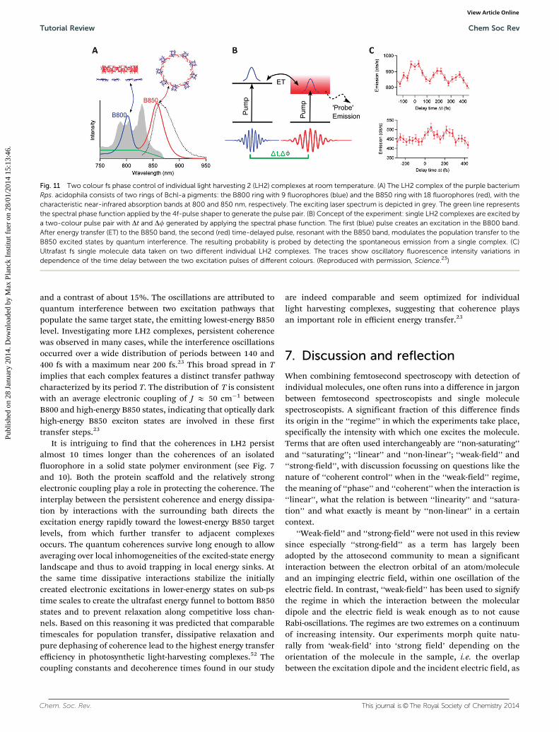
We address energy transfer in the light-harvesting 2 (LH2)complex of purple photosynthetic bacteria Rhodopseudomonasacidophila. LH2 is a ring shaped pigment–protein complexcomposed of 9 identical subunits, each containing an a- and
a b-helix which coordinate 1 carotenoid and 3 bacterioChloro-phyll a (bChl a) pigments. The specific binding of the subunitsresults in the formation of two bChl pigment rings comprising9 and 18 bChls, respectively. The 9 weakly interacting bChlsabsorb at B800 nm (B800 ring), and the 18 strongly excitoni-cally interacting bChls absorb at 850–860 nm (B850 ring),Fig. 11A. Light energy absorbed by the carotenoids or theB800 ring is transferred in about 1 ps to the B850 ring fromwhich the emission occurs with a fluorescence quantum yieldof about 10% at room temperature. Single LH2 has beenstudied extensively, both at cryogenic and room tempera-tures.48–50 The spectroscopic features, including the excitoniccoupling in the B850 band, are well documented. The B800–B850 energy transfer is governed by an electronic coupling withintermediate strength, which raises interest in the potentialrole of quantum coherence in the transfer. For instance,recently a highly optimized 2D spectroscopy of LH2 in solutionwas performed by the Engel group, searching for the character-istics of the transfer mechanism in LH2.51
To address B800–B850 energy transfer in single LH2 com-plexes we designed a specific two-colour experiment.23 Laserand 4f-shaper were identical to those described in previousSection 5, but a more red-shifted spectral band was selectedThe broad-band 15 fs pulse (grey spectrum in Fig. 11A) wasphase-shaped into two time-delayed transform limited pulseswith different carrier frequencies that overlap with the B800and B850 absorptions of LH2, respectively (Fig. 11B). This wasachieved by pure phase shaping: a linear phase ramp wasselectively applied to the appropriate spectral band in a pulseshaper (green line in Fig. 11A). The slope and off-set give fullcontrol over both the delay time, Dt, and the relative carrierenvelope phase, Df, between both pulses. We detected thefluorescence signal, that is, the total population probability ofthe lowest-energy and emitting B850 exciton state after inter-action with both pulses.
Fig. 11C shows two typical two-colour time delay traces ofindividual LH2 complexes for phase difference Df between thetwo excitation pulses equal to zero. Both complexes exhibitoscillations up to at least 400 fs, with periods of around 200 fs
Fig. 10 Time-phase control of single-molecule wave packets. (A) Single-molecule fluorescence intensity as a function of the time delay between twoin-phase (Df = 0) and in-antiphase (Df = p) excitation pulses. The excitation intensity was constant for all (Dt, Df) combinations except for the (excluded)points near zero delay with Df = p. While the main frequency component is around 30 THz (1000 cm�1), the influence of other frequency components isseen. (B) Time-phase fluorescence excitation maps with a four-pulse sequence, with a Dt time delay and a Df phase shift between each consecutivepulse in the sequence. The maximum/minimum fluorescence ratio of B3 is achievable through optimization of coherent excitation. The maximumdevelops as 23 fs/p in the time-phase space. (Reproduced with permission from Nature.21)
Chem Soc Rev Tutorial Review
Publ
ishe
d on
28
Janu
ary
2014
. Dow
nloa
ded
by M
ax P
lanc
k In
stitu
t fue
r on
28/
01/2
014
15:1
3:46
. View Article Online
Chem. Soc. Rev. This journal is©The Royal Society of Chemistry 2014
and a contrast of about 15%. The oscillations are attributed toquantum interference between two excitation pathways thatpopulate the same target state, the emitting lowest-energy B850level. Investigating more LH2 complexes, persistent coherencewas observed in many cases, while the interference oscillationsoccurred over a wide distribution of periods between 140 and400 fs with a maximum near 200 fs.23 This broad spread in Timplies that each complex features a distinct transfer pathwaycharacterized by its period T. The distribution of T is consistentwith an average electronic coupling of J E 50 cm�1 betweenB800 and high-energy B850 states, indicating that optically darkhigh-energy B850 exciton states are involved in these firsttransfer steps.23
It is intriguing to find that the coherences in LH2 persistalmost 10 times longer than the coherences of an isolatedfluorophore in a solid state polymer environment (see Fig. 7and 10). Both the protein scaffold and the relatively strongelectronic coupling play a role in protecting the coherence. Theinterplay between the persistent coherence and energy dissipa-tion by interactions with the surrounding bath directs theexcitation energy rapidly toward the lowest-energy B850 targetlevels, from which further transfer to adjacent complexesoccurs. The quantum coherences survive long enough to allowaveraging over local inhomogeneities of the excited-state energylandscape and thus to avoid trapping in local energy sinks. Atthe same time dissipative interactions stabilize the initiallycreated electronic excitations in lower-energy states on sub-pstime scales to create the ultrafast energy funnel to bottom B850states and to prevent relaxation along competitive loss chan-nels. Based on this reasoning it was predicted that comparabletimescales for population transfer, dissipative relaxation andpure dephasing of coherence lead to the highest energy transferefficiency in photosynthetic light-harvesting complexes.52 Thecoupling constants and decoherence times found in our study
are indeed comparable and seem optimized for individuallight harvesting complexes, suggesting that coherence playsan important role in efficient energy transfer.23
7. Discussion and reflection
When combining femtosecond spectroscopy with detection ofindividual molecules, one often runs into a difference in jargonbetween femtosecond spectroscopists and single moleculespectroscopists. A significant fraction of this difference findsits origin in the ‘‘regime’’ in which the experiments take place,specifically the intensity with which one excites the molecule.Terms that are often used interchangeably are ‘‘non-saturating’’and ‘‘saturating’’; ‘‘linear’’ and ‘‘non-linear’’; ‘‘weak-field’’ and‘‘strong-field’’, with discussion focussing on questions like thenature of ‘‘coherent control’’ when in the ‘‘weak-field’’ regime,the meaning of ‘‘phase’’ and ‘‘coherent’’ when the interaction is‘‘linear’’, what the relation is between ‘‘linearity’’ and ‘‘satura-tion’’ and what exactly is meant by ‘‘non-linear’’ in a certaincontext.
‘‘Weak-field’’ and ‘‘strong-field’’ were not used in this reviewsince especially ‘‘strong-field’’ as a term has largely beenadopted by the attosecond community to mean a significantinteraction between the electron orbital of an atom/moleculeand an impinging electric field, within one oscillation of theelectric field. In contrast, ‘‘weak-field’’ has been used to signifythe regime in which the interaction between the moleculardipole and the electric field is weak enough as to not causeRabi-oscillations. The regimes are two extremes on a continuumof increasing intensity. Our experiments morph quite natu-rally from ‘weak-field’ into ‘strong field’ depending on theorientation of the molecule in the sample, i.e. the overlapbetween the excitation dipole and the incident electric field, as
Fig. 11 Two colour fs phase control of individual light harvesting 2 (LH2) complexes at room temperature. (A) The LH2 complex of the purple bacteriumRps. acidophila consists of two rings of Bchl-a pigments: the B800 ring with 9 fluorophores (blue) and the B850 ring with 18 fluorophores (red), with thecharacteristic near-infrared absorption bands at 800 and 850 nm, respectively. The exciting laser spectrum is depicted in grey. The green line representsthe spectral phase function applied by the 4f-pulse shaper to generate the pulse pair. (B) Concept of the experiment: single LH2 complexes are excited bya two-colour pulse pair with Dt and Df generated by applying the spectral phase function. The first (blue) pulse creates an excitation in the B800 band.After energy transfer (ET) to the B850 band, the second (red) time-delayed pulse, resonant with the B850 band, modulates the population transfer to theB850 excited states by quantum interference. The resulting probability is probed by detecting the spontaneous emission from a single complex. (C)Ultrafast fs single molecule data taken on two different individual LH2 complexes. The traces show oscillatory fluorescence intensity variations independence of the time delay between the two excitation pulses of different colours. (Reproduced with permission, Science.23)
Tutorial Review Chem Soc Rev
Publ
ishe
d on
28
Janu
ary
2014
. Dow
nloa
ded
by M
ax P
lanc
k In
stitu
t fue
r on
28/
01/2
014
15:1
3:46
. View Article Online
This journal is©The Royal Society of Chemistry 2014 Chem. Soc. Rev.
shown in e.g. Fig. 7. It therefore makes little sense to make ahard statement about the ‘‘regime’’ in which these experi-ments take place.
The same largely holds for the other terms. In the pictureposited above, ‘‘saturation’’ would mean an intensity so highthat the molecule undergoes fast oscillation between groundand excited states, without an increase in intensity significantlyincreasing the excited state population probability. In pulsedexperiments this explanation of the term lacks clarity, but inthe same vein as weak-field and strong-field, it can easily beexplained in terms of the Bloch vector and Rabi oscillations,where ‘‘saturating’’ would then simply mean having a Rabi-frequency significantly higher than the decoherence rate, and‘‘non-saturating’’ the opposite.
‘‘Linear’’ and ‘‘nonlinear’’ have the added complication thatboth terms can be used in two different ways: they can meanthe same as ‘‘weak-field’’ and ‘‘strong-field’’ above, or they canbe used to signify an interaction with one, vs. multiple photons.This is a subtle difference, but it would be possible to e.g. have a1-photon interaction in the strong field regime, or a 2-photoninteraction in the weak-field regime and have both be describedas either ‘‘linear’’ or ‘‘non-linear’’.
We have therefore opted to consistently adopt a semi-classical picture in which the interaction with light is describedin terms of fields (where every interaction with either ‘‘one’’ or‘‘multiple’’ photons is probabilistic), and we have tried to avoidthe terms ‘‘linear’’ and ‘‘non-linear’’. Since the strong-fieldassociation with attoscience is quite influential, we also chosenot to use ‘‘weak-field’’ and ‘‘strong-field’’, and have opted forthe terms ‘‘saturating’’ or ‘‘non-saturating’’ when we needed todelineate the two extremes of interaction between the moleculeand the electromagnetic field.
A consistent application of the semi-classical picture usedthroughout in this review supplies a model that naturallyencompasses all dynamics observed in our experiments. How-ever, the discussion of ‘‘regime’’ of the experiment often leadsto other questions pertaining to coherence and phase, observedand induced coherence, and the distinguishability of differenttypes of couplings and coherences in molecules. Below, we willdiscuss some of these issues.
Time vs. frequency domain description and the role of thespectral phase
From a purely theoretical view-point, measurements in the timedomain and in the frequency domain can be thought to beequivalent, straightforwardly linked through the Fourier trans-formation. It is true that ultrafast pulses can be described bothin the time-domain and frequency domain without loss ofinformation. In a naıve fashion, this could lead one to believethat pulse shaping experiments yield results equivalent to thosethat can be obtained using tuneable CW lasers. Yet this notionignores the most important aspect of the spectral description ofultrafast pulses: the spectral phase. Generally speaking, it istherefore impossible to describe the results of ultrafast experi-ments in terms of CW spectroscopy: the spectral phase is an
integral part of the experiment and determines the dynamicsinduced in molecules using ultrashort pulses.
In fact to consider an experiment with pulses equivalent toan experiment with tuneable CW light, one has to meet verystrict conditions: (1) the experiment is performed in the ‘‘non-saturating’’ (i.e. linear, weak-field etc.) regime, (2) only Fourierlimited pulses are used (i.e. all shaping is amplitude shapingand the spectral phase is flat throughout), (3) the experiment isnot limited by the amount of read-out photons available. Onlyin this limiting case, an experiment with ultrafast pulses can bethought of as spectroscopy in a different set of bases. Note thatcondition (3) is vital here, since the only way in which these twoexperiments contain the same information is when both haveenough readout photons to encompass the entire time (fre-quency) space. However, these conditions are typically notimposed on ultrafast experiments and are certainly never metin single molecule experiments, where even if conditions (1)and (2) are met, the limited amount of photocycles availablebefore photobleaching will ensure that a time domain experi-ment would result in a complementary dataset to a frequencydomain experiment.
Phrased differently, this also shows the richness of ultrafastexperiments compared to CW experiments; a description in thefrequency domain shows that with tuneable CW lasers, spectralamplitude is the only accessible parameter, whereas withultrafast pulse shaping, both spectral amplitude and phaseare independently addressable. This changes the experimentfrom taking place in a 1D parameter space to a 2D parameterspace and leads to proportionally more access to information.
Electronic vs. vibrational coherence
In pump probe experiments, as described in Sections 4, 5 and 6,one induces a coherent superposition with the first pulse, to beprobed with the second pulse. Typically this probes coherencein the exciton basis, i.e. between eigen-states of a quantumsystem, indicative of quantum dynamics in the system underinvestigation induced by interaction with coherent light. Thecoherence can be both vibrational and electronic; it depends onthe system in question, the selected pump and probe wave-lengths, and the signal observed, which is being probed. Forinstance, in the experiments in Section 4, only an electronicground state and an electronic excited state were involved, andthe response measured (Fig. 7) is indicative of the decay ofelectronic coherence. In Section 5, a degenerate broadbandpump probe experiment was performed, where the fast oscilla-tions were indicative of vibrational coherence, while the decay-ing envelope around the oscillations is again the decay ofelectronic coherence (Fig. 10).
Using the two colour method described in Section 6, acoherent contribution to the energy transfer between B800and B850, mediated by electronic coupling, was measured inLH2. Although it is a matter of theoretical debate whether themethod presented there is also suited to probe vibrationalcoherence, the oscillations in Fig. 11C can confidently beattributed to electronic coherence: any vibrational signalwould have to stem from Franck–Condon active, i.e. optically
Chem Soc Rev Tutorial Review
Publ
ishe
d on
28
Janu
ary
2014
. Dow
nloa
ded
by M
ax P
lanc
k In
stitu
t fue
r on
28/
01/2
014
15:1
3:46
. View Article Online
Chem. Soc. Rev. This journal is©The Royal Society of Chemistry 2014
accessible, vibrations. LH2 has been extensively analysed insteady state spectroscopy, both at cryogenic and biologicaltemperatures, and no vibrational mode with an energy corres-ponding to the 200 fs period we measured has been identifiedto couple to the electronic transition. Conversely, this periodcould be described by an electronic B800–B850 coupling con-stant J = 50 cm�1 which is well in the range of values measuredover the years. In other words, here we measured electroniccoherence between electronically excited eigenstates of the LH2complex, that is induced by the coupling J and not by specificexcitation conditions. As such this is an intrinsic property ofthe molecular assembly. This constitutes a fundamentallydifferent situation from the (much shorter lived) ground –excited state electronic coherence described in Section 4 andthe vibrational wave packets in Section 5, which can only becreated by a specific excitation with an external field.
To conclude, the coherence measured in molecular systemscan be both of vibrational and electronic nature; which is probeddepends on the system in question, the selected pump and probewavelengths, i.e. the molecular levels involved, the spectral band-widths of pump and probe pulses, and possible (electronic)interactions between chromophores within an assembly.
8. Conclusions
We have presented an overview of recent advances in thedetection of ultrafast dynamics of single molecules. Exampleson electronic coherence, vibrational wave-packet interference,vibrational relaxation decay and coherent electronic energytransfer, illustrate the versatility of the technology. Most impor-tantly single (bio)molecules in their natural ambient conditioncan be addressed which is crucial for applications in biologyand molecular photonics. Particularly the detection of persis-tent coherences in individual light harvesting complexes providesan unprecedented look into the functioning of photosyntheticenergy transfer, with guidelines for efficient energy flow throughmolecular systems.
Probably the main conclusion that can be drawn from thisset of investigations is that coherence, both induced andintrinsic, is relevant to the interaction of light with complexmolecules and molecular assemblies at room temperature.Single molecule experiments greatly facilitate the observationof coherent effects in complex systems under these conditions.On a fundamental level, they provide the opportunity to inves-tigate molecular dynamics under conditions previously unat-tainable; they allow observing of quantum mechanics ‘‘at work’’in everyday circumstances; and they shed light on the (quan-tum) physics underlying heavily evolved molecular systems. It isimportant to realize that, due to intermolecular heterogeneity,single molecule experiments provide a realistic look of thecoherence times in each unit; bulk experiments would tendto underestimate coherence times due to inhomogeneousaveraging. Equally important is realizing that the ergodicityprinciple (i.e. time-averaging equals ensemble averaging) doesnot hold for investigations on systems in functional interaction
with a structured environment, and for measurements basedon limited photocounts and observation times. As such, whilebulk experiments are indispensable for calibrating structurallydetermined responses of complex molecular systems, singlemolecule research provides an approach to discover the func-tional characteristics of complex molecules. Further femto-second single molecule investigations like the ones presentedhere, in combination with theoretical calculations, have thepotential to resolve the dynamics and photophysics of complexmolecular systems previously out of reach of physical study.
All schemes presented here rely on fluorescence detection.Many interesting systems do fluoresce and can be studied on afs time scale, yet the dependence on fluorescence comes withrapid photo-reaction and blinking, both of which complicatelong term systematic observations. Thus it remains importantto endeavour for alternative detection schemes such as absorp-tion, stimulated emission, Raman scattering or enhancementby nanoantennas. Hopefully, in the near future, fs spectroscopywith single molecule sensitivity will form part of the extensivepalette of molecular spectroscopic methodologies.
Acknowledgements
Presented work has been financed by MICINN the defunctSpanish ministry of science and innovation (CSD2007-046-NanoLight.es, MAT2006-08184, FIS2009-08203), MINECO theSpanish ministry of economy and competitiveness (FIS2012-35527), the Ramon y Cajal program, the ERC Advanced Grantno. 247330, European Union FP7 project Bio-Light-Touch, theDutch Foundation for Fundamental Research of Matter (FOMprogram 42 SMDNO) and Fundacio CELLEX (Barcelona). DBacknowledges support by a Rubicon Grant of the NetherlandsOrganization for Scientific Research (NWO), and RH by theGerman Research Foundation (DFG, GRK 1640). We thankF. Kulzer, T. H. Taminiau and R. Sapienza for discussions andassistance with the experimental setup. NvH thanks MarıaF. Garcıa-Parajo, Kobus Kuipers and Herman Offerhaus forstimulating discussions. We thank Klaus Mullen and RichardCogdell for providing samples. We also appreciate technical assis-tance of Peter Fendel with the Octavius laser system, and thecollaboration with Biophotonics Solutions Inc. in developingin-focus dispersion control schemes and double-pass pulse shaping.
References
1 W. E. Moerner and L. Kador, Optical detection and spectro-scopy of single molecules in a solid, Phys. Rev. Lett., 1989,62, 2535.
2 M. Orrit and J. Bernard, Single pentacene moleculesdetected by fluorescence excitation in a p-terphenyl crystal,Phys. Rev. Lett., 1990, 65, 2716–2719.
3 W. E. Moerner and M. Orrit, Science, 1999, 283, 1670.4 W. P. Tamarat, A. Maali, B. Lounis and M. Orrit, J. Phys.
Chem. A, 2000, 104, 1; W. E. Moerner, J. Phys. Chem. B, 2002,106, 910.
Tutorial Review Chem Soc Rev
Publ
ishe
d on
28
Janu
ary
2014
. Dow
nloa
ded
by M
ax P
lanc
k In
stitu
t fue
r on
28/
01/2
014
15:1
3:46
. View Article Online
This journal is©The Royal Society of Chemistry 2014 Chem. Soc. Rev.
5 A. Yildiz, J. N. Forkey, S. A. McKinney, T. Ha, Y. E. Goldmanand P. R. Selvin, Science, 2003, 300, 2061.
6 J. J. Macklin, J. K. Trautman, T. D. Harris and L. E. Brus,Science, 1996, 272, 255.
7 J. A. Veerman, M. F. Garcia-Parajo, L. Kuipers and N. F. vanHulst, Phys. Rev. Lett., 1999, 83, 2155.
8 K. D. Weston, P. J. Carson, H. Metiu and S. K. Buratto,J. Chem. Phys., 1998, 109, 7474.
9 R. A. L. Vallee, N. Tomczak, L. Kuipers, G. J. Vancso andN. F. van Hulst, Phys. Rev. Lett., 2003, 91, 38301.
10 X. S. Xie and J. K. Trautman, Annu. Rev. Phys. Chem., 1998,49, 441.
11 G. S. Engel, T. R. Calhoun, E. L. Read, T.-K. Ahn, T. Mancal,Y.-C. Cheng, R. E. Blankenship and G. R. Fleming, Evidencefor wavelike energy transfer through quantum coherence inphotosynthetic systems, Nature, 2007, 446(7137), 782–786.
12 E. Collini, C. Y. Wong, K. E. Wilk, P. M. G. Curmi, P. Brumerand G. D. Scholes, Coherently wired light-harvesting inphotosynthetic marine algae at ambient temperature, Nat-ure, 2010, 463(7281), 644–647.
13 T. Guenther, C. Lienau, T. Elsaesser, M. Glanemann, V. M. Axt,T. Kuhn, S. Eshlaghi and A. D. Wieck, Phys. Rev. Lett., 2002,89, 057401; T. Unold, K. Mueller, C. Lienau, T. Elsaesser andA. D. Wieck, Phys. Rev. Lett., 2004, 92, 157401.
14 V. Westphal, L. Kastrup and S. W. Hell, Appl. Phys. B: LasersOpt., 2003, 77, 377.
15 M. Dyba and S. Hell, Phys. Rev. Lett., 2002, 88, 163901.16 L. Kastrup and S. Hell, Angew. Chem., Int. Ed., 2004, 43, 6646.17 E. M. H. P. van Dijk, J. Hernando, J. J. Garcıa-Lopez, M. Crego-
Calama, D. N. Reinhoudt, L. Kuipers, M. F. Garcıa-Parajo andN. F. van Hulst, Single molecule pump–probe detection resolvesultrafast pathways in individual and coupled quantum systems,Phys. Rev. Lett., 2005, 94, 78302.
18 E. M. H. P. van Dijk, J. Hernando, M. F. Garcıa-Parajo andN. F. van Hulst, Single-molecule pump–probe experimentsreveal variations in ultrafast energy redistribution, J. Chem.Phys., 2005, 123, 64703.
19 J. Hernando, E. M. H. P. van Dijk, J. P. Hoogenboom,J. J. Garcıa-Lopez, M. Crego-Calama, D. N. Reinhoudt,M. F. Garcıa-Parajo and N. F. van Hulst, Effect of disorderon ultrafast exciton dynamics probed by single moleculespectroscopy, Phys. Rev. Lett., 2006, 97, 216403.
20 R. Hildner, D. Brinks, F. D. Stefani and N. F. van Hulst,Electronic coherences and vibrational wave-packets in sin-gle molecules studied with femtosecond phase-controlledspectroscopy, Phys. Chem. Chem. Phys., 2011, 13, 1888–1894.
21 D. Brinks, F. D. Stefani, F. Kulzer, R. Hildner, T. H.Taminiau, Y. Avlasevich, K. Mullen and N. F. van Hulst,Visualizing and controlling vibrational wave packets of singlemolecules, Nature, 2010, 465(7300), 905–908; D. Brinks,R. Hildner, F. D. Stefani and N. F. van Hulst, Coherentcontrol of single molecules at room temperature, FaradayDiscuss., 2011, 153, 51–60.
22 R. Hildner, D. Brinks and N. F. van Hulst, Femtosecondcoherence and quantum control of single molecules atroom temperature, Nat. Phys., 2011, 7, 172–177.
23 R. Hildner, D. Brinks, J. B. Nieder, R. J. Cogdell and N. F. vanHulst, Science, 2013, 340, 1448–1451.
24 T. Brixner, et al., Two-dimensional spectroscopy of electro-nic couplings in photosynthesis, Nature, 2005, 434, 625.
25 G. Panitchayangkoon, et al., Long-lived quantum coherencein photosynthetic complexes at physiological temperature,Proc. Natl. Acad. Sci. U. S. A., 2010, 107, 12766.
26 D. Hayes, G. B. Griffin and G. S. Engel, Engineering coherenceamong excited states in synthetic heterodimer systems,Science, 2013, 340, 1431–1434.
27 S. Chong, W. Min and X. S. Xie, Ground-state depletionmicroscopy: detection sensitivity of single-molecule opticalabsorption at room temperature, J. Phys. Chem. Lett., 2010,1, 3316–3322.
28 P. Kukura, M. Celebrano, A. Renn and V. Sandoghdar,Single-molecule sensitivity in optical absorption at roomtemperature, J. Phys. Chem. Lett., 2010, 1, 3323–3327.
29 M. Celebrano, P. Kukura, A. Renn and V. Sandoghdar,Single-molecule imaging by optical absorption, Nat. Photo-nics, 2011, 5, 95–98.
30 A. Gaiduk, M. Yorulmaz, P. V. Ruijgrok and M. Orrit, Room-temperature detection of a single molecule’s absorption byphotothermal contrast, Science, 2010, 330, 353–356.
31 S. Nie and S. R. Emory, Probing single molecules and singlenanoparticles by surface-enhanced Raman scattering,Science, 1997, 275, 1102–1106.
32 W. Min, C. W. Freudiger, S. Lu and X. S. Xie, Coherentnonlinear optical imaging: beyond fluorescence micro-scopy, Annu. Rev. Phys. Chem., 2011, 62, 507–530.
33 M. J. Winterhalder, A. Zumbusch, M. Lippitz and M. Orrit,J. Phys. Chem. B, 2011, 115, 5425–5430.
34 W. Min, et al., Imaging chromophores with undetectablefluorescence by stimulated emission microscopy, Nature,2009, 461, 1105–1109.
35 M. J. Rosker, F. W. Wise and C. L. Tang, Femtosecondrelaxation dynamics of large molecules, Phys. Rev. Lett.,1986, 57, 321–324.
36 D. J. Nesbitt and R. W. Field, Vibrational energy flow inhighly excited molecules: role of intramolecular vibrationalredistribution, J. Phys. Chem., 1996, 100, 12735–12756.
37 T. Kasajima, S. Akimoto, S. Sato and I. Yamazaki, Vibra-tional energy relaxation of S1 perylene in solution, J. Phys.Chem. A, 2004, 108, 3268–3275; T. Kiba, S. Sato, S. Akimoto,T. Kasajima and I. Yamazaki, Solvent-assisted intra-molecular vibrational energy redistribution of S1 perylenein ketone solvents, J. Photochem. Photobiol., A, 2006, 178,201–207.
38 J. Hofkens, T. Vosch, M. Maus, F. Kohn, M. Cotlet, T. Weil,A. Herrmann, K. Mullen and F. C. D. Schryver, Chem. Phys.Lett., 2001, 333, 255.
39 Y. Silberberg and D. Meshulach, Coherent quantum controlof two-photon transitions by a femtosecond laser pulse,Nature, 1998, 396(6708), 239–242; H. Rabitz, R. de Vivie-Riedle, M. Motzkus and K. Kompa, Whither the future ofcontrolling quantum phenomena?, Science, 2000, 288(5467),824–828.
Chem Soc Rev Tutorial Review
Publ
ishe
d on
28
Janu
ary
2014
. Dow
nloa
ded
by M
ax P
lanc
k In
stitu
t fue
r on
28/
01/2
014
15:1
3:46
. View Article Online
Chem. Soc. Rev. This journal is©The Royal Society of Chemistry 2014
40 J. L. Herek, W. Wohlleben, R. J. Cogdell, D. Zeidler andM. Motzkus, Quantum control of energy flow in lightharvesting, Nature, 2002, 417(6888), 533–535.
41 V. I. Prokhorenko, A. M. Nagy, S. A. Waschuk, L. S. Brown,R. R. Birge and R. J. D. Miller, Coherent control of retinalisomerization in bacteriorhodopsin, Science, 2006,313(5791), 1257–1261.
42 D. G. Kuroda, C. P. Singh, Z. Peng and V. D. Kleiman,Mapping excited-state dynamics by coherent control of adendrimer’s photoemission efficiency, Science, 2009,326(5950), 263–267.
43 M. Aeschlimann, M. Bauer, D. Bayer, T. Brixner, F. J. Garcıade Abajo, W. Pfeiffer, M. Rohmer, C. Spindler and F. Steeb,Adaptive subwavelength control of nano-optical fields,Nature, 2007, 446(7133), 301–304; M. Aeschlimann, et al.,Coherent two-dimensional nanoscopy, Science, 2011, 333,1723–1726.
44 D. Brinks, M. Castro-Lopez, R. Hildner and N. F. van Hulst,Plasmonic antennas as design elements for coherent ultra-fast nanophotonics, Proc. Natl. Acad. Sci. U. S. A., 2013, 110,18386–18390.
45 Y. S. Avlasevich, et al., Novel core-expanded rylenebis-(dicarboximide) dyes bearing pentacene units: facile syn-thesis and photophysical properties, Chem.–Eur. J., 2007,13, 6555–6561.
46 V. V. Lozovoy, I. Pastirk and M. Dantus, Multiphotonintrapulse interference (MIIPS); Characterization and com-pensation of the spectral phase of ultrashort laser pulses,Opt. Lett., 2004, 29, 775–777.
47 D. Brinks, R. Hildner, F. D. Stefani and N. F. van Hulst, Beatingspatio-temporal coupling: implications for pulse shaping andcoherent control experiments, Opt. Express, 2011, 19, 26486;N. Accanto, J. B. Nieder, L. Piatkowski, M. Castro-Lopez,F. Pastorelli, D. Brinks and N. F. van Hulst, Phase control offemtosecond pulses on the nanoscale using second harmonicnanoparticles, Light: Sci. Appl., 2014, 3, e143.
48 M. A. Bopp, A. Sytnik, T. D. Howard, R. J. Cogdell andR. M. Hochstrasser, The dynamics of structural deforma-tions of immobilized single light -harvesting complexes,Proc. Natl. Acad. Sci. U. S. A., 1999, 96, 11271.
49 A. M. van Oijen, M. Ketelaars, J. Kohler, T. J. Aartsma andJ. Schmidt, Unraveling the electronic structure of individualphotosynthetic pigment–protein complexes, Science, 1999,285, 400; C. Hofmann, M. Ketelaars, M. Matsushita,H. Michel, T. J. Aartsma and J. Kohler, Phys. Rev. Lett.,2003, 90, 013004.
50 G. S. Schlau-Cohen, Q. Wang, J. Southall, R. J. Cogdell andW. E. Moerner, Single-molecule spectroscopy reveals photo-synthetic LH2 complexes switch between emissive states,Proc. Natl. Acad. Sci. U. S. A., 2013, 110(27), 10899–10903.
51 E. Harel and G. S. Engel, Quantum coherence spectroscopyreveals complex dynamics in bacterial light-harvestingcomplex 2 (LH2), Proc. Natl. Acad. Sci. U. S. A., 2012, 109, 706.
52 P. Rebentrost, M. Mohseni, I. Kassal, S. Lloyd and A. Aspuru-Guzik, New J. Phys., 2009, 11, 033003; M. B. Plenio andS. F. Huelga, New J. Phys., 2008, 10, 113019; M. Mohseni,P. Rebentrost, S. Lloyd and A. Aspuru-Guzik, J. Chem. Phys.,2008, 129, 174106.
Tutorial Review Chem Soc Rev
Publ
ishe
d on
28
Janu
ary
2014
. Dow
nloa
ded
by M
ax P
lanc
k In
stitu
t fue
r on
28/
01/2
014
15:1
3:46
. View Article Online
Related Documents
![[Sattler K.D. (Ed.)] Handbook of Nanophysics Nano(BookFi.org)](https://static.cupdf.com/doc/110x72/55cf98ca550346d03399aebe/sattler-kd-ed-handbook-of-nanophysics-nanobookfiorg.jpg)