ARTICLE Received 20 Feb 2014 | Accepted 4 Apr 2014 | Published 13 May 2014 Chd5 orchestrates chromatin remodelling during sperm development Wangzhi Li 1,2 , Jie Wu 1,3 , Sang-Yong Kim 1 , Ming Zhao 4 , Stephen A. Hearn 1 , Michael Q. Zhang 5,6 , Marvin L. Meistrich 4 & Alea A. Mills 1 One of the most remarkable chromatin remodelling processes occurs during spermiogenesis, the post-meiotic phase of sperm development during which histones are replaced with sperm-specific protamines to repackage the genome into the highly compact chromatin structure of mature sperm. Here we identify Chromodomain helicase DNA binding protein 5 (Chd5) as a master regulator of the histone-to-protamine chromatin remodelling process. Chd5 deficiency leads to defective sperm chromatin compaction and male infertility in mice, mirroring the observation of low CHD5 expression in testes of infertile men. Chd5 orches- trates a cascade of molecular events required for histone removal and replacement, including histone 4 (H4) hyperacetylation, histone variant expression, nucleosome eviction and DNA damage repair. Chd5 deficiency also perturbs expression of transition proteins (Tnp1/Tnp2) and protamines (Prm1/2). These findings define Chd5 as a multi-faceted mediator of histone- to-protamine replacement and depict the cascade of molecular events underlying this process of extensive chromatin remodelling. DOI: 10.1038/ncomms4812 1 Cold Spring Harbor Laboratory, Cold Spring Harbor, New York 11724, USA. 2 Molecular and Cellular Biology Program, Stony Brook University, Stony Brook, New York 11794, USA. 3 Department of Applied Mathematics and Statistics, Stony Brook University, Stony Brook, New York 11794, USA. 4 Department of Experimental Radiation Oncology, MD Anderson Cancer Center, Houston, Texas 77030, USA. 5 Department of Molecular and Cell Biology, Center for Systems Biology, The University of Texas at Dallas, Richardson, Texas 75080, USA. 6 MOE Key Laboratory of Bioinformatics and Bioinformatics Division, Center for Synthetic and System Biology, TNLIST/Department of Automation, Tsinghua University, Beijing 100084, China. Correspondence and requests for materials should be addressed to A.A.M. (email: [email protected]). NATURE COMMUNICATIONS | 5:3812 | DOI: 10.1038/ncomms4812 | www.nature.com/naturecommunications 1 & 2014 Macmillan Publishers Limited. All rights reserved.

Welcome message from author
This document is posted to help you gain knowledge. Please leave a comment to let me know what you think about it! Share it to your friends and learn new things together.
Transcript

ARTICLE
Received 20 Feb 2014 | Accepted 4 Apr 2014 | Published 13 May 2014
Chd5 orchestrates chromatin remodellingduring sperm developmentWangzhi Li1,2, Jie Wu1,3, Sang-Yong Kim1, Ming Zhao4, Stephen A. Hearn1, Michael Q. Zhang5,6,
Marvin L. Meistrich4 & Alea A. Mills1
One of the most remarkable chromatin remodelling processes occurs during spermiogenesis,
the post-meiotic phase of sperm development during which histones are replaced with
sperm-specific protamines to repackage the genome into the highly compact chromatin
structure of mature sperm. Here we identify Chromodomain helicase DNA binding protein 5
(Chd5) as a master regulator of the histone-to-protamine chromatin remodelling process.
Chd5 deficiency leads to defective sperm chromatin compaction and male infertility in mice,
mirroring the observation of low CHD5 expression in testes of infertile men. Chd5 orches-
trates a cascade of molecular events required for histone removal and replacement, including
histone 4 (H4) hyperacetylation, histone variant expression, nucleosome eviction and DNA
damage repair. Chd5 deficiency also perturbs expression of transition proteins (Tnp1/Tnp2)
and protamines (Prm1/2). These findings define Chd5 as a multi-faceted mediator of histone-
to-protamine replacement and depict the cascade of molecular events underlying this process
of extensive chromatin remodelling.
DOI: 10.1038/ncomms4812
1 Cold Spring Harbor Laboratory, Cold Spring Harbor, New York 11724, USA. 2 Molecular and Cellular Biology Program, Stony Brook University, Stony Brook,New York 11794, USA. 3 Department of Applied Mathematics and Statistics, Stony Brook University, Stony Brook, New York 11794, USA. 4 Department ofExperimental Radiation Oncology, MD Anderson Cancer Center, Houston, Texas 77030, USA. 5 Department of Molecular and Cell Biology, Center forSystems Biology, The University of Texas at Dallas, Richardson, Texas 75080, USA. 6 MOE Key Laboratory of Bioinformatics and Bioinformatics Division,Center for Synthetic and System Biology, TNLIST/Department of Automation, Tsinghua University, Beijing 100084, China. Correspondence and requests formaterials should be addressed to A.A.M. (email: [email protected]).
NATURE COMMUNICATIONS | 5:3812 | DOI: 10.1038/ncomms4812 | www.nature.com/naturecommunications 1
& 2014 Macmillan Publishers Limited. All rights reserved.
Spermatogenesis is an intricate biological process thattransforms diploid spermatogonial stem cells into haploidspermatozoa in seminiferous tubules of testis. It consists of
three major phases: mitosis, meiosis and spermiogenesis1.Spermatogonial stem cells first multiply by repeated rounds ofmitosis and differentiate into primary spermatocytes, whichsubsequently undergo meiosis and become haploid roundspermatids. Round spermatids then mature into highlyspecialized spermatozoa through spermiogenesis, the final phaseof spermatogenesis1. During spermiogenesis, round spermatidsundergo a number of characteristic changes including elongationand condensation of the nucleus, formation of the acrosome andflagellum, and removal of cytoplasm2. In mouse, spermatogenesisis subdivided into twelve stages (stages I–XII), whereasspermiogenesis is further divided into 16 steps (steps 1–16)mainly defined by changes in acrosome structure and nuclearmorphology of the maturing spermatids1,3–5.
Extensive chromatin remodelling occurs during spermiogen-esis, which results in the majority of nucleosomal histones beingreplaced by sperm-specific basic proteins, initially transitionproteins and ultimately protamines6. Protamines are distinctfrom histones, packaging the sperm genome into a distinct toroidchromatin structure7. This dramatic histone-to-protamineremodelling repackages the sperm genome into a chromatinstructure that is sixfold or more compact than that ofsomatic cells, and is essential for normal sperm development6,8.Given its extensive degree of chromatin remodelling,spermiogenesis offers a unique process to study mechanisms ofchromatin remodelling. However, this process is currentlyunderstudied and still poorly understood, mainly due to thecomplexity of the process itself and lack of in vitro experimentalsystems for studying it. In particular, chromatin remodellers arebelieved to be essential for facilitating the extensive degree ofchromatin remodelling during spermiogenesis, but their roles inthis process are not well elucidated. In this study, we discoveredthat Chromodomain helicase DNA-binding protein 5 (Chd5)plays an orchestrating role in the histone-to-protamineremodelling process during spermiogenesis. Chd5 is a memberof the CHD family of chromatin remodellers, which we identifiedas a dosage-sensitive tumour suppressor9. While recent studiesreveal that Chd5 binds unmodified histone 3 (H3) via its dualplant homeodomains10,11 and that this interaction is essential fortumour suppression10, the ability of Chd5 to mediate chromatindynamics in the context of normal cells is not well understood.We find that Chd5 is highly expressed during spermiogenesis andplays essential roles during sperm development. Inactivation ofChd5 in mice leads to sperm chromatin compaction defects andmale infertility. We reveal that Chd5 both mediates a cascade ofmolecular events for histone removal and modulates thehomeostasis of transition proteins and protamines, identifyingChd5 as a master regulator of the histone-to-protaminechromatin remodelling process during spermiogenesis.
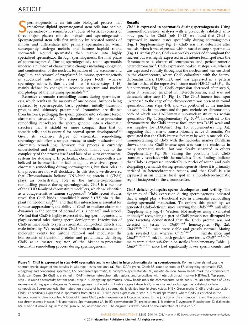
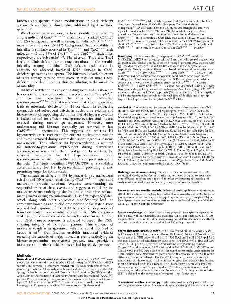
ResultsChd5 is expressed in spermatids during spermiogenesis. Usingimmunofluorescence analyses with a previously validated anti-body specific for Chd5 (refs 10,12) we found that Chd5 isexpressed in mouse testes specifically during spermiogenesis(Fig. 1, Supplementary Fig. 1). Chd5 was first detectable aftermeiosis, when it was expressed within nuclei of step 4 spermatids(Fig. 1). At this phase, Chd5 was weakly expressed throughout thenucleus but was highly expressed in an intense focal spot near thechromocentre, a cluster of centromeres and pericentromericheterochromatin13. Chd5 expression peaked at steps 7–8, when itwas expressed robustly throughout the nucleus and was enrichedin the chromocentre, where Chd5 colocalized with the hetero-chromatin mark H3K9me3, and was expressed in a patternsimilar to that of the repressive histone mark H3K27me3 (Fig. 1b,Supplementary Fig. 2). Chd5 expression decreased after step 9,when it remained enriched in heterochromatin, and was notdetectable after step 10 (Fig. 1). The Chd5-intense focal spotjuxtaposed to the edge of the chromocentre was present in roundspermatids from steps 4–8, and was positioned at the junctionbetween the chromocentre and the post-meiotic sex chromosome,both of which are DAPI-intense sub-nuclear structures withinspermatids (Fig. 1, Supplementary Fig. 3a)14. In contrast to thechromocentre, the Chd5-intense focal spot was DAPI-weak andwas negative for H3K9me3 (Fig. 1b, Supplementary Fig. 3a),suggesting that it marks transcriptionally active chromatin. Wespeculated that the Chd5-intense foci may be within nucleoli. Co-immunostaining of Chd5 with the nucleolar marker fibrillarinshowed that the Chd5-intense spot was near the nucleolus inmany spermatid nuclei, but was clearly separated in others(Supplementary Fig. 3b), raising the possibility that Chd5transiently associates with the nucleolus. These findings indicatethat Chd5 is expressed specifically in nuclei of round and earlyelongating spermatids during spermiogenesis where it is primarilyenriched in heterochromatic regions, and that Chd5 is alsoexpressed in an intense focal spot in a non-heterochromaticregion juxtaposed to the chromocentre.
Chd5 deficiency impairs sperm development and fertility. Thedynamics of Chd5 expression during spermiogenesis indicatedthat it might play a functional role in chromatin remodellingduring spermatid maturation. To explore this possibility, wegenerated Chd5-deficient mice carrying the Chd5Aam1 null allele(Supplementary Fig. 4). Western blot analyses using a validatedantibody10 recognizing a part of Chd5 protein not disrupted bygene targeting demonstrated that the Chd5 protein was notdetected in testes of Chd5Aam1 homozygotes (Fig. 2a).Chd5Aam1� /� mice were viable and grossly normal. Matingtests revealed that whereas Chd5Aam1� /� female mice andChd5Aam1þ /� mice of both genders were fertile, Chd5Aam1� /�
males were either sub-fertile or sterile (Supplementary Table 1).Chd5Aam1� /� mice had significantly lower sperm counts, and
Figure 1 | Chd5 is expressed in step 4–10 spermatids and is enriched in heterochromatin during spermiogenesis. Roman numerals indicate the
spermatogenic stages of the tubules in wild-type testes sections. (a) Blue, DAPI; green, Chd5; RS, round spermatid; ES, elongating spermatid; ECS,
elongating and condensing spermatid; CS, condensed spermatid; P, pachytene spermatocyte; Mi, meiotic division. Arrow heads mark the chromocentre.
Scale bar, 10mm. (b) Chd5 is enriched in DAPI-intense heterochromatic regions, and colocalizes with heterochromatin marker H3K9me3. Top panel,
step 7–8 round spermatids; bottom panel, step 9–10 elongating spermatids. Arrow heads mark the chromocentre. Scale bar, 5 mm. (c) Schematic of Chd5
expression during spermatogenesis. Spermatogenesis is divided into twelve stages (stage I–XII) in mouse and each stage has a distinct cellular
composition. Spermiogenesis, the maturation process of haploid spermatids, is divided into 16 steps (steps 1–16). Green marks Chd5 protein expression.
Chd5 is specifically expressed in spermatids from steps 4–10, with peak expression in step 7–8 round spermatids, where Chd5 is enriched in the
heterochromatic chromocentre. A focus of intense Chd5 protein expression is located adjacent to the junction of the chromocentre and the post-meiotic
sex chromosomes in steps 4–8 spermatids. Spermatogonia (A, In, B); spermatocyte (Pl, preleptotene; L, leptotene; Z, zygotene; P, pachytene; D, diakinesis;
Mi, meiotic division); Ag, acrosomic granule; Ac, acrosomic cap. The diagram is drawn based on the illustration of Hess et al.71
ARTICLE NATURE COMMUNICATIONS | DOI: 10.1038/ncomms4812
2 NATURE COMMUNICATIONS | 5:3812 | DOI: 10.1038/ncomms4812 | www.nature.com/naturecommunications
& 2014 Macmillan Publishers Limited. All rights reserved.

III III IV V VI VII VIII IX X XI XIIMouse spermatogenesis stages
Mito
sis
1 2 3 4 5 6 7 8 9 10 11 12
13 14 15 15 15 15 16 16
P P P P P P P P P P D Mi
Pl Pl L L Z ZB BInInInA A A A A A
c Ag
II–III
II–III
II–III
IV VII–VIII
IV VII–VIII
IV
RS RSCS
ECSES
MiP
VII–VIII
IV VII–VIII
VII–VIII
DAPI Chd5 H3K9me3 MergeH3K9me3& Chd5
VII–VIII VII–VIII VII–VIII VII–VIII
X
X
X
IX
IX
IX
IX–X IX–X IX–X IX–X IX–X
IX
XII
XII
XII
XII
a
b
c
Spe
rmio
gene
sis
Mei
osis
NATURE COMMUNICATIONS | DOI: 10.1038/ncomms4812 ARTICLE
NATURE COMMUNICATIONS | 5:3812 | DOI: 10.1038/ncomms4812 | www.nature.com/naturecommunications 3
& 2014 Macmillan Publishers Limited. All rights reserved.

0
1
2
3
4
+/+ –/–
1.00
0.280.09 0.05
0.00.20.40.60.81.01.2
Normal Few latespermatids
Nospermatids
Sertoli cellonly
+/+ –/– +/+ –/–
Chd5
β-Actin
DF
I fol
d ch
ange
+/+
+/+
–/–
–/– +/+ +/+–/– –/–
Human testis pathology
c
f
ba
d
e
Rel
ativ
e C
HD
5ex
pres
sion
+/+ –/–
250150
5037
KD
II–III
I VI
VII
–VII
IX
X
XI
XIIIV–V
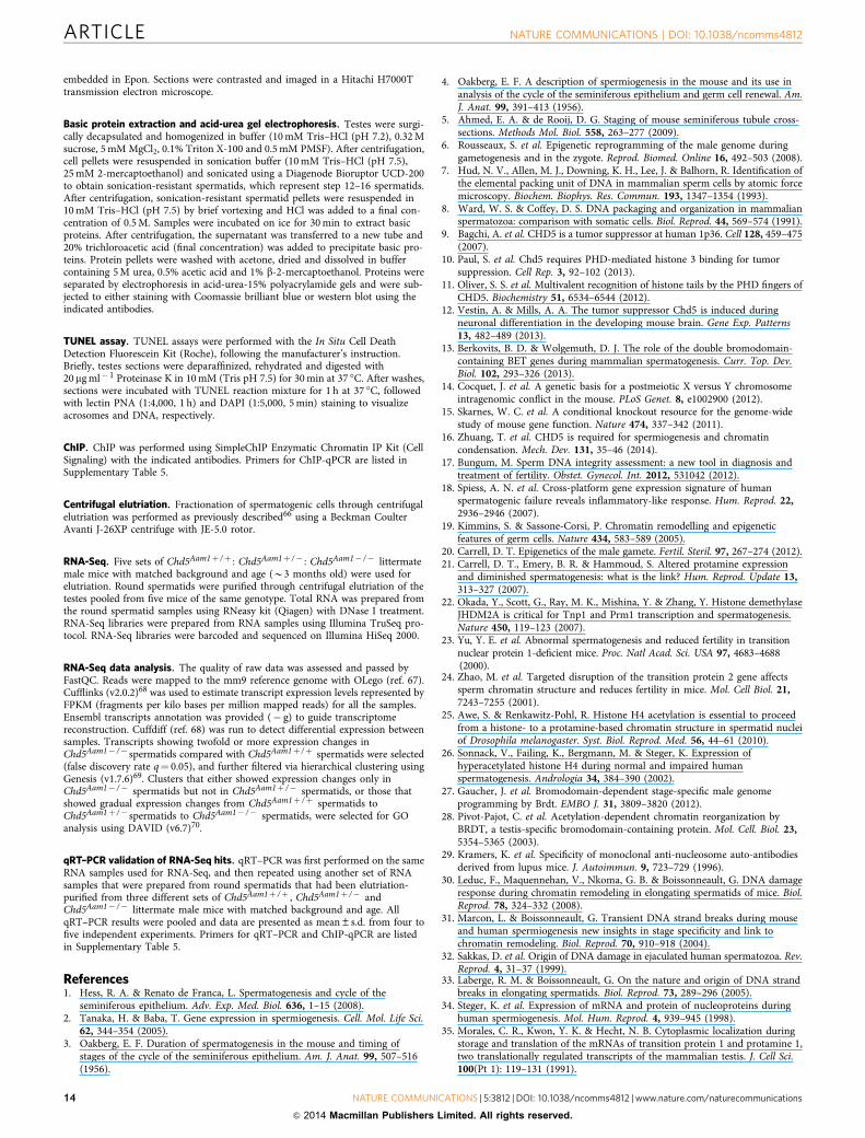
Figure 2 | Chd5 deficiency leads to defective spermatogenesis and chromatin condensation. (a) Western blot analyses indicates that Chd5 protein is not
detectable in Chd5Aam1� /� (� /� ) testis. A validated antibody10 raised against amino acids 1,524–1,705 of mouse Chd5 (which is not disrupted by gene
targeting), was used for western blotting. b-Actin serves as a loading control. (b) Representative abnormal head morphology of Chd5Aam1� /� sperm. Scale
bar, 20mm. (c) SCSA revealed impaired chromatin integrity of Chd5Aam1� /� sperm. DFI, DNA Fragmentation Index (see Methods). Data are presented as
mean±s.d. from four independent experiments. (d) Transmission electron microscopy analyses of sperm from Chd5Aam1þ /þ and Chd5Aam1� /� caudal
epididymi. Chromatin is homogenously condensed in Chd5Aam1þ /þ sperm, but appears loose and uneven with fibrillar texture and contains abnormal
vacuoles in Chd5Aam1� /� sperm nuclei. Scale bar, 1 mm. (e) Staged comparison of periodic acid–Schiff (PAS)-stained Chd5Aam1þ /þ and Chd5Aam1� /�
testes. Roman numerals indicate the stages of the seminiferous tubules. A decrease in the number of elongated spermatids, especially at stages VII and
VIII, is evident in Chd5Aam1� /� tubules. Arrows in stage IX and X mark abnormal retention of condensed spermatids. Scale bar, 10mm. (f) Relative CHD5
expression in human testis with normal versus clinically defined abnormal spermatogenesis. Data are derived from published microarray data set
(ArrayExpress: E-TABM-234) of 39 human testis biopsy samples from 29 men with highly defined testicular pathologies and 10 men with normal
spermatogenesis. RNA was prepared from the testis biopsies and analysed for gene expression using Affymetrix GeneChip. Data were analysed through
NextBio18. Arrow indicates increased severity of spermatogenic defect.
ARTICLE NATURE COMMUNICATIONS | DOI: 10.1038/ncomms4812
4 NATURE COMMUNICATIONS | 5:3812 | DOI: 10.1038/ncomms4812 | www.nature.com/naturecommunications
& 2014 Macmillan Publishers Limited. All rights reserved.

the sperm that were produced had compromised motility and ahigher proportion of morphological abnormalities (Table 1, Fig. 2band Supplementary Table 2). Using in vitro fertilization (IVF), wefound that Chd5Aam1� /� sperm failed to fertilize wild-typeoocytes (Supplementary Table 3). Although it might be expectedthat some functional sperm from sub-fertile Chd5Aam1� /� miceshould be able to fertilize oocytes, none of the sperm obtainedfrom three different Chd5Aam1� /� mice were able to generateblastocysts in vitro. This may be because each of the three malestested happened to be sterile rather than sub-fertile, or that IVFconditions compromised the sperm that would have been able tofertilize oocytes under in vivo conditions. A possible explanationfor the range of severity of the fertility phenotype in different micewas genetic background, as 129Sv embryonic stem cells were usedto generate the Chd5Aam1 allele, with mice being backcrossed forover four generations onto the C57BL/6 background beforeheterozygotes were intercrossed. To determine whether geneticbackground affected fertility, we established a second Chd5-deficient mouse model (carrying the Chd5Tm1b null allele15) thatwas in a 100% pure C57BL/6 genetic background, and assessedmale fertility (Supplementary Fig. 5, Supplementary Table 4). Wefound that Chd5Tm1b� /� male mice were also either sterile orsub-fertile. Another Chd5-deficient mouse model in a pure 129Ebackground also showed that homozygous male mice exhibitedvariable pathology ranging from absence of sperm to near-normalsperm count, although six homozygous males tested did notproduce any progeny over the 2-month period analysed16. Thesedata suggest that Chd5 deficiency compromises sperm productionand male fertility and that this phenotype exhibits inherentvariability in different individuals.
To assess chromatin integrity in sperm from Chd5-deficientmice, we used the sperm chromatin structure assay (SCSA)(Fig. 2c). SCSA revealed that DNA fragmentation was enhancedin Chd5Aam1� /� sperm, reflecting a compromise in chromatinintegrity17. Consistent with this finding, transmission electronmicroscopy showed that whereas chromatin within nuclei ofwild-type sperm is homogeneously condensed, less-condensedchromatin with a punctate texture and uneven density, as well asthe presence of abnormal vacuoles, were observed in nuclei ofChd5Aam1� /� sperm (Fig. 2d). These findings show that Chd5deficiency leads to defective chromatin compaction in sperm.
Since compromised fertility can be caused by a deregulation ofsex hormones, we investigated this possibility (SupplementaryFig. 6). However, we did not detect a significant alteration in sexhormones in Chd5Aam1� /� male mice relative to controls.Histological analyses of testes revealed that seminiferous tubulesof Chd5Aam1� /� mice contained fewer elongated spermatidsrelative to controls, with an abnormal retention of condensedspermatids within stage IX and X tubules (Fig. 2e, Supplementary
Fig. 7). The extent of histological abnormalities varied amongindividual Chd5Aam1� /� mice (Supplementary Fig. 7), inagreement with the variable severity of infertility observed inmale Chd5-deficient mice. In contrast to post-meiotic defects,we did not observe differences in spermatogenic cells (spermato-gonia, spermatocytes, round spermatids) or in somatic cells(Sertoli cells, Leydig cells) in Chd5Aam1� /� testes (Fig. 2e,Supplementary Fig. 7). These findings indicate that Chd5deficiency disrupts the elongation and condensation steps ofpost-meiotic sperm maturation, consistent with Chd5’s peak ofexpression in step 7–8 round spermatids, the phase immediatelypreceeding extensive chromatin remodelling.
To determine whether our findings from Chd5-deficient micemight be relevant to human cases of male infertility, we analyseda previously established gene expression data set of testes biopsiesfrom 39 men (29 men with highly defined testicular pathologyand 10 men with normal spermatogenesis)18. This analysisrevealed that men with spermatogenic defects had lower CHD5expression relative to controls and that the clinical grade ofspermatogenic defect correlated inversely with CHD5 expression(Fig. 2f). While this is a correlation rather than evidence forcausality, it suggests that future efforts should be made todetermine whether compromised CHD5 contributes to maleinfertility in humans.
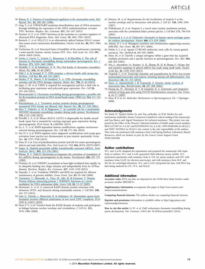
Chd5 deficiency disrupts histone-to-protamine replacement.During mammalian spermiogenesis, the majority of canonicalhistones are removed and replaced by histone variants andtransition proteins, which are subsequently replaced by prota-mines6,7,19,20. This histone-to-protamine replacement processrepackages the sperm genome at least sixfold more compact thanits somatic counterpart8. To define the mechanism whereby Chd5deficiency compromises chromatin compaction duringspermiogenesis, we used western blotting to assess expression ofa panel of somatic histones, transition proteins and protamines inelutriation-purified spermatids at different steps of spermio-genesis (Supplementary Fig. 8). The core nucleosomal histones(H1, H2A, H2B, H3 and H4) were quickly depleted after theround spermatid steps in wild-type testes, with minimal retentionin condensing and condensed spermatids (Fig. 3a). In contrast,these core histones were retained to a higher extent inChd5Aam1� /� differentiated spermatids, which impliedineffective histone removal. In addition, transition proteins(Tnp1 and Tnp2) and protamines (Prm1 and Prm2)had elevated expression in differentiated Chd5Aam1� /�
spermatids (Fig. 3b,c), likely contributing to impaired fertility ofChd5Aam1� /� mice, as precise control of levels of Prm1 andPrm2 are critical for male fertility21,22. Prm2 is first translated as aprecursor protein that is subsequently processed into maturePrm2 through a multistep proteolytic cleavage. In Chd5Aam1� /�
spermatids, there was enhanced expression of the Prm2 precursorand partially processed Prm2 was present, indicating a defect inPrm2 processing, which may be because the overproduction ofPrm2 in Chd5Aam1� /� spermatids exceeds the processingcapacity of the cells. In addition, both Tnp1 and Tnp2deficiency have been shown to increase levels of the Prm2precursor and partially processed forms of Prm2 (refs 23,24).Thus, the abnormal elevation of Tnp1 and Tnp2 inChd5Aam1� /� spermatids may also contribute to defectivePrm2 processing. Consistent with the observation inspermatids, western blotting of lysates from mature sperm alsorevealed enhanced retention of core nucleosomal histones andelevated Prm1 and Prm2 expression in Chd5Aam1� /� sperm(Fig. 3d). Furthermore, immunofluorescent analyses of testesshowed enhanced expression of Tnp1, Tnp2, Prm1 and Prm2 in
Table 1 | Reduced sperm counts and motility inChd5Aam1� /� mice.
Genotype N Sperm/caudalepididymis
(� 106)
All motilesperm (%)
Progressivemotile sperm (%)
Chd5Aam1þ /þ 7 5.52±1.13 62.3±5.4 48.6±11.3Chd5Aam1þ /� 4 6.51±0.68 66.0±3.8 52.8±2.3Chd5Aam1� /� 11 2.74±0.68 42.4±10.3 21.0±5.7P(þ /þ versus þ /� ) 0.103 0.228 0.375P(þ /þ versus � /� ) 0.0002* 0.0055* 0.0004*
N indicates the number of mice used for the indicated sperm analyses. P-value is two-tailStudent’s t-test result between the indicated genotypes.*indicates statistical significance. Data are presented as mean±s.d. þ /þ , Chd5Aam1þ /þ ;� /� , Chd5Aam1� /� ; þ /� , Chd5þ /� .
NATURE COMMUNICATIONS | DOI: 10.1038/ncomms4812 ARTICLE
NATURE COMMUNICATIONS | 5:3812 | DOI: 10.1038/ncomms4812 | www.nature.com/naturecommunications 5
& 2014 Macmillan Publishers Limited. All rights reserved.
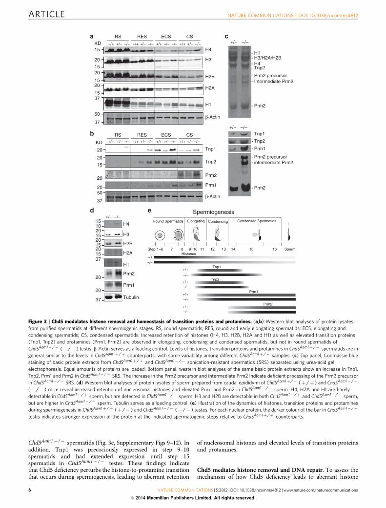
Chd5Aam1� /� spermatids (Fig. 3e, Supplementary Figs 9–12). Inaddition, Tnp1 was precociously expressed in step 9–10spermatids and had extended expression until step 15spermatids in Chd5Aam1� /� testes. These findings indicatethat Chd5 deficiency perturbs the histone-to-protamine transitionthat occurs during spermiogenesis, leading to aberrant retention
of nucleosomal histones and elevated levels of transition proteinsand protamines.
Chd5 mediates histone removal and DNA repair. To assess themechanism of how Chd5 deficiency leads to aberrant histone
+/+ +/+ +/+ +/++/– +/– +/– +/––/–
+/+ +/+ +/+ +/++/– +/– +/– +/––/– –/– –/– –/–
–/– –/– –/–
RS RES ECS CS
H4
H3
H2B
H2A
H1
β-Actin
aKD15
201520
1520
1537
50
37
Tnp1
Tnp2
Prm2
Prm1
β-Actin
b RS RES ECS CS
20
20
15
20
2050
37
KD
+/+
+/+
–/–
–/–
- H1- H3/H2A/H2B - H4 - Tnp2
- Prm2 precursor- Intermediate Prm2
- Prm2
c
- Tnp1
- Tnp2
- Prm1
- Prm2 precursor- intermediate Prm2
- Prm2
d e Spermiogenesis
Round Spermatids Elongating Condensing Condensed Spermatids
Step 1–6
+/+
–/–
+/+–/–
+/+–/–
+/+–/–
+/+–/–
7 8 9 10Histones
Tnp1
Tnp2
Prm1
Prm2
11 12 13 14 15 16 Sperm
+/+ –/–
H4
H3
H2B
H2A
H1
Prm2
Prm1
Tubulin
2015
20
20
37
2015
2015
1510
37
Figure 3 | Chd5 modulates histone removal and homeostasis of transition proteins and protamines. (a,b) Western blot analyses of protein lysates
from purified spermatids at different spermiogenic stages. RS, round spermatids; RES, round and early elongating spermatids; ECS, elongating and
condensing spermatids; CS, condensed spermatids. Increased retention of histones (H4, H3, H2B, H2A and H1) as well as elevated transition proteins
(Tnp1, Tnp2) and protamines (Prm1, Prm2) are observed in elongating, condensing and condensed spermatids, but not in round spermatids of
Chd5Aam1� /� (� /� ) testis. b-Actin serves as a loading control. Levels of histones, transition proteins and protamines in Chd5Aam1þ /� spermatids are in
general similar to the levels in Chd5Aam1þ /þ counterparts, with some variability among different Chd5Aam1þ /� samples. (c) Top panel, Coomassie blue
staining of basic protein extracts from Chd5Aam1þ /þ and Chd5Aam1� /� sonication-resistant spermatids (SRS) separated using urea-acid gel
electrophoresis. Equal amounts of proteins are loaded. Bottom panel, western blot analyses of the same basic protein extracts show an increase in Tnp1,
Tnp2, Prm1 and Prm2 in Chd5Aam1� /� SRS. The increase in the Prm2 precursor and intermediate Prm2 indicate deficient processing of the Prm2 precursor
in Chd5Aam1� /� SRS. (d) Western blot analyses of protein lysates of sperm prepared from caudal epididymi of Chd5Aam1þ /þ (þ /þ ) and Chd5Aam1� /�
(� /� ) mice reveal increased retention of nucleosomal histones and elevated Prm1 and Prm2 in Chd5Aam1� /� sperm. H4, H2A and H1 are barely
detectable in Chd5Aam1þ /þ sperm, but are detected in Chd5Aam1� /� sperm. H3 and H2B are detectable in both Chd5Aam1þ /þ and Chd5Aam1� /� sperm,
but are higher in Chd5Aam1� /� sperm. Tubulin serves as a loading control. (e) Illustration of the dynamics of histones, transition proteins and protamines
during spermiogenesis in Chd5Aam1þ /þ (þ /þ ) and Chd5Aam1� /� (� /� ) testes. For each nuclear protein, the darker colour of the bar in Chd5Aam1� /�
testis indicates stronger expression of the protein at the indicated spermatogenic steps relative to Chd5Aam1þ /þ counterparts.
ARTICLE NATURE COMMUNICATIONS | DOI: 10.1038/ncomms4812
6 NATURE COMMUNICATIONS | 5:3812 | DOI: 10.1038/ncomms4812 | www.nature.com/naturecommunications
& 2014 Macmillan Publishers Limited. All rights reserved.
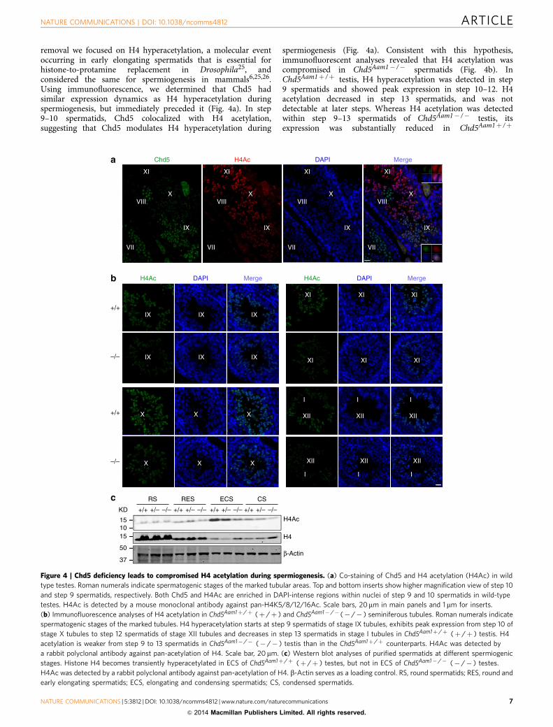
removal we focused on H4 hyperacetylation, a molecular eventoccurring in early elongating spermatids that is essential forhistone-to-protamine replacement in Drosophila25, andconsidered the same for spermiogenesis in mammals6,25,26.Using immunofluorescence, we determined that Chd5 hadsimilar expression dynamics as H4 hyperacetylation duringspermiogenesis, but immediately preceded it (Fig. 4a). In step9–10 spermatids, Chd5 colocalized with H4 acetylation,suggesting that Chd5 modulates H4 hyperacetylation during
spermiogenesis (Fig. 4a). Consistent with this hypothesis,immunofluorescent analyses revealed that H4 acetylation wascompromised in Chd5Aam1� /� spermatids (Fig. 4b). InChd5Aam1þ /þ testis, H4 hyperacetylation was detected in step9 spermatids and showed peak expression in step 10–12. H4acetylation decreased in step 13 spermatids, and was notdetectable at later steps. Whereas H4 acetylation was detectedwithin step 9–13 spermatids of Chd5Aam1� /� testis, itsexpression was substantially reduced in Chd5Aam1þ /þ
III
XIIXIIXII
III
XIIXIIXII
XIXIXI
XIXIXI
XXX
+/+
–/–
KDH4Ac
H4
β-Actin
RS RES ECS CS
151015
50
37
+/+
a
b
c
–/–
XXX
IXIXIX
IXIXIX
H4Ac DAPI MergeH4Ac DAPI Merge
+/+ +/– –/– +/+ +/– –/– +/+ –/–+/– +/+ –/–+/–
Chd5 H4Ac DAPI Merge
VII VII VII VII
VIII VIII VIII VIIIX X X X
IX IX IX IX
XIXI XI XI
Figure 4 | Chd5 deficiency leads to compromised H4 acetylation during spermiogenesis. (a) Co-staining of Chd5 and H4 acetylation (H4Ac) in wild
type testes. Roman numerals indicate spermatogenic stages of the marked tubular areas. Top and bottom inserts show higher magnification view of step 10
and step 9 spermatids, respectively. Both Chd5 and H4Ac are enriched in DAPI-intense regions within nuclei of step 9 and 10 spermatids in wild-type
testes. H4Ac is detected by a mouse monoclonal antibody against pan-H4K5/8/12/16Ac. Scale bars, 20mm in main panels and 1mm for inserts.
(b) Immunofluorescence analyses of H4 acetylation in Chd5Aam1þ /þ (þ /þ ) and Chd5Aam1� /� (� /� ) seminiferous tubules. Roman numerals indicate
spermatogenic stages of the marked tubules. H4 hyperacetylation starts at step 9 spermatids of stage IX tubules, exhibits peak expression from step 10 of
stage X tubules to step 12 spermatids of stage XII tubules and decreases in step 13 spermatids in stage I tubules in Chd5Aam1þ /þ (þ /þ ) testis. H4
acetylation is weaker from step 9 to 13 spermatids in Chd5Aam1� /� (� /� ) testis than in the Chd5Aam1þ /þ counterparts. H4Ac was detected by
a rabbit polyclonal antibody against pan-acetylation of H4. Scale bar, 20mm. (c) Western blot analyses of purified spermatids at different spermiogenic
stages. Histone H4 becomes transiently hyperacetylated in ECS of Chd5Aam1þ /þ (þ /þ ) testes, but not in ECS of Chd5Aam1� /� (� /� ) testes.
H4Ac was detected by a rabbit polyclonal antibody against pan-acetylation of H4. b-Actin serves as a loading control. RS, round spermatids; RES, round and
early elongating spermatids; ECS, elongating and condensing spermatids; CS, condensed spermatids.
NATURE COMMUNICATIONS | DOI: 10.1038/ncomms4812 ARTICLE
NATURE COMMUNICATIONS | 5:3812 | DOI: 10.1038/ncomms4812 | www.nature.com/naturecommunications 7
& 2014 Macmillan Publishers Limited. All rights reserved.

spermatids. Furthermore, western blotting of lysates fromelutriation-purified spermatid fractions showed that whereasmore total H4 was retained, H4Ac was severely compromisedin differentiated Chd5Aam1� /� spermatids (Fig. 4c). Consistentwith the findings in Chd5Aam1� /� mice, Chd5Tm1b� /�
testes also showed compromised histone H4 acetylation(Supplementary Fig. 13). These findings indicate that acetylatedH4 is compromised in Chd5-deficient testes.
Following H4 hyperacetylation, acetylated histone tails arerecognized by Brdt, a testis-specific member of the BRDbromodomain-containing protein family that has been shownto induce eviction of nucleosomes6,27,28. However, how Brdtmediates nucleosome eviction is not clear. Likely, Brdt recruitschromatin remodelers to remodel and evict hyperacetylatednucleosomes. We therefore asked whether the compromised H4acetylation and Chd5 deficiency perturbed nucleosome evictionin Chd5Aam1� /� spermatids. Using an antibody specific forintact nucleosomes29, we found that nucleosomes were detectablethrough step 11 of spermatid maturation in wild-type testes, butwere depleted at later steps (Fig. 5a). However, we found thatnucleosomes were aberrantly retained in Chd5Aam1� /�
spermatids as late as step 14 (Fig. 5b), indicating thatnucleosomes were ineffectively evicted. Thus, both H4acetylation and nucleosome eviction are compromised by Chd5deficiency.
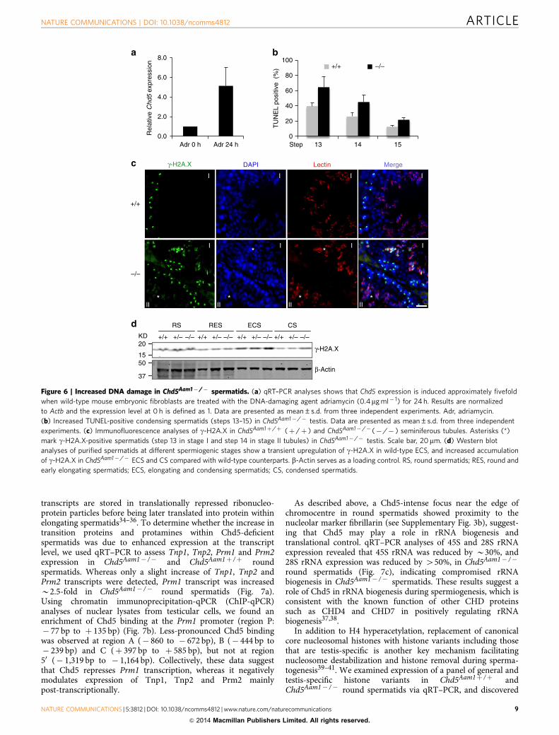
Nucleosome eviction generates DNA supercoiling tension thatneeds to be relieved. Previous studies indicate that topoisomeraseII beta (Top2b) catalyses resolution of such supercoils inelongating spermatids, during which double-strand breaks(DSBs) are generated. A DNA damage response is then triggeredto repair the DSBs in order to maintain genome integrity30–33.We found that Chd5 expression is induced by DNA damage(Fig. 6a), suggesting that Chd5 plays a role in the DNA damageresponse as well. Indeed, TUNEL assays revealed an increase ofDNA breaks in differentiated Chd5Aam1� /� spermatids, mostnotably at steps 13–14 (Fig. 6b), a time point when most DNAbreaks are repaired in wild-type testes. In agreement with thisfinding, immunofluorescent analyses showed that whereasg-H2A.X (a marker for the DSB-activated DNA damageresponse) is cleared in wild-type spermatids after step 12, it isdetected in Chd5Aam1� /� spermatids as late as step 14 (Fig. 6c).Western blot analyses further confirmed an increase of g-H2A.Xin differentiated Chd5Aam1� /� spermatids (Fig. 6d). Together,these findings indicate that Chd5 deficiency impairs H4hyperacetylation, nucleosome eviction, and DNA damage repairduring spermiogenesis.
Chd5 loss alters gene expression in spermatids. Tnp1, Tnp2,Prm1 and Prm2 are transcribed in round spermatids, but their
Nucleosome DAPI Lectin Merge
+/+
–/–
*
*
II
II
*
*
II
II
*
*
II
II
*
*
II
II
Mi
XIIXI
*
Mi
XIIXI
*
Mi
XIIXI
*
Mi
XIIXI
*
a
b
Figure 5 | Chd5 deficiency leads to inefficient nucleosome eviction during spermiogenesis. Roman numerals indicate the spermatogenic stages of tubular
areas. (a) Nucleosomes are detected in step 11 spermatids (indicated by arrow head) of stage XI seminiferous tubules, but are depleted afterwards
and not detectable in step 12 spermatids (indicated by *) of stage XII seminiferous tubule in wild-type testes. Green, nucleosomes; Red, lectin (visualizing
acrosome for staging seminiferous tubules); Blue, DAPI; Mi, meiotic figure, a hallmark of stage XII tubules. (b) Immunostaining for nucleosomes shows that
nucleosomes are retained in as late as step 14 spermatids of stage II seminiferous tubules in Chd5Aam1� /� (� /� ) testes, while being absent in the
Chd5Aam1þ /þ (þ /þ ) counterpart. Asterisks (*) mark nucleosome-positive condensing spermatids (step 14) in Chd5Aam1� /� tubules. Scale bars, 10mm.
ARTICLE NATURE COMMUNICATIONS | DOI: 10.1038/ncomms4812
8 NATURE COMMUNICATIONS | 5:3812 | DOI: 10.1038/ncomms4812 | www.nature.com/naturecommunications
& 2014 Macmillan Publishers Limited. All rights reserved.
transcripts are stored in translationally repressed ribonucleo-protein particles before being later translated into protein withinelongating spermatids34–36. To determine whether the increase intransition proteins and protamines within Chd5-deficientspermatids was due to enhanced expression at the transcriptlevel, we used qRT–PCR to assess Tnp1, Tnp2, Prm1 and Prm2expression in Chd5Aam1� /� and Chd5Aam1þ /þ roundspermatids. Whereas only a slight increase of Tnp1, Tnp2 andPrm2 transcripts were detected, Prm1 transcript was increasedB2.5-fold in Chd5Aam1� /� round spermatids (Fig. 7a).Using chromatin immunoprecipitation-qPCR (ChIP-qPCR)analyses of nuclear lysates from testicular cells, we found anenrichment of Chd5 binding at the Prm1 promoter (region P:� 77 bp to þ 135 bp) (Fig. 7b). Less-pronounced Chd5 bindingwas observed at region A (� 860 to � 672 bp), B (� 444 bp to� 239 bp) and C (þ 397 bp to þ 585 bp), but not at region50 (� 1,319 bp to � 1,164 bp). Collectively, these data suggestthat Chd5 represses Prm1 transcription, whereas it negativelymodulates expression of Tnp1, Tnp2 and Prm2 mainlypost-transcriptionally.
As described above, a Chd5-intense focus near the edge ofchromocentre in round spermatids showed proximity to thenucleolar marker fibrillarin (see Supplementary Fig. 3b), suggest-ing that Chd5 may play a role in rRNA biogenesis andtranslational control. qRT–PCR analyses of 45S and 28S rRNAexpression revealed that 45S rRNA was reduced by B30%, and28S rRNA expression was reduced by 450%, in Chd5Aam1� /�
round spermatids (Fig. 7c), indicating compromised rRNAbiogenesis in Chd5Aam1� /� spermatids. These results suggest arole of Chd5 in rRNA biogenesis during spermiogenesis, which isconsistent with the known function of other CHD proteinssuch as CHD4 and CHD7 in positively regulating rRNAbiogenesis37,38.
In addition to H4 hyperacetylation, replacement of canonicalcore nucleosomal histones with histone variants including thosethat are testis-specific is another key mechanism facilitatingnucleosome destabilization and histone removal during sperma-togenesis39–41. We examined expression of a panel of general andtestis-specific histone variants in Chd5Aam1þ /þ andChd5Aam1� /� round spermatids via qRT–PCR, and discovered
0
20
40
60
80
100
13 14 15
+/+ –/–
TU
NE
L po
sitiv
e (
%)
Step
a b
+/+ +/– –/– +/– –/– +/– –/– +/– –/–+/+ +/+ +/+
RS RES ECS CS
β-Actin
γ-H2A.X
d
c
Rel
ativ
e C
hd5
expr
essi
on
0.0
2.0
4.0
6.0
8.0
Adr 0 h Adr 24 h
KD20
1550
37
γ-H2A.X DAPI Lectin Merge
*
*
I
II
I*
*
I
II
I*
*
I
II
I*
*
I
II
I
+/+
–/–
Figure 6 | Increased DNA damage in Chd5Aam1� /� spermatids. (a) qRT–PCR analyses shows that Chd5 expression is induced approximately fivefold
when wild-type mouse embryonic fibroblasts are treated with the DNA-damaging agent adriamycin (0.4mg ml� 1) for 24 h. Results are normalized
to Actb and the expression level at 0 h is defined as 1. Data are presented as mean±s.d. from three independent experiments. Adr, adriamycin.
(b) Increased TUNEL-positive condensing spermatids (steps 13–15) in Chd5Aam1� /� testis. Data are presented as mean±s.d. from three independent
experiments. (c) Immunofluorescence analyses of g-H2A.X in Chd5Aam1þ /þ (þ /þ ) and Chd5Aam1� /� (� /� ) seminiferous tubules. Asterisks (*)
mark g-H2A.X-positive spermatids (step 13 in stage I and step 14 in stage II tubules) in Chd5Aam1� /� testis. Scale bar, 20mm. (d) Western blot
analyses of purified spermatids at different spermiogenic stages show a transient upregulation of g-H2A.X in wild-type ECS, and increased accumulation
of g-H2A.X in Chd5Aam1� /� ECS and CS compared with wild-type counterparts. b-Actin serves as a loading control. RS, round spermatids; RES, round and
early elongating spermatids; ECS, elongating and condensing spermatids; CS, condensed spermatids.
NATURE COMMUNICATIONS | DOI: 10.1038/ncomms4812 ARTICLE
NATURE COMMUNICATIONS | 5:3812 | DOI: 10.1038/ncomms4812 | www.nature.com/naturecommunications 9
& 2014 Macmillan Publishers Limited. All rights reserved.

that expression of the H2B variant Hist1h2bc was elevated overfivefold in Chd5Aam1� /� round spermatids (Fig. 7d). Althoughlittle is known about the functions of Hist1h2bc, its substantialincrease in Chd5Aam1� /� round spermatids suggests that itmight impact histone variant exchanges during spermiogenesis.In addition, an B30% decrease in expression of histone H1variant Hist1h1e, histone H3 variant Hist2h3c1 and testis-specificH1 variant H1t were consistently observed in Chd5Aam1� /�
spermatids (Fig. 7d). These findings indicate that Chd5 deficiencyalters transcript levels of specific histone variants duringspermiogenesis.
In order to gain insight into the global gene expression changesresulting from Chd5 deficiency and to identify candidate Chd5targets, we performed RNA sequencing (RNA-Seq) (Fig. 8). Chd5is mainly expressed in round spermatids, where transcription ismost active during spermiogenesis, with transcription globallybeing ceased afterwards. Using RNA-Seq, we compared mRNAexpression profiles of round spermatids that had been elutriation-purified from five sets of Chd5Aam1þ /þ , Chd5Aam1þ /� andChd5Aam1� /� littermates. Expression of 14,206 transcripts wasdetected in at least one of the three genotypes. Two-hundred andsixty-one transcripts, or 1.8% of all the transcripts detected,showed a twofold or greater expression change in Chd5Aam1� /�
round spermatids compared with Chd5Aam1þ /þ counterparts(false discovery rate q¼ 0.05). Among the 261 transcripts, 156transcripts or 59.8% were downregulated, whereas 105
transcripts, or 40.2%, were upregulated in Chd5Aam1� /� roundspermatids. Together, these results suggest that Chd5 deficiencyleads to a rather limited alteration in global gene expression inround spermatids, and that Chd5 both activates and suppressesgene expression in round spermatids with a slight preference foractivation. Gene ontology (GO) analysis of the gene expressionchanges showed clustering of GO terms including chromosomeorganization, response to DNA damage, acetylation, alternativesplicing, nuclear export, protein transport, ubl (ubiquitin)conjugation, intracellular transport, endocytosis, cell cycle andMAPK pathway (Supplementary Fig. 14). Since Chd5Aam1þ /�
male mice were fertile, we reasoned that genes that hadexpression changes only in Chd5Aam1� /� spermatids but notin Chd5Aam1þ /� spermatids, or genes that had gradualexpression changes dependent on Chd5 dosage, would becandidates most likely to contribute to the infertility ofChd5Aam1� /� male mice. We thus further filtered the 261transcripts through clustering with manual examination andidentified a list of 155 transcripts that had expression changesonly in Chd5Aam1� /� spermatids, or that had gradual expressionchanges that correlated with Chd5 dosage (Fig. 8a, SupplementaryFig. 15). Among the list, 90 transcripts were downregulated and65 transcripts, including Hist1h2bc, were upregulated inChd5Aam1� /� spermatids (Fig. 8a). qRT–PCR analyses forexpression of 11 genes from this list, which included the positivecontrol Hist1h2bc and represented a range of expression changes,
0.0
1.0
2.0
3.0
4.0
5.0
5′ A B P C
IgGa-Chd5
Rel
ativ
e m
RN
A e
xpre
ssio
n
+1 100 bp
585 bp1,319 –860 –444 –77Prm1
5′ A B P C
a b
0.0
0.5
1.0
1.5
2.0
2.5
3.0
Tnp1 Tnp2 Prm1 Prm2
+/+–/–
+/+–/–
+/+–/–
0.0
0.2
0.4
0.6
0.8
1.0
1.2
45S rRNA 28S rRNA
c d
Rel
ativ
e m
RN
A e
xpre
ssio
n
Rel
ativ
e m
RN
A e
xpre
ssio
n F
old
of e
nric
hmen
t
His1h2bc Hist1h1e Hist2h3c1 H1t
5.5
3.5
1.2
1.0
0.8
0.6
0.4
0.2
0.0
Figure 7 | Altered transcription of Prm1 and histone variants in Chd5-deficient spermatids. (a) qRT–PCR analyses of transition protein and protamine
genes in Chd5Aam1þ /þ and Chd5Aam1� /� round spermatids. Results are normalized to Actb expression. Prm1 expression is increased by B2.5-fold in
Chd5Aam1� /� round spermatids. Data are presented as mean±s.d. from four independent experiments. (b) Top panel, diagram of mouse Prm1
gene and location of primer sets used for ChIP-qPCR. Bottom panel, ChIP-qPCR analyses reveals enrichment of Chd5 at promoter region P (� 77 bp to
þ 135 bp) of Prm1 gene. The results are normalized to IgG control and are shown as fold of enrichment. Data are presented as mean±s.d. from four
to five independent experiments. (c) qRT–PCR analyses of rRNAs shows compromised expression of 28S and 45S ribosomal RNAs in Chd5Aam1� /�
(� /� ) round spermatids. Results are normalized to Actb expression. Data are presented as mean±s.d. from four to five independent experiments.
(d) qRT–PCR analyses of histone variants revealed an approximately fivefold increase in expression of histone H2B variant Hist1h2bc and modest decreases
in expression of histone variants Hist1h1e, Hist2h3c1 and H1t in Chd5Aam1� /� (� /� ) round spermatids. Results are normalized to Actb expression.
Data are presented as mean±s.d. from three to five independent experiments.
ARTICLE NATURE COMMUNICATIONS | DOI: 10.1038/ncomms4812
10 NATURE COMMUNICATIONS | 5:3812 | DOI: 10.1038/ncomms4812 | www.nature.com/naturecommunications
& 2014 Macmillan Publishers Limited. All rights reserved.
was used to validate the expression changes revealed by RNA-Seq,attesting to the reliability of the RNA-Seq data and analyses(Fig. 8b). GO analysis of this list showed clustering of a subset ofGO terms revealed from the original list, which includechromosome organization, response to DNA damage stimulus,alternative splicing, ubl conjugation, regulation of DNA meta-bolic process, cell cycle and MAPK signalling (Fig. 8c). A numberof candidates implicated in acetylation (1700019G17Rik), DNAdamage response (Wrnip1), RNA processing and translationalcontrol (Cstf2t) and nuclear structure maintenance (Syne3) werefurther verified using qRT–PCR (Fig. 8d, Supplementary Fig. 16).Altogether, these GO terms are consistent with the phenotypic
impacts of Chd5 deficiency on chromatin compaction,DNA damage response and post-transcriptional modulation oftransition proteins and protamines during spermiogenesis.Alterations in gene expression within these GO terms are likelycontributing, at least partially, to the spermatogenic abnormalitiesof Chd5Aam1� /� male mice.
DiscussionIn this study, we show that Chd5 mediates a cascade of molecularevents underlying histone removal during spermiogenesisincluding H4 hyperacetylation, histone variant expression,
a b
c
d
47
9
9
8
7
6
5
3
0 10 20 30 40 50
Alternative splicing
ubl conjugation
Cell cycle
Cellular response to stress
Chromosome organization
Response to DNA damage stimulus
MAPK signalling pathway
Regulation of DNA metabolic process 4.70E–02
4.30E–02
2.80E–02
3.30E–02
1.00E–02
2.90E–02
1.70E–02
1.10E–04
P valueNumber of genes classifiedGO terms
65
90
1.00.0
+\+
+\–
–\–
0
0.2
0.4
0.6
0.8
1
1.2 +/+ –/–
1700
019G
17Rik
Wrn
ip1
Cstf2t
Syne3
Rel
ativ
e m
RN
A e
xpre
ssio
n
0
1
2
3
4
5
6 +/+ –/–
Scara
5
Trpm
8Pnp
2Acs
l5
Klk1b9
Car2
Gxylt1
Sik1Prm
1
Zfp61
8
Hist1h
2bc
Figure 8 | RNA-Seq reveals global gene expression changes in Chd5Aam1� /� spermatids. (a) Heat map presentation of gene expression in
Chd5Aam1þ /þ , Chd5Aam1þ /� and Chd5Aam1� /� round spermatids revealed by RNA-Seq. Green to red (0 to 1) represents the gradient increase of
expression levels. Sixty-five transcripts show upregulation, and 90 transcripts show downregulation, in Chd5Aam1� /� round spermatids. (b) qRT–PCR
validation of RNA-Seq revealed gene expression changes in round spermatids. Results are normalized to Actb expression and data are presented as
mean±s.d. from four to five independent experiments. (c) GO analysis of the 155 transcripts that either show expression changes only in Chd5Aam1� /�
spermatids but not in Chd5Aam1þ /� spermatids, or that show gradual expression change from Chd5Aam1þ /þ spermatids to Chd5Aam1þ /� spermatids to
Chd5Aam1� /� spermatids. Numbers next to bars indicate the number of genes classified to the corresponding GO term. P-value is calculated using
modified Fisher’s exact test by DAVID (v6.7)70. (d) qRT–PCR verification of compromised expression of genes implicated in histone acetylation,
DNA damage response, RNA processing and nuclear integrity in Chd5Aam1� /� spermatids. Results are normalized to Actb expression and Data are
presented as mean±s.d. from four to five independent experiments.
NATURE COMMUNICATIONS | DOI: 10.1038/ncomms4812 ARTICLE
NATURE COMMUNICATIONS | 5:3812 | DOI: 10.1038/ncomms4812 | www.nature.com/naturecommunications 11
& 2014 Macmillan Publishers Limited. All rights reserved.
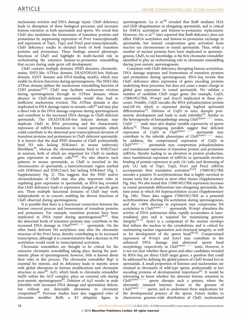
nucleosome eviction and DNA damage repair. Chd5 deficiencyleads to disruption of these biological processes and increaseshistone retention in both spermatids and sperm. We reveal thatChd5 also modulates the homeostasis of transition proteins andprotamines by suppressing expression of Prm1 transcriptionallyand expression of Tnp1, Tnp2 and Prm2 post-transcriptionally.Chd5 deficiency results in elevated levels of both transitionproteins and protamines. These findings unravel pleiotropicfunctions of Chd5 and highlight its multi-faceted role inorchestrating the extensive histone-to-protamine remodellingthat occurs during male germ cell development.
Chd5 contains multiple domains (PHD domains, chromodo-mains, SNF2-like ATPase domain, DEAD/DEAH-box Helicasedomain, SANT domain and DNA-binding motifs), which mayenable its diverse functions during spermiogenesis. The SNF2-likeATPase domain defines the nucleosome remodelling function ofCHD proteins42,43. Chd5 may facilitate nucleosome evictionduring spermiogenesis through its ATPase domain, whoseabsence in Chd5-deficient spermatids would thus lead toinefficient nucleosome eviction. The ATPase domain is alsoimplicated in DNA damage repair in somatic cells44 and may playa direct role in the DNA damage response during spermiogenesisand contribute to the increased DNA damage in Chd5-deficientspermatids. The DEAD/DEAH-box helicase domain mayimplicate Chd5 in RNA processing and the characteristicrepression of mRNA translation in round spermatids, whichcould contribute to the abnormal post-transcriptional elevation oftransition proteins and protamines in Chd5-deficient spermatids.It is known that the dual PHD domains of Chd5 preferentiallybind H3 tails lacking H3K4me3 in mouse embryonicfibroblasts10, whereas the chromodomains bind to H3K27me3in neurons, both of which are important for Chd5 to mediategene expression in somatic cells10,45. We also observe suchpatterns in mouse spermatids, as Chd5 is enriched in thechromocentre of spermatids, a heterochromatic region markedwith H3K9me3 and H3K27me3, but lacking H3K4me3 (Fig. 1,Supplementary Fig. 2). This suggests that the PHD and/orchromodomains of Chd5 may also play important roles inregulating gene expression in spermatids, as RNA-Seq revealedthat Chd5 deficiency leads to expression changes of specific genesets. These multiple functional domains of Chd5 may workindependently or in concert, enabling the diverse functions ofChd5 observed during spermiogenesis.
It is possible that there is a functional connection between thehistone removal process and homeostasis of transition proteinsand protamines. For example, transition proteins have beenimplicated in DNA repair during spermiogenesis46,47, thusthe abnormal levels of transition proteins may contribute to theincreased DNA damage in Chd5Aam1� /� spermatids. On theother hand, deficient H4 acetylation may alter the chromatinstructure of the Prm1 locus, thereby contributing to its increasedtranscription, although it is counterintuitive that a decrease in H4acetylation would result in transcriptional activation.
Chromatin remodellers are thought to be critical for theextensive chromatin remodelling taking place during the post-meiotic phase of spermiogenesis; however, little is known abouttheir roles in this process. The chromatin remodeller Brg1 isessential for meiosis, and its deficiency leads to meiotic arrestwith global alterations in histone modifications and chromatinstructure in mice48. Acf1, which binds to chromatin remodellerSnf2h within the ACF complex, plays an essential role duringpost-meiotic spermiogenesis49. Deletion of Acf1 results in maleinfertility with increased DNA damage and spermiation defects,but without any detectable alterations in chromatincomposition49. Previous studies have also suggested roles ofchromatin modifier Rnf8, a E3 ubiquitin ligase, in
spermiogenesis. Lu et al.50 revealed that Rnf8 mediates H2Aand H2B ubiquitination in elongating spermatids, and is criticalfor H4K16 acetylation and histone-to-protamine replacement.However, Sin et al.51 later reported that Rnf8 deficiency does notaffect H4K16 acetylation and histone-to-protamine exchange inspermatids, but instead compromises gene activation frominactive sex chromosomes in round spermatids. Thus, while anumber of nuclear proteins have been implicated in spermato-genesis, Chd5, to our knowledge, is the first chromatin remodelleridentified to play an orchestrating role in chromatin remodellingduring post-meiotic spermiogenesis.
Consistent with Chd5 deficiency disrupting histone acetylation,DNA damage response and homeostasis of transition proteinsand protamines during spermiogenesis, RNA-Seq reveals thatChd5 deficiency alters expression of genes encoding proteinsunderlying these processes, but does not cause a major change inglobal gene expression in round spermatids. We validate anumber of candidate Chd5 target genes (for example, Cstf2t,1700019G17Rik, Wrnip1 and Syne3) implicated in these pro-cesses. Notably, Cstf2t encodes the RNA polyadenylation proteintauCstF-64, which is expressed during haploid spermatiddifferentiation52. Deletion of Cstf2t in mice disrupts post-meiotic development and leads to male infertility52. Similar tothe heterogeneity of histopathology among Chd5Aam1� /� testes,Cstf2t� /� male mice also display variable expressivity of spermdefects52. These intriguing parallels suggest that deficientexpression of Cstf2t in Chd5Aam1� /� spermatids maycontribute to the infertile phenotypes of Chd5Aam1� /� mice.In addition, the compromised expression of Cstf2t inChd5Aam1� /� spermatids may compromise polyadenylationand translational repression of transition protein and protaminemRNAs, thereby leading to an elevation in protein production,since translational repression of mRNAs in spermatids involvesbinding of protein repressors to poly (A) tails, and shortening ofpoly (A) tails of Tnp1, Tnp2, Prm1 and Prm2 mRNAsaccompanies their translation activation53,54. 1700019G17Rikencodes a putative N-acetyltransferase that is highly enriched inmouse testis but is absent in most other tissues (SupplementaryFig. 16a). We also found that 1700019G17Rik expression increasesas round spermatids differentiate into elongating spermatids, thetime point at which H4 hyperacetylation occurs (SupplementaryFig. 16b). These data suggest 1700019G17Rik as a potentialacetyltransferase affecting H4 acetylation during spermiogenesis,and the 480% decrease in expression may compromise H4acetylation in Chd5Aam1� /� spermatids. Wrnip1 stimulates theactivity of DNA polymerase delta, rapidly accumulates at laser-irradiated sites and is required for maintaining genomeintegrity55–57. Syne3 is a component of the nuclear envelopethat tethers the nucleus to the cytoskeleton, and is critical formaintaining nuclear organization and structural integrity, as wellas for development of the sperm head58–62. Compromisedexpression of Wrnip1 and Syne3 may contribute to theenhanced DNA damage and abnormal sperm headmorphology, respectively, in Chd5Aam1� /� testis. However, itis not yet clear whether these genes and other candidates revealedby RNA-Seq are direct Chd5 target genes, a question that couldbe addressed by defining the global pattern of Chd5-bound loci inspermatids. A small proportion of histones and nucleosomes areretained in chromatin of wild-type sperm, preferentially at lociencoding proteins of developmental importance63. It would beinteresting to know whether the aberrant histone retention inChd5Aam1� /� sperm disrupts such a pattern, where theaberrantly retained histones locate in the genome ofChd5Aam1� /� sperm, and to understand these implications forthe developmental potency of the sperm. Future studies tocharacterize genome-wide distribution of Chd5, nucleosomal
ARTICLE NATURE COMMUNICATIONS | DOI: 10.1038/ncomms4812
12 NATURE COMMUNICATIONS | 5:3812 | DOI: 10.1038/ncomms4812 | www.nature.com/naturecommunications
& 2014 Macmillan Publishers Limited. All rights reserved.
histones and specific histone modifications in Chd5-deficientspermatids and sperm should shed additional light on thesequestions.
We observed variation ranging from sterility to sub-fertilityamong individual Chd5Aam1� /� male mice in a mixed C57BL/6and 129S background, as well as among individual Chd5Tm1b� /�
male mice in a pure C57BL/6 background. Such variability ininfertility is similarly observed in Tnp1� /� and Tnp2� /� malemice, as B40 and 89% of Tnp1� /� and Tnp2� /� male mice,respectively, are sub-fertile23,24. The aberrant Tnp1 and Tnp2levels in Chd5-deficient testes may contribute to the variableinfertility among individual Chd5-deficient male mice. Inaddition, we observed increased DNA damage in Chd5-deficient spermatids and sperm. The intrinsically variable extentof DNA damage may be more severe in testes of some Chd5-deficient mice than in others, thus contributing to the variabilityof infertility.
H4 hyperacetylation in early elongating spermatids is shown tobe essential for histone-to-protamine replacement in Drosophila25
and has been considered the same for mammalianspermiogenesis6,25,26. Our study shows that Chd5 deficiencyleads to substantial deficiency in H4 acetylation in elongatingspermatids and subsequent defects in nucleosome eviction andhistone removal, supporting the notion that H4 hyperacetylationis indeed critical for efficient nucleosome eviction and histoneremoval during mouse spermiogenesis. However, mostnucleosomes and histones eventually get removed in lateChd5Aam1� /� spermatids. This suggests that whereas H4hyperacetylation is important for efficient nucleosome evictionand histone removal during mammalian spermiogenesis, it seemsnon-essential. Thus, whether H4 hyperacetylation is requiredfor histone-to-protamine replacement during mammalianspermiogenesis warrants further investigation. In addition, theenzymes responsible for H4 hyperacetylation duringspermiogenesis remain unidentified and are of great interest inthe field. Our study identifies 1700019G17Rik as a candidateacetyltransferase for H4 hyperacetylation, providing a newpromising target for future study.
The cascade of defects in H4 hyperacetylation, nucleosomeeviction and DNA break repair during Chd5Aam1� /� spermatidmaturation provide functional evidence demonstrating thesequential order of these events, and suggest a model for themolecular events underlying the histone-to-protamine replace-ment process during spermiogenesis: H4 is first hyperacetylated,which along with other epigenetic modifications, leads tochromatin loosening and nucleosome eviction to facilitate histoneremoval and exposure of the DNA to allow for deposition oftransition proteins and eventually protamines. DSBs are gener-ated during nucleosome eviction to resolve supercoiling tension,and DNA damage response is activated to repair the DSBs,ensuring integrity of the sperm genome. This sequence ofmolecular events is in agreement with the model proposed byLeduc et al.30. Our findings establish functional evidencerevealing the cascade of major molecular events underlying thehistone-to-protamine replacement process, and provide afoundation to further elucidate this critical but elusive process.
MethodsGeneration of Chd5-deficient mouse models. To generate the Chd5Aam1 mousemodel, Chd5 locus was disrupted in AB2.2 ES cells using the MHPN20h05 MICERvector64, and targeted ES cells were injected into C57BL/6 blastocysts throughstandard procedures. All animals were housed and utilized according to the ColdSpring Harbor Institutional Animal Care and Use Committee (IACUC) and theAssociation for Accreditation of Laboratory Animal Care International (AAALAC)policies. Progeny resulting from germline transmission were backcrossed to wild-type C57BL/6 mice, and Chd5Aam1þ /� mice were intercrossed to obtainhomozygotes. To generate the Chd5Tm1b mouse model, ES clones with
Chd5Tm1a(EUCOMM)Wtsi allele, which has exon 2 of Chd5 locus flanked by LoxPsites, were obtained from EUCOMM (European Conditional MouseMutagenesis)65. ES cells were from the C57BL/6N-A/a background and wereinjected into albino B6 (C57BL/6J-Tyr c-2J) blastocysts through standardprocedures. Progeny resulting from germline transmission, designated asChd5Tm1aþ /� mice harboured a Chd5 allele with exon 2 flanked by LoxP sites.Chd5Tm1aþ /� mice were mated to CMV-Cre mice in the C57BL/6 background toobtain Chd5Tm1bþ /� mice (which had a Chd5 allele with exon 2 excised), andChd5Tm1bþ /� mice were intercrossed to obtain Chd5Tm1b� /� progeny.
Genotyping. For Southern blot genotyping of the Chd5Aam1 model, theMHPN20h05 MICER vector was cut with AflII and the 2.6 kb excised fragment wasgel-purified and used as a probe. Southern blotting of genomic DNA digested withBglII yielded the expected 7.8 and 10.4 kb endogenous and targeted alleles,respectively. Genotypes were differentiated based on dosage of the targeted allele(Chd5Aam1þ /þ , 0 copies; Chd5Aam1þ /� , 1 copy; Chd5Aam1� /� , 2 copies). Allgenotypes had two copies of the endogenous band, which serve as an internalloading control and reference for dosage. For PCR-based genotyping, relativedosage of the neo-cassette in different genotypes (Chd5Aam1þ /þ , 0 copies;Chd5Aam1þ /� , 1 copy; Chd5Aam1� /� , 2 copies) was quantified using qPCR, withNeo-cassette dosage being normalized to dosage of Actb. Genotyping of Chd5Tm1b
mice was performed by PCR using primers (Supplementary Fig. 16) that amplify a674-bp endogenous band specific for the wild-type Chd5 allele and a 456-bptargeted band specific for the targeted Chd5Tm1b allele.
Antibodies. Antibodies used for western blot, immunoflurorescence and ChIPare as follows: anti-H3K27me3 (Cell Signaling no. 9756, 1:100 for IF, that is,immunofluorescence), anti-H2A (Cell Signaling no. 2578, 1:300 for WB, that is,Western blotting; for uncropped images, see Supplementary Fig. 17), anti-H4 (CellSignaling no. 2935, 1:800 for WB), anti-g-H2A.X (Cell Signaling no. 9718, 1:200 forIF; 1:1,500 for WB), anti-H3K9me3 (Active Motif no. 39385, 1:100 for IF), anti-H1(Active Motif no. 39707, 1:800 for WB), anti-H2B (Active Motif no. 39125, 1:800for WB), anti-H4Ac-pan (Active Motif no. 39243, 1:1,000 for WB; 1:200 for IF),anti-H3 (Abcam no. ab1791, 1:15,000 for WB), anti-Chd5 (Santa Cruz Bio-technology no. sc-68389, 1:1,500 for WB; 1:200 for IF), anti b-Actin (Sigma no.A2228, 1:2,000 for WB), anti-H4K5/8/12/14Ac (Millipore no. 05-1335, 1:100 for IF), anti-lectin PNA Alex Fluor 568 (Invitrogen no. L32458, 1:4,000 for IF), anti-Prm1 (Briar Patch Biosciences, Hup1N, 1:300 for WB; 1:150 for IF), antiPrm2(Briar Patch Biosciences, Hup2B, 1:800 for WB; 1: 200 for IF), anti-Tnp1 (gift fromDr Stephen Kistler, University of South Carolina, 1:500 for WB; 1: 100 for IF ),anti-Tnp2 (gift from Dr Stephen Kistler, University of South Carolina, 1:1,000 forWB; 1: 200 for IF) and anti-nucleosome (mab no. 32, gift from Dr Jo H.M. Berden,Radboud University Nijmegen Medical Center, 1:300 for IF).
Histology and immunostaining. Testes were fixed in Bouin’s fixative or 4%paraformaldehyde, embedded in paraffin and sectioned at 5 mm. Sections weredeparaffinized in xylene and subjected to either PAS staining for histologicalanalyses or immunofluorescent staining using the indicated antibodies.
Sperm counts and motility analysis. Individual caudal epididymi were minced in200 ml HTF medium (Irvine Scientific). After 30 min incubation at 37 �C, the tissuepieces were separated from sperm by pipetting and passaging through a 70-mmfilter. Sperm counts and motility assessment were performed using the DRM-600CELL-VU Sperm Counting Cytometer.
Sperm morphology. Air-dried smears were prepared from sperm suspended inPBS, stained with haematoxylin, and examined using light microscopy at � 100magnification. Head, neck and tail morphology was determined independently foreach mouse, with separate counts of at least 100 cells per sample.
Sperm chromatin structure assay. SCSA was carried out as previously descri-bed24 using a LSR II flow cytometer (Becton Dickinson). Briefly, a 0.2 ml aliquot ofsperm nuclei in TNE buffer (0.1 M Tris, 0.15 M NaCl and 1 mM EDTA (pH 7.4))was mixed with 0.4 ml acid detergent solution (0.15 M NaCl, 0.08 N HCl and 0.1%Triton X-100, pH 1.4). After 30 s, 1.2 ml acridine orange staining solution(6 mg ml� 1 acridine orange, 0.1 M citric acid, 0.2 M Na2HPO4, 1 mM EDTA and0.15 M NaCl, pH 6.0) were added to the denatured sperm nuclei. After staining for3 min, samples were measured for green and red fluorescence using LSR II with a488-nm excitation wavelength. For the SCSA assay, acid-treated sperm werestained with acridine orange, which emits red or green fluorescence when bindingto single-stranded or double-stranded DNA, respectively. Sperm with impairedchromatin generate more single-stranded DNA after denaturation with acidtreatment, and therefore emit more red fluorescence. DNA Fragmentation Index(DFI) is defined as the percentage of red/greenþ red fluorescence.
Transmission electron microscopy. Testes were fixed with 2% paraformaldehydeand 2% glutaraldehyde in 0.1 M sodium phosphate buffer (pH 7.4), dehydrated and
NATURE COMMUNICATIONS | DOI: 10.1038/ncomms4812 ARTICLE
NATURE COMMUNICATIONS | 5:3812 | DOI: 10.1038/ncomms4812 | www.nature.com/naturecommunications 13
& 2014 Macmillan Publishers Limited. All rights reserved.
embedded in Epon. Sections were contrasted and imaged in a Hitachi H7000Ttransmission electron microscope.
Basic protein extraction and acid-urea gel electrophoresis. Testes were surgi-cally decapsulated and homogenized in buffer (10 mM Tris–HCl (pH 7.2), 0.32 Msucrose, 5 mM MgCl2, 0.1% Triton X-100 and 0.5 mM PMSF). After centrifugation,cell pellets were resuspended in sonication buffer (10 mM Tris–HCl (pH 7.5),25 mM 2-mercaptoethanol) and sonicated using a Diagenode Bioruptor UCD-200to obtain sonication-resistant spermatids, which represent step 12–16 spermatids.After centrifugation, sonication-resistant spermatid pellets were resuspended in10 mM Tris–HCl (pH 7.5) by brief vortexing and HCl was added to a final con-centration of 0.5 M. Samples were incubated on ice for 30 min to extract basicproteins. After centrifugation, the supernatant was transferred to a new tube and20% trichloroacetic acid (final concentration) was added to precipitate basic pro-teins. Protein pellets were washed with acetone, dried and dissolved in buffercontaining 5 M urea, 0.5% acetic acid and 1% b-2-mercaptoethanol. Proteins wereseparated by electrophoresis in acid-urea-15% polyacrylamide gels and were sub-jected to either staining with Coomassie brilliant blue or western blot using theindicated antibodies.
TUNEL assay. TUNEL assays were performed with the In Situ Cell DeathDetection Fluorescein Kit (Roche), following the manufacturer’s instruction.Briefly, testes sections were deparaffinized, rehydrated and digested with20mg ml� 1 Proteinase K in 10 mM (Tris pH 7.5) for 30 min at 37 �C. After washes,sections were incubated with TUNEL reaction mixture for 1 h at 37 �C, followedwith lectin PNA (1:4,000, 1 h) and DAPI (1:5,000, 5 min) staining to visualizeacrosomes and DNA, respectively.
ChIP. ChIP was performed using SimpleChIP Enzymatic Chromatin IP Kit (CellSignaling) with the indicated antibodies. Primers for ChIP-qPCR are listed inSupplementary Table 5.
Centrifugal elutriation. Fractionation of spermatogenic cells through centrifugalelutriation was performed as previously described66 using a Beckman CoulterAvanti J-26XP centrifuge with JE-5.0 rotor.
RNA-Seq. Five sets of Chd5Aam1þ /þ : Chd5Aam1þ /� : Chd5Aam1� /� littermatemale mice with matched background and age (B3 months old) were used forelutriation. Round spermatids were purified through centrifugal elutriation of thetestes pooled from five mice of the same genotype. Total RNA was prepared fromthe round spermatid samples using RNeasy kit (Qiagen) with DNase I treatment.RNA-Seq libraries were prepared from RNA samples using Illumina TruSeq pro-tocol. RNA-Seq libraries were barcoded and sequenced on Illumina HiSeq 2000.
RNA-Seq data analysis. The quality of raw data was assessed and passed byFastQC. Reads were mapped to the mm9 reference genome with OLego (ref. 67).Cufflinks (v2.0.2)68 was used to estimate transcript expression levels represented byFPKM (fragments per kilo bases per million mapped reads) for all the samples.Ensembl transcripts annotation was provided (� g) to guide transcriptomereconstruction. Cuffdiff (ref. 68) was run to detect differential expression betweensamples. Transcripts showing twofold or more expression changes inChd5Aam1� /� spermatids compared with Chd5Aam1þ /þ spermatids were selected(false discovery rate q¼ 0.05), and further filtered via hierarchical clustering usingGenesis (v1.7.6)69. Clusters that either showed expression changes only inChd5Aam1� /� spermatids but not in Chd5Aam1þ /� spermatids, or those thatshowed gradual expression changes from Chd5Aam1þ /þ spermatids toChd5Aam1þ /� spermatids to Chd5Aam1� /� spermatids, were selected for GOanalysis using DAVID (v6.7)70.
qRT–PCR validation of RNA-Seq hits. qRT–PCR was first performed on the sameRNA samples used for RNA-Seq, and then repeated using another set of RNAsamples that were prepared from round spermatids that had been elutriation-purified from three different sets of Chd5Aam1þ /þ , Chd5Aam1þ /� andChd5Aam1� /� littermate male mice with matched background and age. AllqRT–PCR results were pooled and data are presented as mean±s.d. from four tofive independent experiments. Primers for qRT–PCR and ChIP-qPCR are listedin Supplementary Table 5.
References1. Hess, R. A. & Renato de Franca, L. Spermatogenesis and cycle of the
seminiferous epithelium. Adv. Exp. Med. Biol. 636, 1–15 (2008).2. Tanaka, H. & Baba, T. Gene expression in spermiogenesis. Cell. Mol. Life Sci.
62, 344–354 (2005).3. Oakberg, E. F. Duration of spermatogenesis in the mouse and timing of
stages of the cycle of the seminiferous epithelium. Am. J. Anat. 99, 507–516(1956).
4. Oakberg, E. F. A description of spermiogenesis in the mouse and its use inanalysis of the cycle of the seminiferous epithelium and germ cell renewal. Am.J. Anat. 99, 391–413 (1956).
5. Ahmed, E. A. & de Rooij, D. G. Staging of mouse seminiferous tubule cross-sections. Methods Mol. Biol. 558, 263–277 (2009).
6. Rousseaux, S. et al. Epigenetic reprogramming of the male genome duringgametogenesis and in the zygote. Reprod. Biomed. Online 16, 492–503 (2008).
7. Hud, N. V., Allen, M. J., Downing, K. H., Lee, J. & Balhorn, R. Identification ofthe elemental packing unit of DNA in mammalian sperm cells by atomic forcemicroscopy. Biochem. Biophys. Res. Commun. 193, 1347–1354 (1993).
8. Ward, W. S. & Coffey, D. S. DNA packaging and organization in mammalianspermatozoa: comparison with somatic cells. Biol. Reprod. 44, 569–574 (1991).
9. Bagchi, A. et al. CHD5 is a tumor suppressor at human 1p36. Cell 128, 459–475(2007).
10. Paul, S. et al. Chd5 requires PHD-mediated histone 3 binding for tumorsuppression. Cell Rep. 3, 92–102 (2013).
11. Oliver, S. S. et al. Multivalent recognition of histone tails by the PHD fingers ofCHD5. Biochemistry 51, 6534–6544 (2012).
12. Vestin, A. & Mills, A. A. The tumor suppressor Chd5 is induced duringneuronal differentiation in the developing mouse brain. Gene Exp. Patterns13, 482–489 (2013).
13. Berkovits, B. D. & Wolgemuth, D. J. The role of the double bromodomain-containing BET genes during mammalian spermatogenesis. Curr. Top. Dev.Biol. 102, 293–326 (2013).
14. Cocquet, J. et al. A genetic basis for a postmeiotic X versus Y chromosomeintragenomic conflict in the mouse. PLoS Genet. 8, e1002900 (2012).
15. Skarnes, W. C. et al. A conditional knockout resource for the genome-widestudy of mouse gene function. Nature 474, 337–342 (2011).
16. Zhuang, T. et al. CHD5 is required for spermiogenesis and chromatincondensation. Mech. Dev. 131, 35–46 (2014).
17. Bungum, M. Sperm DNA integrity assessment: a new tool in diagnosis andtreatment of fertility. Obstet. Gynecol. Int. 2012, 531042 (2012).
18. Spiess, A. N. et al. Cross-platform gene expression signature of humanspermatogenic failure reveals inflammatory-like response. Hum. Reprod. 22,2936–2946 (2007).
19. Kimmins, S. & Sassone-Corsi, P. Chromatin remodelling and epigeneticfeatures of germ cells. Nature 434, 583–589 (2005).
20. Carrell, D. T. Epigenetics of the male gamete. Fertil. Steril. 97, 267–274 (2012).21. Carrell, D. T., Emery, B. R. & Hammoud, S. Altered protamine expression
and diminished spermatogenesis: what is the link? Hum. Reprod. Update 13,313–327 (2007).
22. Okada, Y., Scott, G., Ray, M. K., Mishina, Y. & Zhang, Y. Histone demethylaseJHDM2A is critical for Tnp1 and Prm1 transcription and spermatogenesis.Nature 450, 119–123 (2007).
23. Yu, Y. E. et al. Abnormal spermatogenesis and reduced fertility in transitionnuclear protein 1-deficient mice. Proc. Natl Acad. Sci. USA 97, 4683–4688(2000).
24. Zhao, M. et al. Targeted disruption of the transition protein 2 gene affectssperm chromatin structure and reduces fertility in mice. Mol. Cell Biol. 21,7243–7255 (2001).
25. Awe, S. & Renkawitz-Pohl, R. Histone H4 acetylation is essential to proceedfrom a histone- to a protamine-based chromatin structure in spermatid nucleiof Drosophila melanogaster. Syst. Biol. Reprod. Med. 56, 44–61 (2010).
26. Sonnack, V., Failing, K., Bergmann, M. & Steger, K. Expression ofhyperacetylated histone H4 during normal and impaired humanspermatogenesis. Andrologia 34, 384–390 (2002).
27. Gaucher, J. et al. Bromodomain-dependent stage-specific male genomeprogramming by Brdt. EMBO J. 31, 3809–3820 (2012).
28. Pivot-Pajot, C. et al. Acetylation-dependent chromatin reorganization byBRDT, a testis-specific bromodomain-containing protein. Mol. Cell. Biol. 23,5354–5365 (2003).
29. Kramers, K. et al. Specificity of monoclonal anti-nucleosome auto-antibodiesderived from lupus mice. J. Autoimmun. 9, 723–729 (1996).
30. Leduc, F., Maquennehan, V., Nkoma, G. B. & Boissonneault, G. DNA damageresponse during chromatin remodeling in elongating spermatids of mice. Biol.Reprod. 78, 324–332 (2008).
31. Marcon, L. & Boissonneault, G. Transient DNA strand breaks during mouseand human spermiogenesis new insights in stage specificity and link tochromatin remodeling. Biol. Reprod. 70, 910–918 (2004).
32. Sakkas, D. et al. Origin of DNA damage in ejaculated human spermatozoa. Rev.Reprod. 4, 31–37 (1999).
33. Laberge, R. M. & Boissonneault, G. On the nature and origin of DNA strandbreaks in elongating spermatids. Biol. Reprod. 73, 289–296 (2005).
34. Steger, K. et al. Expression of mRNA and protein of nucleoproteins duringhuman spermiogenesis. Mol. Hum. Reprod. 4, 939–945 (1998).
35. Morales, C. R., Kwon, Y. K. & Hecht, N. B. Cytoplasmic localization duringstorage and translation of the mRNAs of transition protein 1 and protamine 1,two translationally regulated transcripts of the mammalian testis. J. Cell Sci.100(Pt 1): 119–131 (1991).
ARTICLE NATURE COMMUNICATIONS | DOI: 10.1038/ncomms4812
14 NATURE COMMUNICATIONS | 5:3812 | DOI: 10.1038/ncomms4812 | www.nature.com/naturecommunications
& 2014 Macmillan Publishers Limited. All rights reserved.
36. Kleene, K. C. Patterns of translational regulation in the mammalian testis. Mol.Reprod. Dev. 43, 268–281 (1996).
37. Ling, T. et al. CHD4/NuRD maintains demethylation state of rDNA promotersthrough inhibiting the expression of the rDNA methyltransferase recruiterTIP5. Biochem. Biophys. Res. Commun. 437, 101–107 (2013).
38. Zentner, G. E. et al. CHD7 functions in the nucleolus as a positive regulator ofribosomal RNA biogenesis. Hum. Mol. Genet. 19, 3491–3501 (2010).
39. Bonisch, C. et al. H2A.Z.2.2 is an alternatively spliced histone H2A.Z variantthat causes severe nucleosome destabilization. Nucleic Acids Res. 40, 5951–5964(2012).
40. Tachiwana, H. et al. Structural basis of instability of the nucleosome containinga testis-specific histone variant, human H3T. Proc. Natl Acad. Sci. USA 107,10454–10459 (2010).
41. Govin, J., Caron, C., Lestrat, C., Rousseaux, S. & Khochbin, S. The role ofhistones in chromatin remodelling during mammalian spermiogenesis. Eur. J.Biochem. 271, 3459–3469 (2004).
42. Marfella, C. G. & Imbalzano, A. N. The Chd family of chromatin remodelers.Mutat. Res. 618, 30–40 (2007).
43. Hall, J. A. & Georgel, P. T. CHD proteins: a diverse family with strong ties.Biochem. Cell Biol. 85, 463–476 (2007).
44. Stanley, F. K., Moore, S. & Goodarzi, A. A. CHD chromatin remodellingenzymes and the DNA damage response. Mutat. Res. 750, 31–44 (2013).
45. Egan, C. M. et al. CHD5 is required for neurogenesis and has a dual role infacilitating gene expression and polycomb gene repression. Dev. Cell 26,223–236 (2013).
46. Boissonneault, G. Chromatin remodeling during spermiogenesis: a possible rolefor the transition proteins in DNA strand break repair. FEBS Lett. 514, 111–114(2002).
47. Kierszenbaum, A. L. Transition nuclear proteins during spermiogenesis:unrepaired DNA breaks not allowed. Mol. Reprod. Dev. 58, 357–358 (2001).
48. Kim, Y., Fedoriw, A. M. & Magnuson, T. An essential role for a mammalianSWI/SNF chromatin-remodeling complex during male meiosis. Development139, 1133–1140 (2012).
49. Dowdle, J. A. et al. Mouse BAZ1A (ACF1) is dispensable for double-strandbreak repair but is essential for averting improper gene expression duringspermatogenesis. PLoS Genet. 9, e1003945 (2013).
50. Lu, L. Y. et al. RNF8-dependent histone modifications regulate nucleosomeremoval during spermatogenesis. Dev. Cell 18, 371–384 (2010).
51. Sin, H. S. et al. RNF8 regulates active epigenetic modifications and escape geneactivation from inactive sex chromosomes in post-meiotic spermatids. GenesDev. 26, 2737–2748 (2012).
52. Dass, B. et al. Loss of polyadenylation protein tauCstF-64 causes spermatogenicdefects and male infertility. Proc. Natl Acad. Sci. USA 104, 20374–20379 (2007).
53. Steger, K. Haploid spermatids exhibit translationally repressed mRNAs. Anat.Embryol. (Berl.). 203, 323–334 (2001).
54. Kleene, K. C. Poly(A) shortening accompanies the activation of translation offive mRNAs during spermiogenesis in the mouse. Development 106, 367–373(1989).
55. Nomura, H. et al. WRNIP1 accumulates at laser light irradiated sites rapidly viaits ubiquitin-binding zinc finger domain and independently from its ATPasedomain. Biochem. Biophys. Res. Commun. 417, 1145–1150 (2012).
56. Hayashi, T. et al. Vertebrate WRNIP1 and BLM are required for efficientmaintenance of genome stability. Genes Genet. Syst. 83, 95–100 (2008).
57. Tsurimoto, T., Shinozaki, A., Yano, M., Seki, M. & Enomoto, T. HumanWerner helicase interacting protein 1 (WRNIP1) functions as a novelmodulator for DNA polymerase delta. Genes Cells 10, 13–22 (2005).
58. Morimoto, A. et al. A conserved KASH domain protein associates withtelomeres, SUN1, and dynactin during mammalian meiosis. J. Cell Biol. 198,165–172 (2012).
59. Gob, E., Schmitt, J., Benavente, R. & Alsheimer, M. Mammalian sperm headformation involves different polarization of two novel LINC complexes. PLoSONE 5, e12072 (2010).
60. Nery, F. C. et al. TorsinA binds the KASH domain of nesprins and participatesin linkage between nuclear envelope and cytoskeleton. J. Cell Sci. 121,3476–3486 (2008).
61. Ketema, M. et al. Requirements for the localization of nesprin-3 at thenuclear envelope and its interaction with plectin. J. Cell Sci. 120, 3384–3394(2007).
62. Wilhelmsen, K. et al. Nesprin-3, a novel outer nuclear membrane protein,associates with the cytoskeletal linker protein plectin. J. Cell Biol. 171, 799–810(2005).
63. Hammoud, S. S. et al. Distinctive chromatin in human sperm packages genesfor embryo development. Nature 460, 473–478 (2009).
64. Adams, D. J. et al. Mutagenic insertion and chromosome engineering resource(MICER). Nat. Genet. 36, 867–871 (2004).
65. Pettitt, S. J. et al. Agouti C57BL/6N embryonic stem cells for mouse geneticresources. Nat. Methods 6, 493–495 (2009).
66. Zhao, M. et al. Utp14b: a unique retrogene within a gene that has acquiredmultiple promoters and a specific function in spermatogenesis. Dev. Biol. 304,848–859 (2007).
67. Wu, J., Anczukow, O., Krainer, A. R., Zhang, M. Q. & Zhang, C. OLego: fastand sensitive mapping of spliced mRNA-Seq reads using small seeds. NucleicAcids Res. 41, 5149–5163 (2013).
68. Trapnell, C. et al. Transcript assembly and quantification by RNA-Seq revealsunannotated transcripts and isoform switching during cell differentiation. Nat.Biotechnol. 28, 511–515 (2010).
69. Sturn, A., Quackenbush, J. & Trajanoski, Z. Genesis: cluster analysis ofmicroarray data. Bioinformatics 18, 207–208 (2002).
70. Huang da, W., Sherman, B. T. & Lempicki, R. A. Systematic and integrativeanalysis of large gene lists using DAVID bioinformatics resources. Nat. Protoc.4, 44–57 (2009).
71. Hess, R et al. in: Molecular Mechanisms in Spermatogenesis, Ch. 1 (Springer,2008).
AcknowledgementsWe thank W. Stephen Kistler for anti-Tnp antibodies, Jo H.M. Berden for anti-nucleosome antibodies, Emma Vernersson-Lindahl for critical reading of the manuscript,and Lisa Bianco and Aigoul Nourjanova for technical assistance. This project was sup-ported by the Office of the Director, National Institutes of Health under award numbersR01CA127383 (to A.A.M.) and R21OD018332 (to A.A.M.), NIH HG001696 (to M.Q.Z.),and NSFC 91019016 (to M.Q.Z.); the content is the sole responsibility of the authors.This work was performed with assistance from Cold Spring Harbour Laboratory SharedResources, which are funded, in part, by the Cancer Center Support Grant5P30CA045508.
Author contributionsW.L. and A.A.M. designed the experiments and prepared the manuscript with inputfrom co-authors. W.L. and A.A.M. generated Chd5-deficient mouse models. W.L.performed experiments with assistance from S.-Y.K. for sperm analyses and IVF, withassistance from S.A.H. for electron microscopy, and with assistance from M.Z. andM.L.M. for centrifugal elutriation. W.L. and A.A.M. interpreted the data, with RNA-Seqdata being analysed by J.W., W.L. and M.Q.Z.
Additional informationAccession codes: RNA-seq data are deposited in the NCBI Short Read Archive underaccession number SRPO40711.
Supplementary Information accompanies this paper at http://www.nature.com/naturecommunications
Competing financial interests: The authors declare no competing financial interests.
Reprints and permission information is available online at http://npg.nature.com/reprintsandpermissions/
How to cite this article: Li, W. et al. Chd5 orchestrates chromatin remodelling duringsperm development. Nat. Commun. 5:3812 doi: 10.1038/ncomms4812 (2014).
NATURE COMMUNICATIONS | DOI: 10.1038/ncomms4812 ARTICLE
NATURE COMMUNICATIONS | 5:3812 | DOI: 10.1038/ncomms4812 | www.nature.com/naturecommunications 15
& 2014 Macmillan Publishers Limited. All rights reserved.
Related Documents