RESEARCH Open Access Expression of the neuron-specific protein CHD5 is an independent marker of outcome in neuroblastoma Idoia Garcia 1 , Gemma Mayol 1 , Eva Rodríguez 1 , Mariona Suñol 2 , Timothy R Gershon 3 , José Ríos 4 , Nai-Kong V Cheung 5 , Mark W Kieran 6 , Rani E George 6 , Antonio R Perez-Atayde 7 , Carla Casala 1 , Patricia Galván 1 , Carmen de Torres 1 , Jaume Mora 1* , Cinzia Lavarino 1* Abstract Background: The chromodomain, helicase DNA-binding protein 5 (CHD5) is a potential tumor suppressor gene located on chromosome 1p36, a region recurrently deleted in high risk neuroblastoma (NB). Previous data have shown that CHD5 mRNA is present in normal neural tissues and in low risk NB, nevertheless, the distribution of CHD5 protein has not been explored. The aim of this study was to investigate CHD5 protein expression as an immunohistochemical marker of outcome in NB. With this purpose, CHD5 protein expression was analyzed in normal neural tissues and neuroblastic tumors (NTs). CHD5 gene and protein expression was reexamined after induction chemotherapy in a subset of high risk tumors to identify potential changes reflecting tumor response. Results: We provide evidence that CHD5 is a neuron-specific protein, absent in glial cells, with diverse expression amongst neuron types. Within NTs, CHD5 immunoreactivity was found restricted to differentiating neuroblasts and ganglion-like cells, and absent in undifferentiated neuroblasts and stromal Schwann cells. Correlation between protein and mRNA levels was found, suggesting transcriptional regulation of CHD5. An immunohistochemical analysis of 90 primary NTs highlighted a strong association of CHD5 expression with favorable prognostic variables (age at diagnosis <12 months, low clinical stage, and favorable histology; P < 0.001 for all), overall survival (OS) (P < 0.001) and event-free survival (EFS) (P < 0.001). Multivariate analysis showed that CHD5 prognostic value is independent of other clinical and biologically relevant parameters, and could therefore represent a marker of outcome in NB that can be tested by conventional immunohistochemistry. The prognostic value of CHD5 was confirmed in an independent, blinded set of 32 NB tumors (P < 0.001). Reactivation of CHD5 expression after induction chemotherapy was observed mainly in those high risk tumors with induced tumor cell differentiation features. Remarkably, these NB tumors showed good clinical response and pro- longed patient survival. Conclusions: The neuron-specific protein CHD5 may represent a marker of outcome in NB that can be tested by conventional immunohistochemistry. Re-establishment of CHD5 expression induced by chemotherapy could be a surrogate marker of treatment response. * Correspondence: [email protected]; [email protected] 1 Developmental Tumor Biology Laboratory, Hospital Sant Joan de Déu, Fundación Sant Joan de Déu, Barcelona, Spain Full list of author information is available at the end of the article Garcia et al. Molecular Cancer 2010, 9:277 http://www.molecular-cancer.com/content/9/1/277 © 2010 Garcia et al; licensee BioMed Central Ltd. This is an Open Access article distributed under the terms of the Creative Commons Attribution License (http://creativecommons.org/licenses/by/2.0), which permits unrestricted use, distribution, and reproduction in any medium, provided the original work is properly cited.

Welcome message from author
This document is posted to help you gain knowledge. Please leave a comment to let me know what you think about it! Share it to your friends and learn new things together.
Transcript
RESEARCH Open Access
Expression of the neuron-specific protein CHD5 isan independent marker of outcome inneuroblastomaIdoia Garcia1, Gemma Mayol1, Eva Rodríguez1, Mariona Suñol2, Timothy R Gershon3, José Ríos4,Nai-Kong V Cheung5, Mark W Kieran6, Rani E George6, Antonio R Perez-Atayde7, Carla Casala1, Patricia Galván1,Carmen de Torres1, Jaume Mora1*, Cinzia Lavarino1*
Abstract
Background: The chromodomain, helicase DNA-binding protein 5 (CHD5) is a potential tumor suppressor genelocated on chromosome 1p36, a region recurrently deleted in high risk neuroblastoma (NB). Previous data haveshown that CHD5 mRNA is present in normal neural tissues and in low risk NB, nevertheless, the distribution ofCHD5 protein has not been explored. The aim of this study was to investigate CHD5 protein expression as animmunohistochemical marker of outcome in NB. With this purpose, CHD5 protein expression was analyzed innormal neural tissues and neuroblastic tumors (NTs). CHD5 gene and protein expression was reexamined afterinduction chemotherapy in a subset of high risk tumors to identify potential changes reflecting tumor response.
Results: We provide evidence that CHD5 is a neuron-specific protein, absent in glial cells, with diverse expressionamongst neuron types. Within NTs, CHD5 immunoreactivity was found restricted to differentiating neuroblasts andganglion-like cells, and absent in undifferentiated neuroblasts and stromal Schwann cells. Correlation betweenprotein and mRNA levels was found, suggesting transcriptional regulation of CHD5. An immunohistochemicalanalysis of 90 primary NTs highlighted a strong association of CHD5 expression with favorable prognostic variables(age at diagnosis <12 months, low clinical stage, and favorable histology; P < 0.001 for all), overall survival (OS)(P < 0.001) and event-free survival (EFS) (P < 0.001). Multivariate analysis showed that CHD5 prognostic value isindependent of other clinical and biologically relevant parameters, and could therefore represent a marker ofoutcome in NB that can be tested by conventional immunohistochemistry. The prognostic value of CHD5 wasconfirmed in an independent, blinded set of 32 NB tumors (P < 0.001).Reactivation of CHD5 expression after induction chemotherapy was observed mainly in those high risk tumors withinduced tumor cell differentiation features. Remarkably, these NB tumors showed good clinical response and pro-longed patient survival.
Conclusions: The neuron-specific protein CHD5 may represent a marker of outcome in NB that can be tested byconventional immunohistochemistry. Re-establishment of CHD5 expression induced by chemotherapy could be asurrogate marker of treatment response.
* Correspondence: [email protected]; [email protected] Tumor Biology Laboratory, Hospital Sant Joan de Déu,Fundación Sant Joan de Déu, Barcelona, SpainFull list of author information is available at the end of the article
Garcia et al. Molecular Cancer 2010, 9:277http://www.molecular-cancer.com/content/9/1/277
© 2010 Garcia et al; licensee BioMed Central Ltd. This is an Open Access article distributed under the terms of the Creative CommonsAttribution License (http://creativecommons.org/licenses/by/2.0), which permits unrestricted use, distribution, and reproduction inany medium, provided the original work is properly cited.
IntroductionNeuroblastic tumors (NTs) are embryonal cancers aris-ing from neural crest derived sympathetic nervous sys-tem precursors. These neoplasms are the mostcommon extracranial solid tumors in childhood andaccount for approximately 15% of all pediatric oncol-ogy deaths [1].Neuroblastoma (NB), the most undifferentiated form
of NTs, embodies a heterogeneous spectrum of diseaseswhereby patients with similar clinicopathological fea-tures exhibit radically different outcomes ranging fromspontaneous regression to inexorable progression. Sincetreatment strategies vary from a “watchful waiting”approach to multimodal intensive regimens, precise riskassessment is critical for therapeutic decisions. Variouscombinations of prognostic markers have been usedwith success for risk group distinction, including clini-cal, histologic and genetic factors, yet there remain caseswhere established indicators of aggressiveness havedemonstrated limited clinical utility. Additional para-meters are therefore needed for a more precise identifi-cation and therapeutic targeting of high risk NBpatients.There is an apparent link between NB aggressiveness
and specific genetic aberrations. One of the most recur-rent genetic alterations described is the deletion of theshort arm of chromosome 1 found in approximately35% of NB [2]. The high incidence of chromosome 1pdeletion in human cancer [2], with 1p36 deletion beingthe most common alteration [3], has led to an extensivesearch for 1p36 tumor suppressor genes. Recent findingshave identified the CHD5 gene as a candidate tumorsuppressor [4,5] mapping to the smallest region of dele-tion (SRD) described in NB, 1p36.31 [6]. Evidence sup-porting CHD5 as a tumor suppressor is the recentlyreported strong promoter methylation and transcrip-tional silencing of the remaining allele in 1p deleted NBcell lines [5]. Nevertheless, low or absent CHD5 expres-sion levels have been found in NB cell lines lacking pro-moter methylation [7], 1p deletion, or inactivatingmutations [6], suggesting other mechanisms by whichCHD5 expression may be inhibited.CHD5 is one of the nine members of the chromodo-
main helicase DNA-binding (CHD) family of enzymesthat belong to the ATP-dependent chromatin remodel-ing protein SNF2 superfamily [8]. CHD protein struc-ture is characterized by two N-terminal chromodomainsand a SNF2-like ATPase central domain that defines thechromodomain remodeling proteins [9,10]. The mem-bers of this evolutionarily conserved class of proteinsplay a critical role in organizing the chromatin structureand accordingly, in chromatin based transcriptional reg-ulation of genes.
The aberrant expression of some of the CHD geneshas been associated with human disease processes likeCHARGE syndrome, Hodgkin’s lymphoma or dermato-myositis [8]. CHD5 mRNA expression, restricted to neu-ronal-derived tissues and the adrenal gland in normaltissues [10], is basically absent in NB primary tumorswith high risk features, MYCN amplification, advancedstage and 1p monosomy [5].The distribution of CHD5 protein in NTs and normal
neural tissues has not been explored. Like neural tissue,NTs consist of two main cell populations, neuroblasticcells and Schwann-like cells. The malignant potential ofthese tumors is inherently dependent on the proportionof immature neuroblastic cells and the abundance ofSchwann cell stromal component, Schwannian stroma-poor undifferentiated NB being the most malignant.CHD5 expression remains to be investigated in thesetwo cell populations. In the present study, we analyzedby immunohistochemistry normal neural derived tissuesand NTs to visualize CHD5 protein distribution withinthe different cell populations. Because impaired CHD5expression is associated with high risk NB tumors, weasked whether CHD5 protein expression might serve asan immunohistochemical marker of outcome in NB. Itis known that gene expression pattern can change withtreatment, for this reason, CHD5 gene and proteinexpression was re-examined after induction treatment ina set of paired cases.
Material and MethodsPatients and tumor samplesA total of 90 primary tumor specimens (63 NB, 14ganglioneuroblastomas (GNB) and 13 ganglioneuromas(GN)) (Additional file 1) were obtained at diagnosisfrom two institutions (Hospital Sant Joan de Déu(HSJD) of Barcelona and Memorial Sloan-KetteringCancer Center (MSKCC) of New York) together with 12high risk NB cases with available paired diagnostic andpost-chemotherapy tumor specimens. An independentset of 32 NB tumors was obtained from Children’s Hos-pital of Boston and Dana-Farber Cancer Institute (CHB/DFCI) for data validation analysis. Non-tumor samples(fetal brain, adult cerebral cortex, adult cerebellum,adrenal gland, bone marrow, spinal cord and sympa-thetic ganglion) were also included in this study.NB risk assessment was defined by the International
Neuroblastoma Staging System (INSS) [11]. NB stages 1,2, 3 (MYCN non-amplified) and 4s were uniformly treatedwithout use of cytotoxic therapy, when possible. Stage 4and stage 3 MYCN amplified NB patients were treatedaccording to N5, N6 or N7 protocols. This study wasapproved by the Institutional Review Boards and informedconsent was obtained before collection of samples.
Garcia et al. Molecular Cancer 2010, 9:277http://www.molecular-cancer.com/content/9/1/277
Page 2 of 14
Tumors were assessed by a pathologist (M.S.), onlytumors with >70% viable tumor cell content wereincluded in the study.Seven NB cell lines (LA-N-1, SKNSH-SY5Y, SK-N-Be
(2)C, SKNSH-EP1, SK-N-JD, SK-N-LP and SK-N-AS)were used in this study. NB cell lines were cultured inRPMI-1640 supplemented with 10% fetal bovine serum(FBS), 2 mM L-glutamine and penicillin (100 U/ml) andstreptomycin (100 μg/ml) (GIBCO, Invitrogen, US) at37°C in 5% CO2 atmosphere.
In vivo studyNB cell lines SK-N-JD, SK-N-LP and SK-N-AS wereharvested and resuspended in phosphate buffered saline(PBS) solution and BD Matrigel Basement MembraneMatrix (BD Biosciences, US). One hundred microlitersof cell suspension containing 8 × 10⁶ cells were subcuta-neously inoculated into the right flank of six-week oldCD-1 Nude (nu/nu) mice (Charles River Laboratories,Europe). Mice were killed when NB cell lines developedtumors that exceeded 1.5 cm3. Tumors were removedsurgically, fixed in 10% formalin and embedded in paraf-fin for histological examination.
ImmunohistochemistryImmunohistochemical (IHC) analysis was performed onformalin-fixed, paraffin-embedded (FFPE) tissues usingrabbit-polyclonal anti-CHD5 antibody (Strategic Diag-nostics, DE) at a 1:1000 dilution for 1 hour; mouse-poly-clonal anti-Neurofilament protein, 68kD (NF68)antibody (Zymed, US) 1:300 dilution, 1 hour andmouse-polyclonal anti-Glial fibrillary acidic protein(GFAP) antibody (Novocastra, UK) 1:200 dilution, 2min. Two different anti-CHD5 antibody batches(T00251-A1 and T00251-A02, Strategic Diagnostics,DE) have been tested in this study. Normal humanbrain was used as positive control.Slides were examined by a pathologist (M.S.) using an
Olympus BX41 light microscopy to assess staining andscore both percentage of positive cells and stainingintensity (0, negative; 1, weak; 2, strong and 3, veryintense staining). Integer values were assigned to theproportion of positive cells (<25% = 1; 25-75 = 2; >75%= 3). Intensity and positive cell values were multipliedto provide a single score for each case.Double fluorescent immunostaining: Paraformaldehyde
(4%, pH 7.4) fixed cryosections, blocked with bovineserum albumin (BSA) 1% for 1 hour, were incubatedovernight at 4°C with a rabbit-polyclonal anti-CHD5antibody (H-185) (Santa Cruz, US) at 1:1000 dilution,followed by anti-rabbit IgG Cy3-conjugated antibody,(Sigma, US) 1:400 dilution for 45 min. Sections weresubsequently incubated with anti-NF68 antibody (1:300dilution) 1 hour or anti-GFAP antibody (1:200 dilution)
2 min, and stained with anti-mouse IgG FITC-conju-gated antibody (Sigma, US) 1:700 dilution, 45 min.Nuclei were counterstained with 4’6-diamino-2-pheny-lindole (DAPI) (Sigma, US), 1:5000 dilution, 5 min.Paraformaldehyde fixed bone marrow aggregates were
incubated with anti-GD2 antibody (BD Biosciences, US)1:800 dilution 1 hour and stained with anti-mouse IgG-FITC antibody at 1:700 dilution, 45 min, or with anti-CHD5 antibody as described above.Immunoreactivity was evaluated with a Leica epifluor-
escence DM5000B microscope (Leica Microsystems,US).
Western blot analysisProteins were extracted from cell lines and homogenizedtissue in lysis buffer (20 mM Tris pH 8.8, 80 mM NaCl,1% NP-40 and protease inhibitors). Protein concentra-tions were quantified using the Bradford method (Bio-Rad laboratories, US) and 30 μg of protein were resolvedon an 8% SDS-PAGE. Membranes were incubated withpolyclonal anti-CHD5 antibody (1:2000; Strategic Diag-nostics, DE) and monoclonal anti b-actin antibody(1:5000; Sigma, US) and detected with donkey anti-rab-bit IgG HRP-conjugated antibody (1:2500; Affinity BioR-eagents, Inc., US) and goat anti-mouse IgG HRP-conjugated antibody (1:5000; Sigma, US) respectively.Antibody conjugates were visualized by enhanced che-miluminescence (ECL, Amersham Life Science, US).
RNA isolation and cDNA synthesisTotal RNA was isolated from snap frozen samples andcell lines using Tri Reagent (Sigma, US), following man-ufacturers’ protocols. cDNA was synthesized from 1 μgtotal RNA using random primers and M-MLV reversetranscriptase (Promega, US) as previously described [12].
Quantitative Real-time Polymerase Chain Reaction (qRT-PCR)Quantification of transcript levels, using the ΔΔCT rela-tive quantification method, were performed on an ABIPrism 7000 Sequence Detection System with TaqMan®Assay-on-Demand Gene Expression products (AppliedBiosystems, US), as previously reported [12].
Statistical analysisComparisons between immunohistochemical resultswere performed by means of the log-rank test. qRT-PCR transcript levels were normalized by z-score trans-formation to enable a correlation analysis with theimmunostaining score values. Correspondence betweenimmunoreactivity and mRNA expression levels withinthe same samples was examined using the Spearman’scorrelation coefficient analysis. Statistical analyses forqualitative variables were performed by means of the
Garcia et al. Molecular Cancer 2010, 9:277http://www.molecular-cancer.com/content/9/1/277
Page 3 of 14
Fisher’s exact test and U Mann-Whitney test for quanti-tative or ordinal variables. Overall survival (OS) andevent-free survival (EFS) probabilities were estimatedusing the Kaplan-Meier method. Multivariate Coxregression models were used to examine the prognosticsignificance of CHD5, INSS stage, age at diagnosis,MYCN status and 1p LOH. Each variable consisted oftwo groups: “INSS stage” consisted of: (1) ST1, 2, 3 and4s, and (2) ST4; “age” (at diagnosis): (1) ≤ 12 months (2)> 12 months; “MYCN“: (1) MYCN non-amplified (2)MYCN amplified; “LOH": (1) no LOH (2) LOH. Predic-tive Positive and Negative Values (PPV and NPV) wereused for a descriptive comparison between CHD5expression and MYCN and 1p LOH. All reportedP-values are two-sided. P-values ≤0.05 were consideredstatistically significant. Statistical analysis was performedwith SPSS 15.0 package (SPSS, Chicago, IL).
ResultsCHD5 protein expression in normal neural tissues isrestricted to neuronal cellsIn normal human neural tissue sections (brain cortex,cerebellum, spinal cord and sympathetic ganglion),CHD5 immunoreactivity was found restricted to neu-rons, whereas glial cells were consistently negative (Fig-ure 1A, C, D and 1E). CHD5 expression pattern wasconfirmed by immunostaining with neuronal (NF68)and glial (GFAP) cell markers. Frozen brain sectionsanalyzed by double immunofluorescence showed co-localization of CHD5 and NF68 in neurons. No CHD5protein expression was observed in GFAP positive glialcells (Figure 1G and 1H).Intensity and intracellular localization of CHD5 stain-
ing in the cerebral cortex varied among neuron typesbut did not exhibit a layer-related expression (Figure1A). Nuclear labeling was intense in morphologicallysmall neurons with scarce cytoplasm present in all corti-cal layers identified by size and location as interneurons.Larger neurons with triangular shaped soma, includingpyramidal neurons present in cortical layers III, IV andV, exhibited essentially negative or lower intensity ofnuclear staining and diffuse cytoplasm reactivity (Figure1A). In the cerebellum, Purkinje cells and deep nucleineurons exhibited intense nuclear and diffuse cytoplasmstaining. Cerebellar granular layer neurons lackedimmunoreactivity (Figure 1C).Spinal cord specimens were characterized by intense
positive neuron processes, predominantly located in theexternal white matter, and large motoneuron cell bodieswith positive cytoplasm and mostly negative nuclearstaining (Figure 1D). All glial cells, including the epen-dymal cells lining the central canal of the spinal cord,were negative for CHD5 expression (Figure 1D*). In thesympathetic ganglia, neuron cell bodies showed intense
nuclear and diffuse cytoplasm reactivity, while the stro-mal cell component was found negative for CHD5 (Fig-ure 1E).Adrenal gland specimens exhibited weak CHD5
expression, mainly in the nucleus of the medullary cells.Neuroblastic aggregates found in fetal adrenal glands(19-20 weeks) were essentially negative, although fewintermixed positive cells were identified in larger neuro-blastic islets (Figure 1F).CHD5 expression was evaluated in brain cortex speci-
mens and in NB cell lines by immunoblot analysis.CHD5 protein (250-260 kDa) was detected only in braincortex specimens, both in the total protein extract andin the nuclear fraction. No CHD5 protein was detectedin the cytoplasmic fraction of all the analyzed specimensor in NB cell lines (Figure 1B).These results identify CHD5 as a neuron-specific pro-
tein, absent in glial cells, with a diverse expression pat-tern amongst neuron types. Human immatureneuroblastic aggregates in the developing adrenal glandare mostly negative for CHD5.
CHD5 protein is expressed in the neuroblastic componentof low clinical risk NTsCHD5 immuno-localization was investigated in a totalof 90 primary NTs (63 NB including 24 stage 4, 8 stage4s and 31 loco-regional NB; 14 GNB and 13 GN)(Table 1).Stage 4 NB cases, all histologically undifferentiated
high risk NB, appeared predominantly (20/24) negativeor with <25% neuroblastic cells with faint CHD5 nuclearreactivity (Figure 2A and 2E; Table 1, Additional file 1).Only 3/24 undifferentiated NB tumors exhibited weaknuclear reactivity in 25-75% of cells, and one hadintense nuclear staining in >75% of tumor cells. In con-trast, stage 4s NB, histologically undifferentiated lowrisk tumors, showed consistently (8/8) very intenseCHD5 nuclear positivity in >75%, generally >90% of theneuroblasts (Figure 2F, Table 1, Additional file 1). Thisclinically low risk NB is, nevertheless, a highly prolifera-tive metastatic tumor. Thus, for 2 stage 4s NB tumors,CHD5 expression was also evaluated in the liver andbone marrow metastases. Intense CHD5 immunopositiv-ity, equivalent to the primary tumor, was observed in>75% neuroblasts disseminated in the liver. Intriguingly,bone marrow neuroblastic aggregates, identified usingan antibody against the ganglioside GD2 ubiquitouslyexpressed in NB (data not shown), lacked CHD5 immu-noreactivity (Figure 2H), similar to stage 4 bone marrowsmears (Figure 2G).Loco-regional tumors (stage 1, 2, and 3) displayed
more heterogeneous expression patterns (Figure 2B and2C; Additional file 1), with staining values being highestin differentiating NB, where intense nuclear staining was
Garcia et al. Molecular Cancer 2010, 9:277http://www.molecular-cancer.com/content/9/1/277
Page 4 of 14
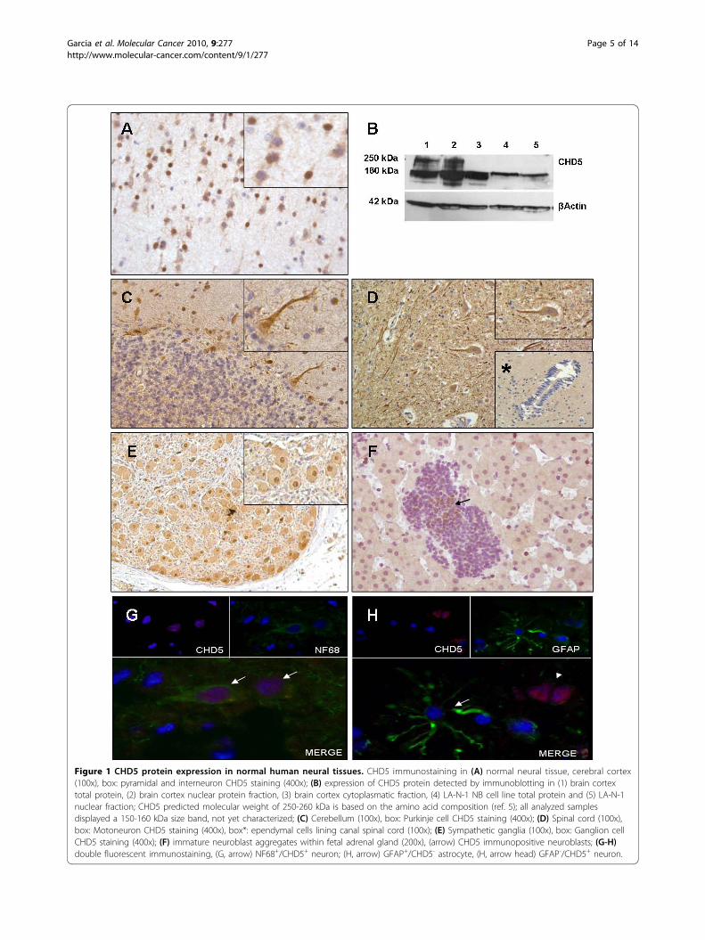
Figure 1 CHD5 protein expression in normal human neural tissues. CHD5 immunostaining in (A) normal neural tissue, cerebral cortex(100x), box: pyramidal and interneuron CHD5 staining (400x); (B) expression of CHD5 protein detected by immunoblotting in (1) brain cortextotal protein, (2) brain cortex nuclear protein fraction, (3) brain cortex cytoplasmatic fraction, (4) LA-N-1 NB cell line total protein and (5) LA-N-1nuclear fraction; CHD5 predicted molecular weight of 250-260 kDa is based on the amino acid composition (ref. 5); all analyzed samplesdisplayed a 150-160 kDa size band, not yet characterized; (C) Cerebellum (100x), box: Purkinje cell CHD5 staining (400x); (D) Spinal cord (100x),box: Motoneuron CHD5 staining (400x), box*: ependymal cells lining canal spinal cord (100x); (E) Sympathetic ganglia (100x), box: Ganglion cellCHD5 staining (400x); (F) immature neuroblast aggregates within fetal adrenal gland (200x), (arrow) CHD5 immunopositive neuroblasts; (G-H)double fluorescent immunostaining, (G, arrow) NF68+/CHD5+ neuron; (H, arrow) GFAP+/CHD5- astrocyte, (H, arrow head) GFAP-/CHD5+ neuron.
Garcia et al. Molecular Cancer 2010, 9:277http://www.molecular-cancer.com/content/9/1/277
Page 5 of 14
observed in >75% of neuroblastic cells (13/32) (Figure2B; Additional file 1), and lowest in stage 3 MYCNamplified NB composed mainly of undifferentiated neu-roblasts with undetectable immunoreactivity, similar tostage 4 NB cases (Figure 2C, Table 1, Additional file 1).GNB (14/14) and GN (13/13) tumors exhibited gang-
lion-like cells with intense nuclear and diffuse cytoplasmstaining. Absence of nuclear staining and feeble cyto-plasmic reactivity was observed in Schwann-like cells(Figure 2D; Additional file 1). The undifferentiated neu-roblastic component of GNB lacked CHD5 staining(Table 1, Additional file 1).The described immunohistochemical assays were per-
formed using two different batches of the anti-CHD5antibody (T00251-A1 and T00251-A02). Both batchesperformed consistently across many repeats, further sup-porting the validity of our results (Additional file 2A).The specificity of the anti-CHD5 antibody was furthervalidated on mouse xenografts of human NB cell lines(SK-N-JD, SK-N-LP and SK-N-AS). All the xenograftswere found to be negative for CHD5 staining (Addi-tional file 2B).Altogether, CHD5 protein was expressed in the
nucleus of neuroblastic cells of clinical low risk NTs. Instage 4s NB, CHD5 negative neuroblast bone marrowmetastasis imply the existence of intratumoral cloneswith CHD5 differential expression in an otherwise histo-logically homogeneous tumor subtype.
CHD5 transcript levels are associated with proteinexpressionCHD5 protein expression was contrasted with genetranscript levels. Quantification of CHD5 mRNA innon-tumoral frozen tissue samples using qRT-PCR iden-tified high expression in fetal brain and adult cerebralcortex, as reported previously [10]. Normal bone mar-row specimens lacked CHD5 expression.
CHD5 mRNA levels were analyzed for 84 primaryNTs obtained at diagnosis (23 stage 4; 7 stage 4s; 34loco-regional NB; 9 GNB and 11 GN); 55 of thesetumors were also analyzed by immunohistochemistry.High risk undifferentiated NB tumors, stage 4 and
stage 3 MYCN-amplified NB displayed significantlylower mRNA expression levels than stage 1, 2, 3 (P <0.001) and stage 4s NB (P = 0.001) (Additional file 3).The highest mean expression values, similar to normalfetal brain, were found for stage 4s NB. GN specimensdisplayed consistently low CHD5 transcript levels,whereas, GNB tumors were characterized by highly vari-able expression attributable to the presence of CHD5negative component, Schwann-like stroma and undiffer-entiated neuroblasts, besides the positive ganglion-likecells that compose these tumors.Correlation between CHD5 immunoreactivity and
mRNA expression levels within the same samples wasexamined in a set of 34 consecutive NB tumors. Immu-nohistochemical and qRT-PCR analyses were carriedout on the same portion of the tumor specimen, withsimilar cell composition and a high tumor cell content(>70% as recommended for PCR studies). CHD5 nuclearimmunoreactivity was assigned a staining score (Addi-tional file 1) and gene expression values were z-scoretransformed. A significant correlation was observedbetween mRNA and protein levels (Spearman’s rho =0.774; P < 0.001), low CHD5 protein scores were consis-tently associated with low mRNA levels (negative z-score values), and high IHC scores with high mRNAexpression (positive z-score values) (Additional file 4).Interestingly, very intense nuclear staining displayed bylow risk tumors, mostly stage 4s and infant stage 1 NB,was not associated with the highest transcript levels(Additional file 4, cases # 1-6, 30, 31 and 33).These results reveal a correspondence between CHD5
protein and mRNA expression, suggesting a potentialregulation of CHD5 expression at the transcriptionallevel.
CHD5 protein expression is associated with patientoutcome in NBCHD5 nuclear immunoreactivity was assigned a stainingscore (Additional file 1) and compared to clinical andbiological variables currently used for NB risk classifica-tion. High CHD5 staining values were found to be sig-nificantly associated with INSS stages 1, 2, 3 (MYCNnon-amplified) and 4s NB (n = 63), age at diagnosis <12m (n = 63) and favorable tumor histology (n = 63); P <0.001 for all the tested variables.To assess whether CHD5 expression was associated
with patient outcome, immunoreactivity scores werecompared to overall survival (OS) and event-free survi-val (EFS) for all 63 NB tumors. The median score value
Table 1 CHD5 inmunostaining in Neuroblastic tumors
Percentage of CHD5 immunopositive neuroblasticcells
n <25% 25-75% >75%
St 1,2,3 31 10/31 (32.2%) 9/31 (29%) 12/31 (38.7%)
St 4 24 19/24 (79.1%) 4/24 (16.6%) 1/24 (4.1%)
St 4s 8 – – 8/8 (100%)
GNB 14 14/14* (100%) – 14/14** (100%)
GN 13 – – 13/13 (100%)
Total 90 29 13 48
Percentage of CHD5 positive (nuclear staining) tumor cells within eachneuroblastic tumor group evaluated using predetermined cutoff values (<25%;25-75%; >75%). Detailed data regarding percentage of positive tumor cellsand staining intensity are reported in Additional file 1. For GNB tumors, theundifferentiated neuroblastic (*) and the ganglionar (**) cell populations werescored separately. GNB = ganglioneuroblastoma, GN = Ganglioneuroma.
Garcia et al. Molecular Cancer 2010, 9:277http://www.molecular-cancer.com/content/9/1/277
Page 6 of 14
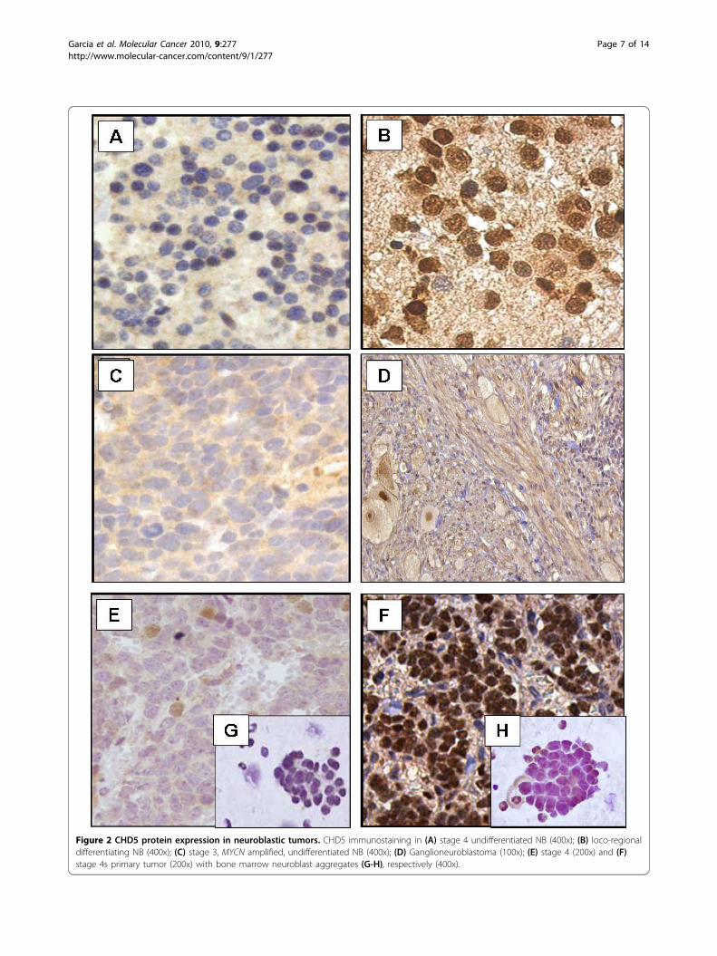
Figure 2 CHD5 protein expression in neuroblastic tumors. CHD5 immunostaining in (A) stage 4 undifferentiated NB (400x); (B) loco-regionaldifferentiating NB (400x); (C) stage 3, MYCN amplified, undifferentiated NB (400x); (D) Ganglioneuroblastoma (100x); (E) stage 4 (200x) and (F)stage 4s primary tumor (200x) with bone marrow neuroblast aggregates (G-H), respectively (400x).
Garcia et al. Molecular Cancer 2010, 9:277http://www.molecular-cancer.com/content/9/1/277
Page 7 of 14
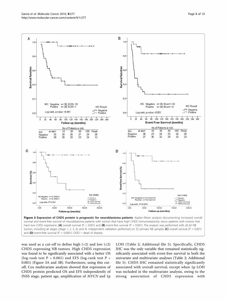
was used as a cut-off to define high (>2) and low (≤2)CHD5 expressing NB tumors. High CHD5 expressionwas found to be significantly associated with a better OS(log-rank test P < 0.001) and EFS (log-rank test P <0.001) (Figure 3A and 3B). Furthermore, using this cut-off, Cox multivariate analysis showed that expression ofCHD5 protein predicted OS and EFS independently ofINSS stage, patient age, amplification of MYCN and 1p
LOH (Table 2; Additional file 5). Specifically, CHD5IHC was the only variable that remained statistically sig-nificantly associated with event-free survival in both theunivariate and multivariate analyses (Table 2; Additionalfile 5). CHD5 IHC remained statistically significantlyassociated with overall survival, except when 1p LOHwas included in the multivariate analysis, owing to thestrong association of CHD5 expression with
Figure 3 Expression of CHD5 protein is prognostic for neuroblastoma patients. Kaplan-Meier analysis documenting increased overallsurvival and event-free survival of neuroblastoma patients with tumors that have high CHD5 immunoreactivity versus patients with tumors thathave low CHD5 expression, (A) overall survival (P < 0.001) and (B) event-free survival (P < 0.001). The analysis was performed with all 63 NBtumors, including all stages (stage 1, 2, 3, 4s and 4). Independent validation performed on 32 primary NB samples (C) overall survival (P = 0.001)and (D) event-free survival (P < 0.0001). DOD = dead of disease.
Garcia et al. Molecular Cancer 2010, 9:277http://www.molecular-cancer.com/content/9/1/277
Page 8 of 14
chromosome 1p status (Table 2; Additional file 5). ThePredictive Value of CHD5 staining was evaluated andcompared to MYCN and 1p LOH (Table 3). CHD5expression showed the highest Negative PredictiveValue (NPV) for overall survival status (96.4%) andevent free survival (85.7%). MYCN status and 1p LOHshowed a NPV of 81.6% and 78.9%, respectively, forthe overall survival status, and of 65.3% and 63.2%,respectively, for the event free survival. The PositivePredictive Value (PPV) of CHD5 expression (overallsurvival status: 54.3%; event free survival: 71.4%), i.e.
proportion of events or deaths in patients with lowCHD5 expressing tumors, was intermediate betweenMYCN (66.7% and 77.8%, respectively) and 1p LOH(46.7% and 60%, respectively) values. CHD5 IHCshowed high sensitivity and accuracy rate for the pre-diction of OS (95% and 73%, respectively) and EFS(86.20% and 77.80%) (Table 4).The prognostic value of CHD5 expression was validatedon an independent, blinded set of 32 FFPE primary NBtumors of patients diagnosed and treated at the Chil-dren’s Hospital of Boston (n = 21) and HSJD of
Table 2 Cox regression analysis
Overall Survival
Variable HR and 95%CI p-value Variable HR and 95%CI p-value Interaction p-value&
CHD5 IHC 21.28 (2.84-159.39) 0.003
CHD5 IHC 18.67 (2.27-153.88) 0.007 0.951
INSS 4.15 (1.63-10.58) 0.003 INSS 1.24 (0.46-3.29) 0.673
CHD5 IHC 15.17 (1.89-121.73) 0.011 0.931
Age (>12 m) 5.56 (1.61-19.19) 0.007 Age (>12 m) 2.14 (0.59-7.75) 0.249
CHD5 IHC 12.29 (1.56-96.49) 0.017 0.952
MYCN (*) 14.27 (4.28-47.58) < 0.001 MYCN(*) 8.08 (2.43-26.93) 0.001
CHD5 IHC 14.33 (1.84-111.48) 0.011 0.946
LOH(**) 2.88 (1.04-7.96) 0.042 LOH(**) 1.75 (0.63-4.89) 0.287
Event Free Survival
Variable HR and 95%CI p-value Variable HR and 95%CI p-value Interaction p-value&
CHD5 IHC 8.14 (2.82-23.5) <0.001
CHD5 IHC 7.01 (2.09 to 23.51) 0.002 0.939
INSS 3.7 (1.73-7.88) 0.001 INSS 1.26 (0.53 to 2.97) 0.605
CHD5 IHC 7.04 (2.24 to 22.09) 0.001 0.638
Age (>12 m) 3.13 (1.33-7.39) 0.009 Age (>12 m) 1.36 (0.54 to 3.44) 0.515
CHD5 IHC 5.97 (1.98 to 17.98) 0.001 0.933
MYCN (*) 4.58 (1.84-11.42) 0.001 MYCN 2.72 (1.07 to 6.88) 0.035
CHD5 IHC 6.02 (1.98 to 18.34) 0.002 0.492
LOH(**) 2.12 (0.92-4.91) 0.079 LOH 1.24 (0.52 to 2.95) 0.622
Cox regression analysis of the NB cohort using CHD5 protein expression and clinical and biologically relevant variables (INSS stage, patient age at diagnosis,MYCN amplification status and chromosome 1p LOH). Expression of CHD5 was statistically significantly associated with overall survival and event-free survival inboth the univariate and multivariate analyses. All NB patients (n = 63) where included in the study except for the studies with MYCN amplification (n = 58) and1p LOH (n = 53), due to the undetermined status in some patients. IHC = Immunohistochemical analysis; INSS = International Neuroblastoma Staging System; HR= hazard ratio; CI = confidence interval. P-values are two sided. (&) Interaction P-values obtained from Cox regression model: IHC + Co-factor+IHC*Co-factor. (*)data available for 58 patients; (**) data available for 53 patients.
Table 3 Analysis of the Predictive Value was performed for a descriptive comparison between CHD5 expression andMYCN and 1p LOH
Overall Survival Event Free Survival
Alive Dead PPV NPV No event Event PPV NPV
CHD5 IHC High 27 (62.8%) 1 (5%) 54.30% 96.40% 24 (70.6%) 4 (13.8%) 71.40% 85.70%
Low 16 (37.2%) 19 (95%) 10 (29.4%) 25 (86.2%)
MYCN Non amplified 40 (93%) 9 (60%) 66.70% 81.60% 32 (94.1%) 17 (70.8%) 77.80% 65.30%
Amplified 3 (7%) 6 (40%) 2 (5.9%) 7 (29.2%)
1p LOH No 30 (78.9%) 8 (53.3%) 46.70% 78.90% 24 (80%) 14 (60.9%) 60% 63.20%
LOH 8 (21.1%) 7 (46.7%) 6 (20%) 9 (39.1%)
Garcia et al. Molecular Cancer 2010, 9:277http://www.molecular-cancer.com/content/9/1/277
Page 9 of 14
Barcelona (n = 11). Kaplan-Meier analysis and a log-rank test showed a statistically significant difference inOS (log-rank test P = 0.001) and EFS (log-rank test P <0.0001) between patients with high and low CHD5expression scores (Figure 3C and 3D). Tumors withhigh IHC scores were associated with longer survival(mean 73 months) in comparison with low expressingtumors (mean 46 months).These results suggest that CHD5 protein expression is
a potential prognostic marker of outcome in NBpatients.
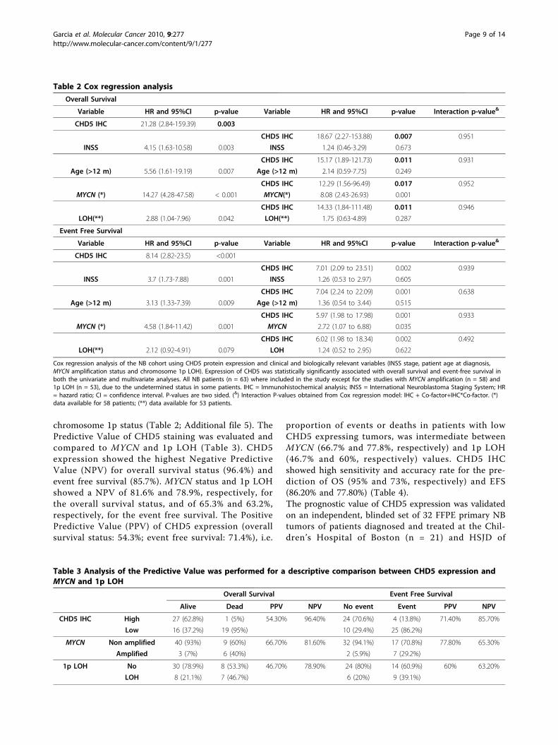
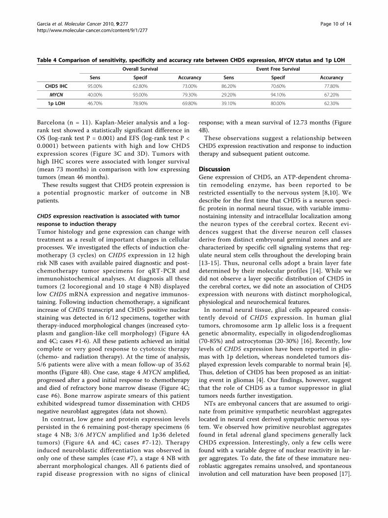
CHD5 expression reactivation is associated with tumorresponse to induction therapyTumor histology and gene expression can change withtreatment as a result of important changes in cellularprocesses. We investigated the effects of induction che-motherapy (3 cycles) on CHD5 expression in 12 highrisk NB cases with available paired diagnostic and post-chemotherapy tumor specimens for qRT-PCR andimmunohistochemical analyses. At diagnosis all thesetumors (2 locoregional and 10 stage 4 NB) displayedlow CHD5 mRNA expression and negative immunos-taining. Following induction chemotherapy, a significantincrease of CHD5 transcript and CHD5 positive nuclearstaining was detected in 6/12 specimens, together withtherapy-induced morphological changes (increased cyto-plasm and ganglion-like cell morphology) (Figure 4Aand 4C; cases #1-6). All these patients achieved an initialcomplete or very good response to cytotoxic therapy(chemo- and radiation therapy). At the time of analysis,5/6 patients were alive with a mean follow-up of 35.62months (Figure 4B). One case, stage 4 MYCN amplified,progressed after a good initial response to chemotherapyand died of refractory bone marrow disease (Figure 4C;case #6). Bone marrow aspirate smears of this patientexhibited widespread tumor dissemination with CHD5negative neuroblast aggregates (data not shown).In contrast, low gene and protein expression levels
persisted in the 6 remaining post-therapy specimens (6stage 4 NB; 3/6 MYCN amplified and 1p36 deletedtumors) (Figure 4A and 4C; cases #7-12). Therapyinduced neuroblastic differentiation was observed inonly one of these samples (case #7), a stage 4 NB withaberrant morphological changes. All 6 patients died ofrapid disease progression with no signs of clinical
response; with a mean survival of 12.73 months (Figure4B).These observations suggest a relationship between
CHD5 expression reactivation and response to inductiontherapy and subsequent patient outcome.
DiscussionGene expression of CHD5, an ATP-dependent chroma-tin remodeling enzyme, has been reported to berestricted essentially to the nervous system [8,10]. Wedescribe for the first time that CHD5 is a neuron speci-fic protein in normal neural tissue, with variable immu-nostaining intensity and intracellular localization amongthe neuron types of the cerebral cortex. Recent evi-dences suggest that the diverse neuron cell classesderive from distinct embryonal germinal zones and arecharacterized by specific cell signaling systems that reg-ulate neural stem cells throughout the developing brain[13-15]. Thus, neuronal cells adopt a brain layer fatedetermined by their molecular profiles [14]. While wedid not observe a layer specific distribution of CHD5 inthe cerebral cortex, we did note an association of CHD5expression with neurons with distinct morphological,physiological and neurochemical features.In normal neural tissue, glial cells appeared consis-
tently devoid of CHD5 expression. In human glialtumors, chromosome arm 1p allelic loss is a frequentgenetic abnormality, especially in oligodendrogliomas(70-85%) and astrocytomas (20-30%) [16]. Recently, lowlevels of CHD5 expression have been reported in glio-mas with 1p deletion, whereas nondeleted tumors dis-played expression levels comparable to normal brain [4].Thus, deletion of CHD5 has been proposed as an initiat-ing event in gliomas [4]. Our findings, however, suggestthat the role of CHD5 as a tumor suppressor in glialtumors needs further investigation.NTs are embryonal cancers that are assumed to origi-
nate from primitive sympathetic neuroblast aggregateslocated in neural crest derived sympathetic nervous sys-tem. We observed how primitive neuroblast aggregatesfound in fetal adrenal gland specimens generally lackCHD5 expression. Interestingly, only a few cells werefound with a variable degree of nuclear reactivity in lar-ger aggregates. To date, the fate of these immature neu-roblastic aggregates remains unsolved, and spontaneousinvolution and cell maturation have been proposed [17].
Table 4 Comparison of sensitivity, specificity and accuracy rate between CHD5 expression, MYCN status and 1p LOH
Overall Survival Event Free Survival
Sens Specif Accurancy Sens Specif Accurancy
CHD5 IHC 95.00% 62.80% 73.00% 86.20% 70.60% 77.80%
MYCN 40.00% 93.00% 79.30% 29.20% 94.10% 67.20%
1p LOH 46.70% 78.90% 69.80% 39.10% 80.00% 62.30%
Garcia et al. Molecular Cancer 2010, 9:277http://www.molecular-cancer.com/content/9/1/277
Page 10 of 14
The immunoreactivity observed in a small proportion ofneuroblasts within these islets could suggest the estab-lishment of CHD5 expression prior to their disappear-ance; however, no evident differentiating features wereobserved in these immunopositive cells that suggestedthe activation of the maturation process.In NTs, CHD5 is essentially expressed in the nucleus
of differentiating neuroblastic cells and ganglion cells,and absent in the Schwannian stromal component.However, the most intense immunoreactivity wasobserved in stage 4s NB, a rare subgroup of histologi-cally undifferentiated, highly proliferative, metastatictumors with a high incidence of spontaneous regression,affecting young infants. Accurate distinction of
spontaneously regressing infant NB from high risk infantstage 4 can be difficult, but critical for therapeutic deci-sions. In our hands, the intensely positive CHD5 nuclearstaining enabled a clear distinction of stage 4s NB fromstage 4 NB, which was consistently immunonegative.These results are consistent with our previous geneexpression profiling study, where similar differentialCHD5 expression profiles were observed amongstinfants with disseminated NB subgroups [18]. Thus,CHD5 immunohistochemical staining may be clinicallyuseful for a more accurate characterization of dissemi-nated infant NB.In NB, CHD5 nuclear staining was strongly asso-
ciated with established favorable prognostic variables
Figure 4 Chemotherapy-induced expression of CHD5 in 12 high risk NB tumors. (A) CHD5 immunoreactivity pre- and post-chemotherapy(400x); (B) Kaplan-Meier analysis for OS was performed using the mean between pre-treatment and post-treatment gene expression levels ascut-off to divide tumors which reactivate CHD5 and tumors that maintain low expression levels; (C) CHD5 transcript levels quantified by qRT-PCR:CHD5 expression levels (Black) at diagnosis, (Grey) post-chemotherapy. Fetal brain CHD5 expression represents normal neural tissue values.
Garcia et al. Molecular Cancer 2010, 9:277http://www.molecular-cancer.com/content/9/1/277
Page 11 of 14
like low clinical stage, age at diagnosis <12 months andfavorable histology. Our findings suggest that CHD5protein expression may accurately define NB riskgroups and may, therefore, be a prognostic marker.Evidence is provided by the statistically significantassociation found between high CHD5 immunoreactiv-ity and favorable OS and EFS. These results are consis-tent with recent studies reporting a strong associationof CHD5 mRNA levels with patient outcome in NB[5,10]. Furthermore, Cox multivariate analyses suggestthat the prognostic value of CHD5 protein expressionis independent of other clinical and biological variablescurrently used in risk stratification of NB patients andcould therefore represent an immunohistochemicalmarker of prognosis in NB.Currently, risk stratification of NB patients is per-
formed by combining different markers with strongprognostic impact, including patients’ age at diagnosis,tumor stage, genomic amplification of the oncogeneMYCN, copy number alterations of chromosomalregions 1p, 11q and 17q, tumor DNA content [1,19] andShimada histological score [20]. However, despite elabo-rate risk stratification strategies, outcome prediction inneuroblastoma is still deficient. In recent years, toimprove risk assessment additional prognostic indicatorssuch as gene-expression signatures [21-23], combinedgenomic and molecular signatures [24] or expressionlevels of single candidate genes, e.g., Trk (NTRK) familyof neurotrophin receptors [25,26], FYN [27], PRAME[28] and ZNF423 [29], have been associated with NBclinical behavior. Expression of the Trk family receptorshas been the most extensively characterized marker inNB and has been found to be consistently correlatedwith the biology and clinical behavior of NB. Based onour results, there is an apparent similarity between theexpression patterns of CHD5 and TRKA in NB and theirpatterns of association with NB disease outcome. TRKAexpression has been reported to be high in biologicallyfavorable NB tumors and inversely associated withMYCN amplification [30]. The prognostic value of theimmunohistochemical detection of TrkA has also beenexamined and reported to be high, especially in combi-nation with Ha-Ras expression pattern [31,32]. FurtherIHC studies have correlated the lack of TrkA expressionwith metastatic malignant NB [33]. However, in the lat-ter study, 34% of the patients with stage 4 NB displayedTrkA expression, a subset of which died of aggressivemetastatic disease despite TrkA expression [33,34]. Inour study, the majority of stage 4 NB either lackedCHD5 immunoreactivity (83%) or exhibited weaknuclear staining (13%), a high risk phenotype accordingto our scoring system. Only one stage 4 tumor wasfound to be clearly immunoreactive for CHD5; at thetime of analysis the patient is alive, 29 months from
diagnosis. These observations further confirm CHD5 asa powerful prognostic marker that could complementother known markers such as age at diagnosis, stage,MYCN status, cellular DNA content, 1p deletion andtumor histology. However, the potential clinical use ofthis marker must be tested in larger, prospectivecohorts.It is known that tumor histology and gene expression can
change with treatment as a result of important changes incellular processes, e.g., induced tumor differentiation, DNArepair, apoptosis and tissue necrosis. Undifferentiated NBoccasionally exhibit neuroblastic maturation in response tochemotherapy. Assessment of CHD5 gene and proteinexpression in NB post-therapy specimens revealed thattumors with evident neuroblastic maturation showed bothCHD5 gene and protein reactivation. Notably, none ofthese tumors harbored 1p deletion. Conversely, in tumorswhere minimal or no morphological changes wereobserved in the post-treatment specimens, low CHD5expression persisted. These observations suggest the exis-tence of a subset of tumors within high risk NB whereCHD5 expression can be reactivated from the silencedstate by standard chemotherapy. Remarkably, when post-therapy reactivation was observed, CHD5 expression waslargely associated with disease response to cytotoxic induc-tion therapy and subsequently with longer patient OS. All12 patients included in the study received the same treat-ment, nevertheless some tumors failed to respond. At pre-sent, treatment response in NB is routinely evaluated bymonitoring urine levels of catecholamine and its metabo-lites (VMA/HVA ratio) and by estimating the decrease inthe size of measurable lesions with conventional imagingmodalities, such as computed tomography (CT) or mag-netic resonance imaging (MRI). At the time of second-looksurgery, the degree of induced tumor cell differentiationand the extent of necrosis can also be useful to estimatetreatment response. However, no biological markers fortumor chemotherapy responsiveness have been reported inNB. The use of such biomarkers would make chemother-apy more effective for individual patients by allowingtimely changes of therapy in the case of nonrespondingtumors. Furthermore, markers reflecting tumor responsecan function as surrogates of long-term outcome. Takinginto account the small cohort of cases that may have led toan overestimation of the data, our findings would suggestthat restoration of CHD5 expression could be a surrogatemarker of treatment response that can be clinically usefulto identify patients that do not benefit from conventionaltreatment. These results warrant further investigation in alarger cohort of uniformly treated patients.In summary, we report that the differential expression
of the neuron-specific protein CHD5 accurately definesNB risk groups and may represent a marker of outcomein neuroblastoma that can be tested by conventional
Garcia et al. Molecular Cancer 2010, 9:277http://www.molecular-cancer.com/content/9/1/277
Page 12 of 14
immunohistochemistry. In high risk NB patients, re-establishment of CHD5 expression following chemother-apy should be tested prospectively as a surrogate markerof treatment response.
Additional material
Additonal file 1: Clinical and biological characteristics of 90 NTspecimens included in the study. INSS = International NeuroblastomaStaging System; Diagnosis: NB = neuroblastoma, GNB =ganglioneuroblastoma, GN = ganglioneuroma; Disease status: A = alive,D = dead; EFS = Event free survival; qRT-PCR = Quantitative real-timePCR; IHC = immunohistochemistry; n.a = not available dataImmunohistochemical analysis of CHD5 expression in NTs Resultsare displayed as percentage of CHD5 immunopositive cells present ineach tumor specimen. Staining intensity: 0 = negative; 1 = weak staining;2 = strong staining, 3 = very intense staining. Proportion of positive cellsvalues (<25% = 1; 25-75 = 2; >75% = 3). Intensity and positive cell valueswere multiplied together to provide a single score for each case.
Additonal file 2: A. Immunohistochemical staining of FFPE sectionsof two immunopositive neuroblastic tumors using two differentbatches of the anti-CHD5 antibody (T00251-A1 and T00251-A02); B.Immunochemical assay with the anti-CHD5 antibody (StrategicDiagnostics, DE) on mouse xenografts derived from human NB celllines. The specificity of the anti-CHD5 antibody was validated byimmunohistochemical assays on FFPE sections of mouse xenografts ofhuman NB cell lines (SK-N-JD, SK-N-LP and SK-N-AS). In these NB celllines CHD5 gene expression is very low or absent (data not shown),similar to previously reported data (ref. 5, ref. 10). Two different anti-CHD5 antibody batches (T00251-A1 and T00251-A02, StrategicDiagnostics, DE) were tested. Ganglioneuroblastoma FFPE tissue sectionswere used as positive control samples. All the analyzed xenographs werecomposed nearly exclusively (>95%) of neuroblastic cells exhibiting noCHD5 nuclear staining and faint cytoplasmic staining (when present).Only few (<5%) immunopositve cells were observed in the SK-N-LPxenograft. However, viable tumor cells in the SK-N-LP xenograft wherenegative for CHD5 nuclear staining, similar to SK-N-JD and SK-N-AS.These results were comparable to the immunostaining pattern observedin undifferentiated high risk NB tumors. The GNB ganglionar cellsshowed intense nuclear and diffused cytoplasm immunostaining.
Additional file 3: CHD5 mRNA expression levels in NTs Results aredisplayed as mean expression levels of NT subgroups obtained from twoindependent analyses. HR = high risk NB (Stage 4 and Stage 3 MYCNamplified); LR = low risk NB (stage 1, stage 2 and stage 3 MYCN non-amplified); GNB= ganglioneuroblastoma; GN = ganglioneuroma.Quantification was performed relative to normal fetal brain. Error barsillustrate the variability amongst the samples of each NT subgroup.
Additional file 4: Comparison of CHD5 mRNA and proteinexpression. The lineal graph shows comparison between CHD5 mRNAlevels and protein immunoreactivity in 34 NB cases. Low CHD5 proteinscores were associated with lower mRNA levels (negative z-score values),and high IHC scores with high mRNA expression (positive z-score values),(Spearman’s correlation analysis rho = 0.774; P < 0.001). Low risk tumors,stage 4s and infant stage 1 NB tumors showed very intense nuclearstaining in comparison to the observed transcript levels (cases # 1-6, 30,31 and 33).
Additional file 5: Cox multivariate análisis. Cox multivariate regressionanalysis has been performed using clinical and biological variablescurrently used in risk stratification of NB patients (INSS stage, age atdiagnosis, MYCN status and 1p LOH) in combination with the CHD5 IHC.The analysis has been performed sequentially, adding one variable ateach step, in order to assess how the presence of each variableinfluences the performance of CHD5. CHD5 IHC remained statisticallysignificantly associated with overall survival in all the analyses, exceptwhen the 1p LOH parameter is included in the overall survival analysis.This is due to the strong association of the expression of CHD5, locatedon 1p36, with chromosome 1p status. All the rest of variables, except for
MYCN amplification, were not statistically significant. For event freesurvival analysis, CHD5 IHC is the only variable that remained statisticallysignificant along the whole analysis, even in the presence of 1p LOH. IHC= Immunohistochemical analysis; INSS = International NeuroblastomaStaging System; HR = hazard ratio; CI = confidence interval. P-values aretwo sided.
AcknowledgementsAuthors thank Dr. B. Spengler (Fordham University, New York) and Dr. N.K.V.Cheung (MSKCC, New York) for annotated NB cell lines; the Neural TissueBank (Hospital Clínic, Barcelona) for normal brain samples and theDepartment of Audiovisual Systems (HSJD, Barcelona) for technicalassistance. This study was supported by grants from the Spanish Ministry ofHealth (PI070286), Spanish Society against Cancer (Asociación EspañolaContra el Cáncer, 2007), the Catalan government (2005SGR00605;2006FI00404), and the generous donations from Margarita del Pozo andAlicia Pueyo Foundations.
Author details1Developmental Tumor Biology Laboratory, Hospital Sant Joan de Déu,Fundación Sant Joan de Déu, Barcelona, Spain. 2Department of Pathology,Hospital Sant Joan de Déu, Barcelona, Spain. 3Department of Neurology,University of North Carolina, Chapel Hill, USA. 4Laboratory of Biostatistics &Epidemiology, Universitat Autònoma de Barcelona; Clinical PharmacologyService, IDIBAPS, Hospital Clinic, Barcelona, Spain. 5Department of Pediatrics,Memorial Sloan-Kettering Cancer Centre, New York, USA. 6Division ofPediatric Hematology/Oncology, Dana-Farber Cancer Institute and Children’sHospital of Boston, USA. 7Department of Pathology, Children’s Hospital ofBoston, USA.
Authors’ contributionsCL, IG and JM are responsible for the initial conception and overallhypothesis of this study. IG, GM and CL are responsible for the design ofthis manuscript, including the original draft and subsequent revisions. IG,GM, ER, MS, TG, JR, NKC, CdT, JM MK, RG, AAP and CL were involved withthe interpretation of data, draft and revision of this manuscript. CdTprovided guidance for many of the experiments. NKC, TG, MK, RG and AAPare responsible for the procurement and cryopreservation of NBT tissuespecimens derived from MSKCC and CHB/DFCI. ER, IG, GM, JM and CL wereresponsible for the procurement and cryopreservation of NBT tissuespecimens derived from the Spanish institutions. ER, IG GM, CL and MS areresponsible of inmunohistochemical analyses. MS evaluated tumourspecimens for staging classification, tumour content. JM and CL areresponsible for patient clinico-biological database management. CC, GM, PGand ER are responsible for the in vivo study. IG, GM and CL are responsiblefor the quantitative PCR experiments and inmunoblotting. JR and CL areresponsible of statistical analyses. All authors were involved in the draftingand revisions for this manuscript. All authors read and approved the finalmanuscript.
Competing interestsThe authors declare that they have no competing interests.
Received: 25 January 2010 Accepted: 15 October 2010Published: 15 October 2010
References1. Maris JM, Hogarty MD, Bagatell R, Cohn SL: Neuroblastoma. Lancet 2007,
369:2106-2120.2. Weith A, Brodeur GM, Bruns GA, Matise TC, Mischke D, Nizetic D, Seldin MF,
van Roy N, Vance J: Report of the second international workshop onhuman chromosome 1 mapping 1995. Cytogenet Cell Genet 1996,72:114-144.
3. Fong CT, Dracopoli NC, White PS, Merrill PT, Griffith RC, Housman DE,Brodeur GM: Loss of heterozygosity for the short arm of chromosome 1in human neuroblastoma: correlation with N-myc amplification. Proc NatlAcad Sci USA 1989, 86:3753-3757.
Garcia et al. Molecular Cancer 2010, 9:277http://www.molecular-cancer.com/content/9/1/277
Page 13 of 14
4. Bagchi A, Papazoglu C, Wu Y, Capurso D, Brodt M, Francis D, Bredel M,Vogel H, Mills AA: CHD5 is a tumor suppressor at human 1p36. Cell 2007,128:459-475.
5. Fujita T, Igarashi J, Okawa ER, Gotoh T, Manne J, Kolla V, Kim J, Zhao H,Pawel BR, London WB, Maris JM, White PS, Brodeur GM: CHD5, a tumorsuppressor gene deleted from 1p36.31 in neuroblastomas. J Natl CancerInst 2008, 100:940-949.
6. Okawa ER, Gotoh T, Manne J, Igarashi J, Fujita T, Silverman KA, Xhao H,Mosse YP, White PS, Brodeur GM: Expression and sequence analysis ofcandidates for the 1p36.31 tumor suppressor gene deleted inneuroblastomas. Oncogene 2008, 27:803-810.
7. Mulero-Navarro S, Esteller M: Chromatin remodeling factor CHD5 issilenced by promoter CpG island hypermethylation in human cancer.Epigenetics 2008, 3:210-215.
8. Marfella CG, Imbalzano AN: The Chd family of chromatin remodelers.Mutat Res 2007, 618:30-40.
9. Schuster EF, Stöger R: CHD5 defines a new subfamily of chromodomain-SWI2/SNF2-like helicases. Mammalian Genome 2002, 13:117-119.
10. Thompson PM, Gotoh T, Kok M, White PS, Brodeur GM: CHD5, a newmember of the chromodomain gene family, is preferentially expressedin the nervous system. Oncogene 2003, 22:1002-1011.
11. Brodeur GM, Pritchard J, Berthold F, Carlsen NL, Castel V, Castelberry RP, DeBernardi B, Evans AE, Favrot M, Hedborg F: Revision of the Internationalcriteria for neuroblastoma diagnosis, staging and response to treatment.J Clin Oncol 1993, 11:1466-1477.
12. Lavarino C, Garcia I, Mackintosh C, Cheung NKV, Domenech G, Ríos J,Perez N, Rodríguez E, De Torres C, Gerald WL, Tuset E, Acosta S, Beleta H,de Alava E, Mora J: Differential expression of genes mapping torecurrently abnormal chromosomal regions characterize neuroblastictumours with distinct ploidy status. BMC Med Genomics 2008, 1:36.
13. Anderson SA, Kaznowski CE, Horn C, Rubenstein JL, McConnell SK: Distinctorigins of neocortical projection neurons and interneurons in vivo. CerebCortex 2002, 12:702-709.
14. Hevner RF, Daza RA, Rubenstein JL, Stunnenberg H, Olavarria JF, Englund C:Beyond laminar fate: toward a molecular classification of corticalprojection/pyramidal neurons. Dev Neurosci 2003, 25:139-151.
15. Gilbertson RJ, Ellison DW: The origins of medulloblastoma subtypes. AnnuRev Pathol 2008, 3:341-365.
16. Barbashina V, Salazar P, Holland EC, Rosenblum MK, Ladanyi M: Alleliclosses at 1p36 and 19q13 in gliomas: correlation with histologicclassification, definition of a 150-kb minimal deleted region on 1p36,and evaluation of CAMTA1 as a candidate tumor suppressor gene. ClinCancer Res 2005, 11:1119-1128.
17. de Preter K, Vandesompele J, Heimann P, Yigit N, Beckman S, Schramm A,Eggert A, Stallings RL, Benoit Y, Renard M, De Paepe A, Laureys G,Påhlman S, Speleman F: Human fetal neuroblast and neuroblastomatranscriptome analysis confirms neuroblast origin and highlightsneuroblastoma candidate genes. Genome Biol 2006, 7(9):R84.
18. Lavarino C, Cheung NK, Garcia I, Domenech G, de Torres C, Alaminos M,Rios J, Gerald WL, Kushner B, LaQuaglia M, Mora J: Specific geneexpression profiles and chromosomal abnormalities are associated withinfant disseminated neuroblastoma. BMC Cancer 2009, 3:9-44.
19. Ambros PF, Ambros IM, Brodeur GM, Haber M, Khan J, Nakagawara A,Schleiermacher G, Speleman F, Spitz R, London WB, Cohn SL, Pearson AD,Maris JM: International consensus for neuroblastoma moleculardiagnostics: report from the International Neuroblastoma Risk Group(INRG) Biology Committee. Br J Cancer 2009, 100:1471-1482.
20. Shimada H, Ambros IM, Dehner LP, Hata J, Joshi VV, Roald B, Stram DO,Gerbing RB, Lukens JN, Matthay KK, Castleberry RP: The InternationalNeuroblastoma Pathology Classification (the Shimada system). Cancer1999, 86:364-72.
21. Oberthuer A, Berthold F, Warnat P, Hero B, Kahlert Y, Spitz R, Ernestus K,König R, Haas S, Eils R, Schwab M, Brors B, Westermann F, Fischer M:Customized oligonucleotide microarray gene expression-basedclassification of neuroblastoma patients outperforms current clinical riskstratification. J Clin Oncol 2006, 24(31):5070-5078.
22. De Preter K, Vermeulen J, Brors B, Delattre O, Eggert A, Fischer M, Janoueix-Lerosey I, Lavarino C, Maris JM, Mora J, Nakagawara A, Oberthuer A,Ohira M, Schleiermacher G, Schramm A, Schulte JH, Wang Q,Westermann F, Spleleman F, Vandesompele J: Accurate outcome
prediction in neuroblastoma across independent data sets using amultigene signature. Clin Cancer Res 2010, 16(5):1532-1541.
23. Oberthuer A, Hero B, Berthold F, Juraeva D, Faldum A, Kahlert Y,Asgharzadeh S, Seeger R, Scaruffi P, Tonini GP, Janoueix-Lerosey I,Delattre O, Schleiermacher G, Vandesompele J, Vermeulen J, Speleman F,Noguera R, Piqueras M, Bénard J, Valent A, Avigad S, Yaniv I, Weber A,Christiansen H, Grundy RG, Schardt K, Schwab M, Eils R, Warnat P,Kaderali L, Simon T, Decarolis B, Theissen J, Westermann F, Brors B,Fischer M: Prognostic impact of gene expression-based classification ofneuroblastoma. J Clin Oncol 2010, 28(21):3506-15.
24. Tomioka N, Oba S, Ohira M, Misra A, Fridlyand J, Ishii S, Nakamura Y,Isogai E, Hirata T, Yoshida Y, Todo S, Kaneko Y, Albertson DG, Pinkel D,Feuerstein BG, Nakagawara A: Novel risk stratification of patients withneuroblastoma by genomic signature, which is independent ofmolecular signature. Oncogene 2008, 27:441-449.
25. Nakagawara A, Arima-Nakagawara M, Scavarda NJ, Azar CG, Cantor AB,Brodeur GM: Association between high levels of expression of the TRKgene and favorable outcome in humana neuroblastoma. N Engl J Med1993, 328:847-854.
26. Brodeur GM, Minturn JE, Ho R, Simpson AM, Iyer R, Varela CR, Light JE,Kolla V, Evans AE: Trk receptor and inhibition in neuroblastomas. Clin CanRes 2009, 15(10):3244-3250.
27. Berwanger B, Hartmann O, Bergmann E, Bernard S, Nielsen D, Krause M,Kartal A, Flynn D, Wiedemeyer R, Schwab M, Schäfer H, Christiansen H,Eilers M: Loss of a FYN-regulated differentiation and growth arrestpathway in advanced stage neuroblastomas. Cancer Cell 2002, 2(5):377-86.
28. Oberthuer A, Hero B, Spitz R, Berthold F, Fischer M: The tumor-associatedantigen PRAME is universally expressed in high-stage neuroblastomaand associated with poor outcome. Clin Can Res 2004, 10(13):4307-13.
29. Huang S, Laoukili J, Epping MT, Koster J, Hölzel M, Westerman BA,Nijkamp W, Hata A, Asgharzadeh S, Seeger RC, Versteg R, Beijersbergen RL,Bernards R: ZNF423 is critically required for retinoic acid-induceddifferentiation and is a marker of neuroblastomas outcome. Cancer Cell2009, 15:328-340.
30. Nakagawara A, Arima M, Azar CG, Scavarda NJ, Brodeur GM: Inverserelationship between trk expression and N-myc amplification in humananeuroblastomas. Cancer Res 1992, 52:1364-1368.
31. Tanaka T, Hiyama E, Sugimoto T, Sawada T, Tanabe M, Ida N: trkA geneexpression in neuroblastoma. Can Res 1995, 76:1086-1095.
32. Tanaka T, Sugimoto T, Sawada T: Prognostic discrimination amongneuroblastomas according to Ha-ras/trk A gene expression. Can Res 1998,83:1626-1633.
33. Krammer K. Gerald W, LeSauteur L, Saragovi HU, Cheung N-KV: Prognosticvalue of TrkA protein detection by monoclonal antibody 5C3 inneuroblastoma. Clin Can Res 1996, 2:1361-67.
34. Krammer K. Gerald W, LeSauteur L, Saragovi HU, Cheung N-KV: Monoclonalantibody to human Trk-A: Diagnostic and therapeutic potential inneuroblastoma. Eur J Cancer 33(12):2090-2091.
doi:10.1186/1476-4598-9-277Cite this article as: Garcia et al.: Expression of the neuron-specificprotein CHD5 is an independent marker of outcome in neuroblastoma.Molecular Cancer 2010 9:277.
Submit your next manuscript to BioMed Centraland take full advantage of:
• Convenient online submission
• Thorough peer review
• No space constraints or color figure charges
• Immediate publication on acceptance
• Inclusion in PubMed, CAS, Scopus and Google Scholar
• Research which is freely available for redistribution
Submit your manuscript at www.biomedcentral.com/submit
Garcia et al. Molecular Cancer 2010, 9:277http://www.molecular-cancer.com/content/9/1/277
Page 14 of 14
Related Documents