Characterisation of biofilm associated with bacterial vaginosis Karakterisering van biofilm geassocieerd met bacteriële vaginose

Welcome message from author
This document is posted to help you gain knowledge. Please leave a comment to let me know what you think about it! Share it to your friends and learn new things together.
Transcript
Characterisation of biofilm
associated with
bacterial vaginosis
Karakterisering van biofilm
geassocieerd met
bacteriële vaginose
Voor mama
Colophon
Liselotte Hardy was supported by the European and Developing Countries Clinical Trials
Partnership (Grant number: SP.2011.41304.043) and the Combined Highly Active Anti-Retroviral
Microbicides project under EU FP7 (Grant number: 242135).
Cover design: Jan Moesen
Pencil drawings: Karolien Vanmerhaeghe
ISBN:
© 2016 | Liselotte Hardy | Ghent | Belgium
All rights reserved. No part of this work may be reproduced in any form or by any means,
electronically, mechanically, by print, or otherwise, without prior written permission of the
author.
Characterisation of biofilmassociated with bacterial vaginosis
Liselotte Hardy
Dissertation submitted to fulfill the requirements for
the degree of Doctor in Medical Sciences, 2016
Supervisors:
Prof. Dr. Mario Vaneechoutte, Ghent University
Dr. Tania Crucitti, Institute of Tropical Medicine
Dr. Vicky Jespers, Institute of Tropical Medicine
Faculty of Medicine and Health Sciences, Ghent University
De Pintelaan 185, B-9000 Ghent
Chairman of the examination commission
Prof. Dr. Joris Delanghe, Ghent University
Members of the examination commission
Dr. Sarah Joseph, University College London
Prof. Dr. Stijn Deborggraeve, Institue of Tropical Medicine
Dr. Therese Delvaux, Institute of Tropical Medicine
Prof. Dr. Geert Claeys, Ghent University
Prof. Dr. Tom Van de Wiele, Ghent University
Prof. Dr. Hans Verstraelen, Ghent University

“If you don’t like bacteria, you’re on the wrong planet.”
– Stewart Brand

Dankwoord
Toen ik een dikke tien jaar geleden het dankwoord voor mijn masterthesis schreef, was ik
ervan overtuigd dat dat de eerste en laatste keer zou zijn. Ik wou absoluut niet (nooit,
never, jamais) aan een doctoraat beginnen en heb destijds zelfs tegen Jan gezegd dat hij
mij moest tegenhouden als ik dat voornemen ooit zou vergeten. Tja. . .
Na een kleine omweg via de farmaceutische industrie, kwam ik terecht in mijn gedroomde
werkomgeving: het befaamde Instituut voor Tropische Geneeskunde in Antwerpen, en dan
ook nog eens gecombineerd met verschillende onderzoeksinstituten in Afrika. Ik ben Vicky
nog altijd enorm dankbaar voor de kans die ze me destijds gegeven heeft om, ondanks mijn
geringe ervaring, een internationaal project te coordineren en me onder te dompelen in de
wereld van reproductieve gezondheid. Toen ze me op een tropische avond in Kigali, bij een
geımproviseerde pick-nick aan het zwembad, vroeg wat mijn plannen voor de toekomst
waren, wist ik het eigenlijk onbewust al lang. Jaja, ik wou meer onderzoek doen naar dat
vaginale microbioom en dan ook nog eens in de vorm van een doctoraat (na 5 jaar was ik
mijn goede voornemen dus al lang vergeten). Maar dan toch liever in het labo dan achter
een computerscherm. Op een kort mailtje naar Tania kreeg ik een heel enthousiaste reactie
en ik werd ook daar met open armen ontvangen (zowel in het labo als in haar bureau).
Hetzelfde enthousiasme vond ik ook bij Mario, toen ik, toch wel met enige stress, hem
vroeg om officieel mijn promotor te zijn. Dat gedeeld enthousiasme ben ik blijven voelen
bij mijn promotoren, of het nu over resultaten, congressen, nieuwe ideeen, de lay-out van
mijn PDF’s, fietsen of reizen ging. Bedankt hiervoor, jullie hebben me de beste begeleiding
gegeven, alledrie op een andere, maar aanvullende manier!
Dankzij jullie heb ik ondertussen ook een heus netwerk kunnen opbouwen, bestaande uit
stuk voor stuk interessante wetenschappers op het ITG, binnen Belgie, Europa en Afrika.
Voor mijn doctoraat mocht ik gebruik maken van kostbare stalen vrijwillig gedoneerd door
lieve Rwandese vrouwen. Deze dames kan ik natuurlijk nooit genoeg bedanken: zonder
hun had ik hier namelijk niet veel te vermelden. Ook het Rinda Ubuzima team in Kigali
v
en Janneke in Liverpool, merci!
De weg naar dat doctoraat zou natuurlijk niet hetzelfde geweest zijn zonder mijn collega’s
van het SOA/HIV-labo, de HIV/SOA epidemiologie-eenheid, en de verre collega’s van
het LBR in Gent. Bedankt om me op te nemen in de groep als respectievelijk enige
doctoraatsstudent, enige biomedica, en enige “Antwerpse collega”. Said en Vicky verdienen
hier toch wel een speciale vermelding, voor hun ontelbare uren FISH’en en qPCR’en in
het labo. Zonder jullie zeer gewaardeerde bijdrage had ik een paar jaar extra nodig gehad
om tot dit resultaat te komen. En dan de collega’s die meer waren dan collega’s: Celine,
Irith, Jasna, Odin, Severine en Tine, voor de lunches, koffiepauzes, after-work drankjes, en
de vriendschap.
Die ontspanning werd ook verzorgd door de vrienden buiten het werk: de Limburgers, de
Q-ladies en de Meelstraatmeisjes. Lien, bedankt om op het aller-einde mijn thesis nog
eens na te lezen en en je onversaagd een weg doorheen dat woud van referenties te banen,
op zoek naar misplaatste punten en komma’s. Karolien, bedankt voor de mooie figuren, ze
geven dit boekje absoluut meer cachet. En Jonathan, bedankt voor de vele luistersessies.
Ik weet dat ik veel heb zitten doordrammen, maar gelukkig ging dit meestal wel vergezeld
door een koffie of een cocktail, of twee.
En dan natuurlijk mijn familie, mama, papa, zus, voor jullie steun. Mama, iets meer dan
een jaar te laat opdat jij het nog had kunnen meemaken, maar voor jou was ik toch al
lang doctor/dokter.
Traditioneel de laatste in het rijtje (en dus de belangrijkste?): mijn lief, echtgenoot, meest
kritische taaloloog, eenkoppig presentatie-oefenpubliek, IT’er, en vormgever. Jan, wat zou
ik toch maar zonder jou doen? Bedankt om zelfs geen poging te hebben gedaan om mij
tegen te houden, dat was toch zinloos geweest.
vi

Table of contents
Dankwoord v
List of figures ix
List of tables xi
Abbreviations xiii
1 General introduction 1
2 State of the art 3
2.1 The healthy vaginal ecosystem . . . . . . . . . . . . . . . . . . . . . . . . . 3
2.2 Bacterial vaginosis: a dysbiosis of the vaginal
microbiome . . . . . . . . . . . . . . . . . . . . . . . . . . . . . . . . . . . 11
2.3 Gardnerella vaginalis . . . . . . . . . . . . . . . . . . . . . . . . . . . . . . 28
2.4 Atopobium vaginae . . . . . . . . . . . . . . . . . . . . . . . . . . . . . . . 35
2.5 Microbial biofilm . . . . . . . . . . . . . . . . . . . . . . . . . . . . . . . . 38
2.6 Treatment of biofilm infections . . . . . . . . . . . . . . . . . . . . . . . . . 47
2.7 Biofilm in bacterial vaginosis . . . . . . . . . . . . . . . . . . . . . . . . . . 49
3 Rationale and objectives 51
4 Experimental work 53
4.1 The Ring Plus study . . . . . . . . . . . . . . . . . . . . . . . . . . . . . . 53
4.2 Unravelling the bacterial vaginosis-associated
biofilm: technical preparation . . . . . . . . . . . . . . . . . . . . . . . . . 57
vii
4.3 A fruitful alliance: the synergy between Atopobium vaginae and Gardnerella
vaginalis . . . . . . . . . . . . . . . . . . . . . . . . . . . . . . . . . . . . . 75
4.4 The importance of Gardnerella vaginalis sialidase for bacterial vaginosis-
associated biofilm . . . . . . . . . . . . . . . . . . . . . . . . . . . . . . . . 85
4.5 Association of vaginal dysbiosis and biofilm with contraceptive vaginal ring
biomass in African women . . . . . . . . . . . . . . . . . . . . . . . . . . . 95
5 General discussion 107
5.1 Visualising vaginal biofilm with fluorescence in situ hybridisation . . . . . . 108
5.2 G. vaginalis: mostly harmless? . . . . . . . . . . . . . . . . . . . . . . . . . 110
5.3 Production of sialidase by G. vaginalis . . . . . . . . . . . . . . . . . . . . 112
5.4 With a little help from my friends: A. vaginae . . . . . . . . . . . . . . . . 113
5.5 Treatment of BV . . . . . . . . . . . . . . . . . . . . . . . . . . . . . . . . 115
5.6 The impact of the vaginal biofilm on intravaginal contraceptive rings . . . 116
5.7 Final conclusions: the characterisation of biofilm associated with BV . . . 118
5.8 Directions for future research . . . . . . . . . . . . . . . . . . . . . . . . . 120
Summary 123
Samenvatting 125
Bibliography 127
Appendix: Methods 161
A1.1 Vaginal samples . . . . . . . . . . . . . . . . . . . . . . . . . . . . . . . . . 161
A1.2 Ring samples . . . . . . . . . . . . . . . . . . . . . . . . . . . . . . . . . . 165
Appendix: Curriculum vitae 167
Appendix: Papers as published 173
viii
List of figures
2.1 Legend . . . . . . . . . . . . . . . . . . . . . . . . . . . . . . . . . . . . . . 4
2.2 Lactobacillus-dominated vaginal microbiome . . . . . . . . . . . . . . . . . 7
2.3 Stages in vaginal maturation . . . . . . . . . . . . . . . . . . . . . . . . . . 9
2.4 Bacterial vaginosis-associated vaginal microbiome . . . . . . . . . . . . . . 12
2.5 Nugent score . . . . . . . . . . . . . . . . . . . . . . . . . . . . . . . . . . 16
2.6 FISH technique . . . . . . . . . . . . . . . . . . . . . . . . . . . . . . . . . 23
2.7 Structure of PNA versus DNA . . . . . . . . . . . . . . . . . . . . . . . . . 24
2.8 Taxonomic ranking of G. vaginalis . . . . . . . . . . . . . . . . . . . . . . . 28
2.9 Clue cell after Gram stain . . . . . . . . . . . . . . . . . . . . . . . . . . . 31
2.10 Bacterial use of sialic acid . . . . . . . . . . . . . . . . . . . . . . . . . . . 33
2.11 Taxonomic ranking of A. vaginae . . . . . . . . . . . . . . . . . . . . . . . 35
2.12 The biofilm cycle . . . . . . . . . . . . . . . . . . . . . . . . . . . . . . . . 40
2.13 Mechanisms of bacterial adhesion: fimbriae and capsule . . . . . . . . . . . 41
2.14 Communication between microbes: quorum sensing . . . . . . . . . . . . . 46
2.15 Biofilm in bacterial vaginosis: what we knew before . . . . . . . . . . . . . 49
4.1 Dispersed bacteria versus biofilm . . . . . . . . . . . . . . . . . . . . . . . 64
4.2 Distribution of samples according to fluorescence in situ hybridisation . . . 65
4.3 Gardnerella vaginalis biofilm . . . . . . . . . . . . . . . . . . . . . . . . . . 66
4.4 Superimposed images of polymicrobial biofilm . . . . . . . . . . . . . . . . 67
4.5 Polymicrobial biofilm in different panes . . . . . . . . . . . . . . . . . . . . 68
4.6 Superimposed images of bacterial vaginosis biofilm . . . . . . . . . . . . . . 80
4.7 Superimposed images of bacterial vaginosis biofilm . . . . . . . . . . . . . . 91
4.8 Visualisation of biomass on intravaginal ring surface after FISH . . . . . . 104
ix

4.9 Visualisation of biomass on intravaginal ring surface by scanning electron
microscopy . . . . . . . . . . . . . . . . . . . . . . . . . . . . . . . . . . . . 105
5.1 Biofilm in bacterial vaginosis: what we know now . . . . . . . . . . . . . . 119
x

List of tables
2.1 Nugent score . . . . . . . . . . . . . . . . . . . . . . . . . . . . . . . . . . 17
2.2 Performance of point-of-care tests, compared to Nugent score . . . . . . . . 18
2.3 Animal models used to mimic the human vaginal environment . . . . . . . 26
4.1 Baseline characteristics of Ring Plus participants, stratified by randomisa-
tion group . . . . . . . . . . . . . . . . . . . . . . . . . . . . . . . . . . . . 54
4.2 Ring Plus study procedures for both randomisation groups . . . . . . . . . 55
4.3 Probe specifications . . . . . . . . . . . . . . . . . . . . . . . . . . . . . . . 59
4.4 Specificity peptide nucleic acid probes using cultured bacteria . . . . . . . 60
4.5 Performance of AtoITM1 and Gard162 probes . . . . . . . . . . . . . . . . 69
4.6 Presence of Atopobium vaginae and Gardnerella vaginalis, FISH versus qPCR 70
4.7 FISH results stratified by Nugent score . . . . . . . . . . . . . . . . . . . . 81
4.8 Association between bacterial presence and Nugent score . . . . . . . . . . 81
4.9 Characteristics of vaginal samples . . . . . . . . . . . . . . . . . . . . . . . 89
4.10 Association between FISH, Nugent score and sialidase qPCR . . . . . . . . 90
4.11 Participants’ vaginal microbiome FISH . . . . . . . . . . . . . . . . . . . . 100
4.12 Association of the vaginal microbial status with contraceptive vaginal ring
biomass . . . . . . . . . . . . . . . . . . . . . . . . . . . . . . . . . . . . . 102
1 Probe specifications . . . . . . . . . . . . . . . . . . . . . . . . . . . . . . . 162
2 Master mix composition for qPCR . . . . . . . . . . . . . . . . . . . . . . . 163
3 Primers and conditions for qPCR . . . . . . . . . . . . . . . . . . . . . . . 164
xi


Abbreviations
AEEA 8-amino-3,6-dioxaoctanoicacid
ARDRA amplified ribosomal DNA restriction analysis
BAP biofilm-associated protein
BV bacterial vaginosis
BVAB BV-associated bacterium
CDC Centers for Disease Control and Prevention
cfu colony forming units
CLASI combinatorial labelling and spectral imaging
CVF cervicovaginal fluid
CVR contraceptive vaginal ring
DAPI 6-diamidine-2-phenylindole dihydrochloride
DGGE denaturing gradient gel electrophoresis
eDNA extracellular DNA
EPS extracellular polymeric substances
FISH fluorescence in situ hybridisation
geq genome equivalents
HPLC high performance liquid chromatography
xiii
hsp70 heat shock protein 70
HSV-2 herpes simplex virus type 2
IgA immunoglobuline A
IVR intravaginal ring
LEA lauramide arginine ethyl ester
NGS next-generation sequencing
NO nitric oxide
PBS phosphate buffered saline
PCR polymerase chain reaction
PID pelvic inflammatory disease
PNA peptide nucleic acid
qPCR quantitative real-time PCR
RAPD random amplified polymorphic DNA
ROS reactive oxygen species
STI sexually transmitted infections
UTI urinary tract infection
xiv
1
CHAPTER 1
General introduction
The lower reproductive tract is a highly versatile part of the female reproductive system,
populated by a range of bacterial species that can have a profound effect on the health of
women and their newborns as opposed to a mere passageway for menstrual fluid, sperm
and neonates.
We have knowledge of these bacterial species, at least of the most important ones — or
should we say “the most abundant ones”? Or even “the ones that are the easiest to
cultivate”? A substantial percentage of microorganisms found in the human body are not
cultivable using standard techniques [1], so there is a very good chance we are missing out
on more than a few of the vaginal bacteria. To deal with this limitation, an increasing
range of culture-independent methods is being developed and deployed, revealing more
and more detail of the vaginal microbiome. In spite of that, more research is needed to
fully understand the ins and outs of the vaginal environment.
When I first started studying the vaginal microbiome, while coordinating the “Microbicides
Biomarkers” study in Africa [2], I became intrigued by the high prevalence of bacterial
vaginosis (BV) in this representative cohort of African women. Moreover, this problem was
not unique for the women residing in sub-Saharan Africa: we also found a BV prevalence
of 30% in women visiting a local sexually transmitted infections (STI) clinic and HIV
testing and counselling centre in Antwerp, Belgium [3]. Additionally, a small number of
young Belgian adolescent girls, another group whose vaginal microbiome we studied in the
previous years, were also diagnosed with BV [4]. It was fascinating that BV was common
in both high-risk women and young adolescent girls.
1

General introduction
1
BV is a major cause of preterm birth and increases the possibility of getting infected with
STIs [5]. This is an immense problem, especially in the group of women at increased risk
for STIs, not coincidentally the group of women in whom BV is most prevalent [6].
Effective treatment and prevention for BV are still beyond our reach, unfortunately.
Currently available antibiotics can relieve the symptoms temporarily, but after a while the
bacteria revive and recolonise the vagina, causing recurrent symptoms. This recurrence of
symptoms is typical of chronic infections and it has been hypothesised that the development
of a biofilm is at the root of this process. The biofilm creates a safe harbour for non-
commensal bacteria [7]. The bacteria in this biofilm are sticking together in a self-produced
matrix and are less sensitive to the effects of antibiotic therapy and the host immune
system [8].
Very limited research has been performed on this BV-associated biofilm. The research
group of Swidsinki [9] was one of the first in the world, and certainly the first group in
Europe, to study the vaginal biofilm and bring new techniques such as fluorescence in situ
hybridisation (FISH) into the field of BV research. I considered myself very lucky to be
able to visit this group, to get trained in FISH, and to be able to discuss this intriguing
concept with dr. Swidsinski in person. From there on, I tried to ameliorate the FISH
technique, studying not only Gardnerella vaginalis, but also its apparent partner in crime:
Atopobium vaginae. I employed a more stable type of probes and fluorophores, providing
me with clear images of the biofilm attaching to the vaginal epithelium. We used this
technique and other molecular methods at the Institute of Tropical Medicine (ITM) to try
and understand why G. vaginalis can be detected in the vaginal environment of women
with BV, as well as in women without any signs or symptoms of a vaginal imbalance.
I applied the developed techniques to samples of the Ring Plus study. This study,
coordinated by the ITM, involved a group of Rwandan women with a high BV prevalence
who were introduced to the concept of vaginal rings and were enrolled to use contraceptive
vaginal rings for three months. We considered it would be important, next to knowing
how these women perceived the vaginal rings, to study the effect of these rings on the
vaginal microbiome and vice versa. Once they hit the market, vaginal rings should be
safe in these highly vulnerable populations, considering future possible applications of the
rings for prevention of HIV and treatment of STIs.
I was able to study the BV-associated biofilm and its effect on contraceptive vaginal rings
in the STI/HIV Reference Laboratory at the ITM, in collaboration with the Laboratory
Bacteriology Research at Ghent University under the guidance of my promotors, all three
of them specialists in their own discipline, working together for a common cause. I am
truly proud to be able to present the result of all of this hard work.
2
2CHAPTER 2
State of the art
The state of the vaginal environment affects the likelihood of conception, the probability
of a successful pregnancy and the risk of acquiring sexually transmitted infections (STIs).
The healthy vaginal epithelium serves as a highly protective barrier against STIs in sexually
active women. The vagina consists of a stratified squamous epithelium of about 28 layers
overlying a loose connective tissue stroma [10]. Apical epithelial cells are covered by
a glycocalyx layer that hydrates the luminal surface and may act to prevent microbial
attachment [11]. Also, the stratum corneum on the luminal surface consists of several
layers of dead cells that, besides being uninfectable, are shed continuously (each four
hours one cell layer is lost [10,12]), thereby reducing the ability of pathogens to migrate
deeper into the epithelium. However, disruptions in this protective layer could facilitate
the invasion of pathogens [13].
2.1 The healthy vaginal ecosystem
Before the establishment of molecular techniques, the definition of a healthy vaginal
environment was based on the absence of vaginal complaints, mainly by having a low
vaginal pH and not having vaginal discharge or other symptoms of inflammation. However,
since the introduction of molecular techniques, it became apparent that there is no such
thing as a single “healthy” vaginal microbiome [14]. It has been shown that lactobacilli
are predominantly present in the vaginal microbiomes of healthy women of reproductive
age [3, 15–18]. However, a considerable percentage of women with an apparent healthy
3
State of the art
2
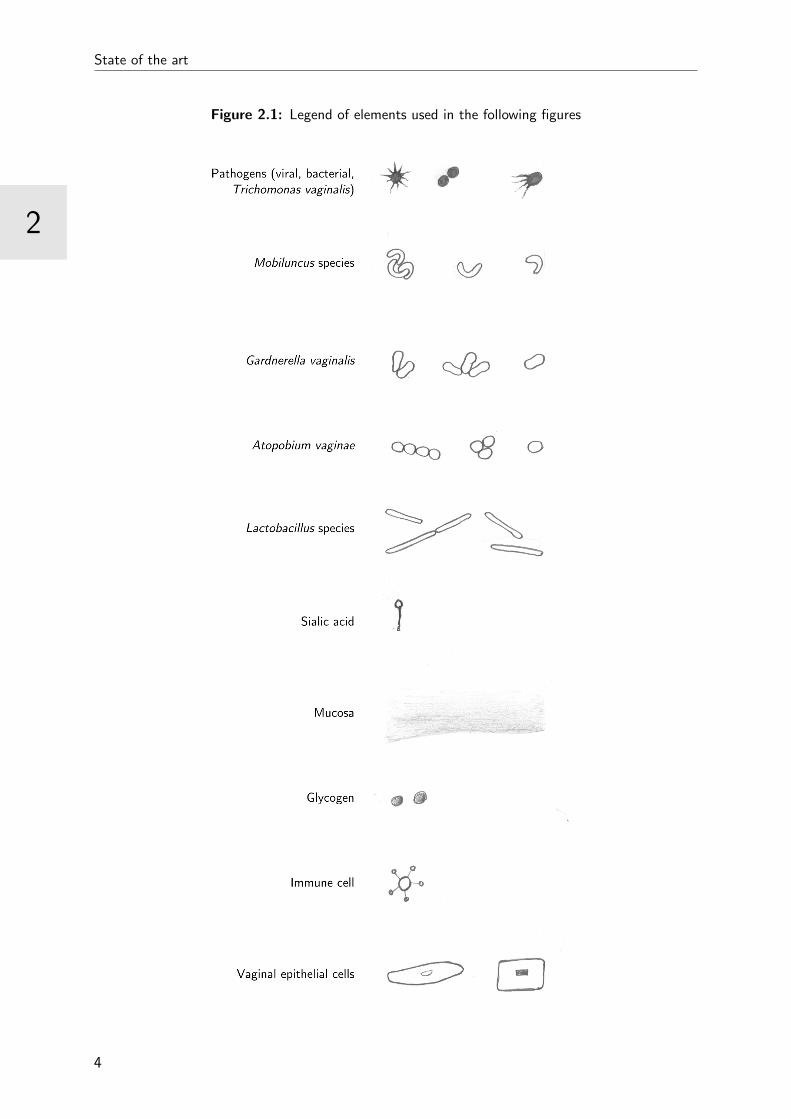
Figure 2.1: Legend of elements used in the following figures
4
2
State of the art
vaginal microbiome (i.e. without symptoms of a vaginal disturbance) also have non-
Lactobacillus-species in their microbiome, e.g. Gardnerella vaginalis, Atopobium vaginae,
Prevotella spp., Streptococcus spp., Staphylococcus spp. and Escherichia coli [19].
2.1.1 Composition of the cervicovaginal fluid
The vaginal epithelium is kept moist by a cervicovaginal fluid (CVF) that is a mixture of
plasma transudate percolating through the vaginal wall and endocervical mucus produced
by goblet cells [20]. In normal circumstances, outside of coitus, the CVF also contains
mucous secretions from Bartholin’s and Skene’s glands, exfoliated epithelial cells, residual
urine, and fluids from the upper reproductive tract such as endometrial and tubal fluids.
The exact composition of the CVF is variable and depends on the levels of the hormones
oestrogen and progesterone, sexual stimulation and the state of the microbiotic community
[21–23].
Cervical mucus is an important component of the CVF because it forms a mechanical and
chemical mucosal barrier that prevents invasion of microbes and viruses. Mucus consists
mostly of water (92-98%) and also contains glycoproteins, ions and antimicrobial proteins
and polypeptides [11]. These glycoproteins, e.g. lactoferrin, lysozyme, immunoglobulins
and defensins, have a broad-spectrum antibacterial activity [24]. The cervical gel-forming
mucins play a more important role in the defence against pathogens by controlling the
physical clearance of microbes. The mucins determine the amount and viscosity of the
mucosal flow and therefore are in charge of the first line of defence against intruders, that
washes pathogens out of the vagina [25]. It is important to note that this barrier function
is not infallible: its efficiency also depends on the physicochemical [26] and microbial
environment [27].
Mucins have a linear protein backbone (apomucin) that is highly O-glycosylated by
oligosaccharide chains containing blood group structures. The O-linked chain starts with a
N-acetylgalactosamine, α-linked with a serine or threonine, which is further extended with
various monosaccharides. At the terminus, an α-linked sugar residue can be found: sialic
acid, N-acetylgalactosamine (blood group A), or galactose (blood group B) associated with
subterminal fucose (blood group O). Mucin monomers are linked together with disulfide
bonds and form mucin multimers [28]. The capacity of bacteria to degrade mucins by
means of microbial enzymes or mucinases, including sialidases, glycosidases, proteases,
and sulphatases, is often a fundamental step in the disruption of the defensive mucosal
barrier, which constitutes a direct interface between the internal and external environment
of the vagina [24,29].
5

State of the art
2
2.1.2 The lactobacilli-dominated vaginal microbiome
Lactobacillus species are the predominant resident bacteria of the healthy vaginal econiche.
The first lactobacillus was isolated in 1894 by Doderlein from the vagina of a healthy
pregnant woman [29]. The genus Lactobacillus comprises a phenotypically heterogeneous
group of aerotolerant or anaerobic, catalase-negative, Gram-positive, non-spore-forming,
rod-shaped bacteria. This genus is embedded within the lactic acid bacteria, which
are functionally related through their ability to produce lactic acid [30]. A lactobacilli-
dominated vaginal microbiome supports the mucosal barrier (Figure 2.2).
Both the vaginal mucosa and the Lactobacillus species are sources of lactic acid in vaginal
secretions, which results in an acidic (pH 3.8-4.5) vaginal environment. Under the influence
of oestrogen, the vaginal epithelial cells will lyse glycogen into glucose. Glucose is further
metabolised into L-lactic acid via pyruvic acid using α-amylase [31]. Lactobacilli use a
similar glycolysis mechanism to convert extracellular glucose into D- and L-lactic acid
isomers, that have a different arrangement of the same chemical components around a
central carbon atom [32–34]. The D/L ratio of lactic acid isomers found in the vaginal fluid
strongly indicates that the lactobacilli are mainly responsible for vaginal acidity [33]. This
low pH exerts selective antimicrobial activity against nonresident species of bacteria (and
viruses and fungi) while favouring the presence of Lactobacillus species [23]. In addition to
acidification of the vaginal fluid, the proliferation of non-advantageous bacterial species
is also suppressed through Lactobacillus’ production of broad-spectrum antimicrobial
peptides (or bacteriocins) and hydrogen peroxide (whose value is still being debated), and
by competing for receptor sites on the vaginal epithelium [35–37].
The most frequently isolated species of lactobacilli from the vaginal microbiome are L.
crispatus, L. iners, L. gasseri, and L. jensenii. Furthermore L. acidophilus, L. brevis, L.
casei, L. delbrueckii, L. fermentum, L. plantarum, L. rhamnosus, L. reuteri, L. salivarius,
and L. vaginalis have frequently been isolated from women without vaginal complaints
as well [3, 18, 38, 39]. G. vaginalis and Prevotella spp. are also often present in the
healthy vaginal microbiome, although in relatively low concentrations [3, 15,40,41]. The
Lactobacillus composition of the vaginal microbiome varies among women of geographic
locations, and ethnicities.
Two species from the Lactobacillus genus deserve close scrutiny: L. crispatus and L. iners.
L. crispatus is associated with a healthy vaginal microbiome and is likely to mediate vaginal
protection against STIs through the mechanisms listed above [17, 27, 42–44]. L. iners,
however, can be found both in the Lactobacillus-dominated vaginal microbiome and in the
vaginal microbiome that is dominated by other anaerobic organisms. Moreover, in a study
by Ferris et al. [45], L. iners was predominant in all bacterial vaginosis (BV) patients
after treatment with metronidazole. On top of that, a more recent study by Petricevic
6
2
State of the art
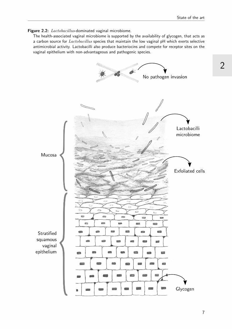
Figure 2.2: Lactobacillus-dominated vaginal microbiome.The health-associated vaginal microbiome is supported by the availability of glycogen, that acts asa carbon source for Lactobacillus species that maintain the low vaginal pH which exerts selectiveantimicrobial activity. Lactobacilli also produce bacteriocins and compete for receptor sites on thevaginal epithelium with non-advantageous and pathogenic species.
7
State of the art
2
et al. [46] observed an association between preterm delivery and the vaginal presence
of L. iners, as the only Lactobacillus, in the first trimester of pregnancy. It has been
suggested that L. iners is a dominant part of the vaginal microbiome at the transitional
stage between health and dysbiosis 1, caused by treatment or by physiological changes. A
L. crispatus-dominated vaginal microbiome might shift to a L. iners-dominated vaginal
microbiome but is less likely to transition directly to a dysbiotic state [45, 47,48].
2.1.3 Variability of the vaginal environment
The composition of the vaginal microbiome can be influenced by exogenous factors, such
as antibiotic treatment, sexual intercourse [4,49], personal hygiene (vaginal douching) [50],
cigarette smoking [51] and stress [52]. Furthermore, the vaginal microbiome is impacted
by a range of endogenous factors as well. Apart from ethnicity, innate immunity, and
menses, a woman’s hormone levels, oestrogen in particular, have a major effect on the
composition of the vaginal microbiome.
Maturation of the vaginal environment
Throughout the different stages of life, the vaginal environment is subjected to many
alterations due to changes in oestrogen levels (Figure 2.3). After birth, the vaginal
epithelium of the female newborn is rich in glycogen, due to the maternal oestrogen. This
results in a low pH in which the maternal vaginal microbiome, that was acquired during
passage through the maternal birth canal, can survive. However, shortly after birth, the
decline in the maternally derived oestrogen level results in the thinning of the epithelium
and the rise of the newborn’s vaginal pH, in which the acidophilic bacteria no longer benefit
from the selective advantage. During childhood, the vagina is predominantly colonised
by a variety of anaerobic bacteria other than lactobacilli originating from the skin and
the gastrointestinal tract [21,53]. But with the onset of menarche, the increased level of
oestrogen stimulates the maturing epithelial cells to release glycogen, which indirectly
supplies lactobacilli with nutrients. The lactobacilli degrade glucose released from glycogen
into lactic acid and again create an acidic environment, restricting the growth of pathogenic
microorganisms [21, 33]. Thus, at fertile age, the normal pH of the lactobacilli-dominated
vagina is 3.5 ± 0.3 [54]. After a long period of adulthood marked by a stable pH and vaginal
environment, the onset of menopause and its associated decrease in free oestrogen might
offer less protection from dysbiosis and possible colonisation by enteric bacteria [55–57]. In
contrast, menopausal women are protected from the potentially negative effects of menses
on the vaginal microbiome [15].
1Dysbiosis: a microbial imbalance in the body.
8

2
State of the art
Figure 2.3: Stages in vaginal maturation
Menstrual cycle
The menstrual cycle is governed by hormonal changes and creates an ever-changing vaginal
environment. The first half of the menstrual cycle, or follicular phase, is characterised by
gradually increasing oestrogen levels which provoke an increased amount of cervical mucus
that is thin and watery to allow sperm penetration. In the second half of the menstrual
cycle, or the luteal phase, which is predominated by increased progesterone levels, the
cervical mucus becomes scant, thick and opaque and is less penetrable to sperm [24].
During menses, there seems to be an interindividual variability, with some women main-
taining a consistent vaginal microbiome, others having fluctuations timed with menses (less
lactobacilli, more anaerobic bacteria) and some having random fluctuations without appar-
ent cause [48,58–60]. Several reports observed an overgrowth of L. iners can be observed
during menses, while the concentration of L. crispatus decreases [41,48,59,61].
Use of hormonal contraceptives
Hormonal contraceptives are being used by millions of women worldwide, the most widely
used being oral combined (oestrogen and progestogen) contraceptives and progestin-only
9
State of the art
2
injectables [62, 63]. Both contraceptive methods seem to have a protective effect on
the vaginal microbiome and favour the presence of Lactobacillus spp. in the vaginal
ecosystem [64]. The high oestrogen level and subsequent higher availability of glycogen
induced by oral contraceptives probably facilitates growth of lactobacilli and subsequent
lactic acid production [65]. For progestin-only injectables, this protective effect may be
due to a lack of menses, but hard evidence is still lacking [65,66].
Combined contraceptive vaginal rings (CVRs) are a common alternative to these widely
used oral contraceptives. Currently, only two contraceptive rings are commercially available:
the widely available NuvaRing (etonogestrel/ethinyl estradiol) and the progestogen-only
Progering, only available in South-America [67]. In our research, we have focussed on
the combined contraceptive ring (NuvaRing). An early clinical trial with the combined
3-ketodesogestrel/ethinyl estradiol ring could not demonstrate significant changes in the
vaginal microbiome and presence of inflammatory cells, before and after use of the contra-
ceptive ring for either 21, 28, 42, or 56 days [68]. However, another study using a combined
etonogestrel/ethinyl estradiol ring (the current NuvaRing) reported a 2.7-fold increase
in the concentration of H2O2-producing Lactobacillus species compared to users of oral
contraception [69]. This finding was confirmed by De Seta et al. in a study in 60 volunteers
that used either this contraceptive etonogestrel/ethinyl estradiol ring or combined oral
contraceptives with the same steroids (desogestrel and etonogestrel/ethinyl estradiol). The
ring users experienced a significant increase in vaginal lactobacilli concentration after three
and six months of use [70]. It has been suggested that this positive effect on the vaginal
lactobacilli population is mainly due to the local availability of ethinyl estradiol, which
promotes a glycogen-rich environment [71]. In a more recent study, a combined CVR
containing NesteroneTM and ethinyl estradiol, that was used continuously for one year did
not have any effect on the prevalence of Lactobacillus species, but no data on the species
concentration was available [72].
10
2
State of the art
2.2 Bacterial vaginosis: a dysbiosis of the vaginal
microbiome
BV was defined in 1984 as follows: “A replacement of the lactobacilli of the vagina by
characteristic groups of bacteria accompanied by changed properties of the vaginal fluid”
[73] (Figure 2.4). The condition is characterised by a change in the microbial composition
of the vagina: the Lactobacillus spp., associated with a healthy vaginal microbiome,
are outnumbered by other microaerophilic and anaerobic organisms [15, 74, 75]. The
term bacterial vaginosis was recommended because “vaginitis” suggests an inflammatory
reaction of the vaginal epithelium and a high level of polymorphonuclear cells in the
vaginal discharge, which is usually absent [76–80]. In the preceding years, several attempts
have been made to rephrase and rename these conditions, from non-specific vaginitis
over vaginal bacteriosis to anaerobic vaginosis, but none of the alternatives was generally
accepted [81].
Although BV generally is not associated with symptoms of inflammation, significant
variations in the cervicovaginal immune response have been demonstrated [80, 82]. BV
is positively associated with proinflammatory cytokines/chemokines (IL-α, IL-1β, IL-6,
IL-12 (p70), and IL-8) and negatively associated with protective antimicrobial proteins
SLPI (produced by epithelial and immune cells) and elafin and the chemokine IP-10, that
functions as a chemoattractant for various immune cells to the site of infection [82–84].
Furthermore, the increased diversity in the vaginal bacterial population results in increasing
vaginal levels of antimicrobial effectors produced by leukocytes, such as nitric oxide
(NO) [85] and heat shock protein 70 (hsp70) [86]. However, BV has also been associated
with a suppressed number of leukocytes, which can explain the absence of apparent
symptoms of inflammation [87].
BV is the most prevalent vaginal disorder in women of reproductive age worldwide, but
disproportionately afflicts women of African descent [88], and is the most common cause
of vaginal complaints [89]. BV prevalences vary considerably, but are generally highest in
Sub-Saharan Africa (between 20-60%) and lowest in Europe and Asia (less than 20%),
although there are exceptions to this rule [90].
More than half of the women diagnosed with BV do not report symptoms, but others
may have malodorous “fishy-smelling”, white, thin and homogeneous discharge and can
experience vaginal itching or burning feeling [91]. The change in discharge is caused by the
overgrowth of non-Lactobacillus bacteria. These bacteria produce enzymes (e.g. sialidases,
prolidases, mucinases) that cause a degradation of the cervicovaginal mucus and increased
discharge. The malodour is a consequence of the production of volatile polyamines, such
as putrescine and cadaverine, by the BV-associated bacteria [75,92].
11
State of the art
2
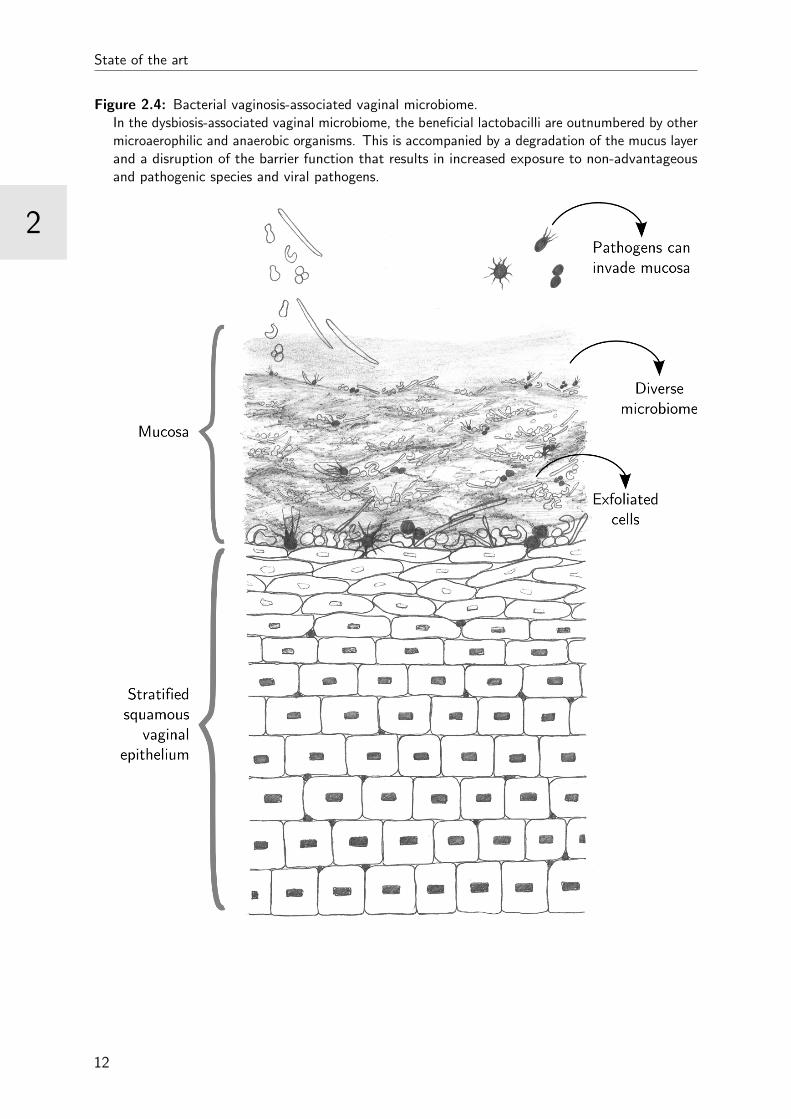
Figure 2.4: Bacterial vaginosis-associated vaginal microbiome.In the dysbiosis-associated vaginal microbiome, the beneficial lactobacilli are outnumbered by othermicroaerophilic and anaerobic organisms. This is accompanied by a degradation of the mucus layerand a disruption of the barrier function that results in increased exposure to non-advantageousand pathogenic species and viral pathogens.
12
2
State of the art
2.2.1 BV-associated complications
Aside from being the cause of unpleasant symptoms, BV can also generate an entire array
of serious gynaecological and obstetric complications.
In pregnant women, BV has been associated with chorioamnionitis [93, 94], premature
rupture of membranes [95], intra-amniotic infections [96,97], premature labour and delivery
[98–104], spontaneous abortion [98,101,104,105] and low birth weight [100,106]. These
adverse pregnancy outcomes have been linked to the presence of BV-associated anaerobes,
although the exact mechanism of action is still not clear. High vaginal concentrations of A.
vaginae (>106 colony forming units (cfu)/ml) and G. vaginalis (>107 cfu/ml) significantly
increase the risk of preterm delivery, according to Menard et al. [107]. Moreover, BV-
associated bacteria including Mycoplasma hominis, Prevotella spp., and G. vaginalis are
often isolated from the chorioamnion in preterm labor [108] and can pose a risk for intra-
amniotic infections [93, 109]. Moreover, both G. vaginalis and Prevotella spp. produce
sialidase, an enzyme implicated in preterm birth [110–112]. In addition, BV-associated
microorganisms and their toxins are capable of crossing the placenta which could lead
to brain injury in foetuses and long-term neurodevelopmental disorders in children, such
as hyperactivity, academic difficulties in school and severe handicaps such as cerebral
palsy 2 and periventricular leucomalacia 3 [93,113–118]. BV is also a risk factor for the
development of postpartum maternal infections [101], postabortion endometritis and pelvic
infection following gynaecological surgery [119–121].
BV has been associated with histological endometritis [122] and pelvic inflammatory
disease (PID) in nonpregnant women. The ascent of pathogenic bacteria such as Chlamydia
trachomatis, Mycoplasma genitalium, or Neisseria gonorrhoeae from the lower to the upper
genital tract can lead to PID [123]. These pathogenic bacteria are often accompanied by
BV-associated bacteria [124,125].
The disturbed BV-associated vaginal microbiome is associated with increased incidences of
STIs [6,126,127] and more specifically with herpes simplex virus type 2 (HSV-2) [128,129].
BV also creates a more permissive environment for acquiring HIV [130–132]. The presence
of BV-associated bacteria in the vagina directly leads to an upregulation of HIV-replication
[109,133–135]. The risk of HIV acquisition is even higher in women, as the female genital
tract is twice as sensitive to HIV compared to the the male counterpart [136,137].
2Cerebral palsy: a group of permanent movement disorders that appear in early childhood.3Periventricular leucomalicia: a form of white-matter brain injury that is characterised by the necrosis
of white matter near the lateral ventricles.
13
State of the art
2
2.2.2 The bacteria involved in bacterial vaginosis
In a lactobacilli-dominated vagina, a relative low α-diversity, or within-subject diversity,
is seen, while the species diversity between different subjects is higher. Moreover, little
distinction can be found in the distribution of species between different vaginal sites
(mid-vagina, posterior fornix and vaginal introitus) [138]. However, when the Lactobacillus
species are being outnumbered by BV-associated species, an increased taxonomic richness
can be found, with an even higher inter-subject variability. No single dominant taxon can
be found in the BV microbiome; BV patients harbour a diverse array of vaginal bacteria,
many of which are only present at low relative abundance [139].
The typical spectrum of microorganisms involved in BV is well-described using con-
ventional cultivation as well as molecular methods. G. vaginalis and Prevotella spp.
are consistently found in the disturbed vaginal microbiome, but they are also present
in lower bacterial loads in the healthy vaginal microbiome [3, 15, 40, 41]. Other fre-
quently found BV-associated bacteria are A. vaginae, bacteria species from the Lach-
nospiraceae family (including BV-associated bacterium (BVAB) 1-3) and species in the
following genera: Bacteroides, Clostridiales, Eggerthella, Escherichia/Shigella, Dialister,
Fusobacterium, Gemella, Leptotrichia, Megasphaera, Mobiluncus, Mycoplasma, Parvi-
monas, Porphyromonas, Staphylococcus, Sneathia, Streptococcus, Ureaplasma, and Veil-
lonella [3, 15,18,19,40,41,80,140–153].
Although this collection of involved bacteria seems to indicate high complexity, the
vaginal microbial communities are relatively “simple” at the taxonomic level, especially
when compared to more diverse microbial communities such as the oral and intestinal
microbiota [138]. However, the gut and mouth can also act as extravaginal reservoirs of
vaginal microbiome bacteria. Lactobacilli and BV-associated bacteria are often found
in the rectum [154], and lactobacilli are found in the oral cavity [4, 155]. In adolescent
girls with a healthy vaginal microbiome, nearly no oral G. vaginalis or A. vaginae was
detected [4], but in women who developed BV, G. vaginalis was consistently found in the
oral cavity [156]. Furthermore, Jespers et al. [4] demonstrated that the anorectal presence
of G. vaginalis and A. vaginae was significantly higher in sexually experienced adolescent
girls. And this is not without consequences: Marrazzo et al. showed that women with high
quantities of oral or rectal G. vaginalis, or rectal Megasphaera, Leptotrichia, or Sneathia
spp., were more likely to develop clinical BV; in contrast, women who had L. crispatus in
the rectum were more likely to maintain their healthy vaginal environment [156]. El Aila
et al. also showed strong correspondence between rectal and vaginal microbes [154].
In this thesis, the main focus will be on only two bacteria out of the full array of BV-
associated bacterial species. Although this might seem to simplify a complex condition,
it also allows putting the following apparent important players in the spotlight. Firstly,
14
2
State of the art
G. vaginalis certainly deserves close scrutiny since it is present in up to 97.5% of cases
of BV [40–42,157], and in 50% to 70% of BV-free women, although in lower abundances
[40,41,158]. This finding leads to the suspicion that G. vaginalis actually consists of several
species with distinct roles in BV pathogenesis [159–162]. The second player is A. vaginae,
only recently discovered and still quite unknown. The involvement of A. vaginae in BV
was only established in 2004 [163–167] but the bacterium is rarely detected in the normal
vaginal microbiome [3, 168–174]. This finding leads to the suggestion that A. vaginae is a
better marker for BV than G. vaginalis and thus warrants some extra attention in this
thesis.
2.2.3 Diagnosis and detection of BV
From a diagnostic point of view, a dysbiosis such as BV is very different from most
infectious diseases: there is no single infectious agent that causes the condition. The
condition is diagnosed based on symptoms and on the abundance of a few typical BV-
associated microorganisms, which implies that BV is not diagnosed in asymptomatic
women. In a clinical setting, BV is generally diagnosed using microscopic evaluation of
vaginal fluid, while there is a wide array of methods available and in development to detect
and investigate BV in research settings.
Amsel criteria
The most widely used method for BV diagnosis in clinical practice is based on symptoms;
a positive diagnosis requires that the patient has three out of the following four Amsel
criteria [91]:
1. Thin, white homogenous discharge
2. Vaginal pH greater than 4.5
3. Detection of clue cells 4 in vaginal wet smear
4. Positive whiff test: presence of amine odour after addition of 10% KOH to vaginal
discharge on a glass slide
Unfortunately, this method is flawed for various reasons. The evaluation of the discharge
and whiff test are subjective, and could lead to misdiagnosis. The detection of clue cells in
the vaginal fluid is a subjective procedure that requires a well-trained microscopist. While
the measurement of the vaginal pH is not subjective or technically difficult (since there
is a wide range of commercially available pH tests), the vaginal pH can be influenced by
4Clue cells: Squamous epithelial cells whose surfaces are heavily coated with bacteria.
15

State of the art
2
intravaginal washing, menstruation, and intercourse [55,175] and is therefore not always
reliable. Nevertheless, the Amsel criteria remain the best option for clinicians to quickly
diagnose BV in their clinical practice.
Nugent score
The gold standard in BV research, the Nugent score, is a grading method that was first
described by Spiegel et al. [176] and later modified by Nugent et al. [158] to include
an extra category. It relies on the microscopic evaluation of Gram stained 5 smears of
vaginal fluid, that divides bacteria into two groups (Gram-positive and Gram-negative)
based on the properties of their cell wall. The Nugent score is based on the presence and
relative amounts of three bacterial cell types (often designated as “morphotypes”) in the
vaginal fluid: Gram-positive rods (corresponding to lactobacilli), Gram-negative/variable
pleiomorphic rods (supposed to correspond to G. vaginalis and Bacteroides species) and
curved rods (supposed to correspond to Mobiluncus species) (Table 2.1, Figure 2.5).
The approach is used in research settings to classify vaginal smears into three categories:
normal, intermediate or BV. A high abundance of Gram-positive rods, or at least the
absence of G. vaginalis or Mobiluncus, gives a Nugent score of 0-3 which is considered
normal. A Nugent score of 7-10 leads to the diagnosis of BV and is marked by a high
abundance of Gardnerella or Mobiluncus morphotypes and the absence of Gram-positive
rods. A third state between these two extreme poles is the intermediate flora, with a
Nugent score of 4-6.
Figure 2.5: Nugent score.Gram-stained smears of vaginal fluid, photographed at 100x magnification. A: Nugent 0-3 or normalstate characterised by Gram-positive rods scattered around the vaginal epithelial cells; B: Nugent 4-6or intermediate state showing a mixture or Gram-positive rods and Gram-negative pleimorphic rods;C: Nugent 7-10 or BV state with a typical dense network of Gram-negative/variable pleiomorphicrods.
5Gram staining: The process is done stepwise. Gram-positive bacteria retain the first dye (crystalviolet) due to their thick peptidoglycan layer, and are microscopically visible as blue cells. Gram-negativebacteria lose this colouring after a decolouring step, but retain the second stain (safranin or fuchsine) andcan be seen as red or pink cells.
16

2
State of the art
Table 2.1: Nugent score
ScoreLactobacillus
morphotypes
Gardnerella andBacteroides spp.
morphotype
CurvedGram-variable
rods
No morphotypes present 4+ 0 0<1 morphotype present 3+ 1+ 1+ or 2+1-4 morphotypes present 2+ 2+ 3+ or 4+5-30 morphotypes present 1+ 3+>30 morphotypes present 0 4+
Total score = Lactobacilli + Gardnerella and Bacteroides spp. + curved rods
This method is less suited to the clinical setting because the Gram staining and microscopic
evaluation require a certain level of technical knowledge and expertise. Furthermore, the
staining is time-consuming, and it would not be feasible to perform this technique instantly
when the clinician should decide on treatment of BV. But due to the greater reproducibility
and objectivity, compared to the Amsel criteria, the Nugent score is still used as the
standard in BV research.
There is an ongoing discussion on the designation of the correct bacterial species to these
different morphotypes. Srinivasan et al. proposed, based on the results of pyrosequencing,
to reclassify the pleiomorphic rods (G. vaginalis/Bacteroides species) as G. vaginalis,
Prevotella spp. and Porphyromonas spp. and suggested that the curved rods (Mobiluncus
species) were more likely to be BVAB1 [177]. Alternative grading systems based on Gram
stained smears have also been developed. Ison and Hay have simplified the Nugent scoring
system and have added a fourth category based on the presence of only Gram-positive
cocci and another category to indicate that no bacteria are present [178]. This system was
subsequently developed into the Claeys criteria by Verhelst et al. [179] who differentiated
between the different Lactobacillus cell types and added a new category for the presence
of diphtheroid bacilli cell types (irregular-shaped Gram-positive rods).
Commercial point-of-care tests
Several rapid diagnostics tests for BV have been commercialised and quite good sensitivities
and specificities have been reported (Table 2.2). However, rapid tests have little coverage in
the clinical practice, which might question the value of these reported performances.
A first type of tests is based on the symptoms of BV: increased pH and fishy odour.
The self-test pH glove is based on the monitoring of pH: it was developed with a focus
on pregnant women who are instructed to visit their clinician if their vaginal pH rises
above 4.7 [180]. Other tests detect trimethylamine in vaginal fluid, which is responsible
17
State of the art
2
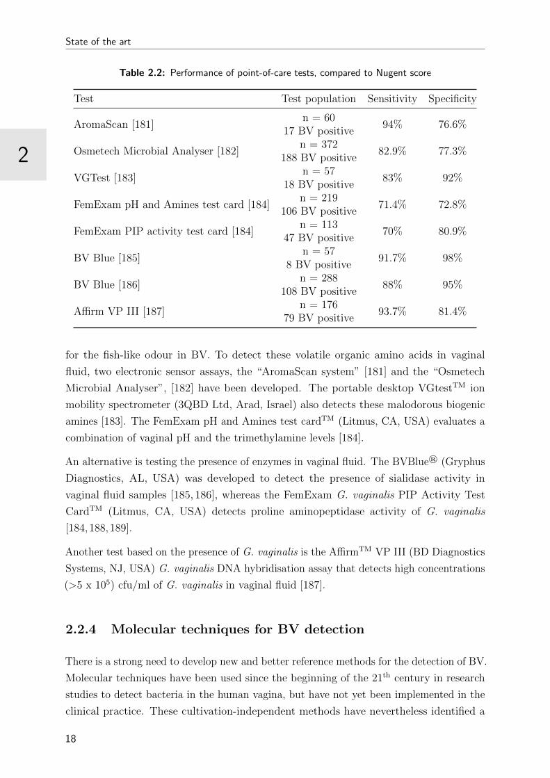
Table 2.2: Performance of point-of-care tests, compared to Nugent score
Test Test population Sensitivity Specificity
AromaScan [181]n = 60
17 BV positive94% 76.6%
Osmetech Microbial Analyser [182]n = 372
188 BV positive82.9% 77.3%
VGTest [183]n = 57
18 BV positive83% 92%
FemExam pH and Amines test card [184]n = 219
106 BV positive71.4% 72.8%
FemExam PIP activity test card [184]n = 113
47 BV positive70% 80.9%
BV Blue [185]n = 57
8 BV positive91.7% 98%
BV Blue [186]n = 288
108 BV positive88% 95%
Affirm VP III [187]n = 176
79 BV positive93.7% 81.4%
for the fish-like odour in BV. To detect these volatile organic amino acids in vaginal
fluid, two electronic sensor assays, the “AromaScan system” [181] and the “Osmetech
Microbial Analyser”, [182] have been developed. The portable desktop VGtestTM ion
mobility spectrometer (3QBD Ltd, Arad, Israel) also detects these malodorous biogenic
amines [183]. The FemExam pH and Amines test cardTM (Litmus, CA, USA) evaluates a
combination of vaginal pH and the trimethylamine levels [184].
An alternative is testing the presence of enzymes in vaginal fluid. The BVBlue® (Gryphus
Diagnostics, AL, USA) was developed to detect the presence of sialidase activity in
vaginal fluid samples [185, 186], whereas the FemExam G. vaginalis PIP Activity Test
CardTM (Litmus, CA, USA) detects proline aminopeptidase activity of G. vaginalis
[184,188,189].
Another test based on the presence of G. vaginalis is the AffirmTM VP III (BD Diagnostics
Systems, NJ, USA) G. vaginalis DNA hybridisation assay that detects high concentrations
(>5 x 10⁵) cfu/ml of G. vaginalis in vaginal fluid [187].
2.2.4 Molecular techniques for BV detection
There is a strong need to develop new and better reference methods for the detection of BV.
Molecular techniques have been used since the beginning of the 21th century in research
studies to detect bacteria in the human vagina, but have not yet been implemented in the
clinical practice. These cultivation-independent methods have nevertheless identified a
18

2
State of the art
number of novel, fastidious and uncultivable bacterial species.
Polymerase chain reaction
The first study to characterise the vaginal microbiome using broad-range polymerase chain
reaction (PCR) combined with denaturing gradient gel electrophoresis (DGGE), was carried
out in 2002 by Burton and Reid [190] to profile the total vaginal bacterial population. A
limitation of such a broad-range method is that it tends to sample only the most prevalent
bacteria and is likely to miss low-abundance or minority species [191]. Verhelst et al. [179]
and Fredricks et al. [167] combined PCR with culturing of bacteria, which increases the
sensitivity. The above studies have played a critical role in defining the bacteriology and
identifying key organisms in BV and have paved the way for the detection of these bacteria
by specific conventional or quantitative real-time PCR (qPCR). Several PCR assays using
primers against the 16S-23S rRNA spacer region or 16S rRNA were developed for detection
of vaginal bacteria that represent either the normal vaginal microbiome (lactobacilli), or
are characteristic for BV (e.g. G. vaginalis [192] and A. vaginae [163,164,172]). Due to
the polybacterial nature of BV, PCR [40,170] and qPCR [3,59,168,193,194] assays for
detection of a panel of key vaginal bacteria have been developed.
The use of qPCR as a diagnostic tool has been studied by several groups [45, 168, 170–
174,193,195], but it remains difficult to apply in a clinical setting due to the expensive
equipment and long turnaround time. Hence this technique is currently still better suited
to a research setting. Another hindrance to the use of qPCR as a diagnostic tool is
that it requires a preselection of the expected organisms. Since the aetiology of BV is
still unknown and there is still some uncertainness about the relative importance of the
different players, one might miss an important bacterial species when only focussing on
one or a set of specific bacteria with qPCR. Jespers et al. [174] proposed a DNA tool
based on log-transformed counts of the bacterial cells of G. vaginalis, A. vaginae and the
Lactobacillus genus for the detection of BV in a research environment, which also could
possibly lead to a next-generation point-of-care test for BV.
Microarray
Simultaneously measuring the expression of a large amount of genes can be achieved using
a DNA microarray. A microarray is a multiplex lab-on-a-chip, in which a selection of
genomic sequences (or probes) are spotted onto a solid substrate. After hybridisation of
the sample onto the microarray, the relative abundance of nucleic acid sequences in the
sample can be determined. A phylogenetic microarray targeting the 16S rDNA or 16S
rRNA could be useful to assess the relative presence of multiple bacteria in the vaginal
19
State of the art
2
microbiome in a semi-quantitative manner. An additional advantage is the fact that the
composition of the microarray can be modified according to needs (for example for use in
different ethnic groups). Moreover, it is possible to select multiple genomic sequences for
each bacterium represented in the microarray.
A few groups have been working on the development of a tailored microarray platform
which would be used as a fast, low-cost diagnostic device [17,196–199]. However, like qPCR,
the technique is handicapped by the inability to detect unknown species that were not
included in the set-up and, unlike qPCR, it is not fully quantitative. The technology often
also requires a rather large sample volume (in terms of micrograms of DNA), which requires
PCR-based amplification and thus can introduce bias into the samples. In addition, there
have been reports of cross-hybridisation between similar sequences and concerns regarding
the reproducibility of microarray data [200]. Moreover, Cruciani et al. [197] reported a
low efficiency of their microarray in the amplification of members of the Bifidobacteriaceae
family, which includes G. vaginalis. This is a major limit of the technique, since G.
vaginalis plays a key role in the vaginal niche.
Sequencing
With sequencing the precise order of the nucleotides of a DNA or RNA strand is determined
step by step. There are different ways to sequence a sample, but in general the genomic
material of a sample is broken into smaller pieces that are individually sequenced and
afterwards reassembled. For most approaches an in vitro cloning step is needed to amplify
the genomic material, in order to increase the sensitivity of the technique. Conventional
sequencing of cultured clinical isolates may provide a framework, but is unable to truly un-
cover the bacterial diversity in the vaginal microbiome. Bacteria present in low abundance,
that could provide important information about the genetic and functional diversity of
the vaginal microbiome and that may be relevant in the pathogenesis of BV, are less likely
to be detected with this technique [5, 151].
To deal with this disadvantage, next-generation sequencing (NGS) has been carried out by
different groups in order to discern VMB clusters in different study populations covering
different ethnicities This has been carried out by different groups to detect both known and
unknown sequences without prior knowledge of the bacterial species in the sample. Because
the material is directly sequenced and not dependent on user-defined sequences (compared
to qPCR and microarray technologies), there are no experimental bias or microarray
cross-hybridisation issues to deal with. Furthermore, a small sample volume (in terms of
nanograms of material) is sufficient for NGS [200]. But despite these strengths, NGS is
still a rather costly technique that requires expensive equipment and highly skilled people
to prepare the samples and to analyse the massive amount of data collected. Furthermore,
20

2
State of the art
notwithstanding the big amount of data collected, only a small fraction of this data is
trustworthy and useful for analysis.
This technology can be used to discern vaginal microbiome clusters in different study
populations covering different ethnicities. The study by Ravel et al. [18] has served as the
reference of a large-scale clustering study of the bacterial communities of asymptomatic
North-American women of four ethnic groups. They found four lactobacilli-dominated
vaginal microbiome communities, with either L. crispatus, L. gasseri, L. iners, or L.
jensenii as the main contributor, that were identified mostly in Asian and Caucasian
women. A fifth vaginal microbiome group with lower proportions of lactic acid bacteria
and higher proportions of strictly anaerobic organisms was overrepresented in Hispanic
and African American women [18]. In addition to this, other studies using similar methods
to study a variety of study populations, reported similar and additional clusters (overall
between three to nine clusters were found) [15, 19, 41, 143–148,199,201,202]. The majority
of studies found one cluster dominated by L. crispatus and one by L. iners. In contrast,
clusters dominated by L. jensenii, L. gasseri and G. vaginalis were found less frequently,
but several clusters combining G. vaginalis with lactobacilli were described. Furthermore,
all studies found at least one (but more often more) cluster that contained mixtures of
anaerobes with or without Lactobacillus species. Typically, this cluster contained L. iners
and G. vaginalis and a group of other anaerobes. Clusters dominated by aerobes (including
Streptococcus spp., Staphylococcus spp., Escherichia/Shigella spp. and Proteus spp.) were
only reported in two studies [147, 201]. The most abundant taxa (other than Lactobacillus
spp.), present in at least 50% of the studies were [15]:
• Actinobacteria: A. vaginae, Eggerthella spp., G. vaginalis, Mobiluncus spp.
• Firmicutes: Dialister spp., Gemella spp., Lacnospiraceae (including BVAB1-3),
Megasphaera spp., Parvimonas spp., Staphylococcus spp., Streptococcus spp., Veil-
lonella spp.
• Fusobacteria: Sneathia spp., Leptotrichia spp.
• Proteobacteria: Escherichia/Shigella spp.
• Sfingobacteria: Prevotella spp., Porphyromonas spp., Bacteroides spp.
• Tenericutes: Mycoplasma spp., Ureaplasma spp.
Fluorescence in situ hybridisation
Another molecular technique proposed for detection of BV is fluorescence in situ hybridis-
ation (FISH) using specific fluorescent probes against the 16S rRNA of vaginal bacteria.
Hybridisation-based techniques such as FISH have been developed for use in cytogenetics,
21

State of the art
2
to detect the presence or absence of nucleic acid sequences on chromosomes. FISH is
also being used to identify microorganisms in the field of microbial ecology, and allows
visualising the distribution of a certain bacterium in a bacterial biofilm.
FISH is based on the hybridisation of a fluorescently labeled DNA or RNA sequence (or
probe) with a target sequence in a biological sample and can be performed on a microscopic
glass slide (Figure 2.6). Before hybridisation can occur, the double-stranded genomic
sequences are denatured into single strands, using heat or chemicals. This denaturation is
the result of the destruction of the hydrogen bonds between the two nucleotide strands and
is needed to allow for binding the labeled probes to the complementary target sequences
through new hydrogen bonds. After a wash step that stops the hybridisation reaction
and removes the unbounded probes, the location of hybridised probes can be detected
immediately using a fluorescence microscope.
This technique is widely used in microbial ecology and can give valuable information using
FISH: the identification of microorganisms and visualisation of the distribution of specific
species within a sample (for example in a biofilm). However, like most molecular techniques,
FISH will only detect the user-defined bacterial species. Furthermore, similar as with
other microscopic techniques, the interpretation of the FISH results can be subjective,
and a well-trained eye is needed to differentiate between a genuine signal and background
fluorescence. The technique can be used as a multiplex test, to detect an array of bacterial
species with multiple probes at once, but it is restricted to non-overlapping spectra of
fluorophores. Researchers have tried to circumvent this limitation to expand the number of
distinguishable taxa in a single FISH experiment, for example by combinatorial labelling
and spectral imaging (CLASI). CLASI FISH involves labelling microbes of interest with
combinations of probes coupled with spectral imaging to allow the use of fluorophores with
highly overlapping excitation and emission spectra in order to the simultaneously identify
tens to potentially hundreds of microbial taxa in a single microscope image [203].
FISH using DNA probes was first used in BV research by Swidsinski et al. [9] for the
detection of lactobacilli, G. vaginalis and A. vaginae. Fredricks et al. [167] used extra DNA
FISH probes for Mobiluncus spp., BVAB-1, BVAB-2 and BVAB-3 to analyse the vaginal
microbiome. To improve FISH efficiency, Machado et al. [204,205] started using peptide
nucleic acid (PNA) probes that have significant advantages over DNA probes to detect
G. vaginalis and lacobacilli in vaginal smears. PNA is an artificially synthesised polymer
that is similar to DNA, but with a backbone composed of repeating N-(2-aminoethyl)-
glycine units (linked by peptide bonds) instead of the deoxyribose sugar backbone of
DNA [206] (Figure 2.7). Compared to DNA/RNA probes, PNA probes have higher binding
strength, are not easily recognised by nucleases and proteases (and thus less susceptible to
degradation) and they have a superior penetration through the cell wall and hydrophobic
bilayer of the target organism [206,207]. Furthermore, they have a neutral backbone (no
22
2
State of the art
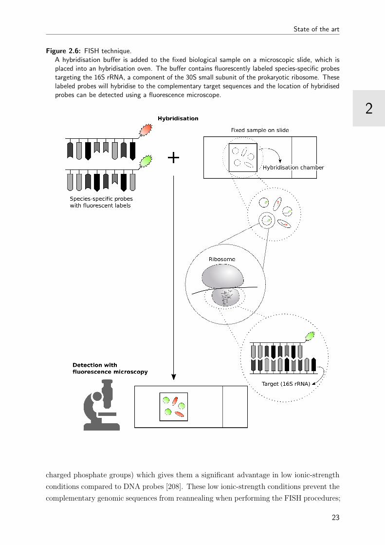
Figure 2.6: FISH technique.A hybridisation buffer is added to the fixed biological sample on a microscopic slide, which isplaced into an hybridisation oven. The buffer contains fluorescently labeled species-specific probestargeting the 16S rRNA, a component of the 30S small subunit of the prokaryotic ribosome. Theselabeled probes will hybridise to the complementary target sequences and the location of hybridisedprobes can be detected using a fluorescence microscope.
charged phosphate groups) which gives them a significant advantage in low ionic-strength
conditions compared to DNA probes [208]. These low ionic-strength conditions prevent the
complementary genomic sequences from reannealing when performing the FISH procedures;
23
State of the art
2
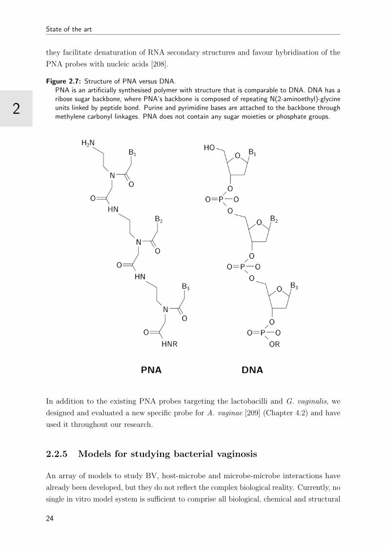
they facilitate denaturation of RNA secondary structures and favour hybridisation of the
PNA probes with nucleic acids [208].
Figure 2.7: Structure of PNA versus DNA.PNA is an artificially synthesised polymer with structure that is comparable to DNA. DNA has aribose sugar backbone, where PNA’s backbone is composed of repeating N(2-aminoethyl)-glycineunits linked by peptide bond. Purine and pyrimidine bases are attached to the backbone throughmethylene carbonyl linkages. PNA does not contain any sugar moieties or phosphate groups.
In addition to the existing PNA probes targeting the lactobacilli and G. vaginalis, we
designed and evaluated a new specific probe for A. vaginae [209] (Chapter 4.2) and have
used it throughout our research.
2.2.5 Models for studying bacterial vaginosis
An array of models to study BV, host-microbe and microbe-microbe interactions have
already been developed, but they do not reflect the complex biological reality. Currently, no
single in vitro model system is sufficient to comprise all biological, chemical and structural
24
2
State of the art
human features. Different types of in vitro cell models exist, ranging from simple, relatively
inexpensive models to more complex, costly systems.
• Monolayer vaginal epithelial cell culture models can be grown in standard
tissue culture equipment using primary and immortalised vaginal epithelial cell lines.
They have been used for the evaluation of epithelial immune responses and the
impact and safety of products on the vaginal epithelium [210]. Bacteria can be
co-cultured on monolayer cell cultures to investigate surface interactions between
bacteria and cells, but bacterial growth is limited in this kind of model [211,212].
• Cell culture insert multilayer models are established on insert systems. Growing
cells on plastic insert with a porous membrane and exposing the upper cell layer to
oxygen produces polarised, differentiated, 3D multilayer cultures. The separated
apical and basal chamber facilitate studying of secretion of host products and the
set-up can be used for cell migration assays. In this model, bacterial growth is also
supported by carbon sources produced by the vaginal epithelium. Furthermore, the
multilayer model allows for the development of biofilm, consistent with the in vivo
situation. The system can also be enhanced by adding immune cells, but currently
it still lacks other cell types and underlying structures that are associated with the
vaginal mucosa [213,214].
• Rotating wall vessel bioreactor-derived 3D cell culture models are gener-
ated when human vaginal epithelial cells are combined with collagen-coated micro-
carrier beads under constant low fluid shear in a fluid-filled rotating wall vessel
bioreactor. The fully differentiated aggregates can be seeded into multiwell plates
to study the innate immune system, epithelial barrier function (including mucus
production), and epithelial-specific responses to introduced organisms. So far, no
immune cells have been incorporated in this model system and it takes 28 days to
culture a fully differentiated system [215,216].
Using an in vivo animal model would be a more advanced approach to study BV patho-
genesis, bacterial interactions, adverse pregnancy outcomes and the safety and efficacy of
candidate products for prevention and treatment of BV. Already in 1961, Gardner and
Dukes [217] unsuccessfully attempted to establish models for vaginal infection using mice,
guinea pigs, rats, and rabbits. This work was succeeded by several other attempts in
small-animal systems and nonhuman primates (Table 2.3). It has to be noted that the
typical characteristics of the human vagina, such as the Lactobacillus-dominance, high
availability of glycogen and lactic acid, and the low pH, are not reflected in these animal
systems [210,218]. This poses significant limitations and questions the relevance of these
systems as a model for vaginal dysbiosis.
25

State of the art
2
Table 2.3: Animal models used to mimic the human vaginal environment
Animal Use Reference
Nonhuman primates Microbicide safety/efficacy testing [219,220]Bacterial biofilm formation on vaginal rings [221]Safety of probiotics [222]Vaginal colonisation with G. vaginalis [223]
Grivet monkey Model for bacterial vaginosis [224]Rabbit Contraceptive safety/efficacy testing [225]
Foetal infection with G. vaginalis [226]Mouse Model for group B streptococci colonisation [227]
Microbicide safety/efficacy testing [220,228]Model for bacterial vaginosis [229]Antifungal safety/efficacy testing [230]Basic research: role of mucus sialoglycans [231]Inhibition of G. vaginalis colonisation by DNAse [232]Model for non-BV and BV vagina [233]Efficacy of probiotics [234]Model for Mycoplasma hominis infection [235]
2.2.6 Bacterial vaginosis treatment
The regimen of symptomatic treatment for non-pregnant women according to the Centers
for Disease Control (CDC) in 2015 [236] was:
• Metronidazole 500 mg orally twice a day for 7 days
• or Metronidazole gel 0.75%, one full applicator (5 g) intravaginally, once a day for 5
days
• or Clindamycin cream 2%, one full applicator (5 g) intravaginally at bedtime for 7
days
Alternative regimens are:
• Tinidazole 2 g orally once daily for 2 days
• or Tinidazole 1 g orally once daily for 5 days
• or Clindamycin 300 mg orally twice daily for 7 days
• or Clindamycin ovules 100 mg intravaginally once at bedtime for 3 days
It is also recommended to treat all symptomatic pregnant women, using Metronidazole 500
mg orally twice daily for 7 days [236], which has been demonstrated to reduce bacterial
overgrowth but not the number of preterm deliveries [237].
Unfortunately, the currently available treatments have been shown to have poor initial cure
26
2
State of the art
rates in 10% to 15% of patients and relapse rates of up to 80% in those who show initial
response [238,239]. In addition, these ineffective treatments can increase drug resistance in
G. vaginalis, Prevotella, Bacteroides and Peptostreptococcus spp. [240–242]. Alternatives
for these failing antibiotic treatments are increasingly being explored, using probiotics,
prebiotics, synbiotics, antiseptics, desinfectants, vaginal acidifying and buffering agents and
combinations of different therapies, but until now, none has been successful [47,243].
27

State of the art
2
2.3 Gardnerella vaginalis
2.3.1 What’s in a name?
G. vaginalis was isolated for the first time in 1953 from men with prostatitis and women
with cervicitis by Leopold [244] and was described as a small, nonmotile, nonencapsulated,
pleomorphic Gram-negative rod. It was named Haemophilus vaginalis by Gardner and
Dukes in 1955 [245]. Later it was renamed Corynebacterium vaginale by Zinner and
Turner [246], because it did not require hemin and nicotinamide adenine dinucleotide to
grow, unlike Haemophilus species; because of its diphtheroid (i.e. corynebacterium-like)
cell morphology; and because it had a tendency to retain violet dye after Gram staining,
unlike the Gram-negative Haemophilus species (which implies that the Haemophilus species
lose the crystal violet dye).
In 1980, Greenwood and Pickett [247] suggested a new genus for “Corynebacterium vaginale”
and proposed the name Gardnerella vaginalis. This was supported by Piot et al. [248,249].
Years later, Van Esbroeck et al. [250] made an attempt to place G. vaginalis in the
Gram-positive genus Bifidobacterium, based on phylogenetic analysis, but despite the
high level of similarity it shares with this genus, the difference in G+C content6 between
G. vaginalis (42 mole %) and the genus Bifidobacterium (55-67 mole %) is too large to
consider G. vaginalis as a genuine Bifidobacterium species. To date G. vaginalis remains
the sole member of the genus Gardnerella, that is part of the Bifidobacteriaceae family
(Figure 2.8).
Figure 2.8: Taxonomic ranking of G. vaginalis
Class
Order
Family
Genus
Species
Actinobacteria
Bifidobacteriales
Bifidobacteriaceae
Gardnerella
vaginalis
G. vaginalis was defined as a facultative anaerobic, small (0.4 by 1.0 to 1.5 µm), rod-
shaped, nonencapsulated, catalase, oxidase, and β-glucosidase negative, Gram-variable7
6G+C content: the percentage of guanine and cytosine in the chromosome7Gram-variable: the bacterium appears Gram-positive during the exponential growth phase, but
Gram-negative as the culture ages because the thinning of the peptidoglycan layer results in an inabilityto retain the crystal-violet aggregates.
28
2
State of the art
bacterium with a laminated cell wall, that produces acetic acid as the major end product
of fermentation of carbohydrates [80, 247]. G. vaginalis can, due to its cell wall struc-
ture, phylogenetically be classified as a Gram-positive bacterium, even though its thin
peptidoglycan layer results in the Gram-variability [157].
2.3.2 Biotypes
Piot et al. [251] assigned G. vaginalis to eight different biotypes, based on the activity of
three enzymes: β-galactosidase, lipase (breakdown of lipids) and hippurate hydrolase. In
this study, 359 strains were tested and the most common types were type 1 (β-galactosidase-
positive, lipase-positive and hippurate-positive), type 2 (β-galactosidase-negative, lipase-
positive and hippurate-positive) and type 5 (β-galactosidase-negative, lipase-negative and
hippurate-positive). No specific differences were found in biotypes between strains isolated
from women with and without BV, but up to 14% of women with BV harboured at least two
different biotypes. However, a study [252] using 261 strains did find a significant difference
in the distribution of biotypes from women with and without BV, with a predominance of
lipase-positive strains (biotypes 1, 2, 3 and 4) in women with BV. Furthermore, women
that acquired BV were likely to have a shift in biotype, suggesting that G. vaginalis isolates
recovered from these women represented newly acquired strains rather than overgrowth
of previously colonising biotypes [252]. This finding was confirmed by Numanovic et
al. [253], but once again contradicted by Aroutcheva et al. [254], who found a diversity
of biotypes in the BV group. In summary, the interrelationship of the different biotypes
with the occurrence of BV is still controversial and unproven. A clear view is probably
hampered also because several G. vaginalis isolates of the reference set used by Piot et
al. [251] appear to be L. iners that were misidentified as G. vaginalis (Mario Vaneechoutte,
personal communication).
To cope with the limitations of the phenotypic G. vaginalis biotyping scheme, modified
techniques were implemented, albeit still based on bacterial culture [255]. More recently,
molecular approaches such as random amplified polymorphic DNA (RAPD) and amplified
ribosomal DNA restriction analysis (ARDRA) identified three to four different G. vaginalis
genotypes in clinical isolates [256]. Additionally, more recent advances in next generation
sequencing technology were used to differentiate G. vaginalis strains and subgroups
according to sequence variations in 16S rRNA and the cpn60 genes, and based on whole
genome analysis [161, 162, 257, 258]. Ahmed et al. [161] suggested dividing G. vaginalis
into four clades after the genomic analysis of 17 G. vaginalis isolates. Each clade was
a molecular subgroup with a distinct gene pool and genomic properties, and the clades
possibly correspond to four different species that differ in metabolic capabilities and
virulence [159, 161]. A study by Balashov et al. [259] made an attempt using vaginal
29
State of the art
2
samples to describe the four clades of G. vaginalis by subtyping multiplex qPCR. Clade
1 and 4 were the most prevalent clades in the vaginal specimens, and multiple clades in
one specimen were found as well. The presence of clade 1, clade 3, and the presence of
multiple clades correlated with BV, while the detection of clade 2 was associated with an
intermediate microbiome and clade 4 did not show any correlation with BV [259].
2.3.3 Virulence factors
G. vaginalis is present in up to 97.5% of cases of BV [40–42,157]. However, the presence
of G. vaginalis in the vagina of women with Nugent scores of 0-3 indicates that its mere
presence can not be used as a specific marker for BV. Indeed, G. vaginalis is also present
in 50% to 70% of women with a Nugent score of 0-3 [40,41,158].
G. vaginalis is equipped with a number of virulence factors which facilitate vaginal biofilm
formation and thus contribute to G. vaginalis’ pathogenic potential. Multiple studies
show that G. vaginalis is more virulent in vitro than many of the other bacterial species
frequently isolated from BV patients [260–262]. Because of its virulence potential and its
abundance in the vaginal microbiome, G. vaginalis is believed to be the initial coloniser
of the vaginal mucosa and epithelium, serving as the scaffolding to which other bacteria
can attach [9, 209,260,263–265]. The ability to adhere to vaginal epithelial cells has been
suspected ever since Gardner and Dukes [245] postulated the value of clue cells for the
diagnosis of BV. Clue cells are squamous epithelial cells whose surfaces are heavily coated
with bacteria (Figure 2.9). By adhering to the vaginal cells, G. vaginalis could colonise the
epithelium, possibly minimising contact with potentially deleterious extracellular enzymes
and local antibodies, and reducing the possibility of being flushed away with vaginal
fluid.
Biofilm formation
G. vaginalis can form a biofilm [9] on the vaginal epithelium, which has been suggested to
increase its tolerance to lactic acid and hydrogen peroxide produced by lactobacilli [260].
The association of G. vaginalis in a biofilm could also explain its decreased susceptibility to
antimicrobial treatment and the recurrent symptoms [266,267]. The section on microbial
biofilms will elaborate on their properties.
30

2
State of the art
Figure 2.9: Clue cell after Gram stain.Clue cells are characterised by the dotted pattern which are bacteria covering the epithelial cell.
Pili and fimbriae
A first step in vaginal colonisation is adherence to the epithelium cells, which can be
facilitated by fimbriae8 or pili. Pili9 with a diameter between 3.0 and 7.5 nm, radiating
from the surface of G. vaginalis cells, could be observed by electron microscopy in some G.
vaginalis strains (but not all). Fresh isolates were more heavily piliated than laboratory
strains that had been subcultured several times [223,268].
Exopolysaccharide
Adherence to vaginal epithelial cells and clustering of G. vaginalis cells is facilitated by the
production of an electron-dense, fibrillar extracellular polymeric substances (EPS) layer.
This microcapsular material can be seen as weblike strands connecting cells lying closely
together. This fibrillar layer was visualised with electron microscopy after staining with
ruthenium red to visualise the polysaccharide component [269].
Iron acquisition
Next to being an essential growth factor, iron also plays an important role in the virulence
of bacterial pathogens. Therefore, a number of bacteria have developed high-affinity iron
acquisition mechanisms, such as secretion of high-affinity iron chelators or siderophores,
8Fimbria: A filamentous structure composed of proteins that extend from the surface of a cell and canbe involved in attachment or can assist in disease processes
9Pilus: A bacterial surface structure, similar to a fimbria but longer, that is present on the cell surfacein one or two copies. Type IV pili mediate twitching motility and can be involved in biofilm development
31
State of the art
2
the expression of cell-surface receptors that directly bind to iron-containing compounds,
and haemolysis [270]. All three mechanisms are used by G. vaginalis: it can produce
siderophores [270] and it can bind iron carriers, e.g. catalase [271], lactoferrin [272], trans-
ferrin [272], haeme [273], and haemoglobin [274]. Additionally, by producing vaginolysin, it
induces haemolysis and the release of iron-rich compounds from red blood cells [263].
Vaginolysin
The G. vaginalis vaginolysin is a spore-forming, cytolytic exotoxin [263]. G. vaginalis
vaginolysin is a member of the cholesterol-dependent cytolysin family of toxins and is
selective for human cells, through recognition of the complement regulatory molecule
CD59 [263, 275, 276]. Vaginolysin was previously called a haemolysin because it lyses
endothelial cells, neutrophils, and erythrocytes [277–281]. This ability to lyse neutrophils
might explain the relative absence of neutrophils in BV [282].
Vaginolysin can also trigger the immune system, which is upregulated in BV [82,283]. This
upregulation is mediated by the human epithelial cells inducing interleukin-8 production,
and through a specific secretory immunoglobulin A (IgA) targeting the G. vaginalis
vaginolysin [84,281,284].
Sialidase
Some genotypes of G. vaginalis can produce sialidase [160]. Sialidase, also known as
neuraminidase, is a common virulence factor in various organisms such as the influenza
virus [285] and a large number of bacterial species, such as Propionibacterium acnes [286],
Pseudomonas aeruginosa [287], Streptococcus pneumoniae [288] and Vibrio cholerae [289]
and has been strongly linked with the production of biofilm in vitro [290–292]. Increased
sialidase activity was detected in the vaginal fluid of women diagnosed with BV [293]. This
finding was also used in the development of a quick test for diagnosis of BV, i.e. BVBlue®,
that is based on the detection of sialidase activity in vaginal fluid [294]. However, it is
important to acknowledge that other BV-associated bacteria (e.g. Prevotella spp.) are
also known to produce sialidase, hence the BVBlue® test is not specific for G. vaginalis
sialidase.
Sialidase facilitates the destruction of the protecting mucus layer in the vagina by hydrolysis
of sialic acid, which is the most distal sugar moiety on the glycans of mucous epithelial
membranes [293, 295]. After cleaving off the sialic acid by sialidase, the sialic acid can
serve as a nutrient [296] and the exposed glycoconjugates can serve as receptors for the
bacterial cells [295]. G. vaginalis could benefit from this mechanism by attaching to the
vaginal epithelium to initiate the formation of a biofilm. Furthermore, sialidase helps to
32
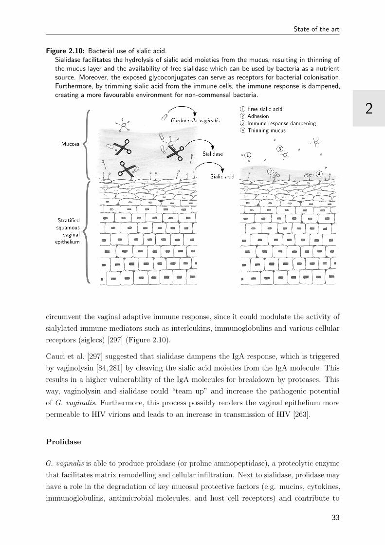
2
State of the art
Figure 2.10: Bacterial use of sialic acid.Sialidase facilitates the hydrolysis of sialic acid moieties from the mucus, resulting in thinning ofthe mucus layer and the availability of free sialidase which can be used by bacteria as a nutrientsource. Moreover, the exposed glycoconjugates can serve as receptors for bacterial colonisation.Furthermore, by trimming sialic acid from the immune cells, the immune response is dampened,creating a more favourable environment for non-commensal bacteria.
circumvent the vaginal adaptive immune response, since it could modulate the activity of
sialylated immune mediators such as interleukins, immunoglobulins and various cellular
receptors (siglecs) [297] (Figure 2.10).
Cauci et al. [297] suggested that sialidase dampens the IgA response, which is triggered
by vaginolysin [84,281] by cleaving the sialic acid moieties from the IgA molecule. This
results in a higher vulnerability of the IgA molecules for breakdown by proteases. This
way, vaginolysin and sialidase could “team up” and increase the pathogenic potential
of G. vaginalis. Furthermore, this process possibly renders the vaginal epithelium more
permeable to HIV virions and leads to an increase in transmission of HIV [263].
Prolidase
G. vaginalis is able to produce prolidase (or proline aminopeptidase), a proteolytic enzyme
that facilitates matrix remodelling and cellular infiltration. Next to sialidase, prolidase may
have a role in the degradation of key mucosal protective factors (e.g. mucins, cytokines,
immunoglobulins, antimicrobial molecules, and host cell receptors) and contribute to
33

State of the art
2
the exfoliation and detachment of vaginal epithelial cells. It has been suggested that
prolidase could modulate immune mediators causing an inefficient immune cascade after
the initial IL-1β rise, which would explain why women with BV does not inflammatory
signs [111,298].
Phospholipase A2 activity
G. vaginalis can exert phospholipase A2 activity, which triggers the labour process [299,300].
Normal labour is initiated by amniotic and chorionic phospholipase A2, by liberation of
arachidonic acid esters from the phospholipids of these membranes, leading to the synthesis
of prostaglandins by the placental membranes. In two studies, phospholipase A2 activity
from bacteria was shown to be several times higher than that of the human counterpart,
and correlated with preterm labour in women with BV [299,300].
2.3.4 Epidemiology
G. vaginalis overgrowth is found in nearly all cases of BV [40–42,157], and has shown a
high sensitivity (100%) but low specificity (49%) for BV diagnosis [80]. Nevertheless, its
presence has been associated with three out of four Amsel criteria: amine odour, elevated
pH, and the presence of clue cells [41].
G. vaginalis is seldom found in children, except in case of sexual abuse [301–304], although
G. vaginalis can be found in young girls [4, 305, 306]. G. vaginalis is more frequently
found in adolescent girls and sexually inexperienced girls, but at significantly lower rates
compared to sexually active adolescents [4, 307–309]. In sexually active adult women, G.
vaginalis is a common part of the normal vaginal microbiome [3,15].
G. vaginalis is one of the bacterial species that can be involved in urinary tract infections
(UTI), possible because of the presence of vaginal squamous epithelium in the region of the
bladder trigone in postpubertal women [157,310,311]. It has been found in the rectum and
in the oral cavity, which both can act as extravaginal reservoirs [4,154–156]. Carriage of G.
vaginalis is common in men as well, and it is more often found in the genital tract than in
the urinary tract [157,311]. The sole presence of G. vaginalis does not give rise to symptoms
in most men, but the bacterium might invade the prostate or bladder [312,313] and become
more pathogenic, especially in patients who have undergone a urological procedure [157].
Seminal colonisation by G. vaginalis also occurs [305,311,314–317] but there is no evidence
that it could affect the semen quality in men [318]. Furthermore, bloodstream infections
involving G. vaginalis occur far more frequently in women than in men, often after surgical
intervention, postpartum endometritis, and septic abortion [157,310].
34
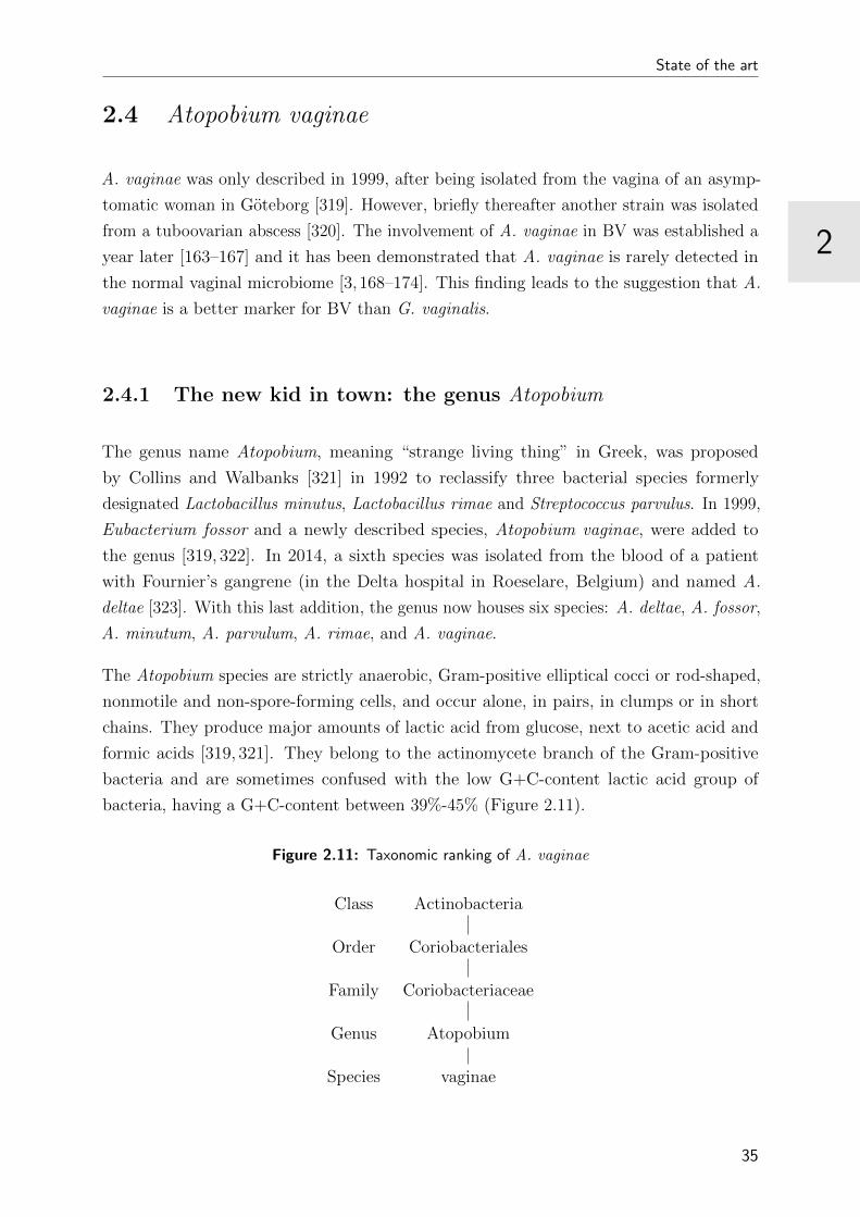
2
State of the art
2.4 Atopobium vaginae
A. vaginae was only described in 1999, after being isolated from the vagina of an asymp-
tomatic woman in Goteborg [319]. However, briefly thereafter another strain was isolated
from a tuboovarian abscess [320]. The involvement of A. vaginae in BV was established a
year later [163–167] and it has been demonstrated that A. vaginae is rarely detected in
the normal vaginal microbiome [3,168–174]. This finding leads to the suggestion that A.
vaginae is a better marker for BV than G. vaginalis.
2.4.1 The new kid in town: the genus Atopobium
The genus name Atopobium, meaning “strange living thing” in Greek, was proposed
by Collins and Walbanks [321] in 1992 to reclassify three bacterial species formerly
designated Lactobacillus minutus, Lactobacillus rimae and Streptococcus parvulus. In 1999,
Eubacterium fossor and a newly described species, Atopobium vaginae, were added to
the genus [319, 322]. In 2014, a sixth species was isolated from the blood of a patient
with Fournier’s gangrene (in the Delta hospital in Roeselare, Belgium) and named A.
deltae [323]. With this last addition, the genus now houses six species: A. deltae, A. fossor,
A. minutum, A. parvulum, A. rimae, and A. vaginae.
The Atopobium species are strictly anaerobic, Gram-positive elliptical cocci or rod-shaped,
nonmotile and non-spore-forming cells, and occur alone, in pairs, in clumps or in short
chains. They produce major amounts of lactic acid from glucose, next to acetic acid and
formic acids [319, 321]. They belong to the actinomycete branch of the Gram-positive
bacteria and are sometimes confused with the low G+C-content lactic acid group of
bacteria, having a G+C-content between 39%-45% (Figure 2.11).
Figure 2.11: Taxonomic ranking of A. vaginae
Class
Order
Family
Genus
Species
Actinobacteria
Coriobacteriales
Coriobacteriaceae
Atopobium
vaginae
35
State of the art
2
2.4.2 Epidemiology
Atopobium species occur in the oral cavity (A. rimae, A. parvulum), the vagina (A.
vaginae) [322], or the pharynx of horses (A. fossor) [324], but can also rarely be isolated
from human infections: dental infections (A. rimae, A. parvulum), sepsis (A. rimae [325],
A. deltae [323]), abdominal wounds, and pelvic abscesses (A. minutum) [319].
The identification and differentiation of Atopobium species from other non-spore-forming
Gram-positive bacilli is often laborious and carries the risk of misidentification, especially in
the presence of coexisting colonising organisms, due to its variable cell morphology [80,326].
In addition, it is difficult to isolate and culture this bacterium. Therefore it is likely that
the incidence of colonisation and infection of the female genital tract by A. vaginae was
underestimated in the past.
A. vaginae has been reported to be the cause of foetal death and bacteraemia in the mother
due to an intrauterine infection after a chorionic villus sample [327]. It has also been
reported to be the source of intrapartum bacteraemia in a patient showing a disturbed
vaginal microbiome with overgrowth of G. vaginalis and C. albicans [326].
PID is one of the possible sequelae of BV. It is a condition in which tuboovarian abscesses
can be formed involving the oviduct and possibly other segments of the upper genital
tract [123, 328]. PID is caused by ascending vaginal and endocervical infections. Being
one of the main bacteria involved in BV, A. vaginae has an indirect role in PID [170,171].
Furthermore, A. vaginae has also been recovered from patients with salpingitis [329] and
as the sole microorganism from a patient with a tuboovarian abscess [320]. In conclusion,
A. vaginae may be partly responsible for the association between BV and PID.
2.4.3 A. vaginae in BV
A. vaginae has been associated with vaginal discharge, elevated pH and the presence of
clue cells, adding up to three out of four Amsel criteria [41]. The involvement of A. vaginae
in BV has only recently been established [163,165–167]. Since then, several researchers
have demonstrated that the occurrence of BV may correspond better with the presence of
high concentrations of A. vaginae than with high quantities of G. vaginalis in the vaginal
tract [170, 171]. In 2005, Swidsinski et al. described for the first time the presence of
a vaginal biofilm in which G. vaginalis and A. vaginae co-existed [9]. In this thesis, we
elaborate on the presence of this species in vaginal biofilms in the section “Experimental
work”.
The involvement of A. vaginae in BV has a major impact on treatment of the condition.
Apart from being part of a vaginal biofilm, which decreases the susceptibility to antibiotic
36

2
State of the art
treatment (elaborated on in the next section), it can also be resistant to metronidazole,
the standard treatment for BV. De Backer et al. [330] demonstrated that susceptibility to
metronidazole varied significantly across various A. vaginae strains in vitro. Another in
vitro study [331,332] showed that A. vaginae is susceptible to clindamycin and nifuratel.
Unfortunately there is a scarcity of in vivo data, but in a study with topical metronidazole
gel by Ferris et al. [163], a high concentration of A. vaginae before treatment was associated
with complete or partial failure of treatment for BV. Bradshaw et al. [170] found that rates
of recurrence of BV were higher when A. vaginae was present in the vaginal microbiome in
addition to G. vaginalis, even though there was a reduction in A. vaginae after treatment
with metronidazole.
Similarly to G. vaginalis, A. vaginae can also be a strong trigger of inflammation and
vaginal epithelial innate immune responses [212, 333–335]. In cervicovaginal epithelial
cells in vitro, A. vaginae activates the major proinflammatory transcription factor NF-
κB [212] and it significantly boosts the expression of chemokines in vaginal and cervical
epithelial cells, including IL-8 [211, 212, 333, 334], MIP-3 (CCL20) [335], and RANTES
(CCL5) [212,333]. In vivo studies have shown that the detection of A. vaginae in vaginal
specimens correlated with higher levels of the same inflammatory markers that were also
associated with G. vaginalis [82], and that A. vaginae was among the most abundant
taxa in the vaginal microbiome types distinguished by the highest levels of cervicovaginal
inflammatory markers [336].
Since A. vaginae has been isolated only 17 years ago and its association with BV was
demonstrated even more recently, we still know little regarding this microorganism and its
relation with the commensal and pathogenic vaginal microbiome. A considerable amount
of research needs to be done before denouncing this microorganism as one of the main
causes of BV.
37
State of the art
2
2.5 Microbial biofilm
Microbes rarely exist as single-species planktonic forms, but thrive in complex polymicro-
bial10 sessile biofilm communities [337,338].
A bacterial biofilm is a structured community of bacterial cells, adherent to an inert
surface or living biological tissue and enclosed in a mucous substance (“slime”), which is
a self-produced matrix of EPS [339]. This community is characterised by a complicated
internal architecture: e.g. channels for circulating nutrients [340], genetically identical
cells in separate areas of the biofilm that exhibit different patterns of gene expression [341]
and extracellular DNA (eDNA) [232,342,343].
This biofilm mode of growth facilitates an enhanced tolerance to adverse conditions, which
allows survival in hostile environments and offers protection against chemical disinfection,
antimicrobial treatment, and human immune responses [7,339,344,345]. Mixed-species
biofilms are the dominant form of microbial organisation in nature, and are also prominent
in the human body [7]. It has been estimated that 65% to 80% of human infections are
associated with biofilm, for example Pseudomonas aeruginosa colonising the lungs of cystic
fibrosis patients or Staphylococcus epidermidis infecting wounds on the skin [346].
2.5.1 Bacterial biofilm infections
The first multispecies biofilm was observed by Antonie van Leeuwenhoek more than
300 years ago, when studying “animalculi” in dental plaque, but was not recognised as
such [347]. For a long time, microbial infection in the human body was envisioned as caused
by single-species planktonic bacteria. This view, while explaining the pathogenesis of acute
infections, was inadequate for understanding chronic and recurrent conditions [348]. To
deal with the shortcoming of this classic view, Costerton et al. [339, 349,350] investigated
the association of a bacterial biofilm with these chronic infections.
Biofilm infections share clinical characteristics, regardless of the location in the human
body where the biofilm develops. Although colonisation often occurs on inert surfaces, e.g.
medical devices and prostheses, or on dead tissue, biofilm can also develop on living tissue,
as in the case of endocarditis [339], and stagnated mucosal epithelium-associated mucus,
as in the case of cystic fibrosis [7]. Biofilms grow slowly and consequently symptoms
are also produced in a slow fashion [351]. Biofilm communities are rarely fully destroyed
by the host defence mechanisms. Sessile bacterial cells in a biofilm release antigens
resulting in an increase in antibody production. However, due to the biofilm structure, the
produced antibodies are not capable of killing the biofilm bacteria and accumulate in the
10Polymicrobial: diverse in species and/or strain content.
38
2
State of the art
surrounding tissues, which results in immune complex-related damage to these tissues [352].
Neutrophils are also attracted to the biofilm, where they continuously release antimicrobial
granule contents and reactive oxygen species (ROS) that promote collagen degradation
and subsequent host tissue injury as well [353]. On top of that, as antibiotic therapy fails
to kill the biofilm, only symptoms caused by the planktonic cells released from the biofilm
are reversed after treatment [339, 354]. As a result, even after multiple cycles of antibiotic
therapy, biofilm infections continue to cause recurring symptoms [341].
2.5.2 Stages in the biofilm life cycle
Biofilm formation is facilitated by a regulated switch between the planktonic lifestyle and
the sedentary multicellular state of bacteria. The biofilm life cycle includes three major
steps: attachment, growth of colonies (development), and detachment of planktonic cells
(Figure 2.12).
Attachment
Zobell [355] demonstrated in vitro in 1943 already that growth on a surface is more
favourable to bacteria than drifting in the surrounding aqueous phase.
When environmental stress (such as antibiotics) favours biofilm formation, the first and
most important step of the biofilm cycle is initiated. Motile and nonmotile bacterial
species deploy different methods to localise a surface to colonise. In case of motile species,
the individual bacterium will localise a surface and initiate a lifestyle switch, losing its
motility.
Nonmotile species increase expression of adhesins on their outer surface, a mechanism
that makes them “sticky” and thus promotes cell-cell and cell-surface adherence when
the bacteria encounter another cell or a surface [356,357]. Bacterial adhesion to a surface
has been described as a two-phase process. The initial encounter with a surface usually
leads to transient adherence, because of weak reversible bonds called van der Waals forces,
that can be repulsive or attractive. If the repulsive forces are greater than the attractive
forces, the bacteria will detach again [358]. However, if the bacterial colonists are not
immediately disengaged from the surface, they anchor themselves permanently using their
cell adhesion molecules 11 which results in a stable surface association [356,359].
Attachment to a surface can be mediated by the bacterial capsule, fimbriae, and fibrillae
through adhesins (Figure 2.13).
11Cell adhesion molecules: proteins on the bacterial surface that bind cells, or adhesins.
39
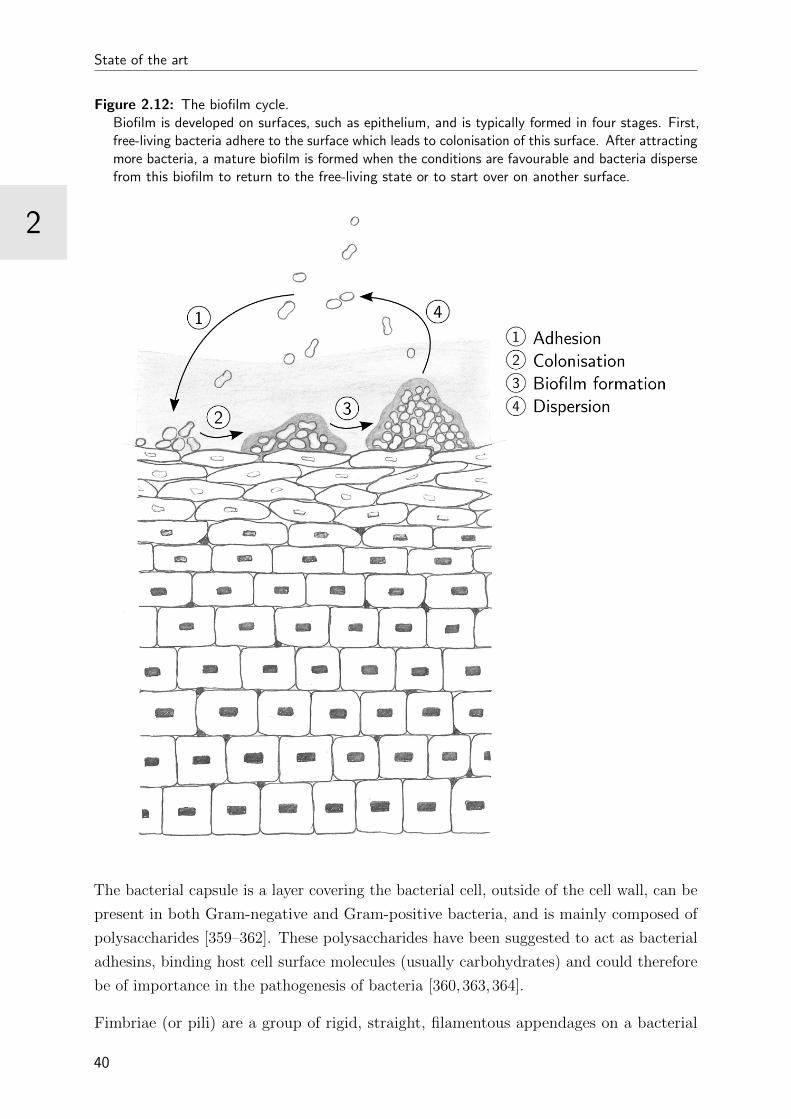
State of the art
2
Figure 2.12: The biofilm cycle.Biofilm is developed on surfaces, such as epithelium, and is typically formed in four stages. First,free-living bacteria adhere to the surface which leads to colonisation of this surface. After attractingmore bacteria, a mature biofilm is formed when the conditions are favourable and bacteria dispersefrom this biofilm to return to the free-living state or to start over on another surface.
The bacterial capsule is a layer covering the bacterial cell, outside of the cell wall, can be
present in both Gram-negative and Gram-positive bacteria, and is mainly composed of
polysaccharides [359–362]. These polysaccharides have been suggested to act as bacterial
adhesins, binding host cell surface molecules (usually carbohydrates) and could therefore
be of importance in the pathogenesis of bacteria [360,363,364].
Fimbriae (or pili) are a group of rigid, straight, filamentous appendages on a bacterial
40

2
State of the art
Figure 2.13: Mechanisms of bacterial adhesion: fimbriae and capsule.Attachment to surfaces is facilitated by fimbriae and the bacterial capsule. Fimbriae (or pili) area group of short, rigid, straight, filamentous appendages on a bacterial surface, prominent inGram-negative bacteria. The bacterial capsule is a covering layer outside of the cell wall, whichcan be present in both Gram-negative and Gram-positive bacteria.
surface, prominent in Gram-negative bacteria and no more than 4 to 7 nm in diameter
and 0.2 to 20 nm in length [359]. Fimbriae are polymers, composed primarily of identical
protein subunits called pilin [365], that arise from proteins in the outer surface of the outer
membrane and can be present in several hundred to one thousand copies per cell [366,367].
Bacterial adhesion is mediated by fimbriae through associated adhesins, by adhesive
subunits, or by fimbriae-dependent surface hydrophobicity [359]. Bacterial fimbriae have
been shown to be related to virulence: Gram-negative bacteria possessing fimbriae are
more infectious than their non-fimbriated variants [368,369].
Fibrillae are amorphous surface adhesive structures that lack the regular filamentous forms
of fimbriae, and are anchored in the cell wall. Fibrillae are more common in Gram-positive
than in Gram-negative bacteria and have for example been observed at the surface of
various streptococci as short, stubby appendages [359].
Several generalisations concerning bacterial adhesion and adhesins can be made [359]:
41
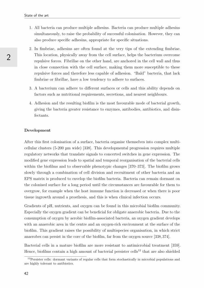
State of the art
2
1. All bacteria can produce multiple adhesins. Bacteria can produce multiple adhesins
simultaneously, to raise the probability of successful colonisation. However, they can
also produce specific adhesins, appropriate for specific situations.
2. In fimbriae, adhesins are often found at the very tips of the extending fimbriae.
This location, physically away from the cell surface, helps the bacterium overcome
repulsive forces. Fibrillae on the other hand, are anchored in the cell wall and thus
in close connection with the cell surface, making them more susceptible to these
repulsive forces and therefore less capable of adhesion. “Bald” bacteria, that lack
fimbriae or fibrillae, have a low tendency to adhere to surfaces.
3. A bacterium can adhere to different surfaces or cells and this ability depends on
factors such as nutritional requirements, secretions, and nearest neighbours.
4. Adhesion and the resulting biofilm is the most favourable mode of bacterial growth,
giving the bacteria greater resistance to enzymes, antibodies, antibiotics, and disin-
fectants.
Development
After this first colonisation of a surface, bacteria organise themselves into complex multi-
cellular clusters (5-200 µm wide) [338]. This developmental progression requires multiple
regulatory networks that translate signals to concerted switches in gene expression. The
modified gene expression leads to spatial and temporal reorganisation of the bacterial cells
within the biofilms and to observable phenotypic changes [370–373]. The biofilm grows
slowly through a combination of cell division and recruitment of other bacteria and an
EPS matrix is produced to envelop the biofilm bacteria. Bacteria can remain dormant on
the colonised surface for a long period until the circumstances are favourable for them to
overgrow, for example when the host immune function is decreased or when there is poor
tissue ingrowth around a prosthesis, and this is when clinical infection occurs.
Gradients of pH, nutrients, and oxygen can be found in this microbial biofilm community.
Especially the oxygen gradient can be beneficial for obligate anaerobic bacteria. Due to the
consumption of oxygen by aerobic biofilm-associated bacteria, an oxygen gradient develops
with an anaerobic area in the centre and an oxygen-rich environment at the surface of the
biofilm. This gradient raises the possibility of multispecies organisation, in which strict
anaerobes can persist in the core of the biofilm, far from the oxygen source [338,374].
Bacterial cells in a mature biofilm are more resistant to antimicrobial treatment [359].
Hence, biofilms contain a high amount of bacterial persister cells12 that are also shielded
12Persister cells: dormant variants of regular cells that form stochastically in microbial populations andare highly tolerant to antibiotics.
42
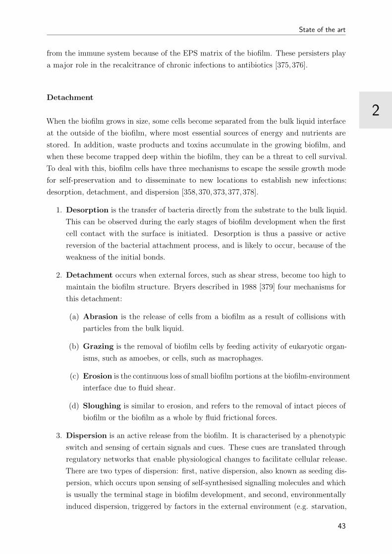
2
State of the art
from the immune system because of the EPS matrix of the biofilm. These persisters play
a major role in the recalcitrance of chronic infections to antibiotics [375,376].
Detachment
When the biofilm grows in size, some cells become separated from the bulk liquid interface
at the outside of the biofilm, where most essential sources of energy and nutrients are
stored. In addition, waste products and toxins accumulate in the growing biofilm, and
when these become trapped deep within the biofilm, they can be a threat to cell survival.
To deal with this, biofilm cells have three mechanisms to escape the sessile growth mode
for self-preservation and to disseminate to new locations to establish new infections:
desorption, detachment, and dispersion [358,370,373,377,378].
1. Desorption is the transfer of bacteria directly from the substrate to the bulk liquid.
This can be observed during the early stages of biofilm development when the first
cell contact with the surface is initiated. Desorption is thus a passive or active
reversion of the bacterial attachment process, and is likely to occur, because of the
weakness of the initial bonds.
2. Detachment occurs when external forces, such as shear stress, become too high to
maintain the biofilm structure. Bryers described in 1988 [379] four mechanisms for
this detachment:
(a) Abrasion is the release of cells from a biofilm as a result of collisions with
particles from the bulk liquid.
(b) Grazing is the removal of biofilm cells by feeding activity of eukaryotic organ-
isms, such as amoebes, or cells, such as macrophages.
(c) Erosion is the continuous loss of small biofilm portions at the biofilm-environment
interface due to fluid shear.
(d) Sloughing is similar to erosion, and refers to the removal of intact pieces of
biofilm or the biofilm as a whole by fluid frictional forces.
3. Dispersion is an active release from the biofilm. It is characterised by a phenotypic
switch and sensing of certain signals and cues. These cues are translated through
regulatory networks that enable physiological changes to facilitate cellular release.
There are two types of dispersion: first, native dispersion, also known as seeding dis-
persion, which occurs upon sensing of self-synthesised signalling molecules and which
is usually the terminal stage in biofilm development, and second, environmentally
induced dispersion, triggered by factors in the external environment (e.g. starvation,
43
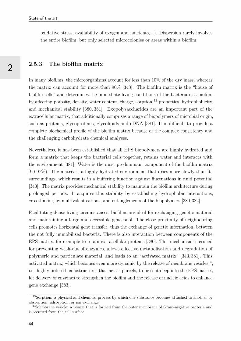
State of the art
2
oxidative stress, availability of oxygen and nutrients,...). Dispersion rarely involves
the entire biofilm, but only selected microcolonies or areas within a biofilm.
2.5.3 The biofilm matrix
In many biofilms, the microorganisms account for less than 10% of the dry mass, whereas
the matrix can account for more than 90% [343]. The biofilm matrix is the “house of
biofilm cells” and determines the immediate living conditions of the bacteria in a biofilm
by affecting porosity, density, water content, charge, sorption 13 properties, hydrophobicity,
and mechanical stability [380, 381]. Exopolysaccharides are an important part of the
extracellular matrix, that additionally comprises a range of biopolymers of microbial origin,
such as proteins, glycoproteins, glycolipids and eDNA [381]. It is difficult to provide a
complete biochemical profile of the biofilm matrix because of the complex consistency and
the challenging carbohydrate chemical analyses.
Nevertheless, it has been established that all EPS biopolymers are highly hydrated and
form a matrix that keeps the bacterial cells together, retains water and interacts with
the environment [381]. Water is the most predominant component of the biofilm matrix
(90-97%). The matrix is a highly hydrated environment that dries more slowly than its
surroundings, which results in a buffering function against fluctuations in fluid potential
[343]. The matrix provides mechanical stability to maintain the biofilm architecture during
prolonged periods. It acquires this stability by establishing hydrophobic interactions,
cross-linking by multivalent cations, and entanglements of the biopolymers [380,382].
Facilitating dense living circumstances, biofilms are ideal for exchanging genetic material
and maintaining a large and accessible gene pool. The close proximity of neighbouring
cells promotes horizontal gene transfer, thus the exchange of genetic information, between
the not fully immobilised bacteria. There is also interaction between components of the
EPS matrix, for example to retain extracellular proteins [380]. This mechanism is crucial
for preventing wash-out of enzymes, allows effective metabolisation and degradation of
polymeric and particulate material, and leads to an “activated matrix” [343,381]. This
activated matrix, which becomes even more dynamic by the release of membrane vesicles14;
i.e. highly ordered nanostructures that act as parcels, to be sent deep into the EPS matrix,
for delivery of enzymes to strengthen the biofilm and the release of nucleic acids to enhance
gene exchange [383].
13Sorption: a physical and chemical process by which one substance becomes attached to another byabsorption, adsorption, or ion exchange.
14Membrane vesicle: a vesicle that is formed from the outer membrane of Gram-negative bacteria andis secreted from the cell surface.
44
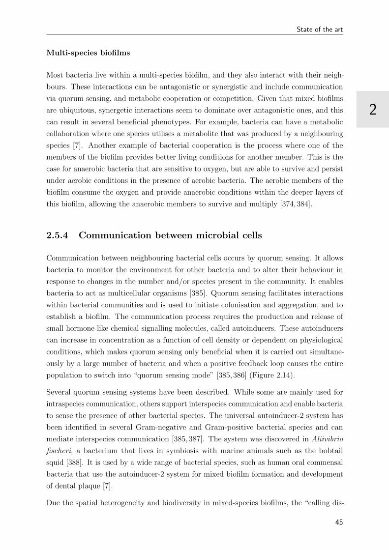
2
State of the art
Multi-species biofilms
Most bacteria live within a multi-species biofilm, and they also interact with their neigh-
bours. These interactions can be antagonistic or synergistic and include communication
via quorum sensing, and metabolic cooperation or competition. Given that mixed biofilms
are ubiquitous, synergetic interactions seem to dominate over antagonistic ones, and this
can result in several beneficial phenotypes. For example, bacteria can have a metabolic
collaboration where one species utilises a metabolite that was produced by a neighbouring
species [7]. Another example of bacterial cooperation is the process where one of the
members of the biofilm provides better living conditions for another member. This is the
case for anaerobic bacteria that are sensitive to oxygen, but are able to survive and persist
under aerobic conditions in the presence of aerobic bacteria. The aerobic members of the
biofilm consume the oxygen and provide anaerobic conditions within the deeper layers of
this biofilm, allowing the anaerobic members to survive and multiply [374,384].
2.5.4 Communication between microbial cells
Communication between neighbouring bacterial cells occurs by quorum sensing. It allows
bacteria to monitor the environment for other bacteria and to alter their behaviour in
response to changes in the number and/or species present in the community. It enables
bacteria to act as multicellular organisms [385]. Quorum sensing facilitates interactions
within bacterial communities and is used to initiate colonisation and aggregation, and to
establish a biofilm. The communication process requires the production and release of
small hormone-like chemical signalling molecules, called autoinducers. These autoinducers
can increase in concentration as a function of cell density or dependent on physiological
conditions, which makes quorum sensing only beneficial when it is carried out simultane-
ously by a large number of bacteria and when a positive feedback loop causes the entire
population to switch into “quorum sensing mode” [385,386] (Figure 2.14).
Several quorum sensing systems have been described. While some are mainly used for
intraspecies communication, others support interspecies communication and enable bacteria
to sense the presence of other bacterial species. The universal autoinducer-2 system has
been identified in several Gram-negative and Gram-positive bacterial species and can
mediate interspecies communication [385,387]. The system was discovered in Aliivibrio
fischeri, a bacterium that lives in symbiosis with marine animals such as the bobtail
squid [388]. It is used by a wide range of bacterial species, such as human oral commensal
bacteria that use the autoinducer-2 system for mixed biofilm formation and development
of dental plaque [7].
Due the spatial heterogeneity and biodiversity in mixed-species biofilms, the “calling dis-
45

State of the art
2
Figure 2.14: Communication between microbes: quorum sensing.Bacteria produce small, diffusible signal molecules, that accumulate in confined surroundings whenthe bacterial population increases until it reaches a threshold. When the threshold is exceeded,there will be concerted gene modulation throughout the population which can result in biofilmformation, for example. Quorum sensing allows bacteria to switch between two distinct geneexpression programs: one that is favoured at low cell density for individual behaviour, and anotherthat is favoured at high cell density for group behaviour.
tance” can be an important limitation in quorum sensing. Egland et al. [389] demonstrated
that signalling occurred mainly within cell clusters, rather than across them. Therefore it
is suggested that the distance between bacteria may be more important than the amount of
cells present in the environment. Sufficient accumulation of autoinducers is thus regulated
by cell aggregation rather than by population density [390].
46
2
State of the art
2.6 Treatment of biofilm infections
2.6.1 Resistance versus tolerance
Resistance is the act of fighting against something that is attacking you. Antibiotic
resistance is the resistance of a bacterium to an antibiotic used for treatment or prevention
of an infection caused by that bacterium. Bacteria can be naturally resistant, or can
acquire resistance through genetic mutation, or by gene transfer from another bacterium.
These acquired genetic mutations often involve familiar mechanisms of resistance, such as
efflux pumps, modifying enzymes, and target mutations [8].
However, it has been demonstrated that bacteria living in a biofilm are not per se resistant
to antibiotics, but have a higher tolerance towards antimicrobial treatment [8,267,375].
Tolerance is being defined as the organism’s ability not to be harmed by a drug over
a continuous period of time. As such, it is a coping strategy against antimicrobial
treatment.
2.6.2 Increased tolerance towards antibiotics in biofilm cells
Living in a biofilm results in increased tolerance of bacterial cells towards antibiotic
treatment. Biofilm-associated bacteria can become 10 to 1000 times less susceptible to
antibiotics compared to planktonic cells [391, 392]. There are multiple reasons for this,
including decreased and slower penetration of relatively large antibiotic compounds through
the viscous matrix. The matrix components can also chemically neutralise antimicrobial
compounds [393]. However, the biofilm matrix does not form an entirely impermeable
barrier for all antibiotics, as demonstrated by mathematical models [394], so there must be
other mechanisms that play a role. Bacteria can develop an increased tolerance towards
antibiotics without undergoing genetic changes, and these bacterial cells are known as
persisters. The first description of this phenotype has been done by Hobby et al. in
1942 [395]: they discovered that 1% of Staphylococcus aureus cells were not killed by
penicillin. Persister cells usually comprise about 1% of biofilm cell, are in a state of
dormancy, and exhibit a reduced growth rate and a decreased metabolism [396]. This
implies that the persister cells do not undergo cellular activities, which can be corrupted
by antibiotics [396].
Moreover, persister cells can also lead to relapses after treatment. The biofilm contains
both regular and persister cells which are occasionally shed off into the surrounding tissue
and bloodstream. Shedded regular cells are killed by the antibiotics and the immune
system is able to eliminate shedded persister cells. In the meanwhile, persister cells in
the core of the biofilm survive because they are shielded from the immune system by the
47
State of the art
2
matrix. When the concentration of antibiotic compounds drops, the persister cells can
repopulate the biofilm, causing a relapse in infection [376].
2.6.3 New treatment strategies for biofilms
Treating a biofilm infection is challenging, due to biofilm bacteria’s intrinsic resistance, or
due to increased tolerance towards the available antimicrobials and the innate immune
system. Therefore, new targets for therapy and prevention of biofilm infections have
been studied [375]. Prevention strategies include manipulation of abiotic and biotic
surfaces with anti-biofilm components [397] and external stimulation of the innate immune
response [398,399]. New treatments could be targeted at diminishing the biofilm buildup
or damaging the established biofilm (e.g. matrix, persister cells), which could be done
by interfering with quorum sensing signalling [400]. Another promising approach results
from the research on pilicides and curlicides, novel compounds currently being developed
against Escherichia coli’s fimbriae that are involved in biofilm formation [401]. Other
anti-biofilm agents that are being studied are inspired by the human immune system: for
example the cationic human host defence peptide LL-37, suppressing stress responses [402]
or nitric oxide promoting biofilm dispersal through intracellular secondary messenger cyclic
di-GMP [403,404].
48

2
State of the art
2.7 Biofilm in bacterial vaginosis
The ability of G. vaginalis, the most representative bacteria in BV, to colonise human
cells has already been established in the eighties [223, 405]. However, it was not until
2005 that Swidsinski et al. [9] used FISH to demonstrate the presence of a polymicrobial
biofilm adhering to the vaginal epithelial cells in BV. After this first visualisation, other
researchers have developed probes for other associated bacteria to visualise microorganisms
involved in BV [204,209,406]. The presence of epithelial cells covered with bacteria, or
clue cells, is one of the Amsel criteria used in clinical settings to diagnose the condition.
Such coating of epithelial cells with multiple layers of bacteria is exactly what one expects
to see in case of biofilm formation.
Figure 2.15: Biofilm in bacterial vaginosis: what we knew before
In reality, we have been looking at clue cells for decades, without realising that we
were dealing with biofilm formation. Furthermore, BV treatment is very challenging,
due to recurrence and relapses after antibiotic therapy, as is the case in other biofilm-
associated infections. Little is known about the exact mechanisms of biofilm formation
in BV: the genes responsible, the communication strategies (quorum sensing, metabolic
49
State of the art
2
communication), and the genetic exchanges between the biofilm-associated bacteria (Figure
2.15). Furthermore, although it has been established that BV is a polymicrobial condition,
which involves a polymicrobial biofilm, currently we do not know the exact bacterial
composition of this biofilm and the importance of the separate members.
We do know that G. vaginalis is an important player in BV, even though it is also
prevalent in the healthy vaginal microbiome. Moreover, G. vaginalis has multiple virulence
factors that may contribute to the development of a biofilm. Specifically, the presence
of fimbriae [157] and the ability to produce sialidase [111, 160] play a major role in the
colonisation of the vaginal epithelial cells, and its potential to produce EPS [157] could
be important in the maturation of the biofilm. It is therefore very convincing that G.
vaginalis is the initial coloniser that functions as the scaffolding to which other bacteria
attach in order to establish a mature biofilm and benefit from the synergy between the
different members of the biofilm. One of these secondary colonisers could be A. vaginae, an
obligate anaerobic bacteria, that more recently has been associated with BV. One reason
for the proliferation of A. vaginae may be the presence of an oxygen gradient within the
biofilm. By embedding itself within the biofilm, A. vaginae can take advantage of the
anaerobe environment and proliferate in a mutualistic relationship with G. vaginalis.
Microorganisms are able to attach to the surface of indwelling medical devices, and cover
these surfaces with biomass [407, 408]. Currently little information is available on whether
a vaginal device, such as a contraceptive ring, influences vaginal biofilm formation, and on
whether the dysbiosis state of the vaginal microbiome could result in a denser biomass
on the CVR. At this moment, only two studies have investigated biofilm formation on
the contraceptive NuvaRing. Miller et al. [409] examined a NuvaRing after four weeks of
use by one healthy volunteer and observed only cellular debris, but no bacterial growth
on the surface of the ring using scanning electron microscopy (although at a very low
magnification). Another study demonstrated biofilm formation on the NuvaRing by
Candida albicans and C. tropicalis in vitro [410]. Studies using other types of vaginal
rings in women and female pig-tailed macaques showed evidence of biomass formation on
the rings using microscopic techniques [221,411]. More research on this topic is urgently
needed, since there is an ongoing trend to incorporate products for BV prevention (lactic
acid, probiotics) in future rings.
In this thesis, we study G. vaginalis as one of the main initiators of a vaginal biofilm, and
investigate the role of G. vaginalis’ sialidase gene as a virulence factor for the development
of this vaginal biofilm (Chapter 4.4). We further investigate the association of G. vaginalis
with A. vaginae, a second relevant bacterial species in BV (Chapter 4.2 and 4.3). In
addition, we assess the association between the state of the vaginal microbiome and
the colonisation of CVRs and describe the composition of this CVR biomass (Chapter
4.5).
50
3
CHAPTER 3
Rationale and objectives
Despite the 10 years of research since the first description of a biofilm in bacterial vaginosis
(BV), still little is known about the role of the BV-associated bacteria in the process
of biofilm development. G. vaginalis has been proposed as the main initiator of BV
development by establishing the first attachment to the vaginal epithelium and thus acting
as the base of the biofilm [141,264]. This hypothesis still lacks hard evidence and various
researchers are looking into this mechanism. The study leading to this thesis also aspired
to unravel this process of biofilm development in women with BV.
The specific objectives of this thesis are:
1. To further characterise the biofilm associated with BV.
2. To identify, quantify and visualise the microorganisms involved in the BV biofilm,
and more specifically G. vaginalis and A. vaginae, using qPCR and FISH.
3. To study the association of A. vaginae with G. vaginalis in the biofilm.
4. To research the association between the presence of the sialidase gene of G. vaginalis
and the development of a biofilm on the vaginal epithelium.
5. To study the effect of vaginal dysbiosis and vaginal biofilm on vaginal contraceptive
rings (CVRs).
The next chapter of this thesis, representing four different research papers, deals with
these questions.
51

Rationale and objectives
3
Unraveling the bacterial vaginosis biofilm: technical preparation To initiate
the characterisation of the biofilm in BV, we looked for appropriate tools. We opted to
use FISH to visualise bacteria of interest using microscope slides with vaginal fluid. After
testing and validating existing FISH probes, we established that the available probe for A.
vaginae was not specific enough and we decided to design a new, specific one. Chapter 4.2
describes the design and validation of the new AtoITM1 probe. FISH was employed using
this new probe together with an existing probe for G. vaginalis and a broad-spectrum
bacterial probe as positive control.
A fruitful alliance: the synergy between A. vaginae and G. vaginalis In chapter
4.3 we demonstrate our newly set-up technique on vaginal specimens collected for the Ring
Plus study. The Ring Plus study was set up to study the safety and acceptability of a
CVR, used by 120 women for a period of three months. We studied the possible synergy
between G. vaginalis, which is present in the healthy vaginal microbiome in addition to the
BV microbiome, and A. vaginae, a bacterium that may be more specific for the diagnosis
of BV.
Sialidase-producing versus non-sialidase-producing G. vaginalis in biofilm Be-
cause G. vaginalis is also present in the healthy microbiome, we investigated differences
between different types of G. vaginalis. One possibly important factor is the production of
sialidase, which may facilitate adhesion to the vaginal epithelial cells and thus the first
step in biofilm formation. In chapter 4.4 we assess the association between the presence of
a sialidase-producing gene in G. vaginalis and the occurrence of BV-associated biofilm of
G. vaginalis on the vaginal epithelium.
The impact of the vaginal biofilm on intravaginal rings Microbes are known to
adhere to all surfaces and especially to inert surfaces of medical devices that are brought
into the human body. Nowadays more and more research is being done on the use of
intravaginal rings (IVR) for contraception, and for prevention and treatment of STIs, but
little data are available on the effect of an IVR on the vaginal microbiome and vice versa.
Chapter 4.5 deals with the association between the vaginal microbiome state and the
buildup of biomass on CVR.
52

4
CHAPTER 4
Experimental work
4.1 The Ring Plus study
The data used in this chapter was obtained using vaginal samples of the Ring Plus study,
a multidisciplinary research project at the Rinda Ubuzima in Kigali, Rwanda coordinated
by the Institute of Tropical Medicine. This open-label study aimed to determine the safety
of a contraceptive intravaginal ring (IVR) and to investigate the feasibility, acceptability
and adherence to vaginal ring use in a group of Rwandan women. Chapter 4.2 was based
on the cross-sectional baseline data of this study, whereas chapter 4.3, 4.4 and 4.5 were
based on the longitudinal data of all visits of the Ring Plus study.
A total of 120 sexually active, HIV-negative women between 18 and 35 years old were
randomised to an intermittent or a continuous regimen of ring use. The intermittent group
used the contraceptive ring for three weeks, followed by one week without IVR before
starting with a new ring. The continuous group used the IVR for three weeks, skipped
the one-week-break, and immediately inserted a new ring for the next three weeks. The
investigational product in this study was the NuvaRing® (Organon, Oss, the Netherlands).
The ring contained 11.7 mg etonogestrel and 2.7 mg ethiynylestradiol which was released
at an average amount of 0.102 mg and 0.0115 mg per 24 hours respectively, over a period
of three weeks.
At the baseline visit, demographic information was collected, and vaginal, urinary and
53
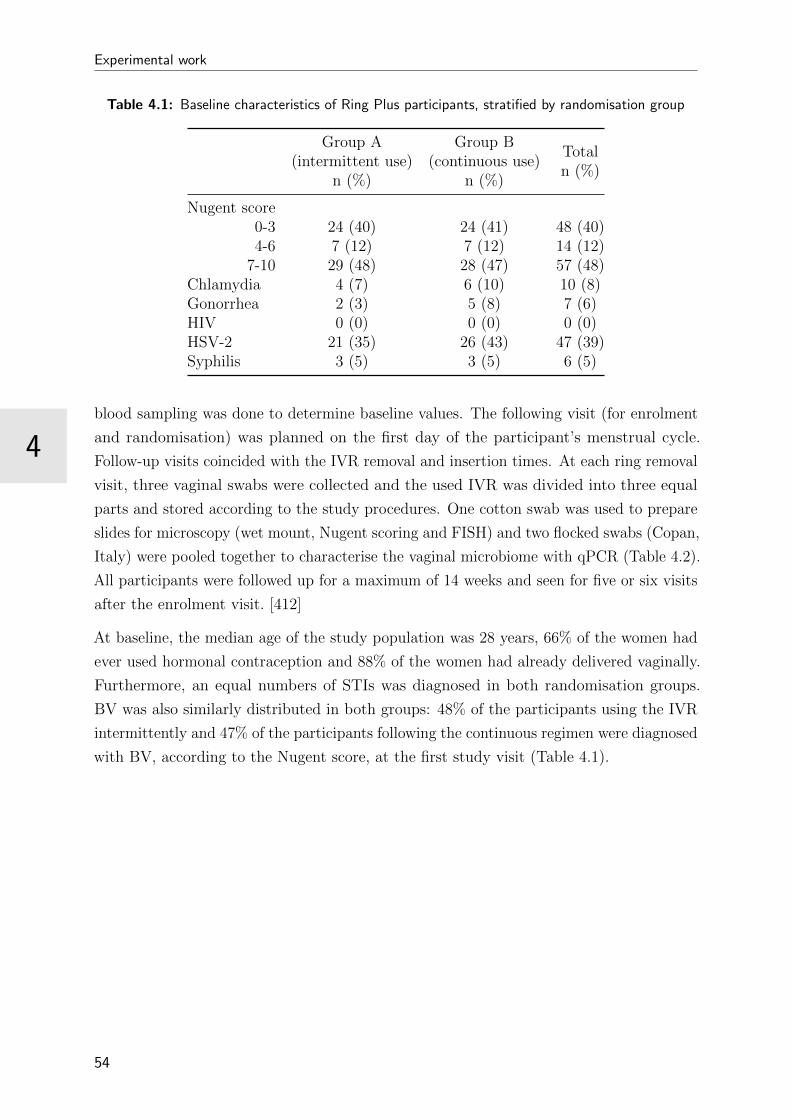
Experimental work
4
Table 4.1: Baseline characteristics of Ring Plus participants, stratified by randomisation group
Group A(intermittent use)
n (%)
Group B(continuous use)
n (%)
Totaln (%)
Nugent score0-3 24 (40) 24 (41) 48 (40)4-6 7 (12) 7 (12) 14 (12)
7-10 29 (48) 28 (47) 57 (48)Chlamydia 4 (7) 6 (10) 10 (8)Gonorrhea 2 (3) 5 (8) 7 (6)HIV 0 (0) 0 (0) 0 (0)HSV-2 21 (35) 26 (43) 47 (39)Syphilis 3 (5) 3 (5) 6 (5)
blood sampling was done to determine baseline values. The following visit (for enrolment
and randomisation) was planned on the first day of the participant’s menstrual cycle.
Follow-up visits coincided with the IVR removal and insertion times. At each ring removal
visit, three vaginal swabs were collected and the used IVR was divided into three equal
parts and stored according to the study procedures. One cotton swab was used to prepare
slides for microscopy (wet mount, Nugent scoring and FISH) and two flocked swabs (Copan,
Italy) were pooled together to characterise the vaginal microbiome with qPCR (Table 4.2).
All participants were followed up for a maximum of 14 weeks and seen for five or six visits
after the enrolment visit. [412]
At baseline, the median age of the study population was 28 years, 66% of the women had
ever used hormonal contraception and 88% of the women had already delivered vaginally.
Furthermore, an equal numbers of STIs was diagnosed in both randomisation groups.
BV was also similarly distributed in both groups: 48% of the participants using the IVR
intermittently and 47% of the participants following the continuous regimen were diagnosed
with BV, according to the Nugent score, at the first study visit (Table 4.1).
54

4
Experimental work
Table 4.2: Ring Plus study procedures for both randomisation groups.CM/AE: Concomitant Medication/Adverse Events; IDI: In-Depth Interview; IAQ: InterviewerAdministered Questionnaire; FGD: Focus Group Discussion; VIA: Visual Inspection with Aceticacid
Intermittent users Continuous usersRing in/out Procedures Ring in/out Procedures
Prescreening
HIV/STI counsellingInformed consentCollect contact-and menses-information
HIV/STI counsellingInformed consentCollect contact-and menses-information
IDI IDI IDI
Baseline(Week 0)
Current medicationMedical historyCounsellingVaginal/physical examBlood/vaginal (swabs+CVL)/urine samplesIAQ
Current medicationMedical historyCounsellingVaginal/physical examBlood/vaginal (swabs+CVL)/urine samplesIAQ
Enrolment(Week 1)
V1Ring 1in
CounsellingRandomisationPhysical examUrine sample
V1(+-3 d)
Ring 1in
CounsellingRandomisationPhysical examUrine sample
Week 4V2(+2 d)
Ring 1out
CM/AE/CounsellingVaginal/physical examVaginal (swabs)/urine samplesIAQ
V2(+- 3d)
Ring 1 out2 in
CM/AE/CounsellingVaginal/physical examVaginal (swabs)/urine samplesIAQ
Week 5V3(-2 d)
Ring 2in
CM/AE/counselling
Week 7 V3 (+-3d)Ring 2 out3 in
CM/AE/CounsellingVaginal/physical examVaginal (swabs)/urine samplesIAQFGD
Week 8V4(+2 d)
Ring 2out
CM/AE/CounsellingVaginal/physical examVaginal (swabs)/urine samplesIAQFGD
Week 9V5(-2 d)
Ring 3in
CM/AE/counselling
Week 10V 4(+-3 d)
Ring 3 out4 in
CM/AE/CounsellingVaginal/physical examVaginal (swabs)/urine samplesIAQ
Week 12V6(+2 d)
Ring 3out
CM/AE/CounsellingVaginal/physical examVIAVaginal (swabs+CVL)/urine samplesACASIIAQ
Week 13V5(+-3 d)
Ring 4out
CM/AE/CounsellingVaginal/physical examVIAVaginal (swabs+CVL)/urine samplesACASIIAQ
Week 14V7(+-3 d)
FGDIDI
V6(+-3 d)
FGDIDI
55

4
4.2 Unravelling the bacterial vaginosis-associated
biofilm: technical preparation
Abstract
Bacterial vaginosis, a condition defined by increased vaginal discharge without significant
inflammation, is characterised by a change in the bacterial composition of the vagina.
Lactobacillus spp., associated with a healthy vaginal microbiome, are outnumbered by
BV-associated organisms. These bacteria could form a polymicrobial biofilm which allows
them to persist in spite of antibiotic treatment. In this study, we examined the presence of
Gardnerella vaginalis and Atopobium vaginae in vaginal biofilms using Peptide Nucleic Acid
probes targeting these bacteria. For this purpose, we developed three new PNA probes for
A. vaginae. The most specific A. vaginae probe, AtoITM1, was selected and then used in
an assay with two existing probes, Gard162 and BacUni-1, to evaluate multiplex FISH
on clinical samples. Using quantitative polymerase chain reaction as the gold standard,
we demonstrated a sensitivity of 66.7% (95% confidence interval: 54.5% - 77.1%) and a
specificity of 89.4% (95% confidence interval: 76.1% - 96%) of the new AtoITM1 probe.
FISH enabled us to show the presence of a polymicrobial biofilm in bacterial vaginosis, in
which A. vaginae is part of a G. vaginalis-dominated biofilm. We showed that the presence
of this biofilm is associated with high bacterial loads of A. vaginae and G. vaginalis.
Adapted from:
Hardy L, Jespers V, Dahchour N, Mwambarangwe L, Musengamana V, Vaneechoutte
M and Crucitti T. Unravelling the Bacterial Vaginosis-Associated Biofilm: a Multiplex
Gardnerella vaginalis and Atopobium vaginae Fluorescence In Situ Hybridization Assay
Using Peptide Nucleic Acid Probes. PLoS ONE 10(8): e0136658 (authentic paper in
appendix).
57

Experimental work
4
4.2.1 Introduction
Bacterial vaginosis (BV), a condition characterised by increased vaginal discharge without
significant inflammation, is highly prevalent in women of reproductive age. It increases the
risk for acquisition and transmission of sexually transmitted infections, including HIV, and
is associated with preterm birth in pregnant women [74,75]. BV is a dysbiotic condition
of unknown aetiology and is characterised by a change in the microbial composition of the
vagina. Lactobacillus spp., associated with a healthy vaginal microbiome, are outnumbered
by an array of BV-associated organisms including Gardnerella vaginalis [15,74,75]. However,
several studies suggest that the mere presence of G. vaginalis is not sufficient for the
diagnosis of BV. Indeed, G. vaginalis is also present in 50% to 70% of women without BV
according to Nugent score [40,41,158]. G. vaginalis expresses various virulence factors such
as vaginolysin [263] and sialidase [160]. It can also produce a biofilm [9], thereby increasing
its tolerance to lactic acid and hydrogen peroxide produced by lactobacilli [413,414] and to
antimicrobial treatment [266,267]. Furthermore, it has been suggested that its adherence
and biofilm-forming capacities allow G. vaginalis to initiate the colonisation and scaffolding
of the vaginal epithelium to which other species can attach subsequently [260,264].
As was first shown a decade ago, Atopobium vaginae is one of the many other species that
are characteristic of BV [163,164,167,171,330]. In one study, A. vaginae was detected in
80% of samples testing positive for G. vaginalis and made up 40% of the total biofilm mass
dominated by G. vaginalis [9]. This association was confirmed in a study by Bradshaw et
al. [170]: 93% of samples containing A. vaginae also contained G. vaginalis, whereas only
10% tested positive for G. vaginalis when A. vaginae was absent [322]. In contrast to G.
vaginalis, A. vaginae is rarely part of the healthy vaginal microbiome and is considered a
more specific marker of BV than G. vaginalis [163,165,170].
It is postulated that a biofilm provides bacteria with a competitive advantage over
planktonic bacteria and that polymicrobial biofilms may offer additional advantages over
single-species biofilms. Mechanisms that have been described in previous studies include
metabolic cooperation, increased resistance to antibiotics or host immune responses [7] and
an enlarged gene pool with more efficient sharing of genetic material compared to mono-
species biofilms [348]. Polymicrobial coexistence is the dominant form in environmental
biofilms, but is also prominent in the human body [7]. A well-known example is dental
biomass: anaerobic bacteria, which are sensitive to oxygen, can survive and persist under
the aerobic conditions in the oral cavity due to the consummation of oxygen by aerobic
bacteria in the dental biofilm [374].
On the basis of these previous findings, we hypothesise that a polymicrobial biofilm
consisting of A. vaginae and G. vaginalis and other bacteria not discussed in this study
may serve as a marker of BV. Thus, better visualisation of the structure of vaginal biofilms
58
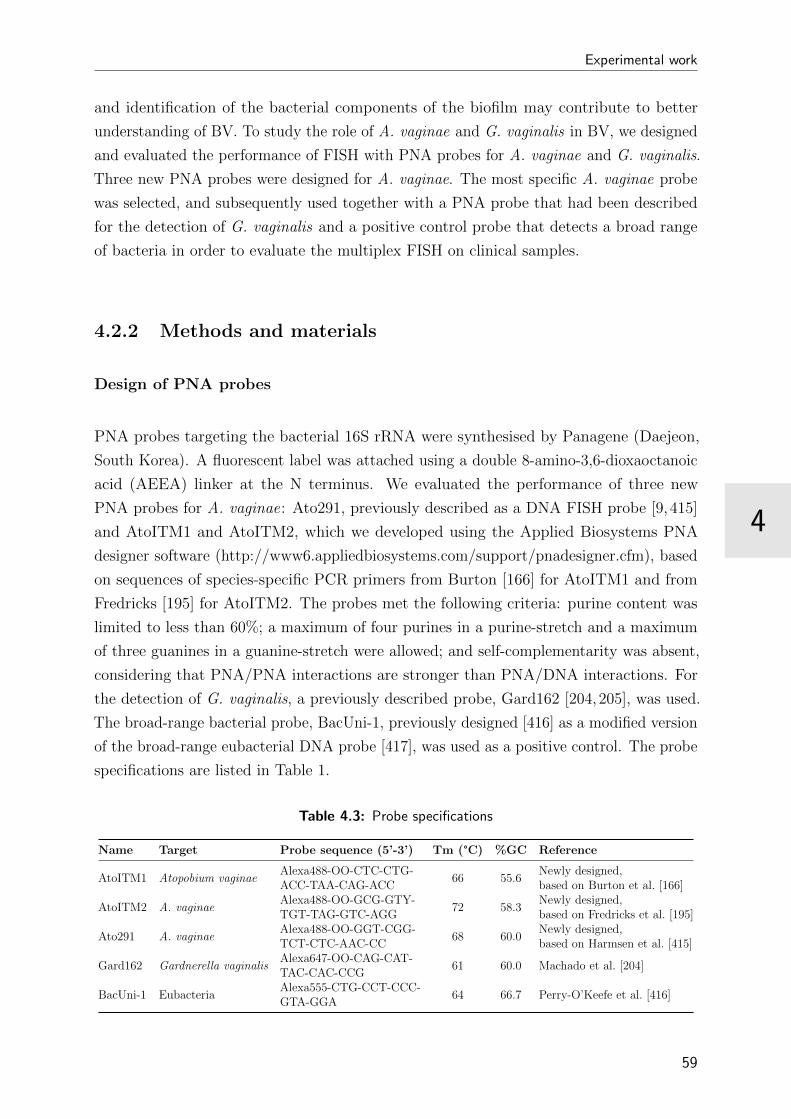
4
Experimental work
and identification of the bacterial components of the biofilm may contribute to better
understanding of BV. To study the role of A. vaginae and G. vaginalis in BV, we designed
and evaluated the performance of FISH with PNA probes for A. vaginae and G. vaginalis.
Three new PNA probes were designed for A. vaginae. The most specific A. vaginae probe
was selected, and subsequently used together with a PNA probe that had been described
for the detection of G. vaginalis and a positive control probe that detects a broad range
of bacteria in order to evaluate the multiplex FISH on clinical samples.
4.2.2 Methods and materials
Design of PNA probes
PNA probes targeting the bacterial 16S rRNA were synthesised by Panagene (Daejeon,
South Korea). A fluorescent label was attached using a double 8-amino-3,6-dioxaoctanoic
acid (AEEA) linker at the N terminus. We evaluated the performance of three new
PNA probes for A. vaginae: Ato291, previously described as a DNA FISH probe [9, 415]
and AtoITM1 and AtoITM2, which we developed using the Applied Biosystems PNA
designer software (http://www6.appliedbiosystems.com/support/pnadesigner.cfm), based
on sequences of species-specific PCR primers from Burton [166] for AtoITM1 and from
Fredricks [195] for AtoITM2. The probes met the following criteria: purine content was
limited to less than 60%; a maximum of four purines in a purine-stretch and a maximum
of three guanines in a guanine-stretch were allowed; and self-complementarity was absent,
considering that PNA/PNA interactions are stronger than PNA/DNA interactions. For
the detection of G. vaginalis, a previously described probe, Gard162 [204,205], was used.
The broad-range bacterial probe, BacUni-1, previously designed [416] as a modified version
of the broad-range eubacterial DNA probe [417], was used as a positive control. The probe
specifications are listed in Table 1.
Table 4.3: Probe specifications
Name Target Probe sequence (5’-3’) Tm (°C) %GC Reference
AtoITM1 Atopobium vaginaeAlexa488-OO-CTC-CTG-ACC-TAA-CAG-ACC
66 55.6Newly designed,based on Burton et al. [166]
AtoITM2 A. vaginaeAlexa488-OO-GCG-GTY-TGT-TAG-GTC-AGG
72 58.3Newly designed,based on Fredricks et al. [195]
Ato291 A. vaginaeAlexa488-OO-GGT-CGG-TCT-CTC-AAC-CC
68 60.0Newly designed,based on Harmsen et al. [415]
Gard162 Gardnerella vaginalisAlexa647-OO-CAG-CAT-TAC-CAC-CCG
61 60.0 Machado et al. [204]
BacUni-1 EubacteriaAlexa555-CTG-CCT-CCC-GTA-GGA
64 66.7 Perry-O’Keefe et al. [416]
59

Experimental work
4
Bacterial culture techniques for evaluation performance PNA probes
The performance of the PNA probes was evaluated using clinical isolates, obtained from
the collections of the Institute of Tropical Medicine (ITM) and Ghent University. The five
most frequently detected Lactobacillus species [3,15], representing the non-BV microbiome,
were chosen as a negative control to assure that the probes would not cross-hybridise
with the normal microbiome (Table 4.4). Furthermore we selected the most frequent
BV-associated bacteria (8 A. vaginae strains, 5 G. vaginalis strains) as a negative control
for G. vaginalis and A. vaginae respectively in addition to 2 Prevotella melaninogenica
strains (Table 4.4).
Table 4.4: Specificity testing in duplicate of peptide nucleic acid (PNA) probes using culturedbacteria
Species Strain AtoITM1 AtoITM2 Ato291 Gard162 BacUni-1
Atopobium vaginae CCUG 38953T +1 + + -2 +Atopobium vaginae UG080499 + + + - +Atopobium vaginae UG071164 + + + - +Atopobium vaginae UG020349 + + + - +Atopobium vaginae UG160373 + + + - +Atopobium vaginae UG550940 + + + - +Atopobium vaginae UG030313 + + + - +Atopobium vaginae UG030312 + + + - +Gardnerella vaginalis UG860108 - - - + +Gardnerella vaginalis UG030406 - - - + +Gardnerella vaginalis UG860107 - + + + +Gardnerella vaginalis LMG 7832T - + + + +Gardnerella vaginalis UG030407 - + + + +Lactobacillus iners LMG 18914T - - + - +Lactobacillus vaginalis LMG 12891T - - + - +Lactobacillus jensenii LMG 6414T - - + - +Lactobacillus crispatus LMG 9479T - - + - +Lactobacillus gasseri LMG 9203T - - + - +Prevotella melaninogenica UG160361 - - + - +Prevotella melaninogenica UG040818 - - - - +
The signal was considered positive if it had a positive counterpart in the DAPI stain and displayed apositive signal simultaneously with the broad-range probe. The signal was considered negative ifno signal was seen with the species-specific probe.1(+) Presence of hybridisation2(−) Absence of hybridisation
This small test panel is a limitation of the study and more expansive testing will be
required to assure that there is no cross-reactivity with other bacteria. The strains from
frozen stocks in skimmed milk (-80 °C) were cultured on Columbia agar base (Becton
Dickinson Biosciences, Erembodegem, Belgium) + 5% horse blood and grown under
anaerobic conditions (10% hydrogen, 10% carbon dioxide and 80% nitrogen), using an
anaerobic incubator (Whitley DG250) at 37 °C for 48 h and bacteria were streaked onto
fresh plates every 48-72 hours. To determine the limit of detection, FISH was performed
60
4
Experimental work
using the AtoITM1, Gard162 and BacUni-1 probe on serial tenfold dilutions using the
fixative used throughout the study: Carnoy solution (6:3:1, ethanol:chloroform:glacial acetic
acid [418,419]). Carnoy solution was chosen as a fixative solution because of its proven
effectiveness in the stabilisation of and minimal shrinkage in tissue structure [419, 420].
The concentration of the dilutions was estimated based on the turbidity of the sample
compared to McFarland Standards (Bio-Merieux SA, Marcy l’Etoile, France) and ranged
from 1.2x10⁹ to 1.2x10³ cells per ml. The suspensions were vortexed briefly and 5 µl was
spotted into the hybridisation chamber; a 5 mm2 area marked with a PAP Pen (Sigma
Aldrich, St. Louis, USA) that creates a hydrophobic border on a Superfrost Plus slide
(Menzel-Glaser, Braunschweig, Germany). The spotted suspensions were dried before
performing FISH. Experiments were performed in duplicate.
Clinical samples
Ethics statement Vaginal samples were collected from 119 women participating in a
clinical trial in Rwanda studying the vaginal microbiome and acceptability of a contracep-
tive ring (the ’Ring Plus’ study, ClinicalTrials.gov identifier NCT01796613) (data analysis
on-going) [412]. Participants were between 18 and 35 years old and provided written in-
formed consent for participation in the study. The Ring Plus study and consent procedure
were approved by the Rwanda National Ethics Committee, Rwanda; the Institutional
Review Board of the ITM Belgium; and the ethics committee of the University Teaching
Hospital in Antwerp, Belgium.
Vaginal sample collection and preparation Vaginal sampling was carried out by
the study clinician as part of the study procedures. Two Copan flocked swabs (Copan,
Brescia, Italy) and one cotton swab were brushed against the lateral walls of the vagina.
The cotton swab was immediately rolled on a Superfrost Plus slide (Menzel-Glaser) which
was heat-fixed by passing twice through a flame. The Superfrost Plus (Menzel-Glaser)
slides were stored for maximum six months and shipped to ITM at room temperature and
fixed for a minimum of 12 hours at ITM, submerged in Carnoy solution [418,419]. The
Copan flocked swabs were eluted by vortexing each swab for at least 15 seconds in 1.2 ml
of diluted phosphate buffered saline (PBS) (pH 7.4 - 1:9, PBS:saline). The two eluates
were combined and divided into three aliquots, which were stored at -80 °C. The swab
eluates were shipped frozen (-191 °C) in a dry shipper to the ITM to determine the total
bacterial load of A. vaginae and G. vaginalis by means of qPCR.
Urine sample collection and preparation According to an earlier described proce-
dure [305], first-void urine was collected by the participants and 2 ml was transferred
61

Experimental work
4
immediately to a 15 ml tube containing 2 ml of Carnoy solution. The sample was fixed
overnight and after centrifugation (10 minutes at 3200 g), the supernatant was decanted
and the pellet was treated for a second time with 0.75 µl of Carnoy solution. The samples
were stored between 2-8 °C and shipped at room temperature to the ITM. Before applying
FISH, the urine samples were vortexed briefly and 5 µl was spotted into the hybridisation
chamber on a Superfrost Plus slide (Menzel-Glaser).
Quantitative polymerase chain reaction for quantification of bacteria in vaginal
samples
DNA was extracted from 250 µl of the vaginal swab eluate using the Abbott m2000sp
automated extraction platform (Abbott, Maidenhead, UK), according to the manufacturer’s
instructions. The volume of 200 µl DNA extract was stored at -80 °C until testing. qPCR
was performed for each bacteria species separately, to avoid competition between the
primers. The 25 µl PCR mixture contained 12.5 µl Rotor-Gene SYBR Green RT-PCR
Master mix (Qiagen, Venlo, the Netherlands), 5 µl DNA extract, 0.5 µM of A. vaginae or
1 µM of G. vaginalis forward and reverse primers (Integrated DNA Technologies, Leuven,
Belgium) and RNase-free water provided with the Rotor-Gene SYBR Green PCR kit. The
primers for A. vaginae and G. vaginalis and the amplification reactions (Rotor Gene Q
MDx 5 plex, Qiagen) have been described before [3].
Quantification was done using standard curves, constructed using DNA extracts from
A. vaginae (CCUG 38953T) and G. vaginalis (LMG 7832T), grown at 35 °C ± 2 °C on
Columbia agar base (Becton Dickinson) + 5% horse blood, under anaerobic conditions.
DNA concentrations were determined using NanoDrop (Thermo Fisher Scientific, Erem-
bodegem, Belgium) and the number of genomes was calculated using the described genome
sizes and G+C content of the strains. A total of six tenfold dilutions of the DNA stocks
were prepared in high performance liquid chromatography (HPLC) grade water. Both the
standard curve and samples were run in duplicate. The bacterial load was expressed as
genome equivalents (geq)/ml.
Peptide nucleic acid fluorescence in situ hybridisation procedure
Multiplex hybridisation was performed on a Superfrost Plus slide (Menzel-Glaser) in a
5 mm2 quadrant hybridisation area marked with a PAP pen (Sigma Aldrich, St. Louis,
USA), a liquid-repellent slide marker. The slide was covered with a cover slip after addition
of a hybridisation buffer that contained 200 nM of each probe: species-specific probes
for A. vaginae (AtoITM1 or AtoITM2 or Ato291) and G. vaginalis (Gard162), and the
broad-range BacUni-1 probe. The hybridisation solution consisted of 10% (wt/vol) dextran
62
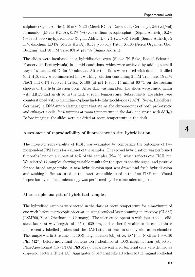
4
Experimental work
sulphate (Sigma Aldrich), 10 mM NaCl (Merck KGaA, Darmstadt, Germany), 2% (vol/vol)
formamide (Merck KGaA), 0.1% (wt/vol) sodium pyrophosphate (Sigma Aldrich), 0.2%
(wt/vol) polyvinylpyrrolidone (Sigma Aldrich), 0.2% (wt/vol) Ficoll (Sigma Aldrich), 5
mM disodium EDTA (Merck KGaA), 0.1% (vol/vol) Triton X-100 (Acros Organics, Geel,
Belgium) and 50 mM Tris-HCl at pH 7.5 (Sigma Aldrich).
The slides were incubated in a hybridisation oven (Shake ’N Bake, Boekel Scientific,
Feasterville, Pennsylvania) in humid conditions, which were achieved by adding a small
tray of water, at 60 °C for 60 minutes. After the slides were rinsed with double-distilled
(dd) H20, they were immersed in a washing solution containing 5 mM Tris base, 15 mM
NaCl and 0.1% (vol/vol) Triton X-100 (at pH 10) for 15 min at 60 °C on the rocking
shelves of the hybridisation oven. After this washing step, the slides were rinsed again
with ddH20 and air-dried in the dark at room temperature. Subsequently, the slides were
counterstained with 6-diamidine-2-phenylindole dihydrochloride (DAPI) (Serva, Heidelberg,
Germany), a DNA-intercalating agent that stains the chromosomes of both prokaryotic
and eukaryotic cells, for 5 minutes at room temperature in the dark and rinsed with ddH20.
Before imaging, the slides were air-dried at room temperature in the dark.
Assessment of reproducibility of fluorescence in situ hybridisation
The inter-run repeatability of FISH was evaluated by comparing the outcomes of two
independent FISH runs for a subset of the samples. The second hybridisation was performed
6 months later on a subset of 15% of the samples (N=17), which reflects one FISH run.
We selected 17 samples showing variable results for the species-specific signal and positive
for the broad-range probe. A new hybridisation spot was drawn and fresh hybridisation
and washing buffer was used on the exact same slides used in the first FISH run. Visual
inspection by confocal microscopy was performed by the same microscopist.
Microscopic analysis of hybridised samples
The hybridised samples were stored in the dark at room temperature for a maximum of
one week before microscopic observation using confocal laser scanning microscopy (CLSM)
(LSM700, Zeiss, Oberkochen, Germany). The microscope operates with four stable, solid-
state lasers at wavelengths of 405 to 639 nm, and is therefore able to detect all three
fluorescently labelled probes and the DAPI stain at once in one hybridisation chamber.
The sample was first scanned at 100X magnification (objective: EC Plan-Neofluar 10x/0.30
Ph1 M27), before individual bacteria were identified at 400X magnification (objective:
Plan-Apochromat 40x/1.3 Oil Ph3 M27). Separate scattered bacterial cells were defined as
dispersed bacteria (Fig 4.1A). Aggregates of bacterial cells attached to the vaginal epithelial
63
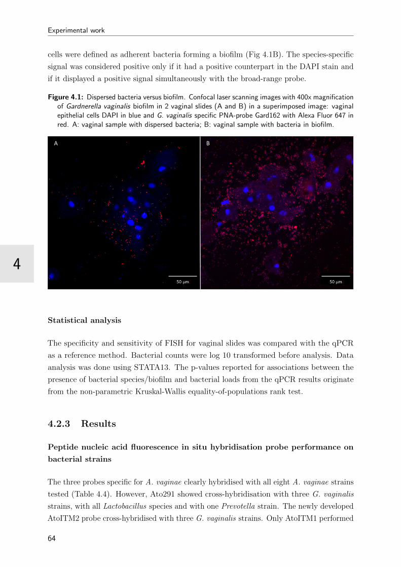
Experimental work
4
cells were defined as adherent bacteria forming a biofilm (Fig 4.1B). The species-specific
signal was considered positive only if it had a positive counterpart in the DAPI stain and
if it displayed a positive signal simultaneously with the broad-range probe.
Figure 4.1: Dispersed bacteria versus biofilm. Confocal laser scanning images with 400x magnificationof Gardnerella vaginalis biofilm in 2 vaginal slides (A and B) in a superimposed image: vaginalepithelial cells DAPI in blue and G. vaginalis specific PNA-probe Gard162 with Alexa Fluor 647 inred. A: vaginal sample with dispersed bacteria; B: vaginal sample with bacteria in biofilm.
Statistical analysis
The specificity and sensitivity of FISH for vaginal slides was compared with the qPCR
as a reference method. Bacterial counts were log 10 transformed before analysis. Data
analysis was done using STATA13. The p-values reported for associations between the
presence of bacterial species/biofilm and bacterial loads from the qPCR results originate
from the non-parametric Kruskal-Wallis equality-of-populations rank test.
4.2.3 Results
Peptide nucleic acid fluorescence in situ hybridisation probe performance on
bacterial strains
The three probes specific for A. vaginae clearly hybridised with all eight A. vaginae strains
tested (Table 4.4). However, Ato291 showed cross-hybridisation with three G. vaginalis
strains, with all Lactobacillus species and with one Prevotella strain. The newly developed
AtoITM2 probe cross-hybridised with three G. vaginalis strains. Only AtoITM1 performed
64

4
Experimental work
Figure 4.2: Distribution of samples according to fluorescence in situ hybridisation (FISH). Aggregatesof bacterial cells attached to the vaginal epithelial cells, were defined as biofilm. Separate scatteredbacterial cells, without the presence of biofilm, were defined as dispersed only bacteria.
without false positive results and was selected for further evaluation on the clinical samples.
The Gard162 probe was able to identify all five G. vaginalis test strains and showed no
cross-hybridisation with any of the other 15 species tested (Table 4.4). All bacterial strains
tested hybridised with the broad-range BacUni-1 probe. According to the FISH results of
the serial dilutions, the limit of detection for AtoITM1, Gard162 and BacUni-1 probes
was 1.2 x 10⁵ cells per ml.
Detection of Atopobium vaginae and Gardnerella vaginalis in clinical samples
by Peptide nucleic acid fluorescence in situ hybridisation
In a small pilot study, a total of 10 paired vaginal slides and urine samples were tested
to evaluate the suitability of each type of sample for FISH analysis. Six out of 10 urine
samples could not be assessed due to the low presence of vaginal epithelial cells in the
urine, whereas this problem was experienced in only 2 vaginal samples. Therefore, it was
decided to continue analyses on vaginal slides only.
Using the AtoITM1 PNA-probe, A. vaginae was visualised as dispersed entities, without
the presence of biofilm, in 27/119 (22.7%) of the samples. A. vaginae biofilm was present
in 26/119 (21.8%) samples. A. vaginae FISH was negative in the remaining 66/119 (55.5%)
samples. PNA-FISH using Gard162 detected dispersed-only G. vaginalis in 31/119 (26%)
samples, G. vaginalis biofilm in 58/119 (48.7%) samples (e.g., Fig 4.2) and 30/119 (25.3%)
samples were negative for G. vaginalis. Of the 89 G. vaginalis FISH-positive samples
(dispersed or biofilm), 36 samples (41%) were negative for A. vaginae. However, all samples
with A. vaginae biofilm showed a G. vaginalis biofilm as well (e.g., Fig 4.3, 4.4, 4.5).
65
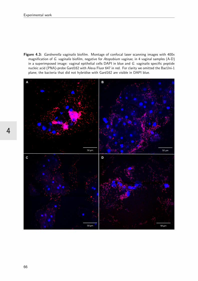
Experimental work
4
Figure 4.3: Gardnerella vaginalis biofilm. Montage of confocal laser scanning images with 400xmagnification of G. vaginalis biofilm, negative for Atopobium vaginae, in 4 vaginal samples (A-D)in a superimposed image: vaginal epithelial cells DAPI in blue and G. vaginalis specific peptidenucleic acid (PNA)-probe Gard162 with Alexa Fluor 647 in red. For clarity we omitted the BacUni-1plane; the bacteria that did not hybridise with Gard162 are visible in DAPI blue.
66
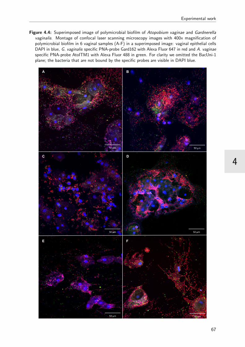
4
Experimental work
Figure 4.4: Superimposed image of polymicrobial biofilm of Atopobium vaginae and Gardnerellavaginalis. Montage of confocal laser scanning microscopy images with 400x magnification ofpolymicrobial biofilm in 6 vaginal samples (A-F) in a superimposed image: vaginal epithelial cellsDAPI in blue, G. vaginalis specific PNA-probe Gard162 with Alexa Fluor 647 in red and A. vaginaespecific PNA-probe AtoITM1 with Alexa Fluor 488 in green. For clarity we omitted the BacUni-1plane; the bacteria that are not bound by the specific probes are visible in DAPI blue.
67
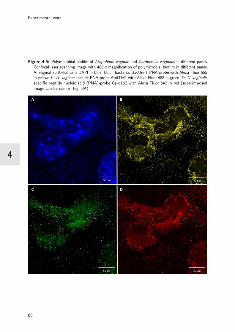
Experimental work
4
Figure 4.5: Polymicrobial biofilm of Atopobium vaginae and Gardnerella vaginalis in different panes.Confocal laser scanning image with 400 x magnification of polymicrobial biofilm in different panes,A: vaginal epithelial cells DAPI in blue, B: all bacteria, BacUni-1 PNA-probe with Alexa Fluor 555in yellow, C: A. vaginae specific PNA-probe AtoITM1 with Alexa Fluor 488 in green, D: G. vaginalisspecific peptide nucleic acid (PNA)-probe Gard162 with Alexa Fluor 647 in red (superimposedimage can be seen in Fig. 3A).
68

4
Experimental work
Characterisation of vaginal samples by quantitative polymerase chain reac-
tion
A total of 119 vaginal samples were available for qPCR analysis. A. vaginae was present in
72 (60%) of the samples with a mean log of 7.55 ± 1.34 geq/ml. G. vaginalis was detected
in 95 (80%) of the samples with a mean log of 7.38 ± 1.11 geq/ml.
Performance of probes in vaginal samples
Quantitative PCR was used as the reference method for detection and quantification of A.
vaginae and G. vaginalis. The signal of the species-specific probes was only considered
positive if a positive counterpart was seen in the DAPI stain and with the universal
BacUni-1 probe. When assessing the results with FISH probe AtoITM1 against the qPCR
outcomes for A. vaginae for 119 vaginal samples, FISH results were false negative for 24
samples and false positive for 5 samples, resulting in a sensitivity of 66.7% (95% confidence
interval (CI): 54.5% - 77.1%) and a specificity of 89.4% (95% CI: 76.1% - 96%) (Table
4.5). The mean log for the true positive samples (positive with qPCR and FISH) was 7.73
geq/ml, as compared to a mean log of 7.19 geq/ml for the false negative FISH results
(p=0.399). For Gard162, the G. vaginalis probe, 13 FISH results were false negative and
six false positive. The sensitivity was 86.3% (95% CI: 77.4% - 92.2%) and the specificity
75.0% (95% CI: 52.9% - 89.4%) (Table 4.5). The mean log for the true positive results for
G. vaginalis was 7.61 geq/ml compared to a mean log of 5.94 geq/ml for the false negative
results (p<0.001).
Table 4.5: Performance of Atopobium vaginae (AtoITM1) and Gardnerella vaginalis (Gard162)peptide nucleic acid probes, compared to quantitative polymerase chain reaction (qPCR) results,for 119 vaginal slides
FISH qPCRA. vaginae positive A. vaginae negative G. vaginalis positive G. vaginalis negative Total
AtoITM1 positive 48 (66.7%) 5 (10.6%) 53AtoITM1 negative 24 (33.3%) 42 (89.4%) 66Gard162 positive 82 (86.3%) 6 (25.0%) 88Gard 162 negative 13 (13.7%) 18 (75.0%) 31Total 72 47 95 24
Assessment of the repeatability was done using 17 samples. After the first hybridisation, all
samples showed a signal for the BacUni-1 probe, 5 and 9 samples out of 17 for the AtoITM1
and Gard162 probe respectively. The results of the second FISH with the BacUni-1 and
Gard162 probe were in full agreement with the first run. For the AtoITM1 probe, only
one sample had a different result in the second run (negative at first, but positive in the
second run).
69
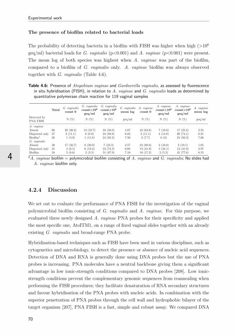
Experimental work
4
The presence of biofilm related to bacterial loads
The probability of detecting bacteria in a biofilm with FISH was higher when high (>106
geq/ml) bacterial loads for G. vaginalis (p<0.001) and A. vaginae (p<0.001) were present.
The mean log of both species was highest when A. vaginae was part of the biofilm,
compared to a biofilm of G. vaginalis only. A. vaginae biofilm was always observed
together with G. vaginalis (Table 4.6).
Table 4.6: Presence of Atopobium vaginae and Gardnerella vaginalis, as assessed by fluorescencein situ hybridisation (FISH), in relation to A. vaginae and G. vaginalis loads as determined byquantitative polymerase chain reaction for 119 vaginal samples
TotalG. vaginalis
count 0
G. vaginaliscount<106
geq/ml
G. vaginaliscount>106
geq/ml
G. vaginalismean log
A. vaginaecount 0
A. vaginaecount<106
geq/ml
A. vaginaecount>106
geq/ml
A. vaginaemean log
Detected byPNA FISH
N (%) N (%) N (%) geq/ml N (%) N (%) N (%) geq/ml
A. vaginaeAbsent 66 20 (30.3) 13 (19.7) 33 (50.0) 4.87 42 (63.6) 7 (10.6) 17 (25.8) 2.55Dispersed only 27 3 (11.1) 0 (0.0) 24 (88.9) 6.82 3 (11.1) 4 (14.8) 20 (74.1) 6.21Biofilm1 26 1 (3.9) 1 (11.8) 24 (92.3) 7.50 2 (7.7) 0 (0) 24 (92.3) 7.66G. vaginalisAbsent 30 17 (56.7) 6 (20.0) 7 (23.3) 2.57 24 (80.0) 3 (10.0) 3 (10.1) 1.05Dispersed only 31 2 (6.5) 6 (19.4) 23 (74.2) 6.68 13 (41.9) 5 (16.1) 13 (41.9) 3.97Biofilm 58 5 (8.6) 2 (3.5) 51 (87.9) 7.18 10 (17.2) 3 (5.2) 45 (77.6) 6.55
1A. vaginae biofilm = polymicrobial biofilm consisting of A. vaginae and G. vaginalis; No slides hadA. vaginae biofilm only.
4.2.4 Discussion
We set out to evaluate the performance of PNA FISH for the investigation of the vaginal
polymicrobial biofilm consisting of G. vaginalis and A. vaginae. For this purpose, we
evaluated three newly designed A. vaginae PNA probes for their specificity and applied
the most specific one, AtoITM1, on a range of fixed vaginal slides together with an already
existing G. vaginalis and broad-range PNA probe.
Hybridisation-based techniques such as FISH have been used in various disciplines, such as
cytogenetics and microbiology, to detect the presence or absence of nucleic acid sequences.
Detection of DNA and RNA is generally done using DNA probes but the use of PNA
probes is increasing. PNA molecules have a neutral backbone giving them a significant
advantage in low ionic-strength conditions compared to DNA probes [208]. Low ionic-
strength conditions prevent the complementary genomic sequences from reannealing when
performing the FISH procedures; they facilitate denaturation of RNA secondary structures
and favour hybridisation of the PNA probes with nucleic acids. In combination with the
superior penetration of PNA probes through the cell wall and hydrophobic bilayer of the
target organism [207], PNA FISH is a fast, simple and robust assay. We compared DNA
70
4
Experimental work
and PNA probes (data not reported) and can confirm that PNA FISH is faster and more
robust than DNA FISH. In the current study, PNA FISH proved to be highly efficient
for the identification and visualisation of the spatial arrangement of A. vaginae and G.
vaginalis in the BV-associated biofilms. Moreover, PNA FISH showed excellent inter-assay
repeatability for all three probes used.
Peptide nucleic acid fluorescence in situ hybridisation probe performance on
bacterial strains
For the design of the PNA probes, we opted for Alexa fluorochromes, which have similar
spectral properties as other fluorochromes, such as cyanine dyes, but are brighter and
more resistant to photo bleaching [421].
PNA probes that specifically target A. vaginae have not been described before. The
only probe thus far reported was a DNA probe (Ato291) [415]. The probe was designed
to detect bacteria belonging to the Atopobium cluster in faecal samples. However, in
silico evaluation of the specificity of the Ato291 probe showed cross-hybridisation with
other bacteria belonging to the Coriobacteriaceae, a family of Actinobacteria, to which A.
vaginae belongs. This is not surprising, since the probe was originally designed on the basis
of sequences of Coriobacteriaceae strains isolated from faeces and clinical material. The
probe has been used for the detection of A. vaginae in vaginal samples by Swidsinski et
al. [9], but their findings have not yet been confirmed by other groups. In our experiments,
using a PNA equivalent of the Ato291 probe, we showed a low specificity of the Ato291
probe on vaginal clinical isolates. The Ato291 probe cross-hybridised with three out
of five G. vaginalis strains and all five Lactobacillus species. Therefore, we designed
two new probes for A. vaginae targeting the 16S rRNA-gene, based on published PCR
primers [166,195] and we adjusted the sequences to fit the requirements for PNA probes.
One of the new probes, AtoITM1, did not cross-react to any of the tested species and was
further used for detection of A. vaginae in vaginal slides.
Gard162 is the first PNA FISH probe designed specifically for G. vaginalis and has
extensively been tested by Machado et al. on a variety of cultured bacterial strains and
clinical samples [204, 205]. Using this probe, we obtained clear hybridisation for all G.
vaginalis isolates tested and observed no cross-reaction with strains of the other species
tested, confirming the findings of Machado et al. [204].
71
Experimental work
4
Peptide nucleic acid fluorescence in situ hybridisation probe performance on
clinical samples
Vaginal slides proved to be a valid sample type for imaging of the composition of the vaginal
microbiome, if processed directly after sampling, as shown by Peltroche-Llacsahuanga
et al. [422]. Collection of a vaginal swab is an easy and cheap sampling method, with a
minimal burden on the study participant or patient. After heat fixation, we recorded that
the slides can be stored at room temperature for up to at least six months and can be
easily transported. A high quality vaginal sample can be obtained by thinly rolling the
swab onto the slide. A thick vaginal smear on the contrary where the material is smeared
onto the slide is not useful for FISH and microscopic visualisation.
The probes were also applied to 119 vaginal slides from women for whom the bacterial
loads of G. vaginalis and A. vaginae had been quantified by qPCR. qPCR was used in this
study as the reference method to evaluate the performance of the FISH probes, although
comparison of these methods is subject to some limitations. qPCR is highly sensitive and
was performed on a homogenised DNA extract representing half of the full sample. FISH
however was carried out on 0.5 mm2 of a vaginal slide, which could be heterogeneous. This
is inevitably an underrepresentation of the vaginal sample. Both techniques were also
performed using two different vaginal swabs, but the first collected specimen was used to
prepare the slide as per study protocol.
After hybridisation for 60 minutes and washing for 15 minutes, both at 60 °C, the AtoITM1
probe gave only five false positive results, compared to qPCR results, resulting in a
specificity of 90% for this set of samples. However, 24 samples that were positive according
to the qPCR were not detected by FISH, which gives a sensitivity of 67%. These results
are comparable to the sensitivity observed for vaginal samples using FISH for detection
of Group B Streptococcus [422]. The authors of this study obtained a higher sensitivity
after extraction of the swabs by centrifugation of the swab head in a NaCl solution [422],
but this method would probably disrupt the epithelial biofilm and thus prevent us from
investigating the organisation of the bacterial biofilm.
The relatively low sensitivity of the A. vaginae FISH assay cannot be explained by the
bacterial load as measured by qPCR; the mean log of the true positive and false negative
samples was not significantly different (log 7.73 and 7.19 geq/ml respectively). One possible
explanation could be the typical structure of a biofilm, whereby an oxygen gradient exists
from the top to the centre of the biofilm [423]. Anaerobic bacteria such as A. vaginae are
possibly found more embedded in the biofilm, to take advantage of the anaerobicity. It
could be that the PNA probes are not able to fully penetrate into the inner parts of the
biofilm; or that if the PNA probes do penetrate, the fluorescence could be masked and not
detected due to low resolution of the equipment.
72

4
Experimental work
For G. vaginalis, 6 false positive results and 13 false negative results were found compared
to 95 positive and 24 negative samples according to qPCR. This implies a sensitivity of
86% and specificity 75% for the detection of G. vaginalis by the Gard162 probe using
our FISH protocol for this set of samples. This is lower than reported by Machado [204],
who, using the same probe, showed a full agreement between qPCR and FISH results
for 13 vaginal samples. We were not able to elucidate why these discordant results were
obtained.
Bacterial loads and the presence of a biofilm
Our study shows that higher bacterial loads of G. vaginalis and A. vaginae, as detected
by qPCR, are associated with a higher probability of presence of a bacterial biofilm.
Both bacterial species are important constituents of the vaginal epithelial biofilm [9,332].
No samples contained A. vaginae in the absence of G. vaginalis, but almost half of
the G. vaginalis-positive samples did not contain A. vaginae according to FISH results.
Both bacteria were seen in a dispersed and an adherent state, but A. vaginae was always
accompanied by G. vaginalis. The mere presence of A. vaginae did not simply predispose to
a polymicrobial biofilm, but when A. vaginae was part of the biofilm, compared to a biofilm
of only G. vaginalis, both bacterial species were present in higher concentrations.
We hypothesise that G. vaginalis is one of the main initiators of a vaginal biofilm, when
it is present in high amounts. This vaginal biofilm creates a favourable environment for
anaerobic bacteria, such as A. vaginae. One reason for the appearance of A. vaginae may
be the presence of an oxygen gradient within the biofilm. By embedding itself within the
biofilm, A. vaginae can take advantage of the anaerobicity, proliferates and exists in a
mutualistic relationship with G. vaginalis.
4.2.5 Conclusion
Our study confirms that PNA FISH is a valuable tool for detecting and visualising biofilm-
associated organisms in vaginal slides. This study describes the design and evaluation
of a new PNA probe, AtoITM1, which can be included in multiplex FISH in BV biofilm
research. Using the new probe, we have demonstrated the presence of a polymicrobial
biofilm, with A. vaginae taking part in a G. vaginalis dominated biofilm.
73


4
Experimental work
4.3 A fruitful alliance: the synergy between Atopobium
vaginae and Gardnerella vaginalis
Abstract
Bacterial vaginosis is characterised by a change in the microbial composition of the vagina.
The BV-associated organisms outnumber the health-associated Lactobacillus species and
form a polymicrobial biofilm on the vaginal epithelium, possibly explaining the difficulties
with antibiotic treatment. A better understanding of vaginal biofilm with emphasis on
Atopobium vaginae and Gardnerella vaginalis may contribute to a better diagnosis and
treatment of BV. To this purpose, we evaluated the association between the presence
of both bacteria by fluorescence in situ hybridisation and BV by Nugent scoring in 463
vaginal slides of 120 participants participating in a clinical trial in Rwanda. A bacterial
biofilm was detected in half of the samples using a universal bacterial probe. The biofilm
contained A. vaginae in 54.1% and G. vaginalis in 82.0% of the samples. A. vaginae was
accompanied by G. vaginalis in 99.5% of samples. The odds of having a Nugent score
above 4 was increased for samples with dispersed G. vaginalis and/or A. vaginae present
(odds ratio 4.5; CI: 2-10.3). The probability of having a high Nugent score was even higher
when a combination of adherent G. vaginalis and dispersed A. vaginae was visualised
(odds ratio 75.6; CI: 13.3-429.5) and highest when both bacteria were part of the biofilm
(odds ratio 119; CI: 39.9-360.8). Our study, although not comprehensive at studying the
polymicrobial biofilm in BV, provided a strong indication towards the importance of A.
vaginae and the symbiosis of A. vaginae and G. vaginalis in this biofilm.
Adapted from:
Hardy L, Jespers V, Abdellati S, De Baetselier I, Mwambarangwe L, Musengamana V,
van de Wijgert J, Vaneechoutte M and Crucitti T. A fruitful alliance: the synergy between
Atopobium vaginae and Gardnerella vaginalis in bacterial vaginosis-associated biofilm. STI
2016;0:1–5,pii: sextrans-2015-052475,doi: 10.1136 (authentic paper in appendix).
75

Experimental work
4
4.3.1 Introduction
Bacterial vaginosis (BV) is the most prevalent vaginal disorder in women of reproductive
age. It increases the risk of acquisition and transmission of sexually transmitted infections,
including HIV, and is associated with preterm birth in pregnant women [89, 127, 424].
The condition is characterised by a change in the microbial composition of the vagina:
the Lactobacillus spp., associated with a healthy vaginal microbiome, are outnumbered
by microaerophilic and anaerobic organisms, including Gardnerella vaginalis [15, 41,127,
174,260]. The mere presence of G. vaginalis, however, is not sufficient for the diagnosis
of BV using traditional diagnostic algorithms (see below) because many women without
BV also have G. vaginalis in their vaginal microbiome [15]. BV is, however, associated
with high counts of G. vaginalis using molecular methods and/or the presence of a G.
vaginalis-containing polymicrobial biofilm [9, 15, 41, 42, 174, 260, 425]. Due to its strong
adherence to vaginal epithelial cells and biofilm-forming capacities, it has been suggested
that G. vaginalis initiates the colonisation of the vaginal epithelium and serves as a
scaffolding to which other species subsequently can attach [141,209,425].
One of the species that might attach to the biofilm initiated by G. vaginalis could be
Atopobium vaginae [163, 166]. Several molecular studies have indicated a probable role
for A. vaginae in BV [165, 166, 171], and it has also been suggested that A. vaginae
plays a major part in the establishment of a biofilm, together with G. vaginalis [9, 425].
Considering it has been found in 80-90% of cases of relapse [170] and some strains have
been shown in vitro to be metronidazole resistant [330], it could be of importance in the
recurrence of BV after standard treatment with metronidazole.
The current gold standard in BV research is the microscopic evaluation and scoring of
vaginal slides according to Nugent [158]. The diagnosis of BV is based on the absence of
lactobacilli and the presence of small Gram-negative to Gram-variable rods (G. vaginalis
and Bacteroides spp. morphotypes) and curved Gram-negative rods (Mobiluncus spp.
morphotypes). In fact, bacterial biofilm can also be seen with this method, in the form of
clue cells, which are vaginal epithelial cells covered by layers of adherent Gram-negative
and/or Gram-variable cells, that is, biofilms [426]. Using Gram staining, it is impossible
to distinguish between the different bacterial species in the biofilm. By labelling the cells
with a fluorescent probe, using fluorescence in situ hybridisation (FISH), the structure and
composition of the biofilm can be studied in more detail. To study the potential role of
A. vaginae and the synergy between A. vaginae and G. vaginalis in the biofilm, we used
our newly developed peptide nucleic acid (PNA) A. vaginae probe [209] together with an
existing probe for G. vaginalis [204] and a universal bacterial probe [416] to investigate
the composition of vaginal biofilm and its importance in BV.
76
4
Experimental work
4.3.2 Methods and materials
Clinical samples
Vaginal sample collection and preparation Vaginal samples were collected from
120 women participating in a clinical trial at Rinda Ubuzima in Kigali, Rwanda, studying
the safety and acceptability of a contraceptive vaginal ring (NuvaRing®), including the
effect of the vaginal ring on the vaginal microbiome (the Ring Plus study - Clinicaltrials.gov
NCT01796613) [412]. Participants were between 18 and 35 years old and provided written
informed consent for participation in the study. The Ring Plus study was approved by the
Rwanda National Ethics Committee, Rwanda (Approval number 481/RNEC/2013); and
the ethics committees of the Institute of Tropical Medicine (ITM), Belgium (Approval
number 864/13); the Antwerp University Hospital, Belgium (Approval number 13/7/85);
and the University of Liverpool, UK (Approval number RETG000639IREC).
Depending on the group (continuous or intermittent ring use) to which the participant
was randomised, a total of four or five samples from the same participant were taken over
a period of four menstrual cycles. A total of 463 samples were analysed after Gram stain
and after FISH using light microscopy and confocal laser scanning microscopy (CLSM),
respectively. Vaginal sampling was carried out by the study physician during a speculum
examination in the Rinda Ubuzima research clinic. A cotton swab was brushed against
the lateral walls of the vagina and was transported in its container to the Rinda Ubuzima
laboratory within 20 min. Upon arrival in the laboratory, the swab was used to prepare
a vaginal slide on a regular glass slide for Gram stain and a second vaginal slide on a
Superfrost Plus slide (Menzel-Glaser, Braunschweig, Germany).
All slides were air dried, heat-fixed by passing through a flame twice and then stored in
their appropriate boxes until Gram staining and/or shipment for FISH. The first slide
was Gram stained and examined on-site in the Rinda Ubuzima laboratory in Kigali. The
Superfrost Plus slides were stored and shipped at room temperature to the ITM where
they were fixed for a minimum of 12 h in Carnoy solution (6:3:1, ethanol:chloroform:glacial
acetic acid) [209].
77

Experimental work
4
Microbiological analysis of the vaginal samples
Peptide nucleic acid fluorescence in situ hybridisation PNA FISH was performed
as described earlier [209] using species-specific probes for A. vaginae (AtoITM1) and G.
vaginalis (Gard162), and the broad-range BacUni-1 probe. The hybridised samples were
stored in the dark at room temperature for a maximum of 1 week before microscopic obser-
vation, using CLSM (LSM700, Zeiss, Oberkochen, Germany). Detection and identification
of individual bacteria were done at 400X magnification (objective: Plan-Apochromat
40x/1.3 Oil Ph3 M27). Separate scattered bacterial cells were defined as dispersed bacteria.
Aggregates of bacterial cells, sticking to the vaginal epithelial cells, were defined as adherent
bacteria forming a biofilm. The species-specific signal was considered positive only if it had
a positive counterpart in the DAPI stain and if it displayed a positive signal simultaneously
with the universal probe. Semi-quantification was done for the dispersed and adherent
bacteria in three categories (absent, present in low amount, present in high amount), but
for the analysis only two categories (absent or present) have been used.
Nugent score The status of the vaginal microbiome was assessed at the Rinda Ubuzima
laboratory by Nugent scoring of a Gram-stained vaginal slide [158]. A score of 0-3 was
considered normal vaginal microbiome; a score of 4-6 intermediate microbiome and a score
of 7-10 BV.
Statistical analysis
The clinical study sample size calculation was based on the primary objective to assess
the pre-post changes in the vaginal microbiome and required 60 women in each group to
require 95% power to detect clinically important changes in bacterial counts [412]. Data
analysis was done using STATA10 (StataCorp LP, Texas, USA). While the samples were
collected longitudinally, they were analysed cross-sectionally, with each sample as the unit
of analysis. To study the association between the presence and absence of dispersed and/or
adherent A. vaginae and adherent G. vaginalis in relation to BV status, we categorised
the samples into five categories (Table 4.7), based on combinations of the presence of both
bacteria in dispersed and/or adherent form as visualised by FISH. To increase the statistical
power, we made the vaginal microbiome status binary: Nugent score 0-3 (reference group)
versus Nugent score 4-10 (Table 4.8). A mixed-effects logistic regression model was fitted
with BV as the binary outcome (ie Nugent 0-3 vs Nugent 4-10) and biofilm characteristics
as the main dependent variable. The model was adjusted for woman, randomisation group,
and study visit, because multiple samples per woman at multiple study visits were included
in the analysis. ORs are reported with 95% CI and the p-values are from Χ2 tests (Table
4.8).
78
4
Experimental work
4.3.3 Results
Characterisation of vaginal samples
In total, 463 of 527 samples from 120 women were available for FISH analysis, excluding
13 missing samples and 51 samples not readable due to the absence of epithelial cells on
the slides. In all 463 samples, a positive signal was detected for the universal BacUni-1
probe. In 230 samples (49.7%), only dispersed bacteria were present, while the other 233
slides (50.3%) contained adherent bacteria as well (Table 4.7). A. vaginae and G. vaginalis
were part of this biofilm in 126 (54.1%) and 191 (82.0%) samples respectively. Next, we
visualised A. vaginae with FISH in 195 (42.1%) samples; in 69 samples (14.9% of the
total 463 samples) A. vaginae was present in a dispersed state whereas in 126 samples
(27.2%) the A. vaginae bacteria were seen adherent to epithelial cells (Table 4.7). For
122 (97.0%) of the samples with adherent A. vaginae, concurrent dispersed A. vaginae
bacteria were observed. G. vaginalis was detected by FISH in 291 (62.9%) samples; it was
detected as dispersed-only G. vaginalis in 100 samples (21.6% of the total 463 samples)
and for the remaining 191 samples (41.3%) G. vaginalis was adherent to the epithelial
cells. Furthermore, when combining the results of both bacteria and considering only the
291 G. vaginalis FISH-positive samples, A. vaginae was absent in 98 of the slides (33.7%).
On the contrary, only two (0.5%) of the 195 samples showing A. vaginae (dispersed and/or
adherent) with FISH were negative for G. vaginalis; this included one sample with adherent
A. vaginae.
One-third of the vaginal samples (n=156; 33.7%) was classified as Nugent score 7-10, 10%
as Nugent score 4-6 (n=48; 10.4%) and the remaining 259 samples (55.9%) as Nugent 0-3.
The majority of the samples without A. vaginae (n=201; 75.0%) and without G. vaginalis
(n=155; 90.1%) were categorised as Nugent 0-3, thus indicating a healthy microbiome.
A BV microbiome, defined by a Nugent 7-10 category, was present in 75.4% of samples
with adherent A. vaginae (n=95) and in 69.6% of the slides with adherent G. vaginalis
(n=133). In case of absent G. vaginalis and A. vaginae by FISH (n=170, 36.7%), a healthy
microbiome (Nugent 0-3) was observed for 90.0% of the 170 samples (n=153). Furthermore,
when considering G. vaginalis and A. vaginae adherent samples only (n=126), 75.4% of
the samples were categorised as BV (Nugent 7-10) (FISH experiments in figure 4.6; table
4.7).
The presence of A. vaginae, G. vaginalis and combinations of both bacteria in
dispersed and adherent forms in relation to BV status
The group of FISH samples without A. vaginae and G. vaginalis was used as the reference
group (Table 4.8). Compared with this reference group, the odds of having a Nugent
79
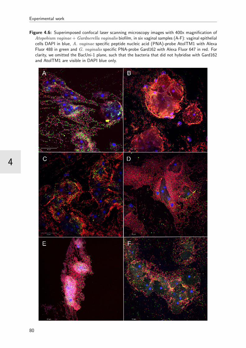
Experimental work
4
Figure 4.6: Superimposed confocal laser scanning microscopy images with 400x magnification ofAtopobium vaginae + Gardnerella vaginalis biofilm, in six vaginal samples (A-F): vaginal epithelialcells DAPI in blue, A. vaginae specific peptide nucleic acid (PNA)-probe AtoITM1 with AlexaFluor 488 in green and G. vaginalis specific PNA-probe Gard162 with Alexa Fluor 647 in red. Forclarity, we omitted the BacUni-1 plane, such that the bacteria that did not hybridise with Gard162and AtoITM1 are visible in DAPI blue only.
80
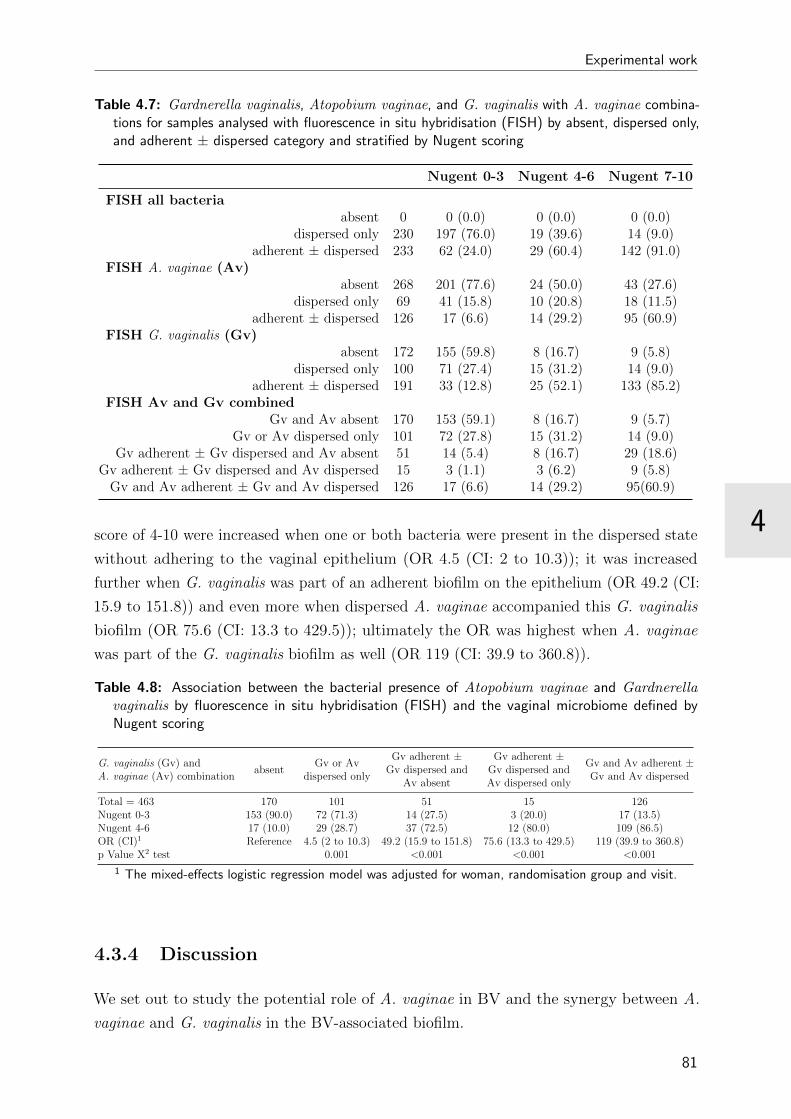
4
Experimental work
Table 4.7: Gardnerella vaginalis, Atopobium vaginae, and G. vaginalis with A. vaginae combina-tions for samples analysed with fluorescence in situ hybridisation (FISH) by absent, dispersed only,and adherent ± dispersed category and stratified by Nugent scoring
Nugent 0-3 Nugent 4-6 Nugent 7-10
FISH all bacteriaabsent 0 0 (0.0) 0 (0.0) 0 (0.0)
dispersed only 230 197 (76.0) 19 (39.6) 14 (9.0)adherent ± dispersed 233 62 (24.0) 29 (60.4) 142 (91.0)
FISH A. vaginae (Av)absent 268 201 (77.6) 24 (50.0) 43 (27.6)
dispersed only 69 41 (15.8) 10 (20.8) 18 (11.5)adherent ± dispersed 126 17 (6.6) 14 (29.2) 95 (60.9)
FISH G. vaginalis (Gv)absent 172 155 (59.8) 8 (16.7) 9 (5.8)
dispersed only 100 71 (27.4) 15 (31.2) 14 (9.0)adherent ± dispersed 191 33 (12.8) 25 (52.1) 133 (85.2)
FISH Av and Gv combinedGv and Av absent 170 153 (59.1) 8 (16.7) 9 (5.7)
Gv or Av dispersed only 101 72 (27.8) 15 (31.2) 14 (9.0)Gv adherent ± Gv dispersed and Av absent 51 14 (5.4) 8 (16.7) 29 (18.6)
Gv adherent ± Gv dispersed and Av dispersed 15 3 (1.1) 3 (6.2) 9 (5.8)Gv and Av adherent ± Gv and Av dispersed 126 17 (6.6) 14 (29.2) 95(60.9)
score of 4-10 were increased when one or both bacteria were present in the dispersed state
without adhering to the vaginal epithelium (OR 4.5 (CI: 2 to 10.3)); it was increased
further when G. vaginalis was part of an adherent biofilm on the epithelium (OR 49.2 (CI:
15.9 to 151.8)) and even more when dispersed A. vaginae accompanied this G. vaginalis
biofilm (OR 75.6 (CI: 13.3 to 429.5)); ultimately the OR was highest when A. vaginae
was part of the G. vaginalis biofilm as well (OR 119 (CI: 39.9 to 360.8)).
Table 4.8: Association between the bacterial presence of Atopobium vaginae and Gardnerellavaginalis by fluorescence in situ hybridisation (FISH) and the vaginal microbiome defined byNugent scoring
G. vaginalis (Gv) andA. vaginae (Av) combination
absentGv or Av
dispersed only
Gv adherent ±Gv dispersed and
Av absent
Gv adherent ±Gv dispersed andAv dispersed only
Gv and Av adherent ±Gv and Av dispersed
Total = 463 170 101 51 15 126Nugent 0-3 153 (90.0) 72 (71.3) 14 (27.5) 3 (20.0) 17 (13.5)Nugent 4-6 17 (10.0) 29 (28.7) 37 (72.5) 12 (80.0) 109 (86.5)OR (CI)1 Reference 4.5 (2 to 10.3) 49.2 (15.9 to 151.8) 75.6 (13.3 to 429.5) 119 (39.9 to 360.8)p Value Χ2 test 0.001 <0.001 <0.001 <0.001
1 The mixed-effects logistic regression model was adjusted for woman, randomisation group and visit.
4.3.4 Discussion
We set out to study the potential role of A. vaginae in BV and the synergy between A.
vaginae and G. vaginalis in the BV-associated biofilm.
81

Experimental work
4
Our study confirms that both A. vaginae and G. vaginalis are important constituents of the
vaginal epithelial biofilm [9, 209]. Adherent A. vaginae and G. vaginalis were visualised in
respectively 54.1% and 82.0% of samples with bacterial biofilm (detected using the universal
BacUni-1 probe), suggesting an important role for both bacteria in this polymicrobial
biofilm. Using FISH, we only found two samples containing A. vaginae (dispersed in
both, adherent in one) in the absence of G. vaginalis, while more than one-third of the G.
vaginalis-positive samples was negative for A. vaginae. This is in accordance with prior
reports on the association of A. vaginae with G. vaginalis [9,164,165,171,209]. We showed
that the presence of both bacteria in the samples, regardless of their existence in a biofilm,
was associated with an elevated or high Nugent score, indicative for vaginal dysbiosis
and BV. The highest probability of having a Nugent score higher than 3 was seen when
both A. vaginae and G. vaginalis were part of a biofilm attaching to the vaginal epithelial
cells.
The association of G. vaginalis with BV was originally described in 1954 by Gardner and
Dukes [427]. The involvement of A. vaginae in BV, however, has only been established 10
years ago [163,165,166]. Swidsinski et al. [9] found vaginal biopsies with vaginal biofilm to
be positive for G. vaginalis and A. vaginae when using fluorescent probes, although in our
hands this A. vaginae probe cross-reacted with other vaginal species as well [209].
The presence of A. vaginae in the BV-associated biofilm could have a major impact
on treatment. Susceptibility to metronidazole, the standard treatment for BV, varied
significantly across various A. vaginae strains in vitro [330]. In vivo data are scarce, but
Bradshaw et al. [170] found that rates of recurrence of BV were higher when A. vaginae was
present in the vaginal microbiome in addition to G. vaginalis. In another study with topical
metronidazole gel by Ferris et al. [163], it was shown that a high concentration of A. vaginae
before treatment was associated with complete or partial failure of treatment for BV. In the
above studies, no distinction was made between dispersed and biofilm-associated bacteria.
Nevertheless, as bacteria in a biofilm are less sensitive to antibiotic treatment [428] and
considering the evidence from our study that the formation of a bacterial biofilm is more
likely to occur when A. vaginae is present in the vaginal microbiome, future design of
studies may want to take this distinction into account when treating BV.
Our study has shed new light on the significance of A. vaginae and the synergy between A.
vaginae and G. vaginalis in vaginal dysbiosis, using highly specific PNA probes for both
bacteria. However, a limitation was that we used multiple samples from the 120 women
of the Ring Plus study. Ideally, we should repeat the study in a larger group of women.
Furthermore, although we assessed the association between bacterial biofilm and vaginal
dysbiosis, more research is needed to unravel the exact mechanisms of biofilm formation
in BV, including the role and the importance of both bacteria studied, to finally define
improved regimens for treatment of BV. Moreover, since BV is a polymicrobial condition,
82

4
Experimental work
new research should study the involvement of other bacteria related to BV.
In conclusion, the presented study uncovered a key piece of the BV puzzle confirming
first, the importance of A. vaginae in BV-associated biofilm and second, showing the joint
presence of A. vaginae and G. vaginalis in a biofilm. Future studies covering a wide array
of BV-associated bacteria may help to further delineate biofilm mechanisms in BV.
83


4
Experimental work
4.4 The importance of Gardnerella vaginalis sialidase
for bacterial vaginosis-associated biofilm
Abstract
Bacterial vaginosis is adifficult-to-treat recurrent condition in which health-associated
lactobacilli are outnumbered by other anaerobic bacteria, such as Gardnerella vaginalis.
Certain genotypes of G. vaginalis can produce sialidase, while others can not. Sialidase
facilitates the destruction of the protective mucus layer on the vaginal epithelium by
hydrolysis of sialic acid on the glycans of mucous membranes. This process facilitates
adhesion of bacterial cells on the epithelium and has been linked with the development
of biofilm in other microorganisms. In this study, using vaginal specimens of 120 women
enrolled in the Ring Plus study, we assessed the association between the G. vaginalis
sialidase gene by quantitative polymerase chain reaction, the diagnosis of bacterial vaginosis
according to Nugent score, and the occurrence of a bacterial vaginosis-associated biofilm
dominated by G. vaginalis by fluorescence in situ hybridisation. We detected the sialidase
gene in 75% of the G. vaginalis-positive samples and found a strong association (p<0.001)
between the presence of a G. vaginalis-dominated biofilm, the diagnosis of BV according
to Nugent and the detection of high loads of the G. vaginalis sialidase gene in the vaginal
specimens. These results could impact the possibilities for diagnosis of bacterial vaginosis,
and in addition might guide research for new treatment.
Adapted from:
Hardy L, Jespers V, Van den Bulck M, Buyze J, Mwambarangwe L, Musengamana V,
Vaneechoutte M, Crucitti T. The importance of Gardnerella vaginalis sialidase for bacterial
vaginosis-associated biofilm. Submitted for publication.
85
Experimental work
4
4.4.1 Introduction
Gardnerella vaginalis has consistently been found in bacterial vaginosis (BV) [15, 40, 41], a
dysbiosis of the vaginal econiche in which the health-associated lactobacilli are outnumbered
by other micro-aerophilic and anaerobic organisms. It has been demonstrated that a vaginal
mucosa polymicrobial biofilm is associated with BV [9,209]. G. vaginalis is able to adhere
to the vaginal epithelial cells and subsequently develop a biofilm on the vaginal wall [9,209],
a mechanism that possibly increases the tolerance of G. vaginalis to lactic acid and
hydrogen peroxide produced by lactobacilli [260] and to antimicrobial treatment [266,267].
It has been suggested that G. vaginalis initiates the colonisation of the vaginal mucosa
and acts as a scaffold to which other species subsequently can attach [260, 264, 414].
However, G. vaginalis can also occur in the healthy vaginal microbiome (although in lower
concentrations) [3, 40, 41], suggesting that the mere presence of G. vaginalis does not
necessarily result in biofilm formation and BV. This observation has led several researchers
to hypothesise that different types of G. vaginalis with different virulence potentials might
exist [160,162,429].
Certain G. vaginalis genotypes can produce sialidase, also known as neuraminidase [160].
Sialidase is a common virulence factor in pathogens such as the Influenza virus [285] and
a large number of bacterial species, such as Propionibacterium acnes [286], Pseudomonas
aeruginosa [287], Streptococcus pneumoniae [288], and Vibrio cholerae [289] and has been
strongly linked with bacterial biofilm development [290–292]. Increased sialidase activity
was also detected in the vaginal fluid of BV patients [293, 430], and is the basis of a
marketed quick test for diagnosis of BV [294].
Sialidase facilitates the destruction of the protective mucus layer in the vagina by hydrolysis
of sialic acid, the most distal sugar moiety on the glycans of mucous epithelial membranes
[293,295]. Cleaving off sialic acid by sialidase provides the bacteria with free sialic acid
that can serve as a nutrient [296], and the exposure of the underlying glycan-binding
site facilitates adhesion of bacterial cells [295, 431]. G. vaginalis could benefit from
this mechanism by attaching to the vaginal epithelium to initiate biofilm development.
Furthermore, sialidase facilitates the circumvention of the vaginal adaptive immune
response, since it modulates the activity of sialylated immune mediators such as interleukins,
immunoglobulins and various cellular receptors, such as sialic acid lectins (Siglecs) [297].
In addition, it has been suggested that, by incorporation of the cleaved sialic acids into
bacterial cell-surface structures, bacteria could disguise themselves as host cells and bypass
the host’s immune response [431,432].
Although other BV-associated bacteria (e.g. Prevotella and Bacteroides species) have
also been shown to produce sialidase in the vagina [293], G. vaginalis is most frequently
isolated, in high concentrations, from vaginal fluid of women with BV [15,40, 41] and has
86

4
Experimental work
a higher tendency to adhere to vaginal epithelial cells compared to other BV-associated
anaerobes [260]. We hypothesised that, like other species [290–292], the genotypes of
G. vaginalis that encode the sialidase gene are associated with the presence of vaginal
biofilms, leading to BV. Therefore, we assessed the association between the presence of
the G. vaginalis sialidase gene and the occurrence of BV-associated biofilm of G. vaginalis
on the vaginal epithelium in vaginal samples of women with and without BV.
Methods and materials
Study participants and ethics statement Vaginal samples were collected from 120
Rwandan women participating in study on the acceptability of using an intravaginal ring
for contraception (NuvaRing®, Merck, New Jersey, USA) and its effect on the vaginal
microbiome (the Ring Plus study [412]). Participants were between 18 and 35 years
old and provided written informed consent for participation in the study. The Ring
Plus study was approved by the Rwanda National Ethics Committee, Rwanda (Approval
number 481/RNEC/2013); and the ethics committees of the Institute of Tropical Medicine
(ITM), Belgium (Approval number 864/13); the Antwerp University Hospital, Belgium
(Approval number 13/7/85); and the University of Liverpool, UK (Approval number
RETG000639IREC).
Vaginal sample collection and preparation
Vaginal samples were collected at the enrolment visit and at each ring insertion visit by the
study clinician. Two Copan flocked® swabs (Copan, Brescia, Italy) and one cotton swab
were brushed against the lateral walls of the vagina. The cotton swab was immediately
used to prepare two vaginal slides: one for Gram staining and one for fluorescence in
situ hybridisation (FISH) on a Superfrost Plus® slide (Menzel-Glaser, Braunschweig,
Germany). Both were heat-fixed by passing twice through a flame. The Superfrost Plus
slides were stored and shipped at room temperature to the ITM, to be fixed for a second
time using Carnoy solution (6:3:1, ethanol:chloroform:glacial acetic acid) [209] for 12 hours
minimum. The Copan flocked swabs were eluted by vortexing for at least 15 seconds in 1.2
ml of diluted phosphate buffered saline (PBS) (pH 7.4 - 1:9, PBS:saline). The eluates were
stored at -80 °C until shipment and shipped to the ITM using a temperature-controlled
dry shipper.
Nugent score of vaginal slides
The status of the vaginal microbiome was assessed at the Rinda Ubuzima laboratory
(Kigali, Rwanda) by Nugent scoring of a Gram stained vaginal slide [158]. A score of
87
Experimental work
4
0-3 was considered as normal vaginal microbiome; a score of 4-6 as intermediate vaginal
microbiome and a score of 7-10 as BV.
Peptide nucleid acid fluorescence in situ hybridisation on vaginal slides
Peptide nucleid acid (PNA) fluorescence in situ hybridisation (FISH) on the vaginal slides
using a species-specific probe for G. vaginalis (Gard162) and the broad-range BacUni-1
probe and imaging was performed as described earlier [209]. Separate scattered bacterial
cells were defined as dispersed bacteria. Aggregates of bacterial cells, sticking to the
vaginal epithelial cells, were defined as adherent bacteria forming a biofilm.
4.4.2 Quantitative polymerase chain reaction for quantification
of bacteria in vaginal samples
Quantitative polymerase chain reaction for Gardnerella vaginalis
The total bacterial load of G. vaginalis was determined by means of quantitative polymerase
chain reaction (qPCR) in the Sexually Transmitted Infections (STI) Reference Laboratory
at the ITM, as described before [3, 209]. The bacterial load was expressed as genome
equivalents (geq)/ml.
Quantitative polymerase chain reaction for Gardnerella vaginalis sialidase The
design of the primer set for amplification of the G. vaginalis sialidase gene was based on
previous work by Lopes dos Santos Santiago et al. [160] and on the sequence of sialidase A
from the fully sequenced G. vaginalis ATCC 14019 strain (reference genome for the Human
Microbiome Project, Baylor College of Medicine, Houston, TX). The previously designed G.
vaginalis sialidase forward primer (GVSI Forward, 5’-GACGACGGCGAATGGCACGA-3’)
[160] was combined with a reverse primer (GVSI Reverse2, 5’-TACAAGCGGCTTTACTC-
TTG-3’) that was newly designed using Primer Blast (National Center for Biotechnology
Information, Bethesda, MD).
For G. vaginalis sialidase gene amplification, the 25 µl PCR mixture contained 12.5 µl
Rotor-Gene SYBR Green qPCR Master mix (Qiagen, Venlo, the Netherlands), 5 µl DNA
extract, 0.75 µM of 5 µM G. vaginalis sialidase forward and reverse primers (Integrated
DNA Technologies, Leuven, Belgium) and RNase-free water provided with the Rotor-Gene
SYBR Green PCR kit.
The amplification reactions were performed using the Rotor Gene Q MDx 5 plex (Qiagen,
Venlo, the Netherlands) and the amplification program (10 min 95 °C, (5 sec 95 °C - 10
88

4
Experimental work
sec 58 °C) x 45) was followed by melting curve analysis. Each sample was run in duplicate
and each run included a standard curve.
Statistical analysis
Bacterial counts were log 10 transformed before analysis. Data analysis was done using
STATA13. The p-values reported for associations between the presence and quantity of
the sialidase gene and BV/qPCR-biofilm results were obtained using mixed effects ordered
logistic regression.
4.4.3 Results
Characterisation of vaginal samples
A total of 527 samples were available for Nugent scoring, 462 samples were analysed by
FISH and 524 samples were used for qPCR to detect G. vaginalis. All 393 G. vaginalis
qPCR-positive samples were tested for the presence of the G. vaginalis sialidase gene by
qPCR (Table 4.9).
Table 4.9: Characteristics of vaginal samples
Test Total Result N (%)
Nugent score 527 0-3 299 (56.7)4-6 53 (10.1)
7-10 175 (33.2)
FISH 462 Gardnerella vaginalis positive 290 (62.8)G. vaginalis biofilm 191 (41.3)
G. vaginalis dispersed only 99 (21.4)
qPCR 524 G. vaginalis positive 393 (75.0)G. vaginalis sialidase positive 294 (56.1)
Nugent score Of the total of 527 samples of 120 participants, 299 (56.7%) had a healthy
microbiome (Nugent score 0-3), 53 (10.1%) were categorised as intermediate (Nugent score
4-6) and 175 (33.2%) were diagnosed as BV (Nugent score 7-10).
Fluorescence in situ hybridisation A subset of 462 samples were analysed with FISH.
The remaining 65 samples could not be analysed mainly due to the absence of epithelial
cells and bacteria on the slides. G. vaginalis was present in 290 samples (62.8%) using
89
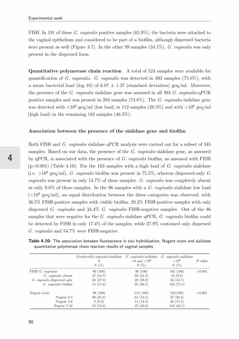
Experimental work
4
FISH. In 191 of these G. vaginalis positive samples (65.9%), the bacteria were attached to
the vaginal epithelium and considered to be part of a biofilm, although dispersed bacteria
were present as well (Figure 4.7). In the other 99 samples (34.1%), G. vaginalis was only
present in the dispersed form.
Quantitative polymerase chain reaction A total of 524 samples were available for
quantification of G. vaginalis. G. vaginalis was detected in 393 samples (75.0%), with
a mean bacterial load (log 10) of 6.97 ± 1.37 (standard deviation) geq/ml. Moreover,
the presence of the G. vaginalis sialidase gene was assessed in all 393 G. vaginalis-qPCR
positive samples and was present in 294 samples (74.8%). The G. vaginalis sialidase gene
was detected with <106 geq/ml (low load) in 112 samples (28.5%) and with >106 geq/ml
(high load) in the remaining 182 samples (46.3%).
Association between the presence of the sialidase gene and biofilm
Both FISH and G. vaginalis sialidase qPCR analysis were carried out for a subset of 345
samples. Based on our data, the presence of the G. vaginalis sialidase gene, as assessed
by qPCR, is associated with the presence of G. vaginalis biofilm, as assessed with FISH
(p<0.001) (Table 4.10). For the 163 samples with a high load of G. vaginalis sialidase
(i.e. >106 geq/ml), G. vaginalis biofilm was present in 75.5%, whereas dispersed-only G.
vaginalis was present in only 14.7% of these samples. G. vaginalis was completely absent
in only 9.8% of these samples. In the 96 samples with a G. vaginalis sialidase low load
(<106 geq/ml), an equal distribution between the three categories was observed; with
36.5% FISH-positive samples with visible biofilm, 29.2% FISH-positive samples with only
dispersed G. vaginalis and 34.4% G. vaginalis FISH-negative samples. Out of the 86
samples that were negative for the G. vaginalis sialidase qPCR, G. vaginalis biofilm could
be detected by FISH in only 17.4% of the samples, while 27.9% contained only dispersed
G. vaginalis and 54.7% were FISH-negative.
Table 4.10: The association between fluorescence in situ hybridisation, Nugent score and sialidasequantitative polymerase chain reaction results of vaginal samples
Gardnerella vaginalis sialidase0
N (%)
G. vaginalis sialidase>0 and <106
N (%)
G. vaginalis sialidase>106
N (%)P-value
FISH G. vaginalis 86 (100) 96 (100) 163 (100) <0.001G. vaginalis absent 47 (54.7) 33 (34.4) 16 (9.8)
G. vaginalis dispersed only 24 (27.9) 28 (29.2) 24 (14.7)G. vaginalis biofilm 15 (17.4) 35 (36.5) 123 (75.5)
Nugent score 98 (100) 112 (100) 181(100) <0.001Nugent 0-3 80 (81.6) 61 (54.5) 37 (20.4)Nugent 4-6 8 (8.2) 14 (12.5) 28 (15.5)
Nugent 7-10 10 (10.2) 37 (33.0) 116 (64.1)
90

4
Experimental work
Figure 4.7: Superimposed confocal laser scanning microscopy images with 400x magnification ofAtopobium vaginae + Gardnerella vaginalis biofilm, in three vaginal samples: vaginal epithelialcells DAPI in blue, A. vaginae specific peptide nucleic acid (PNA)-probe AtoITM1 with AlexaFluor 488 in green and G. vaginalis specific PNA-probe Gard162 with Alexa Fluor 647 in red.
91
Experimental work
4
Association between the presence of the sialidase gene and the diagnosis of
bacterial vaginosis
A subset of 391 samples was available for both Nugent scoring and qPCR based on
the quantification of the G. vaginalis sialidase analysis. The probability of having BV
according to Nugent is increased when the G. vaginalis sialidase gene is present in high
loads (>106 geq/ml) (p<0.001) (Table 4.10). Of the 181 samples with a high load of the
G. vaginalis sialidase gene, 64.1% were BV-positive (Nugent score 7-10), while only 20.4%
had a healthy vaginal microbiome (Nugent score of 0-3) and 15.5% were diagnosed with an
intermediate Nugent score of 4-6. In contrast, BV could be diagnosed in only 10 out of the
98 samples (10.2%) for which no G. vaginalis sialidase gene could be detected. However,
80 samples (81.6%) without the sialidase gene represented a healthy vaginal microbiome
according to Nugent and 8 samples (8.2%) had an intermediate score. Additionally, when
the G. vaginalis sialidase gene was present in low amounts, 54.5% of the 112 samples were
considered healthy according to the Nugent score, 33.0% were categorised as BV, and
12.5% represented an intermediate microbiome.
4.4.4 Discussion
BV is the most prevalent vaginal disorder in women of reproductive age worldwide, and
aside from the discomfort in case of symptomatic BV, it can also generate an array
of serious gynaecological and obstetric complications. The presence of BV-associated
anaerobes in the vaginal environment increases the risk for preterm labor and birth [107].
Furthermore, the presence of sialidase in vaginal fluid has been linked to BV and to preterm
birth as well [111,112]. In a large cohort of 1806 women which included 800 women with
BV and 53 spontaneous preterm births, Cauci et al. [112] showed that the sialidase levels
in the vaginal fluid were significantly associated with all averse pregnancy outcomes.
G. vaginalis plays an important role in BV, since G. vaginalis overgrowth is found in
nearly all cases of BV [157]. However, the presence of G. vaginalis in healthy vaginal
environments [3, 433] contradicts its pathogenic role in BV. To resolve this discrepancy, it
has been suggested that G. vaginalis might actually consist of different species with distinct
roles in BV pathogenesis, which is supported by the genotypic and phenotypic diversity
of the species [159, 161, 162]. Although other BV-associated bacteria (e.g. Prevotella
and Bacteroides species) are able to produce sialidase [293], we decided to investigate
G. vaginalis sialidase in BV, considering that G. vaginalis is most frequently isolated
from vaginal fluids of women suffering from BV [15,40,41,157] and that it has a higher
tendency to adhere to vaginal epithelial cells in vitro compared to other BV-associated
anaerobes [260]. We studied the association between the presence of the G. vaginalis
92

4
Experimental work
sialidase gene, as a proxy for sialidase production, in the vagina and the occurrence of
BV and bacterial biofilm on the vaginal epithelium. To this end, we screened the vaginal
samples of 120 Rwandan women [412] by means of a G. vaginalis sialidase specific qPCR
and assessed the occurrence of BV and biofilm by means of a) light microscopy after Gram
staining and Nugent scoring and b) CLSM after FISH for G. vaginalis and bacteria in
general on vaginal samples.
In this population, the sialidase gene was detected in about 75% of the G. vaginalis-positive
samples. In about 60% of those samples a high load (>106 geq/ml) of the gene was detected.
This high prevalence of the G. vaginalis sialidase gene in our study may be explained by
the cohort of women enrolled for this study and the high prevalence of BV, i.e. in 43.3% of
all samples. Earlier studies have investigated the presence of the G. vaginalis sialidase gene
and the production of sialidase in cultured isolates. Using clinical isolates from Belgian
women, Lopes dos Santos Santiago and colleagues could detect a G. vaginalis sialidase gene
with qPCR in 51% of strains [160]. When using the filter paper spot test for the detection
of sialidase activity, von Nicolai et al. [434] could detect sialidase production in only 1 of
10 clinical isolates. Additionally, Briselden et al. [293] detected sialidase activity in 20% of
105 G. vaginalis isolates (with no difference in isolates from women with and without BV),
and Moncla and Pryke [435] observed sialidase activity in 39% of 31 isolates.
At present, it is not clear whether the sialidase gene is expressed constitutively or not.
Pleckaityte and colleagues [436] detected a sialidase gene in 17 tested G. vaginalis isolates,
but only 10 of these strains actually produced sialidase in vitro. Schellenberg et al. [159]
also found that the gene presence was not predictive of actual sialidase activity using a
qualitative (positive/negative) filter spot assay: out of 77 G. vaginalis isolates positive
for the sialidase gene, 36 produced sialidase [159]. In addition, in currently ongoing
(not yet published) in vitro experiments by our group, we found that only 29 out of 41
sialidase gene-positive G. vaginalis isolates produced sialidase, based on the filter spot
test. Interestingly, we noticed that all but two sialidase-producing strains were isolated
from women with BV according to Nugent. This contradicts what was published by Lopes
dos Santos Santiago et al. [160] who found a 100% correspondence between the mere
presence of the gene and sialidase activity in 19 G. vaginalis isolates. However, all but one
of these isolates were obtained from women with a disturbed microbiome, which might
have introduced a bias [160]. The absence of sialidase activity in sialidase-positive isolates
might be explained by the presence of an alternative gene encoding this activity or the
need for other factors to stimulate the expression of the gene. Possible factors interfering
with the production of sialidase might be the presence of sialic acid on epithelial cells, or a
threshold in G. vaginalis concentration that needs to be reached. In any case, more basic
research is needed to fully understand the sialidase expression pathway.
When looking at the association between the presence of the G. vaginalis sialidase gene
93
Experimental work
4
and the diagnosis of BV by Nugent score, we found that the probability for having BV
(Nugent score 7-10) was increased when a high concentration of the G. vaginalis sialidase
gene was present in the vaginal samples. This was expected, since sialidase production by
G. vaginalis is recognised as a virulence factor [436], and has already been associated with
BV [112]. In our previous work [209], we confirmed the importance of G. vaginalis in the
development of a biofilm on the vaginal epithelium in BV, as established by Swidsinski
et al. [9] in 2005. Ours was the first study to use clinical samples to demonstrate the
significance of G. vaginalis’ ability to produce sialidase and to document its association
with BV and vaginal biofilm. We established a strong association between a high load of
the G. vaginalis sialidase gene, as measured by qPCR using a specific primer set targeting
the G. vaginalis sialidase gene, and G. vaginalis being part of a vaginal epithelium biofilm,
visualised by CLSM after FISH. Sialidase has been linked with biofilm development in other
microorganisms. In Pseudomonas aeruginosa, sialidase (or neuraminidase) contributes
to the initial colonisation of the airway, and colonisation could be blocked in vitro by
viral neuraminidase inhibitors [290]. Likewise in pneumococcal infections, sialidase is
involved in biofilm formation and pathogenesis of respiratory tract infections [291,292].
Also, sialidase producing Propionibacterium acnes isolates were more associated with acne
than sialidase negative isolates [437].
A shortcoming of this study is the absence of isolates. Clinical isolates would have provided
valuable information on ARDRA genotyping and the actual sialidase activity. Being able
to assess sialidase production by G. vaginalis directly in our Ring Plus samples would
have been interesting, but since sialidase activity in our mixed samples could also have
resulted from other vaginal species, it would have confounded the results. Despite this
limitation, we were able to establish that the ability of G. vaginalis to produce sialidase
is linked to the presence of BV and the existence of a vaginal biofilm. This finding may
impact the possibilities for BV diagnosis, but it may also guide future research for new
and better treatments for this recurrent and difficult-to-treat condition.
94
4.5 Association of vaginal dysbiosis and biofilm with
contraceptive vaginal ring biomass in African women
Abstract
We investigated the presence, density and bacterial composition of contraceptive vaginal
ring biomass and its association with the vaginal microbiome. Of 415 rings worn by 120
Rwandese women for three weeks, the biomass density was assessed with crystal violet and
the bacterial composition of biomass eluates was assessed with quantitative polymerase
chain reaction (qPCR). The biomass was visualised after fluorescence in situ hybridisation
(FISH) and with scanning electron microscopy (SEM). The vaginal microbiome was assessed
with Nugent scoring and vaginal biofilm was visualised after FISH. All vaginal rings were
covered with biomass (mean optical density (OD) of 3.35; standard deviation (SD) 0.64).
Lactobacilli were present on 93% of the rings, G. vaginalis on 57%, and A. vaginae on
37%. The ring biomass density was associated with the concentration of A. vaginae
(OD+0.03; 95% confidence interval (CI) 0.01-0.05 for one log increase; p=0.002) and of
G. vaginalis (OD+0.03; (95% CI 0.01-0.05; p=0.013). The density also correlated with
Nugent score: rings worn by women with a BV Nugent score (OD+0.26), and intermediate
score (OD+0.09) had a denser biomass compared to rings worn by participants with a
normal score (p=0.002). Furthermore, presence of vaginal biofilm containing G. vaginalis
(p=0.001) and A. vaginae (p=0.006) correlated with a denser ring biomass (OD +0.24 and
+0.22 respectively). With SEM we observed either a loose network of elongated bacteria
or a dense biofilm. In summary, we found a correlation between vaginal dysbiosis and the
density and composition of the ring biomass, and further research is needed to determine
if these relationships are causal. As multipurpose vaginal rings to prevent pregnancy, HIV,
and other sexually transmitted diseases are being developed, the potential impact of ring
biomass on the vaginal microbiota and the release of active products should be researched
in depth.
Adapted from:
Hardy L, Jespers J, De Baetselier I, Mwambarangwe L, Musengamana V, van de Wijgert
J, Crucitti T. Association of vaginal dysbiosis and biofilm with contraceptive vaginal ring
biomass in African women. Submitted for publication.

Experimental work
4
4.5.1 Introduction
Contraceptive vaginal rings are available in high income countries and Latin America but
not in sub-Saharan Africa [438]. However, vaginal rings are expected to be introduced in
this part of the world in the near future. Most notably, multi-purpose vaginal rings are
being developed for the controlled release of drugs to prevent reproductive tract infections,
such as HIV (dapivirine ring) [439], herpes simplex virus type 2 (HSV-2) [440], bacterial
vaginosis (BV), and pregnancy [441].
Early contraceptive ring studies demonstrated that ring use did not negatively affect the
naturally protective vaginal environment including the presence of lactobacilli [68, 442,
443]. Recent more in-depth work showed an increase in healthy bacteria or lactobacilli
concentrations with ring use [69,70,72]. This effect was thought to be caused by ethinyl
estradiol in the ring [69, 70, 72]. Lactobacilli play an important role in the two main
states of the vaginal microbiome: the health-associated vaginal microbiome dominated by
lactobacilli, and a BV-associated microbiome characterised by a polymicrobial dysbiosis.
In dysbiosis, the lactobacilli disappear and the concentrations of facultative anaerobic
bacteria, such as Gardnerella vaginalis and Atopobium vaginae, increase [15]. The anaerobic
bacteria will often form a vaginal biofilm [9,141,209]. Bacterial biofilms are also known to
develop on indwelling medical devices whenever microorganisms find a surface to attach
to [407]. The potential development of bacterial biofilm on vaginal rings in vivo has yet to
be explored in humans.
We hypothesised that a biomass would develop on vaginal rings, and that rings worn by
women with BV-associated dysbiosis would have higher biomass density than rings worn
by women with dysbiosis. To investigate this hypothesis, we studied the presence, density
and bacterial composition of the biomass on contraceptive vaginal rings and investigated
the association between ring biomass density and the vaginal microbiome.
4.5.2 Methods and materials
This is a laboratory sub-study of the “Ring Plus” contraceptive vaginal ring study performed
at the Rinda Ubuzima (RU) research clinic in Kigali, Rwanda (ClinicalTrials.gov identifier
NCT01796613) [412].
Participants, study product, and clinical sample preparation
The NuvaRing® contraceptive vaginal ring (N.V. Organon, Oss, the Netherlands) was
used over a period of three months by 120 adult female participants [412]. The women
had each ring inserted for three weeks continuously followed by one week off (intermittent
96
4
Experimental work
use) or continuously with no breaks in between the removal of the old/insertion of the
new ring every three weeks (continuous use). In the intermittent use group, women used
three rings each, while the women in the continuous group used four rings each during the
whole study period. Vaginal examination, ring removal, and sample collection were carried
out by the study clinician, as described previously [444]. For this sub-study, vaginal fluid
was rolled on two microscopy slides and airdried for each participant at baseline and at
each ring removal visit. One slide was Gram stained for Nugent scoring, and the other
slide was used to assess the presence of a vaginal biofilm.
All rings worn by study participants were collected after removal at study visits. Each
ring was cut in three equal parts immediately after removal. The part for the biomass
density assessment with crystal violet was submerged in 3 ml of glutaraldehyde for two
weeks, transferred to 3 ml of formaldehyde, and stored at 2-8 °C until testing. This part
was also used for electron microscopy after the crystal violet assay had been completed.
The part for qPCR was stored in diluted phosphate buffered saline (dPBS) (pH 7.4 - 1:9,
PBS:saline) at -20 °C. The final part for fluorescence microscopy was stored in Carnoy
solution (6:3:1, ethanol:chloroform:glacial acetic acid) at 2-8 °C until testing [209]. All
samples, except for the Nugent slide, were shipped from the study site in Rwanda to the
Institute of Tropical Medicine (ITM) in Antwerp, Belgium: the refrigerated samples were
transported at room temperature, while the frozen samples were transported at -196 °C in
a dry shipper.
Laboratory assessment of clinical samples
For this sub-study, all vaginal rings removed and collected throughout the study for both
the intermittent and continuous use groups were analysed as well as all vaginal smears
collected at baseline and each ring removal visit. The vaginal microbiota were characterised
in two ways: Nugent scoring of Gram stained vaginal smears [158] in the on-site RU
laboratory and confocal laser scanning microscopy (CSLM) after peptide nucleic acid
(PNA) fluorescence in situ hybridisation (FISH) of a second vaginal smear to detect vaginal
biofilm at the ITM in Antwerp. In Nugent scoring, a score of 0-3 is considered a normal
vaginal microbiota; a score of 4-6 an intermediate microbiota and a score of 7-10 bacterial
vaginosis. Vaginal biofilms on another vaginal smear were visualised with CSLM after
FISH. This technique was performed as described previously using three probes: the
broad-range BacUni-1 probe detecting all bacteria, and probes detecting two bacterial
species strongly associated with bacterial vaginosis (AtoITM1 for A. vaginae and Gard162
for G. vaginalis) [209,444].
The biomass on contraceptive rings that had been used by participants for three weeks
was stained with crystal violet to determine the optical density (OD) as a proxy for the
97

Experimental work
4
quantity of the biomass. The crystal violet microtitre plate assay [445] was adapted to fit
the ring parts and applied to identify and measure the biomass density on the rings. First,
the ring biomass was stained with crystal violet (0.1% solution) for 10 minutes. Next,
the ring part was rinsed twice and air-dried and the ring biomass staining was solubilised
by submerging the ring in 3 ml of 30% acetic acid in water. From this solution, 125
µl was transferred to a new microtitre plate for OD measurement at 550 nm. Bacterial
compositions of the biomass were assessed by quantitative polymerase chain reaction
(qPCR) of the Lactobacillus genus, G. vaginalis, and A. vaginae. Frozen ring parts were
thawed and vortexed; using this eluate, 200 µl DNA was extracted (Abbott, Maidenhead,
UK) and stored at -80 °C until testing. qPCR was performed for each bacteria genus
or species separately. The PCR mixtures and primers for A. vaginae, G. vaginalis, and
Lactobacillus genus and the amplification reactions (Rotor Gene Q MDx 5 plex, Qiagen,
Venlo, the Netherlands) have been described before [209].
In a random sub-sample of 120 rings, bacterial compositions of the biomass were also
visualised by CSLM after PNA FISH for G. vaginalis, A. vaginae, and Lactobacillus
genus. Slides were prepared for fluorescence microscopy by rinsing the ring with ddH2O,
removing the biomass attached to the ring and spreading it out on the microscopy slide,
passing the slide through a flame twice, and fixating it in Carnoy solution. PNA FISH was
performed as described earlier [3,444]. An additional probe targeting the Lactobacillus genus
(Lac663) [204] was used to visualise the Lactobacillus species in the biomass. Furthermore,
we applied scanning electron microscopy on a random selection of 11 rings to enable a
three-dimensional view of the biomass architecture. The ring parts were dehydrated in an
ethanol line and critical point dried. The parts were cut in pieces of one centimetre each,
mounted on metal specimen stubs, coated with a 16 nm thick platinum film, and imaged
using a JEOL JSM-840 microscope.
Statistical analysis
STATA version 12 was used to analyse data. The numbers and proportions of vaginal
slides by Nugent score category (0-3, 4-6, 7-10) and fluorescence microscopy evaluation
categories were described. Fluorescence microscopy was conducted by one microscopist
(LH) who recorded for each vaginal slide and ring part whether she visualised any bacterial
biofilm (positive fluorescence signal for the “all bacteria” probe), a biofilm incorporating G.
vaginalis and/or A. vaginae (positive fluorescence signals for the relevant species-specific
probe), and/or whether she visualised any of these as dispersed/planktonic bacteria only.
Biofilm was defined as a dense network of bacteria adhering to a surface (the vaginal
epithelial cells), dispersed/planktonic bacteria were defined as scattered bacteria, not
visibly adhering to other bacteria or a surface.
98
4
Experimental work
These results were presented in three ways. First, visualisation of any vaginal biofilm (“all
bacteria” probe positive), of a biofilm containing G. vaginalis and/or A. vaginae (each
of these was assessed for each slide and results are not mutually exclusive). Second, no
biofilm visualised at all, but dispersed/planktonic bacteria were present (for all bacteria,
G. vaginalis, and A. vaginae; not mutually exclusive). Three, no bacteria visualised at all
(for all bacteria, G. vaginalis, and A. vaginae; not mutually exclusive).
Ring biomass density was presented as mean OD with standard deviations; the presence
of Lactobacillus genus, G. vaginalis, and A. vaginae in ring biomass eluates as proportions
with 95% confidence intervals (CI). The bacterial concentrations of Lactobacillus genus,
G. vaginalis, and A. vaginae in ring biomass eluates were expressed as log10 transformed
genome equivalents (geq)/ml. We used mixed effects regression analysis with fixed effects
for participant (due to repeated observations) and randomisation group (intermittent
versus continuous ring use) to evaluate associations between ring biomass density and
Nugent score categories, and with fluorescence microscopy results (presence of G. vaginalis
vaginal biofilm, A. vaginae biofilm, G. vaginalis in a dispersed form only, and A. vaginae
in a dispersed form only).
4.5.3 Results
The mean age of the 120 randomised participants was 28.4 years (95% CI: 25-32), with
61% of women being married, and 57.5% having attained more than just primary school
education. All participants but one completed the study, which implies that 417 rings
and matching vaginal smear duplicates (3 times 60, or 180 from the intermittent group;
4 times 60, or 237 from the continuous group, excluding 3 samples of one discontinued
participant) should have been collected. We were able to collect a total of 415 vaginal
rings and 415 vaginal smears in duplicate. Two sample sets did not reach the laboratories.
A total of 415 vaginal rings were assessed for biomass with the crystal violet assay, 412
ring eluates by qPCR, and sub-samples of 120 rings by FISH and 11 rings with SEM. The
quality of 7 slides was insufficient for Nugent scoring, leaving us with 408 vaginal slides to
score, matching the 415 time-points for which a vaginal ring was available. FISH results
were available for 362 vaginal slides matching the 415 time-points for which a vaginal ring
was available. The quality of 53 samples was not sufficient for FISH.
Vaginal microbiome
Most slides (61.5%) had a normal Nugent score of 0-3, 28.9% a BV Nugent score 7-10, and
9.6% an intermediate score of 4-6. Fluorescence microscopy results are shown in Table
99
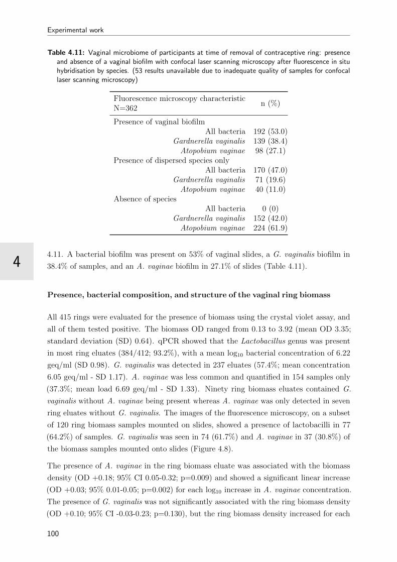
Experimental work
4
Table 4.11: Vaginal microbiome of participants at time of removal of contraceptive ring: presenceand absence of a vaginal biofilm with confocal laser scanning microscopy after fluorescence in situhybridisation by species. (53 results unavailable due to inadequate quality of samples for confocallaser scanning microscopy)
Fluorescence microscopy characteristicN=362
n (%)
Presence of vaginal biofilmAll bacteria 192 (53.0)
Gardnerella vaginalis 139 (38.4)Atopobium vaginae 98 (27.1)
Presence of dispersed species onlyAll bacteria 170 (47.0)
Gardnerella vaginalis 71 (19.6)Atopobium vaginae 40 (11.0)
Absence of speciesAll bacteria 0 (0)
Gardnerella vaginalis 152 (42.0)Atopobium vaginae 224 (61.9)
4.11. A bacterial biofilm was present on 53% of vaginal slides, a G. vaginalis biofilm in
38.4% of samples, and an A. vaginae biofilm in 27.1% of slides (Table 4.11).
Presence, bacterial composition, and structure of the vaginal ring biomass
All 415 rings were evaluated for the presence of biomass using the crystal violet assay, and
all of them tested positive. The biomass OD ranged from 0.13 to 3.92 (mean OD 3.35;
standard deviation (SD) 0.64). qPCR showed that the Lactobacillus genus was present
in most ring eluates (384/412; 93.2%), with a mean log10 bacterial concentration of 6.22
geq/ml (SD 0.98). G. vaginalis was detected in 237 eluates (57.4%; mean concentration
6.05 geq/ml - SD 1.17). A. vaginae was less common and quantified in 154 samples only
(37.3%; mean load 6.69 geq/ml - SD 1.33). Ninety ring biomass eluates contained G.
vaginalis without A. vaginae being present whereas A. vaginae was only detected in seven
ring eluates without G. vaginalis. The images of the fluorescence microscopy, on a subset
of 120 ring biomass samples mounted on slides, showed a presence of lactobacilli in 77
(64.2%) of samples. G. vaginalis was seen in 74 (61.7%) and A. vaginae in 37 (30.8%) of
the biomass samples mounted onto slides (Figure 4.8).
The presence of A. vaginae in the ring biomass eluate was associated with the biomass
density (OD +0.18; 95% CI 0.05-0.32; p=0.009) and showed a significant linear increase
(OD +0.03; 95% 0.01-0.05; p=0.002) for each log10 increase in A. vaginae concentration.
The presence of G. vaginalis was not significantly associated with the ring biomass density
(OD +0.10; 95% CI -0.03-0.23; p=0.130), but the ring biomass density increased for each
100
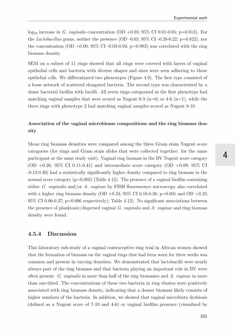
4
Experimental work
log10 increase in G. vaginalis concentration (OD +0.03; 95% CI 0.01-0.05; p=0.013). For
the Lactobacillus genus, neither the presence (OD -0.03; 95% CI -0.28-0.22; p=0.822), nor
the concentration (OD +0.00; 95% CI -0.03-0.04; p=0.983) was correlated with the ring
biomass density.
SEM on a subset of 11 rings showed that all rings were covered with layers of vaginal
epithelial cells and bacteria with diverse shapes and sizes were seen adhering to these
epithelial cells. We differentiated two phenotypes (Figure 4.9). The first type consisted of
a loose network of scattered elongated bacteria. The second type was characterised by a
dense bacterial biofilm with bacilli. All seven rings categorised in the first phenotype had
matching vaginal samples that were scored as Nugent 0-3 (n=6) or 4-6 (n=1), while the
three rings with phenotype 2 had matching vaginal samples scored as Nugent 8-10.
Association of the vaginal microbiome compositions and the ring biomass den-
sity
Mean ring biomass densities were compared among the three Gram stain Nugent score
categories (for rings and Gram stain slides that were collected together: for the same
participant at the same study visit). Vaginal ring biomass in the BV Nugent score category
(OD +0.26; 95% CI 0.11-0.41) and intermediate score category (OD +0.09; 95% CI
-0.12-0.30) had a statistically significantly higher density compared to ring biomass in the
normal score category (p=0.002) (Table 4.12). The presence of a vaginal biofilm containing
either G. vaginalis and/or A. vaginae by FISH fluorescence microscopy also correlated
with a higher ring biomass density (OD +0.24; 95% CI 0.10-0.38; p=0.001 and OD +0.22;
95% CI 0.06-0.37; p=0.006 respectively); Table 4.12). No significant associations between
the presence of planktonic/dispersed vaginal G. vaginalis and A. vaginae and ring biomass
density were found.
4.5.4 Discussion
This laboratory sub-study of a vaginal contraceptive ring trial in African women showed
that the formation of biomass on the vaginal rings that had been worn for three weeks was
common and present in varying densities. We demonstrated that lactobacilli were nearly
always part of the ring biomass and that bacteria playing an important role in BV were
often present: G. vaginalis in more than half of the ring biomasses and A. vaginae in more
than one-third. The concentrations of these two bacteria in ring eluates were positively
associated with ring biomass density, indicating that a denser biomass likely consists of
higher numbers of the bacteria. In addition, we showed that vaginal microbiota dysbiosis
(defined as a Nugent score of 7-10 and 4-6) or vaginal biofilm presence (visualised by
101

Experimental work
4
Table 4.12: Association of the vaginal microbial status with contraceptive vaginal ring biomass.*corrected for participant multiple observations and randomisation group.
Risk factor
Mean change indensity for a
one unit changein risk factor
95%confidence
interval
p-value fromregressionanalysis
Diagnosis bacterial vaginosis 0.002Normal Nugent score (0-3) Ref -
Intermediate Nugent score (4-6) +0.09 -0.12 - 0.30Bacterial vaginosis Nugent score (7-10) +0.26 0.11 - 0.41
Fluorescence microscopy after FISH by speciesGardnerella vaginalis biofilm +0.24 0.10 - 0.38 0.001
Gardnerella vaginalis dispersed only +0.10 -0.04 - 0.24 0.147Atopobium vaginae biofilm +0.22 0.06 - 0.37 0.005
Atopobium vaginae dispersed only +0.09 -0.05 - 0.24 0.195
fluorescence microscopy) were associated with higher ring biomass densities. These findings
suggest that the status of the vaginal microbiome influences the formation or deposit of
biomass on vaginal rings and/or vice versa. Our study was cross-sectional and therefore
does not allow us to determine temporality and causality of these associations.
Only two other human studies and one macaque study have visualised the surfaces of
vaginal rings after use. Miller et al. applied electron microscopy to examine a NuvaRing
used for four weeks by a healthy volunteer, and observed cellular debris but no bacterial
growth on the surface of the ring [409]. We speculated that the magnification of 200X
that they used was too low to visualise bacteria. In comparison, we used magnifications
of 1000X and 4000X in the present study. A second study in human volunteers showed
the presence of biomass on all 48 rings containing an antiretroviral drug that were used
for four weeks [411]. SEM with a magnification of 25X was used to semi-quantify the
biomass density. In this population of women, of whom more than two-third had a normal
Nugent score, the ring biomass density (semi-quantified visually with SEM) was not
associated with the Nugent score category [411]. Gunawardana et al. [221] differentiated
two biomass phenotypes, while visualising the surface of vaginal rings worn by six female
pig-tailed macaques for 28 days with electron microscopy and fluorescence microscopy.
They found large areas of the ring surface covered with tightly packed mats of bacteria and
epithelial cells or thicker interwoven networks of uniform fibres. We also differentiated two
phenotypes. The first type consisted of a loose network of scattered elongated bacteria,
probably lactobacilli, which agrees with the normal Nugent score of the matching vaginal
smears. The second type was characterised by a dense bacterial biofilm with bacilli, also
in agreement with the BV Nugent score of the associated vaginal smear.
At present, contraceptive vaginal rings are commonly used in countries where HIV is
102

4
Experimental work
not endemic and BV prevalence in low. However, multipurpose and long-acting vaginal
rings for the prevention of HIV and pregnancy are being developed specifically for in
HIV-endemic countries, most of which are in sub-Saharan Africa [67, 72, 440, 446–448].
Recently, a vaginal ring containing the antiretroviral drug dapivirine was shown to be
effective for HIV prevention in sub-Saharan African women and this monthly ring will
soon be licensed for that purpose. It will be the first vaginal HIV prevention option for
women at risk of HIV infection [439]. However, BV prevalence in sub-Saharan Africa
is high, and biomass deposit on these HIV prevention rings might promote or maintain
vaginal dysbiosis in ring users and/or hamper active drug release from the rings, resulting
in reduced efficacy [2]. This requires further study. Extensive epidemiological research has
shown that sex hormones, including those released by contraceptive vaginal rings, have a
beneficial effect on the vaginal microbiome [69,70,72]. Our data confirm this: we observed
an improvement in the vaginal microbiome by Nugent score after initiation of NuvaRing
use (data not presented). Incorporating oestrogen and/or progestogens in vaginal rings
may therefore be an important strategy to protect the vaginal microbiome during ring
use in addition to protecting against pregnancy. Other components that are beneficial for
the vaginal microbiome, such as acidifying agents and probiotic lactobacilli, could also be
added to vaginal rings in the future.
Several types of silicones and thermoplastics are used to manufacture vaginal devices. We
have previously shown that the ability of bacteria to adhere to the device surfaces differs by
the type of material used [449]. We studied Neisseria gonorrhoeae biofilm on silicone and
thermoplastic vaginal ring materials and showed the adherence of gonococci was greater
on the silicone ring material as compared to the thermoplastic ring material [449]. The
NuvaRing is composed of a thermoplastic (ethylene-vinyl acetate copolymer) material,
which was less prone to colonisation by gonococci. However, it is unclear if BV-associated
bacteria would behave similarly to gonococci in vitro, and if the in vitro data accurately
predict what would happen in vivo.
In summary, our study showed that biomass easily forms on the contraceptive vaginal
ring within three weeks and that BV-associated bacteria are commonly present in this
biomass. Our study also showed associations between the presence of vaginal dysbiosis
and vaginal biofilm and the the ring biomass density. The temporality and causality of
these relationships deserve further study. Furthermore, we recommend that the design
and development of multipurpose vaginal rings take ring biomass formation into account
by studying the effects on the vaginal microbiota and active product release.
103

Experimental work
4
Figure 4.8: Visualisation of biomass on intravaginal ring surface by confocal laser scanning microscopyafter fluorescence in situ hybridisation at 400x magnification: A. Lactobacilli scattered on vaginalepithelial cells; B. Vaginal epithelial cells covered with bacterial biofilm.
104
4
Experimental work
Figure 4.9: Visualisation of biomass on intravaginal ring surface by scanning electron microscopy at1000x magnification: A. Phenotype 1 - elongated bacteria scattered on vaginal epithelial cells; B.Phenotype 2 - condense biofilm of bacilli on vaginal epithelial cells.
105

5
CHAPTER 5
General discussion
There are two major vaginal microbiome states: the healthy vaginal microbiome which is
dominated by lactobacilli, and the bacterial vaginosis (BV) vaginal microbiome which is
described as polybacterial dysbiosis. The lactobacilli (with the exception of Lactobacillus
iners) are considered to disappear in BV, while the bacterial load of other (facultative)
anaerobic bacteria, such as Gardnerella vaginalis and Atopobium vaginae, increases [15]. It
also has been demonstrated that G. vaginalis and A. vaginae are important constituents of
a biofilm on the vaginal epithelium, that is associated with BV [9,141,209,444]. Moreover,
L. iners has been shown to increase during menstruation [61], during BV [47] and with
sexual exposure [42], but the ambiguous role of this lactobacillus is not the focus of this
research.
In chapter 4.2 and 4.3 we described how we designed and applied a new specific PNA probe
for A. vaginae, AtoITM1, on a range of vaginal slides together with an already existing G.
vaginalis and a broad-range bacterial PNA probe to investigate the role of G. vaginalis
and A. vaginae in vaginal biofilm. The gathered data on biofilm were compared with the
quantitative real-time polymerase chain reaction (qPCR) and Nugent score data to look
for associations between the presence of vaginal biofilm and dysbiosis. Furthermore, we
attempted to differentiate between two types of G. vaginalis: the harmless one, residing
in the healthy vaginal microbiome, and the biofilm-forming one, associated with vaginal
dysbiosis. We looked into the association of G. vaginalis with the anaerobic bacterium A.
vaginae (chapter 4.3). In chapter 4.4 we investigated the role of G. vaginalis’ ability to
produce sialidase in the development of a biofilm on the vaginal epithelium. Finally, we
107
General discussion
5
applied the developed and optimised techniques to investigate the influence of the vaginal
status on the colonisation of intravaginal contraceptive rings. In chapter 4.5 we explored
the adherence of vaginal epithelium and associated microbes to these intravaginal rings
and defined risk factors for a denser ring biomass.
We have succeeded in laying bare more of the biofilm’s characteristics and we have
highlighted the importance of G. vaginalis and A. vaginae, using fluorescence in situ
hybridisation (FISH). Furthermore, we have demonstrated that the presence of the G.
vaginalis sialidase gene is linked with BV and G. vaginalis biofilm and that this vaginal
state is associated with the deposit biomass on vaginal contraceptive rings.
Our study also has several limitations. We used samples of a small group of 120 women
who were followed up for a maximum of 13 weeks. No control or placebo group was
available, which implicates that all participants used a contraceptive ring for the bigger
part of the study. However, we did have a baseline sample for each of the participants as
a reference for their vaginal microbiome without the influence of contraceptive ring. Our
newly designed FISH probe targeting A. vaginae has a rather low sensitivity, which might
result in an underestimation of the presence of A. vaginae in the biofilm-positive samples.
Furthermore, microscopic evaluation after FISH is (like Nugent scoring) a subjective
technique and has some technical difficulties (for example background signal), which we
tried to handle by using evaluation criteria and assessing multiple fields. Due to the study
setup, we did not have access to bacterial cultures which could be used in the assessment
of sialidase production by G. vaginalis.
Finally, we used a range of molecular and microscopic techniques to explore the vaginal
biofilm and the ring biomass, but we could have gained even more information using
other technologies that would enable us to broaden our spectrum. Notwithstanding this
narrow approach, we gained valuable information concerning two main players in BV,
while acknowledging that they are only a small piece of the puzzle.
5.1 Visualising vaginal biofilm with fluorescence in
situ hybridisation
The best-known application of FISH is in cytogenetics: the detection of specific DNA
sequences on chromosomes for the prenatal diagnosis of developmental diseases or for
identification of cancer cells. It is also widely used in microbial ecology to identify specific
microorganisms (for example in complex biofilms).
In our studies, we have used peptide nucleic acid (PNA) probes instead of the DNA alter-
native: PNA has a neutral backbone giving it a significant advantage in low ionic-strength
108
5
General discussion
conditions, that could hinder the complementary genomic sequences from reannealing
while performing FISH [208]. Additionally, the PNA probes are superior in penetrating
the cell wall and hydrophobic cytoplasmic membrane of the target organism [207]. A
wide range of fluorochromes is available, and we selected Alexa fluorochromes, which have
spectral properties similar to other fluorochromes such as cyanine dyes, but are brighter
and more resistant to photo bleaching [421].
5.1.1 Existing probes for FISH in BV research
Already more than 10 years ago, in 2005, Swidsinski et al. [9] published the first work
employing FISH in BV research. They applied FISH on vaginal biopsies using up to 35
different DNA probes targeting mainly bacteria residing in the gastrointestinal tract. Out
of these 35, only three specific probes were positive in the majority of the samples: the
probes targeting Lactobacillus spp. (Lab158), G. vaginalis (GardV, derived from Bif662),
and the group of Atopobium, Coriobacterium, Eggerthella and Collinsella spp. (Ato291) [9].
In the same year, Fredricks et al. [167] demonstrated the use of FISH using the same
probe, Ato291, for A. vaginae and a newly designed one for G. vaginalis (G.vag-198).
They also designed probes for other BV-associated bacteria: the thus far unknown BVAB
1 (Uncxb1-132), BVAB 2 (Uncxb2-1244), and BVAB 3 (Uncxb3-1244), and Mobiluncus
(Mobil-126) [167]. Later, Swidsinski et al. added two probes against Bacteroides/Prevotella
spp. (Bac303) and the Enterobacteriaceae cluster (Ebac21) to the panel [450], modified the
probe targeting G. vaginalis [305] and refined their method on urine samples [305].
The first PNA probes were designed by Machado et al., to detect Lactobacillus spp.
(Lac663) [451] and G. vaginalis (Gard162) [204]. Both probes have been used extensively
in vivo [205] and in vitro [261,265] for BV research.
Using the Gard162 PNA probe, we obtained clear hybridisation for all G. vaginalis isolates
tested and observed no cross-reaction with strains of the other species. Used on clinical
samples and compared with the qPCR results, the G. vaginalis probe had a sensitivity of
86% and specificity 75%, which was lower than reported by Machado et al. [204].
For detection of A. vaginae, Swidsinski et al. [9] employed a DNA probe (Ato291), designed
by Harmsen et al. [415] for the detection of species of the Atopobium cluster in faecal
samples and based on the sequences of Coriobacteriaceae strains isolated from faeces
and clinical material. We used an Ato291 equivalent PNA probe, but it showed low
specificity on vaginal clinical isolates. Consequently, we designed and tested a new probe
for A. vaginae targeting the 16S rRNA-gene, based on published PCR primers [195].
This AtoITM1 probe showed an excellent specificity of 90% but a limited sensitivity of
67%. The relatively low sensitivity of the A. vaginae FISH assay cannot be explained by
109

General discussion
5
the bacterial load as measured by qPCR. One possible explanation could be the typical
structure of a biofilm, where an oxygen gradient exists from the surface to the centre of
the biofilm [423]. This kind of oxygen gradient can develop in bacterial biofilms, due to
the consumption of oxygen by bacteria at the surface of the aggregate. It offers an ideal
opportunity for strict anaerobes, like A. vaginae to persist in the centre of the aggregate,
away from the oxygen source [338]. In the vaginal environment, oxygen diffuses both
from the vaginal smooth muscle into the lumen of the vagina and from the lumen to the
basal side. This possibly results in A. vaginae being closely embedded by G. vaginalis in
the centre of the biofilm and not appearing at the surface or close to the vaginal epithelial
cells. It could be that the PNA probes are not able to fully penetrate into the inner parts
of the biofilm. Another explanation might be that the PNA probes do penetrate, but
that the fluorescence could be masked and not be detected due to the microscope’s low
resolution. Moreover, both explanations are not mutually exclusive.
Vaginal slides, processed directly after sampling, proved to be a valid sample type for imag-
ing the composition of the vaginal microbiome, as shown earlier by Peltroche-Llacsahuanga
et al. [422]. Vaginal swabs are easy to collect, and slides can be stored at room temperature
for at least six months after heat fixation (based on our experience), which makes it an
ideal sample type for BV research. In our opinion, vaginal slides make a better type of
sample to use in BV research than urine samples, that are being used by Swidsinski et
al. [9]. When we compared both types of samples, the majority of the urine samples
did not contain vaginal epithelial cells, which made a proper analysis of these samples
impossible. We did not encounter this problem with the same magnitude when using
vaginal smears, although the quality of the smears (not too thick!) was also an important
success factor for FISH.
FISH proved to be a useful method to identify and localise bacteria, that could easily be
embedded in BV research projects. Despite the value of FISH in research, it requires a
well-equipped laboratory and a skilled and experienced microscopist to interpret the results.
Therefore, translating the technique to the clinical practice, in regard to providing a more
accurate method for diagnosis of BV, is not something for the foreseeable future.
5.2 G. vaginalis: mostly harmless?
The presence of G. vaginalis in the vaginal microbiome of healthy women is contradictory
to its role in BV. In 1955 already, Gardner and Dukes [245] pursued to confirm Koch’s
110
5
General discussion
postulates1 by transferring G. vaginalis into the vagina of a women with a Lactobacillus
dominated vaginal microbiome, but the subject did not develop BV. However, when a
BV patient’s vaginal fluid was transferred into a healthy vagina, BV developed in this
healthy volunteer [245]. One explanation might be that the vaginal fluid of the BV patient
contained biofilm-infested vaginal epithelial cells, that could contain more virulent strains
of G. vaginalis than the laboratory strain used in the first experiment. The existence of
different types of G. vaginalis, more and less virulent ones, has been a popular subject of
study in the BV field.
G. vaginalis can inhabit the genital tract of healthy women [3,142,167,433,452], but there
seems to be a big difference in concentration when compared to the BV vaginal microbiome.
The numbers of G. vaginalis in the healthy vaginal microbiome are several logs lower than
the numbers found in the BV vaginal microbiome [19, 61, 171, 452]. Moreover, certain
strains are more likely to be associated with BV, and these strains are significantly more
cytotoxic than non-BV isolates [258].
The involvement of G. vaginalis in the vaginal epithelial biofilm has been recognised by
Swidsinski et al. [9]. They also showed that only biofilm-forming G. vaginalis isolates were
present in partners of women with BV. This could signify that not the presence of G.
vaginalis in the vagina, but merely the occupancy by biofilm-forming G. vaginalis isolates
is associated with BV and that these strains can be sexually transmitted [305]. Harwich
et al. [258] showed that the BV isolates were more able to adhere to cultured cervical
epithelial cells. This was confirmed by Castro and colleagues [453], who demonstrated
that BV isolates were able to adhere in high densities to a HeLa cell line 2. In another
study by Patterson et al. [260], adherence to vaginal epithelial cells by G. vaginalis was
higher compared to the other BV-associated anaerobes, but no comparison with isolates
from the healthy vaginal microbiome was made.
Results from genomic sequence analysis could also identify significant differences between
strains isolated from the healthy and BV vaginal microbiome. One difference was seen in
the vly gene (encoding vaginolysin) and another in a gene encoding a cell wall-anchored
adhesin, which is a biofilm-associated protein (BAP) [258]. Another comparative genomic
study revealed that BV isolates encoded a large number of proteins, including enzymes
enabling mucin degradation, that were not found in isolates from the healthy vaginal
microbiome [429]. The BV isolates also possessed a broader group of antibiotic resistance
genes, including an aminoglycoside phosphotransferase and the precursors of methicillin
1Koch’s postulates: four criteria to identify the causative agent of a particular disease: 1) themicroorganism must be present in all cases of the disease; 2) the pathogen can be isolated from thediseased host and grown in pure culture; 3) the pathogen from the pure culture must cause the diseasewhen inoculated into a healthy, susceptible subject; 4) the pathogen must be reisolated from the new hostand shown to be the same as the originally inoculated pathogen.
2HeLa cell line: an immortalised cell line derived from cervical cancer cells of Henrietta Lacks
111
General discussion
5
resistance [429].
So, genotypic and phenotypic diversity within G. vaginalis has been described in terms of
virulence factors, in particular the production of biofilm, as well as in the production of
sialidase [160].
5.3 Production of sialidase by G. vaginalis
One of G. vaginalis’ virulence factors is its ability to produce and use sialidase (neu-
raminidase). Sialidase facilitates the destruction of the protective mucus layer in the
vagina by hydrolysis of sialic acid, which is frequently the most distant monosaccharide moi-
ety of the glycoconjugates of mucous epithelial membrane cells and of mucines [293,295,454].
After being exposed by sialidase, these glycoconjugates can serve as receptors for the bac-
terial cells which promotes the adhesion phase of biofilm development [295]. Furthermore,
sialidase helps to circumvent the vaginal innate and adaptive immune response, since it
modulates the activity of sialylated immune mediators such as the complement factors,
interleukins, immunoglobulins and various cellular receptors [297].
Only certain genotypes of G. vaginalis can produce sialidase [160]. We designed and
validated new qPCR primers targeting the sialidase gene of G. vaginalis and linked the
qPCR outcomes with the Nugent score and the FISH results for these samples. The G.
vaginalis sialidase gene was present in about 75% of the G. vaginalis-positive samples, and
in about 60% of those samples a high load (>106 geq/ml) of the gene was detected. This
prevalence is high compared to results from Lopes and colleagues [160] who detected the
G. vaginalis sialidase gene in 51% of the G. vaginalis strains isolated from Belgian women.
However, it may be explained by the fact that the prevalence of vaginal dysbiosis in our
study population is probably higher than in the Belgian group of women (although BV
prevalence was unknown for this last group).
We demonstrated that the presence of a high concentration of the G. vaginalis sialidase
gene was significantly associated with the diagnosis of BV according to the Nugent score
and with the presence of a vaginal epithelium biofilm as detected by FISH. Consequently
we hypothesised that one of the factors that differentiates between the type of G. vaginalis
found in the asymptomatic vaginal microbiome and the BV-associated G. vaginalis is the
presence of the sialidase gene and G. vaginalis’ ability to produce sialidase. This hypothesis
will need to be verified in a larger study, more specifically investigating G. vaginalis in both
types of vaginal microbiome and linking its presence with vaginal complaints. Furthermore,
we suggested that G. vaginalis uses sialidase to initiate adherence to the vaginal epithelium,
which also warrants deeper investigation.
112
5
General discussion
5.4 With a little help from my friends: A. vaginae
Mixed-species biofilms are the dominant form in nature and are prominent in the human
body [7,338]. Bacteria in a polymicrobial biofilm interact with their neighbours, thereby
creating more beneficial living conditions for the members of the biofilm.
G. vaginalis is currently assumed to be one of the dominant members of the bacterial
biofilm in BV. Even though it has been shown to have higher virulence potential than
any of the other tested BV-associated bacteria [260], it is currently still unclear whether
G. vaginalis could be capable of causing BV on its own. Machado et al. [261] recently
demonstrated that G. vaginalis derived a growth benefit from the addition of a second
species to an in vitro adhesion assay. Regardless of which second bacterial species was
added, growth and adhesion of G. vaginalis was increased. This is in agreement with
an earlier study of Pybus and Onderdonk [455], who revealed a symbiotic relationship
between G. vaginalis and P. bivia, which could contribute to the progression of BV. P.
bivia produces ammonia during its growth, which is then utilised by G. vaginalis. On the
other hand, during G. vaginalis growth, amino acids are produced that can be used by
other anaerobes, such as P. bivia [455,456]. Machado et al. [261] also showed a symbiosis
between G. vaginalis and Fusobacterium nucleatum. Although a limited amount of research
has been done on the role of F. nucleatum in BV, it has been shown to play a key role
as bridging species in the establishment of oral biofilms. Foster and Kolenbrander [457]
demonstrated that in vitro F. nucleatum is not capable of developing a biofilm on its
own, but F. nucleatum can become a dominant member of oral multispecies biofilms
by co-aggregating with pathogenic bacteria. In the study of Machado et al. [261], F.
nucleatum was also able to join the initial biofilm formed by G. vaginalis and establish a
symbiotic relationship with G. vaginalis. Synergistic cooperations between G. vaginalis
and M. hominis [458], and G. vaginalis and A. vaginae [444] have also been demonstrated
using clinical samples.
In chapter 4.3, we confirmed the findings of earlier studies [9,209] concerning the importance
of both G. vaginalis and A. vaginae in the vaginal epithelial biofilm. Furthermore, we
demonstrated the significance of A. vaginae and the synergy between A. vaginae and
G. vaginalis in vaginal dysbiosis, using highly specific PNA probes for both species.
Additionally, in chapter 4.2 we showed that higher bacterial loads of G. vaginalis and A.
vaginae, as detected by qPCR, are associated with a higher probability of the presence of
a bacterial biofilm.
We also showed in chapter 4.3 that A. vaginae was almost always accompanied by G.
vaginalis in the BV biofilm: we only found two samples containing A. vaginae in the
absence of G. vaginalis, while more than one-third of the G. vaginalis-positive samples was
negative for A. vaginae. This is in accordance with prior reports on the association of A.
113

General discussion
5
vaginae with G. vaginalis [9,164,165,171,209]. Moreover, the odds ratio of having a Nugent
score higher than 3 (intermediate vaginal microbiome or BV) was highest when both A.
vaginae and G. vaginalis were part of a biofilm on the vaginal epithelium. However, the
presence of both bacteria, regardless of their existence in a biofilm, was also associated
with BV according to Nugent. We also showed in chapter 4.2 that the mere presence of A.
vaginae did not simply predispose to the presence of a polymicrobial biofilm, but when A.
vaginae was part of the biofilm, compared to a biofilm of only G. vaginalis, both bacterial
species were present in higher concentrations.
The involvement of A. vaginae in BV has only recently been established [163, 165, 166]
and we have now confirmed the findings of Swidsinski et al. [9] that A. vaginae is part
of the BV biofilm. A. vaginae can induce an inflammatory response [334], but it did not
demonstrate any specific virulence factors in vitro [261]. The presence of A. vaginae could
have a major impact on treatment since susceptibility to metronidazole, the standard
treatment for BV, varied significantly across various A. vaginae strains in vitro [330]. In
vivo data are still scarce, but Bradshaw et al. [170] found that BV recurrence rates were
higher when A. vaginae was present in the vaginal microbiome in addition to G. vaginalis.
Another study by Ferris et al. [163] demonstrated that a high concentration of A. vaginae
before treatment was associated with complete or partial failure of treatment for BV. In
addition, bacteria in a biofilm are less sensitive to antibiotic treatment [345]. On top of
the observation that some A. vaginae isolates are metronidazole resistant, the association
of this species with BV biofilm can explain the problems with treatment of BV and the
recurrent nature of the condition.
These findings might be of importance for the diagnosis of BV. It has already been
established that the sole presence of G. vaginalis is not a good enough marker to be
used for the detection and diagnosis of BV [40, 41, 158]. However, the presence of both
G. vaginalis and A. vaginae was demonstrated to have a higher predictive value for the
diagnosis of BV [171,174,459]. One might even argue not to use the presence of G. vaginalis
as a marker of BV, but instead target diagnostic methods against the sialidase gene of G.
vaginalis, since the presence of this gene correlated with the detection of a vaginal biofilm.
Multiplex tests have been proposed and tested by different research groups [197–199].
Currently, the detection of specific bacterial species of specific genes is mostly being done
by molecular methods, which still need well-equipped laboratory and trained analysts,
making this not really useful for the clinical practice. However, we are only a small step
away from the development of easy-to-use point-of-care tests, since the knowledge and the
technology are available.
114

5
General discussion
5.5 Treatment of BV
There is still a sustainable amount of information about the BV-associated biofilm to be
uncovered, but even without these details about the involved bacteria and mechanisms,
we are already aware of the impact the biofilm has on treatment of BV. The available
antibiotics do not succeed in curing the condition, as they are still mainly directed toward
alleviation of symptoms [460], and very high BV recurrence rates have been reported [461].
Moreover, the existence and impact of a polymicrobial vaginal biofilm is not taken into
consideration when treating BV at this moment. However, candidates for biofilm disruptors
have already been included in more recent research into anti-BV agents [461].
A possible approach to deal with BV is the restoration of the vaginal environment
by the administration of live microorganisms, or probiotics. The administration of
Lactobacillus strains could facilitate the inhibition of bacterial adhesion to the vaginal
epithelium [204] and the production of antimicrobial compounds, such as lactic acid [32,33]
and bacteriocines [36] can have a bactericidal effect on the BV-associated anaerobes.
Formulations containing probiotic lactobacilli strains are currently being investigated as
a stand-alone therapy or as adjuvants to antibiotic therapy [238, 461]. In vitro studies
have demonstrated that probiotics could be effective against the BV biofilm. Saunders et
al. [462] have shown that L. reuteri RC-14 could disrupt G. vaginalis biofilms. This was
confirmed by McMillan and colleagues [463], who in addition also studied the disruptive
effects of L. rhamnosus on the biofilm in vitro.
To enhance the effect of probiotics, they can be administered in combination with prebi-
otics3. Natural antimicrobials, mainly bacteriocines, could also have a therapeutic effect
against BV [461]. Turovskiy et al. [464] showed that out of a wide range of bacteriocines,
lauramide arginine ethyl ester (LAE) had the strongest bactericidal effect against G.
vaginalis biofilms, while not affecting the lactobacilli. According to Algburi in vivo [465],
LAE could enhance the action of antibiotics clindamycin and metronidazole, when used as
a combination therapy.
A novel approach, DNase, targets the extracellular DNA in the biofilm that ensures its
structural integrity. Moreover, DNase would destroy the biofilm and liberate bacteria, which
could subsequently be killed by antibiotics, when used in combination with DNase [232].
Some other strategies to destroy the biofilm and treat BV, could be acidifying the vaginal
environment [33], synthetic antimicrobial peptides [466], the application of antiseptics [467]
and plant-derived compounds [468], and the destruction of the biofilm matrix [469].
Combination therapies, that combine the disruption of the biofilm matrix with specific
bactericidal effects, or a bactericidal antibiotic combined with the restoration of the vaginal
3Prebiotics: nondigestible carbohydrates that act as nutrition for probiotics
115
General discussion
5
lacobacilli, will likely be most effective.
An approach that is still understudied is the use of bacteriophages 4 in the treatment of
BV, although a few studies in other biofilm-associated infections have already been carried
out [470]. Phage therapy could provide a natural, highly specific and safe approach for
controlling BV-associated bacteria, if the phages are able to reach the biofilm in sufficient
numbers [471,472]. Controlled infection with a mixture of bacteriophages would result in the
killing and lysis of specific targeted bacteria. This process of active penetration of phages
in the biofilm has an impact on the structure of biofilms and promotes the release of new
phage virions, that will continue to infect adjacent bacteria [471,472]. Additionally, certain
bacteriophages can express extracellular polysaccharide (EPS) depolymerase enzymes,
either naturally [473,474] or as a result of bioengineering [475], that could also contribute
to the degradation of the biofilm structure. However, currently no bacteriophages for the
BV-associated bacteria have been described, and the interactions of natural bacteriophages
with the extracellular matrix of the BV biofilm will also need to be studied more extensively
before this approach could be taken into consideration.
And finally, another understudied approach to eradicate the BV biofilm would be the
interaction with quorum sensing, or cell-cell communication. The potential of small
chemical compounds to interfere with the communication between bacterial cells is being
investigated, for example in Pseudomonas aeruginosa for the treatment of cystic fibrosis
patients [400,470]. However, there is still little to no knowledge on quorum sensing in BV
biofilm, let alone on how to interfere with it.
5.6 The impact of the vaginal biofilm on intravaginal
contraceptive rings
Bacteria have been well documented to colonise surfaces and to develop biofilms on inert
material, especially on indwelling medical devices such as catheters or prostheses inside the
human body. We demonstrated that vaginal bacteria, and more specifically the bacteria
involved in BV, can aggregate in a biofilm on vaginal epithelial cells, which may cause
them to become less sensitive to antimicrobial therapy and allows them to persist in the
vagina.
At present, intravaginal rings are available and are used worldwide for contraception,
for treatment of vaginal atrophy [476] and are proposed for prevention and treatment of
sexually transmitted infections, including HIV. A monthly dapivirine vaginal ring was
found safe and effective in HIV prevention in sub-Saharan women and may become an
4Bacteriophage: virus that infects bacteria.
116
5
General discussion
important HIV prevention option for women at risk of HIV infection [439]. Yet not much
is known about the effects of these intravaginal rings on the vaginal microbiome or about
the effects of the microbiome on the intravaginal rings.
In chapter 4.5 we presented the results of our study on the association between the vaginal
microbial status and the biomass deposit on the contraceptive vaginal rings (CVRs). The
density of the biomass on the vaginal rings was measured using the crystal violet assay
and the biomass was visualised with fluorescence microscopy after FISH and with SEM.
We showed that biomass deposit on CVRs is common and that the biomass consists of
vaginal epithelial cells and members of the vaginal microbiome. In conclusion, the CVR
appears to be fully “integrated” in the vaginal microbiome and becomes a part of the
vaginal epithelium. We also demonstrated that the state of the vaginal microbiome has
an important influence on the density of the CVR biomass: the biomass becomes denser
when the Nugent score increases. This was also seen when a vaginal biofilm consisting of
G. vaginalis and/or A. vaginae was visualised after FISH and when high concentrations
of A. vaginae in the vagina were measured with qPCR. In addition, the presence of A.
vaginae and high logs of G. vaginalis or A. vaginae on the CVR were associated with a
high density of the CVR biomass. The presence of Lactobacillus species on the CVR did
not have an effect on the CVR biomass.
Consequently, the BV vaginal microbiome has a profound effect on the development of
biomass on intravaginal rings. When further developing these rings, it should be established
whether the biomass on a vaginal ring can hamper the release of active products from the
rings or whether the ring biomass has a negative influence on the vaginal microbiome. If
future research finds that the biomass deposit on the intravaginal rings poses a threat for
the safety or efficiency of the rings, it might be feasible to improve the design of the rings,
for example by adding an anti-biofilm coating (if available by then). We also know that the
kind of surface material can be an important factor for the growth of biofilm. In a previous
in vitro experiment by our group [449], biofilm development by Neisseria gonorrhoeae
isolates on different types of silicone and thermoplastic vaginal rings was studied. We
showed a difference in adherence of bacterial cells on the two types of rings: biofilm
formation was greater on the silicone rings, compared to the thermoplastic rings. This
will need to be taken into account for further development and application of intravaginal
rings development.
Our understanding and knowledge about the hormonal regulation of the vaginal microbiome,
and more specifically in the context of individual species such as G. vaginalis and A.
vaginae is limited. This thesis did not set out to study this relationship but the interactions
will have implications for the use of IVR and can help in understanding the aetiology and
epidemiology of BV, and treatment of the condition. It is generally accepted knowledge
that hormonal contraception (combined or progesterone only) users have a significantly
117
General discussion
5
reduced risk of BV defined by non-molecular methods [65,66]. Less data is available on
the association of hormones and individual species. The review by van de Wijgert [65]
suggests that high levels of estradiol (due to the phase in the menstrual cycle or availability
in serum of IVF patients) promote lactobacilli, and particularly L. crispatus [18, 60, 70, 71]
which implies a lower presence of BV-associated bacteria. Similarly, pregnancy, a status
of of high estradiol levels, is associated with high levels of lactobacilli, particularly L.
crispatus, and low bacterial diversity [56, 69]. We recently presented data at the ECCMID
conference defining dysbiosis by applying a score of the sum of Lactobacillus genus minus
the sum of G. vaginalis and A. vaginae logs [477]. We showed that the score increased with
evolving pregnancy; G. vaginalis and A. vaginae presence and concentrations diminished
whereas lactobacilli presence and counts increased.
A preliminary analysis of the longitudinal data of the Ring Plus study, which is yet
to be published, demonstrated a protective effect of the contraceptive ring. Over time,
independently of randomisation group, the Nugent score decreased and at the end of the
study there was a higher presence of lactobacilli while G. vaginalis and A. vaginae were less
prevalent compared to the baseline visit. This does not necessarily contradicts the findings
on the association of the dysbiosis biofilm with a higher density of the ring biomass, but
highlights the positive effect of hormones on the vaginal microbiome [65,66]. Exogenous
oestrogen delivered locally in the vagina possibly has a stimulating effect on the lactobacilli
population, by increasing the glycogen content of vaginal epithelial cells [66]. Furthermore,
hormonal contraception reduces the amount of menstrual blood, and therefore the volume
of haemoglobin that serves as an iron source for BV-associated bacteria such as G. vaginalis
and BVAB [66,157].
5.7 Final conclusions: the characterisation of biofilm
associated with BV
In this thesis, we have described the design and optimisation of new probes, primers, and
protocols for FISH and qPCR and the use of light microscopy and CLSM to characterise
the biofilm associated with BV (Figure 5.1). In our attempt to discover why G. vaginalis
can both be a member of the healthy vaginal microbiome and an important player in BV,
we investigated two of G. vaginalis’ virulence factors: biofilm development and presence of
the sialidase gene.
First, we used FISH to visualise the vaginal biofilm that is associated with BV and
confirmed that G. vaginalis is a major constituent of this biofilm and that A. vaginae is
often involved as well. We demonstrated that high bacterial loads of these two bacteria in
the vagina were associated with a higher probability of bacterial biofilm on the vaginal
118

5
General discussion
Figure 5.1: Biofilm in bacterial vaginosis: what we know now
epithelium. Moreover, a polymicrobial biofilm of both G. vaginalis and A. vaginae was
associated with having BV according to Nugent.
Second, we investigated the presence of G. vaginalis’ sialidase gene and its importance in
the development of a biofilm. By doing this, we could determine that the presence of G.
vaginalis’ sialidase gene was linked with the diagnosis of BV according to Nugent and with
the existence of a vaginal biofilm. Sialidase is therefore strongly associated with biofilm
production.
Finally, we applied our developed techniques and acquired knowledge to study the effect
of these vaginal biofilms on intravaginal rings. Growth on the CVRs was common and this
growth reached a higher density when a vaginal biofilm was present or high concentrations
of G. vaginalis or A. vaginae were detected on the CVR. Therefore, we concluded that the
BV vaginal microbiome increased the risk of biomass development on inserted medical
devices, which may be important in terms of safety and efficacy of such devices when used
on a large scale.
119

General discussion
5
5.8 Directions for future research
We have demonstrated that the presence of a biofilm is an important factor in BV, but
there is still much more research to be done to unravel the exact mechanism of biofilm
development in this condition. In our research, we have focused on two prominent bacteria:
G. vaginalis and A. vaginae. However, BV is a polymicrobial condition, and therefore it
is necessary to study other BV-associated bacteria as well and to define their role in the
development of a BV-associated biofilm. Thanks to state-of-the-art molecular techniques,
the spectrum of microorganisms involved in BV has been described quite well. However,
knowledge about their involvement in the BV biofilm and their interactions is lacking.
FISH can elucidate the architecture of the biofilm and is useful to identify the bacteria
involved in this biofilm, but designing and validating a large set of probes specific for every
bacterium involved can be challenging.
An improved, well-designed in vitro three-dimensional human vaginal epithelial cell model
would be an asset to study the development of a biofilm on the vaginal epithelium. This
cell model should ideally consist of human vaginal cells that grow into stratified squamous
multilayer epithelium with tight junctions and integration of immune cells as demonstrated
in the cell culture insert multilayer model [213,214] and the production of mucus as shown
in the bioreactor derived 3D cell model [215,216]. The differentiation of the cell culture
should be relative relatively fast and it should remain viable long enough to allow the
development of bacterial biofilm. With such a cell model, the adhesion capacity and the
interactions between BV-associated bacteria could be studied, but it could also be used to
test prevention and treatment strategies. Besides the use of the cell model in studying
BV-associated bacteria, it could also be used to investigate the role of the health-associated
Lactobacillus species. We did not detect a lactobacilli biofilm using FISH on the vaginal
slides, but there is still uncertainty about biofilm development by lactobacilli in the vagina,
which they could use as a way of competing with the BV-associated bacteria for adherence.
The cell model would enable investigating the role of different vaginal Lactobacillus species
in the development of vaginal biofilm, as well as the potential of lactobacilli to prevent
and resolve the BV-associated biofilm , or recolonise the epithelium after treatment.
Furthermore, the findings in this thesis may be used to fine-tune the diagnosis of BV.
We have already confirmed that the biofilm is of great significance in BV, and that it
can easily be spotted as “clue cells” using microscopy after Gram staining or in a wet
mount. Therefore, it would be advisable to always include the evaluation of clue cells
while assessing vaginal slides according to the Nugent score. In research settings, it will be
important to use molecular techniques such as qPCR to quantify the presence of not only
G. vaginalis, since this bacterium is not specific enough for the diagnosis of BV. Other
BV-associated bacteria, with special attention to A. vaginae, should be taken into account
120

5
General discussion
as well. Ideally, a combination of the presence of BV-associated bacteria and absence of
health-associated lactobacilli should be used to detect BV. This method was proposed
by Jespers et al. [174] and would ideally be engineered as a rapid quantitative molecular
assay.
In addition, the use of sialidase by G. vaginalis should be explored further as well. This
could be done using the above proposed three-dimensional in vitro model, by comparing
the biofilm-developing potential of a sialidase-producing G. vaginalis and a “knock-out”
mutant of this strain. When straightforward evidence for the need of sialidase for adherence
to the epithelial cells is obtained, this knowledge could be used in the development of
treatment and prevention methods (e.g. sialidase inhibitors) for BV, and ultimately other
bacterial infections.
Finally, it will be absolutely necessary to further investigate the effect of the vaginal
microbiome and vaginal biofilm on intravaginal rings and vice versa. Intravaginal rings
have already been used for years to deliver active products such as hormones. Moreover,
their efficacy as a delivery mode for HIV prevention and treatment of STIs and vaginal
conditions is currently being studied. Yet, we still do not know the effect of a foreign body
on the vaginal microbiome: will the vaginal condition deteriorate after long-term exposure
to a potential biofilm-attracting device or will the Lactobacillus dominated microbiome
remain balanced after all? Moreover, it will be necessary to investigate the effect of the
biomass buildup on the product-dispensing capacity of the intravaginal rings: hampering
of release would be very unfavourable for the correct action of therapeutic agents in the
rings.
In the end, BV remains a complicated and atypical condition and there are still several
challenges to be dealt with. In this thesis, we have contributed to solving part of the
puzzle, but joint efforts will be needed to uncover all bacteria involved in the BV-associated
biofilm and use this information to design new and better diagnostic tools and methods of
treatment or to optimise the current ones.
121

General discussion
5
122
S
Summary
The vagina plays a major part in the likelihood of conception and the probability of a
full term delivery, and in the protection against intruding pathogens. The state of the
vaginal environment is very important for a positive outcome. The most favourable state
of the vaginal microbiome is one in which the lactobacilli are abundant, and in which the
vagina is protected and kept moist by cervicovaginal fluid. Conversely, a considerably
unfavourable microbiome state is one where the beneficial lactobacilli are being overruled
by other, more pathogenic, anaerobic bacteria, as seen in bacterial vaginosis (BV).
BV is the most prevalent vaginal disorder worldwide, and is associated with an entire
array of serious gynaecological and obstetric complications and with an increased incidence
of sexually transmitted infections (STIs). At present, little knowledge about the exact
aetiology of BV is available, but the typical spectrum of bacteria that overgrow the vaginal
microbiome in BV has been reasonably well-described. The two main players in this vaginal
microbiome imbalance are Gardnerella vaginalis, often found in the Lactobacillus-dominated
microbiome as well, and Atopobium vaginae. In addition, the presence of a bacterial biofilm
in BV has been established to a certain extent, and is reflected by the presence of clue
cells, epithelial cells covered with bacteria, in the vaginal fluid. This bacterial biofilm is a
polymicrobial group of sessile bacteria adhering to the vaginal epithelium sticking together
in a self-produced mucous matrix. Being part of a biofilm results in an enhanced tolerance
to antimicrobial treatment and to the host immune system.
In this thesis, we delved into the role of G. vaginalis and A. vaginae in the BV biofilm. We
designed new probes for fluorescence in situ hybridisation (FISH) to visualise and identify
G. vaginalis and A. vaginae in the biofilm using vaginal samples on microscopy slides. In
chapter 4.2, we described the probe design and method validation.
Chapter 4.3 reported on the application of this technique on a large set of vaginal slides.
Using FISH, we were able to confirm and visualise the dominant presence of G. vaginalis
123
Summary
S
in the biofilm. Further, we reported on the symbiosis between A. vaginae and G. vaginalis:
the probability of having BV increased when A. vaginae was present in the G. vaginalis-
dominated biofilm.
Because G. vaginalis can also be detected in the health-associated lactobacilli-dominated
microbiome, we explored the role of G. vaginalis’ sialidase gene in chapter 4.4. Sialidase
is an enzyme that can increase the virulence of G. vaginalis, for example by facilitating
adherence to the epithelium after cleaving off the sialic acid on the glycans of mucous
epithelial membranes. In chapter 4.4, we established that the presence of the G. vaginalis
sialidase gene, as measured by quantitative polymerase chain reaction (qPCR) in the
vaginal specimens, was associated with a BV diagnosis and with the existence of a G.
vaginalis-dominated biofilm. The impact of the polymicrobial biofilm and the ability of
G. vaginalis to produce sialidase are important findings which could lead to improved
techniques for BV diagnosis. Furthermore, these results should be taken into account
while designing and developing new strategies for BV treatment and prevention.
The existence of a vaginal biofilm is of importance in relation to medical devices applied
vaginally such as vaginal rings. Currently, vaginal rings are available for contraception and
treatment of vaginal atrophy. Moreover, rings are also being developed and studied for the
controlled release of compounds to prevent and treat STIs. Limited information is available
on the colonisation of these vaginal rings when used continuously for a period of three
months or more. In chapter 4.5, we demonstrated that it was common for vaginal rings to
be covered with biomass consisting of vaginal epithelial cells and associated bacteria after
three weeks use. We also showed that a higher density of this biomass on the contraceptive
rings was related to: BV, a vaginal biofilm with G. vaginalis and A. vaginae, and the
presence of A. vaginae on the contraceptive vaginal ring. More research will be needed to
better understand if this biomass could have an impact on the vaginal microbiome state
and, possibly, on the release of active product from the rings.
The work leading to this thesis has contributed to solving part of the complicated BV
puzzle, but –as always– more research will be needed to determine the role of BV-associated
bacteria to unravel the aetiology of BV.
124

S
Samenvatting
De vagina speelt een belangrijke rol in een vrouwenlichaam: ze bepaalt mee de kans
op bevruchting en op het voldragen van de zwangerschap, en biedt bescherming tegen
binnendringende pathogenen. De toestand van de vaginale omgeving is bepalend voor een
goed resultaat. In de meest gunstige toestand van het vaginale microbioom zijn er voldoende
lactobacillen aanwezig en is de vagina beschermd en bevochtigd door cervicovaginaal vocht.
In de ongunstige toestand van bacteriele vaginose (BV) worden deze goede lactobacillen
verdreven door pathogene anaerobe bacterien.
BV is de meest voorkomende vaginale aandoening wereldwijd en is een risicofactor voor
gynaecologische en verloskundige complicaties en kan leiden tot een verhoogde incidentie
van seksueel overdraagbare aandoeningen. Op dit moment is er nog maar weinig geweten
over de exacte oorzaak van deze aandoening, maar er is wel al een redelijk goede beschrijving
van het typische spectrum van bacterien die voor de overgroei in de vagina zorgen. Twee
hoofdrolspelers in dit onevenwichtige microbioom zijn Gardnerella vaginalis, een bacterie
die ook dikwijls in het Lactobacillus-gedomineerde microbioom te vinden is, en Atopobium
vaginae. Bovendien hebben we voldoende aanwijzingen dat er een bacteriele biofilm
aanwezig is in BV. Dit kan immers gestaafd worden met de aanwezigheid van “clue-cellen”,
epitheelcellen bedekt met bacterien, in het vaginale vocht. Deze bacteriele biofilm bestaat
uit verschillende soorten onbeweeglijke bacterien die zich vasthechten aan het vaginale
epitheel en samengehouden worden in een zelf-geproduceerde slijmerige matrix. Door deel
uit te maken van een biofilm, zijn deze bacterien minder gevoelig voor antibiotica en het
immuunsysteem van de gastheer.
In deze thesis hebben we de rol van G. vaginalis en A. vaginae in de BV-biofilm uitgespit.
We hebben nieuwe probes ontwikkeld voor fluorescentie in situ hybridisatie (FISH) om G.
vaginalis en A. vaginae te identificeren en in beeld te brengen als deel van de biofilm. In
hoofdstuk 4.2 hebben we de ontwikkeling van de probes en de validatie van de techniek
beschreven.
125
Samenvatting
S
In hoofdstuk 4.3 rapporteren we het gebruik van deze techniek voor een grote set van
vaginale stalen. Door middel van visualisatie met behulp van FISH hebben we kunnen
bevestigen dat G. vaginalis dominant aanwezig is in de biofilm. Verder hebben we
ook aangetoond dat er een symbiose is tussen A. vaginae en G. vaginalis: wanneer
A. vaginae deel uitmaakt van de G. vaginalis-gedomineerde biofilm is een BV-diagnose
waarschijnlijker.
G. vaginalis kan echter ook gedetecteerd worden in het door Lactobacillus gedomineerde
gezonde microbioom. Daarom deden we onderzoek naar de rol van het G. vaginalis
sialidase-gen in hoofdstuk 4.4. Het enzyme sialidase kan de virulentie van G. vaginalis
verhogen, bijvoorbeeld door het afknippen van het siaalzuur van glycanen op het slijmerige
epitheel, waardoor de aanhechting aan het vaginale epitheel vergemakkelijkt wordt. In
hoofdstuk 4.4 tonen we aan dat de aanwezigheid van dit G. vaginalis sialidase-gen, gemeten
met kwantitatieve polymerase chain reaction (qPCR) in vaginale stalen, correleert met
BV-diagnose en met het voorkomen van een door G. vaginalis gedomineerde biofilm. De
impact van deze polymicrobiele biofilm en het vermogen van G. vaginalis om sialidase te
produceren zijn twee belangrijke bevindingen die kunnen leiden tot verbeterde technieken
voor BV-diagnose. Deze resultaten zouden ook in gedachten moeten gehouden worden bij
het ontwikkelen van nieuwe strategieen voor de behandeling en preventie van BV.
Een vaginale biofilm kan ook belangrijke gevolgen hebben bij het gebruik van medische
hulpmiddelen die ingebracht worden in de vagina, zoals vaginale ringen. Op dit moment
worden vaginale ringen gebruikt voor contraceptie en de behandeling van vaginale atrofie.
Maar dit soort ringen wordt ook ontwikkeld en bestudeerd voor de gecontroleerde vrijgave
van medicatie om seksueel overdraagbare aandoeningen te voorkomen en behandelen.
Er is slechts beperkte informatie beschikbaar in verband met de kolonisering van deze
vaginale ringen wanneer ze continu gebruikt worden voor een periode van minstens drie
maanden. In hoofdstuk 4.5 tonen we dat het niet uitzonderlijk is dat vaginale ringen na
drie weken gebruik begroeid waren met een biomassa die bestond uit vaginale epitheelcellen
en hiermee geassocieerde bacterien. Verschillende factoren correleerden met een hogere
densiteit van de biomassa op de ring: BV, een vaginale biofilm bestaande uit G. vaginalis
en A. vaginae, en de aanwezigheid van A. vaginae op de contraceptie-ringen. Er is nog
meer onderzoek nodig om te weten of de biomassa een impact heeft op de toestand van
het vaginale microbioom en op de vrijgave van medicatie uit de ringen.
Het werk dat tot deze thesis geleid heeft, heeft een stukje van de gecompliceerde BV-puzzel
opgelost, maar er is –zoals altijd– nog meer onderzoek nodig om de rol van BV-geassocieerde
bacterien te bepalen om de etiologie van BV op te helderen.
126
B
Bibliography
[1] R. I. Amann, W. Ludwig, and K.-H. Schleifer, “Phylogenetic Identification and In SituDetection of Individual Microbial Cells without Cultivation,” Microbiol. Rev., vol. 59, no. 1,pp. 143–169, 1995.
[2] V. Jespers, T. Crucitti, J. Menten, R. Verhelst, M. Mwaura, K. Mandaliya, G. F. Ndayisaba,S. Delany-Moretlwe, H. Verstraelen, L. Hardy, A. Buve, and J. van de Wijgert, “Prevalenceand Correlates of Bacterial Vaginosis in Different Sub-Populations of Women in Sub-SaharanAfrica: A Cross-Sectional Study,” PLoS One, vol. 9, no. 10, p. e109670, 2014.
[3] V. Jespers, J. Menten, H. Smet, S. Poradosu, S. Abdellati, R. Verhelst, L. Hardy, A. Buve,and T. Crucitti, “Quantification of bacterial species of the vaginal microbiome in differentgroups of women, using nucleic acid amplification tests,” BMC Microbiol., vol. 12, no. 1,p. 83, 2012.
[4] V. Jespers, L. Hardy, J. Buyze, J. Loos, A. Buve, and T. Crucitti, “Association of SexualDebut in Adolescents With Microbiota and Inflammatory Markers,” Obstet. Gynecol.,vol. 128, no. 1, pp. 22–31, 2016.
[5] S. Srinivasan and D. N. Fredricks, “The human vaginal bacterial biota and bacterialvaginosis,” Interdiscip. Perspect. Infect. Dis., vol. 2008, p. 750479, 2008.
[6] J. F. Peipert, K. L. Lapane, J. E. Allsworth, C. A. Redding, J. D. Blume, and M. D. Stein,“Bacterial Vaginosis, Race and Sexually Transmitted Infections: Does Race Modify theAssociation?,” Sex. Transm. Dis., vol. 35, no. 4, pp. 363–367, 2008.
[7] S. Elias and E. Banin, “Multi-species biofilms: Living with friendly neighbors,” FEMSMicrobiol. Rev., vol. 36, no. 5, pp. 990–1004, 2012.
[8] P. S. Stewart and J. W. Costerton, “Antibiotic resistance of bacteria in biofilms,” Lancet,vol. 358, no. 9276, pp. 135–138, 2001.
[9] A. Swidsinski, W. Mendling, V. Loening-Baucke, A. Ladhoff, S. Swidsinski, L. P. Hale,and H. Lochs, “Adherent biofilms in bacterial vaginosis,” Obstet. Gynecol., vol. 106, no. 5,pp. 1013–1023, 2005.
[10] D. L. Patton, S. S. Thwin, A. Meier, T. M. Hooton, E. Stapleton, and D. Eschenbach,“Epithelial cell layer thickness and immune cell populations in the normal human vagina at
127
Bibliography
B
different stages of the menstrual cycle,” Am. J. Obstet. Gynecol., vol. 183, no. 4, pp. 967–973,2000.
[11] A. M. Cole, “Innate host defense of human vaginal and cervical mucosae,” Curr. Top.Microbiol. Immunol., vol. 306, pp. 199–230, 2006.
[12] D. J. Anderson, J. Marathe, and J. Pudney, “The structure of the human vaginal stratumcorneum and its role in immune defense,” Am. J. Reprod. Immunol., vol. 71, no. 6,pp. 618–23, 2014.
[13] R. A. Cone, “Vaginal Microbiota and Sexually Transmitted Infections That May InfluenceTransmission of Cell-Associated HIV,” J. Infect. Dis., vol. 210, no. suppl. 3, pp. S616–S621,2014.
[14] A. Buve, V. Jespers, T. Crucitti, and R. N. Fichorova, “The vaginal microbiota andsusceptibility to HIV,” AIDS, vol. 28, no. 16, pp. 2333–2344, 2014.
[15] J. H. H. M. van de Wijgert, H. Borgdorff, R. Verhelst, T. Crucitti, S. Francis, H. Verstraelen,and V. Jespers, “The Vaginal Microbiota: What Have We Learned after a Decade ofMolecular Characterization?,” PLoS One, vol. 9, no. 8, p. e105998, 2014.
[16] K. C. Anukam, E. O. Osazuwa, I. Ahonkhai, and G. Reid, “Lactobacillus vaginal microbiotaof women attending a reproductive health care service in Benin city, Nigeria,” Sex. Transm.Dis., vol. 33, no. 1, pp. 59–62, 2006.
[17] H. Borgdorff, E. Tsivtsivadze, R. Verhelst, M. Marzorati, S. Jurriaans, G. F. Ndayisaba,F. H. Schuren, and J. H. van de Wijgert, “Lactobacillus-dominated cervicovaginal microbiotaassociated with reduced HIV/STI prevalence and genital HIV viral load in African women,”ISME J., pp. 1–13, 2014.
[18] J. Ravel, P. Gajer, Z. Abdo, G. M. Schneider, S. S. K. Koenig, S. L. McCulle, S. Karlebach,R. Gorle, J. Russell, C. O. Tacket, R. M. Brotman, C. C. Davis, K. Ault, L. Peralta, andL. J. Forney, “Vaginal microbiome of reproductive-age women,” Proc. Natl. Acad. Sci. U.S. A., vol. 108, no. suppl. 1, pp. 4680–4687, 2011.
[19] T. Drell, T. Lillsaar, L. Tummeleht, J. Simm, A. Aaspollu, E. Vain, I. Saarma, A. Salumets,G. G. G. Donders, and M. Metsis, “Characterization of the vaginal micro- and mycobiomein asymptomatic reproductive-age Estonian women,” PLoS One, vol. 8, no. 1, p. e54379,2013.
[20] G. Wagner and R. J. Levin, “Vaginal fluid,” in Hum. vagina (E. S. Hafez and T. N. Evans,eds.), pp. 121–137, Elsevier/North-Holland Biomedical Press, 1978.
[21] G. R. Huggins and G. Preti, “Vaginal odors and secretions,” Clin. Obstet. Gynecol., vol. 24,no. 2, pp. 355–377, 1981.
[22] D. H. Owen and D. F. Katz, “A vaginal fluid simulant,” Contraception, vol. 59, pp. 91–95,feb 1999.
[23] E. V. Valore, C. H. Park, S. L. Igreti, and T. Ganz, “Antimicrobial components of vaginalfluid,” Am. J. Obstet. Gynecol., vol. 187, pp. 561–568, sep 2002.
[24] R. Wiggins, S. J. Hicks, P. W. Soothill, M. R. Millar, and A. P. Corfield, “Mucinases andsialidases: their role in the pathogenesis of sexually transmitted infections in the femalegenital tract,” Sex. Transm. Infect., vol. 77, no. 6, pp. 402–408, 2001.
128

B
Bibliography
[25] D. Zopf and S. Roth, “Oligosaccharide anti-infective agents,” Lancet, vol. 347, no. 9007,pp. 1017–1021, 1996.
[26] S. K. Lai, K. Hida, S. Shukair, Y.-y. Wang, A. Figueiredo, R. Cone, T. J. Hope, andJ. Hanes, “Human Immunodeficiency Virus Type 1 Is Trapped by Acidic but Not byNeutralized Human Cervicovaginal Mucus,” J. Virol., vol. 83, no. 21, pp. 11196–11200,2009.
[27] K. L. Nunn, Y.-Y. Wang, D. Harit, M. S. Humphrys, B. Ma, R. Cone, J. Ravel, and S. K.Lai, “Enhanced Trapping of HIV-1 by Human Cervicovaginal Mucus Is Associated withLactobacillus crispatus-Dominant Microbiota,” MBio, vol. 6, no. 5, pp. 1–9, 2015.
[28] B. J. Campbell, “Biochemical and Functional Aspects of Mucus and Mucin-Type Glycopro-teins,” in Bioadhesive Drug Deliv. Syst. Fundam. Nov. Approaches, Dev. (E. Mathiowitz,D. E. Chickering, and C.-M. Lehr, eds.), pp. 85–130, New-York: Marcel Dekker, 98 ed.,1999.
[29] K. K. Holmes, Sexually Transmitted Diseases. New York: Health Professions Division,3rd ed., 1999.
[30] M. E. Stiles and W. H. Holzapfel, “Lactic acid bacteria of foods and their current taxonomy,”Int. J. Food Microbiol., vol. 36, no. 1, pp. 1–29, 1997.
[31] G. T. Spear, A. L. French, D. Gilbert, M. R. Zariffard, P. Mirmonsef, T. H. Sullivan, W. W.Spear, A. Landay, S. Micci, B.-h. Lee, and B. R. Hamaker, “Human α-amylase Present inLower-Genital-Tract Mucosal Fluid Processes Glycogen to Support Vaginal Colonizationby Lactobacillus,” J. Infect. Dis., vol. 210, pp. 1019–1028, 2014.
[32] M. Aldunate, D. Srbinovski, A. C. Hearps, C. F. Latham, P. a. Ramsland, R. Gugasyan,R. a. Cone, and G. Tachedjian, “Antimicrobial and immune modulatory effects of lacticacid and short chain fatty acids produced by vaginal microbiota associated with eubiosisand bacterial vaginosis,” Front. Physiol., vol. 6, no. July, 2015.
[33] E. R. Boskey, R. A. Cone, K. J. Whaley, and T. R. Moench, “Origins of vaginal acidity:high d/l lactate ratio is consistent with bacteria being the primary source,” Hum. Reprod.,vol. 16, pp. 1809–1813, sep 2001.
[34] S. S. Witkin, S. Alvi, A. M. Bongiovanni, I. M. Linhares, and W. J. Ledger, “Lactic acidstimulates interleukin-23 production by pheripheral blood mononuclear cells exposed tobacterial lipopolysaccharide,” FEMS Immunol. Med. Microbiol., vol. 61, pp. 153–158, 2011.
[35] G. Reid, “Probiotic agents to protect the urogenital tract against infection,” Am. J. Clin.Nutr., vol. 73, no. 2 Suppl, pp. 437S–443S, 2001.
[36] A. Aroutcheva, D. Gariti, M. Simon, S. Shott, J. Faro, J. A. Simoes, A. Gurguis, and S. Faro,“Defense factors of vaginal lactobacilli,” Am. J. Obstet. Gynecol., vol. 185, pp. 375–379,2001.
[37] M. I. Petrova, M. van den Broek, J. Balzarini, J. Vanderleyden, and S. Lebeer, “Vaginalmicrobiota and its role in HIV transmission and infection,” FEMS Microbiol. Rev., vol. 37,no. 5, pp. 762–792, 2013.
[38] S. Cribby, M. Taylor, and G. Reid, “Vaginal Microbiota and the Use of Probiotics,”Interdiscip. Perspect. Infect. Dis., vol. 2008, pp. 1–9, 2008.
[39] S. Borges, J. Silva, and P. Teixeira, “The role of lactobacilli and probiotics in maintainingvaginal health,” Arch. Gynecol. Obstet., vol. 289, no. 3, pp. 479–489, 2014.
129
Bibliography
B
[40] D. N. Fredricks, T. L. Fiedler, K. K. Thomas, B. B. Oakley, and J. M. Marrazzo, “Tar-geted PCR for detection of vaginal bacteria associated with bacterial vaginosis,” J. Clin.Microbiol., vol. 45, no. 10, pp. 3270–3276, 2007.
[41] S. Srinivasan, N. G. Hoffman, M. T. Morgan, F. A. Matsen, T. L. Fiedler, R. W. Hall,F. J. Ross, C. O. McCoy, R. Bumgarner, J. M. Marrazzo, and D. N. Fredricks, “Bacterialcommunities in women with bacterial vaginosis: high resolution phylogenetic analysesreveal relationships of microbiota to clinical criteria,” PLoS One, vol. 7, no. 6, p. e37818,2012.
[42] V. Jespers, J. V. D. Wijgert, P. Cools, R. Verhelst, H. Verstraelen, S. Delany-moretlwe,M. Mwaura, G. F. Ndayisaba, K. Mandaliya, J. Menten, L. Hardy, and T. Crucitti, “Thesignificance of Lactobacillus crispatus and L. vaginalis for vaginal health and the negativeeffect of recent sex: a cross-sectional descriptive study across groups of African women,”BMC Infect. Dis., vol. 15, no. 115, pp. 1–14, 2015.
[43] L. M. Breshears, V. L. Edwards, J. Ravel, and M. L. Peterson, “Lactobacillus crispatusinhibits growth of Gardnerella vaginalis and Neisseria gonorrhoeae on a porcine vaginalmucosa model,” BMC Microbiol., pp. 1–12, 2015.
[44] M. I. Petrova, E. Lievens, S. Malik, N. Imholz, and S. Lebeer, “Lactobacillus species asbiomarkers and agents that can promote various aspects of vaginal health,” Front. Physiol.,vol. 6, no. 81, 2015.
[45] M. J. Ferris, J. Norori, M. Zozaya-hinchliffe, and D. H. Martin, “Cultivation-IndependentAnalysis of Changes in Bacterial Vaginosis Flora Following Metronidazole Treatment,” J.Clin. Microbiol., vol. 45, no. 3, pp. 1016–1018, 2007.
[46] L. Petricevic, K. J. Domig, F. J. Nierscher, M. J. Sandhofer, M. Fidesser, I. Krondorfer,P. Husslein, W. Kneifel, and H. Kiss, “Characterisation of the vaginal Lactobacillusmicrobiota associated with preterm delivery,” Nat. Sci. Reports, vol. 4, no. 5136, 2014.
[47] H. Verstraelen and R. Verhelst, “Bacterial vaginosis: an update on diagnosis and treatment,”Expert Rev. Anti. Infect. Ther., vol. 7, no. 9, pp. 1109–1124, 2009.
[48] P. Gajer, R. M. Brotman, G. Bai, J. Sakamoto, U. M. E. Schutte, X. Zhong, S. S. K.Koenig, L. Fu, Z. S. Ma, X. Zhou, Z. Abdo, L. J. Forney, and J. Ravel, “Temporal dynamicsof the human vaginal microbiota,” Sci. Transl. Med., vol. 4, no. 132, pp. 132–152, 2012.
[49] T. L. Cherpes, J. M. Marrazzo, L. a. Cosentino, L. a. Meyn, P. J. Murray, and S. L.Hillier, “Hormonal contraceptive use modulates the local inflammatory response to bacterialvaginosis,” Sex. Transm. Infect., vol. 84, no. 1, pp. 57–61, 2008.
[50] M. A. Klebanoff, T. R. Nansel, R. M. Brotman, J. Zhang, K.-f. Yu, J. R. Schwebke, andW. W. Andrews, “Personal Hygienic Behaviors and Bacterial Vaginosis,” Sex. Transm.Dis., vol. 37, no. 2, pp. 94–99, 2011.
[51] R. M. Brotman, X. He, P. Gajer, D. Fadrosh, E. Sharma, E. F. Mongodin, J. Ravel,E. D. Glover, and J. M. Rath, “Association between cigarette smoking and the vaginalmicrobiota: a pilot study,” BMC Infect. Dis., vol. 14, no. 1, p. 471, 2014.
[52] T. R. Nansel, M. A. Riggs, K.-F. Yu, W. W. Andrews, J. R. Schwebke, and M. A. Klebanoff,“The association of psychosocial stress and bacterial vaginosis in a longitudinal cohort,” Am.J. Obstet. Gynecol., vol. 194, no. 2, pp. 381–6, 2006.
[53] G. B. Hill, K. K. S. Claire, and L. T. Gutman, “Anaerobes Predominate Among the VaginalMicroflora of Prepubertal Girls,” Clin. Infect. Dis., vol. 20, pp. S269–S270, jun 1995.
130
B
Bibliography
[54] D. E. O’Hanlon, T. R. Moench, and R. A. Cone, “Vaginal pH and microbicidal lactic acidwhen lactobacilli dominate the microbiota,” PLoS One, vol. 8, no. 11, p. e80074, 2013.
[55] L. Brabin, S. A. Roberts, E. Fairbrother, D. Mandal, S. P. Higgins, S. Chandiok, P. Wood,G. Barnard, and H. C. Kitchener, “Factors affecting vaginal pH levels among femaleadolescents attending genitourinary medicine clinics.,” Sex. Transm. Infect., vol. 81, no. 6,pp. 483–487, 2005.
[56] P. Mirmonsef, S. Modur, D. Burgad, D. Gilbert, E. Golub, A. L. French, K. McCotter, A. L.Landay, and G. T. Spear, “Exploratory comparison of vaginal glycogen and Lactobacilluslevels in premenopausal and postmenopausal women,” Menopause, vol. 22, no. 7, pp. 702–709, 2015.
[57] M. Farage and H. Maibach, “Lifetime changes in the vulva and vagina,” Arch. Gynecol.Obstet., vol. 273, no. 4, pp. 195–202, 2006.
[58] D. A. Eschenbach, S. S. Thwin, D. L. Patton, T. M. Hooton, A. E. Stapleton, K. Agnew,C. Winter, A. Meier, and W. E. Stamm, “Influence of the normal menstrual cycle onvaginal tissue, discharge, and microflora,” Clin. Infect. Dis., vol. 30, no. 6, pp. 901–907,2000.
[59] S. Srinivasan, C. Liu, C. M. Mitchell, T. L. Fiedler, K. K. Thomas, K. J. Agnew, J. M.Marrazzo, and D. N. Fredricks, “Temporal variability of human vaginal bacteria andrelationship with bacterial vaginosis,” PLoS One, vol. 5, no. 4, p. e10197, 2010.
[60] B. Chaban, M. G. Links, T. Jayaprakash, E. C. Wagner, D. K. Bourque, Z. Lohn, A. Y.Albert, J. van Schalkwyk, G. Reid, S. M. Hemmingsen, J. E. Hill, and D. M. Money, “Char-acterization of the vaginal microbiota of healthy Canadian women through the menstrualcycle,” Microbiome, vol. 2, no. 1, p. 23, 2014.
[61] G. Lopes dos Santos Santiago, I. Tency, H. Verstraelen, R. Verhelst, M. Trog, M. Temmer-man, L. Vancoillie, E. Decat, P. Cools, and M. Vaneechoutte, “Longitudinal qPCR Studyof the Dynamics of L. crispatus, L. iners, A. vaginae, (Sialidase Positive) G. vaginalis, andP. bivia in the Vagina,” PLoS One, vol. 7, no. 9, p. e45281, 2012.
[62] United Nations Population Fund, “The global prgramme to enhance reproductive healthcommodity security: annual report 2013.,” tech. rep., 2013.
[63] Centers for Disease Control and Prevention, “Use of Contraception in the United States:1982-2008,” tech. rep., 2010.
[64] J. Tarleton, L. Haddad, and S. L. Achilles, “Hormonal Contraceptive Effects on the VaginalMilieu: Microbiota and Immunity,” Curr. Obstet. Gynecol. Rep., pp. 1–10, 2016.
[65] J. H. H. M. van de Wijgert, M. C. Verwijs, A. N. Turner, and C. S. Morrison, “Hormonalcontraception decreases bacterial vaginosis but oral contraception may increase candidiasis:implications for HIV transmission.,” AIDS, vol. 27, no. 13, pp. 2141–53, 2013.
[66] L. A. Vodstrcil, J. S. Hocking, M. Law, S. Walker, S. N. Tabrizi, C. K. Fairley, and C. S.Bradshaw, “Hormonal contraception is associated with a reduced risk of bacterial vaginosis:a systematic review and meta-analysis.,” PLoS One, vol. 8, no. 9, p. e73055, 2013.
[67] A. R. Thurman, M. R. Clark, J. A. Hurlburt, and G. F. Doncel, “Intravaginal ringsas delivery systems for microbicides and multipurpose prevention technologies,” Int. J.Womens. Health, vol. 5, no. 1, pp. 695–708, 2013.
131

Bibliography
B
[68] G. C. Davies, L. X. Feng, and J. R. Newton, “The effects of a combined contraceptive vaginalring releasing ethinyloestradiol and 3-ketodesogestrel on vaginal flora,” Contraception,vol. 45, pp. 511–518, 1992.
[69] S. Veres, L. Miller, and B. Burington, “A comparison between the vaginal ring and oralcontraceptives.,” Obstet. Gynecol., vol. 104, no. 3, pp. 555–563, 2004.
[70] F. De Seta, S. Restaino, D. De Santo, G. Stabile, R. Banco, M. Busetti, G. Barbati, andS. Guaschino, “Effects of hormonal contraception on vaginal flora,” Contraception, vol. 86,no. 5, pp. 526–529, 2012.
[71] V. Brache and A. Faundes, “Contraceptive vaginal rings: a review,” Contraception, vol. 82,pp. 418–427, 2010.
[72] Y. Huang, R. B. Merkatz, S. L. Hillier, K. Roberts, D. L. Blithe, R. Sitruk-ware, and M. D.Creinin, “Effects of a One Year Reusable Contraceptive Vaginal Ring on Vaginal Microfloraand the Risk of Vaginal Infection: An Open-Label Prospective Evaluation,” PLoS One,vol. 10, no. 8, p. e0134460, 2015.
[73] L. Westrom, G. Evaldson, K. Holmes, W. van der Meijden, E. Rylander, and B. Fredriksson,“Taxonomy of vaginosis. Bacterial vaginosis - a definition,” in Bact. vaginosis (P.-A. Mardhand D. Taylor-Robinson, eds.), pp. 259–60, Stockholm: Almqvist & Wiksell International,1984.
[74] J. M. Marrazzo, D. H. Martin, D. H. Watts, J. D. Sobel, S. L. Hillier, C. Deal, andD. N. Fredricks, “Bacterial vaginosis: identifying research gaps proceedings of a workshopsponsored by DHHS/NIH/NIAID,” Sex. Transm. Dis., vol. 37, no. 12, pp. 732–744, 2010.
[75] C. A. Spiegel, “Bacterial vaginosis,” Clin. Microbiol. Rev., vol. 4, no. 4, pp. 485–502, 1991.
[76] H. L. Gardner, “”Non-specific” vaginitis: a non-entity,” Scand. J. Infect. Dis. Suppl.,vol. 40, pp. 7–10, 1983.
[77] “Reports from the working groups at the Symposium on Bacterial Vaginosis (Stockholm,January 1984).,” Scand. J. Urol. Nephrol. Suppl., vol. 86, pp. 259–266, 1984.
[78] L. Cristiano, N. Coffetti, G. Dalvai, L. Lorusso, and M. Lorenzi, “Bacterial vaginosis:prevalence in outpatients, association with some micro-organisms and laboratory indices,”Genitourin. Med., vol. 65, pp. 382–387, 1989.
[79] K. K. Holmes, C. Spiegel, R. Amsel, D. A. Eschenbach, K. C. S. Chen, and P. Totten,“Nonspecific vaginosis,” Scand. J. Infect. Dis., vol. 26, no. Suppl., pp. 110–114, 1981.
[80] A. B. Onderdonk, M. L. Delaney, and N. Fichorova, “The Human Microbiome duringBacterial Vaginosis,” Clin. Microbio, vol. 29, no. 2, pp. 223–238, 2016.
[81] E. J. Huth, “Style Notes: Bacterial Vaginosis or Vaginal Bacteriosis?,” Ann. Intern. Med.,vol. 111, no. 7, p. 553, 1989.
[82] J. K. Kyongo, T. Crucitti, J. Menten, L. Hardy, P. Cools, J. Michiels, S. Delany-moretlwe,M. Mwaura, G. Ndayisaba, S. Joseph, R. Fichorova, J. V. D. Wijgert, G. Vanham, K. K.Arien, and V. Jespers, “Cross-Sectional Analysis of Selected Genital Tract ImmunologicalMarkers and Molecular Vaginal Microbiota in Sub-Saharan African Women , with Relevanceto HIV Risk and Prevention,” Clin. Vaccine Immunol., vol. 22, no. 5, pp. 526–538, 2015.
[83] J. Balkus, K. Agnew, R. Lawler, C. Mitchell, and J. Hitti, “Effects of Pregnancy andBacterial Vaginosis on Proinflammatory Cytokine and Secretory Leukocyte ProteaseInhibitor Concentrations in Vaginal Secretions,” J. Pregnancy, p. 385981, 2010.
132
B
Bibliography
[84] S. Cauci, S. Guaschino, S. Driussi, D. De Santo, P. Lanzafame, and F. Quadrifoglio,“Correlation of local interleukin-8 with immunoglobulin A against Gardnerella vaginalishemolysin and with prolidase and sialidase levels in women with bacterial vaginosis,” J.Infect. Dis., vol. 185, no. 11, pp. 1614–1620, 2002.
[85] M. R. Genc , M. L. Delaney, A. B. Onderdonk, S. S. Witkin, and the Microbiology andPrematurity (MAP) Study Group, “Vaginal Nitric Oxide in Pregnant Women with BacterialVaginosis,” Am. J. Reprod. Immunol., vol. 56, pp. 86–90, 2006.
[86] R. Sugiyama, M. Abe, H. Nishitsuji, Y. Murakami, H. Takeuchi, and H. Takaku, “Inductionof heat-shock protein 70 by prostaglandin A 1 inhibits HIV-1 Vif-mediated degradation ofAPOBEC3G,” Antiviral Res., vol. 99, no. 3, pp. 307–311, 2013.
[87] A. R. Thurman, T. Kimble, B. Herold, P. M. M. Mesquita, R. N. Fichorova, H. Y. Dawood,T. Fashemi, N. Chandra, L. Rabe, T. D. Cunningham, S. Anderson, J. Schwartz, andG. Doncel, “Bacterial Vaginosis and Subclinical Markers of Genital Tract Inflammationand Mucosal Immunity,” AIDS Res. Hum. Retroviruses, vol. 31, no. 11, pp. 1139–1152,2015.
[88] D. H. Martin and J. M. Marrazzo, “The Vaginal Microbiome: Current Understanding andFuture Directions,” J. Infect. Dis., vol. 3, no. S1, pp. S36–41, 2016.
[89] D. A. Eschenbach, “History and review of bacterial vaginosis,” Am. J. Obstet. Gynecol.,vol. 169, no. 2, pp. 441–445, 1993.
[90] C. Kenyon, R. Colebunders, and T. Crucitti, “The global epidemiology of bacterial vaginosis:a systematic review,” Am. J. Obstet. Gynecol., vol. 209, no. 6, pp. 505–523, 2013.
[91] R. Amsel, P. A. Totten, C. A. Spiegel, K. C. Chen, D. Eschenbach, and K. K. Holmes,“Nonspecific vaginitis. Diagnostic criteria and microbial and epidemiologic associations,”Am. J. Med., vol. 74, pp. 14–22, 1983.
[92] S. Srinivasan, M. T. Morgan, T. L. Fiedler, D. Djukovic, N. G. Hoffman, and D. Raftery,“Metabolic Signatures of Bacterial Vaginosis,” MBio, vol. 6, no. 2, pp. 1–16, 2015.
[93] R. Goldenberg, J. Hauth, and W. Andrews, “Intrauterine Infection and Preterm Delivery,”N. Engl. J. Med., vol. 342, no. 20, pp. 1500–1507, 2000.
[94] J. A. Svare, H. Schmidt, B. B. Hansen, and G. Lose, “Bacterial vaginosis in a cohort ofDanish pregnant women: prevalence and relationship with preterm delivery, low birthweightand perinatal infections,” BJOG, vol. 113, pp. 1419–1425, 2006.
[95] H. Mikamo, Y. Sato, Y. Hayasaki, K. Kawazoe, Y. X. Hua, and T. Tamaya, “BacterialIsolates From Patients With Preterm Labor With and Without Preterm Rupture of theFetal Membranes,” Infect. Dis. Obstet. Gynecol., vol. 7, pp. 190–194, 1999.
[96] J. Hitti, S. L. Hillier, K. J. Agnew, M. A. Krohn, D. P. Reisner, and D. A. Eschenbach,“Vaginal indicators of amniotic fluid infection in preterm labor,” Obstet. Gynecol., vol. 97,pp. 211–219, feb 2001.
[97] E. R. Newton, J. Piper, and W. Peairs, “Bacterial vaginosis and intraamniotic infection,”Am. J. Obstet. Gynecol., vol. 176, pp. 672–7, 1997.
[98] P. E. Hay, R. F. Lamont, D. Taylor-robinson, D. J. Morgan, C. Ison, R. F. Lamont, andD. J. Morgan, “Abnormal bacterial colonisation of the genital tract and subsequent pretermdelivery and late miscarage,” Br. Med. J., vol. 308, pp. 295–8, 1994.
133

Bibliography
B
[99] E. Holst, R. Goffeng, and B. Andersch, “Bacterial Vaginosis and Vaginal Microorganisms inIdiopathic Premature Labor and Association with Pregnancy Outcome,” J. Clin. Microbiol.,vol. 32, no. 1, pp. 176–186, 1994.
[100] J. C. Hauth, C. Macpherson, J. C. Carey, M. A. Klebanoff, S. L. Hillier, J. M. Ernest, K. J.Leveno, R. Wapner, M. Varner, W. Trout, A. Moawad, and B. Sibai, “Early pregnancythreshold vaginal pH and Gram stain scores predictive of subsequent preterm birth inasymptomatic women,” Am. J. Obstet. Gynecol., vol. 188, no. 3, pp. 831–835, 2003.
[101] H. Leitich, B. Bodner-adler, and M. Brunbauer, “Bacterial vaginosis as a risk factor forpreterm delivery: A meta-analysis,” Am. J. Obstet. Gynecol., vol. 189, no. 1, pp. 139–147,2003.
[102] P. Oakeshott, S. Kerry, S. Hay, and P. Hay, “Bacterial vaginosis and preterm birth: aprospective community-based cohort study,” Br. J. Gen. Pract., vol. 54, pp. 119–122, 2004.
[103] R. Romero, T. Chaiworapongsa, H. Kuivaniemi, and G. Tromp, “Bacterial vaginosis, theinflammatory response and the risk of preterm birth: A role for genetic epidemiology inthe prevention of preterm birth,” Am. J. Obstet. Gynecol., vol. 190, pp. 1509–19, 2004.
[104] H. Leitich and H. Kiss, “Asymptomatic bacterial vaginosis and intermediate flora as riskfactors for adverse pregnancy outcome,” Best Pract. Res. Clin. Obstet. Gynaecol., vol. 21,no. 3, pp. 375–90, 2007.
[105] S. Ralph, A. Rutherford, and J. Wilson, “Influence of bacterial vaginosis on conception andmiscarriage in the first trimester: cohort study,” Br. Med. J., vol. 319, pp. 220–223, 1999.
[106] R. L. Goldenberg and J. F. Culhane, “Infection as a cause of preterm birth,” Clin. Perinatol.,vol. 30, pp. 677–700, dec 2003.
[107] J. P. Menard, C. Mazouni, I. Salem-cherif, F. Fenollar, M. Gamerre, F. Bretelle, andD. Raoult, “High Vaginal Concentrations of Atopobium vaginae and Gardnerella vaginalisin Women,” Obstet. Gynecol., vol. 115, no. 1, pp. 134–140, 2010.
[108] S. L. Hillier, J. Martius, M. Krohn, N. Kiviat, K. K. Holmes, and D. A. Eschenbach,“A Case–Control Study of Chorioamnionic Infection and Histologic Chorioamnionitis inPrematurity,” N. Engl. J. Med., vol. 319, no. 15, pp. 972–978, 1988.
[109] F. B. Hashemi, M. Ghassemi, S. Faro, A. Aroutcheva, and G. T. Spear, “Induction ofHuman Immunodeficiency Virus Type 1 Expression by Anaerobes Associated with BacterialVaginosis,” J. Infect. Dis., vol. 181, pp. 1574–1580, 2000.
[110] S. Cauci, J. McGregor, P. Thorsen, J. Grove, and S. Guaschino, “Combination of vaginalpH with vaginal sialidase and prolidase activities for prediction of low birth weight andpreterm birth,” Am. J. Obstet. Gynecol., vol. 192, no. 2, pp. 489–496, 2005.
[111] S. Cauci, J. F. Culhane, M. Di Santolo, and K. McCollum, “Among pregnant women withbacterial vaginosis, the hydrolytic enzymes sialidase and prolidase are positively associatedwith interleukin-1β,” Am. J. Obstet. Gynecol., vol. 198, no. 1, 2008.
[112] S. Cauci and J. F. Culhane, “High sialidase levels increase preterm birth risk among womenwho are bacterial vaginosis-positive in early gestation,” Am. J. Obstet. Gynecol., vol. 204,no. 142, pp. 1–9, 2011.
[113] D. A. Eschenbach, “Amniotic fluid infection and cerebral palsy: Focus on the fetus,” JAMA,vol. 278, no. 3, pp. 247–248, 1997.
134
B
Bibliography
[114] J. Grether and K. Nelson, “Possible Decrease in Prevalence of Cerebral Palsy in PrematureInfants,” J. Pediatr., p. 133, 2000.
[115] Z. D. Ling, Q. Chang, J. W. Lipton, C. W. Tong, T. M. Landers, and P. M. Carvey, “Com-bined Toxicity of Prenatal Bacterial Endotoxin Exposure and Postnatal 6-Hydroxydopaminein the Adult Rat Midbrain,” Neuroscience, vol. 124, pp. 619–628, 2004.
[116] B. Larroque, P.-Y. Ancel, S. Marret, L. Marchand, M. Andre, C. Arnaud, V. Pierrat,P. M. Curie-paris, J.-C. Roze, J. Messer, G. Thiriez, A. Buguet, J.-C. Picaud, G. Breart,M. Kaminski, and t. E. S. Group, “Neurodevelopmental disabilities and special care of5-year-old children born before 33 weeks of gestation (the EPIPAGE study): a longitudinalcohort study,” Lancet, vol. 371, pp. 813–820, 2008.
[117] M. Hack and A. A. Fanaroff, “Outcomes of children of extremely low birthweight andgestational age in the 1990s,” Semin. Neonatol., vol. 5, pp. 89–106, 2000.
[118] W. M. Gilbert, “The cost of preterm birth: the low cost versus high value of tocolysis,”BJOG An Int. J. Obstet. Gynaecol., vol. 113, no. Suppl. 3, pp. 4–9, 2006.
[119] D. E. Soper, R. C. Bump, and W. G. Hurt, “Bacterial vaginosis and trichomoniasis vaginitisare risk factors for cuff cellulitis after abdominal hysterectomy,” Am. J. Obstet. Gynecol.,vol. 163, no. 3, pp. 1016–1021, 1990.
[120] P. G. Larsson, J. J. Platz-Christensen, U. Forsum, and C. Pahlson, “Clue cells in predictinginfections after abdominal hysterectomy,” Obstet. Gynecol., vol. 77, pp. 450–452, mar 1991.
[121] L. Lin, J. Song, N. Kimber, S. Shott, J. Tangora, A. Aroutcheva, M. B. Mazees, A. Wells,A. Cohen, S. Faro, and L. I. N. E. T. Al, “The role of bacterial vaginosis in infection aftermajor gynecologic surgery,” Infect. Dis. Obstet. Gynecol., vol. 17, pp. 169–174, 1999.
[122] D. H. Watts, D. A. Eschenbach, and G. E. Kenny, “Early postpartum endometritis: the roleof bacteria, genital mycoplasmas, and Chlamydia trachomatis,” Obstet. Gynecol., vol. 73,pp. 52–60, jan 1989.
[123] D. E. Soper, “Pelvic inflammatory disease,” Obstet. Gynecol., vol. 116, no. 2, pp. 419–428,2010.
[124] R. Sweet, “Role of bacterial vaginosis in pelvic inflammatory disease,” Clin. Infect. Dis.,vol. 20, no. Supplement 2, pp. S271–S275, 1995.
[125] C. L. Haggerty, P. A. Totten, G. Tang, S. G. Astete, M. J. Ferris, J. Norori, D. C. Bass,D. H. Martin, B. D. Taylor, and R. B. Ness, “Identification of novel microbes associatedwith pelvic inflammatory disease and infertility,” Sex. Transm. Infect., vol. 0, pp. 1–6,2016.
[126] R. M. Brotman, M. A. Klebanoff, T. R. Nansel, K. F. Yu, W. W. Andrews, J. Zhang, andJ. R. Schwebke, “Bacterial Vaginosis Assessed by Gram Stain and Diminished ColonizationResistance to Incident Gonococcal, Chlamydial, and Trichomonal Genital Infection,” J.Infect. Dis., vol. 202, pp. 1907–1915, 2010.
[127] H. L. Martin, B. A. Richardson, P. M. Nyange, L. Lavreys, S. L. Hillier, B. Chohan,K. Mandaliya, J. Bwayo, and J. Kreiss, “Vaginal Lactobacilli, Microbial Flora, and Risk ofHuman Immunodeficiency Virus Type 1 and Sexually Transmitted Disease Acquisition,” J.Infect. Dis., vol. 180, pp. 1863–1868, 1999.
135
Bibliography
B
[128] T. L. Cherpes, L. A. Meyn, M. A. Krohn, J. G. Lurie, and S. L. Hillier, “Associationbetween Acquisition of Herpes Simplex Virus Type 2 in Women and Bacterial Vaginosis,”Clin. Infect. Dis., vol. 37, pp. 319–325, 2003.
[129] N. Nagot, A. Ouedraogo, M.-c. Defer, R. Vallo, P. Mayaud, and P. V. D. Perre, “Associationbetween bacterial vaginosis and Herpes simplex virus type-2 infection: implications forHIV acquisition studies,” Sex. Transm. Infect., vol. 83, pp. 365–368, 2007.
[130] G. Schmid, L. Markowitz, R. Joesoef, and E. Koumans, “Bacterial vaginosis and HIVinfection,” Sex. Transm. Infect., vol. 76, pp. 3–4, 1995.
[131] B. E. Sha, M. R. Zariffard, Q. J. Wang, H. Y. Chen, J. Bremer, M. H. Cohen, and G. T.Spear, “Female Genital-Tract HIV Load Correlates Inversely with Lactobacillus Speciesbut Positively with Bacterial Vaginosis and Mycoplasma hominis,” J. Infect. Dis., vol. 191,pp. 25–32, 2005.
[132] D. H. Watts, M. Fazarri, H. Minkoff, S. L. Hillier, B. Sha, M. Glesby, A. M. Levine, R. Burk,J. M. Palefsky, M. Moxley, L. Ahdieh-grant, and H. D. Strickler, “Effects of BacterialVaginosis and Other Genital Infections on the Natural History of Human PapillomavirusInfection in HIV-1 – Infected and High-Risk HIV-1 – Uninfected Women,” J. Infect. Dis.,vol. 191, pp. 1129–1139, 2005.
[133] L. Al-Harthi, K. Roebuck, G. Olinger, A. Landay, B. Sha, F. Hashemi, and G. Spear,“Bacterial Vaginosis-Associated Microflora Isolated From the Female Genital Tract ActivatesHIV-1 Expression,” J. Acquir. Immune Defic. Syndr., vol. 21, pp. 194–202, 1999.
[134] J. A. Simoes, F. B. Hashemi, A. A. Aroutcheva, I. Heimler, G. T. Spear, S. Shott, andS. Faro, “Human Immunodeficiency Virus Type 1 Stimulatory Activity by Gardnerellavaginalis: Relationship to Biotypes and Other Pathogenic Characteristics,” J. Infect. Dis.,vol. 184, pp. 22–27, 2001.
[135] M. R. Zariffard, R. M. Novak, N. Lurain, B. E. Sha, P. Graham, and G. T. Spear, “Inductionof Tumor Necrosis Factor – a Secretion and Toll-Like Receptor 2 and 4 mRNA Expressionby Genital Mucosal Fluids from Women with Bacterial Vaginosis,” J. Infect. Dis., vol. 191,pp. 1913–21, 2005.
[136] A. Nicolosi, M. L. Correa Leite, M. Musicco, C. Arici, G. Gavazzeni, and A. Lazzarin,“The efficiency of male-to-female and female-to-male sexual transmission of the humanimmunodeficiency virus: a study of 730 stable couples. Italian Study Group on HIVHeterosexual Transmission.,” Epidemiology, vol. 5, pp. 570–575, nov 1994.
[137] J. R. Machado, M. Vinıcius, C. L. Cavellani, M. Antonia, M. Luiza, R. Monteiro, V. D.Paula, A. Teixeira, R. Rosa, and M. Correa, “Mucosal Immunity in the Female GenitalTract, HIV/AIDS,” Biomed Res. Int., pp. 1–20, 2014.
[138] T. H. M. P. Consortium, “Structure, function and diversity of the healthy human micro-biome,” Nature, vol. 486, pp. 207–214, 2012.
[139] B. B. Oakley, T. L. Fiedler, J. M. Marrazzo, and D. N. Fredricks, “Diversity of HumanVaginal Bacterial Communities and Associations with Clinically Defined Bacterial Vaginosis,”Appl. Environ. Microbiol., vol. 74, no. 15, pp. 4898–4909, 2008.
[140] C. H. Livengood, “Bacterial Vaginosis: An Overview for 2009,” Rev. Obstet. Gynecol.,vol. 2, no. 1, pp. 28–37, 2009.
[141] A. Machado and N. Cerca, “The influence of biofilm formation by Gardnerella vaginalisand other anaerobes on bacterial vaginosis,” J. Infect. Dis., vol. June 16, 2015.
136
B
Bibliography
[142] T. K. Kim, S. M. Thomas, M. Ho, S. Sharma, C. I. Reich, J. a. Frank, K. M. Yeater, D. R.Biggs, N. Nakamura, R. Stumpf, S. R. Leigh, R. I. Tapping, S. R. Blanke, J. M. Slauch,H. R. Gaskins, J. S. Weisbaum, G. J. Olsen, L. L. Hoyer, and B. a. Wilson, “Heterogeneityof vaginal microbial communities within individuals,” J. Clin. Microbiol., vol. 47, no. 4,pp. 1181–1189, 2009.
[143] L. J. Forney, P. Gajer, C. J. Williams, G. M. Schneider, S. S. K. Koenig, S. L. McCulle,S. Karlebach, R. M. Brotman, C. C. Davis, K. Ault, and J. Ravel, “Comparison of self-collected and physician-collected vaginal swabs for microbiome analysis,” J. Clin. Microbiol.,vol. 48, no. 5, pp. 1741–1748, 2010.
[144] R. Hummelen, A. D. Fernandes, J. M. Macklaim, R. J. Dickson, G. B. Gloor, and G. Reid,“Deep Sequencing of the Vaginal Microbiota of Women with HIV,” PLoS One, vol. 5, no. 8,p. e12078, 2010.
[145] J. J. Schellenberg, M. G. Links, J. E. Hill, T. J. Dumonceaux, J. Kimani, W. Jaoko,C. Wachihi, J. N. Mungai, G. A. Peters, S. Tyler, M. Graham, A. Severini, K. R. Fowke,T. B. Ball, and F. A. Plummer, “Molecular Definition of Vaginal Microbiota in East AfricanCommercial Sex Workers,” Appl. Environ. Microbiol., vol. 77, no. 12, pp. 4066–4074, 2011.
[146] B. C. Smith, T. McAndrew, Z. Chen, A. Harari, D. M. Barris, S. Viswanathan, A. C.Rodriguez, P. Castle, R. Herrero, M. Schiffman, and R. D. Burk, “The cervical microbiomeover 7 years and a comparison of methodologies for its characterization,” PLoS One, vol. 7,no. 7, 2012.
[147] D. N. Frank, O. Manigart, V. Leroy, N. Meda, D. Valea, W. Zhang, F. Dabis, N. R.Pace, P. V. D. Perre, and E. N. Janoff, “Altered Vaginal Microbiota Are Associated WithPerinatal Mother-to-Child Transmission of HIV in African Women From Burkina Faso,” J.Acquir. Immune Defic. Syndr., vol. 60, no. 3, pp. 299–306, 2012.
[148] J. E. Lee, S. Lee, H. Lee, Y.-m. Song, K. Lee, M. J. Han, J. Sung, and G. Ko, “Associationof the Vaginal Microbiota with Human Papillomavirus Infection in a Korean Twin Cohort,”PLoS One, vol. 8, no. 5, p. e63514, 2013.
[149] X. Zhou, M. A. Hansmann, C. C. Davis, H. Suzuki, C. J. Brown, U. Schutte, J. D. Pierson,and L. J. Forney, “The Vaginal Bacterial Communities of Japanese Women ResembleThose of Women in Other Racial Groups,” FEMS Immunol. Med. Microbiol., vol. 58, no. 2,pp. 169–181, 2011.
[150] X. Zhou, R. Westman, R. Hickey, M. A. Hansmann, C. Kennedy, T. W. Osborn, and L. J.Forney, “Vaginal Microbiota of Women with Frequent Vulvovaginal Candidiasis,” Infect.Immun., vol. 77, pp. 4130–4135, 2009.
[151] K. Yoshimura, N. Morotomi, K. Fukuda, and M. Nakano, “Intravaginal microbial flora bythe 16S rRNA gene sequencing,” Am. J. Obstet. Gynecol., vol. 205, no. 235, pp. e1–9, 2011.
[152] Z. Woodman, “Can one size fit all? Approach to bacterial vaginosis in sub-Saharan Africa,”Ann. Clin. Microbiol. Antimicrob., vol. 15, no. 16, pp. 1–7, 2016.
[153] S. Srinivasan, M. M. Munch, M. V. Sizova, T. L. Fiedler, C. M. Kohler, N. G. Hoffman,C. Liu, K. J. Agnew, J. M. Marrazzo, S. S. Epstein, and D. N. Fredricks, “More EasilyCultivated Than Identified: Classical Isolation With Molecular Identification of VaginalBacteria,” J. Infect. Dis., vol. 214, no. S1, pp. S21–28, 2016.
137
Bibliography
B
[154] N. A. El Aila, I. Tency, B. Saerens, E. De Backer, P. Cools, G. L. D. S. Santiago,H. Verstraelen, R. Verhelst, M. Temmerman, and M. Vaneechoutte, “Strong correspondencein bacterial loads between vagina and rectum of pregnant women,” Res. Microbiol., vol. 162,no. 5, pp. 506–513, 2011.
[155] L. Petricevic, K. J. Domig, F. Joseph, I. Krondorfer, C. Janitschek, W. Kneifel, and H. Kiss,“Characterisation of the oral, vaginal and rectal Lactobacillus flora in healthy pregnant andpostmenopausal women,” Eur. J. Obstet. Gynecol., vol. 160, no. 1, pp. 93–99, 2012.
[156] J. M. Marrazzo, T. L. Fiedler, S. Srinivasan, K. K. Thomas, C. Liu, D. Ko, H. Xie,M. Saracino, and D. N. Fredricks, “Extravaginal reservoirs of vaginal bacteria as riskfactors for incident bacterial vaginosis,” J. Infect. Dis., vol. 205, no. 10, pp. 1580–1588,2012.
[157] B. W. Catlin, “Gardnerella vaginalis: Characteristics, Clinical Considerations, and Contro-versies,” Clin. Microbiol. Rev., vol. 5, no. 3, pp. 213–237, 1992.
[158] R. P. Nugent, M. A. Krohn, and S. L. Hillier, “Reliability of diagnosing bacterial vaginosisis improved by a standardized method of gram stain interpretation,” J. Clin. Microbiol.,vol. 29, no. 2, pp. 297–301, 1991.
[159] J. J. Schellenberg, T. P. Jayaprakash, N. W. Gamage, M. H. Patterson, M. Vaneechoutte,and J. E. Hill, “Gardnerella vaginalis Subgroups Defined by cpn 60 Sequencing and SialidaseActivity in Isolates from Canada, Belgium and Kenya,” PLoS One, vol. 11, no. 1, pp. 1–12,2016.
[160] G. Lopes Dos Santos Santiago, P. Deschaght, N. El Aila, T. N. Kiama, H. Verstraelen,K. K. Jefferson, M. Temmerman, and M. Vaneechoutte, “Gardnerella vaginalis comprisesthree distinct genotypes of which only two produce sialidase,” Am. J. Obstet. Gynecol.,vol. 204, no. 5, pp. 450.e1–450.e7, 2011.
[161] A. Ahmed, J. Earl, A. Retchless, S. L. Hillier, L. K. Rabe, T. L. Cherpes, E. Powell, B. Janto,R. Eutsey, N. L. Hiller, R. Boissy, M. E. Dahlgren, B. G. Hall, J. W. Costerton, andJ. C. Post, “Comparative Genomic Analyses of 17 Clinical Isolates of Gardnerella vaginalisProvide Evidence of Multiple Genetically Isolated Clades Consistent with Subspeciationinto Genovars,” J. Bacteriol., vol. 194, no. 15, pp. 3922–3937, 2012.
[162] T. P. Jayaprakash, J. J. Schellenberg, and J. E. Hill, “Resolution and characterization ofdistinct cpn60-based subgroups of Gardnerella vaginalis in the vaginal microbiota,” PLoSOne, vol. 7, no. 8, p. e43009, 2012.
[163] M. J. Ferris, A. Masztal, K. E. Aldridge, J. D. Fortenberry, P. L. Fidel, and D. H. Martin,“Association of Atopobium vaginae, a recently described metronidazole resistant anaerobe,with bacterial vaginosis,” BMC Infect. Dis., vol. 4, p. 5, 2004.
[164] R. Verhelst, H. Verstraelen, G. Claeys, G. Verschraegen, J. Delanghe, L. Van Simaey, C. DeGanck, M. Temmerman, and M. Vaneechoutte, “Cloning of 16S rRNA genes amplified fromnormal and disturbed vaginal microflora suggests a strong association between Atopobiumvaginae, Gardnerella vaginalis and bacterial vaginosis,” BMC Microbiol., vol. 4, p. 16, 2004.
[165] H. Verstraelen, R. Verhelst, G. Claeys, M. Temmerman, and M. Vaneechoutte, “Culture-independent analysis of vaginal microflora: The unrecognized association of Atopobiumvaginae with bacterial vaginosis,” Am. J. Obstet. Gynecol., vol. 191, no. 4, pp. 1130–1132,2004.
138
B
Bibliography
[166] J. P. Burton, E. Devillard, P. A. Cadieux, J. A. Hammond, and G. Reid, “Detectionof Atopobium vaginae in Postmenopausal Women by Cultivation-Independent MethodsWarrants Further Investigation,” J. Clin. Microbiol., vol. 42, no. 4, pp. 1829–1831, 2004.
[167] D. N. Fredricks, T. L. Fiedler, and J. M. Marrazzo, “Molecular identification of bacteriaassociated with bacterial vaginosis.,” N. Engl. J. Med., vol. 353, no. 18, pp. 1899–1911,2005.
[168] E. De Backer, R. Verhelst, H. Verstraelen, M. A. Alqumber, J. P. Burton, J. R. Tagg,M. Temmerman, and M. Vaneechoutte, “Quantitative determination by real-time PCR offour vaginal Lactobacillus species, Gardnerella vaginalis and Atopobium vaginae indicatesan inverse relationship between L. gasseri and L. iners,” BMC Microbiol., vol. 7, p. 115,2007.
[169] F. L. Thies, W. Ko, and B. Ko, “Rapid characterization of the normal and disturbedvaginal microbiota by application of 16S rRNA gene terminal RFLP fingerprinting,” J.Med. Microbiol., vol. 56, pp. 755–761, 2007.
[170] C. S. Bradshaw, S. N. Tabrizi, C. K. Fairley, A. N. Morton, E. Rudland, and S. M.Garland, “The association of Atopobium vaginae and Gardnerella vaginalis with bacterialvaginosis and recurrence after oral metronidazole therapy,” J. Infect. Dis., vol. 194, no. 6,pp. 828–836, 2006.
[171] J.-P. Menard, F. Fenollar, M. Henry, F. Bretelle, and D. Raoult, “Molecular quantificationof Gardnerella vaginalis and Atopobium vaginae loads to predict bacterial vaginosis,” Clin.Infect. Dis., vol. 47, no. 1, pp. 33–43, 2008.
[172] J. P. Trama, K. E. Pascal, J. Zimmerman, M. J. Self, E. Mordechai, and M. E. Adelson,“Rapid detection of Atopobium vaginae and association with organisms implicated inbacterial vaginosis,” Mol. Cell. Probes, vol. 22, no. 2, pp. 96–102, 2008.
[173] W. Ke-Di, L. Zhi, and S. Jian-Rong, “Quantification of Atopobium vaginae Loads may bea New Method for the Diagnosis of Bacterial Vaginosis,” Clin Lab, vol. 60, pp. 1501–1508,2014.
[174] V. Jespers, T. Crucitti, J. van de Wijgert, M. Vaneechoutte, S. Delany-Moretlwe,M. Mwaura, S. Agabe, and J. Menten, “A DNA tool for early detection of vaginaldysbiosis in African women,” Res. Microbiol., vol. 167, pp. 133–141, 2016.
[175] C. J. Priestley, B. M. Jones, J. Dhar, and L. Goodwin, “What is normal vaginal flora?,”Genitourin. Med., vol. 73, no. 1, pp. 23–28, 1997.
[176] C. A. Spiegel, R. Amsel, and K. K. Holmes, “Diagnosis of bacterial vaginosis by directgram stain of vaginal fluid.,” J. Clin. Microbiol., vol. 18, no. 1, pp. 170–177, 1983.
[177] S. Srinivasan, M. T. Morgan, C. Liu, F. A. Matsen, N. G. Hoffman, L. Tina, K. J. Agnew,J. M. Marrazzo, and D. N. Fredricks, “More Than Meets the Eye: Associations of VaginalBacteria with Gram Stain Morphotypes Using Molecular Phylogenetic Analysis,” PLoSOne, vol. 8, no. 10, pp. 1–11, 2013.
[178] C. A. Ison and P. E. Hay, “Validation of a simplified grading of Gram stained vaginalsmears for use in genitourinary medicine clinics.,” Sex. Transm. Infect., vol. 78, no. 6,pp. 413–415, 2002.
[179] R. Verhelst, H. Verstraelen, G. Claeys, G. Verschraegen, L. Van Simaey, C. De Ganck,E. De Backer, M. Temmerman, and M. Vaneechoutte, “Comparison between Gram stain
139
Bibliography
B
and culture for the characterization of vaginal microflora: definition of a distinct gradethat resembles grade I microflora and revised categorization of grade I microflora.,” BMCMicrobiol., vol. 5, p. 61, 2005.
[180] U. B. Hoyme and E. Saling, “Efficient prematurity prevention is possible by pH-selfmeasurement and immediate therapy of threatening ascending infection,” Eur. J. Obstet.Gynecol. Reprod. Biol., vol. 115, pp. 148–153, jan 2004.
[181] S. Chandiok, B. A. Crawley, B. A. Oppenheim, P. R. Chadwick, S. Higgins, and K. C.Persaud, “Screening for bacterial vaginosis: a novel application of artificial nose technology,”J. Clin. Pathol., vol. 50, pp. 790–791, 1997.
[182] P. Hay, A. Tummon, M. Ogunfile, A. Adebiyi, and A. Adefowora, “Evaluation of a noveldiagnostic test for bacterial vaginosis: ‘the electronic nose’,” Int. J. STD AIDS, vol. 14,pp. 114–118, 2003.
[183] T. Blankenstein, S. D. Lytton, B. Leidl, E. Atweh, K. Friese, and I. Mylonas, “Point-of-care(POC) diagnosis of bacterial vaginosis (BV) using VGTest™ ion mobility spectrometry(IMS) in a routine ambulatory care gynecology clinic,” Arch. Gynecol. Obstet., vol. 292,no. 2, pp. 355–362, 2015.
[184] B. West, L. Morison, M. Schim van Der Loeff, E. Gooding, A. A. Awasana, E. Demba,and P. Mayaud, “Evaluation of a New Rapid Diagnostic Kit (FemExam) for BacterialVaginosis in Patients With Vaginal Discharge Syndrome in The Gambia,” Sex. Transm.Dis., vol. 30, no. 6, pp. 483–489, 2003.
[185] L. Myziuk, B. Romanowski, and S. C. Johnson, “BVBlue Test for Diagnosis of BacterialVaginosis,” J. Clin. Microbiol., vol. 41, no. 5, pp. 1925–1928, 2003.
[186] C. S. Bradshaw, A. N. Morton, S. M. Garland, L. B. Horvath, I. Kuzevska, and C. K.Fairley, “Evaluation of a Point-of-Care Test, BVBlue, and Clinical and Laboratory Criteriafor Diagnosis of Bacterial Vaginosis,” J. Clin. Microbiol., vol. 43, no. 3, pp. 1304–1308,2005.
[187] A. N. N. M. Briselden and S. L. Hillier, “Evaluation of Affirm VP Microbial IdentificationTest for Gardnerella vaginalis and Trichomonas vaginalis,” J. Clin. Microbiol., vol. 32,no. 1, pp. 148–152, 1994.
[188] J. N. Schoonmaker, B. D. Lunt, D. W. Lawellin, J. I. French, S. L. Hillier, and J. A.McGregor, “A new proline aminopeptidase assay for diagnosis of bacterial vaginosis,” Am.J. Obstet. Gynecol., vol. 165, pp. 737–742, jan 1991.
[189] E. Calderon, R. Rivera, S. Gordillo, and C. Conde-glez, “Evaluation of a Fast Test toIdentify the Presence of Proline Aminopeptidase in Women With Bacterial Vaginosis,”Infect. Dis. Obstet. Gynecol., vol. 5, pp. 226–231, 1997.
[190] J. P. Burton and G. Reid, “Evaluation of the Bacterial Vaginal Flora of 20 PostmenopausalWomen by Direct (Nugent Score) and Molecular (Polymerase Chain Reaction and Dena-turing Gradient Gel Electrophoresis) Techniques,” J. Infect. Dis., vol. 186, pp. 1770–1780,2002.
[191] S. L. Hillier, “The Complexity of Microbial Diversity in Bacterial Vaginosis,” N. Engl. J.Med., vol. 353, no. 18, pp. 1886–1887, 2005.
[192] A. V. Belkum, A. Koeken, P. Vandamme, M. V. Esbroeck, H. Goossens, J. Koopmans,J. Kuijpers, and E. Falsen, “Development of a species-specific polymerase chain reactionassay for Gardnerella vaginalis,” Mol. Cell. Probes, vol. 9, pp. 167–174, 1995.
140

B
Bibliography
[193] M. R. Zariffard, M. Saifuddin, B. E. Sha, and G. T. Spear, “Detection of bacterial vaginosis-related organisms by real-time PCR for Lactobacilli, Gardnerella vaginalis and Mycoplasmahominis,” FEMS Immunol. Med. Microbiol., vol. 34, pp. 277–281, 2002.
[194] K. Fethers, J. Twin, C. K. Fairley, F. J. I. Fowkes, S. M. Garland, G. Fehler, A. M. Morton,J. S. Hocking, S. N. Tabrizi, and C. S. Bradshaw, “Bacterial vaginosis (BV) candidatebacteria: Associations with BV and behavioural practices in sexually-experienced andinexperienced women,” PLoS One, vol. 7, no. 2, p. e30633, 2012.
[195] D. N. Fredricks, T. L. Fiedler, K. K. Thomas, C. M. Mitchell, and J. M. Marrazzo, “Changesin vaginal bacterial concentrations with intravaginal metronidazole therapy for bacterialvaginosis as assessed by quantitative PCR,” J. Clin. Microbiol., vol. 47, no. 3, pp. 721–726,2009.
[196] R. Gautam, H. Borgdorff, V. Jespers, S. C. Francis, R. Verhelst, M. Mwaura, S. Delany-moretlwe, G. Ndayisaba, J. K. Kyongo, L. Hardy, J. Menten, and T. Crucitti, “Correlatesof the molecular vaginal microbiota composition of African women,” BMC Infect. Dis.,vol. 15, no. 86, pp. 1–14, 2015.
[197] F. Cruciani, E. Biagi, M. Severgnini, C. Consolandi, F. Calanni, G. Donders, P. Brigidi,and B. Vitali, “Development of a Microarray-Based Tool To Characterize Vaginal BacterialFluctuations and Application to a Novel Antibiotic Treatment for Bacterial Vaginosis,”Antimicrob. Agents Chemother., vol. 59, no. 5, pp. 2825–2834, 2015.
[198] J. A. M. Dols, P. W. Smit, R. Kort, G. Reid, F. H. J. Schuren, H. Tempelman, T. R.Bontekoe, H. Korporaal, and M. E. Boon, “Microarray-based identification of clinicallyrelevant vaginal bacteria in relation to bacterial vaginosis,” Am. J. Obstet. Gynecol.,vol. 204, pp. 305.e1–7, apr 2011.
[199] J. A. M. Dols, D. Molenaar, J. J. V. D. Helm, M. P. M. Caspers, A. D. K. Angelino-bart,F. H. J. Schuren, A. G. C. L. Speksnijder, H. V. Westerhoff, J. H. Richardus, M. E. Boon,G. Reid, H. J. C. D. Vries, and R. Kort, “Molecular assessment of bacterial vaginosis byLactobacillus abundance and species diversity,” BMC Infect. Dis., vol. 16, no. 180, 2016.
[200] P. J. Hurd and C. J. Nelson, “Advantages of next-generation sequencing versus themicroarray in epigenetic research,” Briefings Funct. genomics proteomics, vol. 8, no. 3,pp. 174–183, 2009.
[201] D. H. Martin, M. Zozaya, R. Lillis, J. Miller, and M. J. Ferris, “The Microbiota of theHuman Genitourinary Tract: Trying to See the Forest Through the Trees,” Trans. Am.Clin. Climatol. Asscociation, vol. 123, pp. 242–256, 2012.
[202] A. Y. K. Albert, B. Chaban, E. C. Wagner, J. J. Schellenberg, M. G. Links, J. V. Schalkwyk,G. Reid, S. M. Hemmingsen, J. E. Hill, D. Money, and VOGUE Research Group, “A Studyof the Vaginal Microbiome in Healthy Canadian Women Utilizing cpn60-Based MolecularProfiling Reveals Distinct Gardnerella Subgroup Community State Types,” PLoS One,vol. 10, no. 8, p. e0135620, 2015.
[203] A. M. Valm, J. L. M. Welch, and G. G. Borisy, “CLASI-FISH: Principles of CombinatorialLabeling and Spectral Imaging,” Syst. Appl. Microbiol., vol. 35, no. 8, pp. 496–502, 2013.
[204] A. Machado, C. Almeida, D. Salgueiro, A. Henriques, M. Vaneechoutte, F. Haesebrouck,M. J. Vieira, L. Rodrigues, N. F. Azevedo, and N. Cerca, “Fluorescence in situ Hybridiza-tion method using Peptide Nucleic Acid probes for rapid detection of Lactobacillus andGardnerella spp.,” BMC Microbiol., vol. 13, no. 82, 2013.
141
Bibliography
B
[205] A. Machado, J. Castro, T. Cereija, C. Almeida, and N. Cerca, “Diagnosis of bacterialvaginosis by a new multiplex peptide nucleic acid fluorescence in situ hybridization method,”PeerJ, vol. 3, p. e780, 2015.
[206] P. E. Nielsen and M. Egholm, “An Introduction to Peptide Nucleic Acid,” Curr. IssuesMol. Biol., vol. 1, no. 2, pp. 89–104, 1999.
[207] B. Williams, H. Stender, and J. M. Coull, “PNA Fluorescent In Situ Hybridization for RapidMicrobiology and Cytogenetic Analysis,” in Methods Mol. Biol., vol. 208, pp. 181–193,2002.
[208] M. Egholm, O. Buchardt, L. Christensen, C. Behrens, S. Freier, D. Driver, R. Berg, S. Kim,B. Norden, and P. Nielsen, “PNA hybridizes to complementary oligonucleotides obeyingthe Watson-Crick hydrogen-bonding rules,” Nature, vol. 365, pp. 566–568, 1993.
[209] L. Hardy, V. Jespers, N. Dahchour, L. Mwambarangwe, V. Musengamana, M. Vaneechoutte,and T. Crucitti, “Unravelling the Bacterial Vaginosis-Associated Biofilm: A MultiplexGardnerella vaginalis and Atopobium vaginae Fluorescence In Situ Hybridization AssayUsing Peptide Nucleic Acid Probes,” PLoS One, vol. 10, no. 8, pp. 1–16, 2015.
[210] M. M. Herbst-Kralovetz, R. B. Pyles, A. J. Ratner, L. K. Sycuro, and C. Mitchell, “NewSystems for Studying Intercellular Interactions in Bacterial Vaginosis,” J. Infect. Dis.,vol. 214, no. S1, pp. S6–13, 2016.
[211] C. R. Eade, C. Diaz, M. P. Wood, K. Anastos, B. K. Patterson, P. Gupta, A. L. Cole,and A. M. Cole, “Identification and Characterization of Bacterial Vaginosis-AssociatedPathogens Using a Comprehensive Cervical-Vaginal Epithelial Coculture Assay,” PLoSOne, vol. 7, no. 11, p. e50106, 2012.
[212] R. N. Fichorova, H. S. Yamamoto, M. L. Delaney, A. B. Onderdonk, and G. F. Don-cel, “Novel vaginal microflora colonization model providing new insight into microbicidemechanism of action,” MBio, vol. 2, no. 6, 2011.
[213] W. A. Rose, C. L. McGowin, R. A. Spagnuolo, T. D. Eaves-Pyles, V. L. Popov, and R. B.Pyles, “Commensal bacteria modulate innate immune responses of vaginal epithelial cellmultilayer cultures,” PLoS One, vol. 7, no. 3, pp. 1–11, 2012.
[214] R. B. Pyles, K. L. Vincent, M. M. Baum, B. Elsom, A. L. Miller, C. Maxwell, T. D.Eaves-Pyles, G. Li, V. L. Popov, R. J. Nusbaum, and M. R. Ferguson, “Cultivatedvaginal microbiomes alter HIV-1 infection and antiretroviral efficacy in colonized epithelialmultilayer cultures,” PLoS One, vol. 9, no. 3, pp. 1–12, 2014.
[215] B. E. Hjelm, A. N. Berta, C. a. Nickerson, C. J. Arntzen, and M. M. Herbst-Kralovetz,“Development and characterization of a three-dimensional organotypic human vaginalepithelial cell model,” Biol. Reprod., vol. 82, no. 3, pp. 617–627, 2010.
[216] a. L. Radtke, a. J. Quayle, and M. M. Herbst-Kralovetz, “Microbial Products Alter theExpression of Membrane-Associated Mucin and Antimicrobial Peptides in a 3-D HumanEndocervical Epithelial Cell Model,” Biol. Reprod., 2012.
[217] C. D. Dukes and H. L. Gardner, “Identification of Haemophilus vaginalis.,” J. Bacteriol.,vol. 81, pp. 277–83, 1961.
[218] S. S. Witkin, “The vaginal microbiome, vaginal anti-microbial defence mechanisms and theclinical challenge of reducing infection-related preterm birth,” BJOG An Int. J. Obstet.Gynaecol., vol. 122, no. 2, pp. 213–218, 2015.
142
B
Bibliography
[219] G. T. Spear, D. Gilbert, M. Sikaroodi, L. Doyle, L. Green, P. M. Gillevet, A. L. Landay, andR. S. Veazey, “Identification of Rhesus Macaque Genital Microbiota by 16S PyrosequencingShows Similarities to Human Bacterial Vaginosis: Implications for Use as an Animal Modelfor HIV Vaginal Infection,” AIDS Res. Hum. Retroviruses, vol. 26, no. 2, 2010.
[220] R. S. Veazey, “Animal models for microbicide safety and efficacy testing,” Curr. Opin.HIV AIDS, p. 1, 2013.
[221] M. Gunawardana, J. A. Moss, T. J. Smith, S. Kennedy, E. Kopin, C. Nguyen, A. M.Malone, L. Rabe, C. Schaudinn, P. Webster, P. Srinivasan, E. D. Sweeney, J. M. Smith, andM. M. Baum, “Microbial biofilms on the surface of intravaginal rings worn in non-humanprimates,” J. Med. Microbiol., vol. 60, no. 6, pp. 828–837, 2011.
[222] D. L. Patton, Y. T. Cosgrove Sweeney, M. A. D. Antonio, L. K. Rabe, and S. L. Hillier,“Lactobacillus crispatus capsules: single-use safety study in the macaca nemestrina model,”Sex. Transm. Dis., vol. 30, no. 7, pp. 568–570, 2003.
[223] A. P. Johnson and H. A. Davies, “Demonstration by electron microscopy of pili onGardnerella vaginalis,” Br. J. Vener. Dis., vol. 60, no. 6, pp. 396–397, 1984.
[224] P. A. Mardh, E. Holst, and B. R. Moller, “The grivet monkey as a model for study ofvaginitis. Challenge with anaerobic curved rods and Gardnerella vaginalis.,” Scand. J. Urol.Nephrol., vol. 86, pp. 201–205, 1984.
[225] A. Jain, L. Kumar, B. Kushwaha, M. Sharma, A. Pandey, V. Verma, V. Sharma, V. Singh,T. Rawat, V. L. Sharma, J. P. Maikhuri, and G. Gupta, “Combining a synthetic spermicidewith a natural trichomonacide for safe, prophylactic contraception,” Hum. Reprod., vol. 29,no. 2, pp. 242–252, 2014.
[226] R. S. McDuffie Jr, M. Kunze, J. Barr, D. Wolf, C.-I. Sze, R. Shikes, M. Sherman, andR. S. Gibbs, “Chronic intrauterine and fetal infection with Gardnerella vaginalis,” Am. J.Obstet. Gynecol., vol. 187, pp. 1263–1266, aug 2002.
[227] A. J. Carey, C. K. Tan, S. Mirza, H. Irving-Rodgers, R. I. Webb, A. Lam, and G. C. Ulett,“Infection and cellular defense dynamics in a novel 17β-estradiol murine model of chronichuman group B streptococcus genital tract colonization reveal a role for hemolysin inpersistence and neutrophil accumulation,” J. Immunol., vol. 192, no. 4, pp. 1718–31, 2014.
[228] M. Deruaz and A. D. Luster, “BLT humanized mice as model to study HIV vaginaltransmission,” J. Infect. Dis., vol. 208 Suppl, no. Suppl 2, pp. S131–6, 2013.
[229] N. M. Gilbert, W. G. Lewis, and A. L. Lewis, “Clinical Features of Bacterial Vaginosis ina Murine Model of Vaginal Infection with Gardnerella vaginalis,” PLoS One, vol. 8, no. 3,2013.
[230] M. Elizondo-Zertuche, E. Robledo-Leal, J. G. Gonzalez, L. A. Cecenas, and G. M. Gonzalez,“Efficacy of ravuconazole in a murine model of vaginitis by Candida albicans,” Rev. Iberoam.Micol., vol. 32, no. 1, pp. 30–33, 2013.
[231] W. G. Lewis, L. S. Robinson, N. M. Gilbert, J. C. Perry, and A. L. Lewis, “Degradation,foraging, and depletion of mucus sialoglycans by the vagina-adapted actinobacteriumGardnerella vaginalis,” J. Biol. Chem., vol. 288, no. 17, pp. 12067–12079, 2013.
[232] S. R. Hymes, T. M. Randis, T. Y. Sun, and A. J. Ratner, “DNase inhibits Gardnerellavaginalis biofilms in vitro and in vivo,” J. Infect. Dis., vol. 207, no. 10, pp. 1491–1497,2013.
143
Bibliography
B
[233] G. S. Teixeira, F. P. Carvalho, R. M. E. Arantes, A. C. Nunes, J. L. S. Moreira, M. Mendonc,R. B. Almeida, and L. M. Farias, “Characteristics of Lactobacillus and Gardnerella vaginalisfrom women with or without bacterial vaginosis and their relationships in gnotobiotic mice,”J. Med. Microbiol., vol. 61, pp. 1074–1081, 2012.
[234] H. M. Joo, Y. J. Hyun, K. S. Myoung, Y. T. Ahn, J. H. Lee, C. S. Huh, M. J. Han, and D. H.Kim, “Lactobacillus johnsonii HY7042 ameliorates Gardnerella vaginalis-induced vaginosisby killing Gardnerella vaginalis and inhibiting NF-kB activation,” Int. Immunopharmacol.,vol. 11, no. 11, pp. 1758–1765, 2011.
[235] D. Taylor-Robinson and P. M. Furr, “Further observations on the murine model of My-coplasma hominis infection,” J. Med. Microbiol., vol. 59, no. 8, pp. 970–975, 2010.
[236] Centers for Disease Control and Prevention, “2015 Sexually Transmitted Diseases TreatmentGuidelines,” 2015.
[237] P. Brocklehurst, A. Gordon, E. Heatley, M. Sj, P. Brocklehurst, A. Gordon, E. Heatley, andS. J. Milan, “Antibiotics for treating bacterial vaginosis in pregnancy,” Cochrane DatabaseSyst. Rev., vol. 1, no. CD000262, 2013.
[238] A. C. Senok, H. Verstraelen, M. Temmerman, and G. a. Botta, “Probiotics for the treatmentof bacterial vaginosis,” Cochrane Database Syst. Rev., no. 4, 2009.
[239] O. O. Oduyebo, R. I. Anorlu, and F. T. Ongunsola, “The effects of antimicrobial therapyon bacterial vaginosis in non-pregnant women,” Cochrane Database Syst. Rev., vol. 3,no. CD006055, 2009.
[240] M. M. Lubbe, L. P. Botha, and J. L. Chalkley, “Comparative Activity of EighteenAntimicrobial Agents against Anaerobic Bacteria Isolated in South Africa,” Eur. J. Clin.Microbiol. Infect. Dis., vol. 18, no. 1, pp. 46–54, 1999.
[241] A. Bryskier, “Anti-anaerobic activity of antibacterial agents,” Expert Opin. Investig. Drugs,vol. 10, no. 2, pp. 239–267, 2001.
[242] A. Liebetrau, A. C. Rodloff, and L. Dubreuil, “In Vitro Activities of a New Des-Fluoro(6) Quinolone, Garenoxacin, against Clinical Anaerobic Bacteria,” Antimicrob.Agents Chemother., vol. 47, no. 11, pp. 3667–3671, 2003.
[243] C. S. Bradshaw and J. D. Sobel, “Current Treatment of Bacterial Vaginosis — Limitationsand Need for Innovation,” J. Infect. Dis., vol. 214, no. S1, pp. S14–20, 2016.
[244] S. Leopold, “Heretofore undescribed organism isolated from the genitourinary system,” U.S. Armed Forces Med. J., vol. 4, pp. 263–266, feb 1953.
[245] H. L. Gardner and C. D. Dukes, “Haemophilus vaginalis vaginitis: a newly defined specificinfection previously classified non-specific vaginitis,” Am. J. Obstet. Gynecol., vol. 69,pp. 962–976, may 1955.
[246] K. Zinnemann and G. C. Turner, “The taxonomic position of “Haemophilus vaginalis”[Corynebacterium vaginale],” J. Pathol. Bacteriol., vol. 85, no. 1, pp. 213–219, 1963.
[247] J. Greenwood and M. Pickett, “Transfer of Haemophilus vaginalis Gardner and Dukes toa New Genus, Gardnerella: G. vaginalis (Gardner and Dukes) comb. nov.,” Int. J. Syst.Bacteriol., vol. 30, no. 1, pp. 170–178, 1980.
[248] P. Piot, E. van Dyck, M. Goodfellow, and S. Falkow, “A taxonomic study of Gardnerellavaginalis (Haemophilus vaginalis) Gardner and Dukes 1955,” J. Gen. Microbiol., vol. 119,pp. 373–396, aug 1980.
144

B
Bibliography
[249] P. Piot, E. van Dyck, P. A. Totten, and K. K. Holmes, “Identification of Gardnerella(Haemophilus) vaginalis,” J. Clin. Microbiol., vol. 15, no. 1, pp. 19–24, 1982.
[250] M. van Esbroeck, P. Vandamme, E. Falsen, M. Vancanneyt, E. Moore, B. Pot, F. Gavini,K. Kersters, and H. Goossens, “Polyphasic approach to the classification and identificationof Gardnerella vaginalis and unidentified Gardnerella vaginalis-like coryneforms present inbacterial vaginosis,” Int. J. Syst. Bacteriol., vol. 46, pp. 675–682, jul 1996.
[251] P. Piot, E. van Dyck, M. Peeters, J. Hale, P. A. Totten, and K. Holmes, “Biotypes ofGardnerella vaginalis,” J. Clin. Microbiol., vol. 20, no. 4, pp. 677–679, 1984.
[252] A. M. Briselden and S. L. Hillier, “Longitudinal Study of the Biotypes of Gardnerellavaginalis,” J. Clin. Microbiol., vol. 28, no. 12, pp. 2761–2764, 1990.
[253] F. Numanovic, M. Hukic, M. Nurkic, M. Gegic, Z. Delibegovic, A. Imamovic, and S. Pasic,“Importance of isolation and biotypization of Gardnerella vaginalis in diagnosis of bacterialvaginosis,” Bosn. J. Basic Med. Sci., vol. 8, pp. 270–276, aug 2008.
[254] A. A. Aroutcheva, J. A. Simoes, K. Behbakht, and S. Faro, “Gardnerella vaginalis Isolatedfrom Patients with Bacterial Vaginosis and from Patients with Healthy Vaginal Ecosystems,”Clin, vol. 33, pp. 1022–7, 2001.
[255] R. Benito, J. A. Vazquez, S. Berron, and A. Fenoll, “A modified scheme for biotypingGardnerella vaginalis,” J. Med. Microbiol., vol. 21, pp. 357–359, 1986.
[256] A. Ingianni, S. Petruzzelli, G. Morandotti, and R. Pompei, “Genotypic differentiation ofGardnerella vaginalis by amplified ribosomal DNA restriction analysis (ARDRA),” FEMSImmunol. Med. Microbiol., vol. 18, pp. 61–66, 1997.
[257] A. M. Eren, M. Zozaya, C. M. Taylor, S. E. Dowd, D. H. Martin, and J. Michael, “Exploringthe Diversity of Gardnerella vaginalis in the Genitourinary Tract Microbiota of MonogamousCouples Through Subtle Nucleotide Variation,” PLoS One, vol. 6, no. 10, p. e26732, 2011.
[258] M. D. Harwich, J. M. Alves, G. A. Buck, J. F. Strauss, J. L. Patterson, A. T. Oki,P. H. Girerd, and K. K. Jefferson, “Drawing the line between commensal and pathogenicGardnerella vaginalis through genome analysis and virulence studies,” BMC Genomics,vol. 11, p. 375, 2010.
[259] S. V. Balashov, E. Mordechai, M. E. Adelson, and S. E. Gygax, “Identification, quantifi-cation and subtyping of Gardnerella vaginalis in noncultured clinical vaginal samples byquantitative PCR,” J. Med. Microbiol., vol. 63, pp. 162–175, 2014.
[260] J. L. Patterson, A. Stull-Lane, P. H. Girerd, and K. K. Jefferson, “Analysis of adherence,biofilm formation and cytotoxicity suggests a greater virulence potential of Gardnerellavaginalis relative to other bacterial-vaginosis-associated anaerobes,” Microbiology, vol. 156,no. 2, pp. 392–399, 2010.
[261] A. Machado, K. K. Jefferson, and N. Cerca, “Interactions between Lactobacillus crispatusand bacterial vaginosis (BV)-associated bacterial species in initial attachment and biofilmformation,” Int. J. Mol. Sci., vol. 14, no. 6, pp. 12004–12012, 2013.
[262] P. Alves, J. Castro, C. Sousa, T. B. Cereija, and N. Cerca, “Gardnerella vaginalis Outcom-petes 29 Other Bacterial Species Isolated From Patients With Bacterial Vaginosis, Usingin an In Vitro Biofilm Formation Model,” J. Infect. Dis., vol. 210, pp. 1–4, 2014.
145
Bibliography
B
[263] S. E. Gelber, J. L. Aguilar, K. L. T. Lewis, and A. J. Ratner, “Functional and phylogeneticcharacterization of vaginolysin, the human-specific cytolysin from Gardnerella vaginalis,”J. Bacteriol., vol. 190, no. 11, pp. 3896–3903, 2008.
[264] H. Verstraelen and A. Swidsinski, “The biofilm in bacterial vaginosis: implications forepidemiology, diagnosis and treatment,” Curr. Opin. Infect. Dis., vol. 26, no. 1, pp. 86–9,2013.
[265] A. Machado, D. Salgueiro, M. Harwich, K. Kay, and N. Cerca, “Quantitative analysis ofinitial adhesion of bacterial vaginosis-associated anaerobes to ME-180 cells,” Anaerobe,vol. 23, pp. 1–4, 2013.
[266] M. E. Olson, H. Ceri, D. W. Morck, A. G. Buret, and R. R. Read, “Biofilm bacteria:Formation and comparative susceptibility to antibiotics,” Can. J. Vet. Res., vol. 66, no. 2,pp. 86–92, 2002.
[267] H. Van Acker, P. Van Dijck, and T. Coenye, “Molecular mechanisms of antimicrobialtolerance and resistance in bacterial and fungal biofilms,” Trends Microbiol., vol. 22, no. 6,pp. 326–333, 2014.
[268] Y. L. Boustouller, A. P. Johnson, and D. Taylor-Robinson, “Pili on Gardnerella vaginalisstudied by electronmicroscopy,” J. Med. Microbiol., vol. 23, no. 4, pp. 327–329, 1987.
[269] J. R. Greenwood, “Current taxonomic status of Gardnerella vaginalis,” Scand. J. Infect.Dis. Suppl., vol. 40, pp. 11–14, 1983.
[270] G. P. Jarosik, C. B. Land, P. Duhon, R. Chandler, and T. Mercer, “Acquisition of Iron byGardnerella vaginalis,” Infect. Immun., vol. 66, no. 10, pp. 5041–5047, 1998.
[271] G. P. Jarosik, “Binding of catalase by Gardnerella vaginalis,” FEMS, vol. 190, pp. 191–194,2000.
[272] G. P. Jarosik and C. B. Land, “Identification of a Human Lactoferrin-Binding Protein inGardnerella vaginalis,” Infect. Immun., vol. 68, no. 6, pp. 3443–3447, 2000.
[273] G. Jarosik and C. Land, “Binding of heme by Gardnerella vaginalis,” J. Basic Microbiol.,vol. 41, no. 1, pp. 37–43, 2001.
[274] G. P. Jarosik, “Identification of a Gardnerella vaginalis hemoglobin-binding protein,” Curr.Microbiol., vol. 42, pp. 49–52, jan 2001.
[275] T. M. Randis, R. Kulkarni, J. L. Aguilar, and A. J. Ratner, “Antibody-Based Detectionand Inhibition of Vaginolysin, the Gardnerella vaginalis Cytolysin,” PLoS One, vol. 4,no. 4, pp. 1–5, 2009.
[276] M. Zilnyte, C. Venclovas, A. Zvirbliene, and M. Pleckaityte, “The Cytolytic Activity ofVaginolysin Strictly Depends on Cholesterol and Is Potentiated by Human CD59,” Toxins(Basel)., vol. 7, pp. 110–128, 2015.
[277] G. Rottini, A. Dobrina, O. Forgiarini, E. Nardon, G. A. Amirante, and P. Patriarca,“Identification and Partial Characterization of a Cytolytic Toxin Produced by Gardnerellavaginalis,” Infect. Immun., vol. 58, no. 11, pp. 3751–3758, 1990.
[278] O. Moran, O. Zegarra-Moran, C. Virginio, and G. Rottini, “Voltage-dependent cationicchannels formed by a cytolytic toxin produced by Gardnerella vaginalis,” FEBS Lett.,vol. 283, pp. 317–320, jun 1991.
146

B
Bibliography
[279] S. Cauci, R. Monte, F. Quadrifoglio, M. Ropele, and G. Menestrina, “Ionic factors regulatingthe interaction of Gardnerella vaginalis hemolysin with red blood cells,” Biochim. Biophys.Acta - Biomembr., vol. 1153, no. 1, pp. 53–58, 1993.
[280] S. Cauci, R. Monte, M. Ropele, C. Missero, T. Not, F. Quadrifoglio, and G. Menestrina,“Pore-forming and haemolytic properties of the Gardnerella vaginalis cytolysin,” Mol.Microbiol., vol. 9, no. 6, pp. 1143–1155, 1993.
[281] S. Cauci, F. Scrimin, S. Driussi, S. Ceccone, R. Monte, L. Fant, and F. Quadrifoglio,“Specific immune response against Gardnerella vaginalis hemolysin in patients with bacterialvaginosis,” Am. J. Obstet. Gynecol., vol. 175, pp. 1601–1605, feb 1996.
[282] R. Verhelst, H. Verstraelen, P. Cools, G. Lopes, M. Temmerman, and M. Vaneechoutte,“Molecular detection of Gardnerella vaginalis,” in Mol. Detect. Hum. Bact. Pathog. (D. Liu,ed.), ch. Gardnerell, pp. 81–94, Boca Raton: Taylor and Francis CRC Press, 2011.
[283] E. J. Vick, H. S. Park, K. A. Huff, K. M. Brooks, A. L. Farone, and M. B. Farone,“Gardnerella vaginalis triggers NLRP3 inflammasome recruitment in THP-1 monocytes,” J.Reprod. Immunol., vol. 106, pp. 67–75, 2014.
[284] T. M. Randis, J. Zaklama, T. J. Larocca, F. C. O. Los, E. L. Lewis, P. Desai, R. Rampersaud,F. E. Amaral, and A. J. Ratner, “Vaginolysin Drives Epithelial Ultrastructural Responsesto Gardnerella vaginalis,” Infect. Immun., vol. 81, no. 12, pp. 4544–4550, 2013.
[285] G. Air and W. Laver, “The Neuraminidase of Influenza Virus,” Proteins, vol. 6, no. 4,pp. 341–356, 1989.
[286] H. Bruggemann, “Insights in the Pathogenic Potential of Propionibacterium acnes FromIts Complete Genome,” Semin. Cutan. Med. Surg., vol. 24, no. 2, pp. 67–72, 2005.
[287] R. Leprat and Y. Michel-Briand, “Extracellular Neuraminidase Production by a Strainof Pseudomonas aeruginosa isolated from cystic fibrosis,” Ann microbiol, vol. 131b, no. 3,pp. 209–202, 1980.
[288] K. Scanlon, W. Diven, and R. Glew, “Purification and Properties of Streptococcus pneumo-niae neuraminidase,” Enzyme, vol. 41, no. 3, pp. 143–150, 1989.
[289] V. Lipovac, G. Bigalli, and A. Rosenberg, “Enzymatic Action of Sialidase of Vibrio choleraeon Brain Gangliosides above and below the Critical Micelle Concentration,” J. Biol. Chem.,vol. 246, no. 24, pp. 7642–7648, 1971.
[290] G. Soong, A. Muir, M. I. Gomez, J. Waks, B. Reddy, P. Planet, P. K. Singh, Y. Kanetko,M. C. Wolfgang, Y. S. Hsiao, L. Tong, and A. Prince, “Bacterial neuraminidase facilitatesmucosal infection by participating in biofilm production,” J. Clin. Invest., vol. 116, no. 8,pp. 2297–2305, 2006.
[291] C. Trappetti, A. Kadioglu, M. Carter, J. Hayre, F. Iannelli, G. Pozzi, P. W. Andrew,and M. R. Oggioni, “Sialic acid: a preventable signal for pneumococcal biofilm formation,colonization, and invasion of the host,” J. Infect. Dis., vol. 199, no. 10, pp. 1497–1505,2009.
[292] D. Parker, G. Soong, P. Planet, J. Brower, A. J. Ratner, and A. Prince, “The NanAneuraminidase of Streptococcus pneumoniae is involved in biofilm formation,” Infect.Immun., vol. 77, no. 9, pp. 3722–3730, 2009.
147

Bibliography
B
[293] A. M. Briselden, B. J. Moncla, C. E. Stevens, and S. L. Hillier, “Sialidases (neuraminidases)in bacterial vaginosis and bacterial vaginosis-associated microflora,” J. Clin. Microbiol.,vol. 30, no. 3, pp. 663–666, 1992.
[294] J. S. Huppert, E. A. Hesse, M. Claire Bernard, J. R. Bates, C. A. Gaydos, and J. A. Kahn,“Accuracy and turst of self-testing for bacterial vaginosis,” J Adolesc Heal., vol. 51, no. 4,pp. 400–405, 2012.
[295] A. Varki, “Multiple changes in sialic acid biology during human evolution,” Glycoconj. J.,vol. 26, no. 3, pp. 231–245, 2009.
[296] E. Severi, D. W. Hood, and G. H. Thomas, “Sialic acid utilization by bacterial pathogens,”Microbiology, vol. 153, no. 9, pp. 2817–2822, 2007.
[297] S. Cauci, R. Monte, S. Driussi, P. Lanzafame, and F. Quadrifoglio, “Impairment of themucosal immune system: IgA and IgM cleavage detected in vaginal washings of a subgroupof patients with bacterial vaginosis,” J. Infect. Dis., vol. 178, no. 6, pp. 1698–1706, 1998.
[298] S. Cauci, P. Thorsen, D. E. Schendel, A. Bremmelgaard, F. Quadrifoglio, and S. Guaschino,“Determination of immunoglobulin A against Gardnerella vaginalis hemolysin, sialidase,and prolidase activities in vaginal fluid: Implications for adverse pregnancy outcomes,” J.Clin. Microbiol., vol. 41, no. 1, pp. 435–438, 2003.
[299] R. Bejar, V. Curbelo, C. Davis, and L. Gluck, “Premature labor. II. Bacterial sources ofphospholipase,” Obstet. Gynecol., vol. 57, pp. 479–482, apr 1981.
[300] B. M. Jones and S. Al-Mushrif, “The determination of phospholipase A2 enzyme activityin the vaginal secretions of pregnant and non-pregnant women with bacterial vaginosis-andin culture exudates of its causative organisms,” J. Obstet. Gynaecol. (Lahore)., vol. 17,no. 3, pp. 290–292, 1997.
[301] D. L. Bartley, L. Morgan, and M. E. Rimsza, “Gardnerella vaginalis in prepubertal girls,”Am. J. Dis. Child., vol. 141, pp. 1014–1017, sep 1987.
[302] J. J. Gardner, “Comparison of the vaginal flora in sexually abused and nonabused girls,”J. Pediatr., vol. 120, pp. 872–877, jun 1992.
[303] A. M. Steele and C. D. S. Lazaro, “Transhymenal cultures for sexually transmissibleorganisms,” Arch. Dis. Child., vol. 71, pp. 423–427, 1994.
[304] D. L. Ingram, S. T. White, P. R. Lyna, K. F. Crews, J. E. Schmid, V. D. Everett, andG. G. Koch, “Gardnerella vaginalis infection and sexual contact in female children,” ChildAbuse Negl., vol. 16, no. 6, pp. 847–853, 1992.
[305] A. Swidsinski, Y. Doerffel, V. Loening-Baucke, S. Swidsinski, H. Verstraelen, M. Vanee-choutte, V. Lemm, J. Schilling, and W. Mendling, “Gardnerella biofilm involves femalesand males and is transmitted sexually,” Gynecol. Obstet. Invest., vol. 70, no. 4, pp. 256–263,2010.
[306] R. J. Hickey, X. Zhou, M. L. Settles, J. Erb, K. Malone, M. A. Hansmann, M. L. Shew,B. Van Der Pol, J. D. Fortenberry, and L. J. Forney, “Vaginal Microbiota of AdolescentGirls Prior to the Onset of Menarche Resemble Those of Reproductive-Age Women,” MBio,vol. 6, no. 2, pp. 00097–15, 2015.
[307] M. A. Shafer, R. L. Sweet, M. J. Ohm-Smith, J. Shalwitz, A. Beck, and J. Schachter,“Microbiology of the lower genital tract in postmenarchal adolescent girls: differences bysexual activity, contraception, and presence of nonspecific vaginitis,” J. Pediatr., vol. 107,pp. 974–981, dec 1985.
148
B
Bibliography
[308] R. C. Bump, L. A. Sachs, and W. J. Buesching, “Sexually transmissible infectious agents insexually active and virginal asymptomatic adolescent girls,” Pediatrics, vol. 77, pp. 488–494,apr 1986.
[309] S. Yen, M.-A. Shafer, J. Moncada, C. J. Campbell, S. D. Flinn, and C. B. Boyer, “Bacterialvaginosis in sexually experienced and non-sexually experienced young women entering themilitary,” Obstet. Gynecol., vol. 102, pp. 927–933, nov 2003.
[310] A. P. Johnson and Y. L. Boustouller, “Extra-vaginal infection caused by Gardnerellavaginalis,” Epidemiol. Infect., vol. 98, pp. 131–137, 1987.
[311] M. H. Lam, D. F. Birch, and K. F. Fairley, “Prevalence of Gardnerella vaginalis in theUrinary Tract,” J. Clin. Microbiol., vol. 26, no. 6, pp. 1130–1133, 1988.
[312] I. D. Sugarman, L. J. Pead, S. R. Payne, and R. M. Maskell, “Bacteriological state of theurine in symptom-free adult males.,” Br. J. Urol., vol. 66, pp. 148–151, aug 1990.
[313] A. Babics and P. Roussellier, “Gardnerella vaginalis: An overlooked pathogen in malepatients?,” Med. Mal. Infect., vol. 45, no. 10, pp. 423–424, 2015.
[314] P. Elsner and A. A. Hartmann, “Gardnerella vaginalis in the male upper genital tract:a possible source of reinfection of the female partner,” Sex. Transm. Dis., vol. 14, no. 2,pp. 122–123, 1987.
[315] S. L. Hillier, L. K. Rabe, C. H. Muller, P. Zarutskie, F. B. Kuzan, and M. A. Stenchever,“Relationship of bacteriologic characteristics to semen indices in men attending an infertilityclinic.,” Obstet. Gynecol., vol. 75, pp. 800–804, may 1990.
[316] N. Kjaergaard, B. Kristensen, E. S. Hansen, S. Farholt, H. C. Schonheyder, N. Uldbjergi,and H. Madsen, “Microbiology of semen specimens from males attending a fertility clinic,”APMIS, vol. 105, pp. 566–570, 1997.
[317] S. Weng, C. Chiu, F. Lin, W. Huang, C. Liang, T. Yang, T. Yang, C. Liu, W. Wu, Y. Chang,T. Chang, and H. Huang, “Bacterial Communities in Semen from Men of Infertile Couples: Metagenomic Sequencing Reveals Relationships of Seminal Microbiota to Semen Quality,”PLoS One, vol. 9, no. 10, 2014.
[318] F. T. Andrade-Rocha, “Colonization of Gardnerella vaginalis in Semen of Infertile Men:Prevalence, Influence on Sperm Characteristics, Relationship with Leukocyte Concentrationand Clinical Significance,” Gynecol. Obstet. Invest., vol. 68, pp. 134–136, 2009.
[319] M. Rodriguez Jovita, M. D. Collins, B. Sjoden, and E. Falsen, “Characterization of a novelAtopobium isolate from the human vagina: description of Atopobium vaginae sp. nov.,” Int.J. Syst. Bacteriol., vol. 49, pp. 1573–1576, 1999.
[320] W. Geißdorfer, C. Bohmer, K. Pelz, C. Schoerner, W. Frobenius, and C. Bogdan, “Tu-boovarian Abscess Caused by Atopobium vaginae following Transvaginal Oocyte Recovery,”J. Clin. Microbiol., vol. 41, no. 6, pp. 2788–2790, 2003.
[321] M. D. Collins and S. Wallbanks, “Comparative sequence analyses of the 16S rRNA genesof Lactobacillus minutus, Lactobacillus rimae and Streptococcus parvulus: proposal for thecreation of a new genus Atopobium,” FEMS Microbiol. Lett., vol. 74, pp. 235–240, aug1992.
[322] P. Cools, H. Verstraelen, M. Vaneechoutte, and R. Verhelst, “Atopobium,” in Mol. Detect.Hum. Bact. Pathog. (D. Liu, ed.), ch. Atopobium, pp. 31–43, Boca Raton: Taylor andFrancis CRC Press, 2011.
149

Bibliography
B
[323] P. Cools, M. Oyaert, M. Vaneechoutte, E. De Laere, and S. Vervaeke, “Atopobium deltaesp. nov., isolated from the blood of a patient with Fournier’s gangrene,” Int. J. Syst. Evol.Microbiol., vol. 64, no. Pt 9, pp. 3140–3145, 2014.
[324] A. Kageyama, Y. Benno, and T. Nakase, “Phylogenic and phenotypic evidence for thetransfer of Eubacterium fossor to the genus Atopobium as Atopobium fossor comb. nov.,”Microbiol. Immunol., vol. 43, no. 5, pp. 389–395, 1999.
[325] E. Angelakis, V. Roux, D. Raoult, and M. Drancourt, “Human Case of Atopobium rimaeBacteremia,” Emerg. Infect. Dis., vol. 15, no. 2, pp. 354–355, 2009.
[326] J. F. W. Chan, S. K. P. Lau, S. O. T. Curreem, K. K. W. TO, S. S. M. Leung, V. C. C. Cheng,K.-Y. Yuen, and P. C. Y. Woo, “First Report of Spontaneous Intrapartum Atopobiumvaginae,” J. Clin. Microbiol., vol. 50, no. 7, pp. 2525–2528, 2012.
[327] M. Knoester, L. E. E. L. O. Lashley, E. Wessels, D. Oepkes, and E. J. Kuijper, “FirstReport of Atopobium vaginae Bacteremia with Fetal Loss after Chorionic Villus Sampling,”J. Clin. Microbiol., vol. 49, no. 4, pp. 1684–1686, 2011.
[328] D. E. Soper, “Pelvic inflammatory disease,” Infect. Dis. Clin. North Am., vol. 8, pp. 821–840,dec 1994.
[329] J. K. Hebb, C. R. Cohen, S. G. Astete, E. A. Bukusi, and P. A. Totten, “Detection of NovelOrganisms Associated with Salpingitis, by Use of 16S rDNA Polymerase Chain Reaction,”J. Infect. Dis., vol. 190, pp. 2109–20, 2004.
[330] E. De Backer, R. Verhelst, H. Verstraelen, G. Claeys, G. Verschraegen, M. Temmerman,and M. Vaneechoutte, “Antibiotic susceptibility of Atopobium vaginae,” BMC Infect. Dis.,vol. 6, p. 51, 2006.
[331] G. Togni, V. Battini, A. Bulgheroni, F. Mailland, M. Caserini, and W. Mendling, “In vitroactivity of nifuratel on vaginal bacteria: Could it be a good candidate for the treatment ofbacterial vaginosis?,” Antimicrob. Agents Chemother., vol. 55, no. 5, pp. 2490–2492, 2011.
[332] F. Polatti, “Bacterial Vaginosis, Atopobium vaginae and Nifuratel,” Curr. Clin. Pharmacol.,vol. 7, no. 1, pp. 36–40, 2012.
[333] R. N. Fichorova, O. R. Buck, H. S. Yamamoto, T. Fashemi, H. Y. Dawood, B. Fashemi,G. R. Hayes, D. H. Beach, Y. Takagi, M. L. Delaney, M. L. Nibert, B. N. Singh, and A. B.Onderdonk, “The villain team-up or how Trichomonas vaginalis and bacterial vaginosisalter innate immunity in concert.,” Sex. Transm. Infect., vol. 89, no. 6, pp. 460–466, 2013.
[334] E. K. Libby, K. E. Pascal, E. Mordechai, M. E. Adelson, and J. P. Trama, “Atopobiumvaginae triggers an innate immune response in an in vitro model of bacterial vaginosis,”Microbes Infect., vol. 10, no. 4, pp. 439–446, 2008.
[335] S. Y. Doerflinger, A. L. Throop, and M. M. Herbst-Kralovetz, “Bacteria in the vaginalmicrobiome alter the innate immune response and barrier properties of the human vaginalepithelia in a species-specific manner,” J. Infect. Dis., vol. 209, no. 12, pp. 1989–1999,2014.
[336] M. N. Anahtar, E. H. Byrne, R. N. Fichorova, D. S. Kwon, M. N. Anahtar, E. H. Byrne,K. E. Doherty, B. A. Bowman, and H. S. Yamamoto, “Cervicovaginal Bacteria Are a MajorModulator of Host Inflammatory Responses in the Female Genital Article,” Immunity,vol. 42, no. 5, pp. 965–976, 2015.
150
B
Bibliography
[337] B. M. Peters, M. A. Jabra-rizk, J. W. Costerton, and M. E. Shirtliff, “PolymicrobialInteractions: Impact on Pathogenesis and Human Disease,” Clin. Microbiol. Rev., vol. 25,no. 1, pp. 193–213, 2012.
[338] A. Stacy, L. Mcnally, S. E. Darch, S. P. Brown, and M. Whiteley, “The biogeography ofpolymicrobial infection,” Nat. Rev. Microbiol., vol. 14, pp. 93–105, 2016.
[339] J. W. Costerton, P. S. Stewart, and E. P. Greenberg, “Bacterial Biofilms: A CommonCause of Persistent Infections,” Science, vol. 284, pp. 1318–1323, 1999.
[340] D. de Beer, P. Stoodley, and Z. Lewandowski, “Liquid flow in heterogeneous biofilms,”Biotechnol. Bioeng., vol. 44, pp. 636–641, aug 1994.
[341] J. W. Costerton, Z. Lewandowski, D. E. Caldwell, D. R. Korber, and H. M. Lappin-Scott,“Microbial Biofilms,” Annu. Rev. Microbiol., vol. 49, no. 1, pp. 711–745, 1995.
[342] C. B. Whitchurch, T. Tolker-Nielsen, P. C. Ragas, and J. S. Mattick, “Extracellular DNArequired for bacterial biofilm formation,” Science, vol. 295, no. 5559, p. 1487, 2002.
[343] H.-C. Flemming and J. Wingender, “The biofilm matrix,” Nat. Rev. Microbiol., vol. 8,no. 9, pp. 623–633, 2010.
[344] L. Hall-Stoodley, J. W. Costerton, and P. Stoodley, “Bacterial biofilms: from the naturalenvironment to infectious diseases.,” Nat. Rev. Microbiol., vol. 2, pp. 95–108, feb 2004.
[345] N. Høiby, T. Bjarnsholt, M. Givskov, S. Molin, and O. Ciofu, “International Journalof Antimicrobial Agents Antibiotic resistance of bacterial biofilms,” Int. J. Antimicrob.Agents, vol. 35, pp. 322–332, 2010.
[346] D. Lebeaux, A. Chauhan, O. Rendueles, and C. Beloin, “From in vitro to in vivo Modelsof Bacterial Biofilm-Related Infections,” Pathogens, vol. 2, no. 2, pp. 288–356, 2013.
[347] C. Dobell, Anthony Van Leeuwenhoek and His Little Animals. New York, NY: HartcourtBrace & Company, 1932.
[348] R. Wolcott, J. W. Costerton, D. Raoult, and S. J. Cutler, “The polymicrobial nature ofbiofilm infection,” Clin. Microbiol. Infect., vol. 19, no. 2, pp. 107–112, 2013.
[349] J. W. Costerton, G. G. Geesey, and K. J. Cheng, “How bacteria stick,” Sci. Am., vol. 238,pp. 86–95, jan 1978.
[350] J. W. Costerton, “The etiology and persistence of cryptic bacterial infections: a hypothesis,”Rev. Infect. Dis., vol. 6 Suppl 3, pp. S608–16, 1984.
[351] K. H. Ward, M. E. Olson, K. Lam, and J. W. Costerton, “Mechanism of persistent infectionassociated with peritoneaI implants,” J. Med. Microbiol., vol. 36, pp. 406–413, 1992.
[352] D. M. G. Cochrane, M. R. W. Brown, H. Anwar, P. H. Weller, K. Lam, and J. W. Costerton,“Antibody response to Pseudomonas aeruginosa surface protein antigens in a rat model ofchronic lung infection,” J. Med. Microbiol., vol. 27, no. 1988, pp. 255–261, 1988.
[353] J. Hirschfeld, “Dynamic Interactions of Neutrophils and Biofilms,” J. Oral Microbiol.,vol. 6, no. 26102, 2014.
[354] T. J. Marrie, J. Nelligan, and J. W. Costerton, “A Scanning and Transmission ElectronMicroscopic Study of an Infected Endocardial Pacemaker Lead fim,” Circulation, vol. 66,no. 6, pp. 1339–1342, 1982.
151
Bibliography
B
[355] C. E. Zobell, “The Effect of Solid Surfaces upon Bacterial Activity,” J. Bacteriol., vol. 46,pp. 39–56, 1943.
[356] K. P. Lemon, A. M. Earl, H. C. Vlamakis, C. Aguilar, and R. Kolter, “Biofilm Developmentwith an Emphasis on Bacillus subtilis,” in Bact. Biofilms (T. Romeo, ed.), pp. 1–16, Berlin:Springer, 2008.
[357] F. Gotz, “Staphylococcus and biofilms,” Mol. Microbiol., vol. 43, no. 6, pp. 1367–1378,2002.
[358] T. Roger, M. Bhakoo, and Z. Zhang, “Bacterial adhesion and biofilms on surfaces,” Prog.Nat. Sci., vol. 18, pp. 1049–1056, 2008.
[359] Y. H. An, R. B. Dickinson, and R. J. Doyle, “Mechanisms of Bacterial Adhesion andPathogenesis of Implant and Tissue Infections,” in Handb. Bact. Adhes. Princ. Methods,Appl. (Y. H. An and R. J. Friedman, eds.), ch. 1, pp. 1–27, Totowa, NJ: Humana PressInc, 2000.
[360] K.-D. Kroncke, I. Orskov, F. Orskov, B. Jann, and K. Jann, “Electron Microscopic Studyof Coexpression of Adhesive Protein Capsules and Polysaccharide Capsules in Escherichiacoli,” Infect. Immun., vol. 58, no. 8, pp. 2710–2714, 1990.
[361] I. W. Sutherland, “Microbial exopolysaccharides – their role in microbial adhesion inaqueous systems.,” Crit. Rev. Microbiol., vol. 10, no. 2, pp. 173–201, 1983.
[362] F. A. Troy, “The chemistry and biosynthesis of selected bacterial capsular polymers.,”Annu. Rev. Microbiol., vol. 33, pp. 519–560, 1979.
[363] M. Fletcher and G. D. Floodgate, “An Electron-microscopic Demonstration of an AcidicPolysaccharide Involved in the Adhesion of a Marine Bacterium to Solid Surfaces,” J. Gen.Microbiol., vol. 74, pp. 325–333, 1973.
[364] A. H. Hogt, J. Dankert, C. E. Hulstaert, and J. A. N. Feijen, “Cell Surface Char-acteristics of Coagulase-Negative Staphylococci and Their Adherence to FluorinatedPoly(Ethylenepropylene),” Infect. Immun., vol. 51, no. 1, pp. 294–301, 1986.
[365] G. W. Jones and R. E. Isaacson, “Proteinaceous bacterial adhesins and their receptors,”Crit. Rev. Microbiol., vol. 10, no. 3, pp. 229–260, 1983.
[366] C. C. Brinton, “The Structure, Function, Synthesis and Genetic Control of Bacterial Piliand a Molecular Model for DNA and RNA Transport in Gram Negative Bacteria,” Trans.N. Y. Acad. Sci., vol. 27, no. 8 Series II, pp. 1003–1054, 1965.
[367] J. P. Duguid, I. W. Smith, G. Dempster, and P. N. Edmunds, “Non-flagellar filamentousappendages (fimbriae) and haemagglutinating activity in Bacterium coli.,” J. Pathol.Bacteriol., vol. 70, pp. 335–348, oct 1955.
[368] J. Hacker, “Role of fimbrial adhesins in the pathogenesis of Escherichia coli infections.,”Can. J. Microbiol., vol. 38, pp. 720–727, jul 1992.
[369] T. J. Marrie and J. W. Costerton, “Scanning Electron Microscopic Study of UropathogenAdherence to a Plastic Surface,” Appl. Environ. Microbiol., vol. 45, no. 3, pp. 1018–1024,1983.
[370] O. E. Petrova and K. Sauer, “Escaping the biofilm in more than one way: desorption,detachment or dispersion,” Curr. Opin. Microbiol., vol. 30, pp. 67–78, 2016.
152
B
Bibliography
[371] R. D. Monds and G. A. O. Toole, “The developmental model of microbial biofilms: tenyears of a paradigm up for review,” Trends Microbiol., vol. 17, no. 2, pp. 73–87, 2009.
[372] O. E. Petrova and K. Sauer, “A Novel Signaling Network Essential for Regulating Pseu-domonas aeruginosa Biofilm Development,” PLoS Pathog., vol. 5, no. 11, p. e1000668,2009.
[373] P. Stoodley, K. Sauer, D. G. Davies, and J. W. Costerton, “Biofilms as Complex Differenti-ated Communities,” Annu. Rev. Microbiol., vol. 56, pp. 187–209, 2002.
[374] D. J. Bradshaw, P. D. Marsh, G. Keith Watson, and C. Allison, “Role of Fusobacteriumnucleatum and coaggregation in anaerobe survival in planktonic and biofilm oral microbialcommunities during aeration,” Infect. Immun., vol. 66, no. 10, pp. 4729–4732, 1998.
[375] U. Romling, S. Kjelleberg, S. Normark, L. Nyman, B. E. Uhlin, and B. Akerlund, “Microbialbiofilm formation: A need to act,” J. Intern. Med., vol. 276, no. 2, pp. 98–110, 2014.
[376] K. Lewis, “Persister Cells,” Annu. Rev. Microbiol., vol. 64, pp. 357–72, 2010.
[377] K. Sauer, A. K. Camper, G. D. Ehrlich, J. W. Costerton, and D. G. Davies, “Pseudomonasaeruginosa Displays Multiple Phenotypes during Development as a Biofilm,” J. Bacteriol.,vol. 184, no. 4, pp. 1140–1154, 2002.
[378] S. L. Chua, Y. Liu, J. Kuok, H. Yam, Y. Chen, R. M. Vejborg, B. Giin, C. Tan, S. Kjelleberg,T. Tolker-nielsen, M. Givskov, and L. Yang, “Dispersed cells represent a distinct stagein the transition form bacterial biofilm to planktonic lifestyles,” Nat. Commun., vol. 5,no. 4462, 2014.
[379] J. D. Breyers, “Modeling biofilm accumulation,” in Physiol. Model. Microbiol. (M. J. Bazinand J. I. Prosser, eds.), pp. 109–144, Boca Raton: CRC Press, volume 2 ed., 1988.
[380] H.-C. Flemming and J. Wingender, “Extracellular polymeric substances: structure, eco-logical functions, technical relevance,” in Encycl. Environ. Microbiol. (G. Bitton, ed.),pp. 1223–1231, New York, NY: Wiley, vol. 3 ed., 2002.
[381] H. C. Flemming, T. R. Neu, and D. J. Wozniak, “The EPS matrix: The ”House of BiofilmCells”,” J. Bacteriol., vol. 189, no. 22, pp. 7945–7947, 2007.
[382] M. M. Klausen, T. R. Thomsen, J. L. Nielsen, L. H. Mikkelsen, and P. H. Nielsen,“Variations in microcolony strength of probe-defined bacteria in activated sludge flocs,”FEMS Microbiol. Ecol., vol. 50, pp. 123–132, 2004.
[383] S. R. Schooling and T. J. Beveridge, “Membrane Vesicles: an Overlooked Component ofthe Matrices of Biofilms,” J. Bacteriol., vol. 188, no. 16, pp. 5945–5957, 2006.
[384] R. L. Sbordone and C. Bortolaia, “Oral microbial biofilms and plaque-related diseases:microbial communities and their role in the shift from oral health to disease,” Clin. OralInvestig., vol. 7, pp. 181–188, 2003.
[385] C. M. Waters and B. L. Bassler, “Quorum Sensing: Communication in Bacteria,” Annu.Rev. Cell Dev. Biol., vol. 21, pp. 319–46, 2005.
[386] W.-L. Ng and B. L. Bassler, “Bacterial Quorum-Sensing Network Architectures,” Annu.Rev. Genet., vol. 43, pp. 197–222, 2009.
[387] M. J. Federle, “Autoinducer-2-Based Chemical Communication in Bacteria: Complexitiesof Interspecies Signaling,” Contrib. Microbiol., vol. 16, pp. 18–32, 2011.
153
Bibliography
B
[388] K. H. Nealson and J. W. Hastings, “Bacterial Bioluminescence: Its Control and EcologicalSignificance,” Microbiol. Rev., vol. 43, no. 4, pp. 496–518, 1979.
[389] P. G. Egland, R. J. Palmer, and P. E. Kolenbrander, “Interspecies communication inStreptococcus gordonii – Veillonella atypica biofilms: Signaling in flow conditions requiresjuxtaposition,” PNAS, vol. 101, no. 48, pp. 16917–16922, 2004.
[390] B. A. Hense, C. Kuttler, J. Muller, M. Rothballer, A. Hartmann, and J.-U. Kreft, “Doesefficiency sensing unify diffusion and quorum sensing?,” Nat. Rev. Microbiol., vol. 5,pp. 230–239, mar 2007.
[391] S. B. I. Luppens, M. W. Reij, R. W. L. V. D. Heijden, F. M. Rombouts, and T. Abee,“Development of a Standard Test To Assess the Resistance of Staphylococcus aureus BiofilmCells to Disinfectants,” Appl. Environ. Microbiol., vol. 68, no. 9, pp. 4194–4200, 2002.
[392] J. N. Anderl, M. J. Franklin, and P. S. Stewart, “Role of Antibiotic Penetration Limitationin Klebsiella pneumoniae Biofilm Resistance to Ampicillin and Ciprofloxacin,” Antimicrob.Agents Chemother., vol. 44, no. 7, pp. 1818–1824, 2000.
[393] C. A. Fux, J. W. Costerton, P. S. Stewart, and P. Stoodley, “Survival strategies of infectiousbiofilms,” Trends Microbiol., vol. 13, no. 1, pp. 34–40, 2005.
[394] P. S. Stewart, “A review of experimental measurements of effective diffusive permeabilitiesand effective diffusion coefficients in biofilms,” Biotechnol. Bioeng., vol. 59, pp. 261–272,aug 1998.
[395] L. Hobby, K. Meyer, and E. Chaffee, “Observations on the Mechanism of Action ofPenicillin,” Proc. Soc. Exp. Biol. Med., vol. 50, pp. 281–285, 1942.
[396] T. K. Wood, S. J. Knabel, and W. Kwan, “Bacterial Persister Cell Formation andDormancy,” Appl. Environ. Microbiol., vol. 79, no. 23, pp. 7116–7121, 2013.
[397] H. J. Busscher, H. C. van der Mei, G. Subbiahdoss, P. C. Jutte, J. J. A. M. Van DenDungen, S. A. J. Zaat, M. J. Schultz, and D. W. Grainger, “Biomaterial-AssociatedInfection: Locating the Finish Line in the Race for the Surface,” Sci. Transl. Med., vol. 4,no. 153, 2012.
[398] P. Luthje, H. Brauner, N. L. Ramos, A. Ovregaard, R. Glaser, A. L. Hirschberg, P. Aspen-strom, and A. Brauner, “Estrogen Supports Urothelial Defense Mechanisms,” Sci. Transl.Med., vol. 5, no. 190, 2013.
[399] A. M. van der Does, P. Bergman, B. Agerberth, and L. Lindbom, “Induction of the humancathelicidin LL-37 as a novel treatment against bacterial infections,” J. Leukoc. Biol.,vol. 92, pp. 735–742, 2012.
[400] T. H. Jakobsen, T. Bjarnsholt, P. Ø. Jensen, M. Givskov, and N. Høiby, “Targeting quorumsensing in Pseudomonas aeruginosa biofilms: current and emerging inhibitors,” FutureMicrobiol., vol. 8, no. 7, pp. 901–921, 2013.
[401] I. Horvath, C. F. Weise, E. K. Andersson, E. Chorell, M. Sellstedt, C. Bengtsson, A. Olofsson,S. J. Hultgren, M. Chapman, M. Wolf-watz, F. Almqvist, and P. Wittung-stafshede,“Mechanisms of Protein Oligomerization: Inhibitor of Functional Amyloids Templatesα-Synuclein Fibrillation,” J. Am. Chem. Soc., vol. 134, pp. 3439–44, 2012.
[402] J. Overhage, A. Campisano, M. Bains, E. C. W. Torfs, B. H. A. Rehm, and R. E. W.Hancock, “Human Host Defense Peptide LL-37 Prevents Bacterial Biofilm Formation,”Infect. Immun., vol. 76, no. 9, pp. 4176–4182, 2008.
154

B
Bibliography
[403] N. Barraud, D. Schleheck, J. Klebensberger, J. S. Webb, D. J. Hassett, S. A. Rice, andS. Kjelleberg, “Nitric Oxide Signaling in Pseudomonas aeruginosa Biofilms MediatesPhosphodiesterase Activity, Decreased Cyclic Di-GMP Levels, and Enhanced Dispersal,”J. Bacteriol., vol. 191, no. 23, pp. 7333–7342, 2009.
[404] N. Liu, Y. Xu, S. Hossain, N. Huang, D. Coursolle, J. A. Gralnick, and E. M. Boon,“Nitric Oxide Regulation of Cyclic di-GMP Synthesis and Hydrolysis in Shewanella woodyi,”Biochemistry, vol. 51, pp. 2087–99, 2012.
[405] T. G. Scott, B. Curran, and C. J. Smyth, “Electron Microscopy of Adhesive Interactionsbetween Gardnerella vaginalis and Vaginal Epithelial Cells, McCoy Cells and Human BloodCells,” J. Gen. Microbiol., no. 135, pp. 475–480, 1989.
[406] D. N. Fredricks, “Molecular methods to describe the spectrum and dynamics of the vaginalmicrobiota,” Anaerobe, vol. 17, no. 4, pp. 191–195, 2011.
[407] R. M. Donlan, “Biofilms and Device-Associated Infections,” Emerg. Infect. Dis., vol. 7,no. 2, pp. 277–281, 2001.
[408] G. Feng, Y. Cheng, S.-y. Wang, D. A. Borca-Tasciuc, R. W. Worobo, and C. I. Moraru,“Bacterial attachment and biofilm formation on surfaces are reduced by small-diameternanoscale pores: how small is small enough?,” Nat. Publ. Gr., vol. 1, no. 15022, 2015.
[409] L. Miller, S. A. MacFarlane, and H. L. Materi, “A scanning electron microscopic study ofthe contraceptive vaginal ring,” Contraception, vol. 71, no. 1, pp. 65–67, 2005.
[410] D. P. Camacho, M. E. L. Consolaro, E. V. Patussi, L. Donatti, A. Gasparetto, and T. I. E.Svidzinski, “Vaginal yeast adherence to the combined contraceptive vaginal ring (CCVR),”Contraception, vol. 76, no. 6, pp. 439–443, 2007.
[411] L. Rabe, L. Meyn, B. A. Chen, L. Panther, C. Hoesley, and S. Hillier, “Effects of a VaginalRing Containing Maraviroc and or Dapivirine Worn for 28 Days on the Vaginal Microflora,”in HIVR4P, 2014.
[412] C. Schurmans, I. De Baetselier, E. Kestelyn, V. Jespers, T. Delvaux, S. K. Agaba, H. vanLoen, J. Menten, J. van de Wijgert, and T. Crucitti, “The ring plus project: safety andacceptability of vaginal rings that protect women from unintended pregnancy,” BMCPublic Health, vol. 15, no. 1, pp. 1–10, 2015.
[413] J. L. Patterson, P. H. Girerd, N. W. Karjane, and K. K. Jefferson, “Effect of biofilmphenotype on resistance of Gardnerella vaginalis to hydrogen peroxide and lactic acid,”Am. J. Obstet. Gynecol., vol. 197, pp. 170.e1–170.e7, oct 2015.
[414] J. R. Schwebke, C. A. Muzny, and W. E. Josey, “Role of Gardnerella vaginalis in thepathogenesis of bacterial vaginosis: a conceptual model.,” J. Infect. Dis., vol. 210, pp. 338–343, aug 2014.
[415] H. J. M. Harmsen, a. C. M. Wildeboer-Veloo, J. Grijpstra, J. Knol, J. E. Degener, andG. W. Welling, “Development of 16S rRNA-based probes for the Coriobacterium groupand the Atopobium cluster and their application for enumeration of Coriobacteriaceae inhuman feces from volunteers of different age groups,” Appl. Environ. Microbiol., vol. 66,no. 10, pp. 4523–4527, 2000.
[416] H. Perry-O’Keefe, H. Stender, A. Broomer, K. Oliveira, J. Coull, and J. J. Hyldig-Nielsenm,“Filter-based PNA in situ hybridization for rapid detection, identification and enumerationof specific micro-organisms,” J. Appl. Microbiol., vol. 90, pp. 180–189, 2001.
155
Bibliography
B
[417] R. I. Amann, B. J. Binder, R. J. Olson, S. W. Chisholm, R. Devereux, and D. A. Stahl,“Combination of 16S rRNA-targeted oligonucleotide probes with flow cytometry for analyzingmixed microbial populations,” Appl Env. Microbiol, vol. 56, no. 6, pp. 1919–1925, 1990.
[418] K. Matsuo, T. Akamatsu, and T. Katsuyama, “Histochemistry of the surface mucous gellayer of the human colon,” Gut, vol. 40, pp. 782–789, 1997.
[419] H. Puchtler, F. Waldrop, H. Conner, and M. Terry, “Carnoy Fixation—Practical andTheoretical Considerations,” Histochemie, vol. 16, no. 4, pp. 361–371, 1968.
[420] M. Shamsi, K. Abdali, N. R. Montazer, P. V. Kumar, and H. R. Tabatabaee, “Comparisonof Carnoy’s solution and 96% Ethyl Alcohol Fixation in Bloody Pap Smears,” Acta Cytol.,vol. 52, no. 2, pp. 187–190, 2007.
[421] N. Panchuk-Voloshina, R. P. Haugland, J. Bishop-Stewart, M. K. Bhalgat, P. J. Millard,F. Mao, W. Y. Leung, and R. P. Haugland, “Alexa dyes, a series of new fluorescent dyesthat yield exceptionally bright, photostable conjugates.,” J. Histochem. Cytochem., vol. 47,no. 9, pp. 1179–1188, 1999.
[422] H. Peltroche-Llacsahuanga, M. J. Fiandaca, S. Von Oy, R. Lutticken, and G. Haase, “Rapiddetection of Streptococcus agalactiae from swabs by peptide nucleic acid fluorescence insitu hybridization,” J. Med. Microbiol., vol. 59, no. 2, pp. 179–184, 2010.
[423] W. Costerton and R. Veeh, “The application of biofilm science to the study and control ofchronic bacterial infections,” J. Clin. Invest., vol. 112, no. 10, pp. 1466–1477, 2003.
[424] S. L. Hillier, J. M. Marrazzo, and K. K. Holmes, “Bacterial vaginosis,” in Sex. Transm.Dis. (K. K. Holmes, P. F. Sparling, W. E. Stamm, P. Piot, J. N. Wasserheit, L. Corey,M. S. Cohen, and D. H. Watts, eds.), pp. 737–768, New York, NY: McGraw-Hill, 2008.
[425] A. Swidsinski, V. Loening-Baucke, W. Mendling, Y. Dorffel, J. Schilling, Z. Halwani, X.-F.Jiang, H. Verstraelen, and S. Swidsinski, “Infection through structured polymicrobialGardnerella biofilms (StPM-GB),” Histol. Histopathol., vol. 29, pp. 567–587, may 2014.
[426] R. L. Cook, G. Reid, D. G. Pond, C. A. Schmitt, and J. D. Sobel, “Clue cells in bacterialvaginosis: immunofluorescent identification of the adherent gram-negative bacteria asGardnerella vaginalis.,” J. Infect. Dis., vol. 160, pp. 490–496, sep 1989.
[427] H. L. Gardner and C. D. Dukes, “New Etiologic Agent in Nonspecific Bacterial Vaginitis,”Science, vol. 120, p. 853, nov 1954.
[428] N. Høiby, T. Bjarnsholt, C. Moser, G. Bassi, T. Coenye, G. Donelli, L. Hall-Stoodley,V. Hola, C. Imbert, K. Kirketerp-Møller, D. Lebeaux, A. Oliver, A. Ullmann, andC. Williams, “ESCMID guideline for the diagnosis and treatment of biofilm infections2014,” Clin. Microbiol. Infect., pp. 1–25, 2015.
[429] C. J. Yeoman, S. Yildirim, S. M. Thomas, A. S. Durkin, M. Torralba, G. Sutton, C. J.Buhay, Y. Ding, S. P. Dugan-Rocha, D. M. Muzny, X. Qin, R. A. Gibbs, S. R. Leigh,R. Stumpf, B. A. White, S. K. Highlander, K. E. Nelson, and B. A. Wilson, “Comparativegenomics of Gardnerella vaginalis strains reveals substantial differences in metabolic andvirulence potential,” PLoS One, vol. 5, no. 8, p. e12411, 2010.
[430] B. J. Moncla, C. A. Chappell, B. M. Debo, and L. A. Meyn, “The Effects of Hormones andVaginal Microflora on the Glycome of the Female Genital Tract: Cervical-Vaginal Fluid,”PLoS One, vol. 11, no. 7, p. e0158687, 2016.
156
B
Bibliography
[431] A. Varki and P. Gagneux, “Multifarious roles of sialic acids in immunity,” Ann. N. Y.Acad. Sci., vol. 1253, no. 1, pp. 16–36, 2012.
[432] H. A. Harvey, W. E. Swords, and M. A. Apicella, “The Mimicry of Human Glycolipids andGlycosphingolipids by the Lipooligosaccharides of Pathogenic Neisseria and Haemophilus,”J. Autoimmun., vol. 16, pp. 257–262, 2001.
[433] R. W. Hyman, M. Fukushima, L. Diamond, J. Kumm, L. C. Giudice, and R. W. Davis,“Microbes on the human vaginal epithelium,” PNAS, vol. 102, no. 22, pp. 7952–7, 2005.
[434] H. von Nicolai, R. Hammann, S. Salehnia, and F. Zilliken, “A newly discovered sialidasefrom Gardnerella vaginalis.,” Zentralbl. Bakteriol. Mikrobiol. Hyg. A., vol. 258, pp. 20–26,oct 1984.
[435] B. J. Moncla and K. M. Pryke, “Oleate lipase activity in Gardnerella vaginalis andreconsideration of existing biotype schemes.,” BMC Microbiol., vol. 9, p. 78, 2009.
[436] M. Pleckaityte, M. Janulaitiene, R. Lasickiene, and A. Zvirbliene, “Genetic and biochemicaldiversity of Gardnerella vaginalis strains isolated from women with bacterial vaginosis,”FEMS Immunol. Med. Microbiol., vol. 65, no. 1, pp. 69–77, 2012.
[437] H. B. Lomholt and M. Kilian, “Population Genetic Analysis of Propionibacterium acnesIdentifies a Subpopulation and Epidemic Clones Associated with Acne,” Plos, vol. 5, no. 8,2010.
[438] S. Ramarao, H. Clark, R. Merkatz, H. Sussman, and R. Sitruk-Ware, “Progesterone vaginalring: Introducing a contraceptive to meet the needs of breastfeeding women,” Contraception,vol. 88, no. 5, pp. 591–598, 2013.
[439] J. M. Baeten, T. Palanee-Phillips, E. R. Brown, K. Schwartz, L. E. Soto-Torres, V. Govender,N. M. Mgodi, F. M. Kiweewa, G. Nair, F. Mhlanga, S. Siva, L.-G. Bekker, N. Jeenarain,Z. Gaffoor, F. Martinson, B. Makanani, A. Pather, L. Naidoo, M. Husnik, B. A. Richardson,U. M. Parikh, J. W. Mellors, M. A. Marwinke, C. W. Hendrix, A. van der Straten, R. G,Z. M. Chirenje, C. Nakabiito, T. E. Taha, J. Jones, A. Mayo, R. Scheckter, J. Berthiaume,E. Livant, C. Jacobson, P. Ndase, R. White, K. Patterson, D. Germuga, B. Galasak,K. Bunge, D. Singh, D. W. Szydlo, E. T. Montgomery, B. S. Mensch, K. Torjesen, C. I.Grossman, N. Chaktoura, A. Nel, Z. Rosenberg, I. McGowan, S. Hillier, and M.-.-A. S.Team, “Use of a Vaginal Ring Containing Dapivirine for HIV-1 Prevention in Women,” N.Engl. J. Med., pp. 1–12, 2016.
[440] M. J. Keller, R. P. Madan, G. Shust, C. a. Carpenter, N. M. Torres, S. Cho, H. Khine, M.-L.Huang, L. Corey, M. Kim, and B. C. Herold, “Changes in the soluble mucosal immuneenvironment during genital herpes outbreaks.,” J. Acquir. Immune Defic. Syndr., vol. 61,no. 2, pp. 194–202, 2012.
[441] R. K. Malcolm, S. D. McCullagh, A. D. Woolfson, S. P. Gorman, D. S. Jones, and J. Cuddy,“Controlled release of a model antibacterial drug from a novel self-lubricating siliconebiomaterial,” J. Control. Release, vol. 97, no. 2, pp. 313–320, 2004.
[442] S. Roy, J. Wilkins, and D. R. Mishell, “The effect of a contaceptive vaginal ring and oralcontraceptives on the vaginal flora,” Contraception, vol. 24, no. 4, pp. 481–491, 1981.
[443] A. Schwan, T. Ahren, and A. Victor, “Effects of contraceptive vaginal ring treatment onvaginal bacteriology and cytology,” Contraception, vol. 28, no. 4, pp. 341–347, 1983.
157

Bibliography
B
[444] L. Hardy, V. Jespers, S. Abdellati, I. D. Baetselier, L. Mwambarangwe, V. Musengamana,J. V. D. Wijgert, M. Vaneechoutte, and T. Crucitti, “A fruitful alliance: the synergybetween Atopobium vaginae and Gardnerella vaginalis in bacterial vaginosis-associatedbiofilm,” Sex. Transm. Infect., vol. 0, pp. 1–5, 2016.
[445] G. A. O’Toole, “Microtiter Dish Biofilm Formation Assay,” J. Vis. Exp., no. 47, pp. 10–11,2011.
[446] R. Karl Malcolm, S. M. Fetherston, C. F. McCoy, P. Boyd, and I. Major, “Vaginal ringsfor delivery of HIV microbicides,” Int. J. Womens. Health, vol. 4, no. 1, pp. 595–605, 2012.
[447] S. M. Fetherston, P. Boyd, C. F. McCoy, M. C. McBride, K. L. Edwards, S. Ampofo,and R. K. Malcolm, “A silicone elastomer vaginal ring for HIV prevention containing twomicrobicides with different mechanisms of action,” Eur. J. Pharm. Sci., vol. 48, no. 3,pp. 406–415, 2013.
[448] Z. F. Rosenberg and B. Devlin, “Future strategies in microbicide development,” Best Pract.Res. Clin. Obstet. Gynaecol., vol. 26, no. 4, pp. 503–513, 2012.
[449] L. Hardy, S. Abdellati, and T. Crucitti, “Biofilm formation by clinical isolates of Neisseriagonorrhoeae,” in Eurobiofilms, (Ghent, Belgium), 2013.
[450] A. Swidsinski, W. Mendling, V. Loening-Baucke, S. Swidsinski, Y. Dorffel, J. Scholze,H. Lochs, and H. Verstraelen, “An adherent Gardnerella vaginalis biofilm persists onthe vaginal epithelium after standard therapy with oral metronidazole,” Am. J. Obstet.Gynecol., vol. 198, no. 1, pp. 1–6, 2008.
[451] A. Machado, C. Almeida, A. Carvalho, F. Boyen, F. Haesebrouck, L. Rodrigues, N. Cerca,and N. F. Azevedo, “Fluorescence in situ hybridization method using a peptide nucleicacid probe for identification of Lactobacillus spp. in milk samples.,” Int. J. Food Microbiol.,vol. 162, pp. 64–70, mar 2013.
[452] C. A. Spiegel, P. Davick, P. A. Totten, K. C. Chen, D. A. Eschenbach, R. Amsel, andK. K. Holmes, “Gardnerella vaginalis and anaerobic bacteria in the etiology of bacterial(nonspecific) vaginosis.,” Scand. J. Infect. Dis. Suppl., vol. 40, pp. 41–46, 1983.
[453] J. Castro, P. Alves, C. Sousa, T. Cereija, A. Franca, and N. Cerca, “Using an in-vitromodel to assess the virulence potential of Bacterial Vaginosis or non-Bacterial VaginosisGardnerella vaginalis isolates,” Nat. Sci. Reports, no. November 2014, pp. 1–10, 2015.
[454] E. Vimr and C. Lichtensteiger, “To sialylate, or not to sialylate: that is the question,”Trends Microbiol., vol. 10, no. 6, pp. 254–257, 2002.
[455] V. Pybus and A. B. Onderdonk, “Evidence for a Commensal, Symbiotic Relationship be-tween Gardnerella vaginalis and Prevotella bivia Involving Ammonia: Potential Significancefor Bacterial Vaginosis,” J. Infect. Dis., vol. 175, pp. 406–413, 1997.
[456] K. C. S. Chen, P. S. Forsyth, T. M. Buchanan, and K. K. Holmes, “Amine Content ofVaginal Fluid from Untreated and Treated Patients with Nonspecific Vaginitis,” J. Clin.Invest., vol. 63, pp. 828–835, 1979.
[457] J. S. Foster and P. E. Kolenbrander, “Development of a Multispecies Oral BacterialCommunity in a Saliva-Conditioned Flow Cell,” Appl. Environ. Microbiol., vol. 70, no. 7,pp. 4340–4348, 2004.
158
B
Bibliography
[458] C. Cox, A. P. Watt, J. P. Mckenna, and P. V. Coyle, “Mycoplasma hominis and Gardnerellavaginalis display a significant synergistic relationship in bacterial vaginosis,” Eur. J. Clin.Microbiol. Infect. Dis., 2016.
[459] D. W. Hilbert, W. L. Smith, S. G. Chadwick, G. Toner, E. Mordechai, M. E. Adelson,T. J. Aguin, J. D. Sobel, and E. Gygax, “Development and Validation of a HighlyAccurate Quantitative Real-Time PCR Assay for Diagnosis of Bacterial Vaginosis,” J. Clin.Microbiol., vol. 54, no. 4, pp. 1017–1024, 2016.
[460] M. Pirotta, K. A. Fethers, and C. S. Bradshaw, “Bacterial vaginosis - More questions thananswers.,” Aust. Fam. Physician, vol. 38, pp. 394–397, jun 2009.
[461] D. Machado, J. Castro, A. Palmeira-de Oliveira, J. Martinez-de Oliveira, and N. Cerca,“Bacterial Vaginosis Biofilms: Challenges to Current Therapies and Emerging Solutions,”Front. Microbiol., vol. 6, no. 1528, 2016.
[462] S. Saunders, A. Bocking, J. Challis, and G. Reid, “Effect of Lactobacillus challenge onGardnerella vaginalis biofilms,” Colloids Surfaces B Biointerfaces, vol. 55, pp. 138–142,2007.
[463] A. McMillan, M. Dell, M. P. Zellar, S. Cribby, S. Martz, E. Hong, J. Fu, A. Abbas, T. Dang,W. Miller, and G. Reid, “Disruption of urogenital biofilms by lactobacilli,” Colloids SurfacesB Biointerfaces, vol. 86, no. 1, pp. 58–64, 2011.
[464] Y. Turovskiy, T. Cheryian, A. Algburi, R. E. Wirawan, P. Takhistov, P. J. Sinko, and M. L.Chikindas, “Susceptibility of Gardnerella vaginalis Biofilms to Natural AntimicrobialsSubtilosin, ε-Poly-L-Lysine, and Lauramide Arginine Ethyl Ester,” Infect. Dis. Obstet.Gynecol., 2012.
[465] A. Algburi, A. Volski, and M. L. Chikindas, “Natural antimicrobials subtilosin andlauramide arginine ethyl ester synergize with conventional antibiotics clindamycin andmetronidazole against biofilms of Gardnerella vaginalis but not against biofilms of healthyvaginal lactobacilli,” Pathog. Dis., vol. 73, pp. 1–12, 2015.
[466] T. A. Hooven, T. M. Randis, S. R. Hymes, R. Rampersaud, and A. J. Ratner, “Retrocy-clin inhibits Gardnerella vaginalis biofilm formation and toxin activity,” J. Antimicrob.Chemother., vol. 67, no. 12, pp. 2870–2872, 2012.
[467] H. Verstraelen, R. Verhelst, K. Roelens, and M. Temmerman, “Antiseptics and disinfectantsfor the treatment of bacterial vaginosis: A systematic review,” BMC Infect. Dis., vol. 12,no. 1, p. 148, 2012.
[468] A. Palmeira-de Oliveira, B. M. Silva, R. Palmeira-de Oliveira, J. Martinez-de Oliveira, andL. Salgueiro, “Are plant extracts a potential therapeutic approach for genital infections?,”Curr. Med. Chem., vol. 20, no. 23, pp. 2914–2928, 2013.
[469] J. B. Xavier, C. Picioreanu, S. A. Rani, M. C. M. V. Loosdrecht, and P. S. Stewart, “Biofilm-control strategies based on enzymic disruption of the extracellular polymeric substancematrix – a modelling study,” Microbiology, vol. 151, pp. 3817–3832, 2005.
[470] M. Simoes, L. C. Simoes, and M. J. Vieira, “A review of current and emergent biofilmcontrol strategies,” LWT - Food Sci. Technol., vol. 43, no. 4, pp. 573–583, 2010.
[471] S. T. Abedon, “Ecology of Anti-Biofilm Agents I: Antibiotics versus Bacteriophages,”Pharmaceuticals, vol. 8, pp. 525–558, 2015.
159

Bibliography
B
[472] S. T. Abedon, “Bacteriophage exploitation of bacterial biofilms: phage preference for lessmature targets?,” Microbiol. Lett., vol. 363, no. fnv246, 2016.
[473] K. A. Hughes, I. W. Sutherland, J. Clark, and M. V. Jones, “Bacteriophage and associatedpolysaccharide depolymerases – novel tools for study of bacterial biofilms,” J. Appl.Microbiol., vol. 85, pp. 583–590, 1998.
[474] K. A. Hughes, I. W. Sutherland, M. V. Jones, and D. Rutherford, “Biofilm susceptibility tobacteriophage attack: the role of phage-borne polysaccharide depolymerase,” Microbiology,vol. 144, pp. 3039–3047, 1998.
[475] T. K. Lu and J. J. Collins, “Dispersing biofilms with engineered enzymatic bacteriophage,”PNAS, vol. 104, no. 27, pp. 11197–11202, 2007.
[476] G. Dezarnaulds and I. S. Fraser, “Vaginal ring delivery of hormone replacement therapy–areview.,” Expert Opin. Pharmacother., vol. 4, pp. 201–212, feb 2003.
[477] V. Jespers, L. Hardy, and J. van de Wijgert, “Dysbiosis around the time of conception inAfrican women; a DNA-tripe-PCR tool applied,” in ECCMID Amsterdam, 2016.
160
A1
Appendix: Methods
A1.1 Vaginal samples
A1.1.1 Fluorescence in situ hybridisation
Preparation of vaginal samples for FISH One cotton swab was brushed against the
lateral walls of the vagina. The cotton swab was immediately rolled (thinly and evenly) on
a Superfrost Plus slide (Menzel-Glaser). This slide was heat-fixed by passing twice through
a flame. The Superfrost Plus slides were stored for maximum six months and shipped to
ITM at room temperature and fixed for a minimum of 12 hours at ITM, submerged in
Carnoy solution (6:3:1, ethan ol:chloroform:glacial acetic acid).
FISH for A. vaginae, G. vaginalis and Lactobacillus genus Multiplex hybridisation
was performed on a Superfrost Plus slide in a 5 mm2 quadrant hybridisation area marked
with a PAP pen (Sigma Aldrich, St. Louis, USA), a liquid-repellent slide marker. The
slide was covered with a cover slip after addition of a hybridisation buffer that contained
200 nM of each probe: species-specific probes for A. vaginae (AtoITM1), G. vaginalis
(Gard162), Lactobacillus genus (Lac663) and the broad-range BacUni-1 probe (Table 1.
The hybridisation solution consisted of:
• 10% (wt/vol) dextran sulphate (Sigma Aldrich)
• 10 mM NaCl (Merck KGaA, Darmstadt, Germany)
• 2% (vol/vol) formamide (Merck KGaA)
• 0.1% (wt/vol) sodium pyrophosphate (Sigma Aldrich)
• 0.2% (wt/vol) polyvinylpyrrolidone (Sigma Aldrich)
161
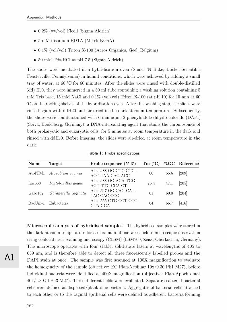
Appendix: Methods
A1
• 0.2% (wt/vol) Ficoll (Sigma Aldrich)
• 5 mM disodium EDTA (Merck KGaA)
• 0.1% (vol/vol) Triton X-100 (Acros Organics, Geel, Belgium)
• 50 mM Tris-HCl at pH 7.5 (Sigma Aldrich)
The slides were incubated in a hybridisation oven (Shake ’N Bake, Boekel Scientific,
Feasterville, Pennsylvania) in humid conditions, which were achieved by adding a small
tray of water, at 60 °C for 60 minutes. After the slides were rinsed with double-distilled
(dd) H20, they were immersed in a 50 ml tube containing a washing solution containing 5
mM Tris base, 15 mM NaCl and 0.1% (vol/vol) Triton X-100 (at pH 10) for 15 min at 60
°C on the rocking shelves of the hybridisation oven. After this washing step, the slides were
rinsed again with ddH20 and air-dried in the dark at room temperature. Subsequently,
the slides were counterstained with 6-diamidine-2-phenylindole dihydrochloride (DAPI)
(Serva, Heidelberg, Germany), a DNA-intercalating agent that stains the chromosomes of
both prokaryotic and eukaryotic cells, for 5 minutes at room temperature in the dark and
rinsed with ddH20. Before imaging, the slides were air-dried at room temperature in the
dark.
Table 1: Probe specifications
Name Target Probe sequence (5’-3’) Tm (°C) %GC Reference
AtoITM1 Atopobium vaginaeAlexa488-OO-CTC-CTG-ACC-TAA-CAG-ACC
66 55.6 [209]
Lac663 Lactobacillus genusAlexa488-OO-ACA-TGG-AGT-TTC-CCA-CT
75.4 47.1 [205]
Gard162 Gardnerella vaginalisAlexa647-OO-CAG-CAT-TAC-CAC-CCG
61 60.0 [204]
BacUni-1 EubacteriaAlexa555-CTG-CCT-CCC-GTA-GGA
64 66.7 [416]
Microscopic analysis of hybridised samples The hybridised samples were stored in
the dark at room temperature for a maximum of one week before microscopic observation
using confocal laser scanning microscopy (CLSM) (LSM700, Zeiss, Oberkochen, Germany).
The microscope operates with four stable, solid-state lasers at wavelengths of 405 to
639 nm, and is therefore able to detect all three fluorescently labelled probes and the
DAPI stain at once. The sample was first scanned at 100X magnification to evaluate
the homogeneity of the sample (objective: EC Plan-Neofluar 10x/0.30 Ph1 M27), before
individual bacteria were identified at 400X magnification (objective: Plan-Apochromat
40x/1.3 Oil Ph3 M27). Three different fields were evaluated. Separate scattered bacterial
cells were defined as dispersed/planktonic bacteria. Aggregates of bacterial cells attached
to each other or to the vaginal epithelial cells were defined as adherent bacteria forming
162
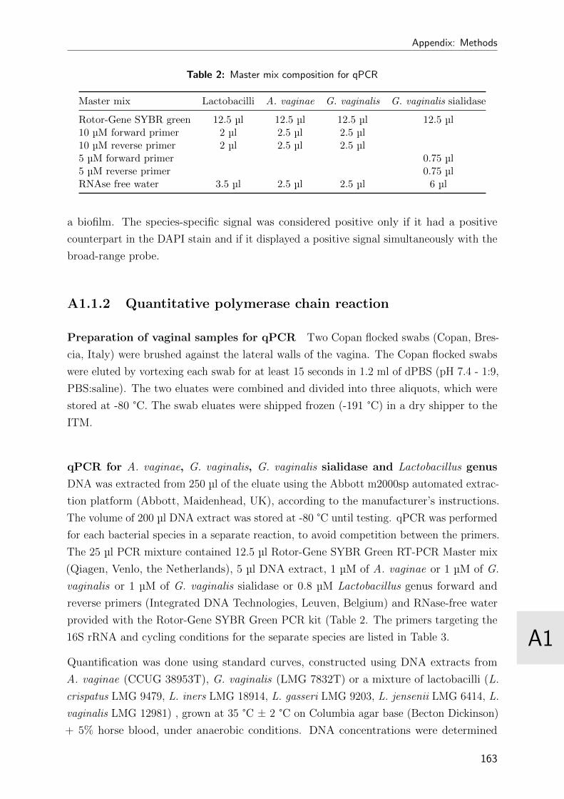
A1
Appendix: Methods
Table 2: Master mix composition for qPCR
Master mix Lactobacilli A. vaginae G. vaginalis G. vaginalis sialidase
Rotor-Gene SYBR green 12.5 µl 12.5 µl 12.5 µl 12.5 µl10 µM forward primer 2 µl 2.5 µl 2.5 µl10 µM reverse primer 2 µl 2.5 µl 2.5 µl5 µM forward primer 0.75 µl5 µM reverse primer 0.75 µlRNAse free water 3.5 µl 2.5 µl 2.5 µl 6 µl
a biofilm. The species-specific signal was considered positive only if it had a positive
counterpart in the DAPI stain and if it displayed a positive signal simultaneously with the
broad-range probe.
A1.1.2 Quantitative polymerase chain reaction
Preparation of vaginal samples for qPCR Two Copan flocked swabs (Copan, Bres-
cia, Italy) were brushed against the lateral walls of the vagina. The Copan flocked swabs
were eluted by vortexing each swab for at least 15 seconds in 1.2 ml of dPBS (pH 7.4 - 1:9,
PBS:saline). The two eluates were combined and divided into three aliquots, which were
stored at -80 °C. The swab eluates were shipped frozen (-191 °C) in a dry shipper to the
ITM.
qPCR for A. vaginae, G. vaginalis, G. vaginalis sialidase and Lactobacillus genus
DNA was extracted from 250 µl of the eluate using the Abbott m2000sp automated extrac-
tion platform (Abbott, Maidenhead, UK), according to the manufacturer’s instructions.
The volume of 200 µl DNA extract was stored at -80 °C until testing. qPCR was performed
for each bacterial species in a separate reaction, to avoid competition between the primers.
The 25 µl PCR mixture contained 12.5 µl Rotor-Gene SYBR Green RT-PCR Master mix
(Qiagen, Venlo, the Netherlands), 5 µl DNA extract, 1 µM of A. vaginae or 1 µM of G.
vaginalis or 1 µM of G. vaginalis sialidase or 0.8 µM Lactobacillus genus forward and
reverse primers (Integrated DNA Technologies, Leuven, Belgium) and RNase-free water
provided with the Rotor-Gene SYBR Green PCR kit (Table 2. The primers targeting the
16S rRNA and cycling conditions for the separate species are listed in Table 3.
Quantification was done using standard curves, constructed using DNA extracts from
A. vaginae (CCUG 38953T), G. vaginalis (LMG 7832T) or a mixture of lactobacilli (L.
crispatus LMG 9479, L. iners LMG 18914, L. gasseri LMG 9203, L. jensenii LMG 6414, L.
vaginalis LMG 12981) , grown at 35 °C ± 2 °C on Columbia agar base (Becton Dickinson)
+ 5% horse blood, under anaerobic conditions. DNA concentrations were determined
163
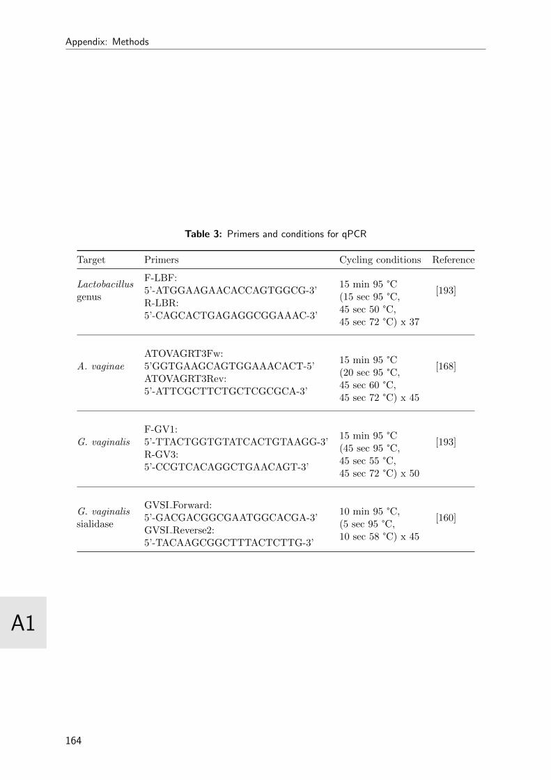
Appendix: Methods
A1
Table 3: Primers and conditions for qPCR
Target Primers Cycling conditions Reference
Lactobacillusgenus
F-LBF:5’-ATGGAAGAACACCAGTGGCG-3’
15 min 95 °C(15 sec 95 °C,45 sec 50 °C,45 sec 72 °C) x 37
[193]R-LBR:5’-CAGCACTGAGAGGCGGAAAC-3’
A. vaginaeATOVAGRT3Fw:5’GGTGAAGCAGTGGAAACACT-5’
15 min 95 °C(20 sec 95 °C,45 sec 60 °C,45 sec 72 °C) x 45
[168]ATOVAGRT3Rev:5’-ATTCGCTTCTGCTCGCGCA-3’
G. vaginalisF-GV1:5’-TTACTGGTGTATCACTGTAAGG-3’
15 min 95 °C(45 sec 95 °C,45 sec 55 °C,45 sec 72 °C) x 50
[193]R-GV3:5’-CCGTCACAGGCTGAACAGT-3’
G. vaginalissialidase
GVSI Forward:5’-GACGACGGCGAATGGCACGA-3’
10 min 95 °C,(5 sec 95 °C,10 sec 58 °C) x 45
[160]GVSI Reverse2:5’-TACAAGCGGCTTTACTCTTG-3’
164
A1
Appendix: Methods
using NanoDrop (Thermo Fisher Scientific, Erembodegem, Belgium) and the number of
genomes was calculated using the described genome sizes and G+C content of the strains.
A total of six tenfold dilutions of the DNA stocks were prepared in high performance liquid
chromatography (HPLC) grade water. Both the standard curve and samples were run
in duplicate. The bacterial load was expressed as genome equivalents (geq)/ml, because
quantification is based on cultured bacteria. The qPCR does not distinguish between living
and dead bacteria, therefore geq is a better term than copies or bacteria/ml, although one
geq is the equivalent of one copy or one bacterium.
A1.2 Ring samples
A1.2.1 Preparation of rings for further analysis
All rings worn by study participants were collected after removal at study visits. Each
ring was cut in three equal parts immediately after removal:
• The first part for the biomass density assessment with crystal violet was submerged
in 3 ml of glutaraldehyde for two weeks, transferred to 3 ml of formaldehyde, and
stored at 2-8 °C until testing.
• The second part for qPCR was stored in diluted phosphate buffered saline (dPBS)
(pH 7.4 - 1:9, PBS:saline) at -20 °C.
• The third part for FISH was stored in Carnoy solution (6:3:1, ethanol:chloroform:glacial
acetic acid) at 2-8 °C until testing.
All samples, except for the Nugent slide, were shipped from the study site in Rwanda
to the ITM in Antwerp, Belgium: the refrigerated samples were transported at room
temperature, while the frozen samples were transported at -196 °C in a dry shipper.
A1.2.2 Fluorescence in situ hybridisation
First the rings were rinsed with ddH2O. After 10 minutes of air-drying (not fully dry!),
the ring-biomass was removed with tweezers, carefully without rupturing the biofilm. The
biofilm was spread out on a Superfrost Plus slide (Menzel-Glaser) and passed through a
flame twice to fixate the slide. A second fixation step was done by submerging the slide
in Carnoy solution for 12 hours. FISH was performed as described above for the vaginal
samples, but the whole fixated sample was used and flown with hybridisation buffer +
probes.
165
Appendix: Methods
A1
A1.2.3 Quantitative polymerase chain reaction
Frozen ring parts were thawed and vortexed; using this eluate, 200 µl DNA was extracted
(Abbott, Maidenhead, UK) and stored at -80 °C until testing. qPCR was performed as
described above for the vaginal samples.
A1.2.4 Crystal violet assay
The biomass on contraceptive rings were stained with crystal violet to determine the
optical density (OD) as a proxy for the quantity of the biomass. The ring was submerged
in water, to rinse off the loose/not attached cells. Then the ring-biomass was stained by
submerging in a small tube containing 3 ml crystal violet (0.1% solution) for 10 to 15
minutes. Next, the ring part was rinsed with water twice and air-dried for two hours. The
dried ring-biomass staining was solubilised by submerging the ring in 3 ml of 30% acetic
acid in water for 10 to 15 minutes. From this solution, 125 µl was transferred to a new
micro-titre plate for OD measurement at 550 nm. OD measurements were done in duplo.
An unworn ring was stained the same way as the used rings and used as the blank; the
OD value of this ring (in duplo) was subtracted from all measured ODs.
166

A2
Appendix: Curriculum vitae
LISELOTTE HARDY
Voorhavenlaan 50 | 9000 Ghent | Belgium
+32 (0)472/344187 | [email protected] | 26 June 1984
Skills and strengths
Technical strengths qPCR, FISH, light microscopy, CLSM, bacterial and cell culture
Clinical research GCP/GLP, writing of and training in protocols and SOPs, reporting
Computer knowledge MS Office, Mac OS X, Windows, Stata, R, LaTeX, ImageJ, Matlab
Project management budgeting and planning, internal and external communication
Languages excellent Dutch and English, average French
Professional experience
Antelope Diagnostics - Ghent University June 2016 - Present
Assay development/Quality management
· Development and validation of novel point-of-care assays
· Introduction quality systems for in vitro diagnostics
AZ Nikolaas - Oncology Department December 2015 - May 2016
Study coordinator/data manager
· Coordinating clinical research studies at the oncology department
· Planning, preparing, realising and evaluating ongoing and new clinical studies
· Data management and administrative tasks
167
Appendix: Curriculum vitae
A2
Institute of Tropical Medicine/Ghent University January 2013 - April 2015
Scientific staff
· PhD research: “Characterisation of bacterial biofilm associated with bacterial vaginosis”
· Developing and implementing Fluorescence in situ Hybridisation (FISH) and quantitative
polymerase chain reaction (qPCR) to detect and identify biofilm-forming bacteria
· Set-up of vaginal epithelial cell model to study biofilm development
· Writing research papers and presenting results at national and international conferences
· Supervising master and bachelor students
Institute of Tropical Medicine November 2009 - January 2013
Project coordinator
· “MMIS study”: a descriptive study on the mucosal microbiome and associated immune
factors in adolescent girls in Antwerp, Integrated Project for the EC 7th Framework
programme
· “Biomarkers study”: a project studying safety biomarkers for the use of microbicides in
Rwanda, Kenya and South Africa, funded by EDCTP
- Writing protocols, SOPs, guidelines and manuals
- Preparing, coordinating and implementing studies
- Laboratory analysis: qPCR, FISH, ELISA, Luminex
- Communication, reporting and presenting study results
Quintiles Belgium December 2007 - November 2009
Clinical Research Associate
· Start-up, monitoring and close-out of clinical studies (phase 2 and 3) according to Good
Clinical Practices (GCP)
· Training in protocols and study procedures
Education
Postgraduate in Tropical Medicine and International Health February 2007
Institute of Tropical Medicine, Antwerp
Master in Biomedical Sciences June 2006
Ghent University
Bachelor in Biomedical Sciences June 2004
Hasselt University/Transnational University Limburg
168
A2
Appendix: Curriculum vitae
Additional training
Language courses
· Advanced academic English: English proficiency for presentations March 2015
University Language Center, University of Ghent
· Writing academic papers in English January 2015
Linguapolis, University of Antwerp
Technical training
· Molecular and physiological regulation of microbial biofilms September 2014
K.U.Leuven
· International practical course on biofilm science July 2014
University of Minho, Portugal
· Postgraduate technical workshop “Medical biofilm techniques” August 2012
Danish Technical University, Kopenhagen
· PhD/Master course “Advanced microscopy and vital imaging” June 2012
University of Maastricht, The Netherlands
Additional training
· Project management January 2016
Ghent University
· Statistical analysis in R for health scientists October 2015
Ghent University
· Specific teacher training June 2007
Ghent University
Conferences attended
· World STI and HIV Congress, 2015, Brisbane
· Belgian Interdisciplinary Biofilm Research meeting, 2014, Louvain-la-Neuve
· Belgian Interdisciplinary Biofilm Research meeting, 2013, Louvain-la-Neuve
· Eurobiofilms, 2013, Ghent
· Nobel conference on biofilm formation, 2013, Stockholm
· World STI and AIDS Congress, 2013, Vienna
· American Society for Microbiology conference on biofilms, 2012, Miami
169
Appendix: Curriculum vitae
A2
Bibliography
Published manuscripts
· Hardy L, Jespers V, Dahchour D, Mwambarangwe L, Musengamana V, Vaneechoutte M,
Crucitti T. Unravelling the Bacterial Vaginosis-Associated Biofilm: a Multiplex Gardnerella
vaginalis and Atopobium vaginae Fluorescence In Situ Hybridization Assay Using Peptide
Nucleic Acid Probes. PLoS ONE 2015, 10(8).
· Hardy L, Jespers V, Abdellati S, De Baetselier I, Mwambarangwe L, Musengamana V,
van de Wijgert J, Vaneechoutte M, Crucitti T. A Fruitful Alliance: the Synergy Between
Atopobium vaginae and Gardnerella vaginalis in Bacterial Vaginosis-Associated Biofilm.
STI 2016;0:1–5.
· Hardy L, Jespers V, Van den Bulck M, Buyze J, Mwambarangwe L, Musengamana V,
Vaneechoutte M, Crucitti T. The Importance of Gardnerella vaginalis Sialidase in Bacterial
Vaginosis-Associated Biofilm. Submitted for review.
· Hardy L, Jespers V, De Baetselier I, Buyze J, Mwambarangwe L, Musengamana V, van
de Wijgert J, Crucitti T. Association of Vaginal Dysbiosis and Biofilm with Contraceptive
Vaginal Ring Biomass in African Women. Submitted for review.
· Jespers V, Hardy L, Buyze J, Loos J, Buve A, Crucitti T. The Influence of Sexual Debut
on Selected Vaginal, Ano-Rectal and Oral Microbiota and Vaginal Inflammatory Markers
in Belgian Adolescent Girls: a Cohort Study. Obstet Gynecol. 2016 Jul;128(1):22-31.
· Cools P, Jespers V, Hardy L, Crucitti T, Delany-Moretlwe S, Mwaura M, Ndayisaba
GF, van de Wijgert J, Vaneechoutte M. Vaginal Carriage and Risk Factors for Carriage
of Group B Streptococci and Escherichia coli, with GBS Serotype Distribution, in Three
African Populations. PLoS ONE 11(1): e0148052.
· Kyongo JK, Crucitti T, Menten J, Hardy L, Cools P, Michiels J, Delany-Moretlwe S,
Mwaura M, Ndayisaba G, Joseph S, Fichorova R, van de Wijgert J, Vanham G, Arien KK,
Jespers V. A Cross-Sectional Analysis of Selected Genital Tract Immunological Markers
and Molecular Vaginal Microbiota in Sub-Saharan African women with Relevance to HIV
Risk and Prevention. Clin Vaccine Immunol 2015 May;22(5):526-38.
· Jespers V, van de Wijgert J, Cools P, Verhelst R, Verstraelen H, Delany-Moretlwe S,
Mwaura M, Ndayisaba GF, Mandaliya K, Menten J, Hardy L, Crucitti T for the Vaginal
Biomarkers Study Group. The Significance of Lactobacillus crispatus and Lactobacillus
vaginalis for Vaginal Health and the Negative Effect of Recent Sex: a Cross-Sectional
Descriptive Study Across Groups of African Women. BMC infectious diseases 2015 15:115.
· Gautam R, Borgdorff H, Jespers V, Francis SC, Verhelst R, Mwaura M, Delany-Moretlwe
S, Ndayisaba G, Kyongo JK, Hardy L, Menten J, Crucitti T, Tsivtsivadze E, Schuren F,
van de Wijgert J for the Vaginal Biomarkers Study Group. Correlates of the Molecular
Vaginal Microbiota Composition of African Women. BMC Infectious Diseases 2015 15:86.
· Jespers V, Crucitti T, Menten J, Verhelst R, Mwaura M, Mandaliya K, Ndayisaba GF,
Delany-Moretlwe S, Verstraelen H, Hardy L, Buve A, van de Wijgert J. Prevalence and
Correlates of Bacterial Vaginosis in Different Sub-Populations of Women in Sub-Saharan
Africa: a Cross-Sectional Study. PLoS One 2015 9(10).
170
A2
Appendix: Curriculum vitae
· Gilles N, Verwijs MC, van Eeckhoudt S, Gasarabwe A, Hardy L, Borgdorff H, Kestelyn
E, Jespers V, van de Wijgert J. Feasibility and Acceptability of a Novel Cervicovaginal
Lavage Self-Sampling Device Among Women in Kigali, Rwanda. Sexually Transmitted
Diseases 2013 7:552-555.
· Jespers V, Menten J, Smet H, Poradosu S, Abdellati S, Verhelst R, Hardy L, Buve A,
Crucitti T. Quantification of Bacterial Species of the Vaginal Microbiome in Different
Groups of Women, Using Nucleic Acid Amplification Tests. BMC Microbiol 2012 12:83.
Oral presentations
· Jespers V, Hardy L, van de Wijgert J. Dysbiosis Around the Time of Conception in
African Women; a DNA-Triple-PCR Tool Applied. ECCMID 2016, Amsterdam.
· Hardy L, Jespers V, De Baetselier I, van de Wijgert J, Crucitti T. The Impact of Vaginal
Bacterial Biofilm on Intravaginal Rings. STI 91:Suppl 2 A53-A54. World STI and HIV
Congress 2015, Brisbane. (Travel scholarship awarded)
· Jespers V, Hardy L, Buve A, Loos J, Tsoumaris A, Buyze J, Crucitti T. The Influence of
Sexual Debut on Selected Vaginal, Rectal and Oral Microbiota and Vaginal Inflammatory
Markers in Belgian Adolescent Girls: a Cohort Study. STI 91:Suppl 2 A34. World STI
and HIV Congress 2015, Brisbane.
· Hardy L. New Insights in Biofilm Associated with Bacterial Vaginosis. Summer School
“Molecular and physiological regulation of microbial biofilms” 2014, Leuven.
· Hardy L, Jespers V. Vaginal Microbiota and Immune Markers. IHAC Reviving Day 2014,
Antwerp.
· Mwaura M, Hardy L, Delany-Moretlwe S, Ndayisaba G, Mandaliya K, Cools P, Verhelst
R, van de Wijgert J, Crucitti T, Jespers J. There is a Need for Multi-purpose Prevention
Technologies Targeting HIV and Common Reproductive Tract Infections: Data from the
Biomarkers Study Team. 7th EDCTP forum 2014, Berlin.
· Hardy L. Microbiome Results from Biomarkers Trials. CHAARM annual consortium
meeting 2014, Camogli.
· Hardy L. New Insights in Diagnostics for STI. Master-after-master and post-graduate
program for clinical biologists UZ Brussels “Sexually transmitted infections anno 2014”,
Brussels.
· Hardy L. Bacterial Biofilm: l’Union Fait la Force. Epi-seminar ITM 2013, Antwerp.
· Jespers V, Hardy L, Kyongo JK, Buve A, Crucitti T. Optimising the Evaluation and
Study of the Vaginal Mucosal Innate Defence System for Prevention Interventions: an
Example from the ITM. 8th ESIDOG Conference 2013, London.
· Kyongo J, Crucitti T, Menten J, Hardy L, Delany-Moretlwe S, Mwaura M, Ndayisaba
G, van de Wijgert J, Vanham G, Buve A, Arien K, Jespers V. Identifying Biomarkers of
Increased HIV Transmission in African Populations. Building Bridges in Reproductive
Immunology 2013, Boston.
· Hardy L. Microbiota and Adolescents. CHAARM annual consortium meeting 2012,
Camogli.
171

Appendix: Curriculum vitae
A2
Poster presentations
· Hardy L, Jespers V, Van den Bulck M, Buyze J, Mwambarangwe L, Musengamana V,
Vaneechoutte M, Crucitti T. The importance of Gardnerella vaginalis sialidase in bacterial
vaginosis-associated biofilm. Antimicrobial resistance in microbial biofilms and options for
treatment 2016, Ghent.
· Hardy L, Jespers V, Buyze J, De Baetselier I, Agabe S, Mwambarangwe L, van de Wijgert
J, Crucitti T. The Ring Plus Project: The effect of a Contraceptive Vaginal Ring on the
Vaginal Microbiota. HIV R4P 2016, Chicago.
· Crucitti T, Hardy L, Agabe S, Kestelyn E, De Baetselier I, Mwambarangwe L, Buyze J,
Ilo Van Nuil J, Delvaux T, Jespers V, van de Wijgert J, Crucitti T. The Ring Plus Project:
Safety and Acceptability of Vaginal Rings that protect Women from Pregnancy. HIV R4P
2016, Chicago.
· Hardy L, Jespers V, Abdellati S, De Baetselier I, Mwambarangwe L, Musengamana V,
van de Wijgert J, Vaneechoutte M, Crucitti T. A Fruitful Alliance: the Synergy Between
Atopobium vaginae and Gardnerella vaginalis in Bacterial Vaginosis-Associated Biofilm.
STI 91:Suppl 2 A126. World STI and HIV Congress 2015, Brisbane.
· Kyongo J, Crucitti T, Menten J, Hardy L, Cools P, Michiels J, Delany-Moretlwe S,
Mwaura M, Ndayisaba G, Joseph S, Fichorova R, van de Wijgert J, Vanham G, Arien
KK, Jespers V. Dynamics of Vaginal Immune Correlates and Microbiota in Women from
Sub-Saharan Africa. STI 91:Suppl 2 A116. World STI and HIV Congress 2015, Brisbane.
· Kyongo J, Crucitti T, Menten J, Hardy L, Cools P, Michiels J, Delany-Moretlwe S,
Mwaura M, Ndayisaba G, Joseph S, Fichorova R, van de Wijgert J, Vanham G, Arien K,
Jespers V. Genital Tract Immunological Markers in Sub-Saharan African Women with
Relevance to HIV risk and Prevention. HIV R4P 2014, Capetown.
· Hardy L. Studies at the Institute of Tropical Medicine Looking at Bacterial Vaginosis-
Associated Biofilm. 4th International practical course on Biofilm science 2014, Braga.
· Hardy L, Abdellati S, Crucitti T. Biofilm Formation by Clinical Isolates of Neisseria
gonorrhoeae. Eurobiofilms 2013, Ghent.
· Hardy L, Jespers V, Dahchour N, Cuylaerts V, Cools P, Abdellati S, Ndayisaba GF,
Delany-Moretlwe S, Mwaura M, Crucitti T. PNA-FISH for the Detection of Vaginal
Gardnerella vaginalis Biofilm. Nobel conference on biofilm formation 2013, Stockholm.
· Hardy L, Jespers V, Menten J, Verhelst R, Mwaura M, Delany-Moretlwe S, Ndayisaba G,
Vaneechoutte M, van de Wijgert J, Crucitti T. Bacterial Species in the Vaginal Microbiome
Correlated by Nugent Score: Cross-Sectional Data from the Microbicide Safety Biomarkers
Study in Kenya, Rwanda, and South Africa. STI 89:Suppl 1 A83. STI & AIDS 2013,
Vienna.
· Hardy L, Jespers V, Abdellati S, Ndayisaba G, Fiat A, Mwambarangwe L, van de Wijgert
J, Crucitti T. Visualisation of Vaginal Epithelial Biofilm in Bacterial Vaginosis with
Fluorescence In Situ Hybridisation. ASM conference on biofilms 2012, Miami.
172

A3
Appendix: Papers as published
173
RESEARCH ARTICLE
Unravelling the Bacterial Vaginosis-Associated Biofilm: A Multiplex Gardnerellavaginalis and Atopobium vaginaeFluorescence In Situ Hybridization AssayUsing Peptide Nucleic Acid ProbesLiselotte Hardy1,2,3*, Vicky Jespers1, Nassira Dahchour3,4, Lambert Mwambarangwe5,Viateur Musengamana5, Mario Vaneechoutte2, Tania Crucitti3
1 Unit of Epidemiology and Control of HIV/STD, Department of Public Health, Institute of Tropical Medicine,Antwerp, Belgium, 2 Laboratory Bacteriology Research, Faculty of Medicine & Health Sciences, University ofGhent, Ghent, Belgium, 3 STI Reference Laboratory, Department of Clinical Sciences, Institute of TropicalMedicine, Antwerp, Belgium, 4 Plantijn Hogeschool, Antwerp, Belgium, 5 Rinda Ubuzima, Kigali, Rwanda
AbstractBacterial vaginosis (BV), a condition defined by increased vaginal discharge without signifi-
cant inflammation, is characterized by a change in the bacterial composition of the vagina.
Lactobacillus spp., associated with a healthy vaginal microbiome, are outnumbered by BV-
associated organisms. These bacteria could form a polymicrobial biofilm which allows them
to persist in spite of antibiotic treatment. In this study, we examined the presence ofGard-nerella vaginalis and Atopobium vaginae in vaginal biofilms using Peptide Nucleic Acid
(PNA) probes targeting these bacteria. For this purpose, we developed three new PNA
probes for A. vaginae. The most specific A. vaginae probe, AtoITM1, was selected and then
used in an assay with two existing probes, Gard162 and BacUni-1, to evaluate multiplex
FISH on clinical samples. Using quantitative polymerase chain reaction (qPCR) as the gold
standard, we demonstrated a sensitivity of 66.7% (95% confidence interval: 54.5% - 77.1%)
and a specificity of 89.4% (95% confidence interval: 76.1% - 96%) of the new AtoITM1
probe. FISH enabled us to show the presence of a polymicrobial biofilm in bacterial vagino-
sis, in which Atopobium vaginae is part of aGardnerella vaginalis-dominated biofilm. We
showed that the presence of this biofilm is associated with high bacterial loads of A. vaginaeandG. vaginalis.
IntroductionBacterial vaginosis (BV), a condition characterized by increased vaginal discharge without sig-nificant inflammation, is highly prevalent in women of reproductive age. It increases the risk
PLOSONE | DOI:10.1371/journal.pone.0136658 August 25, 2015 1 / 16
OPEN ACCESS
Citation: Hardy L, Jespers V, Dahchour N,Mwambarangwe L, Musengamana V, VaneechoutteM, et al. (2015) Unravelling the Bacterial Vaginosis-Associated Biofilm: A Multiplex Gardnerella vaginalisand Atopobium vaginae Fluorescence In SituHybridization Assay Using Peptide Nucleic AcidProbes. PLoS ONE 10(8): e0136658. doi:10.1371/journal.pone.0136658
Editor: A Al-Ahmad, University Hospital of the Albert-Ludwigs-University Freiburg, GERMANY
Received: February 3, 2015
Accepted: August 5, 2015
Published: August 25, 2015
Copyright: © 2015 Hardy et al. This is an openaccess article distributed under the terms of theCreative Commons Attribution License, which permitsunrestricted use, distribution, and reproduction in anymedium, provided the original author and source arecredited.
Data Availability Statement: All relevant data arewithin the paper and its Supporting Information files.
Funding: Funding was received by TC fromEuropean and Developing Countries Clinical TrialsPartnership: SP.2011.41304.043, http://www.edctp.org/; by VJ from Combined Highly Active Anti-Retroviral Microbicides under EU FP7: 242135, http://cordis.europa.eu/fp7/home_en.html; and by VJ fromDormeur Investment Service Ltd. The funders had no
for acquisition and transmission of sexually transmitted infections, including HIV, and is asso-ciated with preterm birth in pregnant women [1–2]. BV is a dysbiotic condition of unknownetiology and is characterized by a change in the microbial composition of the vagina. Lactoba-cillus spp., associated with a healthy vaginal microbiome, are outnumbered by an array of BV-associated organisms including Gardnerella vaginalis [1–3]. However, several studies suggestthat the mere presence of G. vaginalis is not sufficient for the diagnosis of BV. Indeed, G. vagi-nalis is also present in 50% to 70% of women without BV according to Nugent score [4–6]. G.vaginalis expresses various virulence factors such as vaginolysin [7] and sialidase [8]. It canalso produce a biofilm [9], thereby increasing its tolerance to lactic acid and hydrogen peroxideproduced by lactobacilli [10,11] and to antimicrobial treatment [12,13]. Furthermore, it hasbeen suggested that its adherence and biofilm-forming capacities allow G. vaginalis to initiatethe colonization and scaffolding of the vaginal epithelium to which other species can attachsubsequently [14,15].
As was first shown a decade ago, Atopobium vaginae is one of the many other species thatare characteristic of BV [16–20]. In one study, A. vaginae was detected in 80% of samples test-ing positive for G. vaginalis and made up 40% of the total biofilm mass dominated by G. vagi-nalis [9]. This association was confirmed in a study by Bradshaw et al. [21]: 93% of samplescontaining A. vaginae also contained G. vaginalis, whereas only 10% tested positive for G. vagi-nalis when A. vaginae was absent [22]. In contrast to G. vaginalis, A. vaginae is rarely part ofthe healthy vaginal microbiome and is considered a more specific marker of BV than G. vagi-nalis [17,21,23].
It is postulated that a biofilm provides bacteria with a competitive advantage over plank-tonic bacteria and that polymicrobial biofilms may offer additional advantages over single-spe-cies biofilms. Mechanisms that have been described in previous studies include metaboliccooperation, increased resistance to antibiotics or host immune responses [24] and an enlargedgene pool with more efficient sharing of genetic material compared to mono-species biofilms[25]. Polymicrobial coexistence is the dominant form in environmental biofilms, but is alsoprominent in the human body [24]. A well-known example is dental plaque: anaerobic bacte-ria, which are sensitive to oxygen, can survive and persist under the aerobic conditions in theoral cavity due to the consummation of oxygen by aerobic bacteria in the dental biofilm [26].
On the basis of these previous findings, we hypothesize that a polymicrobial biofilm consist-ing of A. vaginae and G. vaginalis and other bacteria not discussed in this study may serve as amarker of BV. Thus, better visualization of the structure of vaginal biofilms and identificationof the bacterial components of the biofilm may contribute to better understanding of BV. Tostudy the role of A. vaginae and G. vaginalis in BV, we designed and evaluated the performanceof fluorescence in situ hybridization (FISH) with peptide nucleic acid (PNA) probes for A. vagi-nae and G. vaginalis. Three new PNA probes were designed for A. vaginae. The most specificA. vaginae probe was selected, and subsequently used together with a PNA probe that had beendescribed for the detection of G. vaginalis and a positive control probe that detects a broadrange of bacteria in order to evaluate the multiplex FISH on clinical samples.
Materials and Methods
Design of PNA probesPNA probes targeting the bacterial 16S rRNA were synthesized by Panagene (Daejeon, SouthKorea). A fluorescent label was attached using a double 8-amino-3,6-dioxaoctanoic acid(AEEA) linker at the N terminus. We evaluated the performance of three new PNA probes forA. vaginae: Ato291, previously described as a DNA FISH probe [9,27] and AtoITM1 andAtoITM2, which we developed using the Applied Biosystems PNA designer software (http://
PNA FISH for Atopobium vaginae andGardnerella vaginalis
PLOSONE | DOI:10.1371/journal.pone.0136658 August 25, 2015 2 / 16
role in study design, data collection and analysis,decision to publish, or preparation of the manuscript.
Competing Interests: Funding was received by TCfrom European and Developing Countries ClinicalTrials Partnership: SP.2011.41304.043, http://www.edctp.org/; by VJ from Combined Highly Active Anti-Retroviral Microbicides under EU FP7: 242135, http://cordis.europa.eu/fp7/home_en.html; and by VJ fromDormeur Investment Service Ltd. This does not alterthe authors’ adherence to PLOS ONE policies onsharing data and materials.
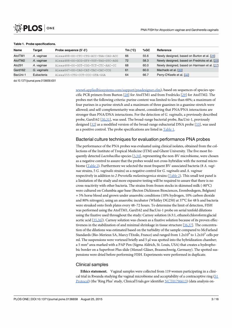
www6.appliedbiosystems.com/support/pnadesigner.cfm), based on sequences of species-spe-cific PCR primers from Burton [28] for AtoITM1 and from Fredricks [29] for AtoITM2. Theprobes met the following criteria: purine content was limited to less than 60%; a maximum offour purines in a purine-stretch and a maximum of three guanines in a guanine-stretch wereallowed; and self-complementarity was absent, considering that PNA/PNA interactions arestronger than PNA/DNA interactions. For the detection of G. vaginalis, a previously describedprobe, Gard162 [30,31], was used. The broad-range bacterial probe, BacUni-1, previouslydesigned [32] as a modified version of the broad-range eubacterial DNA probe [33], was usedas a positive control. The probe specifications are listed in Table 1.
Bacterial culture techniques for evaluation performance PNA probesThe performance of the PNA probes was evaluated using clinical isolates, obtained from the col-lections of the Institute of Tropical Medicine (ITM) and Ghent University. The five most fre-quently detected Lactobacillus species [3,34], representing the non-BV microbiome, were chosenas a negative control to assure that the probes would not cross-hybridize with the normal micro-biome (Table 2). Furthermore we selected the most frequent BV-associated bacteria (8 A. vagi-nae strains, 5 G. vaginalis strains) as a negative control for G. vaginalis and A. vaginaerespectively in addition to 2 Prevotella melaninogenica strains (Table 2). This small test panel isa limitation of the study and more expansive testing will be required to assure that there is nocross-reactivity with other bacteria. The strains from frozen stocks in skimmedmilk (-80°C)were cultured on Columbia agar base (Becton Dickinson Biosciences, Erembodegem, Belgium)+ 5% horse blood and grown under anaerobic conditions (10% hydrogen, 10% carbon dioxideand 80% nitrogen), using an anaerobic incubator (Whitley DG250) at 37°C for 48 h and bacteriawere streaked onto fresh plates every 48–72 hours. To determine the limit of detection, FISHwas performed using the AtoITM1, Gard162 and BacUni-1 probe on serial tenfold dilutionsusing the fixative used throughout the study: Carnoy solution (6:3:1, ethanol:chloroform:glacialacetic acid [35,36]). Carnoy solution was chosen as a fixative solution because of its proven effec-tiveness in the stabilization of and minimal shrinkage in tissue structure [36,37]. The concentra-tion of the dilutions was estimated based on the turbidity of the sample compared to McFarlandStandards (Bio-Merieux SA, Marcy l’Etoile, France) and ranged from 1.2x10⁹ to 1.2x10³ cells perml. The suspensions were vortexed briefly and 5 μl was spotted into the hybridization chamber;a 5 mm² area marked with a PAP Pen (Sigma Aldrich, St. Louis, USA) that creates a hydropho-bic border on a Superfrost Plus slide (Menzel-Gläser, Braunschweig, Germany). The spotted sus-pensions were dried before performing FISH. Experiments were performed in duplicate.
Clinical samplesEthics statement. Vaginal samples were collected from 119 women participating in a clini-
cal trial in Rwanda studying the vaginal microbiome and acceptability of a contraceptive ring (S1Protocol) (the ‘Ring Plus’ study, ClinicalTrials.gov identifier NCT01796613) (data analysis on-
Table 1. Probe specifications.
Name Target Probe sequence (5’-3’) Tm (°C) %GC Reference
AtoITM1 A. vaginae Alexa488-OO-CTC-CTG-ACC-TAA-CAG-ACC 66 55.6 Newly designed, based on Burton et al. [28]
AtoITM2 A. vaginae Alexa488-OO-GCG-GTY-TGT-TAG-GTC-AGG 72 58.3 Newly designed, based on Fredricks et al. [29]
Ato291 A. vaginae Alexa488-OO-GGT-CGG-TCT-CTC-AAC-CC 68 60.0 Newly designed, based on Harmsen et al. [27]
Gard162 G. vaginalis Alexa647-OO-CAG-CAT-TAC-CAC-CCG 61 60.0 Machado et al. [30]
BacUni-1 Eubacteria Alexa555-CTG-CCT-CCC-GTA-GGA 64 66.7 Perry-O'Keefe et al. [32]
doi:10.1371/journal.pone.0136658.t001
PNA FISH for Atopobium vaginae andGardnerella vaginalis
PLOSONE | DOI:10.1371/journal.pone.0136658 August 25, 2015 3 / 16
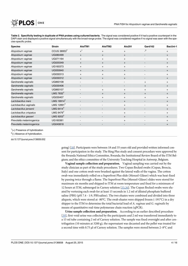
going) [38]. Participants were between 18 and 35 years old and provided written informed con-sent for participation in the study. The Ring Plus study and consent procedure were approved bythe Rwanda National Ethics Committee, Rwanda; the Institutional Review Board of the ITM Bel-gium; and the ethics committee of the University Teaching Hospital in Antwerp, Belgium.
Vaginal sample collection and preparation. Vaginal sampling was carried out by thestudy clinician as part of the study procedures. Two Copan flocked swabs (Copan, Brescia,Italy) and one cotton swab were brushed against the lateral walls of the vagina. The cottonswab was immediately rolled on a Superfrost Plus slide (Menzel-Gläser) which was heat-fixedby passing twice through a flame. The Superfrost Plus (Menzel-Gläser) slides were stored formaximum six months and shipped to ITM at room temperature and fixed for a minimum of12 hours at ITM, submerged in Carnoy solution [35,36]. The Copan flocked swabs were elu-ated by vortexing each swab for at least 15 seconds in 1.2 ml of diluted phosphate bufferedsaline (PBS) (pH 7.4 - 1:9, PBS:saline). The two eluates were combined and divided into threealiquots, which were stored at -80°C. The swab eluates were shipped frozen (-191°C) in a dryshipper to the ITM to determine the total bacterial load of A. vaginae and G. vaginalis bymeans of quantitative real-time polymerase chain reaction (qPCR).
Urine sample collection and preparation. According to an earlier described procedure[39], first-void urine was collected by the participants and 2 ml was transferred immediately toa 15 ml tube containing 2 ml of Carnoy solution. The sample was fixed overnight and after cen-trifugation (10 minutes at 3200 g), the supernatant was decanted and the pellet was treated fora second time with 0.75 μl of Carnoy solution. The samples were stored between 2–8°C and
Table 2. Specificity testing in duplicate of PNA probes using cultured bacteria. The signal was considered positive if it had a positive counterpart in theDAPI stain and displayed a positive signal simultaneously with the broad-range probe. The signal was considered negative if no signal was seen with the spe-cies-specific probe.
Species Strain AtoITM1 AtoITM2 Ato291 Gard162 BacUni-1
Atopobium vaginae CCUG 38953T +1 + + -2 +
Atopobium vaginae UG080499 + + + - +
Atopobium vaginae UG071164 + + + - +
Atopobium vaginae UG020349 + + + - +
Atopobium vaginae UG160373 + + + - +
Atopobium vaginae UG550940 + + + - +
Atopobium vaginae UG030313 + + + - +
Atopobium vaginae UG030312 + + + - +
Gardnerella vaginalis UG860108 - - - + +
Gardnerella vaginalis UG030406 - - - + +
Gardnerella vaginalis UG860107 - + + + +
Gardnerella vaginalis LMG 7832T - + + + +
Gardnerella vaginalis UG030407 - + + + +
Lactobacillus iners LMG 18914T - - + - +
Lactobacillus vaginalis LMG 12891T - - + - +
Lactobacillus jensenii LMG 6414T - - + - +
Lactobacillus crispatus LMG 9479T - - + - +
Lactobacillus gasseri LMG 9203T - - + - +
Prevotella melaninogenica UG160361 - - + - +
Prevotella melaninogenica UG040818 - - - - +
1(+) Presence of hybridization2(-) Absence of hybridization.
doi:10.1371/journal.pone.0136658.t002
PNA FISH for Atopobium vaginae andGardnerella vaginalis
PLOSONE | DOI:10.1371/journal.pone.0136658 August 25, 2015 4 / 16

shipped at room temperature to the ITM. Before applying FISH, the urine samples were vor-texed briefly and 5 μl was spotted into the hybridization chamber on a Superfrost Plus slide(Menzel-Gläser).
Quantitative PCR for quantification of bacteria in vaginal samples. DNA was extractedfrom 250 μl of the vaginal swab eluate using the Abbott m2000sp automated extraction plat-form (Abbott, Maidenhead, UK), according to the manufacturer’s instructions. The volume of200 μl DNA extract was stored at –80°C until testing. qPCR was performed for each bacteriaspecies separately, to avoid competition between the primers. The 25 μl PCR mixture contained12.5 μl Rotor-Gene SYBR Green RT-PCR Master mix (Qiagen, Venlo, the Netherlands), 5 μlDNA extract, 0.5 μM of A. vaginae or 1 μM of G. vaginalis forward and reverse primers (Inte-grated DNA Technologies, Leuven, Belgium) and RNase-free water provided with the Rotor-Gene SYBR Green PCR kit. The primers for A. vaginae and G. vaginalis and the amplificationreactions (Rotor Gene QMDx 5 plex, Qiagen) have been described before [34].
Quantification was done using standard curves, constructed using DNA extracts from A.vaginae (CCUG 38953T) and G. vaginalis (LMG 7832T), grown at 35°C ± 2°C on Columbiaagar base (Becton Dickinson) + 5% horse blood, under anaerobic conditions. DNA concentra-tions were determined using NanoDrop (Thermo Fisher Scientific, Erembodegem, Belgium)and the number of genomes was calculated using the described genome sizes and G+C contentof the strains. A total of six tenfold dilutions of the DNA stocks were prepared in high perfor-mance liquid chromatography (HPLC) grade water. Both the standard curve and samples wererun in duplicate. The bacterial load was expressed as genome equivalents (geq)/ml.
PNA FISH procedureMultiplex hybridization was performed on a Superfrost Plus slide (Menzel-Gläser) in a 5 mm²quadrant hybridization area marked with a PAP pen (Sigma Aldrich, St. Louis, USA), a liquid-repellent slide marker. The slide was covered with a cover slip after addition of a hybridizationbuffer that contained 200 nM of each probe: species-specific probes for A. vaginae (AtoITM1 orAtoITM2 or Ato291) and G. vaginalis (Gard162), and the broad-range BacUni-1 probe. Thehybridization solution consisted of 10% (wt/vol) dextran sulphate (Sigma Aldrich), 10 mMNaCl(Merck KGaA, Darmstadt, Germany), 2% (vol/vol) formamide (Merck KGaA), 0.1% (wt/vol)sodium pyrophosphate (Sigma Aldrich), 0.2% (wt/vol) polyvinylpyrrolidone (Sigma Aldrich),0.2% (wt/vol) Ficoll (Sigma Aldrich), 5 mM disodium EDTA (Merck KGaA), 0.1% (vol/vol) Tri-ton X-100 (Acros Organics, Geel, Belgium) and 50 mM Tris-HCl at pH 7.5 (Sigma Aldrich).
The slides were incubated in a hybridization oven (Shake ‘N Bake, Boekel Scientific, Feaster-ville, Pennsylvania) in humid conditions, which were achieved by adding a small tray of water,at 60°C for 60 minutes. After the slides were rinsed with double-distilled (dd) H20, they wereimmersed in a washing solution containing 5 mM Tris base, 15 mMNaCl and 0.1% (vol/vol)Triton X-100 (at pH 10) for 15 min at 60°C on the rocking shelves of the hybridization oven.After this washing step, the slides were rinsed again with ddH20 and air-dried in the dark atroom temperature. Subsequently, the slides were counterstained with 6-diamidine-2-phenylin-dole dihydrochloride (DAPI) (Serva, Heidelberg, Germany), a DNA-intercalating agent thatstains the chromosomes of both prokaryotic and eukaryotic cells, for 5 minutes at room tem-perature in the dark and rinsed with ddH20. Before imaging, the slides were air-dried at roomtemperature in the dark.
Assessment of reproducibility of FISHThe inter-run repeatability of FISH was evaluated by comparing the outcomes of two indepen-dent FISH runs for a subset of the samples. The second hybridization was performed 6 months
PNA FISH for Atopobium vaginae andGardnerella vaginalis
PLOSONE | DOI:10.1371/journal.pone.0136658 August 25, 2015 5 / 16
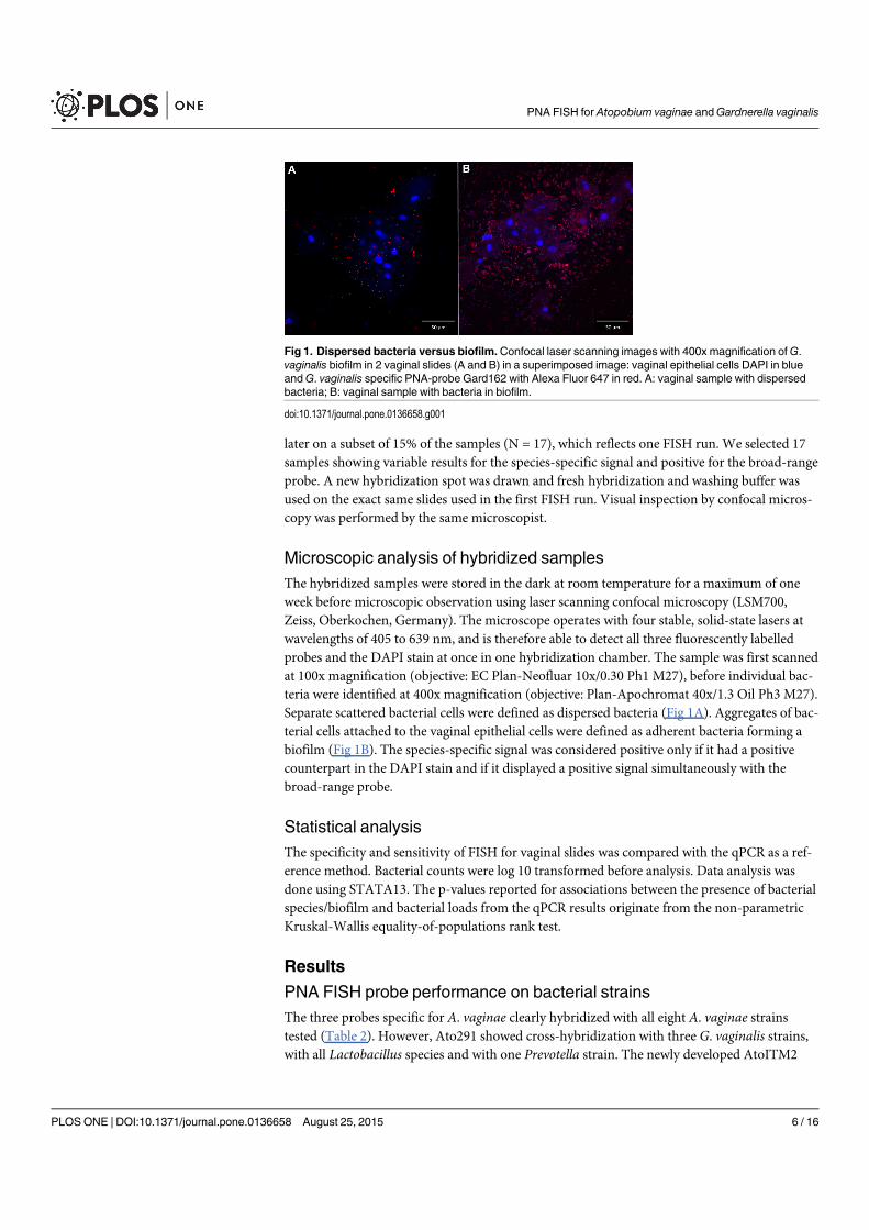
later on a subset of 15% of the samples (N = 17), which reflects one FISH run. We selected 17samples showing variable results for the species-specific signal and positive for the broad-rangeprobe. A new hybridization spot was drawn and fresh hybridization and washing buffer wasused on the exact same slides used in the first FISH run. Visual inspection by confocal micros-copy was performed by the same microscopist.
Microscopic analysis of hybridized samplesThe hybridized samples were stored in the dark at room temperature for a maximum of oneweek before microscopic observation using laser scanning confocal microscopy (LSM700,Zeiss, Oberkochen, Germany). The microscope operates with four stable, solid-state lasers atwavelengths of 405 to 639 nm, and is therefore able to detect all three fluorescently labelledprobes and the DAPI stain at once in one hybridization chamber. The sample was first scannedat 100x magnification (objective: EC Plan-Neofluar 10x/0.30 Ph1 M27), before individual bac-teria were identified at 400x magnification (objective: Plan-Apochromat 40x/1.3 Oil Ph3 M27).Separate scattered bacterial cells were defined as dispersed bacteria (Fig 1A). Aggregates of bac-terial cells attached to the vaginal epithelial cells were defined as adherent bacteria forming abiofilm (Fig 1B). The species-specific signal was considered positive only if it had a positivecounterpart in the DAPI stain and if it displayed a positive signal simultaneously with thebroad-range probe.
Statistical analysisThe specificity and sensitivity of FISH for vaginal slides was compared with the qPCR as a ref-erence method. Bacterial counts were log 10 transformed before analysis. Data analysis wasdone using STATA13. The p-values reported for associations between the presence of bacterialspecies/biofilm and bacterial loads from the qPCR results originate from the non-parametricKruskal-Wallis equality-of-populations rank test.
Results
PNA FISH probe performance on bacterial strainsThe three probes specific for A. vaginae clearly hybridized with all eight A. vaginae strainstested (Table 2). However, Ato291 showed cross-hybridization with three G. vaginalis strains,with all Lactobacillus species and with one Prevotella strain. The newly developed AtoITM2
Fig 1. Dispersed bacteria versus biofilm.Confocal laser scanning images with 400x magnification ofG.vaginalis biofilm in 2 vaginal slides (A and B) in a superimposed image: vaginal epithelial cells DAPI in blueandG. vaginalis specific PNA-probe Gard162 with Alexa Fluor 647 in red. A: vaginal sample with dispersedbacteria; B: vaginal sample with bacteria in biofilm.
doi:10.1371/journal.pone.0136658.g001
PNA FISH for Atopobium vaginae andGardnerella vaginalis
PLOSONE | DOI:10.1371/journal.pone.0136658 August 25, 2015 6 / 16
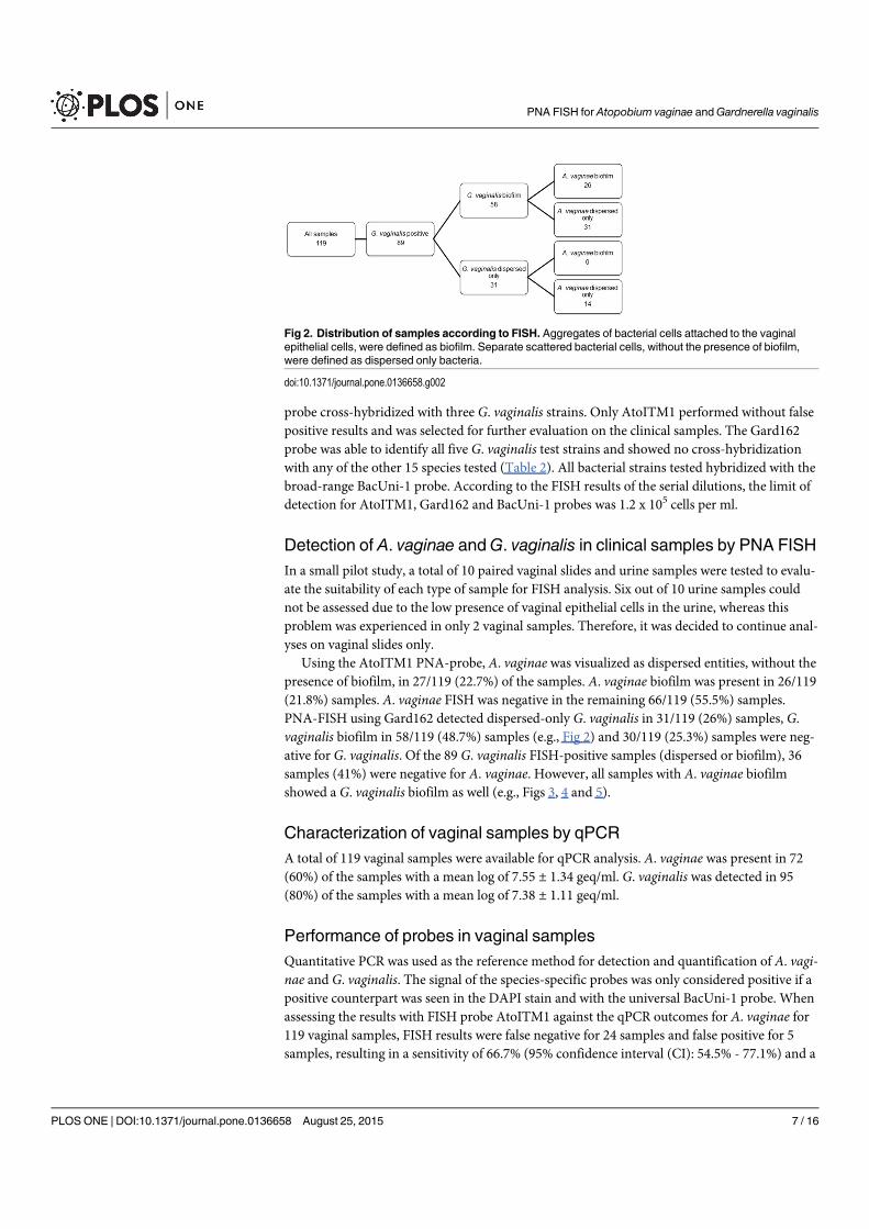
probe cross-hybridized with three G. vaginalis strains. Only AtoITM1 performed without falsepositive results and was selected for further evaluation on the clinical samples. The Gard162probe was able to identify all five G. vaginalis test strains and showed no cross-hybridizationwith any of the other 15 species tested (Table 2). All bacterial strains tested hybridized with thebroad-range BacUni-1 probe. According to the FISH results of the serial dilutions, the limit ofdetection for AtoITM1, Gard162 and BacUni-1 probes was 1.2 x 10⁵ cells per ml.
Detection of A. vaginae andG. vaginalis in clinical samples by PNA FISHIn a small pilot study, a total of 10 paired vaginal slides and urine samples were tested to evalu-ate the suitability of each type of sample for FISH analysis. Six out of 10 urine samples couldnot be assessed due to the low presence of vaginal epithelial cells in the urine, whereas thisproblem was experienced in only 2 vaginal samples. Therefore, it was decided to continue anal-yses on vaginal slides only.
Using the AtoITM1 PNA-probe, A. vaginae was visualized as dispersed entities, without thepresence of biofilm, in 27/119 (22.7%) of the samples. A. vaginae biofilm was present in 26/119(21.8%) samples. A. vaginae FISH was negative in the remaining 66/119 (55.5%) samples.PNA-FISH using Gard162 detected dispersed-only G. vaginalis in 31/119 (26%) samples, G.vaginalis biofilm in 58/119 (48.7%) samples (e.g., Fig 2) and 30/119 (25.3%) samples were neg-ative for G. vaginalis. Of the 89 G. vaginalis FISH-positive samples (dispersed or biofilm), 36samples (41%) were negative for A. vaginae. However, all samples with A. vaginae biofilmshowed a G. vaginalis biofilm as well (e.g., Figs 3, 4 and 5).
Characterization of vaginal samples by qPCRA total of 119 vaginal samples were available for qPCR analysis. A. vaginae was present in 72(60%) of the samples with a mean log of 7.55 ± 1.34 geq/ml. G. vaginalis was detected in 95(80%) of the samples with a mean log of 7.38 ± 1.11 geq/ml.
Performance of probes in vaginal samplesQuantitative PCR was used as the reference method for detection and quantification of A. vagi-nae and G. vaginalis. The signal of the species-specific probes was only considered positive if apositive counterpart was seen in the DAPI stain and with the universal BacUni-1 probe. Whenassessing the results with FISH probe AtoITM1 against the qPCR outcomes for A. vaginae for119 vaginal samples, FISH results were false negative for 24 samples and false positive for 5samples, resulting in a sensitivity of 66.7% (95% confidence interval (CI): 54.5% - 77.1%) and a
Fig 2. Distribution of samples according to FISH. Aggregates of bacterial cells attached to the vaginalepithelial cells, were defined as biofilm. Separate scattered bacterial cells, without the presence of biofilm,were defined as dispersed only bacteria.
doi:10.1371/journal.pone.0136658.g002
PNA FISH for Atopobium vaginae andGardnerella vaginalis
PLOSONE | DOI:10.1371/journal.pone.0136658 August 25, 2015 7 / 16
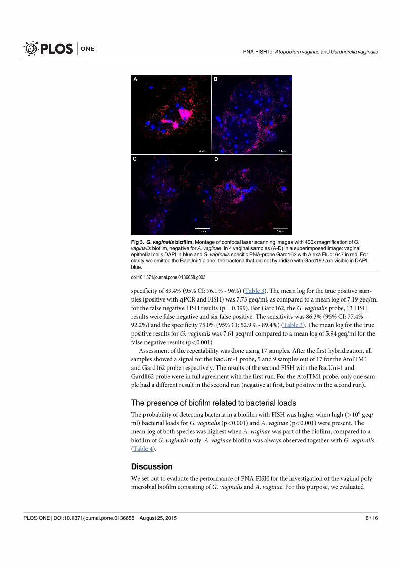
specificity of 89.4% (95% CI: 76.1% - 96%) (Table 3). The mean log for the true positive sam-ples (positive with qPCR and FISH) was 7.73 geq/ml, as compared to a mean log of 7.19 geq/mlfor the false negative FISH results (p = 0.399). For Gard162, the G. vaginalis probe, 13 FISHresults were false negative and six false positive. The sensitivity was 86.3% (95% CI: 77.4% -92.2%) and the specificity 75.0% (95% CI: 52.9% - 89.4%) (Table 3). The mean log for the truepositive results for G. vaginalis was 7.61 geq/ml compared to a mean log of 5.94 geq/ml for thefalse negative results (p<0.001).
Assessment of the repeatability was done using 17 samples. After the first hybridization, allsamples showed a signal for the BacUni-1 probe, 5 and 9 samples out of 17 for the AtoITM1and Gard162 probe respectively. The results of the second FISH with the BacUni-1 andGard162 probe were in full agreement with the first run. For the AtoITM1 probe, only one sam-ple had a different result in the second run (negative at first, but positive in the second run).
The presence of biofilm related to bacterial loadsThe probability of detecting bacteria in a biofilm with FISH was higher when high (>106 geq/ml) bacterial loads for G. vaginalis (p<0.001) and A. vaginae (p<0.001) were present. Themean log of both species was highest when A. vaginae was part of the biofilm, compared to abiofilm of G. vaginalis only. A. vaginae biofilm was always observed together with G. vaginalis(Table 4).
DiscussionWe set out to evaluate the performance of PNA FISH for the investigation of the vaginal poly-microbial biofilm consisting of G. vaginalis and A. vaginae. For this purpose, we evaluated
Fig 3. G. vaginalis biofilm.Montage of confocal laser scanning images with 400x magnification ofG.vaginalis biofilm, negative for A. vaginae, in 4 vaginal samples (A-D) in a superimposed image: vaginalepithelial cells DAPI in blue andG. vaginalis specific PNA-probe Gard162 with Alexa Fluor 647 in red. Forclarity we omitted the BacUni-1 plane; the bacteria that did not hybridize with Gard162 are visible in DAPIblue.
doi:10.1371/journal.pone.0136658.g003
PNA FISH for Atopobium vaginae andGardnerella vaginalis
PLOSONE | DOI:10.1371/journal.pone.0136658 August 25, 2015 8 / 16
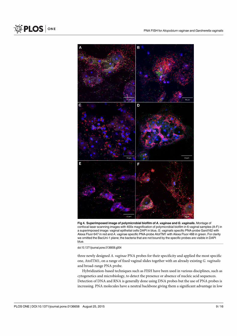
three newly designed A. vaginae PNA probes for their specificity and applied the most specificone, AtoITM1, on a range of fixed vaginal slides together with an already existing G. vaginalisand broad-range PNA probe.
Hybridization-based techniques such as FISH have been used in various disciplines, such ascytogenetics and microbiology, to detect the presence or absence of nucleic acid sequences.Detection of DNA and RNA is generally done using DNA probes but the use of PNA probes isincreasing. PNA molecules have a neutral backbone giving them a significant advantage in low
Fig 4. Superimposed image of polymicrobial biofilm of A. vaginae andG. vaginalis.Montage ofconfocal laser scanning images with 400x magnification of polymicrobial biofilm in 6 vaginal samples (A-F) ina superimposed image: vaginal epithelial cells DAPI in blue,G. vaginalis specific PNA-probe Gard162 withAlexa Fluor 647 in red and A. vaginae specific PNA-probe AtoITM1 with Alexa Fluor 488 in green. For claritywe omitted the BacUni-1 plane; the bacteria that are not bound by the specific probes are visible in DAPIblue.
doi:10.1371/journal.pone.0136658.g004
PNA FISH for Atopobium vaginae andGardnerella vaginalis
PLOSONE | DOI:10.1371/journal.pone.0136658 August 25, 2015 9 / 16
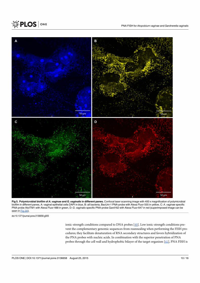
ionic-strength conditions compared to DNA probes [40]. Low ionic-strength conditions pre-vent the complementary genomic sequences from reannealing when performing the FISH pro-cedures; they facilitate denaturation of RNA secondary structures and favors hybridization ofthe PNA probes with nucleic acids. In combination with the superior penetration of PNAprobes through the cell wall and hydrophobic bilayer of the target organism [41], PNA FISH is
Fig 5. Polymicrobial biofilm of A. vaginae andG. vaginalis in different panes.Confocal laser scanning image with 400 x magnification of polymicrobialbiofilm in different panes, A: vaginal epithelial cells DAPI in blue, B: all bacteria, BacUni-1 PNA-probe with Alexa Fluor 555 in yellow, C: A. vaginae specificPNA-probe AtoITM1 with Alexa Fluor 488 in green, D:G. vaginalis specific PNA-probe Gard162 with Alexa Fluor 647 in red (superimposed image can beseen in Fig 3A).
doi:10.1371/journal.pone.0136658.g005
PNA FISH for Atopobium vaginae andGardnerella vaginalis
PLOSONE | DOI:10.1371/journal.pone.0136658 August 25, 2015 10 / 16

a fast, simple and robust assay. We compared DNA and PNA probes (data not reported) andcan confirm that PNA FISH is faster and more robust than DNA FISH. In the current study,PNA FISH proved to be highly efficient for the identification and visualization of the spatialarrangement of A. vaginae and G. vaginalis in the BV-associated biofilms. Moreover, PNAFISH showed excellent inter-assay repeatability for all three probes used.
PNA FISH probe performance on bacterial strainsFor the design of the PNA probes, we opted for Alexa fluorochromes, which have similar spec-tral properties as other fluorochromes, such as cyanine dyes, but are brighter and more resis-tant to photo bleaching [42].
PNA probes that specifically target A. vaginae have not been described before. The onlyprobe thus far reported was a DNA probe (Ato291) [27]. The probe was designed to detect bac-teria belonging to the Atopobium cluster in fecal samples. However, in silico evaluation of thespecificity of the Ato291 probe showed cross-hybridization with other bacteria belonging tothe Coriobacteriaceae, a family of Actinobacteria, to which A. vaginae belongs. This is not sur-prising, since the probe was originally designed on the basis of sequences of Coriobacteriaceaestrains isolated from feces and clinical material. The probe has been used for the detection of A.vaginae in vaginal samples by Swidsinski et al. [9], but their findings have not yet been con-firmed by other groups. In our experiments, using a PNA equivalent of the Ato291 probe, weshowed a low specificity of the Ato291 probe on vaginal clinical isolates. The Ato291 probe
Table 3. Performance of A. vaginae (AtoITM1) andG. vaginalis (Gard162) PNA probes, compared to qPCR results, for 119 vaginal slides.
FISH qPCR
A. vaginae positive A. vaginae negative G. vaginalis positive G. vaginalis negative Total
AtoITM1 positive 48 (66.7%) 5 (10.6%) 53
AtoITM1 negative 24 (33.3%) 42 (89.4%) 66
Gard162 positive 82 (86.3%) 6 (25%) 88
Gard162 negative 13 (13.7%) 18 (75%) 31
Total 72 47 95 24
doi:10.1371/journal.pone.0136658.t003
Table 4. Presence of A. vaginae andG. vaginalis, as assessed by FISH, in relation to A. vaginae andG. vaginalis loads as determined by qPCR for119 vaginal samples.
Total G. vaginaliscount 0
G. vaginaliscount <106
geq/ml
G. vaginaliscount >106
geq/ml
A. vaginaecount 0
A. vaginaecount <106
geq/ml
A. vaginaecount >106
geq/ml
A. vaginaemean log
Detected byPNA FISH
N (%) N (%) N (%) geq/ml N (%) N (%) N (%) geq/ml
A. vaginaeAbsent 66 20 (30.3%) 13 (19.7%) 33 (50.0%) 4.87 42 (63.64%) 7 (10.61%) 17 (25.76%) 2.55
Dispersed only 27 3 (11.1%) 0 (0.0%) 24 (88.9%) 6.82 3 (11.11%) 4 (14.81%) 20 (74.07%) 6.21
Biofilm1 26 1 (3.9%) 1 (11.8%) 24 (92.3%) 7.50 2 (7.69%) 0 (0%) 24 (92.31%) 7.66
G. vaginalisAbsent 30 17 (56.7%) 6 (20.0%) 7 (23.3%) 2.57 24 (80.00%) 3 (10.00%) 3 (10.10%) 1.05
Dispersed only 31 2 (6.5%) 6 (19.4%) 23 (74.2%) 6.68 13 (41.94%) 5 (16.13%) 13 (41.94%) 3.97
Biofilm 58 5 (8.6%) 2 (3.5%) 51 (87.9%) 7.18 10 (17.24%) 3 (5.17%) 45 (77.59%) 6.55
1A. vaginae biofilm = polymicrobial biofilm consisting of A. vaginae and G. vaginalis; No slides had A. vaginae biofilm only.
doi:10.1371/journal.pone.0136658.t004
PNA FISH for Atopobium vaginae andGardnerella vaginalis
PLOSONE | DOI:10.1371/journal.pone.0136658 August 25, 2015 11 / 16
cross-hybridized with three out of five G. vaginalis strains and all five Lactobacillus species.Therefore, we designed two new probes for A. vaginae targeting the 16S rRNA-gene, based onpublished PCR primers [28,29] and we adjusted the sequences to fit the requirements for PNAprobes. One of the new probes, AtoITM1, did not cross-react to any of the tested species andwas further used for detection of A. vaginae in vaginal slides.
Gard162 is the first PNA FISH probe designed specifically for G. vaginalis and has exten-sively been tested by Machado et al. on a variety of cultured bacterial strains and clinical sam-ples [30,31]. Using this probe, we obtained clear hybridization for all G. vaginalis isolates testedand observed no cross-reaction with strains of the other species tested, confirming the findingsof Machado et al. [30].
PNA FISH probe performance on clinical samplesVaginal slides proved to be a valid sample type for imaging of the composition of the vaginalmicrobiome, if processed directly after sampling, as shown by Peltroche-Llacsahuanga et al.[43]. Collection of a vaginal swab is an easy and cheap sampling method, with a minimal bur-den on the study participant or patient. After heat fixation, we recorded that the slides can bestored at room temperature for up to at least six months and can be easily transported. A highquality vaginal sample can be obtained by thinly rolling the swab onto the slide. A thick vaginal‘smear’ on the contrary where the material is smeared onto the slide is not useful for FISH andmicroscopic visualization.
The probes were also applied to 119 vaginal slides from women for whom the bacterial loadsof G. vaginalis and A. vaginae had been quantified by qPCR. qPCR was used in this study as thereference method to evaluate the performance of the FISH probes, although comparison ofthese methods is subject to some limitations. qPCR is highly sensitive and was performed on ahomogenized DNA extract representing half of the full sample. FISH however was carried outon 0.5 mm² of a vaginal slide, which could be heterogeneous. This is inevitably an underrepre-sentation of the vaginal sample. Both techniques were also performed using two different vagi-nal swabs, but the first collected specimen was used to prepare the slide as per study protocol.
After hybridization for 60 minutes and washing for 15 minutes, both at 60°C, the AtoITM1probe gave only five false positive results, compared to qPCR results, resulting in a specificityof 90% for this set of samples However, 24 samples that were positive according to the qPCRwere not detected by FISH, which gives a sensitivity of 67%. These results are comparable tothe sensitivity observed for vaginal samples using FISH for detection of Group B Streptococcus[43]. The authors of this study obtained a higher sensitivity after extraction of the swabs bycentrifugation of the swab head in a NaCl solution [43], but this method would probably dis-rupt the epithelial biofilm and thus prevent us from investigating the organization of the bacte-rial biofilm.
The relatively low sensitivity of theA. vaginae FISH assay cannot be explained by the bacterialload as measured by qPCR; the mean log of the true positive and false negative samples was notsignificantly different (log 7.73 and 7.19 geq/ml respectively). One possible explanation could bethe typical structure of a biofilm, whereby an oxygen gradient exists from the top to the center ofthe biofilm [44]. Anaerobic bacteria such as A. vaginae are possibly found more embedded inthe biofilm, to take advantage of the anaerobicity. It could be that the PNA probes are not able tofully penetrate into the inner parts of the biofilm; or that if the PNA probes do penetrate, thefluorescence could be masked and not detected due to low resolution of the equipment.
For G. vaginalis, 6 false positive results and 13 false negative results were found compared to95 positive and 24 negative samples according to qPCR. This implies a sensitivity of 86% andspecificity 75% for the detection of G. vaginalis by the Gard162 probe using our FISH protocol
PNA FISH for Atopobium vaginae andGardnerella vaginalis
PLOSONE | DOI:10.1371/journal.pone.0136658 August 25, 2015 12 / 16
for this set of samples. This is lower than reported by Machado et al. [30], who, using the sameprobe, showed a full agreement between qPCR and FISH results for 13 vaginal samples. Wewere not able to elucidate why these discordant results were obtained.
Bacterial loads and the presence of a biofilmOur study shows that higher bacterial loads of G. vaginalis and A. vaginae, as detected byqPCR, are associated with a higher probability of presence of a bacterial biofilm. Both bacterialspecies are important constituents of the vaginal epithelial biofilm [9,45]. No samples con-tained A. vaginae in the absence of G. vaginalis, but almost half of the G. vaginalis-positivesamples did not contain A. vaginae according to FISH results. Both bacteria were seen in a dis-persed and an adherent state, but A. vaginae was always accompanied by G. vaginalis. Themere presence of A. vaginae did not simply predispose to a polymicrobial biofilm, but when A.vaginae was part of the biofilm, compared to a biofilm of only G. vaginalis, both bacterial spe-cies were present in higher concentrations.
We hypothesize that G. vaginalis is one of the main initiators of a vaginal biofilm, when it ispresent in high amounts. This vaginal biofilm creates a favorable environment for anaerobicbacteria, such as A. vaginae. One reason for the appearance of A. vaginaemay be the presenceof an oxygen gradient within the biofilm. By embedding itself within the biofilm, A. vaginaecan take advantage of the anaerobicity, proliferates and exists in a mutualistic relationship withG. vaginalis.
ConclusionOur study confirms that PNA FISH is a valuable tool for detecting and visualizing biofilm-associated organisms in vaginal slides. This study describes the design and evaluation of a newPNA probe, AtoITM1, which can be included in multiplex FISH in BV biofilm research. Usingthe new probe, we have demonstrated the presence of a polymicrobial biofilm, with A. vaginaetaking part in a G. vaginalis dominated biofilm.
Supporting InformationS1 Protocol. Final Approved Protocol for "The Ring Plus project: Safety and acceptabilityof vaginal rings that protect women from unintented pregenancy" version 2.0, 16 April2013.(PDF)
AcknowledgmentsWe are grateful to the participants of this study, the study staff of Rinda Ubuzima and the STIreference laboratory team of ITM, Antwerp.
Author ContributionsConceived and designed the experiments: LH VJ TC. Performed the experiments: LH ND LMVM. Analyzed the data: VJ LH. Contributed reagents/materials/analysis tools: MV TC. Wrotethe paper: LH VJ ND LM VMMV TC.
References1. Marrazzo JM, Martin DH, Watts DH, Schulte J, Sobel JD, Hillier SL, et al. (2010) Bacterial vaginosis:
identifying research gaps proceedings of a workshop sponsored by DHHS/NIH/NIAID. Sex Transm Dis37: 732–744. PMID: 21068695
PNA FISH for Atopobium vaginae andGardnerella vaginalis
PLOSONE | DOI:10.1371/journal.pone.0136658 August 25, 2015 13 / 16

2. Spiegel CA (1991) Bacterial vaginosis. Clin Microbiol Rev 4: 485–502. PMID: 1747864
3. van deWijgert JH, Borgdorff H, Verhelst R, Crucitti T, Francis S, Verstraelen H, et al. (2014) The vaginalmicrobiota: what have we learned after a decade of molecular characterization? PLoS One 9:e105998. doi: 10.1371/journal.pone.0105998; PONE-D-14-20877 [pii]. PMID: 25148517
4. Fredricks DN, Fiedler TL, Thomas KK, Oakley BB, Marrazzo JM (2007) Targeted PCR for detection ofvaginal bacteria associated with bacterial vaginosis. J Clin Microbiol 45: 3270–3276. JCM.01272-07[pii]; doi: 10.1128/JCM.01272-07 PMID: 17687006
5. Nugent RP, Krohn MA, Hillier SL (1991) Reliability of diagnosing bacterial vaginosis is improved by astandardized method of gram stain interpretation. J Clin Microbiol 29: 297–301. PMID: 1706728
6. Srinivasan S, Hoffman NG, Morgan MT, Matsen FA, Fiedler TL, Hall RW, et al. (2012) Bacterial com-munities in women with bacterial vaginosis: high resolution phylogenetic analyses reveal relationshipsof microbiota to clinical criteria. PLoS One 7: e37818. doi: 10.1371/journal.pone.0037818; PONE-D-12-04403 [pii]. PMID: 22719852
7. Gelber SE, Aguilar JL, Lewis KL, Ratner AJ (2008) Functional and phylogenetic characterization ofVaginolysin, the human-specific cytolysin fromGardnerella vaginalis. J Bacteriol 190: 3896–3903.JB.01965-07 [pii]; doi: 10.1128/JB.01965-07 PMID: 18390664
8. Lopes dos Santos Santiago G, Deschaght P, El Aila N, Kiama TN, Verstraelen H, Jefferson KK, et al.(2011)Gardnerella vaginalis comprises three distinct genotypes of which only two produce sialidase.Am J Obstet Gynecol 204: 450–457. S0002-9378(11)00019-6 [pii]; doi: 10.1016/j.ajog.2010.12.061
9. Swidsinski A, MendlingW, Loening-Baucke V, Ladhoff A, Swidsinski S, Hale LP, et al. (2005) Adherentbiofilms in bacterial vaginosis. Obstet Gynecol 106: 1013–1023. 106/5/1013 [pii]; doi: 10.1097/01.AOG.0000183594.45524.d2 PMID: 16260520
10. Patterson JL, Girerd PH, Karjane NW, Jefferson KK (2007) Effect of biofilm phenotype on resistance ofGardnerella vaginalis to hydrogen peroxide and lactic acid. Am J Obstet Gynecol 197: 170–177.S0002-9378(07)00263-3 [pii]; doi: 10.1016/j.ajog.2007.02.027 PMID: 17689638
11. Schwebke JR, Muzny CA, JoseyWE (2014) Role ofGardnerella vaginalis in the pathogenesis of bacte-rial vaginosis: a conceptual model. J Infect Dis 210: 338–343. jiu089 [pii]; doi: 10.1093/infdis/jiu089PMID: 24511102
12. Olson ME, Ceri H, Morck DW, Buret AG, Read RR (2002) Biofilm bacteria: formation and comparativesusceptibility to antibiotics. Can J Vet Res 66: 86–92. PMID: 11989739
13. Van Acker H, Van Dijck P, Coenye T (2014) Molecular mechanisms of antimicrobial tolerance andresistance in bacterial and fungal biofilms. Trends Microbiol 22: 326–333. S0966-842X(14)00024-9[pii]; doi: 10.1016/j.tim.2014.02.001 PMID: 24598086
14. Patterson JL, Stull-Lane A, Girerd PH, Jefferson KK (2010) Analysis of adherence, biofilm formationand cytotoxicity suggests a greater virulence potential ofGardnerella vaginalis relative to other bacte-rial-vaginosis-associated anaerobes. Microbiology 156: 392–399. mic.0.034280–0 [pii]; doi: 10.1099/mic.0.034280-0 PMID: 19910411
15. Verstraelen H, Swidsinski A (2013) The biofilm in bacterial vaginosis: implications for epidemiology,diagnosis and treatment. Curr Opin Infect Dis 26: 86–89. doi: 10.1097/QCO.0b013e32835c20cdPMID: 23221767
16. De Backer E, Verhelst R, Verstraelen H, Claeys G, Verschraegen G, Temmerman M, et al. (2006) Anti-biotic susceptibility of Atopobium vaginae. BMC Infect Dis 6: 51. 1471-2334-6-51 [pii]; doi: 10.1186/1471-2334-6-51 PMID: 16542416
17. Ferris MJ, Masztal A, Aldridge KE, Fortenberry JD, Fidel PL Jr, Martin DH (2004) Association of Atopo-bium vaginae, a recently described metronidazole resistant anaerobe, with bacterial vaginosis. BMCInfect Dis 4: 5. doi: 10.1186/1471-2334-4-5; 1471-2334-4-5 [pii]. PMID: 15018635
18. Fredricks DN, Fiedler TL, Marrazzo JM (2005) Molecular identification of bacteria associated with bac-terial vaginosis. N Engl J Med 353: 1899–1911. 353/18/1899 [pii]; doi: 10.1056/NEJMoa043802 PMID:16267321
19. Menard JP, Fenollar F, Henry M, Bretelle F, Raoult D (2008) Molecular quantification ofGardnerellavaginalis and Atopobium vaginae loads to predict bacterial vaginosis. Clin Infect Dis 47: 33–43. doi: 10.1086/588661 PMID: 18513147
20. Verhelst R, Verstraelen H, Claeys G, Verschraegen G, Delanghe J, Van Simaey L, et al. (2004) Cloningof 16S rRNA genes amplified from normal and disturbed vaginal microflora suggests a strong associa-tion between Atopobium vaginae,Gardnerella vaginalis and bacterial vaginosis. BMCMicrobiol 4: 16.doi: 10.1186/1471-2180-4-16; 1471-2180-4-16 [pii]. PMID: 15102329
21. Bradshaw CS, Tabrizi SN, Fairley CK, Morton AN, Rudland E, Garland SM (2006) The association ofAtopobium vaginae andGardnerella vaginaliswith bacterial vaginosis and recurrence after oral metro-nidazole therapy. J Infect Dis 194: 828–836. JID36460 [pii]; doi: 10.1086/506621 PMID: 16941351
PNA FISH for Atopobium vaginae andGardnerella vaginalis
PLOSONE | DOI:10.1371/journal.pone.0136658 August 25, 2015 14 / 16
22. Cools P, Verstraelen H, Vaneechoutte M, Verhelst R (2011) Atopobium. In: Liu Dongyou, editors.Molecular detection of human bacterial pathogens. CRC Press. pp. 31–43.
23. Verstraelen H, Verhelst R, Claeys G, Temmerman M, Vaneechoutte M (2004) Culture-independentanalysis of vaginal microflora: the unrecognized association of Atopobium vaginaewith bacterial vagi-nosis. Am J Obstet Gynecol 191: 1130–1132. S0002937804003990 [pii]; doi: 10.1016/j.ajog.2004.04.013 PMID: 15507931
24. Elias S, Banin E (2012) Multi-species biofilms: living with friendly neighbors. FEMSMicrobiol Rev 36:990–1004. doi: 10.1111/j.1574-6976.2012.00325.x PMID: 22229800
25. Wolcott R, Costerton JW, Raoult D, Cutler SJ (2013) The polymicrobial nature of biofilm infection. ClinMicrobiol Infect 19: 107–112. doi: 10.1111/j.1469-0691.2012.04001.x PMID: 22925473
26. Bradshaw DJ, Marsh PD,Watson GK, Allison C (1998) Role of Fusobacterium nucleatum and coaggre-gation in anaerobe survival in planktonic and biofilm oral microbial communities during aeration. InfectImmun 66: 4729–4732. PMID: 9746571
27. Harmsen HJ, Wildeboer-Veloo AC, Grijpstra J, Knol J, Degener JE, Welling GW (2000) Developmentof 16S rRNA-based probes for theCoriobacterium group and the Atopobium cluster and their applica-tion for enumeration of Coriobacteriaceae in human feces from volunteers of different age groups. ApplEnviron Microbiol 66: 4523–4527. PMID: 11010909
28. Burton JP, Devillard E, Cadieux PA, Hammond JA, Reid G (2004) Detection of Atopobium vaginae inpostmenopausal women by cultivation-independent methods warrants further investigation. J ClinMicrobiol 42: 1829–1831. PMID: 15071062
29. Fredricks DN, Fiedler TL, Thomas KK, Mitchell CM, Marrazzo JM (2009) Changes in vaginal bacterialconcentrations with intravaginal metronidazole therapy for bacterial vaginosis as assessed by quantita-tive PCR. J Clin Microbiol 47: 721–726. JCM.01384-08 [pii]; doi: 10.1128/JCM.01384-08 PMID:19144794
30. Machado A, Almeida C, Salgueiro D, Henriques A, Vaneechoutte M, Haesebrouck F, et al. (2013) Fluo-rescence in situ Hybridization method using Peptide Nucleic Acid probes for rapid detection of Lactoba-cillus andGardnerella spp. BMCMicrobiol 13: 82. 1471-2180-13-82 [pii]; doi: 10.1186/1471-2180-13-82 PMID: 23586331
31. Machado A, Castro J, Cereija T, Almeida C, Cerca N (2015) Diagnosis of bacterial vaginosis by a newmultiplex peptide nucleic acid fluorescence in situ hybridization method. PeerJ 3: e780. doi: 10.7717/peerj.780; 780 [pii]. PMID: 25737820
32. Perry-O'Keefe H, Stender H, Broomer A, Oliveira K, Coull J, Hyldig-Nielsen JJ (2001) Filter-based PNAin situ hybridization for rapid detection, identification and enumeration of specific micro-organisms. JAppl Microbiol 90: 180–189. jam1230 [pii]. PMID: 11168720
33. Amann RI, Binder BJ, Olson RJ, Chisholm SW, Devereux R, Stahl DA (1990) Combination of 16SrRNA-targeted oligonucleotide probes with flow cytometry for analyzing mixed microbial populations.Appl Environ Microbiol 56: 1919–1925. PMID: 2200342
34. Jespers V, Menten J, Smet H, Poradosu S, Abdellati S, Verhelst R, et al. (2012) Quantification of bacte-rial species of the vaginal microbiome in different groups of women, using nucleic acid amplificationtests. BMCMicrobiol 12: 83. 1471-2180-12-83 [pii]; doi: 10.1186/1471-2180-12-83 PMID: 22647069
35. Matsuo K, Ota H, Akamatsu T, Sugiyama A, Katsuyama T (1997) Histochemistry of the surface mucousgel layer of the human colon. Gut 40: 782–789. PMID: 9245933
36. Puchtler H, Waldrop FS, Conner HM, Terry MS (1968) Carnoy Fixation—Practical and Theoretical Con-siderations. Histochemie 16: 361–&. PMID: 4179106
37. Shamsi M, Abdali K, Montazer NR, Kumar PV, Tabatabaee HR (2008) Comparison of Carnoy's solutionand 96% ethyl alcohol fixation in bloody Pap smears. Acta Cytologica 52: 187–190. PMID: 18499991
38. Schurmans C, De Baetselier I, Kestelyn E, Jespers V, Delvaux T, Agaba SK, et al. (2015) The ring plusproject: safety and acceptability of vaginal rings that protect women from unintended pregnancy. BMCPublic Health 15: 348. doi: 10.1186/s12889-015-1680-y; 10.1186/s12889-015-1680-y [pii]. PMID:25880636
39. Swidsinski A, Doerffel Y, Loening-Baucke V, Swidsinski S, Verstraelen H, Vaneechoutte M, et al.(2010)Gardnerella biofilm involves females and males and is transmitted sexually. Gynecol ObstetInvest 70: 256–263. 000314015 [pii]; doi: 10.1159/000314015 PMID: 21051845
40. Egholm M, Buchardt O, Christensen L, Behrens C, Freier SM, Driver DA, et al. (1993) PNA hybridizesto complementary oligonucleotides obeying theWatson-Crick hydrogen-bonding rules. Nature 365:566–568. doi: 10.1038/365566a0 PMID: 7692304
41. Williams B, Stender H, Coull JM (2002) PNA fluorescent in situ hybridization for rapid microbiology andcytogenetic analysis. Methods Mol Biol 208: 181–193. 1-59259-290-2-181 [pii]; doi: 10.1385/1-59259-290-2:181 PMID: 12229288
PNA FISH for Atopobium vaginae andGardnerella vaginalis
PLOSONE | DOI:10.1371/journal.pone.0136658 August 25, 2015 15 / 16
42. Panchuk-Voloshina N, Haugland RP, Bishop-Stewart J, Bhalgat MK, Millard PJ, Mao F, et al. (1999)Alexa dyes, a series of new fluorescent dyes that yield exceptionally bright, photostable conjugates. JHistochem Cytochem 47: 1179–1188. PMID: 10449539
43. Peltroche-Llacsahuanga H, Fiandaca MJ, von OS, Lutticken R, Haase G (2010) Rapid detection ofStreptococcus agalactiae from swabs by peptide nucleic acid fluorescence in situ hybridization. J MedMicrobiol 59: 179–184. jmm.0.013995–0 [pii]; doi: 10.1099/jmm.0.013995-0 PMID: 19833782
44. CostertonW, Veeh R, Shirtliff M, Pasmore M, Post C, Ehrlich G (2003) The application of biofilm sci-ence to the study and control of chronic bacterial infections. J Clin Invest 112: 1466–1477. doi: 10.1172/JCI20365; 112/10/1466 [pii]. PMID: 14617746
45. Polatti F (2012) Bacterial vaginosis, Atopobium vaginae and nifuratel. Curr Clin Pharmacol 7: 36–40.BSP/CCP/E-Pub/0062 [pii]. PMID: 22082330
PNA FISH for Atopobium vaginae andGardnerella vaginalis
PLOSONE | DOI:10.1371/journal.pone.0136658 August 25, 2015 16 / 16
ORIGINAL ARTICLE
A fruitful alliance: the synergy between Atopobiumvaginae and Gardnerella vaginalis in bacterialvaginosis-associated biofilmLiselotte Hardy,1,2,3 Vicky Jespers,1 Said Abdellati,3 Irith De Baetselier,3
Lambert Mwambarangwe,4 Viateur Musengamana,4 Janneke van de Wijgert,4,5
Mario Vaneechoutte,2 Tania Crucitti3
1HIV and Sexual Health Group,Department of Public Health,Institute of Tropical Medicine,Antwerp, Belgium2Laboratory BacteriologyResearch, Faculty of Medicine& Health Sciences, Universityof Ghent, Ghent, Belgium3STI Reference Laboratory,Department of ClinicalSciences, Institute of TropicalMedicine, Antwerp, Belgium4Rinda Ubuzima, Kigali,Rwanda5Institute of Infection andGlobal Health, University ofLiverpool, Liverpool, UK
Correspondence toLiselotte Hardy, Institute ofTropical Medicine,Nationalestraat 155, Antwerp2000, Belgium; [email protected]
Received 20 November 2015Revised 8 February 2016Accepted 20 February 2016
To cite: Hardy L, Jespers V,Abdellati S, et al. SexTransm Infect PublishedOnline First: [please includeDay Month Year]doi:10.1136/sextrans-2015-052475
ABSTRACTObjectives Bacterial vaginosis (BV) is characterised bya change in the microbial composition of the vagina.The BV-associated organisms outnumber the health-associated Lactobacillus species and form a polymicrobialbiofilm on the vaginal epithelium, possibly explaining thedifficulties with antibiotic treatment. A betterunderstanding of vaginal biofilm with emphasis onAtopobium vaginae and Gardnerella vaginalis maycontribute to a better diagnosis and treatment of BV.Methods To this purpose, we evaluated theassociation between the presence of both bacteria byfluorescence in situ hybridisation (FISH) and BV byNugent scoring in 463 vaginal slides of 120 participantsparticipating in a clinical trial in Rwanda.Results A bacterial biofilm was detected in half of thesamples using a universal bacterial probe. The biofilmcontained A. vaginae in 54.1% and G. vaginalis in82.0% of the samples. A. vaginae was accompanied byG. vaginalis in 99.5% of samples. The odds of having aNugent score above 4 were increased for samples withdispersed G. vaginalis and/or A. vaginae present (OR4.5; CI 2 to 10.3). The probability of having a highNugent score was even higher when a combination ofadherent G. vaginalis and dispersed A. vaginae wasvisualised (OR 75.6; CI 13.3 to 429.5) and highestwhen both bacteria were part of the biofilm (OR 119;CI 39.9 to 360.8).Conclusions Our study, although not comprehensive atstudying the polymicrobial biofilm in BV, provided astrong indication towards the importance of A. vaginaeand the symbiosis of A. vaginae and G. vaginalis in thisbiofilm.Trial registration number NCT01796613.
INTRODUCTIONBacterial vaginosis (BV) is the most prevalentvaginal disorder in women of reproductive age. Itincreases the risk of acquisition and transmission ofsexually transmitted infections, including HIV, andis associated with preterm birth in pregnantwomen.1–3 The condition is characterised by achange in the microbial composition of the vagina:the Lactobacillus spp., associated with a healthyvaginal microbiome, are outnumbered by micro-aerophilic and anaerobic organisms, includingGardnerella vaginalis.3–7 The mere presence of
G. vaginalis, however, is not sufficient for the diag-nosis of BV using traditional diagnostic algorithms(see below) because many women without BV alsohave G. vaginalis in their vaginal microbiome.4 BVis, however, associated with high counts of G. vagi-nalis using molecular methods and/or the presenceof a G. vaginalis-containing polymicrobialbiofilm.4–10 Due to its strong adherence to vaginalepithelial cells and biofilm-forming capacities, ithas been suggested that G. vaginalis initiates thecolonisation of the vaginal epithelium and serves asa scaffolding to which other species subsequentlycan attach.10–12
One of the species that might attach to thebiofilm initiated by G. vaginalis could beAtopobium vaginae.13–14 Several molecular studieshave indicated a probable role for A. vaginae inBV,14–16 and it has also been suggested thatA. vaginae plays a major part in the establishmentof a biofilm, together with G. vaginalis.9 10
Considering it has been found in 80–90% of casesof relapse17 and some strains have been shown invitro to be metronidazole resistant,18 it could be ofimportance in the recurrence of BV after standardtreatment with metronidazole.The current gold standard in BV research is the
microscopic evaluation and scoring of vaginal slidesaccording to Nugent.19 The diagnosis of BV isbased on the absence of lactobacilli and the pres-ence of small Gram-negative to Gram-variable rods(G. vaginalis and Bacteroides spp. morphotypes)and curved Gram-negative rods (Mobiluncus spp.morphotypes). In fact, bacterial biofilm can also beseen with this method in the form of clue cells,which are vaginal epithelial cells covered by layersof adherent Gram-negative and/or Gram-variablecells, that is, biofilms.20 Using Gram staining, it isimpossible to distinguish between the different bac-terial species in the biofilm. By labelling the cellswith a fluorescent probe, using fluorescence in situhybridisation (FISH), the structure and compos-ition of the biofilm can be studied in more detail.To study the potential role of A. vaginae and the
synergy between A. vaginae and G. vaginalis in thebiofilm, we used our newly developed peptidenucleic acid (PNA) A. vaginae probe11 togetherwith an existing probe for G. vaginalis21 and a uni-versal bacterial probe22 to investigate the compos-ition of vaginal biofilm and its importance in BV.
Hardy L, et al. Sex Transm Infect 2016;0:1–5. doi:10.1136/sextrans-2015-052475 1
Basic science STI Online First, published on March 10, 2016 as 10.1136/sextrans-2015-052475
Copyright Article author (or their employer) 2016. Produced by BMJ Publishing Group Ltd under licence.
group.bmj.com on March 13, 2016 - Published by http://sti.bmj.com/Downloaded from

MATERIALS AND METHODSClinical samplesVaginal sample collection and preparationVaginal samples were collected from 120 women participatingin a clinical trial at Rinda Ubuzima in Kigali, Rwanda, studyingthe safety and acceptability of a contraceptive vaginal ring(Nuvaring), including the effect of the vaginal ring on thevaginal microbiome (the Ring Plus study—Clinicaltrials.govNCT01796613).23 Participants were between 18 and 35 yearsold and provided written informed consent for participation inthe study. Depending on the group (continuous or intermittentring use) to which the participant was randomised, a total offour or five samples from the same participant were taken overa period of four menstrual cycles. A total of 463 samples wereanalysed after Gram stain and after FISH using light microscopyand confocal laser scanning microscopy (CLSM), respectively.
Vaginal sampling was carried out by the study physician duringa speculum examination in the Rinda Ubuzima research clinic. Acotton swab was brushed against the lateral walls of the vagina andwas transported in its container to the Rinda Ubuzima laboratorywithin 20 min. Upon arrival in the laboratory, the swab was usedto prepare a vaginal slide on a regular glass slide for Gram stainand a second vaginal slide on a Superfrost Plus slide(Menzel-Gläser, Braunschweig, Germany). All slides were air-dried, heat-fixed by passing through a flame twice and then storedin their appropriate boxes until Gram staining and/or shipment forFISH. The first slide was Gram stained and examined on-site inthe Rinda Ubuzima laboratory in Kigali. The Superfrost Plus slideswere stored and shipped at room temperature to the ITM wherethey were fixed for a minimum of 12 h in Carnoy solution (6:3:1,ethanol:chloroform:glacial acetic acid).11
Microbiological analysis of the vaginal samplesPeptide nucleic acid fluorescence in situ hybridisationPNA FISH was performed as described earlier11 using species-specific probes for A. vaginae (AtoITM1) and G. vaginalis(Gard162) and the broad-range BacUni-1 probe. The hybridisedsamples were stored in the dark at room temperature for amaximum of 1 week before microscopic observation, usingCLSM (LSM700, Zeiss, Oberkochen, Germany). Detection andidentification of individual bacteria were done at 400× magnifi-cation (objective: Plan-Apochromat 40x/1.3 Oil Ph3 M27).Separate scattered bacterial cells were defined as dispersed bac-teria. Aggregates of bacterial cells, sticking to the vaginal epithe-lial cells, were defined as adherent bacteria forming a biofilm.The species-specific signal was considered positive only if it hada positive counterpart in the 4’,6-diamidino-2-phenylindole(DAPI) stain and if it displayed a positive signal simultaneouslywith the universal probe. Semi-quantification was done for thedispersed and adherent bacteria in three categories (absent,present in low amount, present in high amount), but for theanalysis only two categories (absent or present) have been used.
Nugent scoreThe status of the vaginal microbiome was assessed at the RindaUbuzima laboratory by Nugent scoring of a Gram stainedvaginal slide.19 A score of 0–3 was considered normal vaginalmicrobiome; a score of 4–6 intermediate microbiome and ascore of 7–10 BV.
Statistical analysisThe clinical study sample size calculation was based on theprimary objective to assess the pre–post changes in the vaginal
microbiome and required 60 women in each group to require95% power to detect clinically important changes in bacterialcounts.23 Data analysis was done using STATA10 (StataCorp LP,Texas, USA). While the samples were collected longitudinally,they were analysed cross-sectionally, with each sample as theunit of analysis. To study the association between the presenceand absence of dispersed and/or adherent A. vaginae and adher-ent G. vaginalis in relation to BV status, we categorised thesamples into five categories (table 1) based on combinations ofthe presence of both bacteria in dispersed and/or adherent formas visualised by FISH. To increase the statistical power, we madethe vaginal microbiome status binary: Nugent score 0–3 (refer-ence group) versus Nugent score 4–10 (table 2). A mixed-effectslogistic regression model was fitted with BV as the binaryoutcome (ie, Nugent 0–3 vs Nugent 4–10) and biofilmcharacteristics as the main dependent variable. The model wasadjusted for woman, randomisation group and study visit,because multiple samples per woman at multiple study visitswere included in the analysis. ORs are reported with 95% CIand the p values are from χ2 tests (table 2).
RESULTSCharacterisation of vaginal samplesIn total, 463 of 527 samples from 120 women were available forFISH analysis, excluding 13 missing samples and 51 samples notreadable due to the absence of epithelial cells on the slides. In all463 samples, a positive signal was detected for the universalBacUni-1 probe. In 230 samples (49.7%), only dispersed bacteriawere present, while the other 233 slides (50.3%) containedadherent bacteria as well (table 1). A. vaginae and G. vaginaliswere part of this biofilm in 126 (54.1%) and 191 (82.0%)samples, respectively. Next, we visualised A. vaginae with FISH
Table 1 Gardnerella vaginalis, Atopobium vaginae and G.vaginalis with A. vaginae combinations for samples analysed withfluorescence in situ hybridisation (FISH) by absent, dispersed onlyand adherent ±dispersed category and stratified by Nugent scoring
TotalNugent0–3
Nugent4–6
Nugent7–10
N N (%) N (%) N (%)
FISH all bacteriaAbsent 0 0 (0.0) 0 (0.0) 0 (0.0)Dispersed only 230 197 (76.0) 19 (39.6) 14 (9.0)Adherent ± dispersed 233 62 (24.0) 29 (60.4) 142 (91.0)
FISH A. vaginae (Av)Absent 268 201 (77.6) 24 (50.0) 43 (27.6)Dispersed only 69 41 (15.8) 10 (20.8) 18 (11.5)Adherent ± dispersed 126 17 (6.6) 14 (29.2) 95 (60.9)
FISH G. vaginalis (Gv)Absent 172 155 (59.8) 8 (16.7) 9 (5.8)Dispersed only 100 71 (27.4) 15 (31.2) 14 (9.0)Adherent ± dispersed 191 33 (12.8) 25 (52.1) 133 (85.2)
FISH Av and Gv combinedGv and Av absent 170 153 (59.1) 8 (16.7) 9 (5.7)Gv or Av dispersed only 101 72 (27.8) 15 (31.2) 14 (9.0)Gv adherent ± Gv dispersedand Av absent
51 14 (5.4) 8 (16.7) 29 (18.6)
Gv adherent ± Gv dispersedand Av dispersed
15 3 (1.1) 3 (6.2) 9 (5.8)
Gv and Av adherent ± Gvand Av dispersed
126 17 (6.6) 14 (29.2) 95 (60.9)
2 Hardy L, et al. Sex Transm Infect 2016;0:1–5. doi:10.1136/sextrans-2015-052475
Basic science
group.bmj.com on March 13, 2016 - Published by http://sti.bmj.com/Downloaded from
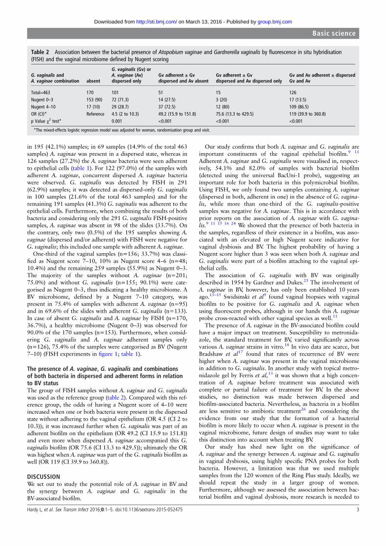
in 195 (42.1%) samples; in 69 samples (14.9% of the total 463samples) A. vaginae was present in a dispersed state, whereas in126 samples (27.2%) the A. vaginae bacteria were seen adherentto epithelial cells (table 1). For 122 (97.0%) of the samples withadherent A. vaginae, concurrent dispersed A. vaginae bacteriawere observed. G. vaginalis was detected by FISH in 291(62.9%) samples; it was detected as dispersed-only G. vaginalisin 100 samples (21.6% of the total 463 samples) and for theremaining 191 samples (41.3%) G. vaginalis was adherent to theepithelial cells. Furthermore, when combining the results of bothbacteria and considering only the 291 G. vaginalis FISH-positivesamples, A. vaginae was absent in 98 of the slides (33.7%). Onthe contrary, only two (0.5%) of the 195 samples showing A.vaginae (dispersed and/or adherent) with FISH were negative forG. vaginalis; this included one sample with adherent A. vaginae.
One-third of the vaginal samples (n=156; 33.7%) was classi-fied as Nugent score 7–10, 10% as Nugent score 4–6 (n=48;10.4%) and the remaining 259 samples (55.9%) as Nugent 0–3.The majority of the samples without A. vaginae (n=201;75.0%) and without G. vaginalis (n=155; 90.1%) were cate-gorised as Nugent 0–3, thus indicating a healthy microbiome. ABV microbiome, defined by a Nugent 7–10 category, waspresent in 75.4% of samples with adherent A. vaginae (n=95)and in 69.6% of the slides with adherent G. vaginalis (n=133).In case of absent G. vaginalis and A. vaginae by FISH (n=170,36.7%), a healthy microbiome (Nugent 0–3) was observed for90.0% of the 170 samples (n=153). Furthermore, when consid-ering G. vaginalis and A. vaginae adherent samples only(n=126), 75.4% of the samples were categorised as BV (Nugent7–10) (FISH experiments in figure 1; table 1).
The presence of A. vaginae, G. vaginalis and combinationsof both bacteria in dispersed and adherent forms in relationto BV statusThe group of FISH samples without A. vaginae and G. vaginaliswas used as the reference group (table 2). Compared with this ref-erence group, the odds of having a Nugent score of 4–10 wereincreased when one or both bacteria were present in the dispersedstate without adhering to the vaginal epithelium (OR 4.5 (CI 2 to10.3)); it was increased further when G. vaginalis was part of anadherent biofilm on the epithelium (OR 49.2 (CI 15.9 to 151.8))and even more when dispersed A. vaginae accompanied this G.vaginalis biofilm (OR 75.6 (CI 13.3 to 429.5)); ultimately the ORwas highest when A. vaginaewas part of the G. vaginalis biofilm aswell (OR 119 (CI 39.9 to 360.8)).
DISCUSSIONWe set out to study the potential role of A. vaginae in BV andthe synergy between A. vaginae and G. vaginalis in theBV-associated biofilm.
Our study confirms that both A. vaginae and G. vaginalis areimportant constituents of the vaginal epithelial biofilm.9 11
Adherent A. vaginae and G. vaginalis were visualised in, respect-ively, 54.1% and 82.0% of samples with bacterial biofilm(detected using the universal BacUni-1 probe), suggesting animportant role for both bacteria in this polymicrobial biofilm.Using FISH, we only found two samples containing A. vaginae(dispersed in both, adherent in one) in the absence of G. vagina-lis, while more than one-third of the G. vaginalis-positivesamples was negative for A. vaginae. This is in accordance withprior reports on the association of A. vaginae with G. vagina-lis.9 11 15 16 24 We showed that the presence of both bacteria inthe samples, regardless of their existence in a biofilm, was asso-ciated with an elevated or high Nugent score indicative forvaginal dysbiosis and BV. The highest probability of having aNugent score higher than 3 was seen when both A. vaginae andG. vaginalis were part of a biofilm attaching to the vaginal epi-thelial cells.
The association of G. vaginalis with BV was originallydescribed in 1954 by Gardner and Dukes.25 The involvement ofA. vaginae in BV, however, has only been established 10 yearsago.13–15 Swidsinski et al9 found vaginal biopsies with vaginalbiofilm to be positive for G. vaginalis and A. vaginae whenusing fluorescent probes, although in our hands this A. vaginaeprobe cross-reacted with other vaginal species as well.11
The presence of A. vaginae in the BV-associated biofilm couldhave a major impact on treatment. Susceptibility to metronida-zole, the standard treatment for BV, varied significantly acrossvarious A. vaginae strains in vitro.18 In vivo data are scarce, butBradshaw et al17 found that rates of recurrence of BV werehigher when A. vaginae was present in the vaginal microbiomein addition to G. vaginalis. In another study with topical metro-nidazole gel by Ferris et al,13 it was shown that a high concen-tration of A. vaginae before treatment was associated withcomplete or partial failure of treatment for BV. In the abovestudies, no distinction was made between dispersed andbiofilm-associated bacteria. Nevertheless, as bacteria in a biofilmare less sensitive to antibiotic treatment26 and considering theevidence from our study that the formation of a bacterialbiofilm is more likely to occur when A. vaginae is present in thevaginal microbiome, future design of studies may want to takethis distinction into account when treating BV.
Our study has shed new light on the significance ofA. vaginae and the synergy between A. vaginae and G. vaginalisin vaginal dysbiosis, using highly specific PNA probes for bothbacteria. However, a limitation was that we used multiplesamples from the 120 women of the Ring Plus study. Ideally, weshould repeat the study in a larger group of women.Furthermore, although we assessed the association between bac-terial biofilm and vaginal dysbiosis, more research is needed to
Table 2 Association between the bacterial presence of Atopobium vaginae and Gardnerella vaginalis by fluorescence in situ hybridisation(FISH) and the vaginal microbiome defined by Nugent scoring
G. vaginalis andA. vaginae combination absent
G. vaginalis (Gv) orA. vaginae (Av)dispersed only
Gv adherent ± Gvdispersed and Av absent
Gv adherent ± Gvdispersed and Av dispersed only
Gv and Av adherent ± dispersedGv and Av
Total=463 170 101 51 15 126Nugent 0–3 153 (90) 72 (71.3) 14 (27.5) 3 (20) 17 (13.5)Nugent 4–10 17 (10) 29 (28.7) 37 (72.5) 12 (80) 109 (86.5)OR (CI)* Reference 4.5 (2 to 10.3) 49.2 (15.9 to 151.8) 75.6 (13.3 to 429.5) 119 (39.9 to 360.8)p Value χ2 test* 0.001 <0.001 <0.001 <0.001
*The mixed-effects logistic regression model was adjusted for woman, randomisation group and visit.
Hardy L, et al. Sex Transm Infect 2016;0:1–5. doi:10.1136/sextrans-2015-052475 3
Basic science
group.bmj.com on March 13, 2016 - Published by http://sti.bmj.com/Downloaded from

unravel the exact mechanisms of biofilm formation in BV,including the role and the importance of both bacteria studied,to finally define improved regimens for treatment of BV.
Moreover, since BV is a polymicrobial condition, new researchshould study the involvement of other bacteria related to BV.
In conclusion, the presented study uncovered a key piece ofthe BV puzzle confirming first, the importance of A. vaginae inBV-associated biofilm and second, showing the joint presence ofA. vaginae and G. vaginalis in a biofilm. Future studies coveringa wide array of BV-associated bacteria may help to further delin-eate biofilm mechanisms in BV.
Handling editor Jackie A Cassell
Acknowledgements We wish to thank the participants of this study, the studystaff of Rinda Ubuzima and the STI reference laboratory team of ITM, Antwerp.
Contributors All authors were involved in the main study that generated the data.For the present study, LH wrote the first draft of the manuscript. VJ, TC, MV andJvdW revised and edited the text. IDB, TC, LH, VJ and JvdW created theexperimental design. SA, LM, VM and LH performed the testing and VJ and LHperformed the data analysis. All authors revised and approved the present version ofthe manuscript.
Figure 1 Superimposed confocallaser scanning images with 400×magnification of Atopobium vaginae+Gardnerella vaginalis biofilm in sixvaginal samples (A–F): vaginalepithelial cells DAPI in blue, A.vaginae-specific peptide nucleic acid(PNA)-probe AtoITM1 with Alexa Fluor488 in green and G. vaginalis-specificPNA-probe Gard162 with Alexa Fluor647 in red. For clarity, we omitted theBacUni-1 plane, such that the bacteriathat did not hybridise with Gard162and AtoITM1 are visible in DAPI blueonly.
Key messages
▸ This study shows that Atopobium vaginae is an importantconstituent of the vaginal biofilm, and is of relevance in thecontext of bacterial vaginosis (BV).
▸ We show that A. vaginae is almost always accompanied byGardnerella vaginalis in BV, but that G. vaginalis can befound without A. vaginae in the vaginal microbiome.
▸ By tackling constituents of the biofilm, the above knowledgecan contribute to a more effective and goal-orientedtreatment and improve women’s reproductive health.
4 Hardy L, et al. Sex Transm Infect 2016;0:1–5. doi:10.1136/sextrans-2015-052475
Basic science
group.bmj.com on March 13, 2016 - Published by http://sti.bmj.com/Downloaded from
Funding This work has been funded by The European & Developing CountriesClinical Trials Partnership (EDCTP) through a project entitled ‘Preparing for clinicaltrials with vaginal rings that protect women from HIV and unintended pregnancy’(grant code SP.2011.41304.043); the University of Liverpool and the ITM.
Competing interests None declared.
Patient consent Obtained.
Ethics approval The Ring Plus study was approved by the Rwanda National EthicsCommittee, Rwanda (Approval number 481/RNEC/2013); the ethics committees ofthe Institute of Tropical Medicine (ITM), Belgium (Approval number 864/13); theAntwerp University Hospital, Belgium (Approval number 13/7/85) and the Universityof Liverpool, UK (Approval number RETG000639IREC).
Provenance and peer review Not commissioned; externally peer reviewed.
Data sharing statement The database relevant to the study has been madeavailable to all collaborators.
Open Access This is an Open Access article distributed in accordance with theCreative Commons Attribution Non Commercial (CC BY-NC 4.0) license, whichpermits others to distribute, remix, adapt, build upon this work non-commercially,and license their derivative works on different terms, provided the original work isproperly cited and the use is non-commercial. See: http://creativecommons.org/licenses/by-nc/4.0/
REFERENCES1 Eschenbach DA. History and review of bacterial vaginosis. Am J Obstet Gynecol
1993;169:441–5.2 Hillier SL, Marrazzo JM, Holmes KK. Bacterial vaginosis. In: Holmes KK, Sparling PF,
Mardh PA, et al., eds. Sexually transmitted diseases. New York: McGraw-Hill,2008;737–68.
3 Martin HL, Richardson BA, Nyange PM, et al. Vaginal lactobacilli, microbial flora,and risk of human immunodeficiency virus type 1 and sexually transmitted diseaseacquisition. J Infect Dis 1999;180:1863–8.
4 van de Wijgert JH, Borgdorff H, Verhelst R, et al. The vaginal microbiota: what havewe learned after a decade of molecular characterization? PLoS ONE 2014;9:e105998.
5 Srinivasan S, Hoffman NG, Morgan MT, et al. Bacterial communities in women withbacterial vaginosis: high resolution phylogenetic analyses reveal relationships ofmicrobiota to clinical criteria. PLoS ONE 2012;7:e37818.
6 Patterson JL, Stull-Lane A, Girerd PH, et al. Analysis of adherence, biofilm formationand cytotoxicity suggests a greater virulence potential of Gardnerella vaginalisrelative to other bacterial-vaginosis-associated anaerobes. Microbiology2010;156:392–9.
7 Jespers V, van de Wijgert J, Cools P, et al. The significance of Lactobacillus crispatusand L. vaginalis for vaginal health and the negative effect of recent sex : across-sectional descriptive study across groups of African women. BMC Infect Dis2015;15:1–14.
8 Jespers V, Crucitti T, van de Wijgert JH, et al. A DNA tool for early detection ofvaginal dysbiosis in African women. Res Microbiol 2016;167:133–41.
9 Swidsinski A, Mendling W, Loening-Baucke V, et al. Adherent biofilms in bacterialvaginosis. Obstet Gynecol 2005;106:1013–23.
10 Swidsinski A, Loening-Baucke V, Mendling W, et al. Infection through structuredpolymicrobial Gardnerella biofilms (StPM-GB). Histol Histopathol 2014;29:567–87.
11 Hardy L, Jespers V, Dahchour N, et al. Unravelling the bacterial vaginosis-associatedbiofilm: a multiplex Gardnerella vaginalis and Atopobium vaginae fluorescence insitu hybridization assay using peptide nucleic acid probes. PLoS ONE 2015;10:e0136658.
12 Machado A, Cerca N. The influence of biofilm formation by Gardnerella vaginalisand other anaerobes on bacterial vaginosis. J Infect Dis 2015;2015;212:1856–61.
13 Ferris MJ, Masztal A, Aldridge KE, et al. Association of Atopobium vaginae, arecently described metronidazole resistant anaerobe, with bacterial vaginosis. BMCInfect Dis 2004;4:5.
14 Burton JP, Devillard E, Cadieux PA, et al. Detection of Atopobium vaginae inpostmenopausal women by cultivation-independent methods warrants furtherinvestigation. J Clin Microbiol 2004;42:1829–31.
15 Verstraelen H, Verhelst R, Claeys G, et al. Culture-independent analysis of vaginalmicroflora: the unrecognized association of Atopobium vaginae with bacterialvaginosis. Am J Obstet Gynecol 2004;191:1130–2.
16 Menard JP, Fenollar F, Henry M, et al. Molecular quantification of Gardnerellavaginalis and Atopobium vaginae loads to predict bacterial vaginosis. Clin Infect Dis2008;47:33–43.
17 Bradshaw CS, Tabrizi SN, Fairley CK, et al. The association of Atopobium vaginaeand Gardnerella vaginalis with bacterial vaginosis and recurrence after oralmetronidazole therapy. J Infect Dis 2006;194:828–36.
18 De Backer E, Verhelst R, Verstraelen H, et al. Antibiotic susceptibility of Atopobiumvaginae. BMC Infect Dis 2006;6:51.
19 Nugent RP, Krohn MA, Hillier SL. Reliability of diagnosing bacterial vaginosis isimproved by a standardized method of gram stain interpretation. J Clin Microbiol1991;29:297–301.
20 Cook RL, Reid G, Pond DG, et al. Clue cells in bacterial vaginosis:immunofluorescent identification of the adherent gram-negative bacteria asGardnerella vaginalis. J Infect Dis 1989;160:490–6.
21 Machado A, Almeida C, Salgueiro D, et al. Fluorescence in situ hybridizationmethod using peptide nucleic acid probes for rapid detection of Lactobacillus andGardnerella spp. BMC Microbiol 2013;13:82.
22 Perry-O’Keefe H, Stender H, Broomer A, et al. Filter-based PNA in situ hybridizationfor rapid detection, identification and enumeration of specific micro-organisms.J Appl Microbiol 2001;90:180–9.
23 Schurmans C, De Baetselier I, Kestelyn E, et al. The ring plus project: safety andacceptability of vaginal rings that protect women from unintented pregnancy. BMCPublic Health 2015;15:348.
24 Verhelst R, Verstraelen H, Claeys G, et al. Cloning of 16S rRNA genes amplifiedfrom normal and disturbed vaginal microflora suggests a strong association betweenAtopobium vaginae, Gardnerella vaginalis and bacterial vaginosis. BMC Microbiol2004;4:16.
25 Gardner HL, Dukes CD. New etiologic agent in nonspecific bacterial vaginitis.Science 1954;120:853.
26 Høiby N, Bjarnsholt T, Moser C, et al. ESCMID guideline for the diagnosis andtreatment of biofilm infections 2014. Clin Microbiol Infect 2015;21(Suppl 1):S1–25.
Hardy L, et al. Sex Transm Infect 2016;0:1–5. doi:10.1136/sextrans-2015-052475 5
Basic science
group.bmj.com on March 13, 2016 - Published by http://sti.bmj.com/Downloaded from
in bacterial vaginosis-associated biofilmGardnerella vaginalis and Atopobium vaginae
A fruitful alliance: the synergy between
Wijgert, Mario Vaneechoutte and Tania CrucittiLambert Mwambarangwe, Viateur Musengamana, Janneke van de Liselotte Hardy, Vicky Jespers, Said Abdellati, Irith De Baetselier,
published online March 10, 2016Sex Transm Infect
http://sti.bmj.com/content/early/2016/03/10/sextrans-2015-052475Updated information and services can be found at:
These include:
MaterialSupplementary
C1.htmlhttp://sti.bmj.com/content/suppl/2016/03/09/sextrans-2015-052475.DSupplementary material can be found at:
References
#BIBLhttp://sti.bmj.com/content/early/2016/03/10/sextrans-2015-052475This article cites 25 articles, 8 of which you can access for free at:
Open Access
http://creativecommons.org/licenses/by-nc/4.0/non-commercial. See: provided the original work is properly cited and the use isnon-commercially, and license their derivative works on different terms, permits others to distribute, remix, adapt, build upon this workCommons Attribution Non Commercial (CC BY-NC 4.0) license, which This is an Open Access article distributed in accordance with the Creative
serviceEmail alerting
box at the top right corner of the online article. Receive free email alerts when new articles cite this article. Sign up in the
CollectionsTopic Articles on similar topics can be found in the following collections
(458)Vulvovaginal disorders (3124)Drugs: infectious diseases
(204)Open access
Notes
http://group.bmj.com/group/rights-licensing/permissionsTo request permissions go to:
http://journals.bmj.com/cgi/reprintformTo order reprints go to:
http://group.bmj.com/subscribe/To subscribe to BMJ go to:
group.bmj.com on March 13, 2016 - Published by http://sti.bmj.com/Downloaded from
Related Documents


