Chapter 41 Physiology, Homeostasis, and Temperature Regulation Biology 102 Tri-County Technical College Pendleton, SC

Chapter 41 Physiology, Homeostasis, and Temperature Regulation Biology 102 Tri-County Technical College Pendleton, SC.
Dec 27, 2015
Welcome message from author
This document is posted to help you gain knowledge. Please leave a comment to let me know what you think about it! Share it to your friends and learn new things together.
Transcript

Chapter 41 Physiology, Homeostasis, and Temperature Regulation
Biology 102
Tri-County Technical College
Pendleton, SC
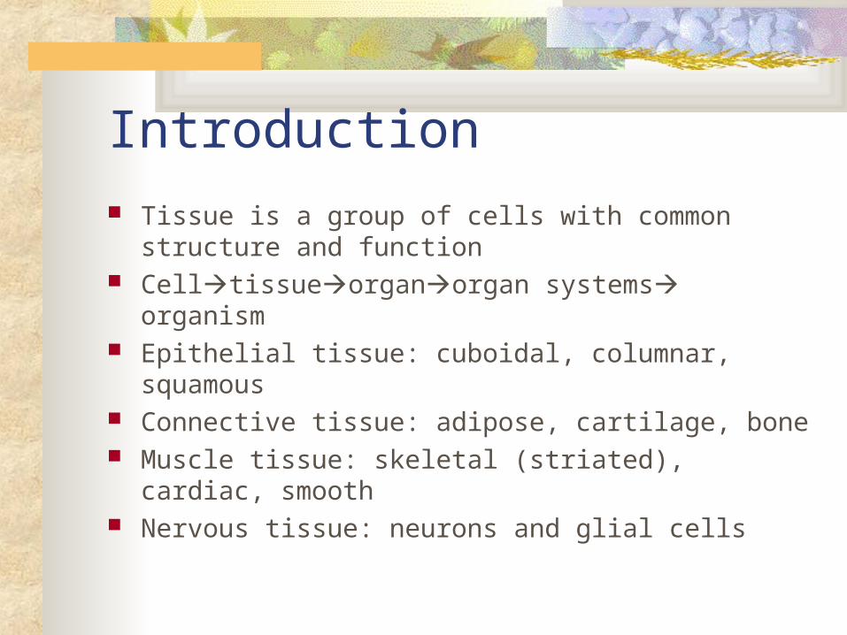
Introduction
Tissue is a group of cells with common structure and function
Celltissueorganorgan systems organism Epithelial tissue: cuboidal, columnar, squamous Connective tissue: adipose, cartilage, bone Muscle tissue: skeletal (striated), cardiac, smooth Nervous tissue: neurons and glial cells

Tissues VisualLining, transport, secretion, and absorption
Support, strength, and elasticity
Movement
Information synthesis, communication, and control

Epithelial Tissue Formed from sheets of tightly packed cells,
covers outside of body & lines organs and body systems
Cells closely joined and riveted by tight junctions
Functions as barrier against mechanical injury, invading organisms, and fluid loss
Free surface exposed to air or fluid

Epithelium, cont.
Cells at base attached to basement membrane (dense layer of extracellular material)
Characterized by number of layers and shape of free surface cells
Simple = one layer thick Stratified = multiple tiers (layers) of cells Pseduostratified = one layer that appears to be
multiple because they vary in length

Epithelium, III
Cell shapes are cuboidal (dice), columnar (bricks on end), or squamous (flat tiles)
Cuboidal epithelium = epithelia of kidney tubules
Columnar epithelium = lining of intestines Squamous = line air sacs of lungs

Connective Tissue
Characterized by sparse cell population in an extensive extracellular matrix
Functions to bind and support other tissues Matrix is web of fibers embedded in
homogenous ground substance Consists of loose weave of 3 types of
proteinaceous fibers: collagenous, elastic, and reticular

Connective, cont.
Collagenous fibers are bundles of fibers containing 3 collagen molecules each, have great tensile strength and resist stretching
Elastic fibers are long threads of protein (elastin); lend tissue resilience to quickly return to original shape
Reticular fibers are branched; form tightly woven fabric joining connective tissue to adjacent tissues

Major types of Connective Tissue Loose connective, adipose, fibrous
connective, cartilage, bone, and blood Adipose tissue is loose connective tissue
specialized to store fat in adipose cells distributed throughout its matrix
Insulates body and stores fuel molecules Each adipose cell has one large fat droplet Adipose tissue covers some internal organs

Connective Types, cont.
Cartilage composed of collagenous fibers embedded in chondroitin sulfate (protein-carbohydrate substance)
Chondrocytes secrete both collagen and chondroitin sulfate
Makes cartilage both strong and flexible Chondrocytes confined to lucunae
(scattered spaces within ground substance)

Connective Types, III
Cartilage composes skeleton of all vertebrate embryos
Retained in some areas: nose, ears, trachea, intervertebral discs, and ends of some bones
BONE is mineralized connective tissue Osteoblasts are bone forming cells Deposit matrix of collagen & calcium phosphate
which hardens into mineral hydroxyapatite

Connective Types, IV
Makes bones hard but not brittle Bone consists of repeating Haversian
systems (concentric layers or lamellae around central canal containing blood vessels/nerves
Once osteoblast trapped in secretion, called osteocyte

Connective Types, V
Osteocytes located in spaces called lucunae surrounded by hard matrix and connected by extensions called canaliculi
In long bones, inner area filled with spongy bone tissue called bone marrow
Skeleton composed mostly on bone tissue Blood is only LIQUID connective tissue
Muscle Tissue
Consists of long, excitable cells capable of contraction
In muscle cell cytoplasm are parallel bundles of microfilaments made of contractile proteins actin and myosin
Most abundant tissue in most animals Three types of muscle tissue: Skeletal,
cardiac, and smooth

Muscle, cont.
Skeletal responsible for voluntary movements Attached to bones by tendons Microfilaments aligned to form banded (striated)
appearance Cardiac forms contractile wall of heart Striated and branched Each joined by intercalated disks (relay
contractile impulse from cell to cell)

Muscle, III
Smooth is not striated Found in walls of internal organs (digestive
tract, bladder) and arteries Spindle-shaped cells contract slowly, can
retain contracted condition longer than skeletal muscle
Responsible for involuntary movements (churning of stomach)

Nervous Tissue Senses stimuli and transmits signals from one
part of animal to another Neuron is nerve cell specialized to conduct
impulse or biochemical signal Cell body (soma); dendrites (extensions that
conduct impulses to cell body), and axon (extension that transmits impulses away from cell body)
Cells of brain and spinal cord (CNS) and nerves (PNS)

Cell Size
Must have enough surface area in contact with aqueous medium to allow for adequate exchange of dissolved oxygen, nutrients, & wastes
Critical factor limiting cell size is surface area to volume ratio
Most complex animals have smaller surface area to volume ratio and thus lack adequate exchange area on outer surface
Instead, highly folded, moist, internal surfaces exchange materials with environment
Cell Size, cont. Circulatory system shuttles materials
between these specialized exchange surfaces
Environmental exchange surfaces are internal and protected from desiccation, so animal can live on land
Cells bathed with internal body fluid Animal can control the quality of the cells
immediate environment

Body Shapes
Single-celled organisms have entire surface area in contact with environment
Two-layered sac has body wall only 2 cell layers thick (body cavity of Hydra)
Flat-shaped body with maximum surface area exposed to aqueous environment (tapeworm)
Highly folded, moist, internal surface (most complex animals)
Interstitial Fluid
Interstitial fluid is the fluid between the cells that comprise the internal environment of vertebrates
Fills spaces between cells Exchanges nutrients and wastes with blood
carried in the capillaries
Homeostasis
Homeostasis is dynamic state of equilibrium in which internal conditions remain relatively stable “Steady state”
Allows organism to maintain (fairly) constant conditions in internal environment even when external environment changes

Feedback Mechansims
Negative feedback is homeostatic mechanism that stops or reduces intensity of original stimulus and consequently causes a change in the variable that is in opposite direction to initial change
Most common homeostatic mechanism in animals Lag time between sensation and response so
variable drifts slightly above and below set point Thermostatic control of room temperature

Feedback, cont.
Positive feedback is homeostatic mechanism that enhances the initial change in a variable
Rarer than negative feedback Controls “episodic events” Examples are childbirth, milk let down,
and blood clotting

Body Temperature
Living cells function over very narrow temperature range
Below 0oC, ice crystals damage cellular structures, possibly fatally Some animals possess “antifreeze” molecules
Nearly all animal cells must remain above 0oC to stay alive
Body Temperature, cont.
Upper temperature limit is less than 45oC for most cells
Biochemical reactions and physiological processes are temperature-sensitive
General rule is that reaction rates 2x or 3x as temperature >s by 10oC
Changes in temperature shifts rates of some some reactions more than others Disrupts balance and integration that processes
require
Body Temperature, III For homeostasis, organisms must be able to
compensate for or prevent changes in temperature Poikilotherm versus homeotherm Ectotherms versus endotherms Behavior, blood flow, heat production, shivering,
decreasing heat loss, evaporation of water, fevers, daily torpor versus hibernation
Related Documents