Copyright © John Wiley and Sons, Inc. All rights rese Chapter 10 Muscular Tissue slides prepared by Curtis DeFriez, Weber State University

Welcome message from author
This document is posted to help you gain knowledge. Please leave a comment to let me know what you think about it! Share it to your friends and learn new things together.
Transcript

Copyright © John Wiley and Sons, Inc. All rights reserved.
Chapter 10
Muscular
Tissue
Lecture slides prepared by Curtis DeFriez, Weber State University
Copyright © John Wiley and Sons, Inc. All rights reserved.
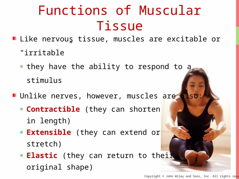
Like nervous tissue, muscles are excitable or
"irritable”
they have the ability to respond to a stimulus
Unlike nerves, however, muscles are also:
Contractible (they can shorten
in length)
Extensible (they can extend or
stretch)
Elastic (they can return to their
original shape)
Functions of Muscular Tissue

Copyright © John Wiley and Sons, Inc. All rights reserved.
Muscle makes up a large percentage of the body’s
weight
Their main functions are to:
Create motion – muscles work with nerves, bones,
and joints to produce body movements
Stabilize body positions and maintain posture
Store substances within the body using sphincters
Move substances by peristaltic contractions
Generate heat through thermogenesis
Functions of Muscular Tissue

Copyright © John Wiley and Sons, Inc. All rights reserved.
Three Types of Muscular Tissue

Copyright © John Wiley and Sons, Inc. All rights reserved.
(b) Cardiac muscle (c) Visceral smooth muscle
(a) Skeletal muscle
Three Types of Muscular Tissue

Copyright © John Wiley and Sons, Inc. All rights reserved.
Skeletal Muscle

Copyright © John Wiley and Sons, Inc. All rights reserved.
Skeletal Muscle

Copyright © John Wiley and Sons, Inc. All rights reserved.
Skeletal muscle fibers are very long “cells” -
next to neurons (which can be over a meter
long),
perhaps the longest in the body
The Sartorious muscle contains
single fibers that are at least
30 cm long
A single skeletal muscle fiber
Skeletal Muscle

Copyright © John Wiley and Sons, Inc. All rights reserved.
Sarcolemma
Motor neuron
Skeletal Muscle
The terminal processes of a
motor neuron in close proximity
to the sarcolemma of a skeletal
muscle fiber

Copyright © John Wiley and Sons, Inc. All rights reserved.
The epimysium, perimysium, and
endomysium all are continuous with
the connective tissues that form
tendons and ligaments (attach
skeletal muscle to bone) and muscle
fascia (connect muscles to other
muscles to form groups of muscles)
Organization of Muscle Tissue

Copyright © John Wiley and Sons, Inc. All rights reserved.
Organization of Muscle Tissue
Organization of a single muscle
belly
Epimysium
Perimysium

Copyright © John Wiley and Sons, Inc. All rights reserved.
Organization of a fasciculus
Organization of Muscle Tissue

Copyright © John Wiley and Sons, Inc. All rights reserved.
Organization of a muscle fiber
Organization of Muscle Tissue

Copyright © John Wiley and Sons, Inc. All rights reserved.
A muscle, a fasciculus, and a fiber all visualized
Organization of Muscle Tissue

Copyright © John Wiley and Sons, Inc. All rights reserved.
In groups of muscles the
epimysium continues to
become thicker, forming
fascia which covers
many muscles
This graphic shows the
fascia lata enveloping
the entire group of
quadriceps and
hamstring muscles in
the thing
Organization of Muscle Tissue

Copyright © John Wiley and Sons, Inc. All rights reserved.
Organization of Muscle
TissueMany large muscle
groups are encased
in both a
superficial
and a deep fascia
Real Anatomy, John Wiley and Sons

Copyright © John Wiley and Sons, Inc. All rights reserved.
Organization of Muscle TissueAn aponeurosis is
essentially a thick
fascia that connects
two muscle bellies.
This epicranial
aponeurosis connects
the muscle bellies of
the occipitalis and
the frontalis to form
“one” muscle: The
occipitofrontalis
Epicranial aponeurosisFrontal belly of
the occipitofrontalis m.

Copyright © John Wiley and Sons, Inc. All rights reserved.
Veins, arteries, and nerves are located in the
deep fascia between muscles of the thigh.
Organization of Muscle Tissue

Copyright © John Wiley and Sons, Inc. All rights reserved.
Beneath the connective tissue endomysium
is found the plasma membrane (called the
sarcolemma) of an individual skeletal
muscle fiber
The cytoplasm (sarcoplasm) of skeletal
muscle fibers is chocked full of
contractile proteins
arranged in myofibrils
The Skeletal Muscle Fiber

Copyright © John Wiley and Sons, Inc. All rights reserved.
You should learn the names of the internal
structures of the muscle fiber
Sarcolemma
Sarcoplasm
Myofibril
T-tubules
Triad (with
terminal cisterns
Sarcoplasmic reticulum
Sarcomere
The Skeletal Muscle Fiber

Copyright © John Wiley and Sons, Inc. All rights reserved.
The Skeletal Muscle Fiber
Increasing the level of magnification, the
myofibrils are seen to be composed
of filaments
Thick filaments
Thing filaments

Copyright © John Wiley and Sons, Inc. All rights reserved.
A scanning electron micrograph of a
sarcomere
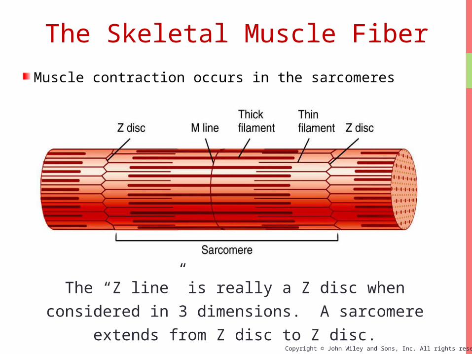
The basic functional unit of skeletal muscle fibers is the sarcomere: An arrangement of thick and thin filaments sandwiched between two Z discs
The Skeletal Muscle Fiber
Copyright © John Wiley and Sons, Inc. All rights reserved.
The “Z line” is really a Z disc when considered in
3 dimensions. A sarcomere extends from Z disc
to Z disc.
Muscle contraction occurs in the sarcomeres
The Skeletal Muscle Fiber
Copyright © John Wiley and Sons, Inc. All rights reserved.
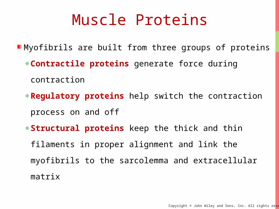
Myofibrils are built from three groups of proteins
Contractile proteins generate force during
contraction
Regulatory proteins help switch the contraction
process on and off
Structural proteins keep the thick and thin
filaments in proper alignment and link the
myofibrils to the sarcolemma and extracellular
matrix
Muscle Proteins
Copyright © John Wiley and Sons, Inc. All rights reserved.
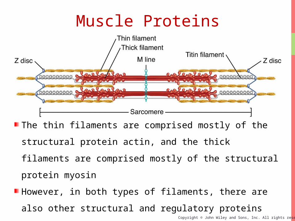
The thin filaments are comprised mostly of the
structural protein actin, and the thick filaments are
comprised mostly of the structural protein myosin
However, in both types of filaments, there are also
other structural and regulatory proteins
Muscle Proteins
Copyright © John Wiley and Sons, Inc. All rights reserved.
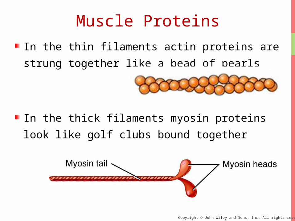
In the thin filaments actin proteins are strung
together like a bead of pearls
In the thick filaments myosin proteins look like
golf clubs bound together
Muscle Proteins
Copyright © John Wiley and Sons, Inc. All rights reserved.
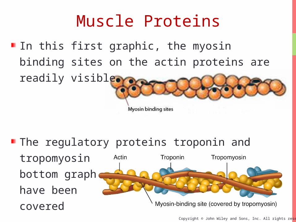
In this first graphic, the myosin binding sites on
the actin proteins are readily visible.
The regulatory proteins troponin and
tropomyosin have been added to the bottom
graphic: The myosin binding sites have been
covered
Muscle Proteins

Copyright © John Wiley and Sons, Inc. All rights reserved.
In this graphic the troponin-tropomyosin
complex has slid down into the “gutters” of the
actin molecule unblocking the myosin binding
site
The troponin-tropomyosin complex can slide
back and forth depending on the presence of
Ca2+
Myosin binding site exposed
Muscle Proteins

Copyright © John Wiley and Sons, Inc. All rights reserved.
Ca2+ binds to troponin which changes the shape of
the troponin-tropomyosin complex and uncovers
the myosin binding sites on actin
Muscle Proteins
Copyright © John Wiley and Sons, Inc. All rights reserved.
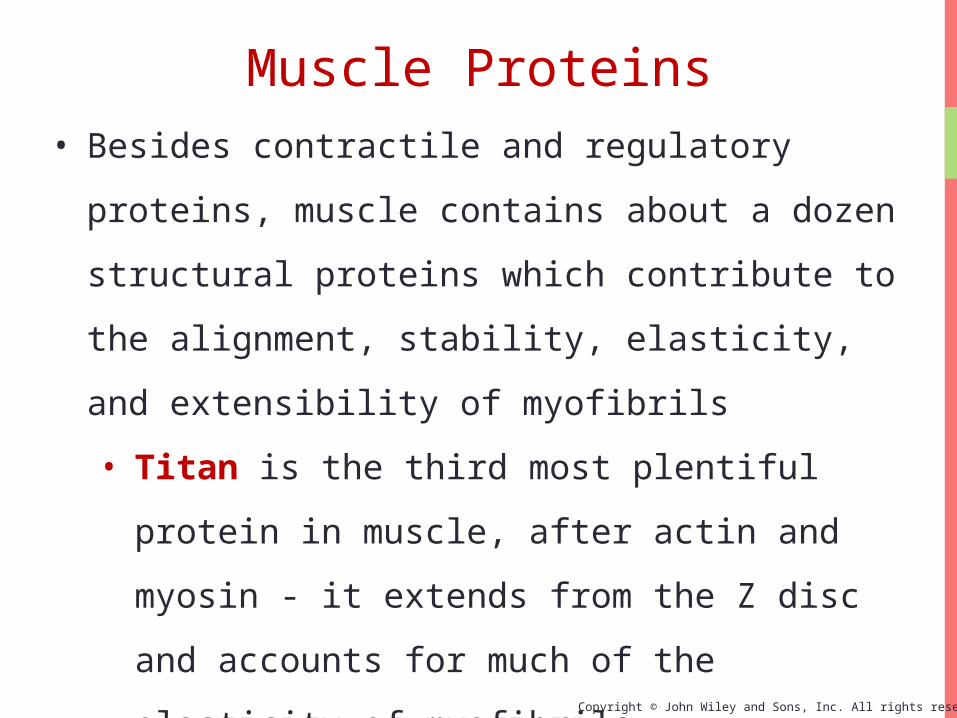
• Besides contractile and regulatory proteins,
muscle contains about a dozen structural
proteins which contribute to the alignment,
stability, elasticity, and extensibility of
myofibrils
• Titan is the third most plentiful protein in
muscle, after actin and myosin - it extends
from the Z disc and accounts for much of the
elasticity of myofibrils
• Dystrophin is discussed later as it relates to
the disease of muscular dystrophy
Muscle Proteins

Copyright © John Wiley and Sons, Inc. All rights reserved.
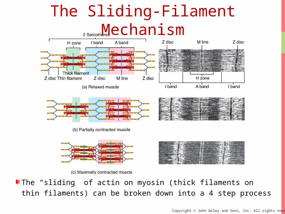
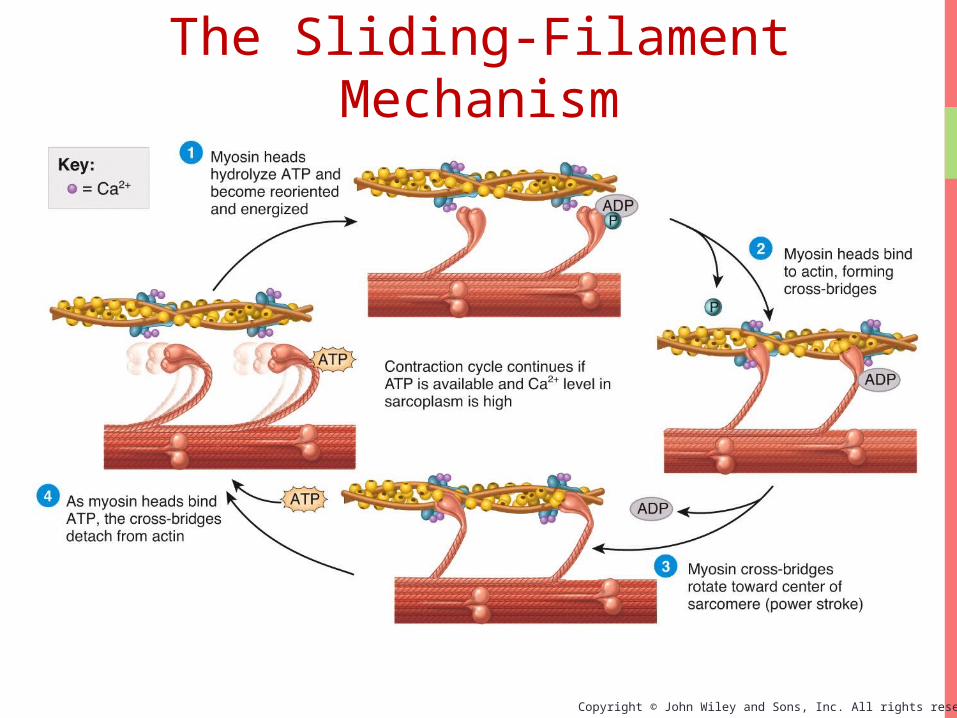
With exposure of the myosin binding sites on actin (the thin filaments)—in the presence of Ca2+ and ATP—the thick and thin filaments “slide” on one another and the sarcomere is shortened
The Sliding-Filament Mechanism
Copyright © John Wiley and Sons, Inc. All rights reserved.
The “sliding” of actin on myosin (thick filaments on thin filaments) can be broken down into a 4 step process
The Sliding-Filament Mechanism

Copyright © John Wiley and Sons, Inc. All rights reserved.
Step 1: ATP hydrolysis
Step 2: Attachment

Copyright © John Wiley and Sons, Inc. All rights reserved.
Step 3: Power Stroke
Step 4: Detachment
Copyright © John Wiley and Sons, Inc. All rights reserved.
The Sliding-Filament Mechanism

Copyright © John Wiley and Sons, Inc. All rights reserved.
Contraction and Movement Overview
Interactions Animation
Contraction and Movement
You must be connected to the internet to run this animation.

Copyright © John Wiley and Sons, Inc. All rights reserved.
Limited contact between actin and myosin
Compressed thick
filaments
Length-Tension RelationshipSarcomere shortening produces tension within a muscle
Copyright © John Wiley and Sons, Inc. All rights reserved.
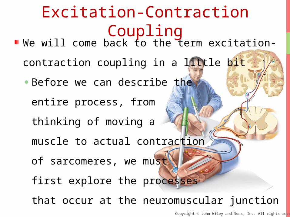
Excitation-Contraction CouplingWe will come back to the term excitation-
contraction coupling in a little bit
Before we can describe the
entire process, from
thinking of moving a
muscle to actual contraction
of sarcomeres, we must
first explore the processes
that occur at the neuromuscular junction

Copyright © John Wiley and Sons, Inc. All rights reserved.
Excitation-Contraction coupling (EC coupling) involves events at the junction between a motor neuron and a skeletal muscle fiber
Neuromuscular Junction

Copyright © John Wiley and Sons, Inc. All rights reserved.
An enlarged view of the neuromuscular junctionThe presynaptic membrane is on the neuron while the postsynaptic membrane is the motor end plate on the muscle cell. The two membranes are separated by a space, or “cleft”
Neuromuscular Junction
Copyright © John Wiley and Sons, Inc. All rights reserved.
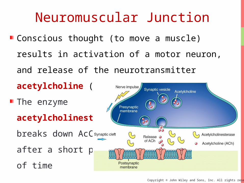
Conscious thought (to move a muscle) results in
activation of a motor neuron, and release of the
neurotransmitter acetylcholine (AcCh) at the
NM junction
The enzyme
acetylcholinesterase
breaks down AcCh
after a short period
of time
Neuromuscular Junction

Copyright © John Wiley and Sons, Inc. All rights reserved.
The plasma membrane on the “far side” of the NMJ
belongs to the muscle cell and is called the motor end
plate
The motor end plate is rich in chemical (ligand) - gated
sodium channels that respond to AcCh. Another way to
say this: The receptors for AcCh are on the ligand-gated
sodium channels on the motor end plate
Neuromuscular Junction

Copyright © John Wiley and Sons, Inc. All rights reserved.
The chemical events at the NMJ transmit the electrical events of a neuronal action potential into the electrical events of a muscle action potential
Neuromuscular Junction

Copyright © John Wiley and Sons, Inc. All rights reserved.
Neuromuscular JunctionInteractions Animation
Neuromuscular Junctions
You must be connected to the internet to run this animation.
Copyright © John Wiley and Sons, Inc. All rights reserved.
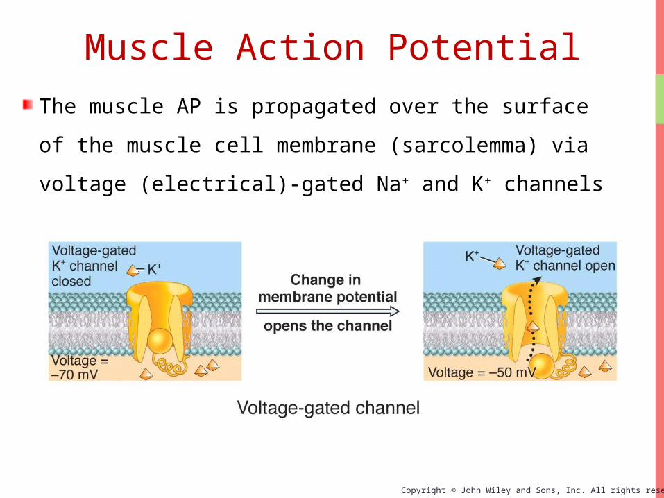
The muscle AP is propagated over the surface
of the muscle cell membrane (sarcolemma) via
voltage (electrical)-gated Na+ and K+ channels
Muscle Action Potential

Copyright © John Wiley and Sons, Inc. All rights reserved.
By placing a micropipette inside a muscle cell,
and then measuring the electrical potential
across the cell membrane, the phases of an
action potential
(AP) can be
graphed (as in this
figure)
Muscle Action Potential

Copyright © John Wiley and Sons, Inc. All rights reserved.
The behavior of the Na+
and K+ channels, at various
points in the AP, are seen
in this graphic
Na+ gates open during the
depolarization phase
K+ gates open during the
repolarization phase
Muscle Action Potential

Copyright © John Wiley and Sons, Inc. All rights reserved.
Generating An Action PotentialThe flow of ions through cell a membrane looks a lot
like a "piece" of electricity flowing through a wire
(but not as fast)
Generating an AP on the muscle membrane involves
the transfer of information from an electrical signal
(down the neuron), to a chemical signal (at the NMJ),
back to an electrical signal (depolarization of the
sarcolemma)
This added complexity (changing from electrical to
chemical back to electrical signals) provides
necessary control of the process

Copyright © John Wiley and Sons, Inc. All rights reserved.
Excitation-Contraction Coupling
Copyright © John Wiley and Sons, Inc. All rights reserved.
Excitation-Contraction Coupling
EC coupling involves putting it all together
The thought process going on in the brain
The AP arriving at the neuromuscular junction
The regeneration of an AP on the muscle
membrane
Release of Ca2+ from the sarcoplasmic reticulum
Sliding of thick on thin filaments in sarcomeres
Generation of muscle tension (work)

Copyright © John Wiley and Sons, Inc. All rights reserved.
Excitation-Contraction Coupling

Copyright © John Wiley and Sons, Inc. All rights reserved.
The brain
The motor neuron
Acetylcholine (ACh)
Acetylcholinesterase
enzyme
Ach receptors on the
motor endplate
Na+-K+ channels on the
sarcolemma
Na+ flow
K+ flow
Regenerate AP
The T-tubules
The SR
Ca2+ release
Troponin/
Tropomyosin
ATP
Myosin binding
Filaments slide
Muscles contract
Role Players in E-C
coupling
Excitation-Contraction Coupling

Copyright © John Wiley and Sons, Inc. All rights reserved.
Contraction of SarcomereInteractions Animation
Contraction of a Sarcomere
You must be connected to the internet to run this animation.

Copyright © John Wiley and Sons, Inc. All rights reserved.
Sources of Muscle EnergyStored ATP
3 seconds
Energy transferred from stored creatine
phosphate
12 seconds
Aerobic ATP production
Anaerobic glucose use
30-40 seconds

Copyright © John Wiley and Sons, Inc. All rights reserved.
Sources of Muscle Energy

Copyright © John Wiley and Sons, Inc. All rights reserved.
Sources of Muscle Energy

Copyright © John Wiley and Sons, Inc. All rights reserved.
Sources of Muscle Energy

Copyright © John Wiley and Sons, Inc. All rights reserved.
In a state of homeostasis, muscle use of
O2 and nutrients is balanced by the production
of manageable levels of waste products like
CO2
Heat - 70-80% of the energy used by
muscles is lost as heat - muscle activity is
important for maintaining body temperature
Lactic acid (anaerobic)
Skeletal Muscle Metabolism

Copyright © John Wiley and Sons, Inc. All rights reserved.
Oxygen Debt, or "Excess Post-Exercise Oxygen
Consumption" (EPOC) is the amount of O2
repayment required after exercise in skeletal
muscle to:
Replenish ATP stores
Replenish creatine phosphate and
myoglobin stores
Convert lactic acid back into pyruvate
so it can be used in the Krebs cycle to replenish
ATP
Skeletal Muscle Metabolism

Copyright © John Wiley and Sons, Inc. All rights reserved.
Skeletal Muscle Metabolism

Copyright © John Wiley and Sons, Inc. All rights reserved.
Muscle Metabolism
Muscle Metabolism
You must be connected to the internet to run this animation.
Copyright © John Wiley and Sons, Inc. All rights reserved.
Cardiac and Smooth Muscle Metabolism
In response to a single AP, cardiac muscle
contracts 10-15 times longer than skeletal
muscle, and must continue to do so, without rest,
for the life of the individual
To meet this constant demand, cardiac muscle
generally uses the rich supply of O2 delivered by
the extensive coronary circulation to generate
ATP through aerobic respiration

Copyright © John Wiley and Sons, Inc. All rights reserved.
Cardiac and Smooth Muscle Metabolism
Like cardiac muscle, smooth muscle (in your deep
organs) is autorhythmic and is not under
voluntary control (your heart beats and your
stomach digests without you thinking about it).
Unlike cardiac (and skeletal muscle) however,
smooth muscle has a low capacity for generating
ATP and does so only through anaerobic
respiration (glycolysis)

Copyright © John Wiley and Sons, Inc. All rights reserved.
Motor Unit is composed of a motor neuron
plus all of the muscle cells it innervates
High precision
• Fewer muscle fibers per neuron
• Laryngeal and extraocular muscles (2-20)
Low precision
• Many muscle fibers per neuron
• Thigh muscles (2,000-3,000)
The Motor Unit
Copyright © John Wiley and Sons, Inc. All rights reserved.
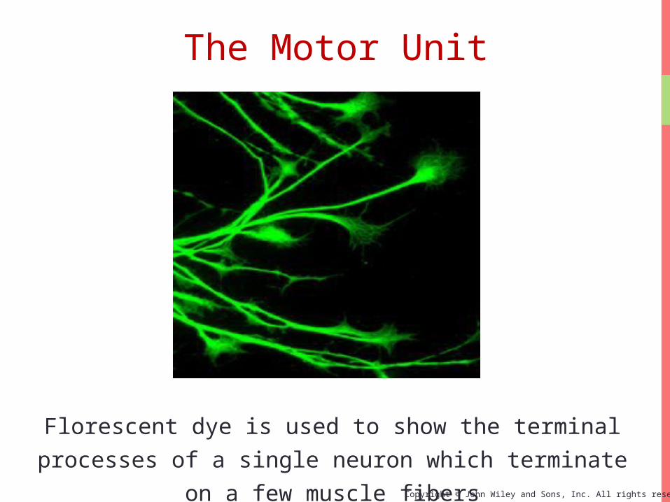
Florescent dye is used to show the terminal
processes of a single neuron which terminate on a
few muscle fibers
The Motor Unit
Copyright © John Wiley and Sons, Inc. All rights reserved.
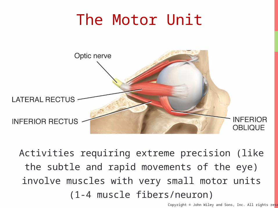
Activities requiring extreme precision (like the subtle
and rapid movements of the eye) involve muscles
with very small motor units (1-4 muscle fibers/neuron)
The Motor Unit
Copyright © John Wiley and Sons, Inc. All rights reserved.
All-or-none principle of muscle contraction
When an individual muscle fiber is stimulated to
depolarization, and an action potential is
propagated along its sarcolemma, it must
contract to it’s full force—it can’t partially
contract
Also, when a single motor unit is recruited to
contract, all the muscle fibers in that motor unit
must all contract at the same time
The Motor Unit

Copyright © John Wiley and Sons, Inc. All rights reserved.
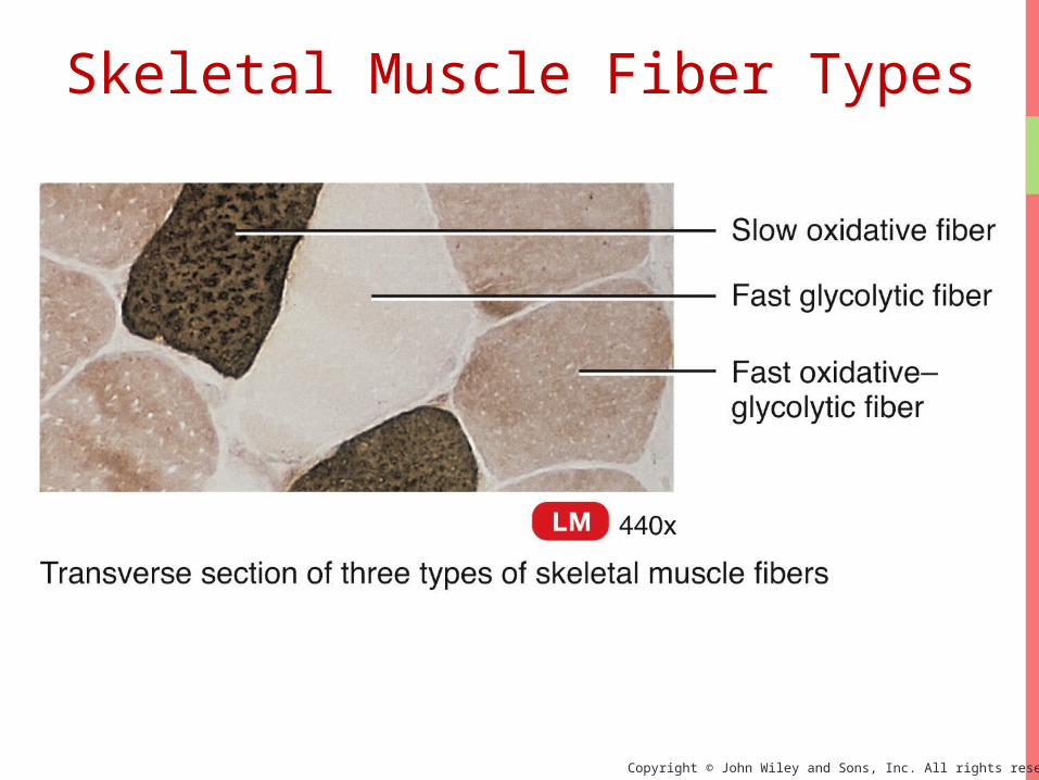
Skeletal muscle fibers are not all alike in appearance
or function. By appearance:
Red muscle fibers (the dark meat in chicken legs)
have a high myoglobin content, more mitochondria,
more energy stores, and a greater blood supply
White muscle fibers (the white meat in chicken
breasts) have less myoglobin, mitochondria, and
blood supply
Skeletal Muscle Fiber Types
Copyright © John Wiley and Sons, Inc. All rights reserved.
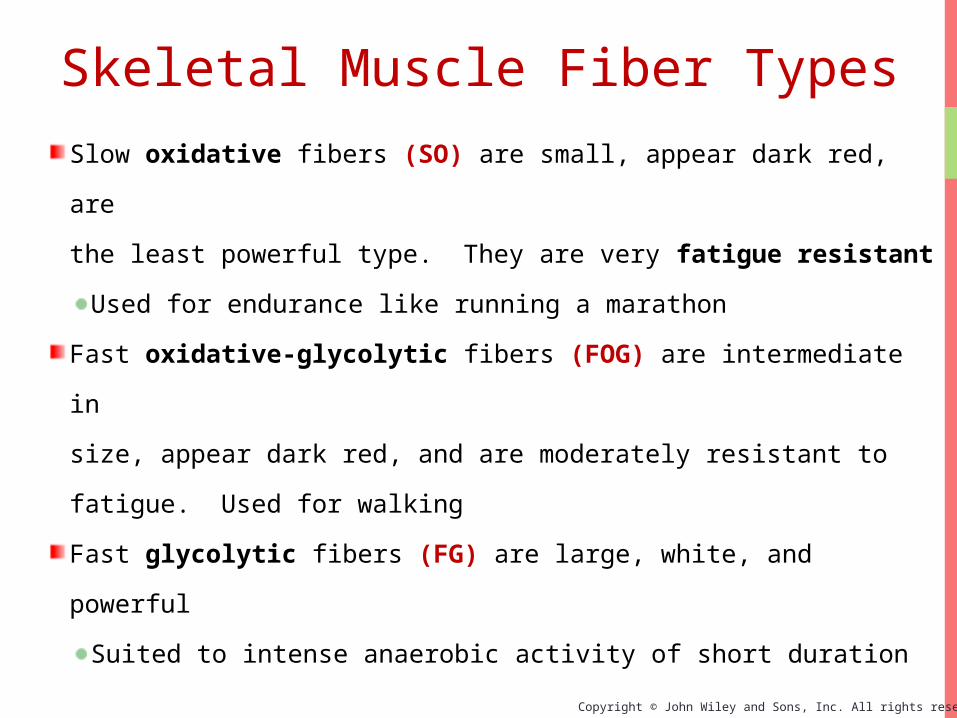
Slow oxidative fibers (SO) are small, appear dark red, are
the least powerful type. They are very fatigue resistant
Used for endurance like running a marathon
Fast oxidative-glycolytic fibers (FOG) are intermediate in
size, appear dark red, and are moderately resistant to fatigue.
Used for walking
Fast glycolytic fibers (FG) are large, white, and powerful
Suited to intense anaerobic activity of short duration
Skeletal Muscle Fiber Types
Copyright © John Wiley and Sons, Inc. All rights reserved.
Skeletal Muscle Fiber Types

Copyright © John Wiley and Sons, Inc. All rights reserved.
Most skeletal muscles are a mixture of all three types
of skeletal muscle fibers; about half the fibers in a
typical skeletal muscle are slow oxidative (SO) fibers
Within a particular motor unit all the skeletal
muscle fibers are the same type
The different motor units in a muscle are recruited
in a specific order depending on the task being
performed (fast anaerobic activity for maximal
force, etc.)
Skeletal Muscle Fiber Types

Copyright © John Wiley and Sons, Inc. All rights reserved.
There is a brief delay called the latent period as
the AP sweeps over the sarcolemma and Ca2+ ions
are released from the sarcoplasmic reticulum (SR)
During the next phase the fiber is actively
contracting
This is followed by relaxation as the Ca2+ ions are
re-sequestered into the SR and myosin binding
sites are covered by tropomyosin
Temporary loss of excitability is call the refractory
period – All muscle fibers in a motor unit will not
respond to a stimulus during this short time
Tension in a Muscle

Copyright © John Wiley and Sons, Inc. All rights reserved.
A twitch is recorded when a stimulus that results in
contraction (force) of a single muscle fiber is
measured over a very brief millisecond time frame
Tension in a Muscle

Copyright © John Wiley and Sons, Inc. All rights reserved.
Applying increased numbers of action potentials to a muscle fiber (or a fascicle, a muscle, or a muscle group) results in fusion of contractions (tetanus) and the performance of useful work
Tension in a Muscle
Copyright © John Wiley and Sons, Inc. All rights reserved.
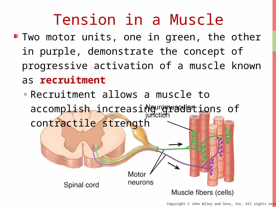
Two motor units, one in green, the other in purple, demonstrate the concept of progressive activation of a muscle known as recruitment
Recruitment allows a muscle to accomplish increasing gradations of contractile strength
Tension in a Muscle

Copyright © John Wiley and Sons, Inc. All rights reserved.
Muscle TensionInteractions Animation
Control of Muscle Tension
You must be connected to the internet to run this animation.

Copyright © John Wiley and Sons, Inc. All rights reserved.
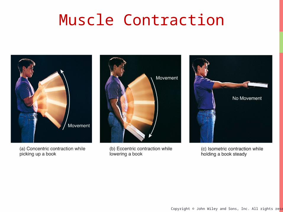
Muscle ContractionIsotonic contractions results in movement
Concentric isotonic is a type of muscle contraction
in which the muscle shorten while generating force
Eccentric isotonic is a contraction in which muscle
tension is less than the resistance (the muscle
lengthens)
Isometric contractions results in no movement
Muscle force and resistance are equal
Supporting objects in a fixed position and posture
Copyright © John Wiley and Sons, Inc. All rights reserved.
Muscle Contraction

Copyright © John Wiley and Sons, Inc. All rights reserved.
Exercise-induced muscle damage
After intense exercise electron micrographs
reveal considerable muscle damage including
torn sarcolemmas and disrupted Z-discs
Blood levels of proteins normally confined
only to muscle (including myoglobin and the
enzyme creatine kinase) increase as they are
released from damaged muscle
Imbalances of Homeostasis

Copyright © John Wiley and Sons, Inc. All rights reserved.
Spasm
A sudden involuntary contraction of a single
muscle within a large group of muscles – usually
painless
Cramp
Involuntary and often painful muscle contractions
Caused by inadequate blood flow to muscles (such
as in dehydration), overuse and injury, and
abnormal blood electrolyte levels
Imbalances of Homeostasis

Copyright © John Wiley and Sons, Inc. All rights reserved.
Disease States and Disorders
Fibrosis (myofibrosis)
Replacement of muscle fibers by excessive
amounts of connective tissues (fibrous scar
tissue)
Myosclerosis
Hardening of the muscle caused by calcification
Both myosclerosis and muscle fibrosis occur as a
result of trauma and various metabolic disorders
Imbalances of Homeostasis
Copyright © John Wiley and Sons, Inc. All rights reserved.
Aging
In part due to decreased levels of physical activity,
with aging humans undergo a slow, progressive loss
of skeletal muscle mass that is replaced largely by
fibrous connective tissue and adipose tissue
Muscle strength at 85 is about half that at age 25
Compared to the other two fiber types, the relative
number of slow oxidative fibers appears to increase
Imbalances of Homeostasis
Related Documents