CHANGESINSURFACEMORPHOLOGYOF CHINESEHAMSTEROVARYCELLS DURINGTHECELLCYCLE KEITHPORTER, DAVIDPRESCOTT,andJEARLFRYE FromtheDepartmentofMolecular,Cellular,andDevelopmentalBiology,Universityof Colorado,Boulder,Colorado 80302 ABSTRACT SynchronizedpopulationsofChinesehamsterovary(CHO)cellsinconfluentculture havebeenexaminedbyscanningelectronmicroscopyandtheirsurfacechangesnotedasthe cellsprogressthroughthecycle .DuringG,itischaracteristicforcellstoshowlargenumbers ofmicrovilli,blebs,andruffles.Exceptfortheruffles,thesetendtodiminishinprominence duringSandthecellsbecomerelativelysmoothastheyspreadthinlyoverthesubstrate . DuringG 2 microvilliincreaseinnumberandthecellsthickeninanticipationofrounding upformitosis .Itappearsthatthechangesobservedherereflectthechangingcapacityof CHOcellsduringthecycletorespondtocontactwithothercellsinthepopulation,because, asnotedinthesucceedingpaper(RubinandEverhart),CHOcellsinsparsenonconfluent culturesdonotshowthesamewiderangeofchangesduringthecellcycle .Normal,non- transformedcellsofequivalenttypeinconfluentcultureareessentiallydevoidofmicrovilli, blebs,andruffles .Therelationofthesesurfaceconfigurationstotheinternalstructureof thecellisdiscussed . INTRODUCTION Progressofacellthroughitslifecyclemustbe Thepresentstudyonthesurfacemorphology basedonacontinuumofcausallyrelatedcellular ofculturedcellswiththescanningelectron events .Thiscontinuumisreflectedintwomajor microscopeisintendedtoaddanotherviewto steps,DNAreplicationandnucleardivision,and thechangingcharacterofthecellthroughthe thesestepsallowthecellcycletobesubdivided courseofitslifecycle .Inparticular,wehave inthefour,well-knownperiods, GI, S,G2,and attemptedtorelatechangesinthesurfacestructure D.TheG,periodisgenerallyassumedtocontain ofcellstoknownstagesinthecycle .Forexample, thoseeventsthatleadtoandpreparethecell the"bubbling"ofthecellsurfaceduringmitosis forDNAreplication(Speriod) .TheG 2 period hasbeendescribedmanytimesfromobservations isbelievedtocontainpreparatorystepsformitosis oflivingcellsbymeansoftime-lapsephotography . (Mperiod) .Inaddition,avarietyofobservations RecentlyFoxetal .(1971)discoveredthatsites havemadeitclearthateventsoftheG,period onthecellsurfacethatbindcertainagglutinins areimportantfortheregulationofthecellcycle, aretransientlyexposedduringmitosis .Onewould sincecellsthatceasetoproliferate,eitherinhopeeventuallytodiscoverthecausalconnection vitroorinvivo,ordinarilydosobecauseofa betweensurfacechangesandspecificprocesses blocktotheirprogressthroughthe GI period . goingoninsidethecell .Inthisregard,anes- THEJOURNALOFCELLBIOLOGY • VOLUME57,1973 • pages 815 -836 815 on June 15, 2014 jcb.rupress.org Downloaded from Published June 1, 1973

Welcome message from author
This document is posted to help you gain knowledge. Please leave a comment to let me know what you think about it! Share it to your friends and learn new things together.
Transcript
CHANGES IN SURFACE MORPHOLOGY OF
CHINESE HAMSTER OVARY CELLS
DURING THE CELL CYCLE
KEITH PORTER, DAVID PRESCOTT, and JEARL FRYE
From the Department of Molecular, Cellular, and Developmental Biology, University ofColorado, Boulder, Colorado 80302
ABSTRACT
Synchronized populations of Chinese hamster ovary (CHO) cells in confluent culturehave been examined by scanning electron microscopy and their surface changes noted as thecells progress through the cycle. During G, it is characteristic for cells to show large numbersof microvilli, blebs, and ruffles. Except for the ruffles, these tend to diminish in prominenceduring S and the cells become relatively smooth as they spread thinly over the substrate .During G 2 microvilli increase in number and the cells thicken in anticipation of roundingup for mitosis. It appears that the changes observed here reflect the changing capacity ofCHO cells during the cycle to respond to contact with other cells in the population, because,as noted in the succeeding paper (Rubin and Everhart), CHO cells in sparse nonconfluentcultures do not show the same wide range of changes during the cell cycle . Normal, non-transformed cells of equivalent type in confluent culture are essentially devoid of microvilli,blebs, and ruffles. The relation of these surface configurations to the internal structure ofthe cell is discussed .
INTRODUCTION
Progress of a cell through its life cycle must be The present study on the surface morphologybased on a continuum of causally related cellular of cultured cells with the scanning electronevents . This continuum is reflected in two major microscope is intended to add another view tosteps, DNA replication and nuclear division, and the changing character of the cell through thethese steps allow the cell cycle to be subdivided course of its life cycle . In particular, we havein the four, well-known periods, GI, S, G2, and attempted to relate changes in the surface structureD. The G, period is generally assumed to contain of cells to known stages in the cycle . For example,those events that lead to and prepare the cell the "bubbling" of the cell surface during mitosisfor DNA replication (S period) . The G2 period has been described many times from observationsis believed to contain preparatory steps for mitosis of living cells by means of time-lapse photography .(M period) . In addition, a variety of observations Recently Fox et al. (1971) discovered that siteshave made it clear that events of the G, period on the cell surface that bind certain agglutininsare important for the regulation of the cell cycle, are transiently exposed during mitosis . One wouldsince cells that cease to proliferate, either in hope eventually to discover the causal connectionvitro or in vivo, ordinarily do so because of a between surface changes and specific processesblock to their progress through the GI period . going on inside the cell. In this regard, an es-
THE JOURNAL OF CELL BIOLOGY • VOLUME 57, 1973 • pages 8 1 5 -836
815
on June 15, 2014jcb.rupress.org
Dow
nloaded from
Published June 1, 1973
pecially important problem is the apparent re-lationship between the cell surface and the regula-tion of cell reproduction in density-dependentinhibition of cell reproduction and the alterationof this relationship in transformed cells . The ex-tent to which contact inhibition is reflected inrelatively gross changes in the surface morphologyshould emerge from these studies with greaterclarity than heretofore.Furthermore, it has become obvious that for
the detailed exploration of changes in surfacemorphology and cell shape associated with thecell cycle, the scanning electron microscope is amost valuable tool.
METHODS
All observations were made on a line of Chinesehamster ovary (CHO) cells obtained from Dr .Donald Petersen who got them originally from T . T.Puck. The cells were cultured in Ham's F-12 medium(North American Biologicals, Inc ., Rockville, Md.)supplemented with 10% fetal calf serum (FlowLaboratories, Inc., Rockville, Md.) . Cells weregrown in glass vessels and subcultured weekly, usinga solution of 0.5% trypsin and 0.1% EDTA tostrip cell monolayers from the glass surface . Regularchecks for pleuropneumonia-like organisms havealways been negative .
Synchronous populations were obtained by themitotic shake-off method (Tobey et al ., 1967). Themitotic index in the shake-off populations used inthese experiments was particularly high, rangingfrom 98 to 99.5%.
The mitotic cells were planted on cover glassescoated with a thin (100 A) carbon film . Cells werefixed at 1, 3, and 5 h after shake-off to provide early,middle, and late G1 cells. Samples of these cells wereexposed to [ 3 H]thymidine for 15 min before fixingand then autoradiographed . These samples allowedus to assess, by light microscopy, whether cells hadcompleted GI and entered the S period .
Because of the variability in the length of G1phase in populations of cultured cells, the mitoticcells used to provide cells in the S phase were firstresynchronized by a single thymidine block (Gala-vazi et al., 1966) . Mitotic populations on coverglasses were exposed to 5 mM thymidine for 10 hafter planting . This is sufficient time for about 95%of the cells to complete the G1 period and reachthe beginning of the S period. The blocked cellswere released by removal of the thymidine with achange of the medium .
Samples were fixed at 1, 3, and 4 h after release ofthe block to provide cells in early, middle, and late S .A sample was fixed at 5 h after release of the block
816
THE JOURNAL OF CELL BIOLOGY • VOLUME 57, 1973
to provide cells in the G2 period .' Again, at eachfixing time samples of cells were exposed to [ 3 H]thy-midine for the final 15 min before fixing in order toassess how many cells were in the S period in any ofthe samples. The fixations were timed on the basis ofpreviously published values (Bostock et al ., 1971)for the length of the cycle subsections for this sameline of cells.
Scanning Microscopy-Methods
The experiments were repeated twice and obser-vations on populations from both experiments wereessentially identical .
Cells for microscopy were rinsed (twice, 2 min each)free of culture media with warm balanced salt solu-tion (Puck's saline G) and then exposed to 2% glu-taraldehyde in 0.1 M s-collidine buffer at pH 7 .1 .After 0.5 h in this they were rinsed twice with warmPuck's saline G for a total of 4 min and postfixed in1 % Os0 4 in 0.3 M s-collidine for 10 min. A briefrinse (5 min) in distilled, deionized H20 was fol-lowed by dehydration with ethyl alcohol of increasingconcentration up to 95%. From here the preparationswere carried through a graded series of amyl acetatein alcohol up to 100% amyl acetate .' Thence theywere transferred into liquid CO2 in a critical-pointdrying apparatus and, after several rinses in C02,the temperature of the closed system was raised to45°C according to the critical-point method of T. F.Anderson (1951) .
The resulting dry specimens were lightly coatedwith carbon and then with gold-palladium andwere ready for microscopy .
A Kent Cambridge S-4 scanning electron micro-scope, operated at 20 kV, was used in all of thesestudies.
OBSERVATIONS
Assessment of Cell Cycle Synchrony
The mitotic index of the populations used forthe electron microscope observations rangedfrom 98 to 99.5 0/0 , and the initial synchrony wasaccordingly excellent . The decay in synchronythrough the ensuing G 1 period was measured by
I The loss of synchrony in the 5 h sample was sub-stantial and interpretations of the results with thescanning microscope are therefore somewhat lessclear. In this sample 58% of the cells were still inDNA replication, 10% were in mitosis, and theremaining 32% were in G2 .2 It has since been found that cells dehydrated inacetone can be taken directly into CO2 thus avoidingthe amyl acetate step in the above procedure .
on June 15, 2014jcb.rupress.org
Dow
nloaded from
Published June 1, 1973
NNNLCNQz0C
100 -
A
10-
0-
i 1 10 1 2 3 4 5
0 1 2 3 4 5
time in hours
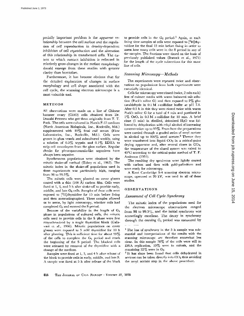
FIGURE 1 A Mitotic cells were collected by the shake-off method and then fixed at 1, 3, or 5 h to providecells in early, middle, and late G1 . 9% of the cells had entered S by 3 h, and 30% had entered S by 5 h .
FIGURE 1 B Mitotic cells were collected and cultured for 10 h in the presence of 5 mM of thymidine toblock DNA synthesis. The cells were released from the thymidine block by washing, and samples werefixed at 1, 3, and 4 h to provide cells in early, mid-late, and late S phase . The sample fixed at 5 h afterrelease from thymidine was composed of 58% late S, 32%c G2, and 10% mitotic cells .
following with time the percentage of cells thatentered DNA replication, as shown in Fig . I A.All of the cells fixed at 1 h were in G1 , and es-sentially all of these could be safely assumed tobe in early G1 . The population fixed at 3 h con-sisted of 9 % S cells and 91 % G I cells and wasaccordingly accepted as a mixture of middle andlate GI stages . The population at 5 h was assumedto consist of equal parts of middle G1 , late G1 ,and early S-phase cells.
The resynchronization of cells at the G 1 -to-Stransition by blocking with 5 mM thymidine for10 h after shake-off led to the cell populationsdescribed in Fig . 1 B . 92 % of the cells fixed at Ih after removal of the thymidine block were inthe early part of the S phase . 99% of the cellsfixed at 3 h after release of the block were in themiddle to late S phase, and the population fixed at4 h represented a slightly later stage of S . Thesample fixed at 5 h after the block consisted of32% G 2 cells, 58% late S-phase cells, and 10%mitotic cells.
In summary, we have highly uniform popula-tions of cells in early G 1, and in early, middle, andlate S. The populations intended to representmiddle and late G1 are less uniform, while the G2
B
O
--o-o-o-o MITOTIC CELLSI
population is in fact a mixture of late S cells,G2 cells, and mitotic stages .
MicroscopyIn other studies of cultured cells with the
scanning microscope we have established to oursatisfaction that critical-point drying of thesespecimens after glutaraldehyde-osmium fixationprovides the observer with a specimen that isfaithful to the original (Porter et al ., 1972). Thecells are far less distorted than by drying fromwater or even from solvents with lower surfacetensions. Nitrous oxide or freon (Freon 13) usedin the same way as CO2 gives results that aresimilar but not better for most purposes .
Except for some shrinkage caused by dehydra-tion in alcohol, the cells showed little evidence ofdistortion . Comparison made by phase-contrastmicroscopy and Nomarski optics of cells beforeand after fixation failed to reveal any other altera-tions .
CHANGES IN MORPHOLOGY WITH PROGRESSTHROUGH THE G1 PHASE OF THE
CELL CYCLE
EARLY GI : Cells fixed I h after shake-off.As expected, the majority of the cells in these
KEITH PORTER, DAVID PRESCOTT, AND ,IEARL FRYE Surface Morphology and the Cell Cycle
817
on June 15, 2014jcb.rupress.org
Dow
nloaded from
Published June 1, 1973
preparations are in very late stages of cytokinesis .Nearly all of them are therefore still rounded upand associated in pairs (Fig. 2) . They are quiteuniform in diameter at 12-13 µm .
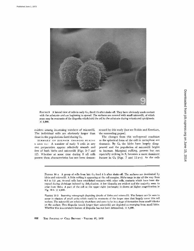
It is evident in the lateral view in Fig . 9 thatthey have attached to the carbon-coated coverglass and have begun to spread over this as sub-strate . It is apparent also in this same micrographthat the cell surfaces are covered with numerousmicrovilli and other small projections .
These same preparations contained a number ofsmooth spheres (2-4 µm) which presumablyrepresent fragments of the cells that have brokenaway during mitosis . Their significance, if any,is not clear. A residue of these spheres persistsinto the middle GI preparations where they ap-pear in a somewhat shriveled form (Fig . 3) .MIDDLE G I : Cells fixed 3 h after shake-off.By this time the cells have obviously lost their
spherical form and some appear almost fully ex-tended (Fig . 3) . The visible (upper) surface iscovered with small spherical blebs intermingledwith numerous microvilli (see below). The ma-jority of the cells is still associated in pairs (seealso Fig . 11 a) with adjacent edges representingthe plane of earlier cleavage . There is some evi-dence of ruffling at the free edges, but in thisearly phase it is not striking .LATE GI : Cells fixed 5 h after shake-off.The cells here (Fig. 4) show a substantially
greater variation in apparent size and form thanwas evident earlier in G I . A few units are thinlyspread and, judging from observations on cellsknown to be in S, these have probably entered theS phase of the cell cycle . Other cells are thickerand less extended over the substrate as thoughstill in middle GI . Sister cells, still closely associ-ated as pairs, are common . Ruffling has increasedin prominence but is confined to margins not incontact with other cells . The surface blebs persiston many cells, and of these some are in intimatecontact with sister units .
818
SUMMARY OF SURFACE CHANGES DURINGGI : At the end of cytokinesis the cells are typi-cally covered with microvilli and other equallysmall protuberances (Fig. 9) . The surface blebsmentioned above are not prominent or evenclearly evident, the greater number appearinglater .
3 h after shake-off the exposed surfaces arecovered with microvilli and blebs (Figs . 3, 10 a,and 10 b) . The latter tend to be concentratedover the central thicker parts of the cell whereasthe microvilli are more widely distributed. Theblebs, which occasionally appear in clusters (Figs .10 b, 11 b, and 11 c), vary in size, some being assmall as 0.2 µm in diameter and others as large as1 .5 µm .The microvilli are small by comparison meas-
uring only about 0.1 µm in diameter . Theirlengths vary greatly, but the majority are in thevicinity of 1 .5 Am (Figs. 11 b and 11 c) . Many ofthe microvilli emerge from the edges of broadflat excrescences in the surface (Figs . 10 b, 11 b,and 11 c) .By the end of G I (5 h after shake-off) the cells
begin to show ruffles (or lamellipodia) at theirfree edges, i .e., along those parts of the marginnot in contact with other cells (Fig . 4) . To alarge extent this restriction in the distribution ofthese extraordinary structures is constant .
Ruffles adopt a variety of appearances . Mostcommonly they comprise thin folds extendingabove the surface as much as 6 µm and havinga thickness equal to the diameter of the micro-villi (0 .1 µm) . In the living cell they have beenobserved to surround and engulf small quantaof the medium (Gey, 1955) (Figs . 13 a-c) .
Toward the end of GI the cells show a widervariation in their surface characteristics thanhad been apparent earlier . Some, for example, aretotally devoid of blebs whereas others show almostas many as at any previous time in GI (Figs .10 a and 11 a) . The number of microvilli tends
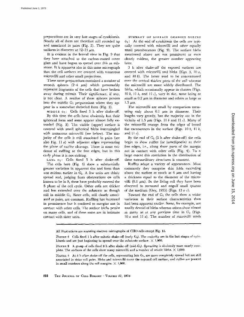
All illustrations are scanning electron micrographs of CHO cells except Fig . 15.
FIGURE 2 Cells fixed 1 h after mitotic shake-off (early G I ) . The majority are in the last stages of cyto-kinesis and are just beginning to spread over the substrate surface . X 1,000 .
FIGURE 3 A group of cells fixed 3 h after shake-off (mid-GI) . Spreading is obviously more nearly com-plete . The surfaces of the cells show many microvilli and a number of zeiotic blebs . X 1,000 .
FIGURE 4 At 5 h after shake-off the cells, representing late GI, are more completely spread but are stillassociated in sister cell pairs . Blebs and microvilli cover the exposed cell surface, and ruffles are presentin small numbers along the cell margins . X 1,000.
THE JOURNAL OF CELL BIOLOGY . VOLUME 57, 1973
on June 15, 2014jcb.rupress.org
Dow
nloaded from
Published June 1, 1973
KEITH PORTER, DAVID PRESCOTT, AND JEARL FRYE Surface Morphology and the Cell Cycle
819
on June 15, 2014jcb.rupress.org
Dow
nloaded from
Published June 1, 1973
to vary inversely with the number of blebs . Sincethe number and prominence of blebs tends todiminish in S and G 2i it is assumed that thosecells showing few, if any, of these structures havemoved into the S phase of the cycle. This as-sumption is supported by the autoradiographicdata .
The relationship of blebs to microvilli, if any,is difficult at this point to define . There is, how-ever, another type of surface extension, a verylong and slender filopodium which seems fre-quently to extend from a bleb (Figs. 10 a and10 b) . These filopodia appear usually to link cellwith cell or, cell with substrate . They are 50-100nm in diameter and may be several micrometersin length. They, or very similar cell extensions,have been observed, in light microscopy of livingcultured cells, to grow quickly, wave around, andfinally make contact as indicated (Gey, 1955 ;Taylor, 1966) .
BEHAVIOR OF CELLS AFTER RESYNCHRONIZATIONWITH THYMIDINE
EARLY S : Cells fixed l h after release from a10 h block with thymidine .
By this time the cells have enlarged noticeablyover those harvested in G1. To some extent thismay reflect the fact that some of them havespread more thinly, but in part it probably resultsfrom growth and increase in cell volume (Fig . 5) .The cells adopt highly variable shapes and seemnot to show any preferred form. The more thinlyspread cells have noticeably fewer microvilli (Figs.5 and 12) .
820 THE JOURNAL OF CELL BIOLOGY - VOLUME 57, 1973
MID-LATE S : Cells fixed 3 h after releasefrom 10 h block .
As the cycle progresses toward G2 the cellsspread more thinly and become essentially epi-thelioid. Frequently, as illustrated in Fig . 6, theyappear in micro colonies and reach confluency .Generally, cell margins in contact show no ruffling(Figs . 6 and 7), but one exception to this rule isevident at the arrow in Fig. 6 (see also Fig . 12) .The exposed surfaces of the cells show only a fewblebs and the majority of the cells are completelydevoid of these structures . It is worthy of notethat the cell at A in Fig. 6 is in contact with ad-jacent cells and is exceptional in showing a num-ber of blebs .LATE S : Cells fixed 4 h after release from
10 h block.A comparison of these cells with those earlier
in S reveals three well-defined changes . Here thecells have thickened and are withdrawing theirmargins to some extent (Fig . 7) . There is in-creasing evidence of ruffling at their edges asthe cells adopt a more independent existence .The surface blebs so evident in G I have essentiallydisappeared .
G2 : Cells fixed 5 h after release from 10 hblock.
By this time a few of the cells have rounded upand entered mitosis (at arrow, Fig . 8) . Others inthe population are thickening and sending outslender filopodia which apparently anticipatethe large number found on the dividing cell(Fig. 14) . Ruffling is prominent along the free,uncontacted edges and, rarely, a few blebs are
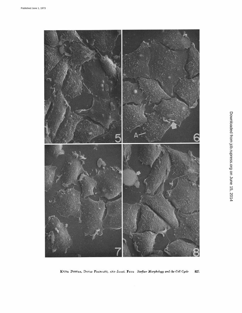
FIGURE 5 The cells here are predominantly in early S . Some are extensively spread and are relativelyfree of microvilli and other surface features. Ruffling at the cell margins is present but not to the extentdisplayed by cells in Figs. 7 and 8 . Other cells, possibly still in GI, show an abundance of microvilli andblebs . X 1,000.
FIGURE 6 The population here is in the S phase of the cycle . The surface blebs are on the wane in fre-quency though microvilli are still fairly prominent . Contact between the cells may have reduced theruffling except at the edges of the colony . X 1,000 .
FIGURE 7 Here the cells are in late S or early G2. They seem to be adopting an independent existencethough some of the separation between them may be a result of shrinkage during dehydration . Thecell margins are extraordinarily active in terms of ruffling . It is also evident that the cells are thickeningin comparison with those shown in Figs . 5 and 6 . X 1,000.
FIGURE 8 The majority of the cells are beginning to pull in their thinner margins in preparation for therounding up characteristic of mitosis . One cell at the upper left has already gone into prophase . Therest are in late G2 . Microvilli and ruffles are common . X 1,000 .
on June 15, 2014jcb.rupress.org
Dow
nloaded from
Published June 1, 1973
KEITH PORTER, DAVID PRESCOTT, AND JEARL FRYE Surface Morphology and the Cell Cycle
821
on June 15, 2014jcb.rupress.org
Dow
nloaded from
Published June 1, 1973
evident among increasing numbers of microvilli .The individual cells are obviously larger thanthose in the populations fixed during G I .
SUMMARY OF SURFACE CHANGES DURINGS AND G2 : A number of early S cells in anyone preparation appear relatively smooth andfree of both blebs and microvilli (Figs . 5-7 and12). Whether at some time during S all cellspossess these characteristics has not been demon-
822
FIGURE 9 A lateral view of cells in early GI, fixed 1 h after shake-off . They have obviously made contactwith the substrate and are beginning to spread . The surfaces are covered with small microvilli, of whichsome may be remnants of the filopodia which hold the cell to the substrate during mitosis and cytokinesis .X 2,300 .
strated by this study (but see Rubin and Everhart,the succeeding paper) .
The changes from this well-spread conditionto the spherical form of the cell in metaphase aredramatic. By G2 the blebs have largely disap-peared and the population of microvilli beginsto increase . Marginal ruffling, present but notespecially striking in S, becomes a more dominatefeature in G 2 (Figs . 7 and 13 a-c) . As the cells
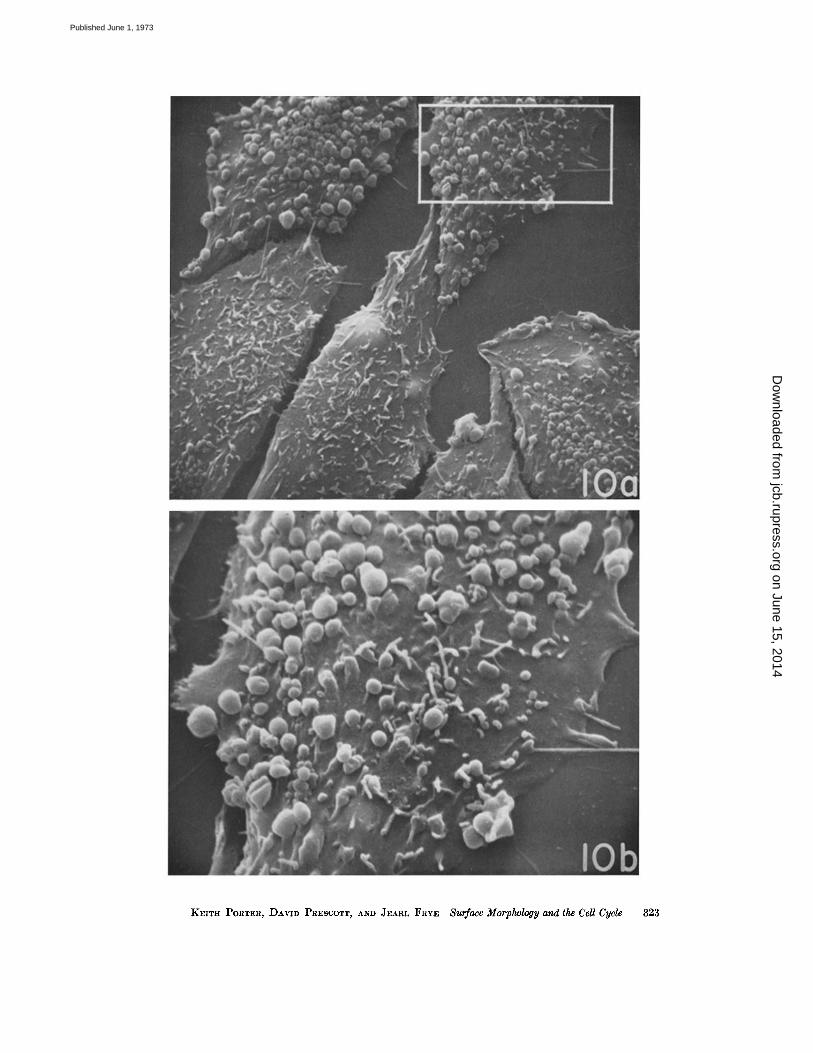
FIGURE 10 a A group of cells from late GI fixed 5 h after shake-off. The surfaces are dominated byblebs and microvilli. A little ruffling is appearing at the cell margins . Blebs range in size all the way from0.2 to 1 .5 fxm . Several cells have established contacts with other cells, contacts which have been dis-rupted during shrinkage induced by dehydration . A few filopodia are evident and the majority seem toarise from blebs. A part of the cell at the upper right (rectangle) is shown at higher magnification inFig . 10 b . X 2,600.
FIGURE 10 b Scanning micrograph depicting details of blebs and microvilli . The former can be seen tooccur in clusters of small units which could be remnants of the larger ones that largely cover this cellsurface . The microvilli are relatively short here and seem to be in a stage of formation from small hillockson the surface. Two filopodia (much longer than microvilli) are depicted as emerging from small blebs .Whether this is a constant feature of filopodia has not been determined . X 6,500.
THE JOURNAL OF CELL BIOLOGY - VOLUME 57, 1973
on June 15, 2014jcb.rupress.org
Dow
nloaded from
Published June 1, 1973
KEITH PORTER, DAVID PRESCOTT, AND JEARL FRYE Surface Morphology and the Cell Cycle
823
on June 15, 2014jcb.rupress.org
Dow
nloaded from
Published June 1, 1973
achieve their maximum size and begin to thickenin G2, the filopodia which characterize the di-viding cell first appear and they increase greatlyin number as the cell enters mitosis (Figs . 8 and14) .
Ruffles are present on free edges of the cellsbut seem to be excluded from margins in intimate(or even less than intimate) contact with othercells . Occasionally, as in Fig. 12, they encroachon the zone of contact, or even invade it (arrow,Fig. 6), but these occurrences are rare .
Some of the contacting cell margins in thesepreparations have been pulled part by shrinkageof the cell bodies which probably takes placeduring dehydration (e .g., in Figs . 4, 7, and 11 a) .Where this has not happened one is able toexamine the contacts in some detail . In some in-stances they show the involvement of small ex-tensions of one cell slightly overlapping the surfaceof the other (Figs . 6 and 12), but this is not afeature in all instances .
There is likewise no easily recognized uni-formity in the ruffles except that they tend tohave minimal thickness consisting of two plasmamembranes and two internally opposed cyto-plasmic cortices (Fig . 13 a) (Abercrombie et al.,1971) . In stereo images (Figs . 13 b and 13 c)they can be seen to extend more or less erectlyfrom the surface to heights measured at 6 µm .
The filopodia which attach the dividing cellto the substrate during mitosis are unique inseveral respects (Fig . 14). They may be verylong, as much as two to three times the diameterof the cell and they frequently arborize at their tip
8 2 4 THE JOURNAL OF CELL BIOLOGY . VOLUME 57, 1973
ends . ,' In these respects these particular extensionsof the surface are distinctive and dissimilar tofilopodia that appear in fewer numbers at otherphases of the cycle .
THE SURFACES OF CELLS FROM PRIMARYEXPLANTS OF CHINESE HAMSTER LUNG
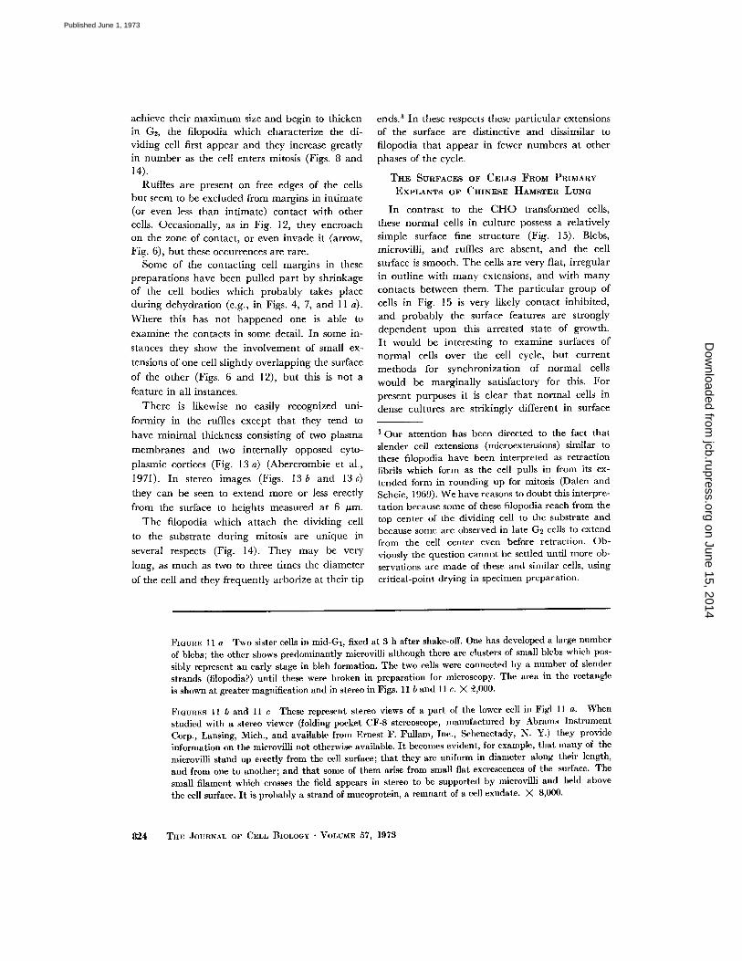
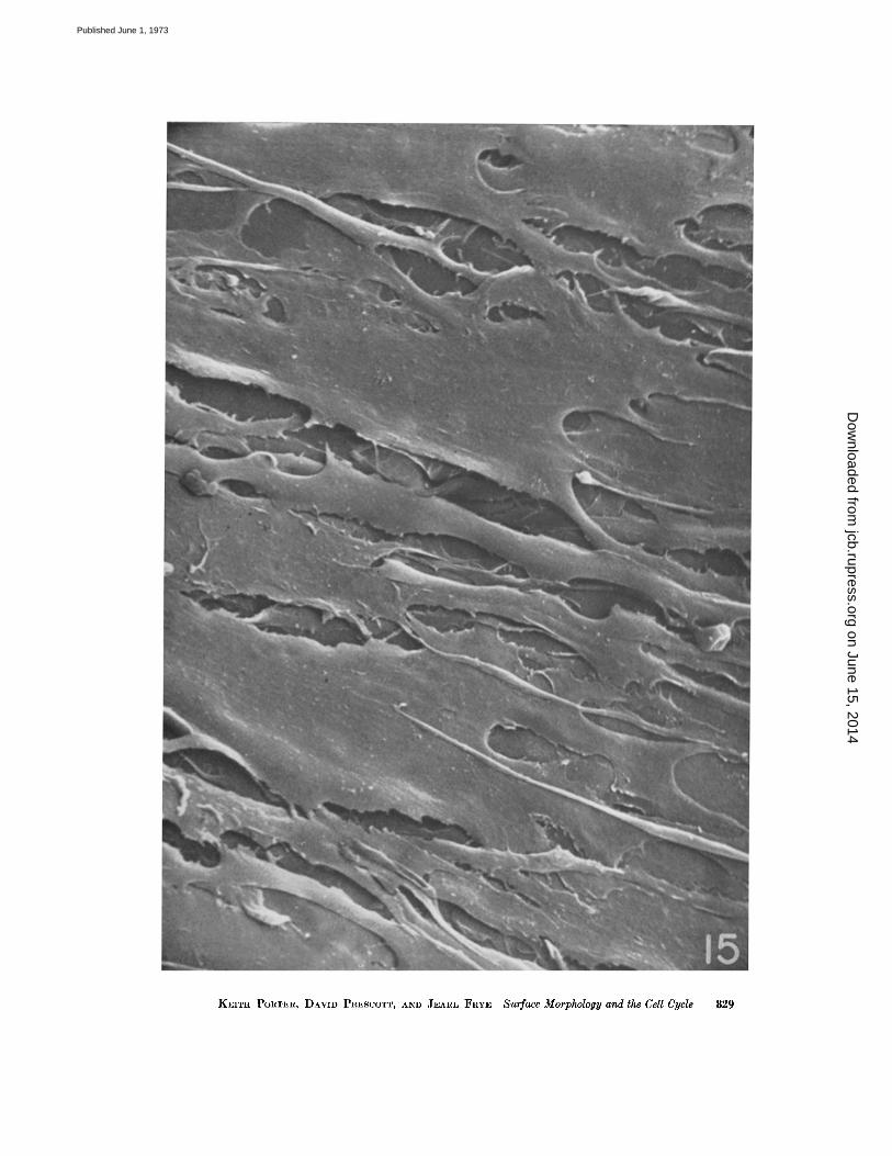
In contrast to the CHO transformed cells,these normal cells in culture possess a relativelysimple surface fine structure (Fig. 15) . Blebs,microvilli, and ruffles are absent, and the cellsurface is smooth . The cells are very flat, irregularin outline with many extensions, and with manycontacts between them. The particular group ofcells in Fig. 15 is very likely contact inhibited,and probably the surface features are stronglydependent upon this arrested state of growth .It would be interesting to examine surfaces ofnormal cells over the cell cycle, but currentmethods for synchronization of normal cellswould be marginally satisfactory for this . Forpresent purposes it is clear that normal cells indense cultures are strikingly different in surface
I Our attention has been directed to the fact thatslender cell extensions (microextensions) similar tothese filopodia have been interpreted as retractionfibrils which form as the cell pulls in from its ex-tended form in rounding up for mitosis (Dalen andScheie, 1969) . We have reasons to doubt this interpre-tation because some of these filopodia reach from thetop center of the dividing cell to the substrate andbecause some are observed in late G2 cells to extendfrom the cell center even before retraction. Ob-viously the question cannot be settled until more ob-servations are made of these and similar cells, usingcritical-point drying in specimen preparation .
FIGURE 11 a Two sister cells in mid-GI, fixed at 8 h after shake-off . One has developed a large numberof blebs ; the other shows predominantly microvilli although there are clusters of small blebs which pos-sibly represent an early stage in bleb formation . The two cells were connected by a number of slenderstrands (filopodia?) until these were broken in preparation for microscopy. The area in the rectangleis shown at greater magnification and in stereo in Figs . 11 b and 11 c . X 2,000 .
FIGURES 11 b and 11 c These represent stereo views of a part of the lower cell in Figl 11 a . Whenstudied with a stereo viewer (folding pocket CF-8 stereoscope, manufactured by Abrams InstrumentCorp., Lansing, Mich., and available from Ernest F . Fullam, Inc ., Schenectady, N . Y.) they provideinformation on the microvilli not otherwise available. It becomes evident, for example, that many of themicrovilli stand up erectly from the cell surface ; that they are uniform in diameter along their length,and from one to another ; and that some of them arise from small flat excrescences of the surface . Thesmall filament which crosses the field appears in stereo to be supported by microvilli and held abovethe cell surface. It is probably a strand of mucoprotein, a remnant of a cell exudate . X 8,000 .
on June 15, 2014jcb.rupress.org
Dow
nloaded from
Published June 1, 1973
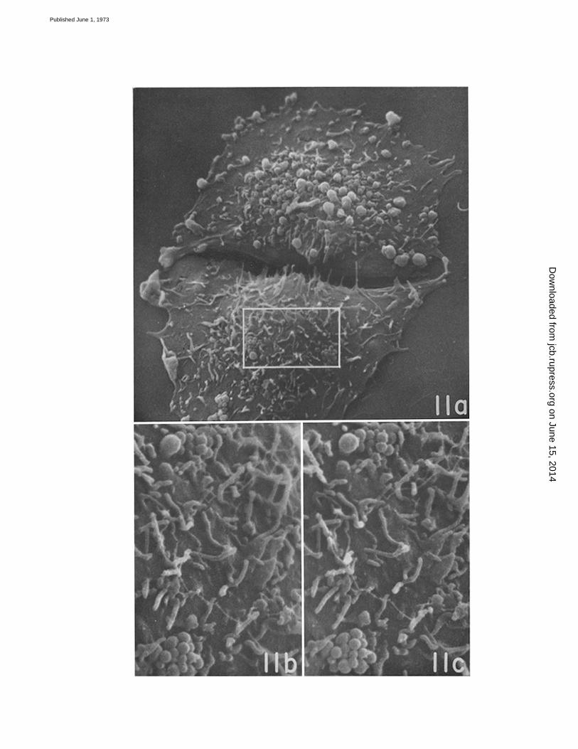
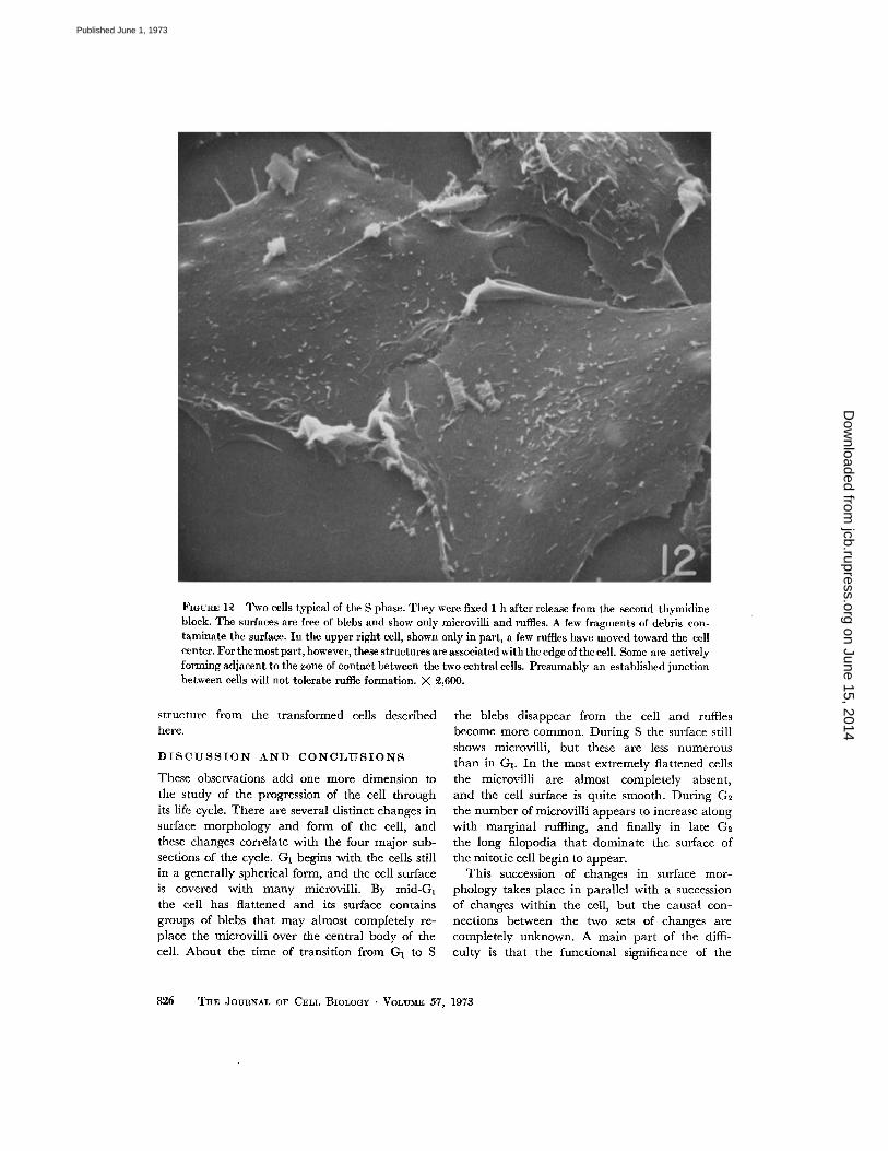
FIGURE 12 Two cells typical of the S phase . They were fixed 1 h after release from the second thymidineblock . The surfaces are free of blebs and show only microvilli and ruffles . A few fragments of debris con-taminate the surface. In the upper right cell, shown only in part, a few ruffles have moved toward the cellcenter . For the most part, however, these structures are associated with the edge of the cell . Some are activelyforming adjacent to the zone of contact between the two central cells . Presumably an established junctionbetween cells will not tolerate ruffle formation. X 2,600 .
structure from the transformed cells described the blebs disappear from the cell and ruffleshere . become more common . During S the surface still
shows microvilli, but these are less numerousthan in GI . In the most extremely flattened cellsthe microvilli are almost completely absent,and the cell surface is quite smooth . During G2the number of microvilli appears to increase alongwith marginal ruffling, and finally in late G2the long filopodia that dominate the surface ofthe mitotic cell begin to appear .
This succession of changes in surface mor-phology takes place in parallel with a successionof changes within the cell, but the causal con-nections between the two sets of changes arecompletely unknown. A main part of the diffi-culty is that the functional significance of the
DISCUSSION AND CONCLUSIONS
These observations add one more dimension tothe study of the progression of the cell throughits life cycle . There are several distinct changes insurface morphology and form of the cell, andthese changes correlate with the four major sub-sections of the cycle . Gl begins with the cells stillin a generally spherical form, and the cell surfaceis covered with many microvilli. By mid-GIthe cell has flattened and its surface containsgroups of blebs that may almost completely re-place the microvilli over the central body of thecell . About the time of transition from GI to S
82 6
THE JOURNAL OF CELL BIOLOGY . VOLUME 57, 1973
on June 15, 2014jcb.rupress.org
Dow
nloaded from
Published June 1, 1973
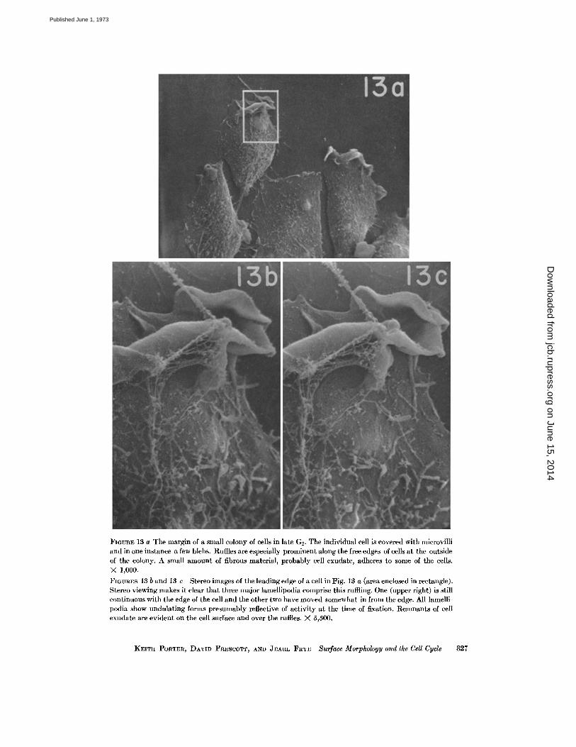
FIGURE 13 a The margin of a small colony of cells in late G2 . The individual cell is covered with microvilliand in one instance a few blebs . Ruffles are especially prominent along the free edges of cells at the outsideof the colony. A small amount of fibrous material, probably cell exudate, adheres to some of the cells.X 1,000 .FIGURES 13 b and 13 c Stereo images of the leading edge of a cell in Fig. 13 a (area enclosed in rectangle) .Stereo viewing makes it clear that three major lamellipodia comprise this ruffling . One (upper right) is stillcontinuous with the edge of the cell and the other two have moved somewhat in from the edge . All lamelli-podia show undulating forms presumably reflective of activity at the time of fixation . Remnants of cellexudate are evident on the cell surface and over the ruffles . X 5,500 .
KEITH PORTER, DAVID PRESCOTT, AND JEARL FRYE Surface Morphology and the Cell Cycle
827
on June 15, 2014jcb.rupress.org
Dow
nloaded from
Published June 1, 1973
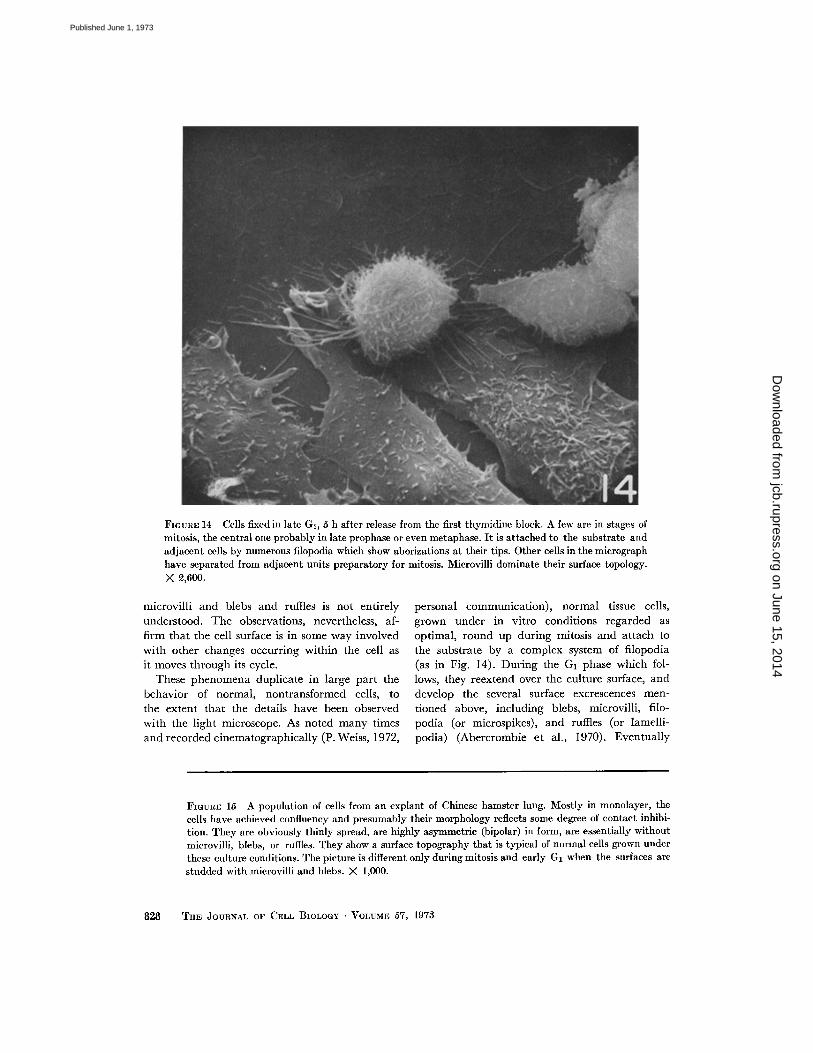
FIGURE 14 Cells fixed in late G2, 5 h after release from the first thymidine block . A few are in stages ofmitosis, the central one probably in late prophase or even metaphase . It is attached to the substrate andadjacent cells by numerous filopodia which show aborizations at their tips . Other cells in the micrographhave separated from adjacent units preparatory for mitosis. Microvilli dominate their surface topology .X 2,600.
microvilli and blebs and ruffles is not entirelyunderstood. The observations, nevertheless, af-firm that the cell surface is in some way involvedwith other changes occurring within the cell asit moves through its cycle .
These phenomena duplicate in large part thebehavior of normal, nontransformed cells, tothe extent that the details have been observedwith the light microscope . As noted many timesand recorded cinematographically (P . Weiss, 1972,
828
personal communication), normal tissue cells,grown under in vitro conditions regarded asoptimal, round up during mitosis and attach tothe substrate by a complex system of filopodia(as in Fig. 14). During the GI phase which fol-lows, they reextend over the culture surface, anddevelop the several surface excrescences men-tioned above, including blebs, microvilli, filo-podia (or microspikes), and ruffles (or lamelli-podia) (Abercrombie et al ., 1970) . Eventually
FIGURE 15 A population of cells from an explant of Chinese hamster lung . Mostly in monolayer, thecells have achieved confluency and presumably their morphology reflects some degree of contact inhibi-tion . They are obviously thinly spread, are highly asymmetric (bipolar) in form, are essentially withoutmicrovilli, blebs, or ruffles . They show a surface topography that is typical of normal cells grown underthese culture conditions . The picture is different only during mitosis and early GI when the surfaces arestudded with microvilli and blebs . X 1,000 .
THE JOURNAL OF CELL BIOLOGY . VOLUME 57, 1973
on June 15, 2014jcb.rupress.org
Dow
nloaded from
Published June 1, 1973
KEITH PORTER, DAVID PRESCOTT, AND JEARL FRYE Surface Morphology and the Cell Cycle
829
on June 15, 2014jcb.rupress.org
Dow
nloaded from
Published June 1, 1973
(and probably in late G1 and S) the blebs andmicrovilli disappear and the cells become thinlyspread and smooth (Follett and Goldman, 1970) .Along margins of contact with other cells theruffles also vanish (Abercrombie and Ambrose,1958) . Finally, in a growing population, the in-dividual cells in late G2 detach from adjacentcells and round up again for the next division .Where factors of population density which in-hibit growth come into play, this flow of struc-tural change is, of course, altered.
The differences between the transformed CHOcells and normal cells of an equivalent type aredifferences4 in the degree to which the cellsspread and lose the various surface structures .In other words, CHO cells, though transformed(and not in the usual sense contact inhibited),do go through a sequence of phenotypic ex-pressions which approach those shown by normalcells.
Two questions of special interest emerge fromthe above observations . Firstly, one should askto what extent these changes in cell form andsurface morphology are a reflection of contactbetween cells of the population ; and secondly,one should inquire into the relation of the formchanges to structural and biochemical phenomenawithin the cell .
The first of these questions is answered by theresults reported in the succeeding paper by Rubinand Everhart. It appears from their study that ifthe population density is so low that contactbetween cells is not to any significant extentachieved, the cells continue to show blebs andretain a form characteristic of GI cells in a partiallyconfluent culture. When this observation isjuxtaposed with those presented here it becomesapparent that CHO cells are susceptible to con-tact influences, and within the limits of theirprescribed capabilities go through structural
4 Cells from explants of hamster lung (Fig . 15) arelike normal chick fibroblasts 5 and normal rat fibro-blasts6 in spreading out thinly during a middleperiod of interphase and for long periods in late G1,where density-dependent inhibition blocks con-tinued growth and reproduction .s Porter, K. R., and G. Weiss . 1973. Chick fibro-blasts during collagen production . A scanning elec-tron microscope study . In preparation.I Porter, K . R., and S . Bandeian . 1973 . Comparativestudy by scanning microscopy of rat sarcoma 4337 andnormal equivalent cells. In preparation.
030
THE JOURNAL OF CELL BIOLOGY . VOLUME 57, 1973
changes approaching those displayed by normalcells. This is not an effect of any media borne,diffusible substance ; direct cell contact is ap-parently required as demonstrated also by Schutzand Mora (1968) for 3T3 mouse embryo fibro-blasts.What intracellular changes in morphology are
associated with the overall form and surfacechanges is more difficult to determine . Thatoriented assemblies of microtubules are involvedis suggested by a number of observations on thedistribution of microtubules in cultured cells .These are consistent in showing that microtubulesare prominent in major cell processes and areoriented parallel to the long axes of the extensions .Colchicine and Colcemid disassemble the micro-tubules, and the cell simultaneously adopts apolygonal (epithelial-like) form or rounds up asthough in preparation for division (Taylor, 1966 ;Goldman, 1971 ; Vasiliev et al., 1970; Goldmanand Follett, 1969; Freed and Lebowitz, 1970) .It has also been observed that nerve cells in thepresence of Colcemid fail to extend their processes(Daniels, 1972), from which the investigatorconcludes that microtubules are "essential to theformation of nerve fibers ."
There is some suggestion that the microtubules,influential in the development of pseudopodiaand bipolar asymmetry in cells, are focused onand possibly oriented and initiated by elementsof the central apparatus (the centrioles and per-sisting aster) . Hence, it is regarded as probablethat one intracellular event associated withshape development after early G I is the assemblyof microtubules in association with the centriolesand centriole-based complex of dense bodies .Their orientation in CHO cells is not bidirectionaland may even be random . That they are there insubstantial numbers has been determined inanother study (Porter et al ., 1973) . When CHOcells are exposed to dibutyryl-cyclic AMP theyacquire a pronounced bipolarity, a response whichis inhibited by colchicine (Hsie and Puck, 1971 ;Johnson et al ., 1971) . In these treated cells themicrotubules are oriented approximately parallelto the long axis of the cells (Porter et al ., 1973) .It appears, therefore, that cAMP is effective ininducing assembly and also orientation of micro-tubules. Whether contact between CHO cells asobserved in these studies contributes to a higherintracellular concentration of cAMP has not beenshown, but in studies of 3T3 and other cell lines,
on June 15, 2014jcb.rupress.org
Dow
nloaded from
Published June 1, 1973
Otten et al. (1971) have found evidence that thecAMP concentration does increase during contactinhibition . It is reasonable to assume thereforethat some of the behavior of cells characteristic ofcontact inhibition is mediated through cAMP(Otten et al ., 1971) .
It has not been determined whether contact suchas observed here between these CHO cells leads tothe establishment of tight junctions or gap junc-tions (nexuses) . However, such low resistant path-ways (ionic couplings) as gap junctions have beenobserved between chick fibroblasts (and a numberof cell lines) grown under in vitro conditions(Pinto da Silva and Gilula, 1972 ; Johnson andSheridan, 1971) . Conceivably then, the establish-ment of contact initiates a membrane propagatedevent (excitation) which spreads over the cellsinvolved and activates the adenyl cyclase re-sponsible for the synthesis of cAMP and secon-darily the initiation of microtubule assembly . Sucha possibility deserves investigation . It is interestingin this regard to note that in invasive carcinoma ofthe cervix where the control of cell form is lost, thenumber of "gap junctions" between the cells isgreatly reduced (McNutt et al., 1971) .
Selected Features of the Cell Surfaces
Though the surfaces of cultured cells have beenimaged before by scanning microscopy (see e.g.,Pugh-Humphreys and Sinclair, 1970 ; Boyde et al .,1972), the literature provides very little commenton the general occurrence and significance ofthe observed structures. There is, however, arapidly developing body of data which relates tosome of these structures, and it therefore seemsappropriate to attempt an integration eventhough the scope of this paper is otherwise limited .
Microvilli
This name is generally reserved for small ex-tensions of the cell surface having a uniformdiameter of about 0 .1 µm and variable lengths .They are of course well known on certain differ-entiated cells such as the adsorptive cells of thesmall intestine and the mesothelial cells liningthe major body cavities . They are less char-acteristic of cells grown in vitro. Where theselatter have been examined with some care (ase.g ., by Follett and Goldman, 1970; and byTaylor, 1966), they have been noted to possess
Aconsistently an axial bundle of 40-60 A fila-ments. Microvilli in Taylor's terminology are"microspikes," but they are very similar to whathave been identified as microvilli . His discoveryof small microtubules (150 A diameter) in micro-villi has apparently not been confirmed .
As reported above, microvilli are prominentsurface elements on CHO cells after mitosis . Theyremain numerous during GI but seem to diminishin prominence during S . Later, as the cells thickenin G5, the microvilli appear again in larger num-bers per unit area of cell surface . Observationsmade by Follett and Goldman (1970) on BHK21/C13 cells over the cell cycle describe a similarfluctuation in numbers of microvilli . The stereoimages (Figs. 11 b and 11 c) depict the microvillias having uniform diameters, as being withoutbranches, and as standing up erectly from thecell surface, at least in many instances . Theyseem frequently to emerge from a larger surfaceexcrescence, but our observations to date areinsufficient to permit any generalization on thispoint .
It is therefore a quality of these transformedCHO cells to show microvilli on their surfacesover most of the cell cycle, even when the cellsare obviously confluent and presumably respond-ing to some level of contact inhibition . This ob-servation is made more interesting by the fact thatnormal, first generation cells cultured from ex-plants of Chinese hamster lung tissue and grownto confluence (Fig. 15) are essentially devoid ofmicrovilli except during mitosis . We are notable to decide at this point whether their presenceon the transformed cell reflects a physiologicaldifference (related to growth rate) or a constantphenotypic expression of the transformation .It would be valuable to test their response tosuch environmental variables as pH and glucoseconcentration .
Microvilli of similar dimensions (but frequentlylonger) are notably prominent on transformedcells besides CHO and especially on strains ofmalignant cells . Micrographs made with thescanning microscope are especially impressive inshowing them on sarcoma 180 cells (Boyde et al .,1972), on Landschutz cells (Pugh-Humphreysand Sinclair, 1970), on HeLa cells (Porter andFonte, 1973; see also Pugh-Humphreys andSinclair, 1970), and on rat sarcoma 4337 . 5 Thenormal equivalent of the latter sarcoma cell isstrikingly free of microvilli during interphase
KEITH PORTER. DAVID PRESCOTT, AND JEARL FRYE Surface Morphology and the Cell Cycle
831
on June 15, 2014jcb.rupress.org
Dow
nloaded from
Published June 1, 1973
periods of the cell cycle. It appears, on the otherhand, that cells normally free of microvilli ac-quire them after transformation with an oncogenicvirus (Boyde et al ., 1972) .
There may in fact be some relationship be-tween the presence of microvilli and the agglutin-ability of cells as though, by providing a greatersurface area or by possessing specific receptorsites, the microvilli facilitate the cell's associationwith agglutinins . In this connection, 3T3 cells,which in vitro show few microvilli during G1, S,and G2 and are then only weakly agglutinable,normally display a higher concentration of recep-tor sites for wheat germ agglutinin during mitosis(Burger, 1971) when their surfaces are coveredwith microvilli (Porter, unpublished observations) .The same cells after transformation by polyomavirus show a marked increase in receptor sitesduring all phases of the cycle (Fox et al ., 1971) aswell as an increase in numbers of microvilli andother surface excrescences. The surfaces of BHK21fibroblasts (normally smooth) develop microvilliand blebs after trypsinization (Follett and Gold-man, 1970), a treatment which simultaneously"exposes" receptor sites for agglutinins . The oc-currence of surface microvilli etc . coincides thenwith the agglutinability of cells by plant lectins ina number of instances and this leads one to wonderif agglutinability, so typical of cells which haveescaped the growth control of contact inhibition,is not a reflection of relatively gross structuralfeatures of the cell surface rather than the induc-tion or exposure of sites with specific chemicalproperties (see also O'Neill and Follett, 1970) .
We have noted also that microvilli on theseCHO cells serve to hold exudates (presumablyglycoproteins) on the cell surface, exudates whichmight bind agglutinins and which are morereadily removed from the relatively smoothsurfaces of the normal, nontransformed cell .
Because microvilli are so small, they are diffi-cult to observe with the light microscope, andobservations on their behavior are neither nu-merous nor reliable. On HeLa cells the microvilliare especially long, and as Gey showed manyyears ago (1955), they can be seen in profilewith good phase optics . He described them assurprisingly rigid extensions of the surface whichmoved about their points of attachment as though
pivoted there . He further described their rapidemergence from the cell and their equally rapid
8.32
THE JOURNAL OF CELL BIOLOGY • VOLUME 57, 1973
disappearance into the cell . They are, in otherwords, remarkably labile, dynamic structures .In the interval since Gey's observations theseHeLa cell microvilli have been better observedby electron microscopy (Pugh-Humphreys andSinclair, 1970 ; Porter and Fonte, 1973). Theyare indeed remarkable in being very long, butin other respects (diameter, etc.) they seem notto differ from other microvilli .Any literature review will turn up a number
of suggestions regarding the function of microvilli .Follett and Goldman (1970), for example, haveproposed that the surface membrane is conservedin the form of microvilli to be available for dra-matic changes in apparent surface area as whenthe cell becomes thinly spread . In specialized(differentiated) cell surfaces such as the brushborder of intestinal adsorptive cells, microvilliare thought to possess "uphill pumps" for theactive transport of sugars. The existence of asimilar device on the microvilli of cultured cellshas not been demonstrated . Chick fibroblaststransformed by Rous sarcoma virus are, however,known to transport glucose at a higher rate(Hatanaka and Hanafusa, 1970) and these trans-formed cells have been shown to have many micro-villi (Boyde et al ., 1972) . Furthermore, there issome indication in HeLa cells that surface micro-villi fluctuate in number directly with glucoseconcentration in the medium (Willoch, 1967) . Sothere is a possibility that microvilli are formed tofacilitate the diffusion of glucose across the surfaceof rapidly growing cells. If this is the case, the re-duction in number of microvilli during the S phaseof CHO cells might imply that sugar (and otherdiffusible metabolites) uptake is least active at thistime, provided of course the surface area is notkept constant by the spreading of the cell whichoccurs during that period . The alternative sug-gestion that microvilli are a device the cell has tokeep its surface area constant while it goes throughseveral form changes is just as valid .
BlebsThese bulbous excrescences of the cell surface,
which are observed to come and go on cellsduring mitosis and cytokinesis, are familiar to
most observers of dividing cells . Less familiar,
perhaps, is their presence on cells during inter-phase. Nevertheless, blebs of a similar nature
on June 15, 2014jcb.rupress.org
Dow
nloaded from
Published June 1, 1973
have been described on several kinds of cellsunder a variety of experimental conditions .Costero and Pomerat (1951), for example, ob-served them along the dendrites of nerve cellsgrown in tissue culture . Their behavior remindedthe authors of boiling of a very viscous fluid,hence the term zeiosis for the phenomena of blebformation and retraction .
Though such blebs may not all belong to onecategory of surface structure, it seems very prob-able that they do . Certainly the observation onthe blebs in CHO cells published by Hsie et al .(1971) and by Puck et al . (1972) repeats soclosely those of Costero and Pomerat (1951) andof Price (1967) that there is little excuse to regardthem as separate structures .
In these scanning images, the blebs dominatethe cell surface during early GI and persist in asmall percentage of cells on into S. Mostly, how-ever, they vanish during the latter half of thecycle. Their shape is spherical when viewedfrom the top, and from the side they appear to
be supported by a stout stalk which has a diameteronly slightly less than the bulb above . They ob-viously vary greatly in size, and we assume thatsome of the smaller ones represent early stagesin the formation of the larger ; conceivably alsothey may be stages in the subsidence of largerones. There was no clear indication in our prepa-rations that these zeiotic blebs are released fromthe cell. Only in the earliest preparations (I hafter shake-off) did we recognize anything thatcould represent a loose bleb, and if cell derived,these may have been released during anaphaseor telophase .
These observations on blebs have not extendedbeyond the recognition of their presence or ab-sence in various phases of the cell cycle . Apparentlyin cells which are isolated, as in low density cul-tures (Rubin and Everhart, 1973), they persistmuch longer, if not through the entire inter-phase. Their disappearance in S and G2 in moredensely populated cultures would seem to be oneresult of cell contact . Nothing in our study sug-gests what they do or why they form. However,transmission microscopy of thin sections of themon some human epithelial cells shows them tocontain large numbers of polyribosomes (Price,1967) . It appears that cytoplasmic componentsother than ribosomes are excluded. Observationsby one of us on the CHO cell blebs, incidental
to another study, make it clear that here too they
contain an impressive concentration of ribosomes(Porter et al., 1973) . That they are observed onnormal cells especially during the earliest phasesof GI should not be overlooked, but their extendedand exaggerated presence on the CHO cells is aunique feature of these transformed cells andone deserving further investigation .?
RufflesAs already noted, these extraordinary structures
of the cell surface are more prominent in somephases of the cycle than in others . They are notfound on cells in mitosis and do not become ob-vious until late G 1 . Then, during S and G2, theyseem to increase in number and probably ac-tivity. It is also evident from this study thatruffles are preferentially associated with the freeedges of cells . This agrees with light microscopeobservations that they in fact originate at theadvancing edges of cells (Abercrombie et al .,1970; Ingram, 1969) . Conversely, where theedge of a cell is confluent with that of another,there are generally no ruffles .
The significance of these observations, es-
pecially the increase in ruffling in G2, is not im-mediately evident. One might assume that thecell is in greatest need of metabolites forgrowth at this time and is using all devices toincrease the area of contact with the environmentincluding internalization of the medium bypinocytosis . To the extent that diffusion acrossthe cell surface is facilitated by the provision ofextra surface, the requirements of the cell aresatisfied . There is probably more involved, how-ever, than this simple statement suggests .
The development of ruffles obviously creatsefairly large areas of membrane . It is easy instereo images (Figs. 13 b and 13 c) to see thattypical lamellipodia are about 0.1 µm thick, avalue which agrees with other measurements
7 According to recent observations (Harris, A . K .,1972, personal communication) on cells in vitro,blebs develop initially at the cell margin and mi-grate centripetally (back from the edge) like ruffles .This and their recession into the cell surface withtime may account for the accumulation of largenumbers of relatively small blebs over the centers ofmany of the GI cells observed here (see Figs . 4, 10 a,and 11 a) .
KEITH PORTER, DAVID PRESCOTT, AND JEARL FRYE Surface Morphology and the Cell Cycle
833
on June 15, 2014jcb.rupress.org
Dow
nloaded from
Published June 1, 1973
made on thin sections of ruffles (Abercrombie etal., 1971) . Their vertical dimension is highlyvariable but is of the order of 6-8 µm, and theycan extend along the cell margin for 8 or 10 µm .A single ruffle may therefore have a surfacearea of ca . 100 grn2 . Since one cell may possessseveral ruffles they can comprise a substantialfraction of the total cell surface at any one time .Much more accurate measurements could bemade from scanning electron microscope imagesin a study devoted primarily to these structuresand their function .
The rate at which lamellipodia form is im-pressive. Observers of this phenomenon agreethat 2-3 min is all it takes for the cell to produceone of these structures, i.e ., before it begins itsmigration back from the free edge (see footnote7) (Gey, 1955 ; Ingram, 1969) . Abercrombieand co-workers (1970) describe this growth asinvolving membrane assembly rather than mem-brane flow . They provide evidence in fact thatmembrane flow is away from the advancing edgerather than toward it . Just how membrane sub-units are fed into the area for assembly becomesa problem . Transmission microscopy of sectionsof ruffles and the associated leading edge of thecell, done also by Abercrombie and his colleagues(1971), provides little or no clue to the origin ofall this membrane. The Golgi complex, demon-strably the source of specialized cell surfaces indifferentiated cells, is not represented . Theirmicrographs show instead a few small vesiclesat the base of the ruffle but little else . Perhaps thephenomena involved are comparable to thoseseen in the rapid elongation of pollen tubes wherenumerous vesicles are fed into the region of thegrowing surface (Rosen, 1968) . Apart from thesevesicles, which conceivably fuse with and con-tribute to the surface, the only source of mem-brane subunits would seem to be the cytoplasmicground substance that floods into the leadingedge as the cell extends its margins . A certainamount of this ground substance, which is de-scribed as "granular" and as showing "vagueand irregular filaments" (Abercrombie et al .,1971 ; Wagner et al ., 1971), is retained betweenthe plasma membranes of the ruffle and is carriedback with the lamellipodia to a point where thestructure fades into the cell surface again . Pos-sibly membrane components are also returned
to the cytoplasm to be fed again (recycled) into
the production of more lamellipodia .
8 3 4 THE JOURNAL OF CELL BIOLOGY • VOLUME 57, 1973
Scanning Microscopy
Scanning microscopy of cells and soft tissueshas not as yet created an important literature .The major reason is that only very recently have afew laboratories developed adequate preparationprocedures and applied them to interesting prob-lems. A study of the lactating mammary gland byNemanic and Pitelka (1971) illustrates well thevalues of the approach for soft tissues, and somepioneering work by Boyde and colleagues (1972)has demonstrated its potential for microscopy ofcultured cells . One can easily agree with theirconclusion that for getting an overall view ofthe topography of cultured cells quickly andeasily there is no equivalent procedure .
Critical-point drying of the specimen is themost significant step in obtaining a relativelyundistorted surface (of cell or tissue) for examina-tion. This important procedure, introduced toelectron microscopy by T . F. Anderson (1951),is simple to do and requires only a small amountof equipment . In our experience, it is better formost purposes than drying from the frozen state(Porter et al,, 1972) . This latter procedure isneeded only when one wants to avoid fixationwith glutaraldehyde or some other reagent .Among the several compounds that may be usedfor critical-point drying (Freon, N 20, CO2),none seems superior to liquid CO2. It is certainlythe least expensive and least toxic .
In the work reported here and by Rubin andEverhart (1973) attention has been placed onchanges in CHO form and surfaces with phasesof the cell cycle. The cells were grown underconditions which can of course be varied ex-perimentally and which would doubtless haveeffects that would be reflected in surface changes .Thus one of the more valuable applications ofscanning microscopy will be that of followingthe response of cells and especially surfaces toenvironmental factors . But the possibilities do notend there by any means . Features of the surface,apart from microvilli etc ., such as the antigenic
sites, may require the development of specialprocedures for their identification, but the pros-pects are good . It will in some instances be neces-
sary, for correlating internal structure with the
surface features, to resort to transmission micros-copy of thin sections, but another distinct possi-
bility is to examine whole cells first in a high (1
MeV) voltage microscope, where high energy
on June 15, 2014jcb.rupress.org
Dow
nloaded from
Published June 1, 1973
electrons should give adequate penetration, andthereafter with the scanning election microscope forsurface characteristics .
The authors are pleased to acknowledge the tech-nical assistance of Mrs . Jan Kirkpatrick, DonnaKelley, and Mr . Gary Weiss.
This work was supported by grants CA-12302-01(National Cancer Institute) to Dr. D. M. Prescottand GB-27965 (National Science Foundation) and5504 RR06084 (National Institutes of Health) toDr. K. R. Porter .
Received for publication 8 November 1972, and in revisedform 1 February 1973.
REFERENCES
ABERCROMBIE, M., and E. J . AMBROSE. 1958 . Inter-ference microscope studies of cell contacts intissue culture . Exp. Cell Res . 15 :332 .
ABERCROMBIE, M ., J. E. M. HEAYSMAN, and S . M .PEGRUM. 1970. The locomotion of fibroblasts inculture . II. Ruffling. Exp . Cell Res. 60:437.
ABERCROMBIE, M ., J . E. M. HEAYSMAN, and S. M .PEGRUM. 1971 . The locomotion of fibroblasts inculture. IV. Electron microscopy of the leadinglamella. Exp. Cell Res. 67:359.
ANDERSON, T . F. 1951 . Techniques for the preserva-tion of three-dimensional structure in preparingspecimens for the electron microscope . Trans.N. Y. Acad. Sci. Ser. II . 13:130.
BOSTOCK, C. J., D. M. PRESCOTT, and J . B. KIRK-PATRICK. 1971 . An evaluation of the double thy-midine block for synchronizing mammalian cellsat the GI-S border . Exp . Cell Res. 68 :163 .
BOYDE, A., R. A. WEISS, and P. VESELY. 1972 .Scanning electron microscopy of cells in culture.Exp . Cell Res. 71 :313 .
BURGER, M. M. 1971 . The significance of surfacestructure changes for growth control undercrowded conditions. Ciba Foundation Symposiumon Growth Control in Cell Cultures. G. E. W.Wolstenholme and J. Knight, editors. J . and A.Churchill, Ltd ., London .
COSTERO, I., and C. M. POMERAT. 1951 . Cultivationof neurons from the adult human cerebral andcerebellar cortex . Am. J. Anat . 89 :405 .
DALEN, H., and P. SCHEIE . 1969. Microextensions ofChang's liver cells as observed throughout theirdivision cycle . Exp. Cell Res. 57 :351 .
DANIELS, M. P. 1972 . Colchicine inhibition of nervefiber formation in vitro. J. Cell Biol. 53 :164.
FOLLETT, E. A. C., and R. D. GOLDMAN . 1970 . Theoccurrence of microvilli during spreading andgrowth of BHK21/C13 fibroblasts. Exp. Cell Res.59:124 .
Fox, T. 0 ., J. R. SHEPPARD, and M. M. BURGER .
1971. Cyclic membrane changes in animal cells :Transformed cells permanently display a surfacearchitecture detected in normal cells only duringmitosis. Proc. Natl. Acad. Sci. U. S. A . 68 :244 .
FREED, J . J., and M. M. LEBOWITZ . 1970 . The as-sociation of a class of saltatory movements withmicrotubules in cultured cells . J. Cell Biol. 45 :334 .
GALAVAZI, G., H. SCHENK, and D. BoOTSMA . 1966 .Synchronization of mammalian cells in vitro byinhibition of the DNA syntheses . Exp. Cell Res.41 :428 .
GEY, G. 0 . 1955 . Some aspects of the constitution andbehavior of normal and malignant cells main-tained in continuous culture . Harvey Lect. Ser. L.154.
GOLDMAN, R. D . 1971 . The role of three cytoplasmicfibers in BHK-21 cell motility. I. Microtubulesand the effects of colchicine. J. Cell Biol. 51 :752.
GOLDMAN, R. D., and E. A. C. FOLLETT. 1969.The structure of the major cell processes of iso-lated BHK21 fibroblasts . Exp. Cell Res. 57 :263 .
HATANAKA, M., and H. HANAFUSA. 1970 . Analysisof a functional change in membrane in the processof cell transformation by Rous sarcoma virus ;alteration in the characteristics of sugar transport .Virology . 41:647.
HsIE, A. W., C. JONES, and T. T. PUCK . 1971 .Further changes in differentiation state accom-panying the conversion of Chinese hamster cellsto fibroblastic form by dibutyryl adenosine cyclic3' :5'-monophosphate and hormones . Proc . Natl .Acad. Sci. U. S. A . 68 :1648 .
HsIE, A. W., and T. T. PUCK . 1971 . Morphologicaltransformation of Chinese hamster cells by di-butyryl adenosine cyclic 3' :5'-monophosphate andtestosterone. Proc. Natl. Acad. Sci. U. S. A. 68 :358 .
INGRAM, V. M. 1969. A side view of moving fibro-blasts. Nature (Lond.) . 222 :641 .
JOHNSON, G. S., R. M. FRIEDMAN, and I. PASTAN .1971. Restoration of several morphological char-acteristics of normal fibroblasts in sarcoma cellstreated with adenosine-3' :5'-cyclic monophos-phate and its derivatives . Proc. Nall. Acad. Sci.U. S. A. 68 :425 .
JOHNSON, R. G., and J . D. SHERIDAN . 1971 . Junctionsbetween cancer cells in culture : Ultrastructure andpermeability. Science (Wash. D. C.) . 174 :717 .
MCNUTT, N . S., R . A. HERSHBERG, and R . S. WEIN-sTEIN. 1971 . Further observations on the occur-rence of nexuses in benign and malignant humancervical epithelium. J. Cell Biol. 51 :805.
NEMANIC, M. K., and D. R. PITELKA . 1971 . Ascanning electron microscope study of the lactatingmammary gland. J. Cell Biol. 48:410.
O'NEILL, C. H., and E . A. C. FOLLETT. 1970. Aninverse relation between cell density and the
KEITH PORTEit, DAVID PRESCOTT, AND JEARL FRYE Surface Morphology and the Cell Cycle
835
on June 15, 2014jcb.rupress.org
Dow
nloaded from
Published June 1, 1973
number of microvilli in cultures of BHK21 ham-ster fibroblasts . J. Cell Sci . 7 :695.
OTTEN, J., G. S . JOHNSON, and I . PASTAN . 1971 .Cyclic AMP levels in fibroblasts : Relationship togrowth rate and contact inhibition of growth .Biochem. Biophys. Res. Commun. 44:1192 .
PINTO DA SILVA, P ., and N. B. GILULA . 1972. Gapjunctions in normal and transformed fibroblastsin culture . Exp. Cell Res. 71:393 .
PORTER, K. R., and V. G. FONTE . 1973. A scanningstudy of HeLa cell surfaces. In press.
PORTER, K. R., D . KELLEY, and P. M. ANDREWS .1972. The preparation of cultured cells and softtissues for scanning electron microscopy . In Pro-ceedings of the Fifth Annual Stereoscan ScanningElectron Microscope Colloquium, Kent Cam-bridge Scientific, Inc., Morton Grove, Ill .
PORTER, K. R., T . T. PUCK, A. W. HSIE, and D .KELLEY . 1973. An electron microscope study ofthe effects of cyclic AMP on CHO cells . In press .
PRICE, Z . H . 1967. The micromorphology of zeioticblebs in cultured human epithelial (HEp) cells .Exp . Cell Res . 48 :82.
PUCK, T. T., C. A . WALDREN, and A . W. HSIE .1972. Membrane dynamics in the action of di-butyryl adenosine 3' :5'-cyclic monophosphateand testosterone on mammalian cells . Proc . Natl.Acad. Sci. U. S. A. 69:1943.
PUGH-HUMPHREYS, R. G. P., and W . SINCLAIR .1970. Ultrastructural studies relating to the sur-face morphology of cultured cells. J. Cell Sci.6:477 .
836 THE JOURNAL OF CELL BIOLOGY . VOLUME 57, 1973
ROSEN, W . G. 1968. Ultrastructure and physiologyof pollen . Annu . Rev. Plant Physiol. 19:435 .
RUBIN, R. W., and L . P. EVERHART . 1973. The effectof cell-to-cell contact on the surface morphologyof Chinese hamster ovary cells . J. Cell Biol. 57:837.
SCHUTZ, L., and P . T. MORA. 1968. The need fordirect cell contact in "contact" inhibition of celldivision in culture . J. Cell. Physiol . 71 :1 .
SEFTON, B . M., and H. RUBIN. 1971 . Stimulation ofglucose transport in cultures of density-inhibitedchick embryo cells . Proc. Natl . Acad. Sci. U. S. A .68:3154 .
TAYLOR, A. C. 1966. Microtubules in the micro-spikes and cortical cytoplasm of isolated cells .J. Cell Biol. 28:155 .
TOBEY, R A., D . C . ANDERSON, and D. F . PETERSEN .1967. Properties of mitotic cells prepared bymechanically shaking monolayer cultures ofChinese hamster cells . J. Cell . Physiol. 70:63.
VASILIEV, J. M ., I . M. GELFAND, L . V. DOMNINA,O. Y. IVANOVA, S. G. KOMM, and L. V. OLSHEV -5KA,JA. 1970. Effect of Colcemid on the locomotorybehaviour of fibroblasts. J. Embryol. Exp . Morphol.24:625 .
WAGNER, R., M . ROSENBERG, and R. ESTENSEN .1971. Endocytosis in Chang liver cells . J. CellBiol . 50:704 .
WILLOCH, M. 1967. Changes in HeLa cell ultra-structure under conditions of reduced glucosesupply. Acta Pathol . Microbiol. Scand. 71:35 .
on June 15, 2014jcb.rupress.org
Dow
nloaded from
Published June 1, 1973
Related Documents