This article was downloaded by: [Dr Alessia D'Agata] On: 11 February 2014, At: 06:06 Publisher: Taylor & Francis Informa Ltd Registered in England and Wales Registered Number: 1072954 Registered office: Mortimer House, 37-41 Mortimer Street, London W1T 3JH, UK Italian Journal of Zoology Publication details, including instructions for authors and subscription information: http://www.tandfonline.com/loi/tizo20 Cellular biomarkers in the mussel Mytilus galloprovincialis (Bivalvia: Mytilidae) from Lake Faro (Sicily, Italy) A. D’Agata a , T. Cappello a , M. Maisano ab , V. Parrino ab , A. Giannetto a , M. V. Brundo c , M. Ferrante d & A. Mauceri ab a Department of Biological and Environmental Sciences, University of Messina, Italy b Centro Universitario CUTGANA, Catania, Italy c Department of Biology “Marcello La Greca”, University of Catania, Italy d Department of Hygiene and Public Health “G.F. Ingrassia”, University of Catania, Italy Published online: 05 Feb 2014. To cite this article: A. D’Agata, T. Cappello, M. Maisano, V. Parrino, A. Giannetto, M. V. Brundo, M. Ferrante & A. Mauceri , Italian Journal of Zoology (2014): Cellular biomarkers in the mussel Mytilus galloprovincialis (Bivalvia: Mytilidae) from Lake Faro (Sicily, Italy), Italian Journal of Zoology, DOI: 10.1080/11250003.2013.878400 To link to this article: http://dx.doi.org/10.1080/11250003.2013.878400 PLEASE SCROLL DOWN FOR ARTICLE Taylor & Francis makes every effort to ensure the accuracy of all the information (the “Content”) contained in the publications on our platform. However, Taylor & Francis, our agents, and our licensors make no representations or warranties whatsoever as to the accuracy, completeness, or suitability for any purpose of the Content. Any opinions and views expressed in this publication are the opinions and views of the authors, and are not the views of or endorsed by Taylor & Francis. The accuracy of the Content should not be relied upon and should be independently verified with primary sources of information. Taylor and Francis shall not be liable for any losses, actions, claims, proceedings, demands, costs, expenses, damages, and other liabilities whatsoever or howsoever caused arising directly or indirectly in connection with, in relation to or arising out of the use of the Content. This article may be used for research, teaching, and private study purposes. Any substantial or systematic reproduction, redistribution, reselling, loan, sub-licensing, systematic supply, or distribution in any form to anyone is expressly forbidden. Terms & Conditions of access and use can be found at http:// www.tandfonline.com/page/terms-and-conditions

Welcome message from author
This document is posted to help you gain knowledge. Please leave a comment to let me know what you think about it! Share it to your friends and learn new things together.
Transcript

This article was downloaded by: [Dr Alessia D'Agata]On: 11 February 2014, At: 06:06Publisher: Taylor & FrancisInforma Ltd Registered in England and Wales Registered Number: 1072954 Registered office: Mortimer House,37-41 Mortimer Street, London W1T 3JH, UK
Italian Journal of ZoologyPublication details, including instructions for authors and subscription information:http://www.tandfonline.com/loi/tizo20
Cellular biomarkers in the mussel Mytilusgalloprovincialis (Bivalvia: Mytilidae) from Lake Faro(Sicily, Italy)A. D’Agataa, T. Cappelloa, M. Maisanoab, V. Parrinoab, A. Giannettoa, M. V. Brundoc, M.Ferranted & A. Mauceriab
a Department of Biological and Environmental Sciences, University of Messina, Italyb Centro Universitario CUTGANA, Catania, Italyc Department of Biology “Marcello La Greca”, University of Catania, Italyd Department of Hygiene and Public Health “G.F. Ingrassia”, University of Catania, ItalyPublished online: 05 Feb 2014.
To cite this article: A. D’Agata, T. Cappello, M. Maisano, V. Parrino, A. Giannetto, M. V. Brundo, M. Ferrante & A. Mauceri ,Italian Journal of Zoology (2014): Cellular biomarkers in the mussel Mytilus galloprovincialis (Bivalvia: Mytilidae) from LakeFaro (Sicily, Italy), Italian Journal of Zoology, DOI: 10.1080/11250003.2013.878400
To link to this article: http://dx.doi.org/10.1080/11250003.2013.878400
PLEASE SCROLL DOWN FOR ARTICLE
Taylor & Francis makes every effort to ensure the accuracy of all the information (the “Content”) containedin the publications on our platform. However, Taylor & Francis, our agents, and our licensors make norepresentations or warranties whatsoever as to the accuracy, completeness, or suitability for any purpose of theContent. Any opinions and views expressed in this publication are the opinions and views of the authors, andare not the views of or endorsed by Taylor & Francis. The accuracy of the Content should not be relied upon andshould be independently verified with primary sources of information. Taylor and Francis shall not be liable forany losses, actions, claims, proceedings, demands, costs, expenses, damages, and other liabilities whatsoeveror howsoever caused arising directly or indirectly in connection with, in relation to or arising out of the use ofthe Content.
This article may be used for research, teaching, and private study purposes. Any substantial or systematicreproduction, redistribution, reselling, loan, sub-licensing, systematic supply, or distribution in anyform to anyone is expressly forbidden. Terms & Conditions of access and use can be found at http://www.tandfonline.com/page/terms-and-conditions

Cellular biomarkers in the mussel Mytilus galloprovincialis (Bivalvia:Mytilidae) from Lake Faro (Sicily, Italy)
A. D’AGATA1, T. CAPPELLO1, M. MAISANO1,2, V. PARRINO1,2, A. GIANNETTO1,M. V. BRUNDO3, M. FERRANTE4, & A. MAUCERI1,2
1Department of Biological and Environmental Sciences, University of Messina, Italy, 2Centro Universitario CUTGANA,Catania, Italy, 3Department of Biology “Marcello La Greca”, University of Catania, Italy, and 4Department of Hygiene andPublic Health “G.F. Ingrassia”, University of Catania, Italy
(Received 26 September 2013; accepted 5 December 2013)
AbstractLake Faro (Sicily, Italy) is a natural confined brackish environment particularly subject to anthropogenic impact, resulting in amixture of xenobiotic substances, i.e. heavy metals and polycyclic aromatic hydrocarbons (PAHs), and characterised by lowhydrodynamics. In order to assess the water quality status of this pond, a multi-biomarker approach was applied on musselMytilus galloprovincialis (Lamarck, 1819) both inhabiting the lake and from a control site (Goro). Different biomarkers wereinvestigated on mussel digestive glands and gills, including biomarkers of exposure (cytochrome P450 4, CYP4), neurotoxicity(acetylcholinesterase, AChE; choline acetyltransferase, ChAT), general stress (lysosomal membrane stability, LMS), andgenotoxicity (micronucleus and comet assays). The results suggest significant responses related to the selected area. A statisticallysignificant inhibition (P < 0.0001) of CYP4 in the digestive gland, and of AChE and ChAT in the gills, was found in specimenscollected fromFaro compared with the control. The lysosomal membrane stability of mussels fromLake Faro was lower than thereference site, while the DNA damages were significantly higher in mussels from the brackish area compared to the control. Thisstudy represents the first time the quality status of Lake Faro is assessed using a multi-biomarker approach on the musselM. galloprovincialis, which proved to be suitable to identify the effects of environmental pollutants at molecular and cellular levels.
Keywords: Mussels, cytochrome, neurotransmitters, lysosomal stability, genotoxicity
Introduction
In recent years, the rapid increase of anthropogenicactivities has caused an incessant influx of organicand inorganic xenobiotics into aquatic environments,including polyaromatic hydrocarbons (PAHs), poly-chlorinated biphenyls (PCBs), organophosphorus(OP) compounds and heavy metals. Bivalves, and inparticular marine mussels, i.e. Mytilus galloprovincialis(Lamarck, 1819), have been widely used as bioindica-tors in pollution monitoring programs, due to theirability to accumulate in their tissues and tolerate highconcentrations of organic and inorganic pollutants(Widdows & Donkin 1992; Fasulo et al. 2008; Ciacciet al. 2012; Cappello et al. 2013a, 2013b).
In this study, specimens of M. galloprovincialiswere collected from a brackish environment, LakeFaro (Sicily, Italy), characterised by the presence of
heavy metals (Giacobbe et al. 1996; Munaò et al.2000; Mauceri et al. 2005; Fasulo et al. 2008) andPAHs (Giacalone et al. 2004; Arpa Sicilia 2007).This site is a typical example of a natural confinedenvironment, where eutrophication can trigger stresswith negative repercussions on the aquatic ecosys-tem. The lake is characterised by euryhaline waters,as there is an injection of fresh water from aquifers,which mixes with salt water through the channelscommunicating with the sea. It represents an exam-ple of a meromictic basin and, therefore, the sedi-ments are not involved in a mixing process. Thepond is also subject to the negative effects of anthro-pogenic impact, associated with a massive construc-tion industry, sewage and fertilisers used in thesurrounding areas, that may affect the ecologicalbalance (Sorgi et al. 2006).
Correspondence: M. Maisano, Department of Biological and Environmental Sciences, University of Messina, Viale F. Stagno d’Alcontres 31, 98166 Messina,Italy. Tel: +39 090 391435. Fax: +39 090 6765556. Email: [email protected]
Italian Journal of Zoology, 2014, 1–12http://dx.doi.org/10.1080/11250003.2013.878400
© 2014 Unione Zoologica Italiana
Dow
nloa
ded
by [
Dr
Ale
ssia
D'A
gata
] at
06:
06 1
1 Fe
brua
ry 2
014

A battery of multiple biomarkers was applied onmussel specimens in two key organs, the digestivegland and gills, including cytochrome P450 (Peterset al. 1999; Solé & Livingstone 2005; Cappello et al.2013a), esterases (Moreira & Guilhermino 2005),lysosomal membrane stability (Da Ros et al. 2002;Castro et al. 2004; Pisoni et al. 2004), micronucleustest and comet assay (Rank et al. 2007; Banni et al.2010).
The digestive gland of mollusks is the main organfor metabolic regulation, participating in the pro-cesses of detoxification and elimination of xenobio-tics (Marigómez et al. 2002; Moore & Allen 2002;Cappello et al. 2013a; D’Agata et al. 2013). Thecytochrome P450 (CYP) enzyme family plays a keyrole in the mechanism of detoxification, beinginvolved in metabolising a wide range of lipophilicorganic endogenous and exogenous compounds.A few studies have been performed on inverte-brates and, to date, only CYP4 was sequenced inM. galloprovincialis. A specific CYP gene,CYP4Y1, was identified in the digestive gland ofM. galloprovincialis, and its expression was foundto be inhibited after exposure to beta-naphthofla-vone (Snyder 1998), and down-regulated in M.galloprovincialis caged in a heavily polluted petro-chemical area (Cappello et al. 2013a).
The gills of the bivalves present several mechan-isms controlling the functions involved in the main-tenance of homeostasis (Gomez-Mendikute et al.2005; Scarfì et al. 2006; Fasulo et al. 2008;Cappello et al. 2013b). The gills are constantly incontact with the surrounding aquatic environment,and thus highly exposed to environmental factorsdue to their large surface and their involvement ingas exchange and feeding (de Oliveira David et al.2008). Therefore, the structural pattern of the gilltissues and their biochemical profiles reflect theadverse environmental impact on the animal(Gregory et al. 2002; Ciacci et al. 2012; Cappelloet al. 2013b). Acetylcholine (ACh) is a neurotrans-mitter used in efferent systems and also in somecentral circuits (Woolf 1991). It is synthesized inthe cytoplasm of cholinergic neurons by the enzymecholine acetyltransferase (ChAT), and degraded atthe synaptic cleft by the enzyme acetylcholinester-ase (AChE). AChE is involved in the transmissionof nerve impulses and has been widely used as anindicator of potential neurotoxicity, both in inverte-brates and lower vertebrates (Peakall 1992; Moraet al. 1999a, 1999b; Ciacci et al. 2012; DeDomenico et al. 2013).
Furthermore, it is well known that the accumula-tion of xenobiotics and their related metabolic
products inside the lysosomes weakens their mem-brane stability and may induce diffusion of hydro-lytic lysosome enzymes into the cytosol. Theassessment of lysosomal membrane stability hasbeen proposed as a rational biomarker of generalstress (Petrovic et al. 2001; Castro et al. 2004), andits use has been evaluated in marine organisms, bothin field and laboratory studies (Da Ros et al. 2002;Castro et al. 2004; Maisano et al. 2013).Inefficient detoxification or increased activation of
xenobiotic compounds is believed to modulate thebiologically active dose available for the molecularend target (DNA), and thus influence the levels ofbiomarkers reflecting exposure to and damage bygenotoxic agents (Brescia et al. 1999). The impactof genotoxic chemicals on the integrity of cellularDNA is the first event after the exposure.Chromosomal damage expressed after cell replica-tion represents an accumulated effect associatedwith long-term exposure (Dixon et al. 2002; Jhaet al. 2005; Jha 2008). The alkaline elution test hasbeen applied in marine invertebrates (Vukmirovicet al. 1994; Bolognesi et al. 1996) and allows theevaluation of a large range of DNA damages in termsof DNA single-strand breaks. The haemocytes havebeen used in such investigations for the assessmentof DNA damage using single cell gel electrophoresisor comet assay (Dixon et al. 2002; Jha et al. 2005;Jha 2008; D’Agata et al. 2013). In addition, themicronucleus (MN) test, one of the most commonlyapplied techniques for identifying genomic altera-tions in animals (Jha et al. 2005; Fasulo et al.2010), has also been performed on mussels as end-point of genotoxicity testing.The purpose of the present study was to evaluate
the water quality status of a natural confined brack-ish environment, Lake Faro, by applying, for the firsttime in this site, a multi-biomarker approach on themussel M. galloprovincialis, chosen as the sentinelorganism. The battery of multiple biomarkersincluded biomarkers of exposure (CYP4), neurotoxi-city (AChE; ChAT), general stress (LMS), and gen-otoxicity (MN and comet assays).
Materials and methods
Study area
Lake Faro (Sicily, Italy) was chosen as the naturalconfined brackish environment for this study. It is ameromictic basin, particularly subject to anthropo-genic impact resulting in the presence of heavymetals and PAHs, and characterised by low hydro-dynamics. In addition, increased population density
2 A. D’Agata et al.
Dow
nloa
ded
by [
Dr
Ale
ssia
D'A
gata
] at
06:
06 1
1 Fe
brua
ry 2
014
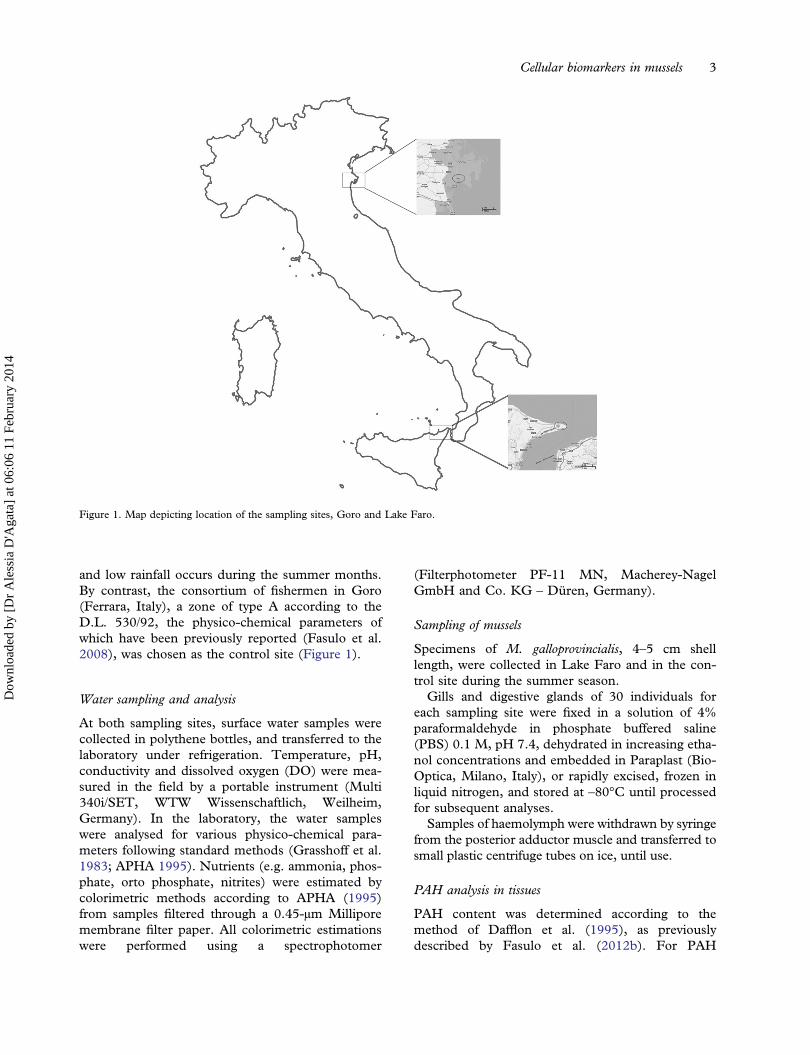
and low rainfall occurs during the summer months.By contrast, the consortium of fishermen in Goro(Ferrara, Italy), a zone of type A according to theD.L. 530/92, the physico-chemical parameters ofwhich have been previously reported (Fasulo et al.2008), was chosen as the control site (Figure 1).
Water sampling and analysis
At both sampling sites, surface water samples werecollected in polythene bottles, and transferred to thelaboratory under refrigeration. Temperature, pH,conductivity and dissolved oxygen (DO) were mea-sured in the field by a portable instrument (Multi340i/SET, WTW Wissenschaftlich, Weilheim,Germany). In the laboratory, the water sampleswere analysed for various physico-chemical para-meters following standard methods (Grasshoff et al.1983; APHA 1995). Nutrients (e.g. ammonia, phos-phate, orto phosphate, nitrites) were estimated bycolorimetric methods according to APHA (1995)from samples filtered through a 0.45-μm Milliporemembrane filter paper. All colorimetric estimationswere performed using a spectrophotomer
(Filterphotometer PF-11 MN, Macherey-NagelGmbH and Co. KG – Düren, Germany).
Sampling of mussels
Specimens of M. galloprovincialis, 4–5 cm shelllength, were collected in Lake Faro and in the con-trol site during the summer season.Gills and digestive glands of 30 individuals for
each sampling site were fixed in a solution of 4%paraformaldehyde in phosphate buffered saline(PBS) 0.1 M, pH 7.4, dehydrated in increasing etha-nol concentrations and embedded in Paraplast (Bio-Optica, Milano, Italy), or rapidly excised, frozen inliquid nitrogen, and stored at –80°C until processedfor subsequent analyses.Samples of haemolymph were withdrawn by syringe
from the posterior adductor muscle and transferred tosmall plastic centrifuge tubes on ice, until use.
PAH analysis in tissues
PAH content was determined according to themethod of Dafflon et al. (1995), as previouslydescribed by Fasulo et al. (2012b). For PAH
Figure 1. Map depicting location of the sampling sites, Goro and Lake Faro.
Cellular biomarkers in mussels 3
Dow
nloa
ded
by [
Dr
Ale
ssia
D'A
gata
] at
06:
06 1
1 Fe
brua
ry 2
014
analysis, the following solvents and reagents wereused: acetonitrile ACN (Romil), water and cyclohex-ane (Chromanorm BDH), acetone (PestinormBDH), potassium hydroxide (KOH), ethanol andexane (Carlo Erba), all of high-performance liquidchromatography (HPLC) grade. The digestiveglands dissected from 15 individuals were pooled inthree samples (each with tissues of five specimens)per each sampling area. Approximately 3 g of eachpooled sample were weighted with an analytical bal-ance Mettler Toledo AT 104 and homogenized in aglass vial using an Ultra-TURRAX IKA T10 basic.The homogenized samples were saponified with10 mL of 1 M KOH in an ethanol solution for 3 hat 80°C in a water bath. Then, 20 mL of cyclohexanewas added and samples mixed by an orbital agitatorfor 10 min using dark glassware (Dafflon et al.1995). The hexanic phase was recovered and thepolar mixture washed once with cyclohexane, andthen discharged. The extracts were filtered, concen-trated under a nitrogen gas stream to about 1 mL,and the concentrated extract was removed with apasteur pipette and loaded into a Varian Bond ElutC18 12-mL cartridge, previously conditioned. Theeluates were dried under nitrogen flow and dissolvedwith 1 mL of acetonitrile before the analysis.
The concentrations of the following 16 PAHsidentified by the Environmental Protection Agency(EPA) as priority pollutants, naphthalene (NA), ace-naphthylene (ACY), acenaphthene (AC), fluorene(FL), phenanthrene (PHE), anthracene (AN), fluor-anthene (FA), pyrene (PY), benzo(a)anthracene(BaA), chrysene (CH), benzo(b)fluoranthene(BbF), benzo(k)fluoranthene (BkF), benzo(a)pyrene(BaP), dibenz(a,h)anthracene (DahA), benzo(g,h,i)perylene (Bghi) and indeno(1,2,3-cd)pyrene (IP),were determined. Quantitative analysis of PAHswas carried out with an HPLC apparatus Pro-Star363 (Varian, Palo Alto, CA) equipped with a 20-mLloop and a fluorescence detector (FLD Pro-Star363). The software used was Star ChromatographyWorkstation version 5.2 (Varian, Palo Alto, CA).
Immunohistochemistry
Sections (4 μm thick) were prepared from paraffin-embedded digestive glands and gills. The sectionswere treated using the indirect immunofluorescencemethod (Mauceri et al. 1999). Non-specific bindingsites for immunoglobulins were blocked by incuba-tions for 1 h with normal goat serum (NGS) in PBS(1:5). The digestive glands sections were incubatedovernight in a humid chamber at 4°C with the pri-mary rabbit polyclonal antibody anti-CYP4 predi-luted (the antibody CYP4 was kindly provided by
Professor Anders Goksøyr of University of Bergen,Norway), while gill sections were incubated by co-localization with a rabbit polyclonal antibody anti-AChE (Chemicon International, Temecula CA)diluted 1:50, and a mouse polyclonal antibody anti-ChAT (Abcam, Cambridge UK), diluted 1:100.After a rinse in PBS for 10 min, the sections wereincubated for 2 h at room temperature withfluorescein isothiocyanate (FITC) conjugated goatanti-rabbit IgG (Sigma) and tetramethylrhodamineisothiocyantate (TRITC) conjugated goat anti-mouse IgG (Sigma), diluted 1:100.Negative controls for the immunohistochemical
labeling were performed by substitution of non-immune sera for the primary antisera.All observations were made on five randomly
selected fields in each slide using a 40× oil-immer-sion objective with a motorized Zeiss Axio ImagerZ1 microscope (Carl Zeiss AG, Werk Göttingen,Germany), equipped with an AxioCam digital cam-era (Zeiss, Jena, Germany) for the acquisition ofimages.
RNA extraction and polymerase chain reaction (PCR)
Total RNA was extracted from the digestive glandsof specimens using TRIzol LS reagent (Invitrogen,Carlsbed, CA, USA) (Chomczynski & Sacchi 1987).The RNA content was quantified using a UV spec-trophotometer (UV Mini 1240 Shimadzu, Milano,Italy).The cDNAwas synthesized using 4 μg of total RNA
and oligo (dt)20 primer (150 pmol/μL) (Invitrogen),with Moloney Murine Leukemia Virus (MMLV)reverse transcriptase (Invitrogen) as prescribed by themanufacturer’s instructions. Two μl of the resultingcDNA were amplified in the PCR reaction.The sequences of primers to CYP4Y1
were designed on the sequence no. AF072855present in Genebank: sense primer 5′-AGGCTTTCACCAGTTCC-3′ and antisense pri-mer 5′- TCCGGCAGAAATGGAGTAAA-3′,amplifying a 172-bp sequence.The actin gene of each examined organism was
used as positive control. The gene was amplifiedusing sequence primers based on the actin cDNAsequence of M. galloprovincialis to obtain a fragmentof 200 bp sequence.PCR was prepared using 2.5 µL of 10× buffer,
0.13 µL of 5 U/µL Poly Taq polymerase (Invitrogen),0.8 µL of 50 mM magnesium chloride (MgCl2), pri-mers (50 µM each), 1 µL of cDNA template, 0.5 µL of10 mM dNTPs (deoxynucleoside triphosphates), andMilli-Q water (Millipore, Vimodrone MI, Italy). Thetotal reaction was performed in a 25-µL volume. The
4 A. D’Agata et al.
Dow
nloa
ded
by [
Dr
Ale
ssia
D'A
gata
] at
06:
06 1
1 Fe
brua
ry 2
014

program used to amplify fragments of cytochromeP450 4Y1 was 95°C for 2 min and 35 cycles at 95°Cfor 30 s, 56°C for 30 s, 72°C for 30 s and a finalextension at 72°C for 5 min.
To perform the reaction we used the MastercyclerEp-Gradient (Eppendorf, Milano, Italy). RT-PCR(Reverse Transcriptase-Polymerase Chain Reaction)products were characterized by electrophoresis onSYBR Safe-stained agarose gel.
Enzyme activities
The activity of AChE was evaluated in the gills of themussels. AChE was determined using the method ofEllman et al. (1961) with slight modifications. Briefly,thiocholine derivatives are hydrolysed by acetylcholi-neterase to yield thiocholine. Subsequent combinationwith 5,5-dithiobis-2-dinitrobenzoic acid (DTNB)forms the yellow anion 5-thio-2-nitrobenzoic acid,which absorbs strongly at 412 nm. The activitieswere expressed as μmol/min/mg.
Lysosomal membrane stability
Haemolymph was analysed for lysosomal membranestability as neutral red retention time (NRRT) (Regoliet al. 2004). Haemolymph collected from the adductormuscle of 30 specimens for each sampling site wasincubated on a glass slide with a freshly prepared neu-tral red working solution (2 µL/mL saline from a stocksolution of 20 mg neutral red dye dissolved in 1 mLdimethyl sulfoxide (DMSO)). The haemocytes weremicroscopically examined (63/100×, Zeiss AxioImager Z1) at 15 min intervals (for up to 90 min) todetermine at what point in time the dye, previouslytaken up by lysosomes, was lost into the cytosol in 50%of granular haemocytes.
Genotoxicity analysis
1. MN assay. Haemolymph of 30 organisms for eachsampling site was spread on slides, transferred to alightproof humidity chamber, and allowed to adhere.After, it was fixed in pure methanol for 10 min,hydrated and then stained with 5% Giemsa solutionfor 15 min. A total of 1000 haemocytes were exam-ined for each specimen under the light microscope(Zeiss Axio Imager Z1) to determine the presence orabsence of micronuclei or other nuclear abnormal-ities (NAs) such as nuclear buds, notched and lobednuclei, bi-nucleated and fragmented-apoptotic cells,as well as micronucleus connected to the mainnucleus, commonly known as “eight-shaped” cells.The NAs were identified using the criteria describedby Fenech et al. (2003) (Barsiene et al. 2012).
2. Comet assay. The comet assay was conducted atpH > 13, according to Steinert et al. (1998), Jha et al.(2005) andD’Agata et al. (2013), with somemodifica-tions (Fasulo et al. 2010). Haemolymph samples werecentrifuged at 2000 rpm for 5 min and pellets resus-pended in Phosphate Buffered Saline (PBS). Tenmicrolitres of the diluted sample were mixed with65 µL of 0.7% low-melting-point (LMP) agarose,then the 75 µL mixture was layered on the precoatedslides on 1% normal melting point agarose. The slideswere covered with a cover slip, and were left for 5 minin a refrigerator to solidify. The cover slip was gentlyremoved and 75 µL of 0.7% low melting agarose wereadded and another cover slip was placed on top. Thesamples were left for another 5 min in the refrigerator.After removing the cover slip, the slides were placed inlysis buffer (2.5MNaCl, 100 mMEDTA-Na, 10mMTris-HCl) for 2 h at 4°C. After lysis, the slides wereplaced in an electrophoresis box for 20 min in runningbuffer, to allow the unwinding to occur.Electrophoresis was performed using the same run-ning buffer at 20 V and 240 mA for 20 min. The slideswere then neutralized with Tris buffer 0.4 M pH 7.5,and stained with Sybr Safe (2.5 µL/mL).The slides were examined with the Zeiss Axio
Imager Z1 fluorescence microscope. To determinewhether visual scoring correlated with computerizedimage analysis, the same cells were also scored forDNA damage using the Comet assay IV software(Perceptive instruments, Suffolk, UK). To quantifythe induced DNA damage, two parameters wereconsidered: the tail length (TL) and the tail moment(TM). TM is a measure of the migrated DNA in thetail, multiplied by the TL (Olive et al. 1990).
Statistical analysis
Immunoreactive cell quantification was performedby counting the positive cells using Axio VisionRelease 4.5 software (Zeiss, Göttingen, Germany).The band intensities of CYP4Y1 were measuredwith Quantity One software (BioRad, Marnes-la-Coquette, France). Results were expressed asmean ± S.D. All the obtained data were statisticallyanalysed with Graph Pad software (Instat, La Jolla,CA, USA) using one-way analysis of variance(ANOVA), and applying the Mann-Whitney U test.
Results
Water physico-chemical analysis
During the summer, in Lake Faro, the water tem-perature at the surface was 27°C, with a salinity of37‰ PSU (Practical Salinity Units). In Goro, the
Cellular biomarkers in mussels 5
Dow
nloa
ded
by [
Dr
Ale
ssia
D'A
gata
] at
06:
06 1
1 Fe
brua
ry 2
014
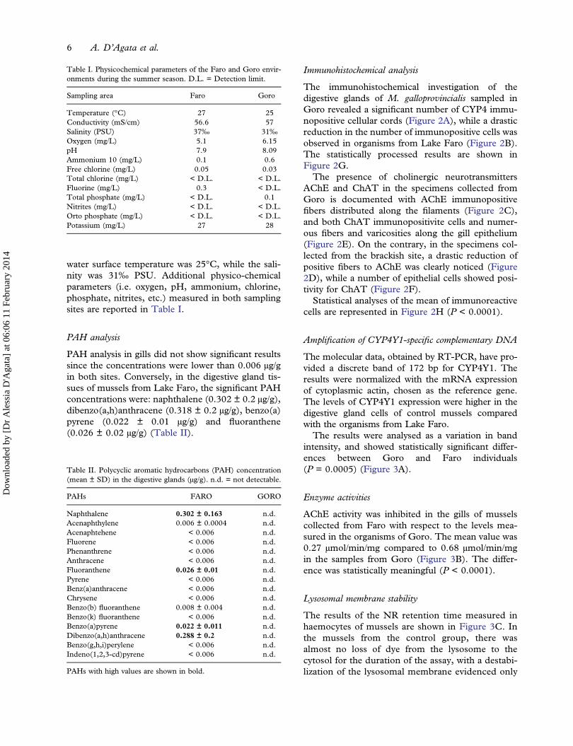
water surface temperature was 25°C, while the sali-nity was 31‰ PSU. Additional physico-chemicalparameters (i.e. oxygen, pH, ammonium, chlorine,phosphate, nitrites, etc.) measured in both samplingsites are reported in Table I.
PAH analysis
PAH analysis in gills did not show significant resultssince the concentrations were lower than 0.006 µg/gin both sites. Conversely, in the digestive gland tis-sues of mussels from Lake Faro, the significant PAHconcentrations were: naphthalene (0.302 ± 0.2 µg/g),dibenzo(a,h)anthracene (0.318 ± 0.2 µg/g), benzo(a)pyrene (0.022 ± 0.01 µg/g) and fluoranthene(0.026 ± 0.02 µg/g) (Table II).
Immunohistochemical analysis
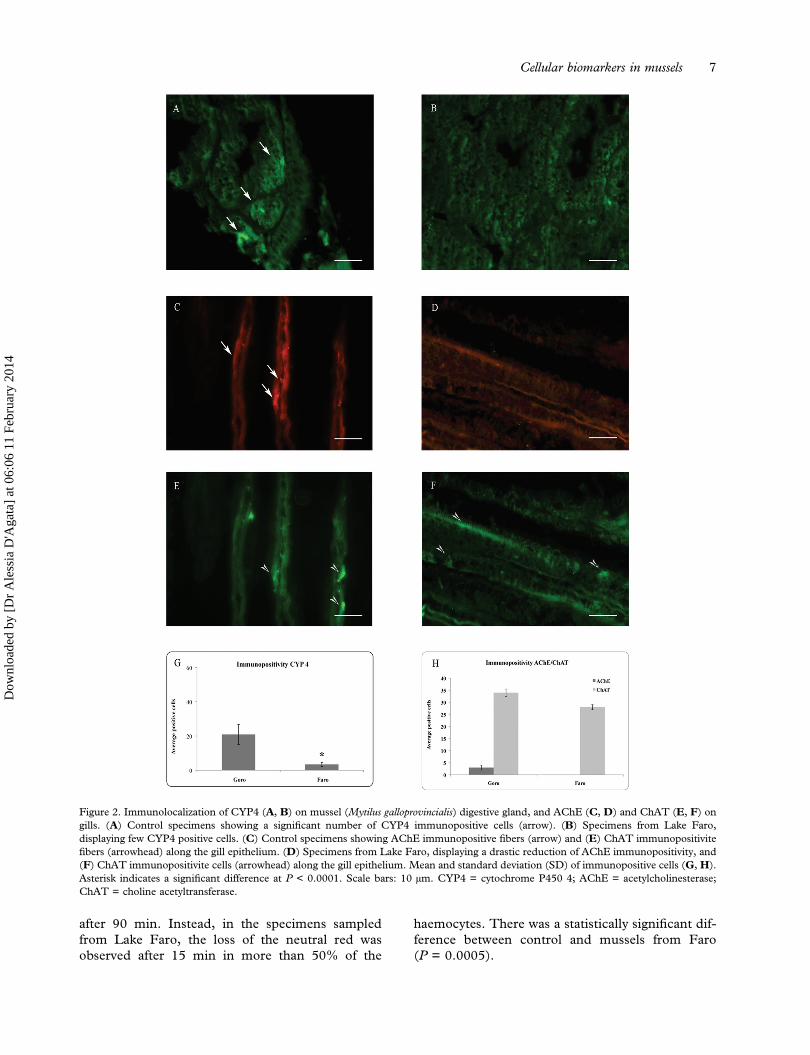
The immunohistochemical investigation of thedigestive glands of M. galloprovincialis sampled inGoro revealed a significant number of CYP4 immu-nopositive cellular cords (Figure 2A), while a drasticreduction in the number of immunopositive cells wasobserved in organisms from Lake Faro (Figure 2B).The statistically processed results are shown inFigure 2G.The presence of cholinergic neurotransmitters
AChE and ChAT in the specimens collected fromGoro is documented with AChE immunopositivefibers distributed along the filaments (Figure 2C),and both ChAT immunopositivite cells and numer-ous fibers and varicosities along the gill epithelium(Figure 2E). On the contrary, in the specimens col-lected from the brackish site, a drastic reduction ofpositive fibers to AChE was clearly noticed (Figure2D), while a number of epithelial cells showed posi-tivity for ChAT (Figure 2F).Statistical analyses of the mean of immunoreactive
cells are represented in Figure 2H (P < 0.0001).
Amplification of CYP4Y1-specific complementary DNA
The molecular data, obtained by RT-PCR, have pro-vided a discrete band of 172 bp for CYP4Y1. Theresults were normalized with the mRNA expressionof cytoplasmic actin, chosen as the reference gene.The levels of CYP4Y1 expression were higher in thedigestive gland cells of control mussels comparedwith the organisms from Lake Faro.The results were analysed as a variation in band
intensity, and showed statistically significant differ-ences between Goro and Faro individuals(P = 0.0005) (Figure 3A).
Enzyme activities
AChE activity was inhibited in the gills of musselscollected from Faro with respect to the levels mea-sured in the organisms of Goro. The mean value was0.27 µmol/min/mg compared to 0.68 µmol/min/mgin the samples from Goro (Figure 3B). The differ-ence was statistically meaningful (P < 0.0001).
Lysosomal membrane stability
The results of the NR retention time measured inhaemocytes of mussels are shown in Figure 3C. Inthe mussels from the control group, there wasalmost no loss of dye from the lysosome to thecytosol for the duration of the assay, with a destabi-lization of the lysosomal membrane evidenced only
Table I. Physicochemical parameters of the Faro and Goro envir-onments during the summer season. D.L. = Detection limit.
Sampling area Faro Goro
Temperature (°C) 27 25Conductivity (mS/cm) 56.6 57Salinity (PSU) 37‰ 31‰Oxygen (mg/L) 5.1 6.15pH 7.9 8.09Ammonium 10 (mg/L) 0.1 0.6Free chlorine (mg/L) 0.05 0.03Total chlorine (mg/L) < D.L. < D.L.Fluorine (mg/L) 0.3 < D.L.Total phosphate (mg/L) < D.L. 0.1Nitrites (mg/L) < D.L. < D.L.Orto phosphate (mg/L) < D.L. < D.L.Potassium (mg/L) 27 28
Table II. Polycyclic aromatic hydrocarbons (PAH) concentration(mean ± SD) in the digestive glands (μg/g). n.d. = not detectable.
PAHs FARO GORO
Naphthalene 0.302 ± 0.163 n.d.Acenaphthylene 0.006 ± 0.0004 n.d.Acenaphtehene < 0.006 n.d.Fluorene < 0.006 n.d.Phenanthrene < 0.006 n.d.Anthracene < 0.006 n.d.Fluoranthene 0.026 ± 0.01 n.d.Pyrene < 0.006 n.d.Benz(a)anthracene < 0.006 n.d.Chrysene < 0.006 n.d.Benzo(b) fluoranthene 0.008 ± 0.004 n.d.Benzo(k) fluoranthene < 0.006 n.d.Benzo(a)pyrene 0.022 ± 0.011 n.d.Dibenzo(a,h)anthracene 0.288 ± 0.2 n.d.Benzo(g,h,i)perylene < 0.006 n.d.Indeno(1,2,3-cd)pyrene < 0.006 n.d.
PAHs with high values are shown in bold.
6 A. D’Agata et al.
Dow
nloa
ded
by [
Dr
Ale
ssia
D'A
gata
] at
06:
06 1
1 Fe
brua
ry 2
014
after 90 min. Instead, in the specimens sampledfrom Lake Faro, the loss of the neutral red wasobserved after 15 min in more than 50% of the
haemocytes. There was a statistically significant dif-ference between control and mussels from Faro(P = 0.0005).
Figure 2. Immunolocalization of CYP4 (A, B) on mussel (Mytilus galloprovincialis) digestive gland, and AChE (C, D) and ChAT (E, F) ongills. (A) Control specimens showing a significant number of CYP4 immunopositive cells (arrow). (B) Specimens from Lake Faro,displaying few CYP4 positive cells. (C) Control specimens showing AChE immunopositive fibers (arrow) and (E) ChAT immunopositivitefibers (arrowhead) along the gill epithelium. (D) Specimens from Lake Faro, displaying a drastic reduction of AChE immunopositivity, and(F) ChAT immunopositivite cells (arrowhead) along the gill epithelium. Mean and standard deviation (SD) of immunopositive cells (G, H).Asterisk indicates a significant difference at P < 0.0001. Scale bars: 10 μm. CYP4 = cytochrome P450 4; AChE = acetylcholinesterase;ChAT = choline acetyltransferase.
Cellular biomarkers in mussels 7
Dow
nloa
ded
by [
Dr
Ale
ssia
D'A
gata
] at
06:
06 1
1 Fe
brua
ry 2
014

Genotoxicity analysis
1. MN assay. The MN test applied on the haemo-cytes of mussels did not reveal the presence ofmicronuclei, but the presence of several nuclearabnormalities in the specimens collected from LakeFaro. In detail, the main abnormalities detected werenuclear blebs and notched lobed. The statistical ana-lysis performed on the average of cells with abnormalnuclei revealed a statistically significant difference(P < 0.0001) between the mussels of Faro andthose of the control, with a frequency of nuclearabnormalities (FNA) of 61‰ in the specimens col-lected from Faro (Figure 3D).
2. Comet assay. A statistically significant increase(P < 0.0001) of DNA damage was observed inhaemocytes from mussels sampled at Faro byapplying the comet assay. The % DNA in thecomet tail was higher (a four-fold increase) com-pared to the control organisms. The ANOVAshowed significant differences between the taillength (P < 0.0001) and the tail moment
(P = 0.0001) values observed in all specimens col-lected from the two sites (Figure 3E).
Discussion
The water quality status of Lake Faro, a naturalconfined brackish environment, was assessed inthis study by applying, for the first time in this site,a multi-biomarker approach on the mussel M. gal-loprovincialis, chosen as the sentinel organism. LakeFaro is particularly subject to anthropogenic impact,resulting in a mixture of xenobiotic substances,including pesticides, organic pollutants, heavymetals and PAHs (Giacobbe et al. 1996; Munaòet al. 2000; Giacalone et al. 2004; Mauceri et al.2005; Scarfì et al. 2006; Arpa Sicilia 2007; Fasuloet al. 2008), and characterised by lowhydrodynamics.In environmental risk assessment, the use of a
battery of biomarkers is strongly recommended(Donnini et al. 2007; Monserrat et al. 2007; Fasuloet al. 2010, 2012a; Iacono et al. 2010) because a
Figure 3. (A) Mean and standard deviation (SD) of the ratio between CYP4Y1 and actin band intensity. Asterisk indicates a significantdifference at P = 0.0005. (B) AChE activity expressed in μmol/min/mg in Mytilus galloprovincialis collected from Goro and Lake Faro.Asterisk indicates a significant difference at P < 0.0001. (C) Neutral red retention time (NRRT) of haemocytes of M. galloprovincialis fromGoro and Faro. In the specimens collected from Lake Faro, the loss of the neutral red was observed after 15 min in more than 50% of thehaemocytes. Asterisk indicates a significant difference at P = 0.0005. (D) Mean frequency of nuclear abnormalities (NA), and (E) tail lengthand tail moment in the haemolymph of mussels collected from Goro and Faro. Asterisk indicates a significant difference at P < 0.0001.CYP4Y1 = cytochrome P450 4Y1; AChE = acetylcholinesterase.
8 A. D’Agata et al.
Dow
nloa
ded
by [
Dr
Ale
ssia
D'A
gata
] at
06:
06 1
1 Fe
brua
ry 2
014

multi-biomarker approach provides a more compre-hensive and integrated view of the biologicalresponses of aquatic organisms, even where thelevels of contaminants are not particularly high(Cravo et al. 2009). The biomarkers applied in thisstudy were selectively chosen to investigate specificbiological responses related to toxic compounds pre-sent in the environment.
In mussels collected from Lake Faro during thesummer season, PAHs, and in particular naphtha-lene, dibenzo(a,h)anthracene, benzo(a)pyrene andfluoranthene, were found in the digestive gland tis-sue, confirming the presence of PAHs in the brackishwater of the pond. An inhibition of CYP4 wasobserved in the digestive glands, according to themolecular analysis with CYP4Y1 mRNA levels sig-nificantly reduced. It is known that the major path-way for lipophilic xenobiotic elimination includesP450-mediated metabolism in the vertebrate liver(Fasulo et al. 2010), or analogous invertebrate tis-sues (Cappello et al. 2013a). Increases in total CYPcontent have been demonstrated in Mytilus sp. afterexposure to organic contaminants (Michel et al.1993), but one CYP4 fragment, named CYP4Y1by the CYP nomenclature committee, has beendescribed in M. galloprovincialis with decreasinggene expression as a result of exposure to naphto-flavone (Snyder 1998; Jonsson et al. 2006), anddown-regulated both at the enzymatic and proteinlevel in the digestive tissue of M. galloprovincialiscaged in a petrochemical area (Cappello et al.2013a).
The neurotoxic effects in mussels were measuredthrough the study of an important neurotransmitter,ACh. Therefore, AChE and ChAT were investigatedin this study, since both enzyme activities are strictlyrelated to the levels of ACh, the main chemicalmediator in the transmission of nerve impulses,with ChAT regulating ACh production and AChEresponsible for its degradation (Walker & Thompson1991; Parsons et al. 1993). The results presented inthis paper show a strong inhibition of AChE in thegills of the specimens collected from Faro withrespect to the control, suggesting the presence ofxenobiotics in the brackish site. Although theAChE activity is inhibited by the presence of neuro-toxic compounds, such as organophosphorus andcarbamate (Day & Scott 1990; Bocquene &Galgani 1998), the responsiveness of AChE tomany other chemical groups, e.g. heavy metals,hydrocarbons and detergents (Guilhermino et al.1998; Fasulo et al. 2010; Ciacci et al. 2012), andalgal toxins (Lehtonen et al. 2003) has also beenacknowledged. Therefore, its inhibition is inter-preted as a very reliable signal of exposure and effect.
As biomarker of biological stress, the stability oflysosomal membranes was also performed on musselhaemolymph. The measure of NRRT showed adecrease in the lysosomal membrane stability of thespecimens collected from Lake Faro after only15 minutes from the start of the observations, con-firming a significant stress to which mussels are sub-jected in the brackish environment. Our results, inagreement with other studies in the field (Regoliet al. 2004; Mamaca et al. 2005; Moore et al.2006, 2008; Nigro et al. 2006; Maisano et al.2013), confirm the usefulness and sensitivity of thelysosomal membrane stability test as a biomarker ofstress for detecting and monitoring sublethal changesin marine organisms.Furthermore, the MN test and comet assay were
used as genotoxicity tests because of heavy metalswith genotoxic potential known to be present in LakeFaro (Fasulo et al. 2008). Both tests revealed thepresence of mutagenic agents in the pond, throughthe presence of a large number of nuclear morpho-logical changes and a substantial damage to DNA, asrevealed by the FNA values, the tail length and tailmoment. DNA damage evaluated by the method ofalkaline comet assay may originate from single- ordouble-strand breaks, adduct formation, alkali-labilesites, or cross-linking DNA-DNA/DNA-protein(Tice et al. 2000).
Conclusion
This study represents the first time the quality statusof Lake Faro has been assessed by using a battery ofbiomarkers on the mussel M. galloprovincialis, whichproved to be suitable to identify the effects of envir-onmental pollutants at both molecular and cellularlevels. The advantage of using the mussel as thesentinel organism is that the changes observed in asessile organism can be attributable to the local andseasonal conditions of the area examined. Indeed,the selected multiple biomarkers applied on M. gal-loprovincialis provided information on the presence ofa mixture of pollutants in Lake Faro, resulting inmetabolic disorders in mussels. Specifically, theobserved inhibition of CYP4 and AChE could belinked to the presence of organic contaminants, andin particular of PAHs detected in the mussel diges-tive gland tissue. The change in the lysosomal stabi-lity is a general indicator of environmental stress,while the increased DNA damage is the result ofexposure to mutagenic and genotoxic compounds.Moreover, the biological responses must also berelated to the seasonal conditions. In fact, as is wellknown, summer represents the most stressful andcritical situation due to higher temperature, and
Cellular biomarkers in mussels 9
Dow
nloa
ded
by [
Dr
Ale
ssia
D'A
gata
] at
06:
06 1
1 Fe
brua
ry 2
014

consequently environmental contamination worsensin a meromictic basin as Lake Faro.
Overall, the results from this work demonstratethe effectiveness of the use of a battery of biomarkersin studies of environmental risk assessment, since amulti-biomarker approach provides a comprehensiveand integrated view of the biological responses ofaquatic organisms.
References
APHA. 1995. Standard methods for the examination of water andwastewater. 19th ed. Washington, DC: American PublicHealth Association.
ARPA SICILIA. 2007. Piano di monitoraggio per la primacaratterizzazione dei corpi idrici superficiali della regionesiciliana. Available: http://www.osservatorioacque.it/documenti/pta/allegati/02/volume%20i%20e%20allegati/allegato%20ii_transizioni.pdf. Accessed July 2013 10.
BanniM,Negri A,Dagnino A, Jebali J, Ameur S, BoussettaH. 2010.Acute effects of benzo[a]pyrene on digestive gland enzymaticbiomarkers and DNA damage onmusselMytilus galloprovincialis.Ecotoxicology and Environmental Safety 73:842–848.
Barsiene J, Rybakovas A, Garnaga G, Andreikenaite L. 2012.Environmental genotoxicity and cytotoxicity studies in musselsbefore and after an oil spill at the marine oil terminal in theBaltic Sea. Environmental Monitoring and Assessment184:2067–2078.
Bocquene G, Galgani F. 1998. Biological effects of contaminants:Cholinesterase inhibition by organophosphate and carbamatecompounds. In: Proceedings of the ICESTechniques inMarineEnvironmental Sciences 22:12. Copenhagen, Denmark: ICES.
Bolognesi C, Rabboni R, Roggieri P. 1996. Genotoxicity biomar-kers in M. galloprovincialis as indicators of marine pollutants.Comparative Biochemistry and Physiology 113:319–323.
Brescia G, Celotti L, Clonfero E, Neumann HG, Forni A, Foà V,Pisoni M, Ferri GM, Assennato G. 1999. The influence ofcytochrome P450 1A1 and glutathione S-transferase M1 gen-otypes on biomarker levels in coke-oven workers. Archives ofToxicology 73:431–439.
Cappello T, Maisano M, D’Agata A, Natalotto A, Mauceri A,Fasulo S. 2013a. Effects of environmental pollution in cagedmussels (Mytilus galloprovincialis). Marine EnvironmentalResearch 91:52–60.
Cappello T, Mauceri A, Corsaro C, Maisano M, Parrino V, LoParo G, Messina G, Fasulo S. 2013b. Impact of environmentalpollution on caged mussels Mytilus galloprovincialis usingNMR-based metabolomics. Marine Pollution Bulletin inpress. doi.org/10.1016/j.marpolbul.2013.10.019.
Castro M, Santos MM, Monteiro NM, Vieira N. 2004. Measuringlysosomal stability as an effective tool for marine coastal envir-onment monitoring. Marine Environmental Research58:741–745.
Chomczynski P, Sacchi N. 1987. Single-step method of RNAisolation by acid guanidium thiocyanate-phenol-chloroformextraction. Analytical Biochemistry 162:156–159.
Ciacci C, Barmo C, Gallo G, Maisano M, Cappello T, D‘Agata A,Leonzio C, Mauceri A, Fasulo S, Canesi L. 2012. Effects ofsub-lethal, environmentally relevant concentrations of hexava-lent chromium in the gills of Mytilus galloprovincialis. AquaticToxicology 120–121:109–118.
Cravo A, Lopes B, Serafim A, Company R, Barreira L, Gomes T,Bebianno MJ. 2009. A multibiomarker approach in Mytilus
galloprovincialis to assess environmental quality. Journal ofEnvironmental Monitoring 11:1673–1686.
D’Agata A, Fasulo S, Dallas LJ, Fisher AS, Maisano M, ReadmanJW, Jha AN. 2013. Enhanced toxicity of ‘bulk’ titanium diox-ide compared to ‘fresh’ and ‘aged’ nano-TiO2 in marine mus-sels (Mytilus galloprovincialis). Nanotoxicology doi:10.3109/17435390.2013.807446.
Da Ros L, Meneghetti F, Nasci C. 2002. Field application oflysosomal destabilization indices in the mussel Mytilus gallopro-vincialis: Biomonitoring and transplantation in the Lagoon ofVenice (north-east Italy). Marine Environmental Research54:817–822.
Dafflon O, Gobet H, Koch H, Bosset JO. 1995. Le dosage deshydrocarbures aromatiques polycycliques dans le poisson, lesproduites carne’s et le fromage par chromatographie liquidea’haute performance. Travaux de chimie alimentaire etd‘hygiène 86:534–555.
Day KE, Scott IM. 1990. Use of acetylcholinesterase activity todetect sublethal toxicity in stream invertebrates exposed to lowconcentrations of organophosphate insecticides. AquaticToxicology 18:101–114.
De Domenico E, Mauceri A, Giordano D, Maisano M, GiannettoA, Parrino V, Natalotto A, D’Agata A, Cappello T, Fasulo S.2013. Biological responses of juvenile European sea bass(Dicentrarchus labrax) exposed to contaminated sediments.Ecotoxicology and Environmental Safety 97:114–123.
de Oliveira David JA, Salaroli RB, Fontanetti CS. 2008. Finestructure of Mytella falcate (Bivalvia) gill filaments. Micron39:329–336.
Dixon DR, Prusky AM, Dixon L, Jha AN. 2002. Marine inverte-brate eco-genotoxicology: A methodological overview.Mutagenesis 17:495–507.
Donnini F, Dinelli F, Sangiorgi F, Fabbri E. 2007. A biologicaland geochemical integrated approach to assess the environ-mental quality of a coastal lagoon (Ravenna, Italy).Environment International 33:919–928.
Ellman GL, Courtney KD, Andres JV, Featherstone RM. 1961. Anew and rapid colometric determination of acetylcholinester-ase activity. Biochemical Pharmacology 7:88–95.
Fasulo S, Brunelli E, Maisano M, Sperone E, Mauceri A, BernabòI, Cappello T, D’Agata A, Tripepi S. 2012a. Toxicity ofForoozan crude oil to ornate wrasse (Thalassoma pavo):Ultrastructure and cellular biomarkers by using integratedapproaches. Italian Journal of Zoology 79:182–199.
Fasulo S, Iacono F, Cappello T, Corsaro C, Maisano M, D‘AgataA, Giannetto A, De Domenico E, Parrino V, Lo Paro G.2012b. Metabolomic investigation of Mytilus galloprovincialis(Lamarck 1819) caged in aquatic environment. Ecotoxicologyand Environmental Safety 84:139–146.
Fasulo S, Marino S, Mauceri A, Maisano M, Giannetto A,D‘Agata A, Parrino V, Minutoli R, De Domenico E. 2010. Amultibiomarker approach in Coris julis living in a natural envir-onment. Ecotoxicology and Environmental Safety 73:1565–1573.
Fasulo S, Mauceri A, Giannetto A, Maisano M, Bianchi N,Parrino V. 2008. Expression of metallothionein mRNAs by insitu hybridization in the gills of Mytilus gallorpovincialis fromnatural polluted environments. Aquatic Toxicology 88:62–68.
Fenech M, Chang WP, Kirsch-Volders M, Holland N, Bonassi S,Zeiger E. 2003. HUMN project: Detailed description of thescoring criteria for the cytokinesis-block micronucleus assayusing isolated human lymphocyte cultures. MutationResearch 534:65–75.
Giacalone A, Gianguzza A, Mannino MR, Orecchio S, PiazzeseD. 2004. Polycyclic aromatic hydrocarbons in sediments of
10 A. D’Agata et al.
Dow
nloa
ded
by [
Dr
Ale
ssia
D'A
gata
] at
06:
06 1
1 Fe
brua
ry 2
014
marine coastal lagoons in Messina, Italy: Extraction and GC/MS analysis, distribution and sources. Polycyclic AromaticCompounds 24:135–149.
Giacobbe MG, Oliva FD, Maimone G. 1996. Environmentalfactors and seasonal occurrence of the DinoflagellateAlexandrium minutum, a PSP potential producer, in aMediterranean lagoon. Estuarine, Coastal and Shelf Science42:539–549.
Gomez-Mendikute A, Elizondo M, Venier P, Cajaraville MP.2005. Characterization of mussel gill cells in vivo and invitro. Cell and Tissue Research 321:131–140.
Grasshoff K, Ehrhardt M, Kremling K. 1983. Methods of seawater analysis. Weinheim: Verlag Chemie.
Gregory MA, Marshall DJ, George RC, Anandraj A, McClurgTP. 2002. Correlations between metal uptake in the soft tissueof Perna perna and gill filament pathology after exposure tomercury. Marine Pollution Bulletin 45:114–125.
Guilhermino L, Barros B, Silva MC, Soares A. 1998. Should theuse of inhibition of cholinesterase as a specific biomarker fororganophosphate and carbamate pesticides be questioned?.Biomarkers 3:157–163.
Iacono F, Cappello T, Corsaro C, Branca C, Maisano M, GioffreG, De Domenico E, Mauceri A, Fasulo S. 2010.Environmental metabolomics and multibiomarker approacheson biomonitoring of aquatic habitats. ComparativeBiochemistry and Physiology Part A: Molecular & IntegrativePhysiology 157:S50–S50.
Jha AN. 2008. Ecotoxicological applications and significance ofthe comet assay. Mutagenesis 23:207–221.
Jha AN, Dogra Y, Turner A, Millward GE. 2005. Impact of lowdoses of tritium on the marine mussel, Mytilus edulis:Genotoxic effects and tissue-specific bioconcentration.Mutation Research 586:47–57.
Jonsson H, Schiedek D, Grøsvik BE, Goksøyr A. 2006. Proteinresponses in blue mussels (Mytilus edulis) exposed to organicpollutants: A combined CYP-antibody/proteomic approach.Aquatic Toxicology 78S:S49–S56.
Lehtonen KK, Kankaanpää H, Leiniö S, Sipiä VO, PflugmacherS, Sandberg-Kilpi E. 2003. Accumulation of nodularin-likecompounds from the cyanobacterium Nodularia spumigenaand changes in acetylcholinesterase activity in the clamMacoma balthica during short-term laboratory exposure.Aquatic Toxicology 64:461–476.
Maisano M, Trapani MR, Parrino V, Parisi MG, Cappello T,D’Agata A, Benenati G, Natalotto A, Mauceri A, CammarataM. 2013. Haemolytic activity and characterization of nemato-cyst venom from Pelagia noctiluca (Cnidaria, Scyphozoa).Italian Journal of Zoology 80:168–176.
Mamaca E, Bechmann RK, Torgrimsen S, Aas E, Bjørnstad A,Baussant T, Le Floch S. 2005. The neutral red lysosomalretention assay and comet assay on haemolymph cells frommussels (Mytilus edulis) and fish (Symphodus melops) exposed tostyrene. Aquatic Toxicology 75:191–201.
Marigómez I, Soto M, Cajaraville MP, Angulo E, Giamberini L.2002. Cellular and subcellular distribution of metals in mol-luscs. Microscopical Research and Technology 56:358–392.
Mauceri A, Fasulo S, Ainis L, Licata A, Lauriano ER, Martinez A.1999. Neuronal nitric oxide synthase (nNOS) expression in theepithelial neuroendocrine cell system and nerve fibers in the gill ofthe catfish,Heteropneustes fossilis. ActaHistochemica 101:437–448.
Mauceri A, Fossi MC, Leonzio C, Ancora S, Minniti F,Maisano M, Lo Cascio P, Ferrando S, Fasulo S. 2005.Stress factors in the gills of Liza aurata (Mugilidae,Teleosts) living in polluted environments. Italian Journal ofZoology 72:285–293.
Michel XR, Suteau P, Robertson LW, Narbonne JF. 1993. Effectsof benzo(a)pyrene 3,3′,4,4′-tetrachlorobiphenyl and2,2’,4,4′,5,5′-hexachlorobiphenyl on the xenobiotic-metaboliz-ing enzymes in the mussel (Mytilus galloprovincialis). AquaticToxicology 27:335–344.
Monserrat JM, Martìnez PE, Geracitano LA, Amado LL, MartinsC, Lopes G, Pinho L, Chaves IS, Ferreira-Cravo M, Ventura-Lima J, Bianchini A. 2007. Pollution biomarkers in estuarineanimals: Critical review and new perspectives. ComparativeBiochemistry and Physiology Part C: Toxicology &Pharmacology 146:221–234.
Moore MN, Allen JI. 2002. A computational model of the diges-tive gland epithelial cells of marine mussels and its simulatedresponses to oil-derived aromatic hydrocarbons. MarineEnvironmental Research 54:579–584.
Moore MN, Allen JI, McVeigh A. 2006. Environmental prognos-tics: An integrated model supporting lysosomal stressresponses as predictive biomarkers of animal health status.Marine Environmental Research 61:278–304.
Moore MN, Koehler A, Lowe D, Viarengo A. 2008. Lysosomesand autophagy in aquatic animals. Methods in Enzymology451:581–620.
Mora P, Fournier D, Norbonne JF. 1999a. Cholinesterases fromthe marine mussels Mytilus galloprovincialis Lmk. and M. edulisL. and from the freshwater bivalve Corbicula fluminea Müller.Comparative Biochemistry and Physiology Part C: Toxicology& Pharmacology 122:353–361.
Mora P, Michel X, Narbonne JF. 1999b. Cholinesterase activity asa potential biomarker in two bivalves. EnvironmentalToxicology and Pharmacology 7:253–260.
Moreira SM, Guilhermino L. 2005. The use of Mytilus gallopro-vincialis acetylcholinesterase and glutathione S-transferasesactivities as biomarkers of environmental contamination alongthe northewest portuguese coast. Environmental Monitoringand Assessment 105:309–325.
Munaò F, Di Pietro A, Scoglio ME, Piperno I. 2000. Elementi intraccia nel complesso lagunare Ganzirri-Faro. Rischio sani-tario. Igiene Moderna 114:243–260.
Nigro M, Falleni A, Del Barga I, Scarcelli V, Lucchesi P, RegoliF, Frenzilli G. 2006. Cellular biomarkers for monitoringestuarine environments: Transplanted versus native mussels.Aquatic Toxicology 77:339–347.
Olive PL, Banath JP, Durand RE. 1990. Heterogeneity in radia-tion-induced DNA damage and repair in tumor and normalcells using the ‘‘comet’’ assay. Radiation Research 122:86–94.
Parsons SM, Prior C, Marshal IG. 1993. Acetylcholine transport,storage and release. Internationa Review of Neurology35:280–390.
Peakall D. 1992. Animal biomarkers as pollution indicators.London: Chapman & Hall.
Peters LD, Shaw JP, Nott M, O‘Hara S, Livingstone DR. 1999.Development of cytochrome P450 as a biomarker of organicpollution in Mytilus sp.: Field studies in United Kingdom (‘SeaEmpress’ oil spill) and the Mediterranean Sea. Biomarkers4:425–441.
Petrovic S, Ozretic B, Krajnovic-Ozretic M, Bobinac D. 2001.Lysosomal membrane stability and metallothioneins in diges-tive gland of mussels (Mytilus galloprovincialis Lam.) as biomar-kers in a field study. Marine Pollution Bulletin 42:1373–1378.
Pisoni M, Cogotzi L, Frigeri A, Corsi I, Bonacci S, Iacocca A,Lancini L, Mastrototaro F, Focardi S, Svelot M. 2004. DNAadducts, benzo(a)pyrene monooxygenase activity, and lysoso-mal membrane stability in Mytilus galloprovincialis from differ-ent areas in Taranto coasta waters (Italy). EnvironmentalResearch 96:163–175.
Cellular biomarkers in mussels 11
Dow
nloa
ded
by [
Dr
Ale
ssia
D'A
gata
] at
06:
06 1
1 Fe
brua
ry 2
014

Rank J, Lehtonen KK, Strand J, Laursen M. 2007. DNA damage,acetylcholinesterase activity and lysosomal stability in nativeand transplanted mussels (Mytilus edulis) in areas close tocoastal chemical dumping sites in Denmark. AquaticToxicology 84:50–61.
Regoli F, Frenzilli G, Bocchetti R, Annarumma F, Scarcelli V,Fattorini D, Nigro M. 2004. Time-course variations of oxyradicalmetabolism, DNA integrity and lysosomal stability in mussels,Mytilus galloprovincialis, during a field translocation experiment.Aquatic Toxicology 68:167–178.
ScarfìK,MauceriA,MaisanoM,Minniti F,BrunoR,FasuloS. 2006.Morpho-functional alterations in branchial epithelium of Mytilusgalloprovincialis (L.) living in natural and polluted environments.Biologia Marina Mediterranea 13:773–776.
SnyderMJ. 1998.CytochromeP450 enzymesbelonging to theCYP4family from marine invertebrates. Biochemical and BiophysicalResearch Communications 249:187–190.
Solé M, Livingstone DR. 2005. Components of the cytochromeP450-dependent monooxygenase system and ‘NADPH-inde-pendent benzo[a]pyrene hydroxylase’ activity in a wide rangeof marine invertebrate species. Comparative Biochemistry andPhysiology 141:20–31.
Sorgi C, Cavallaro M, Brianti E, Ferlazzo M, Giannetto S. 2006.Cryptosporidium and Giardia in mussels (Mytilus galloprovincia-lis) from Faro salt-lake, Sicily. Parassitologia 48:296.
Steinert SA, Streib-Montee R, Leather JM, Chadwick DB. 1998.DNA damage in mussels at sites in San Diego Bay. MutationResearch 399:65–85.
Tice RR, Agurell E, Anderson D, Burlinson B, Hartmann A,Kobayashi H, Miyamae Y, Rojas E, Ryu JC, Sasaki YF.2000. Single cell gel/comet assay: Guidelines for in vitro andin vivo genetic toxicology testing. Environmental andMolecular Mutagenesis 35:206–221.
Vukmirovic M, Bihari N, Zahn RK, Muller W, Batel R. 1994.DNA damage in marine mussel Mytilus galloprovincialis as abiomarker of environmental contamination. Marine EcologyProgress Series 109:165–171.
Walker CH, Thompson HM. 1991. Phylogenetic distribution ofcholinesterases and related esterases. In: Mineau P, editor.Cholinesterase inhibiting insecticides. Their impact on wildlifeand the environment. Chemicals in agriculture. Vol. 2.Amsterdam: Elsevier. pp. 1–17.
Widdows J, Donkin P. 1992. Mussels and environmental con-taminants: Bioaccumulation and physiological aspects. In:Gosling E, editor. The Mussel Mytilus: ecology, physiology,genetics and culture, of developments in aquaculture and fish-eries science. Vol. 25. Amsterdam: Elsevier SciencePublishers. pp. 383–424.
Woolf NJ. 1991. Cholinergic systems in mammalian brain andspinal cord. Progress in Neurobiology 37:475–524.
12 A. D’Agata et al.
Dow
nloa
ded
by [
Dr
Ale
ssia
D'A
gata
] at
06:
06 1
1 Fe
brua
ry 2
014
Related Documents