Vol. 29: 127-135,1997 DISEASES OF AQUATIC ORGANISMS Dis Aquat Org Published May 22 Hemolymph cell types of the mussel Mytil us gall opro vincialis Maria J. Carballal1, M. Carmen Lopezl, Carlos ~ z e v e d o ~ , Antonio Villalbal 'Centro de Investigacidns Marinas. Xunta de Galicia, PO Box 208. E-36600 Vilagarcia de Arousa, Spain '~eparment of Cell Biology, Institute of Biomedical Sciences, University of Oporto, P-4000 Porto, Portugal ABSTRACT. Two types of circulating hemocytes were identified in Mytilus galloprovincialis hemolymph: hyalinocytes and granulocytes. The hyalinocytes are agranular cells and show a central nucleus surrounded by relatively small cytoplasm. The granulocytes are larger than hyalinocytes and have smaller nuclei. Three classes of granulocytes (acidophilic granulocytes, basophilic granulocytes and granulocytes containing both types of granules) were distinguished based on staining properties of their cytoplasmic granules. At the ultrastructural level, the hyahnocytes show characteristics of undif- ferentiated cells. The granulocytes are more differentiated and have membrane-limited cytoplasmic granules. Some granulocytes contain only small granules; others have numerous large granules and some of them contain a mix of granule sizes. Density grad~ent centnfugation allowed the separation of acidophilic granulocytes from the other types. Immunocharacterization demonstrated that the granulo- cytes are an antigenically heterogeneous population. KEY WORDS: Mytilus galloprovincialis Hemocyte types Hemolymph . lsopycnic centrifugation Monoclonal antibodies INTRODUCTION Bivalve hemolymph cells develop a number of func- tions including wound and shell repair, transport and digestion of nutrients, and internal defence (Cheng 1981, 1984, Fisher 1986). In order to understand these functions, it is important to distinguish the hemocyte populations and subpopulations and to characterize them. Two basic cell types are recognized among bivalve hemocytes (reviewed by Cheng 1981, Auffret 1988):agranular (hyalinocytes) and granular (granulo- cytes). Both hemocyte types were observed in every studied species except in Pectinidae, which lack gran- ulocytes (Auffret 1988). Recently, other hemocyte types were identified in Cerastoderma edule (Russell- Pinto et al. 1994) and Scapharca inaequivalvis (Holden et al. 1994). In addition, hemocyte subpopulations whose role is unknown were found. Morphological criteria were considered to be insuffi- cient to characterize mollusc hemocyte types. There- fore, several authors have used other criteria such as the enzyme content (Moore & Lowe 1977, Bayne et al. 1979, Pipe 1990a), antigenic characterization (Yoshino & Granath 1983, 1985, Dikkeboom et al. 1985, 1988, Morvan 1991, Noel et al. 1994) or lectin-related prop- erties (Pipe 1990b). Cell separation techniques have also been applied to obtain pure or enriched fractions of hemocyte populations and subpopulations such as isopycnic centrifugation in a discontinuous density gradient (Cheng et al. 1980, Bachere et al. 1988, Ren- wrantz 1990, Noel et al. 1993, Adema 1994, Ford et al. 1994) and the use of monoclonal antibodies bound to magnetic beads (Morvan 1991). Studies on mussel (mostly Mytilus edulis) hemocyte types have been carried out by several authors (Feng et al. 1977, Moore & Lowe 1977, Bayne et al. 1979, Rasmussen et al. 1985, Pipe 1990a, Renwrantz 1990, Noel et al. 1994), but there is no study on Mytilus galloprovincialis hemocytes. Whether the differences between M. edulis and M. galloprovincialis are suffi- ciently large to warrant full species status for each is a controversial matter. M. galloprovincialis is considered to be a true species by some authors (Sanjuan et al. 1990, 1994, Koehn 1991, McDonald et al. 199 1) and as the subspecies Mytilus edulis galloprovincialis by oth- ers (Gardner 1992, Gosling 1992). Regardless of their O Inter-Research 1997 Resale of full article not permitted

Welcome message from author
This document is posted to help you gain knowledge. Please leave a comment to let me know what you think about it! Share it to your friends and learn new things together.
Transcript

Vol. 29: 127-135,1997 DISEASES OF AQUATIC ORGANISMS Dis Aquat Org
Published May 22
Hemolymph cell types of the mussel Mytil us gall opro vincialis
Maria J. Carballal1, M. Carmen Lopezl, Carlos ~ z e v e d o ~ , Antonio Villalbal
'Centro de Investigacidns Marinas. Xunta de Galicia, PO Box 208. E-36600 Vilagarcia de Arousa, Spain ' ~ e p a r m e n t of Cell Biology, Institute of Biomedical Sciences, University of Oporto, P-4000 Porto, Portugal
ABSTRACT. Two types of circulating hemocytes were identified in Mytilus galloprovincialis hemolymph: hyalinocytes and granulocytes. The hyalinocytes are agranular cells and show a central nucleus surrounded by relatively small cytoplasm. The granulocytes are larger than hyalinocytes and have smaller nuclei. Three classes of granulocytes (acidophilic granulocytes, basophilic granulocytes and granulocytes containing both types of granules) were distinguished based on staining properties of their cytoplasmic granules. At the ultrastructural level, the hyahnocytes show characteristics of undif- ferentiated cells. The granulocytes are more differentiated and have membrane-limited cytoplasmic granules. Some granulocytes contain only small granules; others have numerous large granules and some of them contain a mix of granule sizes. Density grad~ent centnfugation allowed the separation of acidophilic granulocytes from the other types. Immunocharacterization demonstrated that the granulo- cytes are an antigenically heterogeneous population.
KEY WORDS: Mytilus galloprovincialis Hemocyte types Hemolymph . lsopycnic centrifugation Monoclonal antibodies
INTRODUCTION
Bivalve hemolymph cells develop a number of func- tions including wound and shell repair, transport and digestion of nutrients, and internal defence (Cheng 1981, 1984, Fisher 1986). In order to understand these functions, it is important to distinguish the hemocyte populations and subpopulations and to characterize them. Two basic cell types are recognized among bivalve hemocytes (reviewed by Cheng 1981, Auffret 1988): agranular (hyalinocytes) and granular (granulo- cytes). Both hemocyte types were observed in every studied species except in Pectinidae, which lack gran- ulocytes (Auffret 1988). Recently, other hemocyte types were identified in Cerastoderma edule (Russell- Pinto et al. 1994) and Scapharca inaequivalvis (Holden e t al. 1994). In addition, hemocyte subpopulations whose role is unknown were found.
Morphological criteria were considered to be insuffi- cient to characterize mollusc hemocyte types. There- fore, several authors have used other criteria such as the enzyme content (Moore & Lowe 1977, Bayne et al. 1979, Pipe 1990a), antigenic characterization (Yoshino
& Granath 1983, 1985, Dikkeboom et al. 1985, 1988, Morvan 1991, Noel et al. 1994) or lectin-related prop- erties (Pipe 1990b). Cell separation techniques have also been applied to obtain pure or enriched fractions of hemocyte populations and subpopulations such as isopycnic centrifugation in a discontinuous density gradient (Cheng et al. 1980, Bachere et al. 1988, Ren- wrantz 1990, Noel et al. 1993, Adema 1994, Ford et al. 1994) and the use of monoclonal antibodies bound to magnetic beads (Morvan 1991).
Studies on mussel (mostly Mytilus edulis) hemocyte types have been carried out by several authors (Feng et al. 1977, Moore & Lowe 1977, Bayne et al. 1979, Rasmussen et al. 1985, Pipe 1990a, Renwrantz 1990, Noel et al. 1994), but there is no study on Mytilus galloprovincialis hemocytes. Whether the differences between M. edulis and M. galloprovincialis are suffi- ciently large to warrant full species status for each is a controversial matter. M. galloprovincialis is considered to be a true species by some authors (Sanjuan et al. 1990, 1994, Koehn 1991, McDonald et al. 199 1) and as the subspecies Mytilus edulis galloprovincialis by oth- ers (Gardner 1992, Gosling 1992). Regardless of their
O Inter-Research 1997 Resale of full article not permitted

128 Dls Aquat Org
taxonomic rank, each taxon is adapted to different environmental conditions, thus explaining their differ- ent geographical distribution.
Recent findings point out that, in spite of widespread hybridization between Mytilus e d ~ ~ l i s and M. gallo- provincialis in some areas, each taxon maintains ~ t s genetic identity in those areas (Gardner 1995, 1996). As a matter of fact, in hybrid populations (where rep- resentatives of each pure taxa and hybrids coexist) fit- ness advantages for M. galloprovincialis were found, such as higher strength of attachment to the substrate (Gardner & Skibinski 1991, Willis & Skibinski 1992), greater fecundity (Gardner & Skibinski 1990), greater viability (Gardner et al. 1993), and non-susceptibility to the parasitic trematode Prosorrhynchus squamatus (Cousteau et al. 1991). In addition, Hilbish et al. (1994) pointed out that differences in a few genes could be responsible for the significant differences observed in energetic physiology between both taxa.
Evidence from studies on the fine structure of the spermatozoon (Crespo et al. 1990), allozymic loci (San- juan et al. 1994, Quesada e t al. 1995), karyotype (Mar- tinez-Lage et al. 1992) and mitochondrial DNA (San- juAn et a1.1996) indicates that the mussel of Galicia corresponds to the Mytilus galloprovincialis taxon.
Since it was not known if hemocytes of both mussel taxa have different characteristics, the aim of this study was to characterize the circulating hemocytes of Gali- clan mussels, as a preliminary step towards under- standing their defence mechanisms. To this purpose, morphological, antigenic, and differential density cri- teria were used.
MATERIALS AND METHODS
Mussels (shell length 7 to 10 cm) were collected from a culture raft in the Ria d e Arosa (Galicia, NW Spain) and placed in a circulating seawater system in the lab- oratory until the next day. Hemolymph was obtained from the posterior adductor muscle sinus with sterile hypod.ermic needles (23 G) and 2 m1 syringes.
Hemocyte slides. Monolayers of hemocytes were prepared by allowing cells to settle onto glass slides (spontaneous monolayers) or by adhesion of cells to glass slides using a cytocentrifuge (cytospin, smears). In order to prepare spontaneous mon.olayers, hemolymph was withdrawn directly (1:4) in filtered seawater (FSW, 0.22 pm). A drop of this suspension was deposited on clean slides and kept in wet chamber for 30 min. After w a s h ~ n g In FSW, monolayers were fixed for 5 min in 1.25 glutaraldehyde in 0.01 M phosphate buffer and stained with May-Griinwald-Giemsa (Auffret 1989). Monolayers from 10 mussels were made. In order to prepare cytospin smears, hemolymph was
withdrawn and simultaneously diluted 1:3 in anti- aggregant modified Alsever's solution (glucose: 20.8 g ; sodium citrate: 8 g; EDTA: 3.36 g; NaCl: 22.3 g; H 2 0 : 1000 ml) (Bach6re et al. 1988). The number of hemo- cytes in the suspension was estimated in a Malassez hemocytometer. Then, a volume of the suspension con- taining 3 X 10⁴ cells was centrifuged (92 X g, 5 min, 4°C) using a Megafuge 1.0 R (Heraeus) cytocentrifuge. Hemocytes were subsequently fixed and stained with a Hemacolor kit (Merck). Cytospin smears of 540 mus- sels (monthly samples of 30 mussels throughout an 18 mo period) were prepared, since they were used also to determine seasonal variability and the influ- ence of other factors on the hemogram (published else- where).
Hemocyte measurements. Hemocyte dimensions were estimated in a spontaneous monolayer. A total of 174 stained cells were measured under light micro- scopy using an ocular micrometer. Cell size was deter- rmned by measuring the longest axis excluding pseu- dopodia. Nuclear size was estimated by measuring the longest nuclear diameter.
Dimensions of cells, fixed in suspension, were also determined. For this purpose, the hemolymph was withdrawn directly (1 : l ) in a Alsever's solution con- taining 6% formalin to avoid hemocyte degranulation. Hemolymph from 3 mussels was pooled. Hemocytes were fixed for 15 min in this solution and subsequently resuspended in Alsever's solution. Cells were then stained by adding 100 p1 of Trypan Blue. After 30 min, a drop of the solution was deposited on a glass slide and observed with VIDAS 2.1 image analysis system (Kontron, Munich, Germany). The longest diameter of cytoplasm and nucleus of 405 individual hemocytes was measured.
Statistical analysis. Differences in dimensions among hemocyte types were analyzed by ANOVA fol- lowed by Duncan tests for multiple com.parisons (SOLO 2.0, BMDP Statistical Sofware Inc.).
Transmission electron microscopy (TEM). Hemo- lymph of 5 mussels was pooled for this assay. Hemo- lymph of each mussel was withdrawn ( 1 : l ) in fixative solution (5 %, glutaraldehyde in 0.1 M piperazine-N, N'- bis-12-ethanesulfonic acid] (PIPES), pH = 7.2 with 7 % sucrose). After 1 h at 4OC in fixative, cells were pelleted (750 X g, 10 min), washed in buffer and postfixed in l % osmium tetroxide (75 min at 4OC). After washing, pel- lets were pre-embedded in 1.5 "io agar at 40°C and spun (1700 X g, 5 min). Small pieces were cut, dehydrated through graded ethanol solutions, transferred into propylene oxide and finally embedded in Epon resin. Ultrathin sections (50 to 70 nm) were cut and tissues contrasted with aqueous uranyl acetate and lead citrate and observed under a JEOL 100 CXII transmission electron microscope operating at 60 kV.
Carballal et al.: Hemocytes of Mytllus galloprovinc~al~s 129
Separation of hemocyte types by isopycnic centrifu- gation. Blood was obtained and simultaneously diluted (1:l) in Alsever's solution containing 6%) formalin. Hemolymph of 15 mussels was pooled to obtain a sus- pension with ca 40 X 10"ells. After washing twice in Alsever's solution, hemocytes were separated by isopycnic centrifugation in discontinuous density gra- dient of Percoll (Noel et al. 1993). Cells concentrated at each interface were collected separately with a syringe. Cell suspensions from each interface were washed twice in Alsever's solution to remove Percoll. Pellets were resuspended in Alsever's solution and the number of hemocytes was then determined using a Malassez hemocytomer. Cytospin smears of 3 X 10⁴ cells were prepared and stained with hemacolor.
Immunocharacterization. Four different monoclonal
RESULTS
antibodies specific for hemocytes of Mytilus edulis were used. The antibodies were supplied by Dr Danielle Noel (Noel et al. 1991). Hemolymph of 5 mus- sels was withdrawn (1:l) in Alsever's solution con- taining 6 % formalin. Hemocytes were pooled and centrifuged to remove formalin. The pellets were re- suspended in Alsever's solution and cytospin smears of 3 X 10"ells were prepared. The immunoassays were also carried out on cytospin smears of hemocytes sepa- rated in gradients of Percoll. Both types of cytospin smears were fixed in acetone for 5 min, washed in dis- tilled water and used immediately or stored at -20°C for 1 or 2 mo.
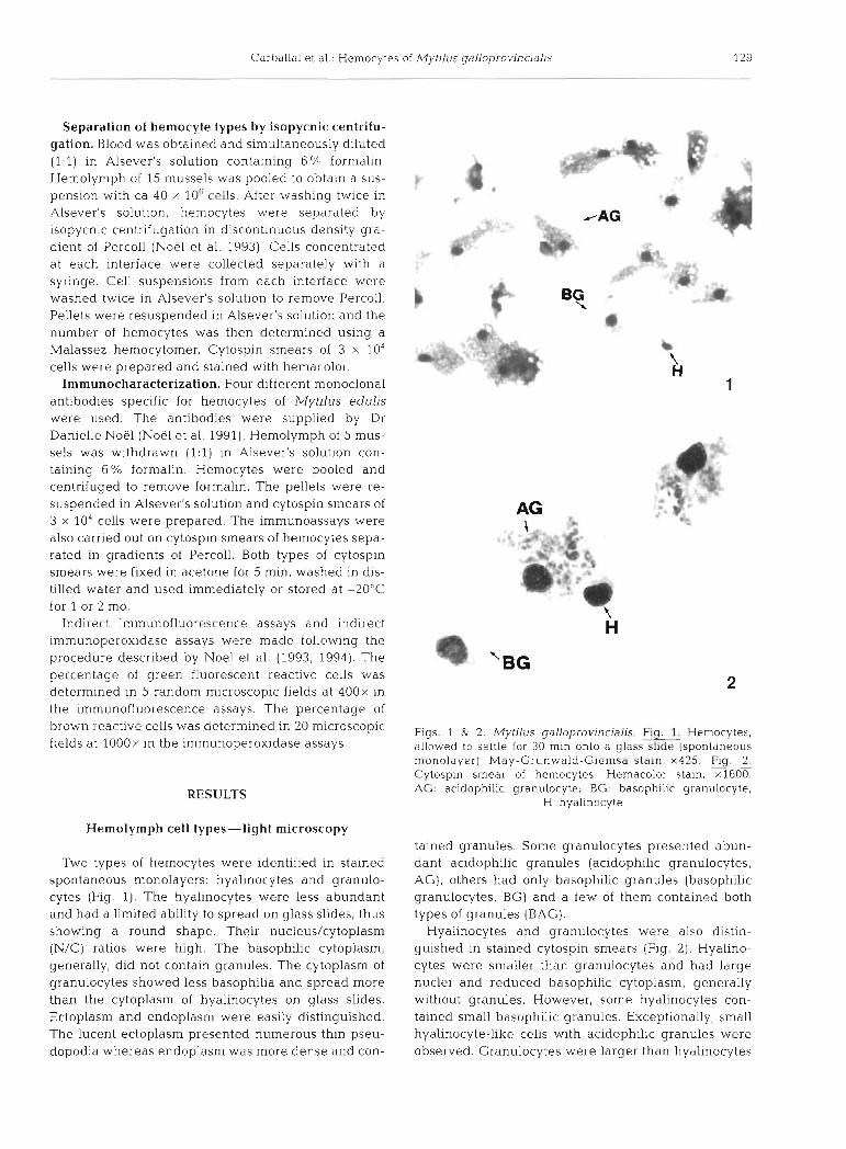
Indirect immunofluorescence assays and indirect immunoperoxidase assays were made following the procedure described by Noel et al. (1993, 1994). The percentage of green fluorescent reactive cells was determined in 5 random microscopic fields at 400x in the immunofluorescence assays. The percentage of brown reactive cells was determined in 20 microscopic F,gs, & 2 , Mytilus galloprovincialjs, Fig, Hemocytes,
fields at lOOOx in the immunoperoxidase assays. allowed to settle for 30 min onto a glass slide (spontaneous monolayer) May-Grunwald-Giemsa stain, x425. Fig. 2. Cytospin smear of hemocytes. Hemacolor stain, x1600. AG: acidophilic granulocyte; BG: basophilic granulocyte;
H: hyalinocyte
Hemolymph cell types-light microscopy
Two types of hemocytes were identified in stained spontaneous monolayers: hyalinocytes and granulo- cytes (Fig. 1). The hyalinocytes were less abundant and had a limited ability to spread on glass slides, thus showing a round shape. Their nucleus/cytoplasm (N/C) ratios were high. The basophilic cytoplasm, generally, did not contain granules. The cytoplasm of granulocytes showed less basophilia and spread more than the cytoplasm of hyalinocytes on glass slides. Ectoplasm and endoplasm were easily distinguished. The lucent ectoplasm presented numerous thin pseu- dopodia whereas endoplasm was more dense and con-
tained granules. Some granulocytes presented abun- dant acidophilic granules (acidophilic granulocytes, AG), others had only basophilic granules (basophilic granulocytes, BG) and a few of them contained both types of granules (BAG).
Hyalinocytes and granulocytes were also distin- guished in stained cytospin smears (Fig. 2 ) . Hyalino- cytes were smaller than granulocytes and had large nuclei and reduced basophilic cytoplasm, generally without granules. However, some hyalinocytes con- tained small basophilic granules. Exceptionally, small hyalinocyte-like cells with acidophilic granules were observed. Granulocytes were larger than hyalinocytes

130 Dis Aquat Org 29: 127-135. 1997
and could be classified according to the staining prop- erties of their cytoplasmic granules in the same way as in spontaneous monolayers. Some BGs contained very few granules. BAGS generally showed more acido- philic than basophilic granules.
Some granulocytes contained refringent orange in- clusions larger than cytoplasmic granules. Large multi- nucleate granulocytes were also observed in a single monolayer of hemocytes. In addition, cells with yello~v- brown granules were seen on several slides. Around 1.5% of the hemocytes (both hyalinocytes and granu- locytes) showed 2 nuclei.
Hemocyte dimensions
Hemocyte dimensions measured in spontaneous monolayers are shown in Table 1. Differences among cell types were found to be significant (p < 0.001) for cell size, nucleus diameter and N/C ratio by means of ANOVA. Neverthel.ess, differences in nucleus diame- ter and N/C ratio between BGs and BAGS were not significant.
Hyalinocytes appeared as the smallest cells with the highest N/C ratio and granulocytes as the largest cells with the lowest N/C ratio.
Hemocytes fixed in suspension had a round shape. This technique permitted distinction between hyalino- cytes and granulocytes, but it was not possible to de- tect different granulocyte types because all granules stained blue. Hemocyte dimensions measured with this technique are shown in Table 2. Differences in cell size, nucleus diameter and N/C ratio between hyalinocytes and granulocytes were significant (p c 0.001; p c 0.05; p c 0.001, respectively).
Table 1 Dimensions of Myt~ lus galloprovincialis hemocytes measured in a spontaneous monolayer. Mean values * stan- dard error and rank of vanation corresponding to cell dia- meter, nucleus diameter and nucleus/cytoplasm (N/C) ratio of each hemocyte type a re shown. H: hyahnocyte; BG: basophilic granulocyte; RAG: granulocyte with baso- philic and acidophilic granules; AG: acidophilic granulocyte;
N: number of measured cells
Cell type Cell diameter (pm1
H (N = 26) 8.73 t 0.54 S to 17
RC; ( N = 42) 43.70 -. 0 91 31 to 55
BAG (N = 35) 44.70 -t 1.34 37 to 66
AG [N = 71) 49 08 5 0.70 37 to 66
Nucleus d ~ a m e t e r (pm)
Table 2. Dimensions of Mytilus galloprov~ncialis hemocytes measured in suspension. Mean t standard error and rank of variation corresponding to cell diameter, nucleus diameter and N/C ratio of each hemocyte type a re shown. H: hya l~n-
ocyte; G: granulocyte; N: number of measured cells
Cell type Cell diameter Nucleus N/C (pm) diameter (pm)
.- -
H ( N = 200) 8.33 ? 0 15 5 05 * 0.10 0.61 + 0.10 5.11 to 12.55 2.46 to 6.95 0.14 to 0 40
G ( N = 205) 10.95 ? 0.16 4 76 * 0.10 0.44 + 0.01 7.30 to 14.74 2 58 to 7.76 0.28 to 0 79
Electron microscopy
Examination of the circulating hemocytes by elec- tron microscopy revealed the occurrence of agranular (hyalinocytes) and granular (granulocytes) hemocytes.
The hyalinocytes appeared as rounded cells with large central nuclei, relatively small cytoplasm, and occasionally with some pseudopodia (Fig. 3). Their cytoplasm contained few organelles, some mitochon- dria, some cisternae and vesicles of rough and smooth endoplasmic reticulum and free ribosomes. Two types of nuclei were observed in the hyalinocytes. Some of them mostly contained euchromatin and others had large clumps of heterochromatin. Some hemocytes similar to hyalinocytes had a more developed cyto- plasm containing long strands of smooth endoplasmic reticulum, numerous free ribosomes and a few small granules (Fig. 4) . The nuclei of the latter frequently contained a nucleolus.
The granulocytes were more abundant than hyalino- cytes. They were ovoid in shape, frequently showing thin pseudopodia, with an eccentric nucleus which contained abundant heterochromatin in the central and peripheral positions. The N/C ratio was lower than in hyalinocytes, and cytoplasmic organelles were more abundant. Granulocytes had abundant mitochondria, cisternae and vesicles of rough and smooth endoplas- rnic reticulum and Golgi complex. In addition, they contained membrane-limited granules with variable electron-dense content. Most of these granules were round or oval in shape and presented a central electron-dense content rounded by an electron-lucent halo. The diameter of the granules ranged from 0.2 to 1.8 pm. Some granulocytes contained only small gran- ules (diameter <0.5 pm) (Fig. 5); others had abundant large granules (diameter >0.5 pm) (Fig. 6) and some of them showed a mix of granule sizes (Fig 7). Cells con- taining small granules had more abundant rnitochon- dria and long strands of rough endoplasmic reticulum around the nucleus. The cytoplasm of cells containing large granules frequently presented vacuoles which

Carballal et a1 Hernocytes of Mytllus gdllopror~~nudl~s
Figs. 3 to 8. ~ y t i l u s galloprovincialis. Hyallnocyte wlth a nucleus containing abundant euchromatln M rnitochondrlon, N: nucleus; R: rough endoplasmic reticulum; V. vacuole. Transmission electron mlcroscopy (TEM). ~ 7 5 0 0 . Hemocyte sun- ilar to hyalinocytes, whose cytoplasm contains long strands of rough endoplasmlc retlculum, abundant free ribosomes and a few small granules (G). Nuc: nucleolus. TEM, x8000. Fig 5 Granulocyte containing small granules P. pseudopod. TEM, x7500.
Granulocyte containing large granules GC Golgi complex TEM, x7500 Granulocyte containing a mix of granule sizes TEM, x8000. Granulocyte containing seronddry lysosomes (LS). W M , x7000

132 Dis Aquat Org 29: 127-135, 1997
included different materials. Some of these vacuoles were secondary lysosomes and residual bodi.es (Fig. 8).
Separation of hemocyte types by isopycnic centrifugation
The hemocyte suspensions were separated into 5 fractions by centrifugation in discontinuous Percoll gradient. The different fractions were formed at the interfaces 10-20%, 20-30%, 30-40% and 40-50% of Percoll and in the pellet. Microscopic examination of different fractions showed that the number of hyalino- cytes and BGs was lower in the interfaces containing a higher percentage of Percoll. However, the number of AGs increased with the density gradient, so that the pellet only contained AGs (Table 3).
Table 3. Percentages of hemocyte types collected at rnterfaces of Percoll gradient and pellet. H: hyalinocyte; BG: basophilic granulocyte; BAG: granulocyte with acidophilic and baso-
philic granules; AG: acidophilic granulocyte
Fraction H (X) BG (%) BAG (%I AG ( l % )
10-20% 29 66 5 0 20-3076 14 76.5 8 1.5 30-4071, 4 4 1 21.5 33.5 40-50% 0 l .S 1 97.5 >50% (pellet) 0 0 0 100
Immunocharacterization with monoclonal antibody
Immunofluorescence assays showed that all mono- clonal antibodies specific for Mytilus edulis hemocytes reacted with M. galloprovincialis hemocytes. The 11 AI-3F9 and 20 D3-3B4 antibodies gave rise to the most intense reactions (Table 4).
The immunoperoxidase staining permitted an easier distinction among hemocyte types than immunofluo- rescence staining. Using this technique, the 13 B9-2E6 antibody showed reaction in the cytoplasm of BGs but
Table 4 . Imm~rnofluorescence staining of Mytllus galloprovjn- cialis hem.ocytes with rn.onoclonal antibod~es raised aga~nst M. edulis hemocytes. +: weak staining; +++ very strong
staining
Antibodv Reactive hernocytes (':L) Intensltv of reaction P P
l 1 AI-3F9 99 +++ 13 B9-2E6 55 +++ 20 D3-3B4 7 9 + C +
18 CS-3H10 51.9 +
not AGs. The remaining antibodies recognized both types of granulocytes. Few hyalinocytes showed reac- tion with 20 D3-3B4, 18 C5-3H10 and l1 AI-3F9 anti- bodies. A higher number of AGs reacted than did BGs. mainly with 20 D3-3B4 and 18 C5-2E6 antibodies. It was impossible to distinguish BAGs clearly using this technique.
Reactivity of hemocytes separated by isopycnic centrifugation in Percoll with 13 B9-2E6
rnonoclonal antibody
Immunofluorescence assays were carried out with this antibody and the hemocytes of 5 fractions obtained by density gradient centrifugation. The highest num- ber of reactive cells was observed at the 20 to 30% interface, where BGs were more abundant (Table 5 ) . The number of reactive cells decreased progressively through the increasing density gradient. No reaction was detected in the pellet which only contained AGs.
Table 5. Irnrnunofluorescence staining of Mytilus galloprovin- cialis hemocytes separated in Percoll gradient using 13
B9-2E6 monoclonal antibody
Fraction Reactive hemocytes (%)
10-20"~- 24.64 20-30"; 49.25 30-40":q 21.21 40-50% 2.78 >50% 0
DISCUSSION
The results of this study reveal the occurrence of 2 hemocyte types in the hemolynlph of Mytilus gallo- provincialis: hyalinocytes and granulocytes. The hya- l~nocytes are agranular cells and present a central large nucleus surrounded by relatively small cyto- plasm. The granulocytes are larger cells with a smaller nucleus and cytoplasmic granules. Moreover, they are more able to spread and produce pseudopodia. AGs, BGs and BAGs can be distinguished according to the staining properties of their granules. The BGs are smaller with larger nuclei than AGs. The BAGs have intermediate sizes.
The results of isopycnic centrifugation showed that hemocytes can be separated by their density. The hyalinocytes and BGs have a lower density than AGs. The latter can be obtained in a pure fraction. There- fore, AGs are the cells with the largest size and the highest density in the mussel hemolymph.

Carballal et al.: Hemocytes of Mytilus galloprovjncialis
Cells containing yellow-brown granules were also observed in hemocyte monolayers. These cells have been descri'bed in other bivalves as brown cells, which have an excretory role (Cheng 1981). Also, the cyto- plasm of some granulocytes presented orange inclu- sions larger than granules. These inclusions are proba- bly phagosomes containing degradation products. In addition, multinucleate giant hemocytes were found on one slide from a mussel heavily infested by a trema- tode, Proctoeces maculatus. These cells have been observed in other molluscs (Sparks & Pauley 1964, Cheng 1984, Auffret 1985). They are considered to be the result of a fusion of granulocytes in some patholog- ical conditions such as postmortem changes or rejec- tion of grafts.
At the ultrastructural level, the hyalinocytes present characteristics of undifferentiated cells: a small volume of cytoplasm that contains few organelles, abundant free ribosomes and a nucleus with abundant euchro- matin. The granulocytes are more differentiated cells and can be classified according to the size of granules. The granulocytes with small granules have more mito- chondria and a well-developed rough endoplasmic reticulum. The granulocytes with large granules show scant rough endoplasmic reticulum and frequently contain secondary lysosomes and residual bodies as a result of their important phagocytic activity (authors' unpubl. data). This last characteristic indicates that they are the most mature cells (Klebanoff et al. 1978).
On the basis of morphological criteria, several authors distinguished granulocyte subpopulations in Mytilus edulis and M. californianus (Feng 1977, Moore & Lowe 1977, Bayne et al. 1979, Rasmussen et al. 1985, Renwrantz 1990). Pipe (1990b) separated the granulo- cytes of the mussel M. edulis in granulocytes with small granules (0.2 to 0.3 pm diameter) and granulo- cytes with large granules (0.5 to 1.5 pm diameter) and did not find clear intermediary types. Noel et al. (1994) demonstrated that the basophilic granulocytes of M. edulis observed by light microscopy correspond to the granulocytes with sillall gran.ules and the acidophilic granulocytes correspond to the granulocytes with large granules. Rasmussen et al. (1985) suggested that small M. edulis granulocytes become larger granulo- cytes with larger granules through maturation. The presence of granulocytes containing a mix of granule sizes in M. galloprovincialis seems to support this hypothesis. Nevertheless, it could also be possible that the granulocytes containing a mix of granules are larger granulocytes synthesizing new granules or a functionally different granulocyte subpopulation.
Monoclonal antibodies have been used for antigenic characterization of the bivalve hemocytes in the case of Crassostrea gigas (Morvan 1991) and Mytilus edulis (Noel et al. 1991, 1994). Noel et al. (1994) demon-
strated that monoclonal antibodies specific for M. edulis hemocytes showed immunofluorescent staining with A4. galloprovincialis, A4. trossulus and M. califor- nianus hemocytes. Our results are very similar to those obtained by Noel, with small differences in the per- centage of reactive cells. These differences are proba- bly caused by different proportions of cell types on the slides.
The results of immunoperoxidase assays suggest that the 13 B9-2E6 monoclonal antibody is specific for BGs. When this monoclonal antibody was assayed with the hemocytes separated through density gradient, the highest number of rective cells was found in the frac- tion containing the highest number of BGs. Further- more, the reaction decreases as the proportion of BGs decreases. The results of these 2 assays demonstrate that Mytilus galloprovincialis hemocytes are an anti- genically heterogeneous population. Similar results were obtained in M, edulis hemocytes (Noel et al. 1994) with the 13 B9-2F8 monoclonal antibody. How- ever, in our case, some hyalinocytes showed reaction with the 20 D3-3B4, 18 C5-3H10, and 11 AI-3F9 mon- oclonal antibodies, whereas Noel et al. (1994) did not observe any reaction in M. edulis hyalinocytes with these 3 antibodies. Therefore, antigenic differences between hyalinocytes and granulocytes observed in M. edu l~s could be considered less important in M. gal- loprovincialis.
Cheng (1981) suggested that AGs are the most mature cells and are probably originated from BGs. Some Mytilus galloprovincjalis granulocytes presented intermediate morphological characteristics (cell size, granular size, staining properties of granules), thus suggesting a maturation process. The fact that 13 B9- 2E6 monoclonal antibody is specific for BCs might lead to a different conclusion. However, as stated by Noel et al. (1994). it is possible that the epitope recognized by this antibody is manifested early and disappears rapidly in the maturation process.
There are several theories on bivalve hemocyte renewal and matmation. Moore & Eble (1977) sug- gested that different hemocyte types are maturing stages within a single cell line. Cheng (1981) proposed an ontogenic model with 2 cell lines, one for hyalino- cytes and another for granulocytes, each originating from a different prohemocyte. Auffret (1988) also sug- gested this last hypothesis for Ostrea edulis and Crass- ostrea gigas hemocytes. Our results do not allow us to confirm any of the previous hypotheses, since the n~orphological variability found by light and electron microscopy in Mytilus galloprovincialis hemocytes could be alternatively explained as transition stages of a single cell line or stages belonging to different cell lines. Further studies are needed to understand bi- valve hemopoiesis.

134 Dis Aquat Org 29: 127-135, 1997
- - - -
The ultrastructural study of Mytilus galloprovincialis hemocytes showed that some hyalinocytes contain a nucleus with abundant euchromatin and others have a nucleus with large clumps of heterochromatin. Both types of hyalinocytes could belong to a different cell line, one for hyalinocytes and another for granulocytes. Another possibility is that the hyalinocytes containing abundant euchromatin give rise to hyalinocytes with more heterochromatin and granulocytes. Also, some hemocytes similar to hyalinocytes had a more devel- oped cytoplasm containing long strands of rough endo- plasmic reticulum, numerous free ribosomes and some small granules. These cells are probably intermediate forms that give rise to granulocytes, since they seem to have an important role in protein synthesis and the granules contain abundant enzymes (authors' unpubl. results)
Some granulocytes with 2 nuclei were found in hemocyte monolayers. This may indicate that granulo- cytes have a separate origin from hyalinocytes. Moore & Lowe (1977) also observed Mytilus edulis granulo- cytes containing 2 nuclei and suggested that there can be a nuclear replication without cell division.
In conclusion, results of our studv show similarities
Auffret M (1988) Bivalve hemocyte morphology. Am Fish Soc Spec Publ 18 169-177
Auffret M (1989) Comparative study of the hemocytes of two oyster species: the European flat oyster, Ostrea edulis, Linnaeus. 1750 and the Pacific oyster, Crassostrea gigas (Thunberg, 1793) J Shellfish Res 2:367-373
Bachere E, Chagot D, Grizel H (1988) Separation of Cras- sostrea gigas hemocytes by density gradient centrifuga- tion and counterflow centrifugal elutriation. Dev Comp Immunol 12549-559
Bayne CJ. Moore MN, Carefoot TH, Thompson RJ (1979) Hemolymph functions in Mytilus californianus: the cyto- chem~stry of hemocytes and their responses to foreign implants and hemolymph factors in phagocytosis. J Inver- tebr Pathol 34 1-20
Cheng TC (1981) Bivalves. In: Ratcliffe NA, Rowley AF (eds) Invertebrate blood cells, Vol 11. Academic Press, London, p 231-300
Cheng TC (1984) A classi.fi.cation of molluscan hemocytes based on functional evidences. In: Cheng TC (ed) Com- parative pathob~ology, Vol 6. Invertebrate blood cells and serum factors. Plenum, New York, p 111-146
Cheng TC, Huang JW, Karadogan H, Renwrantz LR, Yoshino T (1980) Separation of oyster hemocytes by density gradi- ent centrifugatlon and identification of their surface receptors. J lnvertebr Pathol 36:35-40
Cousteau C, Renaud F, Maillard C, Pasteur N, Delay B (1991) Differential susceptibility to a trematode parasite among genotypes of the Mytilus edulis/galloprovincialis complex.
between hemocytes of Mytilus galloprovincialis and Genet Res Camb 57:207-212 Crespo CA, Garcia Caballero T, Beiras A, Espinosa J (1990)
M. edulis. Nevertheless, some new aspects with regard Evidence from sperm ultrastructure that the mussel of to previous studies on M. edulis hemocytes were Galician estuaris is Mytilus galloprovincialis Lamark. uncovered. The occurrence of multinucleate giant J Mollusc Stud 56:127-128
hemocytes, circulating brown cells, and binucleate Dikkeboom R, TiInagel JMGH. blulder EC, Van der Knaap
hyalinocytes was not previously reported. At the ultra- WPW (1987) Monoclonal antibody recognized hemocyte subpopulations in juvenile and adult Lymnaea stagnalis:
stuctural level, 2 nucleus types were observed among functional characteristics and lectin bindina. Dev C o m ~ d
the hyalinocytes, one of them mostly contamed Immunol 12 17-32
euchromatin and the other with abundant heterochro- Dlkkeboom R, Tllnagel JMGH, Van der Knaap WPW (19881
matm. Granulocytes with similar proportion of large Monoclonal antibody recognized hemocyte subpopula- tions In juvenlle and adult Lymnaea stagnahs: functional
and small cytoplasmic granules were found. Some of characteristics and lectin binding. Dev Comp Immunol the monoclonal antibodies reacting with granulocytes 12.17-32 also showed reaction with some hyalinocytes.
Acknowledgememts. We are grateful to Dr Danielle Noel for supplying the monoclonal antibodies against Mytilus edulis hemocytes, to hls Laura Corral, who provided technical assis- tance with ultrastructural studies, and Dr JosP blolarcs for his technical assistance with imagc analysis equipment. The mussel farmer Mr Luis Losada supplled the mussels for this study.
LITERATURE CITED
Adema CM, Mohandas A, Van der Knaap WPW, Smlnia T (1994) Separation ot Lyrnnaea stagnalis hemocytes by dcnslty gradient centrifugation. Dev Comp lmmunol 18 25-31
Auffret M (1985) Morpholog~e comparative des types hemo- cytarles chez quelques mollusques bivalves d'interet com- mcr r~a l . These de doctorat, Universite de Eretagne Occl- dentale, Brest
Dlkkeboorn R , Van der Knaap WPW, Maaskant JJ , De Jonge AJ (1985) Different subpopulations of hemocytes in juve- nile, adult and Trichobilharzia ocellata-infwted Lymnaea stagnahs: a characterization uslng monoclonal antibodies. Z Parasitenkd 71:815-819
Feng SY, Feng JS. Yamasu T (1977) Roles of ~Myt~lus coruscus and Crassostrea gigas blood cells in defense and nutrition. In: Bulla LA Jr, Cheng TC (eds) Compardtive pathobiol- ogy, Vol3. Plenum, London, p 31-67
Fisher WS (1986) Structure and function of oyster hemocytes. In: Brehelin M (ed) Immunity in invertchrdtes. Springer- Verlag. Berlin, p 25-35
Ford SE, Ashton-Alcox KA, Kanaley SA (1994) Comparative cytomctric and microscopic analysis of oyster hemocytes. J lnvertebr Pathol 64:114-122
Gardner JPA (1992) Mytilus galloprovinclalls (Lmk) (Bivalvia, Molluscs): the taxonomic status of the Mediterranean mussel Ophelia 35 (3): 219-243
Gardner JPA (1995) Developmental stability IS not disrupted b>- extensive hybridization and introgresslon among pop- ulat~ons of the manne bivalve molluscs Myt~lus edulis (L . ) and M galloprovrncitrlis (Lmk.) from south-west England. 0101 J Linn Soc 54:71-86
Carballal et al.: Hemocytes of Mytilus galloprovinc~alis
Gardner JPA (1996) The Mytilus edulis species complex in south~vest England: effects of hybridization and introgres- sion upon interlocus associations and morphometric varia- tion. Mar Biol 125:385-399
Gardner JPA, Skibinski DOF (1990) Genotype-dependent fecundity and temporal variation of spawning in hybrid mussel (Mytilus) population. Mdr Biol 105:153-162
Gardner JPA, Skibinski DOF (1991) Biological and physical factors influenc~ng genotype-dependent mortality in hybrid mussel populations Mar Ecol Prog Ser 71:235-243
Gardner JPA, Skibinski DOF, Bajdik CD (1993) Shell growth and viability differences between the marine mussels A4ytilus edulis (L.), Mytilus galloprovincialis (Lmk.), and their hybrids from two sympatric populations in S .W. Eng- land. Biol Bull 185:405-416
Gosling EM (1992) Genetics of Mytilus. In: Gosling E (ed) The mussel Mytilus: ecology, physiology, genetics and culture. Developments in aquaculture and fisheries science, Vol 25. Elsevier, Amsterdam, p 309-382
Hilbish TJ. Bayne BL, Day A (1994) Genetics of physiological differentiation within the marine mussel genus Mytilus. Evolution 48(2): 267-286
Holden JA, Pipe RK, Quaglia A, Ciani G (1994) Blood cells of the arcid clam Scapharca inaequivalvis. J Mar Biol Assoc UK 74:287-299
Klebanoff SJ, Clark RA (1978) The neutrophil: function and clinical disorders. North-Holland Biomedical Press/Else- vier, Amsterdam
Koehn RK (1991) The genetics and taxonomy of species in the genus Mytilus Aquaculture 94:125-145
Martinez-Lage A, Insua A, Martinez Exposito MJ, Mendez J (1992) Estudio comparativo entre una poblacion d e mejil- 1on gallego y otra d e mejillon mediterraneo. Seminario Internacional do Mexillon, Cuadernos da Area d e Cien- cias Mariiias, Vol 6. Sem~nar io de Estudos Galegos (ed), Edicios do Castro, Sada, p 57-63
McDonald JH, Seed R, Koehn RK (1991) Allozyme and mor- phometric characters of three species of Mytilus in the Northern and Southern hemispheres. Mar Biol 111: 323-335
Moore CA, Eble AF (1977) Cytochemical aspects of Merce- naria mercenaria hemocytes. Biol Bull 152:105-119
Moore MN, Lowe DM (1977) The cytology and the cytochem- ~s t ry of the hemocytes of Mytilus edulis and their reponses to experimentally injected particles. J Invertebr Pathol 29: 18-30
Morvan A (1991) Caracterisation antigenique des hemocytes de l'huitre Crassostrea gigas et analyse de leur activite anti-infectieuse oxygene-dependante. Rapport d e D.E.A., Universite de Bordeaux 11
Noel D, Bachere E, Mialhe E (1993) Phagocytosis associated chemiluminescence of heinocyte In Mytilus edulis (Bi- valvia). Dev Comp Immunol 17:483-493
Responsible Subject Editor: A. K. Sparks. Seattle, Washington, USA
Noel D, Boulo V, Chagot D, Mialhe E (1991) Preparation and characterization of n~onoclonal antibodies against neo- plastic hemocytes of Mytilus edulis. Dis Aquat Org 10: 51-58
Noel D, Pipe R , Elston R. Bachere E, Mialhe E (1994) Anti- genic characterization of hemocyte subpop~~lat ions in the mussel Mytilus edulis by means of monoclonal antibodies. Mar Biol 119:549-556
Pipe R K (1990a) Hydrolytic enzymes associated w ~ t h the gl-anular haemocytes of the marlne mussel Mytilus edulis. Histochem J 22:595-603
Pipe RK (1990b) Differential binding of lectins to haemocytes of the mussel Mytilus edulis. Cell Tissue Res 261:261-268
Quesada H , Zapata C , Alvarez G (1995) A multilocus allo- zyme discontinuity in the mussel Mytilus galloprovincialls: the interaction of ecological and life-history factors. Mar Ecol Prog Ser 116:99-115
Rasmussen LPD, Hage E, Karlog 0 (1985) An electron micro- scope study of the circulating leucocytes of the marine mussel Mytilus edulis. J lnvertebr Pathol 45:158-167
Renwrantz L (1990) Internal defence system of Mytilus edulis. In: Stefano GB (ed) Neurobiology of Mytilus edulis. Man- chester University Press, Manchester, p 256-275
Russell-Pinto F. Reimao R, De Sousa M (1994) Haemocytes in Cerastoderma edule (Molluscs Bivalvia): distinct cell types engage in different responses to sheep erythrocytes. Fish Shellfish Immunol 4:383-397
Sanjuan A, Comesaria AS, De Carlos A (1996) Macrogeo- graphic differentation by mtDNA restriction site analysis in the SW. European Mytilus galloprovincialis Lmk. J Exp Mar Biol Ecol 198:89-100
Sanjuan A, Quesada H, Zapata C, Alvarez G (1990) On the occurrence of Mytilus galloprovincialis Lmk. on the NW coast of the Iberian Peninsula. J Exp Mar Biol Ecol 143: 1-14
Sanluan A, Zapata C, Alvarez G (1994) Mytilus galloprov~n- cialis and M. edulis on the coasts of the Iberian Peninsula. Mar Ecol Prog Ser 113:131-146
Sparks AK, Pauley GB (1964) Studies of normal postmorten changes in the oyster, Crassostrea gigas (Thunberg). J Insect Path01 6:78-101
Willis GL, Skibinski DOF (1992) Variation in strength of attachment to the substrate explains differential mortality in hybrid mussel (Mytllus galloprovincialis and M. edu l~s ) populations. Mar Biol 112:403-408
Yoshino TP, Granath WO J r (1983) Identification of antigeni- cally distinct hernocyte subpopulations in ~ i o m ~ h a l a r i a glabrata (Gastropoda] using monoclonal antibodies to surface membrane markers. ce l l Tlsssue Res 232:553-564
Yoshino TP, Granath WO J r (1985) Surface antigens of Biom- phalaria glabrata (Gastropoda) hemocytes: functional het- erogeneity in cell subpopulations recognized by a nlono- clonal antibody. J Invertebr Pathol 45:174-186
Manuscript first received: April 26, 1996 Revised version accepted: January 15, 1997
Related Documents