MYELOID NEOPLASIA Cell transformation by FLT3 ITD in acute myeloid leukemia involves oxidative inactivation of the tumor suppressor protein-tyrosine phosphatase DEP-1/ PTPRJ Rinesh Godfrey, 1 Deepika Arora, 1 Reinhard Bauer, 1,2 Sabine Stopp, 1 Jo ¨rg P. Mu ¨ller, 1 Theresa Heinrich, 1 Sylvia-Annette Bo ¨hmer, 1 Markus Dagnell, 3 Ulf Schnetzke, 4 Sebastian Scholl, 4 Arne O ¨ stman, 3 and Frank-D. Bo ¨ hmer 1 1 Institute of Molecular Cell Biology, Center for Molecular Biomedicine, Jena University Hospital, Jena, Germany; 2 Center for Sepsis Control and Care, Jena University Hospital, Jena, Germany; 3 Cancer Center Karolinska, Karolinska Institutet, Stockholm, Sweden; and 4 Department of Hematology/Oncology, Clinic for Internal Medicine II, Jena University Hospital, Jena, Germany Signal transduction of FMS-like tyrosine kinase 3 (FLT3) is regulated by protein- tyrosine phosphatases (PTPs). We re- cently identified the PTP DEP-1/CD148/ PTPRJ as a novel negative regulator of FLT3. This study addressed the role of DEP-1 for regulation of the acute myeloid leukemia (AML)–related mutant FLT3 inter- nal tandem duplication (ITD) protein. Our experiments revealed that DEP-1 was ex- pressed but dysfunctional in cells trans- formed by FLT3 ITD. This was caused by enzymatic inactivation of DEP-1 through oxidation of the DEP-1 catalytic cysteine. In intact cells, including primary AML cells, FLT3 ITD kinase inhibition reacti- vated DEP-1. DEP-1 reactivation was also achieved by counteracting the high levels of reactive oxygen species (ROS) produc- tion detected in FLT3 ITD–expressing cell lines by inhibition of reduced NAD phos- phate (NADPH)–oxidases, or by overex- pression of catalase or peroxiredoxin-1 (Prx-1). Interference with ROS production in 32D cells inhibited cell transformation by FLT3 ITD in a DEP-1–dependent man- ner, because RNAi-mediated depletion of DEP-1 partially abrogated the inhibitory effect of ROS quenching. Reactivation of DEP-1 by stable overexpression of Prx-1 extended survival of mice in the 32D cell/C3H/HeJ mouse model of FLT3 ITD– driven myeloproliferative disease. The study thus uncovered DEP-1 oxidation as a novel event contributing to cell transfor- mation by FLT3 ITD. (Blood. 2012;119(19): 4499-4511) Introduction Acute myeloid leukemia (AML) is the most frequent leukemia in adults with improving but still limited treatment possibilities, notably in elderly patients. 1,2 It arises by malignant transformation of myeloid progenitor cells. Among the contributing genetic lesions, mutations in the class III receptor tyrosine kinase (RTK) FMS-like tyrosine kinase 3 (FLT3) occur in approximately 30% of patients. 3 The prevalent type of FLT3 mutations are internal tandem duplications (ITD) of amino acid stretches in the juxtamembrane domain or in the tyrosine kinase domain, 4,5 which confer cytokine- independent proliferation and resistance to apoptosis, and causally contribute to AML in combination with additional genetic lesions. 1 Compared with the ligand-activated wild-type (WT) FLT3, FLT3 ITD mutants exhibit not only elevated but also altered signaling quality, with very pronounced activation of signal transducer and activator of transcription (STAT)5 as one characteristic feature. 6,7 FLT3 ITD also causes the production of high levels of reactive oxygen species (ROS). 8,9 Signal transduction of RTKs is regulated by protein-tyrosine phosphatases (PTPs). PTPs prevent ligand-independent RTK activation, and contribute to modulation and termination of ligand-induced signaling. 10 The activity of PTPs is regulated at several different levels. 11 One regulatory principle is the revers- ible oxidation of the PTP active-site cysteine, which leads to reversible inactivation. 12,13 Temporary inactivation of nega- tively regulating PTPs by this mechanism is believed to be important for efficient RTK signal propagation in the cell. 14 A major ROS causing cellular PTP oxidation is hydrogen peroxide (H 2 O 2 ), which can be generated by a dismutase reaction from superoxide anions, the reaction products of NADPH-oxidases. Activation of the NADPH oxidase isoform 1 (NOX1) occurs downstream of RTK activation, and involves activation and membrane translocation of the small guanosine triphosphate (GTP)ase Rac1. 15 ROS generation in the cell is counteracted by efficient ROS decomposing systems. 16 These include peroxire- doxins (Prx), which have a very low K m for H 2 O 2 and can eliminate it even at low concentrations. 17 Relatively little is known about PTPs regulating FLT3 signal transduction. We have previously shown that the nontransmem- brane PTPs PTP1B and SHP-1 can potently dephosphorylate FLT3 on overexpression. Further, PTP1B appears important for suppressing signaling of newly synthesized FLT3. 7,18 SHP-2 acts as a positive regulator, because it is important for Erk activation and proliferation induced by ligand-activated WT FLT3. However, it is dispensable for FLT3 ITD–mediated transformation. 19 We previously performed a shRNA-based screen to identify PTPs regulating WT FLT3. The initial screen assessed the effects of shRNAs for 20 PTPs on FL-induced Erk1/2 activation in WT FLT3-expressing 32D cells. Among several potentially relevant PTPs, we validated and character- ized density-enhanced phosphatase-1 (DEP-1, systematic name PTPRJ, also CD148) as a negative regulator of WT FLT3 autophosphorylation and signaling. 20 Submitted February 11, 2011; accepted March 11, 2012. Prepublished online as Blood First Edition paper, March 20, 2012; DOI 10.1182/blood-2011-02-336446. The online version of this article contains a data supplement. The publication costs of this article were defrayed in part by page charge payment. Therefore, and solely to indicate this fact, this article is hereby marked ‘‘advertisement’’ in accordance with 18 USC section 1734. © 2012 by The American Society of Hematology 4499 BLOOD, 10 MAY 2012 VOLUME 119, NUMBER 19 For personal use only. on March 22, 2016. by guest www.bloodjournal.org From

Welcome message from author
This document is posted to help you gain knowledge. Please leave a comment to let me know what you think about it! Share it to your friends and learn new things together.
Transcript

MYELOID NEOPLASIA
Cell transformation by FLT3 ITD in acute myeloid leukemia involves oxidativeinactivation of the tumor suppressor protein-tyrosine phosphatase DEP-1/ PTPRJRinesh Godfrey,1 Deepika Arora,1 Reinhard Bauer,1,2 Sabine Stopp,1 Jorg P. Muller,1 Theresa Heinrich,1
Sylvia-Annette Bohmer,1 Markus Dagnell,3 Ulf Schnetzke,4 Sebastian Scholl,4 Arne Ostman,3 and Frank-D. Bohmer1
1Institute of Molecular Cell Biology, Center for Molecular Biomedicine, Jena University Hospital, Jena, Germany; 2Center for Sepsis Control and Care, JenaUniversity Hospital, Jena, Germany; 3Cancer Center Karolinska, Karolinska Institutet, Stockholm, Sweden; and 4Department of Hematology/Oncology, Clinic forInternal Medicine II, Jena University Hospital, Jena, Germany
Signal transduction of FMS-like tyrosinekinase 3 (FLT3) is regulated by protein-tyrosine phosphatases (PTPs). We re-cently identified the PTP DEP-1/CD148/PTPRJ as a novel negative regulator ofFLT3. This study addressed the role ofDEP-1 for regulation of the acute myeloidleukemia (AML)–related mutant FLT3 inter-nal tandem duplication (ITD) protein. Ourexperiments revealed that DEP-1 was ex-pressed but dysfunctional in cells trans-formed by FLT3 ITD. This was caused byenzymatic inactivation of DEP-1 through
oxidation of the DEP-1 catalytic cysteine.In intact cells, including primary AMLcells, FLT3 ITD kinase inhibition reacti-vated DEP-1. DEP-1 reactivation was alsoachieved by counteracting the high levelsof reactive oxygen species (ROS) produc-tion detected in FLT3 ITD–expressing celllines by inhibition of reduced NAD phos-phate (NADPH)–oxidases, or by overex-pression of catalase or peroxiredoxin-1(Prx-1). Interference with ROS productionin 32D cells inhibited cell transformationby FLT3 ITD in a DEP-1–dependent man-
ner, because RNAi-mediated depletion ofDEP-1 partially abrogated the inhibitoryeffect of ROS quenching. Reactivation ofDEP-1 by stable overexpression of Prx-1extended survival of mice in the 32Dcell/C3H/HeJ mouse model of FLT3 ITD–driven myeloproliferative disease. Thestudy thus uncovered DEP-1 oxidation asa novel event contributing to cell transfor-mation by FLT3 ITD. (Blood. 2012;119(19):4499-4511)
Introduction
Acute myeloid leukemia (AML) is the most frequent leukemia inadults with improving but still limited treatment possibilities,notably in elderly patients.1,2 It arises by malignant transformationof myeloid progenitor cells. Among the contributing geneticlesions, mutations in the class III receptor tyrosine kinase (RTK)FMS-like tyrosine kinase 3 (FLT3) occur in approximately 30% ofpatients.3 The prevalent type of FLT3 mutations are internal tandemduplications (ITD) of amino acid stretches in the juxtamembranedomain or in the tyrosine kinase domain,4,5 which confer cytokine-independent proliferation and resistance to apoptosis, and causallycontribute to AML in combination with additional genetic lesions.1
Compared with the ligand-activated wild-type (WT) FLT3, FLT3ITD mutants exhibit not only elevated but also altered signalingquality, with very pronounced activation of signal transducer andactivator of transcription (STAT)5 as one characteristic feature.6,7
FLT3 ITD also causes the production of high levels of reactiveoxygen species (ROS).8,9
Signal transduction of RTKs is regulated by protein-tyrosinephosphatases (PTPs). PTPs prevent ligand-independent RTKactivation, and contribute to modulation and termination ofligand-induced signaling.10 The activity of PTPs is regulated atseveral different levels.11 One regulatory principle is the revers-ible oxidation of the PTP active-site cysteine, which leads toreversible inactivation.12,13 Temporary inactivation of nega-tively regulating PTPs by this mechanism is believed to beimportant for efficient RTK signal propagation in the cell.14 A
major ROS causing cellular PTP oxidation is hydrogen peroxide(H2O2), which can be generated by a dismutase reaction fromsuperoxide anions, the reaction products of NADPH-oxidases.Activation of the NADPH oxidase isoform 1 (NOX1) occursdownstream of RTK activation, and involves activation andmembrane translocation of the small guanosine triphosphate(GTP)ase Rac1.15 ROS generation in the cell is counteracted byefficient ROS decomposing systems.16 These include peroxire-doxins (Prx), which have a very low Km for H2O2 and caneliminate it even at low concentrations.17
Relatively little is known about PTPs regulating FLT3 signaltransduction. We have previously shown that the nontransmem-brane PTPs PTP1B and SHP-1 can potently dephosphorylateFLT3 on overexpression. Further, PTP1B appears important forsuppressing signaling of newly synthesized FLT3.7,18 SHP-2acts as a positive regulator, because it is important for Erkactivation and proliferation induced by ligand-activated WTFLT3. However, it is dispensable for FLT3 ITD–mediatedtransformation.19 We previously performed a shRNA-basedscreen to identify PTPs regulating WT FLT3. The initial screenassessed the effects of shRNAs for 20 PTPs on FL-inducedErk1/2 activation in WT FLT3-expressing 32D cells. Amongseveral potentially relevant PTPs, we validated and character-ized density-enhanced phosphatase-1 (DEP-1, systematic namePTPRJ, also CD148) as a negative regulator of WT FLT3autophosphorylation and signaling.20
Submitted February 11, 2011; accepted March 11, 2012. Prepublished online asBlood First Edition paper, March 20, 2012; DOI 10.1182/blood-2011-02-336446.
The online version of this article contains a data supplement.
The publication costs of this article were defrayed in part by page chargepayment. Therefore, and solely to indicate this fact, this article is herebymarked ‘‘advertisement’’ in accordance with 18 USC section 1734.
© 2012 by The American Society of Hematology
4499BLOOD, 10 MAY 2012 � VOLUME 119, NUMBER 19
For personal use only.on March 22, 2016. by guest www.bloodjournal.orgFrom
In this study the role of DEP-1 for regulation of FLT3 ITD, theAML-related mutant version of FLT3 was explored. The experi-ments revealed that DEP-1 was expressed but not functioning as aPTP for FLT3 ITD. The high ROS levels in FLT3 ITD–expressingcells led to partial inactivation of DEP-1 by reversible oxidation.Blocking ROS formation with different approaches restored DEP-1activity and attenuated transformation in vitro and in vivo. Ourfindings reveal the reversible inactivation of a tumor-suppressivePTP as a previously unrecognized mechanism which contributes tocell transformation by FLT3 ITD.
Methods
Cell lines
HEK293, HEK293T cells, and the human AML cell lines, EOL-1, THP-1,RS4-11, MOLM-13, and MV4-11 were purchased from German Collectionof Microorganisms and Cell Cultures (DSMZ). The Phoenix Amphopackaging cell line was a kind gift of Dr G. Nolan (Stanford UniversityMedical Center, Stanford, CA). AML cell lines were maintained inRPMI-1640 (Biochrom) with 10% heat-inactivated fetal bovine serum(FBS) with the exception of RS4-11, which was maintained in Alpha MEM(GIBCO-BRL) with 10% heat inactivated FBS. HEK 293 and PhoenixAmpho were maintained in DMEM/F12 (GIBCO-BRL) with 10% FBS.32D mouse hematopoietic progenitor cells stably expressing murineFLT3-WT or FLT3 ITD were a kind gift of Dr J. Duyster (TechnicalUniversity of Munich, Munich, Germany) and were maintained in HEPES(N-2-hydroxyethylpiperazine-N�-2-ethanesulfonic acid)–buffered RPMI-1640 (Biochrom) with 10% heat-inactivated FBS, 1mM sodium pyruvate,and 2.5 ng/mL murine recombinant IL3 (Peprotech). 32D cells expressinghuman WT FLT3 or FLT3 ITD were previously described,20 and were usedfor FLT3 autophosphorylation experiments.
AML patient samples
Leukemic cells from AML patient blood were isolated by Ficoll (Biochrom)density gradient separation by standard procedures and processed for theexperiments within 2 to 3 hours after taking. The patient study wasapproved by the local ethics committee and each patient gave writteninformed consent. The characteristics of AML patients included in thisstudy are summarized in supplemental Table 1 (available on the Blood Website; see the Supplemental Materials link at the top of the online article).Determination of the FLT3 mutation status is outlined in supplementalMethods.
DNA constructs
Plasmids encoding WT hFLT3, hFLT3 ITD,18 and HA-tagged hDEP-121
were previously described. The human NOX4 expression construct was akind gift of Dr David Lambeth (Emory University Medical School, Atlanta,GA). The construct encoding a fluorescent sensor (HyPer-cyto), capable forhighly specific detection of H2O2, was from Evrogen. A vector encodingdominant negative (dn) STAT5 was a kind gift of Dr Edith Pfitzner (Institutefor Biochemistry, CMB, Jena University, Germany). Retroviral vectorsencoding dnRac-1 and rat Prx-1 were kindly provided by Dr HenrikUngefroren (Department of General and Thoracic Surgery, UniversityHospital Kiel, Kiel, Germany), and Dr Jack van Horssen (VU UniversityMedical Center, Amsterdam, The Netherlands), respectively. A full-lengthPTPRJ-promoter reporter construct was previously described.21 shRNAconstructs for knockdown of endogenous DEP-1 in 32D cells werepreviously reported.20
Antibodies and reagents
Antibodies and reagents are described in supplemental Methods.
Transient transfections and generation of cell lines with alteredgene expression
Transient transfections were carried out using branched polyethylenimine(PEI; Sigma-Aldrich; 40872-7) as previously described.21 Further detailsare described in supplemental Methods.
ROS measurements
Measurements in HEK293 cells. The use of the HyPer vector and thedetection of ROS with carboxy-H2DFFDA and confocal microscopy aredescribed in supplemental Methods.
Detection of ROS with carboxy-H2DFFDA and a fluorescence platereader. This method was used to analyze suspension cells. Cells werewashed with serum-free RPMI-1640 medium, and starved for 4 hours.Inhibitor treatments were made simultaneously during this starvation time.After that, cells were washed twice with Krebs Ringer phosphate glucosebuffer (KRPG; 145mM NaCl, 5.7mM KH2PO4, 4.86mM KCl, 0.54mMCaCl2, 1.22mM MgSO4, 5.5mM glucose), and then resuspended in 1 mLKRPG (2 � 106 cells/assay point). Carboxy-H2DFFDA (20�M) was added,the suspension mixed well, and then incubated in dark for 20 to 30 minutesat room temperature. The subsequent steps were strictly carried out in dark.The cells were washed twice with KRPG and transferred to a 12-well dish.The fluorescence intensity was quantified with a fluorescence microplatereader (TECAN Infinite 200 plate reader) with excitation at 485 nm andemission at 530 nm.
PTP activity measurements
All the PTP activity-related work was carried out in an anaerobic chamber,Glovebox P10 R215T2s (TGS Glovebox Systemtechnik GmbH). Cellswere lysed using deoxygenated lysis buffer in the anaerobic chamber. Thelysates were then subjected to immunoprecipitation of different PTPs. Theimmunoprecipitates were washed and then used for PTP activity assayusing the Tyrosine Phosphatase assay system (Promega Corporation).Control precipitations with nonspecific immunoglobulin (Ig)G were carriedout and assayed in parallel, and the activity values were subtracted fromthose for specific immunoprecipitates. Alternatively, PTP activity wasassayed with a 32P-labeled peptide substrate as previously described.22
General PTP oxidation was assessed using the “modified PTP in-gelassay,”13 and PTEN oxidation was measured based on mobility alterationand with an alkylation method. A detailed description of these methods isprovided in supplemental Methods.
Biotin labeling of the DEP-1 catalytic cysteine
DEP-1 was labeled with biotin using a previously described method23 withseveral modifications. In brief, cells were lysed using deoxygenated lysisbuffer at pH 5.5 containing 5mM EZ-Link iodoacetyl-PEG2-biotin. Afterallowing complete alkylation of reactive thiols, the lysate was used forDEP-1 immunoprecipitation. Finally, biotin labeling was detected usingantibiotin antibodies. A detailed description is provided in supplementalMethods.
Proliferation and colony formation assays
These are described in supplemental Methods.
Animal experiments
Animal experiments were carried out as described.7 Eight- to 10-week-oldC3H/HeJ mice (The Jackson Laboratory), which are syngenic to 32D cells,were used to assess the in vivo development of myeloproliferative disease(MPD). 32D cells (2 � 106) engineered as described in the figure legend ofFigure 7 and expressing equal levels of mFLT3 ITD (sorted for equalreceptor expression) were injected into the lateral tail vein. The experimentswere performed in accordance with the Animal Welfare Guidelines andwere approved by the local ethics committee of the regional government of
4500 GODFREY et al BLOOD, 10 MAY 2012 � VOLUME 119, NUMBER 19
For personal use only.on March 22, 2016. by guest www.bloodjournal.orgFrom
Thuringia. To assess DEP-1 knockdown levels at the end of experiment,spleen cells were fluorescence-activated cell sorter (FACS)–sorted to enrichthe GFP-expressing 32D FLT3 ITD cells, expanded for 2 days in cultureand subjected to immunoblotting and quantitative qRT-PCR (qRT-PCR)based detection of DEP-1 mRNA expression as described in supplemen-tal Methods.
Results
Dysfunction of DEP-1 in FLT3 ITD–expressing cells
We recently identified DEP-1 as a negative regulator of FLT3phosphorylation, and FLT3 signaling among 20 PTPs screened by ashRNA-based knockdown approach in 32D murine myeloid pro-genitor cells expressing WT FLT3.20 Negative control of FLT3signaling is exemplified by hyperphosphorylation of WT FLT3 inDEP-1 depleted cells (Figure 1A).20
To address if DEP-1 also acts as a negative regulator of FLT3ITD we explored the effect of DEP-1 knockdown on FLT3 ITDphosphorylation. Surprisingly, no alteration of the constitutivelyhigh FLT3 ITD phosphorylation could be observed, despite theefficient knockdown of DEP-1 by shRNA expression (Figure 1B).Similarly, DEP-1 knockdown in the FLT3 ITD–expressing humanMV4-11 cell line did not affect the level of FLT3 ITD autophosphor-ylation (Figure 1C). These findings suggested that DEP-1 knock-down may not affect FLT3 ITD mediated transformation. This wastested in FLT3 ITD–expressing 32D cells. Transformation of thesecells is associated with efficient formation of colonies in methylcel-lulose, and by development of a MPD on injection of the cells intosyngeneic C3H mice. Strongly reduced DEP-1 levels did indeed,neither affect colony formation (supplemental Figure 1B) nor MPDdevelopment in vivo (data not shown, see also Figure 7C).
These findings were compatible with the presence of inactiveDEP-1 in FLT3 ITD–expressing cells. A set of experiments wastherefore performed, which analyzed the impact of FLT3 ITD onthe activity of DEP-1.
PTP-activity analysis of immunoprecipitated DEP-1 demonstratedsignificantly reduced DEP-1 activity in FLT3 ITD–expressing 32D cellscompared with parental 32D cells or WT FLT3-expressing 32D cells(Figure 1D). To test a possibly more general character of this phenom-enon and to enable mechanistic studies, we also performed experimentsin HEK293 cells.Also in these cells, DEP-1 activity was clearly reducedin the presence of FLT3 ITD, without noticeable effects on DEP-1protein levels (Figure 1E, supplemental Figure 2A). FLT3 ITD did notaffect the activity of a reporter construct harboring the DEP-1 promoter(supplemental Figure 2B), or DEP-1 mRNA levels assayed by qRT-PCR (supplemental Figure 2C). The analysis of DEP-1 mRNA in AMLpatient samples by qRT-PCR revealed a large variation in expression butno significant differences related to the FLT3 ITD status (supplementalFigure 2D). In normal CD34� progenitor cells we detected compara-tively low DEP-1 mRNA levels (supplemental Figure 2D), precludingassessment of DEP-1 activity from technical reasons. Our observation isconsistent with earlier reports indicating highly variable DEP-1 expres-sion in different hematopoietic cell populations, with high expression inmyeloid and B cells.24,25 Normal CD34� cells and AML cells haverecently been reported to exhibit strikingly different mRNA expressionprofiles.26
Taken together, the described findings supported the notionthat FLT3 ITD causes reduced DEP-1 activity compared withcells expressing WT FLT3 rather than alterations in mRNA orprotein levels.
FLT3 ITD induces high levels of ROS
Given the high sensitivity of PTPs to oxidative inactivation,14 andrecently reported elevated ROS levels in FLT3 ITD–expressingcells,8,9 it appeared possible that DEP-1 inactivation in FLT3 ITDcells was caused by elevated ROS levels. To investigate thispossibility, we first characterized the effects of FLT3 ITD on ROSproduction in different cell models.
High levels of ROS production were detectable with thefluorescent dye 5-(and-6)-carboxy-2�,7�-difluorodihydrofluores-cein diacetate (carboxy-H2DFFDA) in 32D cells stably expressingFLT3 ITD, whereas much lower levels of ROS were detectable inthe corresponding parental 32D cells, or 32D cells stably express-ing FLT3 WT (Figure 2A). In addition, overexpression of FLT3ITD (but not WT FLT3) in HEK293 cells resulted in elevatedfluorescence of a hydrogen peroxide (H2O2) sensitive GFP, ex-pressed from the vector pHyPer,27 which was comparable with asignal induced by overexpression of the constitutively active NOXisoform NOX4 (supplemental Figure 3A).15
Importantly, the human AML cell lines MV4-11 and MOLM13harboring FLT3 ITD also produced high levels of ROS, whereasthe AML lines THP-1, RS4-11, and EOL-1, which express WTFLT3, exhibited lower ROS levels (Figure 2B). Finally, primaryblasts from AML patients expressing FLT3 ITD had a clear trend tohigher ROS-levels than blasts from patients with WT FLT3 (Figure2C). Comparison with FLT3 ITD allele-load revealed some posi-tive correlation in that the 4 patients with highest allele loads alsoshowed highest ROS levels (supplemental Figure 4A black bars). Itshould be noted that these measurements were not side-by-sidecomparisons, which may have obscured larger differences, as theywere observed in a previous study.8
To identify possibilities of interference with FLT3 ITD–inducedROS production, we tested inhibition of several components ofFLT3 ITD–dependent signaling.
Inhibition of FLT3 ITD kinase activity with the selective classIII RTK inhibitor AG1295, treatment of the cells with diphenyleneiodonium (DPI), a flavoprotein inhibitor which potently blocksNOX activity,28 or with the general antioxidant 6-hydroxy-2,5,7,8-tetramethylchroman-2-carboxylic acid (TROLOX) potently sup-pressed FLT3 ITD-driven ROS production (supplemental Figure3B-C). Similar effects were obtained after transfection with a dnform of the small GTPase Rac1 (RacN17) and on treatment withthe PI3-kinase inhibitor wortmannin, but not with UO126, aninhibitor of the MAPK pathway (supplemental Figure 3B-C).Overexpression of a dnSTAT5 protein, or treatment of the cellswith a small-molecule STAT5 antagonist29 also reduced ROSproduction in presence of FLT3 ITD (supplemental Figure 3B-C).
We also tested if activation of WT FLT3 would cause ROSproduction and had any effect on DEP-1 activity. Stimulation ofWT FLT3–expressing 32D cells with FL indeed led to a small, butsignificant increase in detectable ROS, but this was not accompa-nied by an alteration in DEP-1 activity (supplemental Figure 3D).
FLT3 ITD–induced ROS is required for DEP-1 inactivation
The findings described above suggested that FLT3 ITD–induced ROSwas involved in the FLT3 ITD–dependent inactivation of DEP-1. Todirectly test this idea, a set of experiments was performed analyzing ifalterations in ROS production, or ROS scavenging would affect DEP-1activity in FLT3 ITD–expressing cells.
In an initial experiment, catalase overexpression was used as anapproach for ROS scavenging. Interestingly, catalase quenched the
PTP OXIDATION DRIVEN BY FLT3 ITD IN AML 4501BLOOD, 10 MAY 2012 � VOLUME 119, NUMBER 19
For personal use only.on March 22, 2016. by guest www.bloodjournal.orgFrom
Figure 1. Dysfunction of DEP-1 in FLT3 ITD–expressing cells. 32D cells, either expressing WT FLT3 (A) or FLT3 ITD (B), which were stably transduced with nontargeting(control) or DEP-1 targeting shRNA (as indicated), or MV4-11 cells, transiently transfected with a DEP-1–targeting siRNA (C) were starved and stimulated with FLT3 ligand (FL)for the indicated time periods. FLT3 phosphorylation was assessed by immunoblotting in cell lysates with antibodies recognizing phosphorylated FLT3 (pFLT3, pY589, orpY591). Numbers under the blot indicate relative phosphorylation of FLT3, normalized to FLT3 amounts and to the value at 2.5 minutes stimulation in the control sh/siRNA-treated cells(1.0). Blots in the right panel reveal the DEP-1 knockdown in the used 32D cell pools, which were assayed in parallel to the signaling experiments. Note that blot sections are from the samemembranes with identical exposure and image processing, but were rearranged for better clarity. DEP-1 activity was assayed in stably transfected 32D cells (D) or transiently transfectedHEK293 cells (E) by measuring dephosphorylation of a phosphotyrosine-containing phosphopeptide with the malachite green assay in DEP-1 immunoprecipitates (valuescorrected for nonspecific precipitation with IgG controls; means of normalized values for 3 separate experiments � standard deviation (SD), significance tested by t test). Celllysis, immunoprecipitation, and activity assay were performed in an anaerobic chamber. Bottom panels verify equal DEP-1 protein levels.
4502 GODFREY et al BLOOD, 10 MAY 2012 � VOLUME 119, NUMBER 19
For personal use only.on March 22, 2016. by guest www.bloodjournal.orgFrom

inhibitory effect of FLT3 ITD expression on DEP-1 activity (Figure3A), suggesting that H2O2 mediates the inactivation of DEP-1.
Further support for an involvement of oxidative inactivation ofDEP-1 in FLT3 ITD cells was obtained in activity assays performedin the absence or presence of reducing agents. The activity ofDEP-1 isolated from FLT3 ITD–overexpressing HEK293 cellscould partially be recovered by treatment of the immunoprecipi-tates with dithiothreitol (DTT), indicating that the inactivation wasat least partly because of reversible oxidation (Figure 3B).
To directly explore the effect of FLT3 ITD–mediated ROSproduction on the oxidation state of the DEP-1 catalyticcysteine, we used a previously reported alkylation approach.23
In this assay increased PTP oxidation is detected as a reducedsignal from the alkylating agent, which is only reacting with thereduced form of PTPs. As shown in Figure 3C, alkylation ofDEP-1 with a biotin-labeled iodoacetic acid-derivative is greatlydiminished in cells overexpressing FLT3 ITD, compared withWT FLT3–expressing cells. Coexpression of catalase led toenhanced alkylation, consistent with catalase-mediated rescueof PTP activity.
Furthermore, treatment of cells with DPI rescued activity ofDEP-1 in the presence of FLT3 ITD, consistent with a potential roleof NOX-derived ROS in the oxidation of DEP-1 in FLT3 ITD cells.Rescue of DEP-1 activity was detectable toward a syntheticphosphopeptide in DEP-1 immunoprecipitates (Figure 3D). More-over, DEP-1 could dephosphorylate coexpressed FLT3 ITD inintact cells on activation by DPI treatment (Figure 3E). The latterexperiment proved that the phosphotyrosines of FLT3 ITD areaccessible to DEP-1, and that the initially observed defectiveregulation by DEP-1 was not caused by specific structural featuresof FLT3 ITD. It was also possible to force FLT3 ITD dephosphory-lation in HEK293 cells by overexpression of DEP-1 to very high
levels (data not shown). Consistent results were obtained with themore recently described NOX inhibitor VAS2870.30 This com-pound inhibited ROS levels in FLT3 ITD–expressing 32D cells,and concomitantly led to substantial elevation of DEP-1 activity(Figure 3F).
It is probable that further PTPs contribute to regulation of FLT3signaling activity. We therefore explored the effects of FLT3 ITD onactivity of additional PTPs. We first tested PTP oxidation in 32D cellsusing a generic test, the modified in-gel PTP assay.13 In this assay,reversibly oxidized PTPs can be detected based on their activity insodium dodecyl sulfate (SDS)–polyacrylamide gel electrophoresis(PAGE) containing a [32P]labeled substrate. Whereas cell treatment withH2O2 as a positive control caused abundant PTP oxidation, the presenceof FLT3 ITD did not lead to any pronounced PTPoxidation detectable inthis assay (Figure 4A). It should be noted that this assay is not suitable todetect activity of transmembrane PTPs such as DEP-1. We also usedimmunoprecipitation-based assays to assess the activity of PTP1B, aubiquitously expressed nontransmembrane PTP, and of CD45, anabundant hematopoietic transmembrane PTP. Whereas the former wasnot affected in activity by coexpression of FLT3 ITD (Figure 4B), thelatter showed only a trend of weakly reduced activity (Figure 4C). Wealso assessed another member of the PTP family, the lipid phosphataseand tensin homolog (PTEN). It negatively controls the PI3-kinase/AKTpathway, which is important for FLT3 ITD–mediated transformation.31
Oxidation of PTEN has recently been shown in T-cell acute lymphoblas-tic leukemia cells.29 PTEN oxidation can be detected based on a highermobility of the oxidized species in nonreducing SDS-PAGE gels, and bya 2-step alkylation strategy leading to visualization of reversiblyoxidized PTEN as a biotinylated species.32 Both techniques did notreveal oxidized PTEN in FLT3 ITD–expressing cells (Figure 4D-F),whereas H2O2 readily caused PTEN oxidation. Although further PTPsneed to be investigated, we concluded from these experiments that
Figure 2. FLT3 ITD mediates generation of high levels ofROS. ROS production in 32D cells (A) or human myeloid celllines (B) expressing either WT FLT3 or FLT3 ITD (asindicated) was measured with 2 � 106 cells per point using5-(and-6)-carboxy-2�,7�-difluorodihydrofluorescein diacetate(carboxy-H2DFFDA) in a fluorescence plate reader (mean� SD, determination in triplicates, representative experimentof 3 with consistent results). (C) Elevated ROS production inAML patient blasts harboring FLT3 ITD. AML patient blastswere isolated from freshly taken blood samples. ROS levelswere measured in a fluorescence plate reader using carboxy-H2DFFDA. Values for 10 or 7 patients, harboring FLT3 WT orFLT3 ITD, respectively, are shown (significance tested byt test). Further data of the individual patients are provided insupplemental Figure 4.
PTP OXIDATION DRIVEN BY FLT3 ITD IN AML 4503BLOOD, 10 MAY 2012 � VOLUME 119, NUMBER 19
For personal use only.on March 22, 2016. by guest www.bloodjournal.orgFrom
Figure 3. DEP-1 is inactivated by ROS in FLT3 ITD–expressing cells. (A) Rescue of DEP-1 activity by coexpression of catalase (CAT). The experiment wasperformed as in Figure 1E (means of normalized values for 3 separate experiments � SD, significance tested by t test). (B) Partial recovery of DEP-1 activity by DTTtreatment. DEP-1 was immunoprecipitated from HEK293 cells, which were transiently transfected as indicated. The immunoprecipitates were treated with 5mM DTT atroom temperature for 10 minutes, or mock-incubated without DTT as indicated, then washed 3 times with PTP assay buffer, and the activity was subsequently assayedusing the phosphopeptide substrate (values corrected for nonspecific precipitation with IgG controls; means of normalized values for 3 separate experiments � SD,significance tested by t test). (C), FLT3 ITD causes ROS-mediated modification of the DEP-1 catalytic cysteine. HEK293 cells were transiently transfected with theindicated expression constructs, and lysed in the presence of biotinylated iodoacetate (EZ-link idoacetyl-PEG2-biotin). DEP-1 was immunoprecipitated, andincorporation of biotin was assessed by immunoblotting. The blot was reprobed for comparable amounts of DEP-1. A representative experiment (top panel) andquantification of 3 experiments (means of normalized values for the ratio of signals for biotinylated DEP-1 and total DEP-1 � SD, significance tested by t test, bottompanel) is shown. (D-E) DPI reactivates DEP-1 in FLT3 ITD–expressing cells. HEK293 cells were transiently transfected with the indicated expression constructs, andwere left untreated or were treated with 0.5�M DPI for 6 hours before lysis. Thereafter, DEP-1 activity was assessed in immunoprecipitates (D; values corrected fornonspecific precipitation with IgG controls; means of normalized values for 3 separate experiments � SD, significance tested by t test), or phosphorylation of FLT3 ITDwas analyzed by immunoblotting (E). Top panel: representative experiment. Bottom panel: quantification of 3 separate exeriments (means of normalized values for theratio of signals for phosphorylated FLT3 and total FLT3 � SD, significance tested by t test). (F) VAS2870, a NADPH-oxidase inhibitor, reactivates DEP-1 in FLT3ITD–expressing cells. FLT3 ITD–expressing 32D cells were treated with 50�M VAS2870 for 5 hours, thereafter ROS levels were determined using carboxy-H2DFFDAand the DEP-1 activity was assayed measuring dephosphorylation of a phosphotyrosine-containing phosphopeptide with the malachite green assay in DEP-1immunoprecipitates (means of normalized values for 3 separate experiments � SD, significance tested by t test). Cell lysis, immunoprecipitation, and activity assaywere performed in an anaerobic chamber.
4504 GODFREY et al BLOOD, 10 MAY 2012 � VOLUME 119, NUMBER 19
For personal use only.on March 22, 2016. by guest www.bloodjournal.orgFrom

DEP-1 oxidation appears as a relatively selective event. This notion isalso supported by the finding that treatment of FLT3 ITD–expressingMOLM-13 cells with the general PTP inhibitor bV(phen) led to aremarkable elevation of both constitutive, and ligand-induced FLT3autophosphorylation (Figure 4G). Although this is a very drastictreatment, which may cause nonspecific effects, the finding hints toother yet unidentifed PTPs controlling FLT3 phosphorylation, which arestill functional in FLT3 ITD–expressing cells.
Inactivation of DEP-1 in FLT3 ITD–expressing primary AMLcells
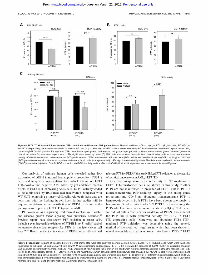
To address whether the model-derived findings presented were clini-cally relevant, we analyzed the possible relationship of ROS productionand DEP-1 activity in leukemic cells isolated from the peripheral bloodof AML patients harboring either WT FLT3 or FLT3 ITD.
Side-by-side comparison of multiple patient samples is ham-pered by the infrequent availability of fresh material, by thedifficulty to cultivate these cells, and by unclear consequences ofcell freezing for redox processes. Moreover, expression of DEP-1is highly variable (supplemental Figure 2D). Presumably by thelatter reason, comparison of basal DEP-1 activity in patient cellsharboring WT FLT3 or FLT3 ITD revealed no statistically signifi-cant differences, however a clear trend to reduced activity in FLT3ITD cells was observed (supplemental Figure 2E). To morerigorously test the impact of FLT3 ITD on DEP-1 activity, wetherefore used a format, where untreated samples were side-by-sidecompared with samples treated with the FLT3 kinase inhibitorAG1295. This compound blocks ROS production downstream ofFLT3 ITD as shown in supplemental Figure 3B-C. This assay wasfirst validated in cell lines.
As shown in Figure 5A, treatment of the FLT3 ITD–harboringcell line MOLM-13 with AG1295 both led to reduction of ROSproduction, and to an elevation of DEP-1 activity. In contrast,AG1295-treatment of EOL-1 cells harboring WT FLT3 did notsignificantly change ROS production and DEP-1 activity was onlyweakly increased (Figure 5B). These data are consistent with thefindings reported in the preceding paragraph, indicating that DEP-1activity is inhibited by FLT3 ITD–driven ROS production.
The same assay was subsequently used on AML patient blasts.Interestingly, AG1295 caused reduced ROS levels and increasedDEP-1 activity in FLT3 ITD–bearing blasts whereas no significantchanges of this type were seen in patients with WT FLT3 (Figure5C; see supplemental Figure 4 for individual patient data).
Taken together, these findings indicated that DEP-1 activity isindeed compromised by FLT3 ITD–driven ROS production in vitroand in vivo.
ROS-mediated DEP-1 inactivation contributes totransformation of 32D cells by FLT3 ITD
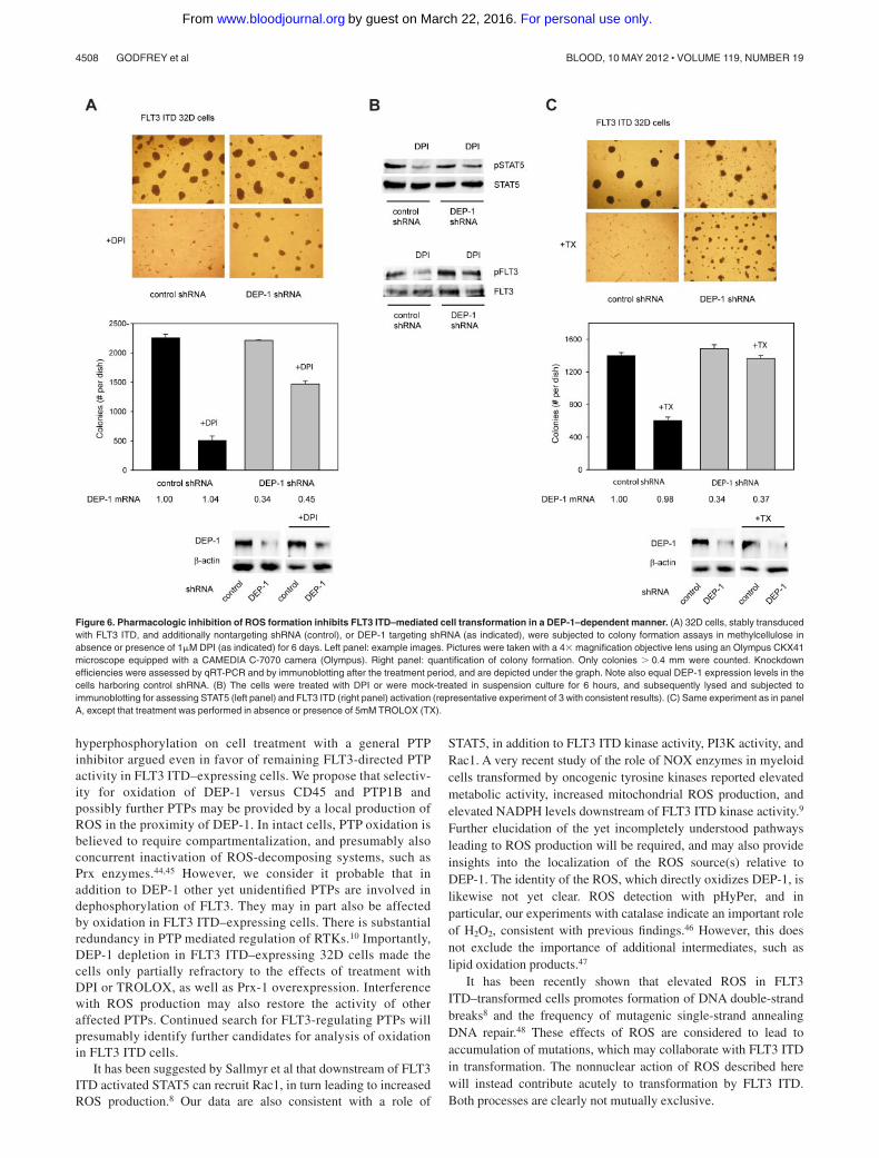
If the described DEP-1 inactivation were important for FLT3ITD–driven transformation, interfering with ROS generation shouldinhibit transformation in the presence of DEP-1. This was testedusing 32D cells stably expressing FLT3 ITD.
According to our preceding analysis, the NOX inhibitor DPIappeared to be a suitable agent because it effectively inhibited ROSproduction and restored DEP-1 activity. Because DPI also inhibitsother flavoproteins and is toxic at high concentrations, we firstcarried out a titration experiment to estimate the minimal dose thatwould inhibit ROS substantially in these cells without severelyimpairing viability (supplemental Figure 5A). DPI (1�M) inhibited
ROS production by approximately 50% but attenuated the growthof FLT3 ITD–expressing cells only partially as revealed byproliferation assays (supplemental Figure 5B). The effect of thisnontoxic dose of DPI was then assayed in colony formation assays.DPI strongly impaired colony formation under these conditions(Figure 6A).
If DEP-1 was involved in mediating this inhibition, one shouldexpect refractoriness of DEP-1 deficient cells to this treatment. Indeed,stable DEP-1 depletion by shRNA partially prevented the inhibition ofcolony formation of FLT3 ITD expressing 32D cells (Figure 6A).
We also analyzed in parallel the effect of treatment with DPIon phosphorylation of STAT5, a prime mediator of transforma-tion by FLT3 ITD. STAT5 phosphorylation was attenuated in thepresence of DPI, providing biochemical evidence for theinterference with a key transforming signaling step. However,the remaining STAT5 activity was obviously sufficient to sustaincell proliferation in suspension. The FLT3 ITD autophosphory-lation was also reduced on DPI treatment in DEP-1 expressingcells, but to a much lesser extent in DEP-1–depleted cells(Figure 6B).
Similar results were obtained using the antioxidant TROLOX tointerfere with the ROS pathway in FLT3 ITD cells. Again,TROLOX inhibited colony formation, and this effect was partiallyrescued by DEP-1 depletion (Figure 6C).
We finally attempted to stably interfere with ROS production, toassess effects on transformation in vivo. Stable overexpression wasachieved for Prx-1, a potent H2O2-scavenging enzyme (supplementalFigure 5C). Prx-1–overexpressing 32D cells formed lesser colonies inpresence of endogenous DEP-1, and this effect was partly abrogated inDEP-1–depleted cells (Figure 7A). We further confirmed that Prx-1overexpression reduced ROS production in the stable cell pools, andcaused elevation of DEP-1 activity (Figure 7B). When injected intosyngeneic mice, the FLT3 ITD–expressing 32D cells caused rapidMPD, regardless of the depletion of DEP-1 (Figure 7C). However, onPrx-1 overexpression the development of MPD was significantlydelayed for cells expressing DEP-1, whereas DEP-1–depleted cellswere unaffected in their capacity to cause MPD (Figure 7C).
These data show that interference with the ROS production canattenuate transformation of 32D cells by FLT3 ITD in a DEP-1–dependent manner.
Discussion
Elimination of tumor suppressors by different molecular mecha-nisms, such as gene deletion, inactivating mutation, or silencing oftranscription is a common phenomenon in oncogenesis. Thesemechanisms have been shown to operate also for tumor-suppressing PTPs, such as PTPRT, SHP-1, PTPRO,33 PTPRD,34,35
and recently TC-PTP.36 We describe here another mechanism ofeliminating a tumor suppressor PTP: the transforming kinase FLT3ITD causes inactivation of the counteracting PTP DEP-1 via aROS-mediated reversible oxidation of the catalytic cysteine. FLT3ITD drives formation of high intracellular ROS levels via yet onlypartially understood pathways. As a consequence, DEP-1 becomesreversibly oxidized and thereby partially inactivated. This isdetectable in experimental cell systems, but also in human AMLcell lines harboring FLT3 ITD. On experimental restoration ofDEP-1 activity by ROS scavenging, or Prx-1 overexpression, thetransforming activity of FLT3 ITD is attenuated as revealed in 32Dcells in vitro and in the related syngeneic mouse model of MPD.
PTP OXIDATION DRIVEN BY FLT3 ITD IN AML 4505BLOOD, 10 MAY 2012 � VOLUME 119, NUMBER 19
For personal use only.on March 22, 2016. by guest www.bloodjournal.orgFrom
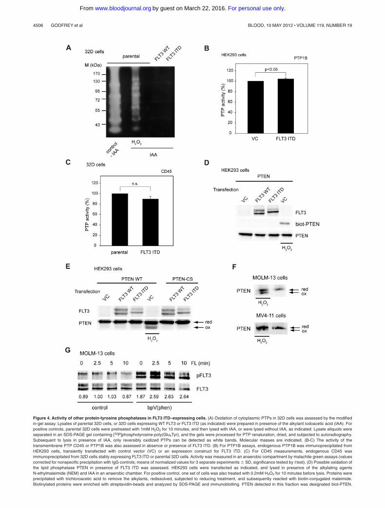
Figure 4. Activity of other protein-tyrosine phosphatases in FLT3 ITD–expressing cells. (A) Oxidation of cytoplasmic PTPs in 32D cells was assessed by the modifiedin-gel assay. Lysates of parental 32D cells, or 32D cells expressing WT FLT3 or FLT3 ITD (as indicated) were prepared in presence of the alkylant iodoacetic acid (IAA). Forpositive controls, parental 32D cells were pretreated with 1mM H2O2 for 10 minutes, and then lysed with IAA, or were lysed without IAA, as indicated. Lysate aliquots wereseparated in an SDS-PAGE gel containing [32P]phosphotyrosine-poly(Glu4Tyr), and the gels were processed for PTP renaturation, dried, and subjected to autoradiography.Subsequent to lysis in presence of IAA, only reversibly oxidized PTPs can be detected as white bands. Molecular masses are indicated. (B-C) The activity of thetransmembrane PTP CD45 or PTP1B was also assessed in absence or presence of FLT3 ITD. (B) For PTP1B assays, endogenous PTP1B was immunoprecipitated fromHEK293 cells, transiently transfected with control vector (VC) or an expression construct for FLT3 ITD. (C) For CD45 measurements, endogenous CD45 wasimmunoprecipitated from 32D cells stably expressing FLT3 ITD or parental 32D cells. Activity was measured in an anaerobic compartment by malachite green assays (valuescorrected for nonspecific precipitation with IgG controls; means of normalized values for 3 separate experiments � SD, significance tested by t test). (D) Possible oxidation ofthe lipid phosphatase PTEN in presence of FLT3 ITD was assessed. HEK293 cells were transfected as indicated, and lysed in presence of the alkylating agentsN-ethylmaleimide (NEM) and IAA in an anaerobic chamber. For positive control, one set of cells was also treated with 0.2mM H2O2 for 10 minutes before lysis. Proteins wereprecipitated with trichloroacetic acid to remove the alkylants, redissolved, subjected to reducing treatment, and subsequently reacted with biotin-conjugated maleimide.Biotinylated proteins were enriched with streptavidin-beads and analyzed by SDS-PAGE and immunoblotting. PTEN detected in this fraction was designated biot-PTEN.
4506 GODFREY et al BLOOD, 10 MAY 2012 � VOLUME 119, NUMBER 19
For personal use only.on March 22, 2016. by guest www.bloodjournal.orgFrom
Our analysis of primary human cells revealed rather lowexpression of DEP-1 in normal hematopoietic progenitor (CD34�)cells, and an apparent up-regulation to similar levels in both FLT3ITD–positive and negative AML blasts by yet undefined mecha-nisms. In FLT3 ITD–expressing AML-cells, DEP-1 activity tendedto be diminished by ROS-mediated inactivation compared withWT FLT3-expressing primary AML cells. Although these data areconsistent with the findings in cell lines, further studies will berequired to determine the contribution of DEP-1 oxidation to thepathogenesis of primary FLT3 ITD–positive AML.
PTP oxidation as a regulatory feed-forward mechanism to enableand enhance growth factor signaling was previously described.17
Previous reports have also shown PTP oxidation in cancer cells,including even irreversible oxidation of PTP1B in A431 cells,37 and ofnontransmembrane and receptor-like PTPs in multiple cancer celllines.38,39 Based on the identification of DEP-1 as an efficient and
relevant PTP for FLT3,20 this study linked PTP oxidation to the activityof a critical oncoprotein in AML, FLT3 ITD.
One obvious question is the selectivity of PTP oxidation inFLT3 ITD–transformed cells. As shown in this study, 2 otherPTPs are not inactivated in presence of FLT3 ITD: PTP1B, anontransmembrane PTP residing largely in the endoplasmicreticulum, and CD45 an abundant transmembrane PTP inhematopoietic cells. Both PTPs have been shown previously tobecome oxidized in intact cells.40-42 PTP1B is even among thePTPs which are most sensitive to oxidation by H2O2.43 Likewise,we did not obtain evidence for oxidation of PTEN, a member ofthe PTP family with preferred activity for PIP3, in FLT3ITD–expressing cells. Moreover, no abundant FLT3 ITD–mediated PTP oxidation was detectable using the genericmethod of the modified in-gel assay, which has been shown toreveal reversible oxidation of some cytoplasmic PTPs.13 FLT3
Figure 5. FLT3 ITD kinase-inhibition rescues DEP-1 activity in cell lines and AML patient blasts. The AML cell lines MOLM-13 (A), or EOL-1 (B), harboring FLT3 ITD, orWT FLT3, respectively, were treated with the FLT3 inhibitor AG1295 (20�M, 3 hours), or DMSO solvent, and subsequently ROS formation was measured in a plate reader usingcarboxy-H2DFFDA (left panels). Endogenous DEP-1 was immunoprecipitated and assayed using a phosphopeptide substrate and malachite green detection (means ofnormalized values for 3 separate experiments � SD, significance tested by t test). (C) AML patient blasts were freshly isolated from blood of patients taken before start oftherapy. AG1295 treatment and measurement of ROS production and DEP-1 activity were performed as in (A-B). Values are based on duplicate (DEP-1 activity) and triplicate(ROS generation) determinations for each patient and means for all patients are presented (� SD, significance tested by t test). The data are normalized to values in vehicle(DMSO)–treated cells (100%). Data for ROS production and DEP-1 activity and the effects of AG1295 for individual patients are shown in supplemental Figure 4.
Figure 4 (continued) Aliquots of fractions before the final affinity step were also analyzed as input controls (lowest panel). (E-F) HEK293 cells, which were transientlytransfected as indicated (E), and MOLM-13 cells or MV4-11 cells expressing endogenously FLT3 ITD (F) were lysed in presence of 40mM NEM in an anaerobic chamber.Samples were fractionated by nonreducing SDS-PAGE. Oxidation of PTEN caused the formation of a band which migrates faster (designated ox) than the reduced form (red).For an additional specificity control, a PTEN cysteine-to-serine mutant (CS), which cannot become oxidized, was also analyzed. (G) MOLM-13 cells were serum-starved andtreated with 100�M bV(phen), a general PTP inhibitor, for 15 minutes. Subsequently, cells were stimulated with FLT3 ligand (FL) for different time as indicated, lysed, and FLT3was immunoprecipitated. Phosphorylation was analyzed by immunoblotting. Numbers under the blot indicate relative phosphorylation of the mature (top) FLT3 band,normalized to total FLT3 and phosphorylation at 2.5 minutes after FL-stimulation (1.0).
PTP OXIDATION DRIVEN BY FLT3 ITD IN AML 4507BLOOD, 10 MAY 2012 � VOLUME 119, NUMBER 19
For personal use only.on March 22, 2016. by guest www.bloodjournal.orgFrom
hyperphosphorylation on cell treatment with a general PTPinhibitor argued even in favor of remaining FLT3-directed PTPactivity in FLT3 ITD–expressing cells. We propose that selectiv-ity for oxidation of DEP-1 versus CD45 and PTP1B andpossibly further PTPs may be provided by a local production ofROS in the proximity of DEP-1. In intact cells, PTP oxidation isbelieved to require compartmentalization, and presumably alsoconcurrent inactivation of ROS-decomposing systems, such asPrx enzymes.44,45 However, we consider it probable that inaddition to DEP-1 other yet unidentified PTPs are involved indephosphorylation of FLT3. They may in part also be affectedby oxidation in FLT3 ITD–expressing cells. There is substantialredundancy in PTP mediated regulation of RTKs.10 Importantly,DEP-1 depletion in FLT3 ITD–expressing 32D cells made thecells only partially refractory to the effects of treatment withDPI or TROLOX, as well as Prx-1 overexpression. Interferencewith ROS production may also restore the activity of otheraffected PTPs. Continued search for FLT3-regulating PTPs willpresumably identify further candidates for analysis of oxidationin FLT3 ITD cells.
It has been suggested by Sallmyr et al that downstream of FLT3ITD activated STAT5 can recruit Rac1, in turn leading to increasedROS production.8 Our data are also consistent with a role of
STAT5, in addition to FLT3 ITD kinase activity, PI3K activity, andRac1. A very recent study of the role of NOX enzymes in myeloidcells transformed by oncogenic tyrosine kinases reported elevatedmetabolic activity, increased mitochondrial ROS production, andelevated NADPH levels downstream of FLT3 ITD kinase activity.9
Further elucidation of the yet incompletely understood pathwaysleading to ROS production will be required, and may also provideinsights into the localization of the ROS source(s) relative toDEP-1. The identity of the ROS, which directly oxidizes DEP-1, islikewise not yet clear. ROS detection with pHyPer, and inparticular, our experiments with catalase indicate an important roleof H2O2, consistent with previous findings.46 However, this doesnot exclude the importance of additional intermediates, such aslipid oxidation products.47
It has been recently shown that elevated ROS in FLT3ITD–transformed cells promotes formation of DNA double-strandbreaks8 and the frequency of mutagenic single-strand annealingDNA repair.48 These effects of ROS are considered to lead toaccumulation of mutations, which may collaborate with FLT3 ITDin transformation. The nonnuclear action of ROS described herewill instead contribute acutely to transformation by FLT3 ITD.Both processes are clearly not mutually exclusive.
Figure 6. Pharmacologic inhibition of ROS formation inhibits FLT3 ITD–mediated cell transformation in a DEP-1–dependent manner. (A) 32D cells, stably transducedwith FLT3 ITD, and additionally nontargeting shRNA (control), or DEP-1 targeting shRNA (as indicated), were subjected to colony formation assays in methylcellulose inabsence or presence of 1�M DPI (as indicated) for 6 days. Left panel: example images. Pictures were taken with a 4� magnification objective lens using an Olympus CKX41microscope equipped with a CAMEDIA C-7070 camera (Olympus). Right panel: quantification of colony formation. Only colonies � 0.4 mm were counted. Knockdownefficiencies were assessed by qRT-PCR and by immunoblotting after the treatment period, and are depicted under the graph. Note also equal DEP-1 expression levels in thecells harboring control shRNA. (B) The cells were treated with DPI or were mock-treated in suspension culture for 6 hours, and subsequently lysed and subjected toimmunoblotting for assessing STAT5 (left panel) and FLT3 ITD (right panel) activation (representative experiment of 3 with consistent results). (C) Same experiment as in panelA, except that treatment was performed in absence or presence of 5mM TROLOX (TX).
4508 GODFREY et al BLOOD, 10 MAY 2012 � VOLUME 119, NUMBER 19
For personal use only.on March 22, 2016. by guest www.bloodjournal.orgFrom
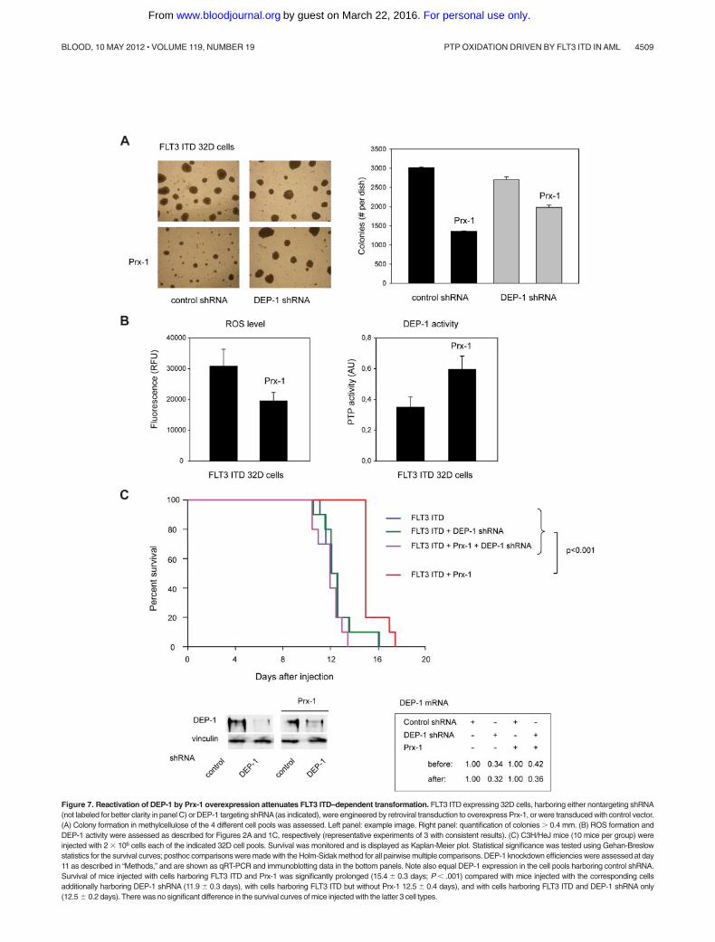
Figure 7. Reactivation of DEP-1 by Prx-1 overexpression attenuates FLT3 ITD–dependent transformation. FLT3 ITD expressing 32D cells, harboring either nontargeting shRNA(not labeled for better clarity in panel C) or DEP-1 targeting shRNA(as indicated), were engineered by retroviral transduction to overexpress Prx-1, or were transduced with control vector.(A) Colony formation in methylcellulose of the 4 different cell pools was assessed. Left panel: example image. Right panel: quantification of colonies � 0.4 mm. (B) ROS formation andDEP-1 activity were assessed as described for Figures 2A and 1C, respectively (representative experiments of 3 with consistent results). (C) C3H/HeJ mice (10 mice per group) wereinjected with 2 � 106 cells each of the indicated 32D cell pools. Survival was monitored and is displayed as Kaplan-Meier plot. Statistical significance was tested using Gehan-Breslowstatistics for the survival curves; posthoc comparisons were made with the Holm-Sidak method for all pairwise multiple comparisons. DEP-1 knockdown efficiencies were assessed at day11 as described in “Methods,” and are shown as qRT-PCR and immunoblotting data in the bottom panels. Note also equal DEP-1 expression in the cell pools harboring control shRNA.Survival of mice injected with cells harboring FLT3 ITD and Prx-1 was significantly prolonged (15.4 � 0.3 days; P � .001) compared with mice injected with the corresponding cellsadditionally harboring DEP-1 shRNA (11.9 � 0.3 days), with cells harboring FLT3 ITD but without Prx-1 12.5 � 0.4 days), and with cells harboring FLT3 ITD and DEP-1 shRNA only(12.5 � 0.2 days). There was no significant difference in the survival curves of mice injected with the latter 3 cell types.
PTP OXIDATION DRIVEN BY FLT3 ITD IN AML 4509BLOOD, 10 MAY 2012 � VOLUME 119, NUMBER 19
For personal use only.on March 22, 2016. by guest www.bloodjournal.orgFrom
ROS quenching has been considered as a therapeutic strategy incancer.49 Further work will be required to establish the relevance ofROS-mediated inactivation of DEP-1 and possibly other PTPs forhuman AML, and whether ROS quenching may be a viabletherapeutic strategy in this disease.
Acknowledgments
The authors are grateful to Drs Nolan, Duyster, Lambeth,Pfitzner, Ronnstrand, and Ungefroren, and the Vasopharmcompany for the kind provision of cells or reagents; and to PeterHerrlich for critical reading of the paper and stimulatingdiscussions throughout this project.
This work was supported by the Deutsche Forschungsgemeinschaft(BO1043/6-3), the Deutsche Krebshilfe (Collaborative grant 108401,
TP2 to F.D.B.), and the European Community (“PTP-NET” MarieCurie Network MRTN-CT-2006-035830, to F.D.B. and A.O.).
Authorship
Contribution: R.G. designed and performed experiments, analyzedthe data, and wrote parts of the paper; D.A., R.B., S.S., T.H., S.A.B.,J.P.M., M.D., and U.S. did experiments; S.S. characterized and providedclinical samples; A.O. contributed to concept and the paper; and F.-D.B.planned and supervised the study, and wrote the paper.
Conflict of interest disclosure: The authors declare no compet-ing financial interests.
The current affiliation for R.G. is Molecular Cardiology andAngiology, University Hospital Munster, Munster, Germany.
Correspondence: F.-D. Bohmer, Institute of Molecular CellBiology, Hans-Knoll-Strasse 2, D-07745 Jena, Germany; e-mail:[email protected].
References
1. Frohling S, Scholl C, Gilliland DG, Levine RL. Ge-netics of myeloid malignancies: pathogenetic andclinical implications. J Clin Oncol. 2005;23(26):6285-6295.
2. Dohner H, Estey EH, Amadori S, et al. Diagnosisand management of acute myeloid leukemia inadults: recommendations from an internationalexpert panel, on behalf of the European Leukemi-aNet. Blood. 2010;115(3):453-474.
3. Meshinchi S, Appelbaum FR. Structural and func-tional alterations of FLT3 in acute myeloid leuke-mia. Clin Cancer Res. 2009;15(13):4263-4269.
4. Breitenbuecher F, Schnittger S, Grundler R, et al.Identification of a novel type of ITD mutations lo-cated in nonjuxtamembrane domains of the FLT3tyrosine kinase receptor. Blood. 2009;113(17):4074-4077.
5. Kayser S, Schlenk RF, Londono MC, et al. Inser-tion of FLT3 internal tandem duplication in thetyrosine kinase domain-1 is associated with resis-tance to chemotherapy and inferior outcome.Blood. 2009;114(12):2386-2392.
6. Grundler R, Miething C, Thiede C, Peschel C,Duyster J. FLT3-ITD and tyrosine kinase domainmutants induce 2 distinct phenotypes in a murinebone marrow transplantation model. Blood. 2005;105(12):4792-4799.
7. Schmidt-Arras D, Bohmer SA, Koch S, et al. An-choring of FLT3 in the endoplasmic reticulum al-ters signaling quality. Blood. 2009;113(15):3568-3576.
8. Sallmyr A, Fan J, Datta K, et al. Internal tandemduplication of FLT3 (FLT3/ITD) induces increasedROS production, DNA damage, and misrepair:implications for poor prognosis in AML. Blood.2008;111(6):3173-3182.
9. Reddy MM, Fernandes MS, Salgia R, Levine RL,Griffin JD, Sattler M. NADPH oxidases regulatecell growth and migration in myeloid cells trans-formed by oncogenic tyrosine kinases. Leukemia.2011;25(2):281-289.
10. Ostman A, Bohmer FD. Regulation of receptortyrosine kinase signaling by protein tyrosinephosphatases. Trends Cell Biol. 2001;11(6):258-266.
11. den Hertog J, Ostman A, Bohmer FD. Protein ty-rosine phosphatases: regulatory mechanisms.FEBS J. 2008;275(5):831-847.
12. Denu JM, Tanner KG. Specific and reversible in-activation of protein tyrosine phosphatases byhydrogen peroxide: evidence for a sulfenic acidintermediate and implications for redox regula-tion. Biochemistry. 1998;37(16):5633-5642.
13. Meng TC, Fukada T, Tonks NK. Reversible oxida-
tion and inactivation of protein tyrosine phospha-tases in vivo. Mol Cell. 2002;9(2):387-399.
14. Tonks NK. Redox redux: revisiting PTPs and thecontrol of cell signaling. Cell. 2005;121(5):667-670.
15. Lambeth JD. NOX enzymes and the biology ofreactive oxygen. Nat Rev Immunol. 2004;4(3):181-189.
16. Stone JR, Yang S. Hydrogen peroxide: a signal-ing messenger. Antioxid Redox Signal. 2006;8(3-4):243-270.
17. Rhee SG, Kang SW, Jeong W, Chang TS, YangKS, Woo HA. Intracellular messenger function ofhydrogen peroxide and its regulation by peroxire-doxins. Curr Opin Cell Biol. 2005;17(2):183-189.
18. Schmidt-Arras DE, Bohmer A, Markova B,Choudhary C, Serve H, Bohmer FD. Tyrosinephosphorylation regulates maturation of receptortyrosine kinases. Mol Cell Biol. 2005;25(9):3690-3703.
19. Moller JP, Schonherr C, Markova B, Bauer R,Stocking C, Bohmer FD. Role of SHP2 for FLT3-dependent proliferation and transformation in32D cells. Leukemia. 2008;22(10):1945-1948.
20. Arora D, Stopp S, Bohmer SA, et al. Protein ty-rosine phosphatase DEP-1 controls receptor ty-rosine kinase FLT3 signaling. J Biol Chem. 2011;286(13):10918-10929.
21. Karagyozov L, Godfrey R, Bohmer SA, et al. Thestructure of the 5�-end of the protein-tyrosinephosphatase PTPRJ mRNA reveals a novelmechanism for translation attenuation. NucleicAcids Res. 2008;36(13):4443-4453.
22. Sandin A, Dagnell M, Gonon A, et al. Hypoxia fol-lowed by re-oxygenation induces oxidation of ty-rosine phosphatases. Cell Signal. 2011;23(5):820-826.
23. Chen K, Kirber MT, Xiao H, Yang Y, Keaney JF Jr.Regulation of ROS signal transduction by NA-DPH oxidase 4 localization. J Cell Biol. 2008;181(7):1129-1139.
24. Lin J, Zhu JW, Baker JE, Weiss A. Regulated ex-pression of the receptor-like tyrosine phospha-tase CD148 on hemopoietic cells. J Immunol.2004;173(4):2324-2330.
25. Arimura Y, Yagi J. Comprehensive expressionprofiles of genes for protein tyrosine phospha-tases in immune cells. Sci Signal. 2010;3(137):rs1.
26. de Jonge HJ, Woolthuis CM, Vos AZ, et al. Geneexpression profiling in the leukemic stem cell-enriched CD34(�) fraction identifies target genesthat predict prognosis in normal karyotype AML.Leukemia. 2011;25(12):1825-1833.
27. Markvicheva KN, Bogdanova EA, Staroverov DB,Lukyanov S, Belousov VV. Imaging of intracellularhydrogen peroxide production with HyPer uponstimulation of HeLa cells with epidermal growthfactor. Methods Mol Biol. 2008;476:79-86.
28. Lambeth JD, Krause KH, Clark RA. NOX en-zymes as novel targets for drug development.Semin Immunopathol. 2008;30(3):339-363.
29. Muller J, Sperl B, Reindl W, Kiessling A, Berg T.Discovery of chromone-based inhibitors of thetranscription factor STAT5. Chembiochem. 2008;9(5):723-727.
30. Freyhaus H, Huntgeburth M, Wingler K, et al.Novel Nox inhibitor VAS2870 attenuates PDGF-dependent smooth muscle cell chemotaxis, butnot proliferation. Cardiovasc Res. 2006;71(2):331-341.
31. Brandts CH, Sargin B, Rode M, et al. Constitutiveactivation of Akt by Flt3 internal tandem duplica-tions is necessary for increased survival, prolif-eration, and myeloid transformation. Cancer Res.2005;65(21):9643-9650.
32. Kwon J, Lee SR, Yang KS, et al. Reversible oxi-dation and inactivation of the tumor suppressorPTEN in cells stimulated with peptide growth fac-tors. Proc Natl Acad Sci U S A. 2004;101(47):16419-16424.
33. Ostman A, Hellberg C, Bohmer FD. Protein-ty-rosine phosphatases and cancer. Nat Rev Can-cer. 2006;6(4):307-320.
34. Solomon DA, Kim JS, Cronin JC, et al. Mutationalinactivation of PTPRD in glioblastoma multiformeand malignant melanoma. Cancer Res. 2008;68(24):10300-10306.
35. Veeriah S, Brennan C, Meng S, et al. The ty-rosine phosphatase PTPRD is a tumor suppres-sor that is frequently inactivated and mutated inglioblastoma and other human cancers. Proc NatlAcad Sci U S A. 2009;106(23):9435-9440.
36. Kleppe M, Lahortiga I, El Chaar T, et al. Deletionof the protein tyrosine phosphatase gene PTPN2in T-cell acute lymphoblastic leukemia. NatGenet. 2010;42(6):530-535.
37. Lou YW, Chen YY, Hsu SF, et al. Redox regula-tion of the protein tyrosine phosphatase PTP1B incancer cells. FEBS J. 2008;275(1):69-88.
38. Boivin B, Zhang S, Arbiser JL, Zhang ZY,Tonks NK. A modified cysteinyl-labeling assayreveals reversible oxidation of protein tyrosinephosphatases in angiomyolipoma cells. Proc NatlAcad Sci U S A. 2008;105(29):9959-9964.
39. Karisch R, Fernandez M, Taylor P, et al. Globalproteomic assessment of the classical protein-tyrosine phosphatome and “Redoxome”. Cell.2011;146(5):826-840.
4510 GODFREY et al BLOOD, 10 MAY 2012 � VOLUME 119, NUMBER 19
For personal use only.on March 22, 2016. by guest www.bloodjournal.orgFrom

40. Lee SR, Kwon KS, Kim SR, Rhee SG. Reversibleinactivation of protein-tyrosine phosphatase 1B inA431 cells stimulated with epidermal growth fac-tor. J Biol Chem. 1998;273(25):15366-15372.
41. Galic S, Hauser C, Kahn BB, et al. Coordinatedregulation of insulin signaling by the protein ty-rosine phosphatases PTP1B and TCPTP. MolCell Biol. 2005;25(2):819-829.
42. Rider DA, Sinclair AJ, Young SP. Oxidative inacti-vation of CD45 protein tyrosine phosphatase maycontribute to T lymphocyte dysfunction in the el-derly. Mech Ageing Dev. 2003;124(2):191-198.
43. Groen A, Lemeer S, van der Wijk T, et al. Differ-
ential oxidation of protein-tyrosine phosphatases.J Biol Chem. 2005;280(11):10298-10304.
44. Winterbourn CC. Reconciling the chemistry andbiology of reactive oxygen species. Nat ChemBiol. 2008;4(5):278-286.
45. Woo HA, Yim SH, Shin DH, Kang D, Yu DY,Rhee SG. Inactivation of peroxiredoxin I by phos-phorylation allows localized H(2)O(2) accumula-tion for cell signaling. Cell. 2010;140(4):517-528.
46. Juarez JC, Manuia M, Burnett ME, et al. Superox-ide dismutase 1 (SOD1) is essential for H2O2-mediated oxidation and inactivation of phospha-tases in growth factor signaling. Proc Natl AcadSci U S A. 2008;105(20):7147-7152.
47. Conrad M, Sandin A, Forster H, et al. 12/15-lipoxygenase-derived lipid peroxides controlreceptor tyrosine kinase signaling throughoxidation of protein tyrosine phosphatases.Proc Natl Acad Sci U S A. 2010;107(36):15774-15779.
48. Fernandes MS, Reddy MM, Gonneville JR, et al.BCR-ABL promotes the frequency of mutagenicsingle-strand annealing DNA repair. Blood. 2009;114(9):1813-1819.
49. Wondrak GT. Redox-directed cancer therapeu-tics: molecular mechanisms and opportunities.Antioxid Redox Signal. 2009;11(12):3013-3069.
PTP OXIDATION DRIVEN BY FLT3 ITD IN AML 4511BLOOD, 10 MAY 2012 � VOLUME 119, NUMBER 19
For personal use only.on March 22, 2016. by guest www.bloodjournal.orgFrom

online March 20, 2012 originally publisheddoi:10.1182/blood-2011-02-336446
2012 119: 4499-4511
Frank-D. BöhmerSylvia-Annette Böhmer, Markus Dagnell, Ulf Schnetzke, Sebastian Scholl, Arne Östman and Rinesh Godfrey, Deepika Arora, Reinhard Bauer, Sabine Stopp, Jörg P. Müller, Theresa Heinrich, phosphatase DEP-1/ PTPRJoxidative inactivation of the tumor suppressor protein-tyrosine Cell transformation by FLT3 ITD in acute myeloid leukemia involves
http://www.bloodjournal.org/content/119/19/4499.full.htmlUpdated information and services can be found at:
(1464 articles)Myeloid Neoplasia Articles on similar topics can be found in the following Blood collections
http://www.bloodjournal.org/site/misc/rights.xhtml#repub_requestsInformation about reproducing this article in parts or in its entirety may be found online at:
http://www.bloodjournal.org/site/misc/rights.xhtml#reprintsInformation about ordering reprints may be found online at:
http://www.bloodjournal.org/site/subscriptions/index.xhtmlInformation about subscriptions and ASH membership may be found online at:
Copyright 2011 by The American Society of Hematology; all rights reserved.of Hematology, 2021 L St, NW, Suite 900, Washington DC 20036.Blood (print ISSN 0006-4971, online ISSN 1528-0020), is published weekly by the American Society
For personal use only.on March 22, 2016. by guest www.bloodjournal.orgFrom
Related Documents