The Open Neuroscience Journal, 2010, 4, 1-12 1 1874-0820/10 2010 Bentham Open Open Access Cell Calcium Extrusion Systems and their Role in Epileptogenesis Jorge Bravo-Martínez a,b , Blanca Delgado-Coello b and Jaime Mas-Oliva* ,b a Departamento de Fisiología, Facultad de Medicina, and b Instituto de Fisiología Celular, Universidad Nacional Autónoma de México, México D.F., México Abstract: The precise control for maintenance of a normal intracellular calcium concentration in eukaryote cells is ac- complished by several systems located at the plasma membrane, as well as several internal membrane systems. Neurons are especially sensitive to changes in these control systems, since when fail and calcium homeostasis disturbed, the cell’s metabolism is immediately modified and a pathological condition emerges. Such a condition has been associated with epi- leptogenesis, and especially to those mechanisms associated to calcium entrance or ON mechanisms. On the other hand, calcium extrusion mechanisms or OFF mechanisms, have been investigated to a lesser extent and therefore remain much less understood. Here, we present a review of these calcium extrusion systems located at the plasma membrane considered to be critical in the process of epileptogenesis; first of all the plasma membrane calcium ATPase (PMCA) as the catalytic moiety of the enzyme that moves calcium outwards in an energy-dependent fashion, and the Na + /Ca 2+ exchanger (NCX) coupled to the (Na + /K + )-ATPase. Based on present knowledge considering the wide range of isoforms found for PMCA and NCX and their specific kinetic characteristics, a hypothesis for their participation on the OFF mechanisms related to the genesis of epilepsy is discussed. Keywords: Epilepsy, epileptogenesis, calcium regulation, PMCA, NCX, calcium extrusion proteins. 1. INTRODUCTION Epilepsy can be defined as a chronic illness of diverse etiology characterized by recurrent crises due to an excessive and synchronic burden of cerebral neurons, eventually asso- ciated with diverse clinical and paraclinical manifestations. Epilepsy is a common pathology; World Health Organiza- tion (WHO) statistics revealed in the year of 2001 a preva- lence of 8.2 per 1,000 individuals in developed countries and 10 per 1,000 in developing countries. During the same year, incidence in developed countries was 50 per 100,000 indi- viduals in the general population, and 100 per 100,000 in developing countries. The analysis we have performed in the present study is related to the 50% of these patients that pre- sent by diverse external causes an acquired epilepsy [1]. One very important period of epilepsy comprises epileptogenesis, i.e., the period in which epilepsy is developed, which can be considered the period between the lesion and the appearance of clinical manifestations. Epileptogenesis includes all phe- nomena that induce normal cells to discharge abnormally, which when repeated in a continuous fashion, produce an epileptic focus. For these phenomena to be expressed in cells, a change is required in the majority of systems control- ling neuronal excitability and inhibitory processes. Such phenomena allow an exaggerated abnormal discharge of neurons provoking hyperexcitability in the long term. During the period of epileptogenesis, there also appear aberrant in- terconnections that promote neuronal synchronization with the consequent clinical manifestations [1]. Calcium is an important regulator in many metabolic pathways, as well as a second messenger; therefore, its intra- *Address correspondence to this author at the Apdo, Postal 70-243, 04510 México, D.F., México; Tel: (+52) (55) 5622-5584, (+52) (55) 5622-5619; Fax: (+52) (55) 5622-5611; E-mail: [email protected] cellular concentration is regulated precisely. Transitory ele- vations in calcium are created during diverse physiological processes such as synaptic transmission, long-term potentia- tion in the learning process, cellular growth and differentia- tion, cytoskeleton maintenance, and genetic expression. In- tracellular calcium is maintained between 50 and 200 nM, which represents four orders of magnitude beneath its ex- tracellular concentration [2,3]. When there is a considerable and irreversible increase in the intracellular concentration of calcium (glutamate-associated citotoxicity), diverse mecha- nisms of cellular death are triggered. At the intermediate point between normal regulation of calcium inflow and in- tracellular calcium cytotoxicity-related cell death, a non- lethal, prolonged, and irreversible intracellular calcium in- crease occurs, triggering a series of abnormal plastic changes termed epileptogenesis. After a lesion takes place at the epi- center, these changes are severe and lead to cell death. None- theless, cells surrounding the epicenter experience less se- vere changes and form the substrate for the development of epilepsy [4]. For an adequate maintenance of calcium levels, cells are provided with specialized mechanisms for increas- ing cytoplasmic calcium concentration termed ON mecha- nisms, while those devoted to extrude calcium are consid- ered the OFF mechanisms. A massive inflow of calcium dur- ing epileptogenesis involves several important consequences, such as changes in neuronal excitability [5], apoptosis induc- tion, and fiber reorganization known as sprouting. The sprouting phenomenon possesses relevance because it pro- motes an important increase of excitatory circuits, mainly those of the recurrent type [1]. Although calcium inflow mechanisms have been extensively studied and their implica- tion in epilepsy is well known, calcium extrusion processes are less understood. In this review, therefore, we focus on the investigation carried out studying mainly the calcium

Welcome message from author
This document is posted to help you gain knowledge. Please leave a comment to let me know what you think about it! Share it to your friends and learn new things together.
Transcript
The Open Neuroscience Journal, 2010, 4, 1-12 1
1874-0820/10 2010 Bentham Open
Open Access
Cell Calcium Extrusion Systems and their Role in Epileptogenesis
Jorge Bravo-Martíneza,b
, Blanca Delgado-Coellob and Jaime Mas-Oliva*
,b
aDepartamento de Fisiología, Facultad de Medicina, and
bInstituto de Fisiología Celular, Universidad Nacional
Autónoma de México, México D.F., México
Abstract: The precise control for maintenance of a normal intracellular calcium concentration in eukaryote cells is ac-
complished by several systems located at the plasma membrane, as well as several internal membrane systems. Neurons
are especially sensitive to changes in these control systems, since when fail and calcium homeostasis disturbed, the cell’s
metabolism is immediately modified and a pathological condition emerges. Such a condition has been associated with epi-
leptogenesis, and especially to those mechanisms associated to calcium entrance or ON mechanisms. On the other hand,
calcium extrusion mechanisms or OFF mechanisms, have been investigated to a lesser extent and therefore remain much
less understood. Here, we present a review of these calcium extrusion systems located at the plasma membrane considered
to be critical in the process of epileptogenesis; first of all the plasma membrane calcium ATPase (PMCA) as the catalytic
moiety of the enzyme that moves calcium outwards in an energy-dependent fashion, and the Na+/Ca
2+ exchanger (NCX)
coupled to the (Na+/K
+)-ATPase. Based on present knowledge considering the wide range of isoforms found for PMCA
and NCX and their specific kinetic characteristics, a hypothesis for their participation on the OFF mechanisms related to
the genesis of epilepsy is discussed.
Keywords: Epilepsy, epileptogenesis, calcium regulation, PMCA, NCX, calcium extrusion proteins.
1. INTRODUCTION
Epilepsy can be defined as a chronic illness of diverse etiology characterized by recurrent crises due to an excessive and synchronic burden of cerebral neurons, eventually asso-ciated with diverse clinical and paraclinical manifestations. Epilepsy is a common pathology; World Health Organiza-tion (WHO) statistics revealed in the year of 2001 a preva-lence of 8.2 per 1,000 individuals in developed countries and 10 per 1,000 in developing countries. During the same year, incidence in developed countries was 50 per 100,000 indi-viduals in the general population, and 100 per 100,000 in developing countries. The analysis we have performed in the present study is related to the 50% of these patients that pre-sent by diverse external causes an acquired epilepsy [1]. One very important period of epilepsy comprises epileptogenesis, i.e., the period in which epilepsy is developed, which can be considered the period between the lesion and the appearance of clinical manifestations. Epileptogenesis includes all phe-nomena that induce normal cells to discharge abnormally, which when repeated in a continuous fashion, produce an epileptic focus. For these phenomena to be expressed in cells, a change is required in the majority of systems control-ling neuronal excitability and inhibitory processes. Such phenomena allow an exaggerated abnormal discharge of neurons provoking hyperexcitability in the long term. During the period of epileptogenesis, there also appear aberrant in-terconnections that promote neuronal synchronization with the consequent clinical manifestations [1].
Calcium is an important regulator in many metabolic pathways, as well as a second messenger; therefore, its intra-
*Address correspondence to this author at the Apdo, Postal 70-243, 04510
México, D.F., México; Tel: (+52) (55) 5622-5584, (+52) (55) 5622-5619;
Fax: (+52) (55) 5622-5611; E-mail: [email protected]
cellular concentration is regulated precisely. Transitory ele-
vations in calcium are created during diverse physiological
processes such as synaptic transmission, long-term potentia-
tion in the learning process, cellular growth and differentia-
tion, cytoskeleton maintenance, and genetic expression. In-
tracellular calcium is maintained between 50 and 200 nM,
which represents four orders of magnitude beneath its ex-
tracellular concentration [2,3]. When there is a considerable
and irreversible increase in the intracellular concentration of
calcium (glutamate-associated citotoxicity), diverse mecha-
nisms of cellular death are triggered. At the intermediate
point between normal regulation of calcium inflow and in-
tracellular calcium cytotoxicity-related cell death, a non-
lethal, prolonged, and irreversible intracellular calcium in-
crease occurs, triggering a series of abnormal plastic changes
termed epileptogenesis. After a lesion takes place at the epi-
center, these changes are severe and lead to cell death. None-
theless, cells surrounding the epicenter experience less se-
vere changes and form the substrate for the development of
epilepsy [4]. For an adequate maintenance of calcium levels,
cells are provided with specialized mechanisms for increas-
ing cytoplasmic calcium concentration termed ON mecha-
nisms, while those devoted to extrude calcium are consid-
ered the OFF mechanisms. A massive inflow of calcium dur-
ing epileptogenesis involves several important consequences,
such as changes in neuronal excitability [5], apoptosis induc-
tion, and fiber reorganization known as sprouting. The
sprouting phenomenon possesses relevance because it pro-
motes an important increase of excitatory circuits, mainly
those of the recurrent type [1]. Although calcium inflow
mechanisms have been extensively studied and their implica-
tion in epilepsy is well known, calcium extrusion processes
are less understood. In this review, therefore, we focus on
the investigation carried out studying mainly the calcium
2 The Open Neuroscience Journal, 2010, Volume 4 Bravo-Martínez et al.
extrusion mechanisms, specifically those located at the plasma membrane.
In hippocampus-cultured cells in which epileptiform ac-
tivity is provoked by lowering magnesium in the perfusion
medium, glutamate was added to the extracellular medium to
measure glutamate-mediated calcium rises; chronic elevated
basal intracellular calcium levels were found [6]. In order to
observe the participation of ON mechanisms (in the induc-
tion phase of epilepsy) during exposure to glutamate, N-
methyl-D-aspartate (NMDA) receptors and voltage-
dependent calcium channels were blocked; thus, calcium
levels diminished to nearly normal levels. When normal and
epileptic cells were challenged with a glutamate stimulus to
increase intracellular calcium levels, restored calcium levels
were quickly observed only in normal cells. In contrast, epi-
leptic cells did not restore calcium levels even after 4 hours,
and depending on the duration of the first crisis, for up to 1
year [6]. These findings evidenced that for epilepsy induc-
tion, ON mechanisms are important, but OFF mechanisms
play an equally important role in this process. Other authors
have seen that intracellular calcium remained elevated in
neurons isolated 1 year after induction of epilepsy with pilo-
carpine, in comparison with normal cells in similarly aged
animals [7]. These findings strongly support the notion that
for epilepsy maintenance, OFF mechanisms are truly impor-
tant, perhaps in a more important fashion than ON mecha-nisms.
In a region that has been damaged, neurons are exposed
for hours to high glutamate concentrations, promoting an
important inflow of calcium into the cell. This phenomenon,
together with a slow removal of calcium, leads to a more
prolonged period of depolarization. These abnormal changes
provide epileptic cells with the ability to produce paroxystic
depolarizations (PD) [8], as described for the first time by
Matsumoto and Ajamone-Marsan [9]. PD are characterized
by a burst of high-frequency action potentials (AP) accom-
panied by a sustained depolarization, which frequently is
followed by hyperpolarization. Calcium participates in the
activation of a phenomenon termed intrinsic burst firing,
associated with synchronization of PD firing among neurons
[10]. Calcium-induced glutamate exocytosis, which in turn
induces the same plastic changes in efferent neurons, extends and propagates the epileptic focus.
2. LONG-TERM CELLULAR EFFECTS OF EPI-
LEPSY
Upon an increase in the discharge of afferent connections
into a neuron, the intensity of the neuron’s response to these
stimuli increases, i.e., potentiation of the synaptic transmis-
sion occurs. An important inflow of calcium and a calcium-
induced expression of early expression genes such as c-jun,
which is implicated in apoptosis-associated gene transcrip-
tion; and c-fos, related with neurotrophic gene transcriptional
activation. Neurotrophins such as nerve growth factor
(NGF), brain-derived neurotrophic factor (BDNF), neurotro-
phin-3 (NT-3), neurotrophin 4/5 (NT4/5) and glial cell line-
derived neurotrophic factor (GDNF) also intervene in the
maturation, survival, and proliferation of specific neuronal
populations [1]. Such activation induces the formation of
new synaptic contacts, many of which are aberrant, causing
the sprouting phenomenon and imposing conditions under which excitability is exacerbated [11].
On the other hand, one consequence of glutamate recep-tor activation comprises an increase in intracellular calcium levels, which are finely regulated by different homeostatic cell mechanisms. Once these mechanisms are exceeded, cell death is induced through two different modalities: the acute (necrotic), or the long-term (apoptotic) death. The acute form is caused, among others, by the cell’s persistent calcium en-trance-mediated depolarization, causing increased sodium and chloride inflow. These ions increase water inflow into the cell, producing swelling and cell lysis [6]. In contrast, the long-term form of cell death consists of an initial increase of calcium considered non-cytotoxic since extrusion mecha-nisms are working. Nevertheless, as calcium extrusion mechanisms are not working properly, intracellular calcium concentration increases, allowing further liberation of this cation from intracellular stores [12]. Moreover, calcium in-flow activates phospholipase A2 [13] and nitrous oxide syn-thetase [14], which result with an increase in arachidonic acid and nitrous oxide. By means of cyclo- and lipoxy-genase, arachidonic acid is transformed to release superoxide (O2
–) and hydroxyl (OH) radicals. When nitrous oxide reacts
with superoxide radicals, peroxynitrite (ONOO–) a highly
reactive molecule is generated [14]. These free radicals de-stroy cytoskeletal proteins, nucleic acids, and membrane lipids [15]. Peroxides, together with an increase in mito-chondrial calcium permeability, alter mitochondrial function and therefore, adenosine triphosphate (ATP) generation. In addition, caspases are activated and these in turn promote the phenomenon of apoptosis. Calcium activates enzymes such as calpain, whose activation has been associated with an ac-tion upon the cytoskeleton, receptor proteins, G proteins, and calcium-dependent proteins.
3. CONTROL OF INTRACELLULAR Ca2+
CONCEN-TRATION
Cell calcium inflow is mediated by several mechanisms that together make up the named “ON component”. Among these, the glutamate receptor-mediated synaptic mechanism is one of the most important since it has been extensively reported that its liberation in epileptogenesis is greatly in-creased. A secondary mechanism is mediated by voltage-dependent calcium channels, that is also activated during epileptic crises [1]. Internal calcium reservoirs such as endo-plasmic reticulum and mitochondria intervene in the phe-nomenon, in a more limited manner.
On the other hand, there are specialized mechanisms to remove cytoplasmic calcium representing the “OFF compo-nent”. Located in intracellular compartments important cal-cium uptake mechanisms are associated with the sarcoendo-plasmic reticulum, the mitochondria, and the Golgi apparatus [16,17]. Also, the presence of calcium buffering proteins such as parvoalbumin, calbindin, and calmodulin (CaM) must be mentioned. However, due to their limited capacity to retain calcium, these soluble proteins participate to a greater extent in modulating calcium signals than in controlling the cation’s cytoplasmic concentration. At the plasma membrane level, both the plasma membrane Ca
2+-ATPase (PMCA) and
the Na+/Ca
2+ exchanger (NCX) work against a high-
concentration gradient; therefore, they are directly or indi-
Calcium Extrusion in Epileptogenesis The Open Neuroscience Journal, 2010, Volume 4 3
rectly ATP-dependent and susceptible to ischemic injury [18]. The PMCA presents a high affinity and low capacity for removing calcium from the cell, pumping the ion against the concentration gradient; whereas the NCX is a low-affinity and high-capacity system. NCX transports sodium and calcium in opposite directions taking advantage of the sodium gradient across the membrane maintained by the Na
+/K
+ ATPase. The exchanger works in situations in which
there is a large intracellular calcium accumulation that re-quires removal in brief periods of time, whereas the PMCA subsequently pumps the remnant calcium until the ion reaches normal values.
Alternative mechanisms of Ca2+
-ATPase regulation are directly coupled with specific signaling pathways according to their presence in excitable or non-excitable cells. Inherent
functions of excitable cells such as neurons require the coor-dinated extrusion of Ca
2+ through the PMCA and the NCX in
the plasma membrane. These transporters work in parallel by means of mechanisms not fully understood.
4. MEMBRANE CALCIUM EXPORTING SYSTEMS AT THE PLASMA MEMBRANE LEVEL
4.1. Plasma Membrane Calcium ATPase (PMCA)
Isoforms
PMCAs are highly conserved ATPases present in eu-karyote cells that are encoded by four genes producing the basic PMCA isoforms (PMCA1–PMCA4). A large variety of isoforms, theoretically >30 PMCA variants, are generated by alternative splicing and differentially expressed according to
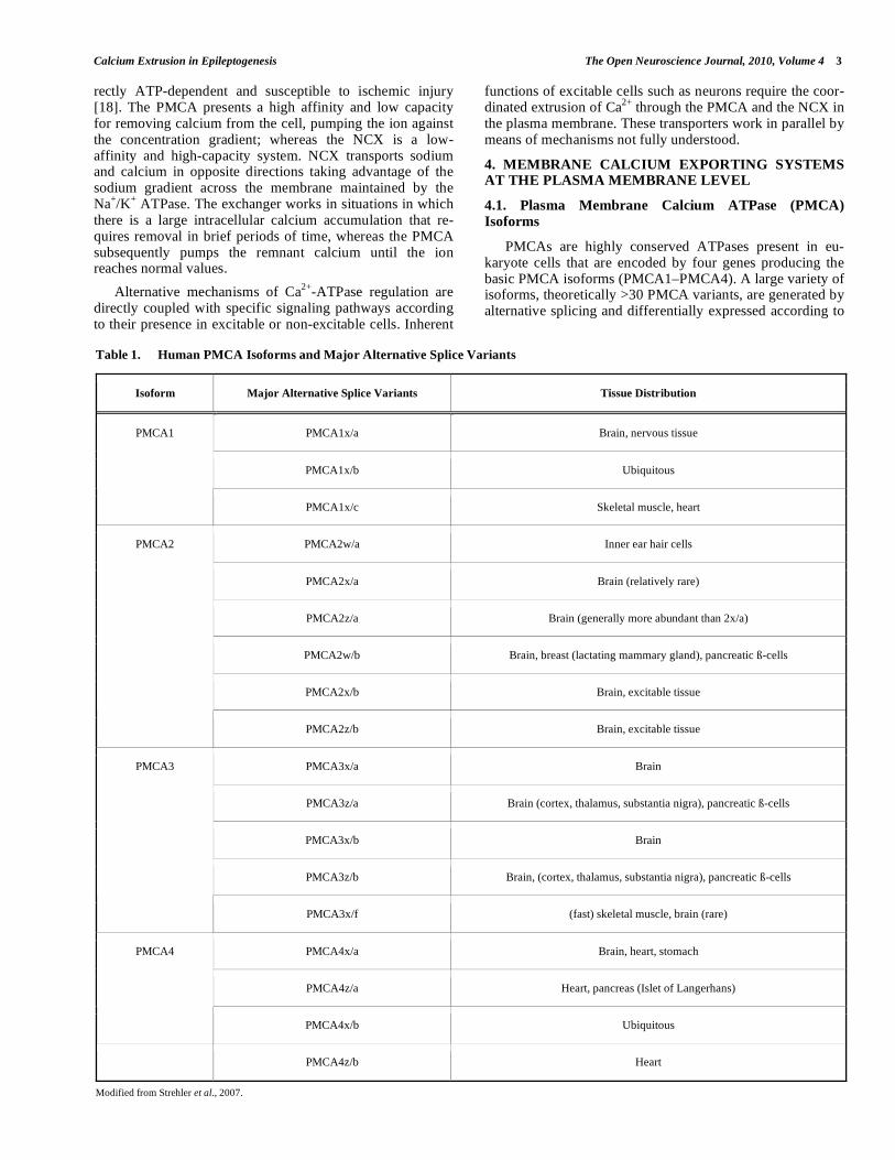
Table 1. Human PMCA Isoforms and Major Alternative Splice Variants
Isoform Major Alternative Splice Variants Tissue Distribution
PMCA1x/a Brain, nervous tissue
PMCA1x/b Ubiquitous
PMCA1
PMCA1x/c Skeletal muscle, heart
PMCA2w/a Inner ear hair cells
PMCA2x/a Brain (relatively rare)
PMCA2z/a Brain (generally more abundant than 2x/a)
PMCA2w/b Brain, breast (lactating mammary gland), pancreatic ß-cells
PMCA2x/b Brain, excitable tissue
PMCA2
PMCA2z/b Brain, excitable tissue
PMCA3x/a Brain
PMCA3z/a Brain (cortex, thalamus, substantia nigra), pancreatic ß-cells
PMCA3x/b Brain
PMCA3z/b Brain, (cortex, thalamus, substantia nigra), pancreatic ß-cells
PMCA3
PMCA3x/f (fast) skeletal muscle, brain (rare)
PMCA4x/a Brain, heart, stomach
PMCA4z/a Heart, pancreas (Islet of Langerhans)
PMCA4
PMCA4x/b Ubiquitous
PMCA4z/b Heart
Modified from Strehler et al., 2007.
4 The Open Neuroscience Journal, 2010, Volume 4 Bravo-Martínez et al.
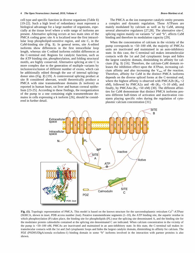
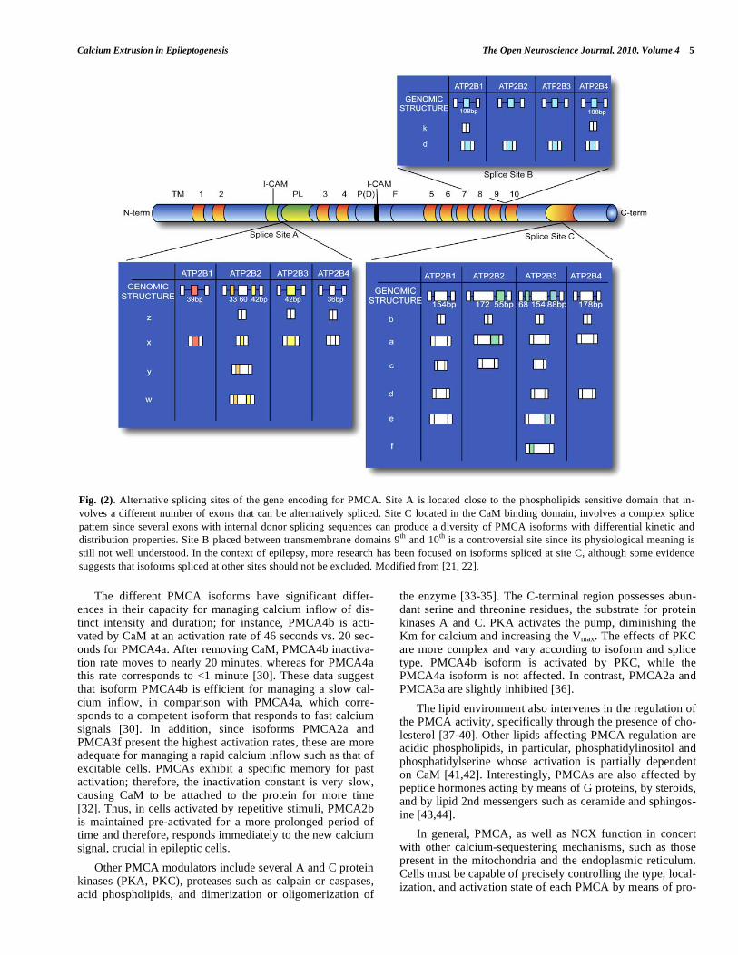
cell type and specific function in diverse organisms (Table 1) [19-22]. Such a high level of redundancy must represent a biological advantage for a large number of organisms, espe-cially at the tissue level where a wide range of isoforms are present. Alternative splicing occurs at two main sites of the PMCA coding gene: site A is localized near the first intracel-lular loop phospholipid-sensitive region, and site C, in the CaM-binding site (Fig. 1). In general terms, site A-edited isoforms show differences in the first intracellular loop length, whereas site C-edited isoforms exhibit differences at the C-terminal end. Regions for catalytic function, such as the ATP binding site, phosphorylation, and folding structural motifs, are highly conserved. Alternative splicing at site C is more complex due to the generation of multiple variants by inclusion/exclusion of different number of exons, which can be additionally edited through the use of internal splicing-donor sites (Fig. 2) [19]. A controversial splicing product at site B considered aberrant, would theoretically produce a PMCA with nine transmembrane domains (k isoform) as reported in human heart, rat liver and human corneal epithe-lium [23-25]. According to these findings, the reorganization of the pump to a one containing eight transmembrane do-mains in cells expressing a k isoform [26], should be consid-ered in further detail.
The PMCA as the ion transporter catalytic entity presents a complex and dynamic regulation. These ATPases are mainly modulated by calcium as well as by CaM, among several alternative regulators [27,28]. The alternative site-C splicing region mainly on variants “a” and “b”, affects CaM binding and therefore its modulation capacity [29].
When the concentration of calcium in the vicinity of the pump corresponds to <50–100 nM, the majority of PMCAs units are inactivated and maintained in an auto-inhibitory state. In this case, the C-terminal tail makes intramolecular contacts with the 1st and 2nd cytoplasmic loops and hides the largest catalytic domain, diminishing its affinity for cal-cium (Fig. 1) [16]. Therefore, the calcium-CaM domain re-leases the inhibition effect upon the ATPase, increasing cal-cium affinity and also increasing the Vmax of the reaction. Therefore, affinity for CaM in the distinct PMCA isoforms depends on the diverse spliced forms at the C-terminal end, where the highest affinity is observed with PMCA2b (KD <2 nM), followed by PMCA2a and -4b (KD <5–10 nM), and finally, by PMCA4a (KD <50 nM) [30]. The different affini-ties for CaM demonstrate that distinct PMCA isoforms pos-sess different half-times of activation and inactivation con-stants playing specific roles during the regulation of cyto-plasmic calcium concentration [31].
Fig. (1). Topologic representation of PMCA. This model is based on the known structure for the sarcoendoplasmic reticulum Ca2+
-ATPase
(SERCA, shown in inset, PDB access number 2eat). Putative transmembrane segments (1–10), the ATP binding site, the aspartic residue in
which phosphorylation (P) takes place, the binding site for phospholipids (PL) near the splicing site denominated A, and the binding site for
the modulator protein calmodulin contained at the splicing site denominated C are indicated. When calcium concentration in the vicinity of
the pump is <50–100 nM, PMCAs are inactivated and maintained in an auto-inhibitory state. In this state, the C-terminal tail makes in-
tramolecular contacts with the 1st and 2nd cytoplasmic loops and hides the largest catalytic domain, diminishing its affinity for calcium. The
PDZ (PSD95/DlgA/zonula occludens-1) binding domain in some “b” isoforms involved in the interaction with partner proteins is also
shown.
Calcium Extrusion in Epileptogenesis The Open Neuroscience Journal, 2010, Volume 4 5
The different PMCA isoforms have significant differ-ences in their capacity for managing calcium inflow of dis-tinct intensity and duration; for instance, PMCA4b is acti-vated by CaM at an activation rate of 46 seconds vs. 20 sec-onds for PMCA4a. After removing CaM, PMCA4b inactiva-tion rate moves to nearly 20 minutes, whereas for PMCA4a this rate corresponds to <1 minute [30]. These data suggest that isoform PMCA4b is efficient for managing a slow cal-cium inflow, in comparison with PMCA4a, which corre-sponds to a competent isoform that responds to fast calcium signals [30]. In addition, since isoforms PMCA2a and PMCA3f present the highest activation rates, these are more adequate for managing a rapid calcium inflow such as that of excitable cells. PMCAs exhibit a specific memory for past activation; therefore, the inactivation constant is very slow, causing CaM to be attached to the protein for more time [32]. Thus, in cells activated by repetitive stimuli, PMCA2b is maintained pre-activated for a more prolonged period of time and therefore, responds immediately to the new calcium signal, crucial in epileptic cells.
Other PMCA modulators include several A and C protein kinases (PKA, PKC), proteases such as calpain or caspases, acid phospholipids, and dimerization or oligomerization of
the enzyme [33-35]. The C-terminal region possesses abun-dant serine and threonine residues, the substrate for protein kinases A and C. PKA activates the pump, diminishing the Km for calcium and increasing the Vmax. The effects of PKC are more complex and vary according to isoform and splice type. PMCA4b isoform is activated by PKC, while the PMCA4a isoform is not affected. In contrast, PMCA2a and PMCA3a are slightly inhibited [36].
The lipid environment also intervenes in the regulation of the PMCA activity, specifically through the presence of cho-lesterol [37-40]. Other lipids affecting PMCA regulation are acidic phospholipids, in particular, phosphatidylinositol and phosphatidylserine whose activation is partially dependent on CaM [41,42]. Interestingly, PMCAs are also affected by peptide hormones acting by means of G proteins, by steroids, and by lipid 2nd messengers such as ceramide and sphingos-ine [43,44].
In general, PMCA, as well as NCX function in concert with other calcium-sequestering mechanisms, such as those present in the mitochondria and the endoplasmic reticulum. Cells must be capable of precisely controlling the type, local-ization, and activation state of each PMCA by means of pro-
Fig. (2). Alternative splicing sites of the gene encoding for PMCA. Site A is located close to the phospholipids sensitive domain that in-
volves a different number of exons that can be alternatively spliced. Site C located in the CaM binding domain, involves a complex splice
pattern since several exons with internal donor splicing sequences can produce a diversity of PMCA isoforms with differential kinetic and
distribution properties. Site B placed between transmembrane domains 9th
and 10th
is a controversial site since its physiological meaning is
still not well understood. In the context of epilepsy, more research has been focused on isoforms spliced at site C, although some evidence
suggests that isoforms spliced at other sites should not be excluded. Modified from [21, 22].
6 The Open Neuroscience Journal, 2010, Volume 4 Bravo-Martínez et al.
teins that interact specifically with the different PMCA iso-forms [45]. As dynamic participants in the regulation of in-tracellular calcium, PMCA isoforms require at the long-term a fine regulation that involves changes at the transcription level, stability of mRNA, alternative splicing, and control of protein translation. At the medium-term, local availability is regulated by directioning toward the specific membrane, internalization, and recycling. This type of regulation is probably due to the intervention of partner proteins belong-ing to the membrane-associated guanylate kinase (MAGUK) family of proteins, which interact with the carboxy end of the 2b and 4b PMCAs [46] at specific sites such as PDZ PSD95/DlgA/zonula occludens-1 (PDZ) domains. Short-term regulation provided by CaM or by differential phos-phorylation is specific for each isoform, allowing dynamic PMCA-function regulation that ranges from seconds to days [47] .
Another important factor comprises the subcellular orga-nization of organelles involved in calcium regulation and its spatial relationship with plasma membrane microdomains. Although the manner in which these microdomains are es-tablished is not well known, it is well established that recep-tors, transporters, and signaling molecules are grouped into multiprotein complexes localized at strategic calcium signal sites [48].
4.2. Tissue Distribution and Local PMCA Expression
All cells express at least one PMCA variant with certain specificity for its expression in time and space. Each cell type possesses distinct splicing regulation mechanisms that determine its specific expression. PMCA1 is found in practi-cally all cell types and has been found from the first stages of embryonic development. PMCA4 is also found in nearly all cell types, but in less abundance with respect to PMCA1 and only detected in late stages of embryonic development [49]. In general terms, PMCAs are more abundant and diverse in brain than in other tissues, particularly variants PMCA2 and PMCA3 [16,50,51].
In the brain, where highly regulated signal transduction events take place, localization of the distinct isoform variants varies greatly among its different structures and cells. For instance, PMCA1 is expressed in cerebral cortex and in the CA1 region of hippocampus [21]. PMCA2 is expressed to a greater degree in cerebellum and also in the cerebral cortex and hippocampus, while PMCA3 is abundant in superficial layers of cerebral cortex and in the cerebellar cortex. PMCA4 expression is reduced in cerebral cortex layers II and VI of pyramidal cells, as well as in cerebellar cortex granular cells. PMCA4 expression in the olfactory bulb, hip-pocampus and striated nucleus is very marginal.
PMCA isoforms also present a distinct distribution at the
subcellular level. For example, in cerebral cortex isoforms 1
and 3 are found in the neuropile (perhaps even in the syn-
apses) and in some dendrites. PMCA2 is abundant in distal
dendrites, while PMCA4 is found as abundant in somato-
dendritic regions [52,53]. These differences reflect func-
tional calcium-regulation differences in the distinct cellular
compartments. The possibility exists that several isoforms
could be integrated into multiprotein complexes, e.g.,
PMCA2 and PMCA4 that might bind to the Na+/H
+ ex-
changer regulator factor and PMCA2 to the MAGUK family
of proteins, that in turn bind to the NMDA receptor. PMCA4
interacts directly with the synthetase I nitrous oxide, known
to decrease its activity in a dose-dependent way when cal-
cium concentration in the microenvironment surrounding the enzyme is low [54].
Additional knowledge is also provided by studies focused
on the association of specific PMCA isoforms to other pro-
teins in caveolae related to calcium handling, since co-
localization of PMCA and these structures has been sug-
gested for different tissues [55,56]. In this sense, the associa-
tion of PMCA isoforms to raft domains has been specially
shown in neurons with the PMCA2 isoform [57]. However,
the relevance of this type of associations in the context of epileptogenesis is still open to investigation.
4.3. Na+/Ca
2+ Exchanger (NCX) Isoforms
The Na+/Ca
2+ exchanger (NCX) is activated when intra-
cellular calcium increases, removing one calcium ion for two
or three sodium ions secondary to the Na+/K
+ ATPase-
generated Na+-gradient formation [18], and suggested that
when altered might contribute to the establishment of epi-
lepsy [58]. The NCX1 gene in humans is localized in chro-
mosome 2 (p23–p22), NCX2 in chromosome 19 (q13.3), and
NCX3 in chromosome 14 (q24.1) (NCBI Entrez Gene data-base).
To date, four Na+/Ca
2+ exchanger genes have been cloned
and functionally characterized. Isoform NCX1 was cloned in
1990 [59,60]; later, isoforms NCX2 and NCX3 were cloned
in mammals. Recently, isoform NCX4 has been found in
teleost, amphibian, and reptilian; and seems to be absent in
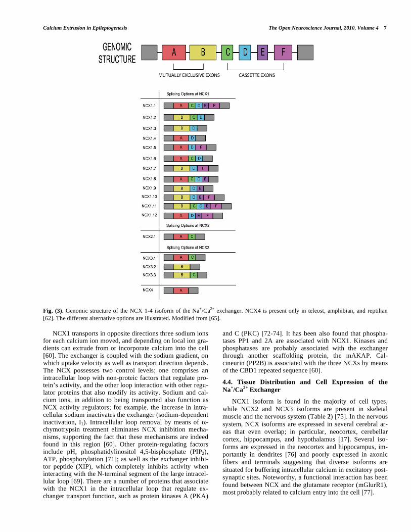
birds and mammals [61,62]. Genes codifying for NCX com-
prise six exons (A–F); NCX1 contains all six, while NCX2
contains only two (B-C) and NCX3 contains four (A, B, C,
E). NCX1 homolog genes have been identified in several
species of microorganisms and have been grouped in the
SLC8 (solute carrier 8) superfamily, from which the most
frequently studied is NCX1 [63,64]. NCX expression levels
are directly correlated to calcium extrusion in distinct cell
types depending on their type of activity; for example, ex-
pression is high in cardiac cells, neurons, and kidney cells. Contrariwise, expression is relatively low in hepatic cells.
The portion corresponding to the intracellular loop ori-
ented toward the extreme C-terminal of NCX produced by
alternative splicing, vary with each one of the isoforms [65].
The 1.1–1.12 variants and the 1.41 were found from NCX1
[66]. NCX3 possesses the 3.1–3.3 variants and only the 2.1
variant is known for NCX2 (Fig. 3).
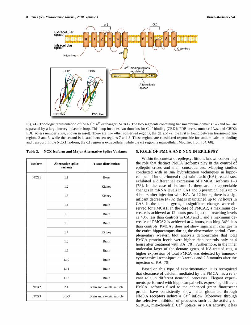
NCX1 presents nine transmembrane domains; domains
1–5 are separated from domains 6–9 by an intracellular loop
representing more than one half of the protein [67]. Two
conserved regions are localized in the intracellular loop; Ca2+
binding domains CBD1 and 2 [68]. The transmembrane re-
gions are responsible for sodium and calcium transport
through the cellular membrane [69]. There are two other
conserved regions among the several NCXs and homologs
termed 1 and 2; the first one found between transmembrane
regions 2 and 3, while the second is located between regions
7 and 8 [64,70]. The 1 region of the NCX1 isoform is ex-tracellular while the 2 region is intracellular (Fig. 4) [67].
Calcium Extrusion in Epileptogenesis The Open Neuroscience Journal, 2010, Volume 4 7
Fig. (3). Genomic structure of the NCX 1-4 isoform of the Na+/Ca
2+ exchanger. NCX4 is present only in teleost, amphibian, and reptilian
[62]. The different alternative options are illustrated. Modified from [65].
NCX1 transports in opposite directions three sodium ions for each calcium ion moved, and depending on local ion gra-dients can extrude from or incorporate calcium into the cell [60]. The exchanger is coupled with the sodium gradient, on which uptake velocity as well as transport direction depends. The NCX possesses two control levels; one comprises an intracellular loop with non-proteic factors that regulate pro-tein’s activity, and the other loop interaction with other regu-lator proteins that also modify its activity. Sodium and cal-cium ions, in addition to being transported also function as NCX activity regulators; for example, the increase in intra-cellular sodium inactivates the exchanger (sodium-dependent inactivation, I1). Intracellular loop removal by means of -chymotrypsin treatment eliminates NCX inhibition mecha-nisms, supporting the fact that these mechanisms are indeed found in this region [60]. Other protein-regulating factors include pH, phosphatidylinositol 4,5-bisphosphate (PIP2), ATP, phosphorylation [71]; as well as the exchanger inhibi-tor peptide (XIP), which completely inhibits activity when interacting with the N-terminal segment of the large intracel-lular loop [69]. There are a number of proteins that associate with the NCX1 in the intracellular loop that regulate ex-changer transport function, such as protein kinases A (PKA)
and C (PKC) [72-74]. It has been also found that phospha-tases PP1 and 2A are associated with NCX1. Kinases and phosphatases are probably associated with the exchanger through another scaffolding protein, the mAKAP. Cal-cineurin (PP2B) is associated with the three NCXs by means of the CBD1 repeated sequence [60].
4.4. Tissue Distribution and Cell Expression of the
Na+/Ca
2+ Exchanger
NCX1 isoform is found in the majority of cell types,
while NCX2 and NCX3 isoforms are present in skeletal
muscle and the nervous system (Table 2) [75]. In the nervous
system, NCX isoforms are expressed in several cerebral ar-
eas that even overlap; in particular, neocortex, cerebellar
cortex, hippocampus, and hypothalamus [17]. Several iso-
forms are expressed in the neocortex and hippocampus, im-
portantly in dendrites [76] and poorly expressed in axonic
fibers and terminals suggesting that diverse isoforms are
situated for buffering intracellular calcium in excitatory post-
synaptic sites. Noteworthy, a functional interaction has been
found between NCX and the glutamate receptor (mGlurR1), most probably related to calcium entry into the cell [77].
8 The Open Neuroscience Journal, 2010, Volume 4 Bravo-Martínez et al.
Fig. (4). Topologic representation of the Na+/Ca
2+ exchanger (NCX1). The two segments containing transmembrane domains 1–5 and 6–9 are
separated by a large intracytoplasmic loop. This loop includes two domains for Ca2+
binding (CBD1; PDB access number 2fws, and CBD2;
PDB access number 2fwu, shown in inset). There are two other conserved regions, the 1 and -2; the first is found between transmembrane
regions 2 and 3, while the second is located between regions 7 and 8. These regions are considered responsible for sodium-calcium binding
and transport. In the NCX1 isoform, the 1 region is extracellular, while the 2 region is intracellular. Modified from [64, 68].
Table 2. NCX Isoform and Major Alternative Splice Variants
Isoform Alternative splice
variants Tissue distribution
1.1 Heart
1.2 Kidney
1.3 Kidney
1.4 Brain
1.5 Brain
1.6 Brain
1.7 Kidney
1.8 Brain
1.9 Brain
1.10 Brain
1.11 Brain
NCX1
1.12 Brain
NCX2 2.1 Brain and skeletal muscle
NCX3 3.1-3 Brain and skeletal muscle
5. ROLE OF PMCA AND NCX IN EPILEPSY
Within the context of epilepsy, little is known concerning the role that distinct PMCA isoforms play in the control of epileptic crises and their consequences. Mapping studies conducted with in situ hybridization techniques in hippo-campus of intraperitoneal (i.p.) kainic acid (KA)-treated rats, exhibited a differential expression of PMCA isoforms 1–3 [78]. In the case of isoform 1, there are no appreciable changes in mRNA levels in CA1 and 3 pyramidal cells up to 4 hours after injection with KA. At 12 hours, there is a sig-nificant decrease (47%) that is maintained up to 72 hours in CA3. In the dentate gyrus, no significant changes were ob-served for PMCA1. In the case of PMCA2, a maximum de-crease is achieved at 12 hours post-injection, reaching levels ca 40% less than controls in CA3 and 1 and a maximum de-crease of PMCA2 is achieved at 4 hours, reaching 34% less than controls. PMCA3 does not show significant changes in the entire hippocampus during the observation period. Com-plementary western blot analysis demonstrates that total PMCA protein levels were higher than controls only at 4 hours after treatment with KA [78]. Furthermore, in the inner molecular layer of the dentate gyrus of KA-treated rats, a higher expression of total PMCA was detected by immuno-cytochemical techniques at 3 weeks and 2.5 months after the injection of KA [79].
Based on this type of experimentation, it is recognized that clearance of calcium mediated by the PMCA has a rele-vant role in different neuronal processes. Elegant experi-ments performed with hippocampal cells expressing different PMCA isoforms fused to the enhanced green fluorescent protein have consistently shown that glutamate through NMDA receptors induce a Ca
2+ inflow. Moreover, through
the selective inhibition of processes such as the activity of SERCA, mitochondrial Ca
2+ uptake, or NCX activity, it has
Calcium Extrusion in Epileptogenesis The Open Neuroscience Journal, 2010, Volume 4 9
been shown that PMCA is the main protein mediating the extrusion of cytoplasmic calcium [80]. These studies have also shown that PMCA isoforms 4b, 2wb, and 2xb are inter-nalized in hippocampal cells after glutamate exposure even at non-toxic concentrations. The observed internalization appears not to be due to damage of the plasma membrane, but instead mediated by calpain through an unknown mecha-nism [80].
New data from our laboratory employing single cells re-corded with a whole-cell configuration and real-time PCR to measure different PMCA mRNAs, show that the expression of the housekeeping isoform PMCA1 importantly increases in cells localized in the hippocampus of chronic pilocarpine-induced epileptic rats [Bravo-Martínez et al., submitted]. In contrast, PMCA3 transcripts show a significant decrease in the epileptic condition [Bravo-Martínez et al., submitted]. These results might be interpreted in the context that differ-ent PMCA isoforms displaying a wide range of affinity con-stants for calcium and CaM might be specifically adjusting to the new condition. It is interesting to note that PMCA1 and PMCA4 as housekeeping isoforms have been considered slow-acting proteins, whereas PMCA2 and PMCA3 isoforms have been considered as fast-acting proteins in the fine tun-ing of cytoplasmic calcium concentrations [81].
On the other hand, it is noteworthy that nowadays less knowledge is available concerning the role of NCX during epileptogenesis. In the hippocampus of KA-epilepticized rats, it was demonstrated by means of immunocytochemistry techniques that the NCX1 isoform diminished in the internal molecular layer of the hippocampus as well as in layer III of the entorhinal cortex; whereas the NCX2 isoform increased mainly in astrocytes. Concerning the NCX3 isoform, it has been shown to diminish in mossy fibers, probably promoting the phenomenon of sprouting, since an increase in intracellu-lar calcium is required for this process to occur [79].
So far, although the specific participation of Ca2+
extrud-ing systems in epileptogenesis such as the one given by the PMCA is still unknown, useful information can be obtained from related studies. For instance, it has been shown that cultured neuroblastome cells exposed to depolarizing KCl concentrations induce the expression of isoform 2x, while during basal conditions it is not found [82]. Interestingly, the effect is observed for several generations and demonstrated that a calcium transient is a necessary condition to express the 2x isoform in addition to the 2w found in non-stimulated cells [82]. Complementary information is added by studies conducted with cultured hippocampal neurons during matu-ration, where an important upregulation at the level of tran-
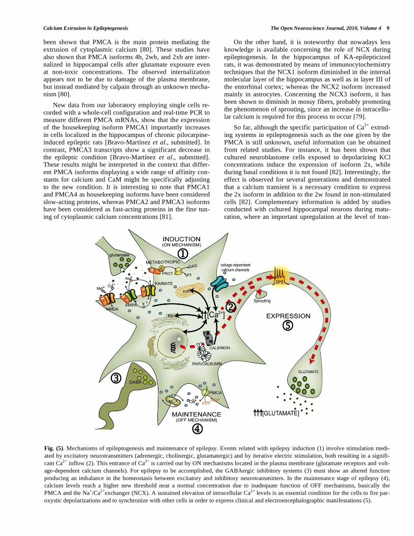
Fig. (5). Mechanisms of epileptogenesis and maintenance of epilepsy. Events related with epilepsy induction (1) involve stimulation medi-
ated by excitatory neurotransmitters (adrenergic, cholinergic, glutamatergic) and by iterative electric stimulation, both resulting in a signifi-
cant Ca2+
inflow (2). This entrance of Ca2+
is carried out by ON mechanisms located in the plasma membrane (glutamate receptors and volt-
age-dependent calcium channels). For epilepsy to be accomplished, the GABAergic inhibitory systems (3) must show an altered function
producing an imbalance in the homeostasis between excitatory and inhibitory neurotransmitters. In the maintenance stage of epilepsy (4),
calcium levels reach a higher new threshold near a normal concentration due to inadequate function of OFF mechanisms, basically the
PMCA and the Na+/Ca
2+exchanger (NCX). A sustained elevation of intracellular Ca
2+ levels is an essential condition for the cells to fire par-
oxystic depolarizations and to synchronize with other cells in order to express clinical and electroencephalographic manifestations (5).
10 The Open Neuroscience Journal, 2010, Volume 4 Bravo-Martínez et al.
scripts and proteins mainly PMCA2 and the NCX2, has been described [83]. During neuronal maturation it is assumed that nervous cells require to improve their Ca
2+ buffering
capacity, and that changes might occur concomitantly with changes within intracellular Ca
2+ buffering systems [83].
6. CONCLUDING REMARKS
Up to date, accumulated knowledge points to the fact that the ultimate control of calcium homeostasis in the neuron is carried out at the level of the plasma membrane by the Ca
2+-
ATPase and the Na+/Ca
2+ exchanger (Fig. 5). In the case of
the epileptic cell, a different cytoplasmic calcium concentra-tion threshold than the one observed in normal cells, is main-tained within manageable levels. This higher intracellular calcium concentration induces the cell to discharge paroxys-tic depolarizations and to synchronize with the remainder of epileptic cells. In parallel, a new equilibrium is reached in challenged cells where the expression of specific PMCA isoforms and other proteins mobilizing calcium lead the cell to overcome the new physiological demand. The temporal screening for the expression of the different isoforms provid-ing the adequate kinetic properties aimed to restore as much as possible the “normal” cytoplasmic calcium levels, is the focus of future investigation. Likewise, the search of alterna-tive regulating mechanisms involved at the transcriptional and/or translational levels of these isoforms will permit a better understanding of the origin and possible control of epileptogenesis.
ACKNOWLEDGEMENTS
Research carried out at our laboratory has been supported by grants from CONACyT-México (47333/A-1) and DGAPA-UNAM (IN228607/19). We are grateful to Maggie Brunner, M.A. for editorial revision.
REFERENCES
[1] McNamara JO, Huang YZ, Leonard AS. Molecular signaling
mechanisms underlying epileptogenesis. Science STKE 2006; re12:
1-22.
[2] DeGraff AS. Epidemiological aspects of epilepsy in Norway. Epi-
lepsia 1974; 15: 291-9.
[3] Putney J. Calcium. In: Siegel GJ, Ed. Basic neurochemistry: mo-
lecular, cellular, and medical aspects. Philadelphia: Lippincott-
Raven Publishers 1999; pp. 453-69.
[4] Sun DA, Sombati S, Blair RE, DeLorenzo RJ. Long-lasting altera-
tions in neuronal calcium homeostasis in an in vitro model of
stroke-induced epilepsy. Cell Calcium 2004; 35: 155-63.
[5] García ML, Strehler EE. Plasma membrane calcium ATPases as
critical regulators of calcium homeostasis during neuronal cell
function. Front Biosci 1999; 4: D869-D82.
[6] Pal S, Limbrick DD, Jr, Rafiq A, DeLorenzo RJ. Induction of spon-
taneous recurrent epileptiform discharges causes long-term changes
in intracellular calcium homeostatic mechanisms. Cell Calcium
2000; 28: 181-93.
[7] Raza M, Pal S, Rafiq A, DeLorenzo RJ. Long-term alteration of
calcium homeostatic mechanisms in the pilocarpine model of tem-
poral lobe epilepsy. Brain Res 2001; 903: 1-12.
[8] De Curtis M, Avanzini G. Interictal spikes in focal epileptogenesis.
Prog Neurobiol 2001; 63: 541-67.
[9] Matsumoto H, Ajmone-Marsan C. Cortical cellular phenomena in
experimental epilepsy: interictal manifestations. Exp Neurol 1964;
9: 286-304.
[10] De Falco FAQ, Bartiromo U, Majello L, Di Geronimo G, Mundo P.
Calcium antagonist nimodipine in intractable epilepsy. Epilepsia
1992; 33: 343-45.
[11] Jarrott B. Epileptogenesis: biochemical aspects. In: Eadie MJ,
Vajda FJE, Eds. Antiepileptic drugs pharmacology and therapeu-
tics. Springer: Berlin 1999; pp. 87-121.
[12] Budd SL. Mechanisms of neuronal damage in brain hy-
poxia/ischemia: focus on the role of mitochondrial calcium accu-
mulation. Pharmacol Ther 1998; 80: 203-29.
[13] Dennis EA. Diversity of group types, regulation, and function of
phospholipase A2. J Biol Chem 1994; 269: 13057-60.
[14] Lipton SA, Choi YB, Sucher NJ, Pan ZH, Stamler JS. Redox state,
NMDA receptors and NO-related species. Trends Pharmacol Sci
1996; 17: 186-7.
[15] Hall ED, Kupina NC, Althaus JS. Peroxynitrite scavengers for the
acute treatment of traumatic brain injury. Ann NY Acad Sci 1999;
890: 462-8.
[16] Strehler EE, Treiman M. Calcium pumps of plasma membrane and
cell interior. Curr Mol Med 2004; 4: 323-35.
[17] Minelli A, Castaldo P, Gobbi P, Salucci S, Magi S, Amoroso S.
Cellular and subcellular localization of Na+–Ca2+ exchanger protein
isoforms, NCX1, NCX2, and NCX3 in cerebral cortex and hippo-
campus of adult rat. Cell Calcium 2007; 41: 221-34.
[18] Tymianski M. Cytosolic calcium concentrations and cell death in
vitro. Adv Neurol 1996; 71: 85-105.
[19] Strehler EE, Zacharias DA. Role of alternative splicing in generat-
ing isoform diversity among plasma membrane calcium pumps.
Physiol Rev 2001; 81: 21-50.
[20] Keeton TP, Burk SE, Shull GE. Alternative splicing of exons en-
coding the calmodulin-binding domains and C termini of plasma
membrane Ca(2+)-ATPase isoforms 1,2,3, and 4. J Biol Chem 1993;
268: 2740-8.
[21] Zacharias DA, Dalrymple SJ, Strehler EE. Transcript distribution
of plasma membrane Ca2+ pump isoforms and splice variants in the
human brain. Mol Brain Res 1995; 28: 263-72.
[22] Kamagate A, Herchuelz A, Bollen A, Van Eylen F. Expression of
multiple plasma membrane Ca2+-ATPases in rat pancreatic islet
cells. Cell Calcium 2000; 27: 231-46.
[23] Santiago-García J, Mas-Oliva J, Saavedra D, Zarain-Herzberg A.
Analysis of mRNA expression and cloning of a novel plasma
membrane Ca2+ATPase splice variant in human. Mol Cell Biochem
1996; 155: 173-82.
[24] Delgado-Coello B, Santiago-García J, Zarain-Herzberg A, Mas-Oliva
J. Plasma membrane Ca2+-ATPase mRNA expression in murine hepa-
tocarcinoma and regenerating liver cells. Mol Cell Biochem 2003;
247: 177-84.
[25] Talarico EF, Jr, Mangini NJ. Alternative splice variants of plasma
membrane calcium-ATPases in human corneal epithelium. Exp Eye
Res 2007; 85: 869-79.
[26] Preiano BS, Guerini D, Carafoli E. Expression and functional char-
acterization of isoforms 4 of the plasma membrane calcium pump.
Biochemistry 1996; 35: 7946-53.
[27] James P, Maeda M, Fischer R, et al. Identification and primary
structure of a calmodulin binding domain of the Ca2+ pump of hu-
man erythrocytes. J Biol Chem 1988; 263: 2905-10.
[28] Enyedi A, Vorherr T, James P, et al. The calmodulin binding do-
main of the plasma membrane Ca2+ pump interacts both with
calmodulin and with another part of the pump. J Biol Chem 1989;
264: 12313-21.
[29] Penniston JT, Enyedi A. Modulation of the plasma membrane Ca2+
pump. J Membr Biol 1998; 165: 101-9.
[30] Caride AJ, Elwess NL, Verma AK, et al. The rate of activation by
calmodulin of isoform 4 of the plasma membrane Ca(2+) pump is
slow and is changed by alternative splicing. J Biol Chem 1999;
274: 35227-32.
[31] Caride AJ, Filoteo AG, Penheiter AR, Pászty K, Enyedi A,
Penniston JT. Delayed activation of the plasma membrane calcium
pump by a sudden increase in Ca2+: fast pumps reside in fast cells.
Cell Calcium 2001; 30: 49-57.
[32] Caride AJ, Penheiter AR, Filoteo AG, Bajzer Z, Enyedi A, Pen-
niston JT. The plasma membrane calcium pump displays memory
Calcium Extrusion in Epileptogenesis The Open Neuroscience Journal, 2010, Volume 4 11
of past calcium spikes. Differences between isoforms 2b and 4b. J
Biol Chem 2001; 276: 39797-804.
[33] Lehotsky J. Plasma membrane Ca(2+)-pump functional specializa-
tion in the brain: complex of isoform expression and regulation by
effectors. Mol Chem Neuropathol 1995; 25: 175-87.
[34] Wang KK, Villalobo A, Roufogalis BD. The plasma membrane
calcium pump: a multiregulated transporter. Trends Cell Biol 1992;
2: 46-52.
[35] Carafoli E. The plasma membrane calcium pump. Structure, func-
tion, regulation. Biochim Biophys Acta 1992; 110: 266-67.
[36] Verma AK, Paszty K, Filoteo AG, Penniston JT, Enyedi A. Protein
kinase C phosphorylates plasma membrane Ca2+ pump isoform 4a
at its calmodulin binding domain. J Biol Chem 1999; 274: 527-31.
[37] Mas-Oliva J, Santiago-García J. Cholesterol effect on thermostabil-
ity of the (Ca2+ + Mg2+)-ATPase from cardiac muscle sarcolemma.
Biochem Int 1990; 21: 233-41.
[38] Ortega A, Santiago-García J, Mas-Oliva J, Lepock JR. Cholesterol
increases the thermal stability of the Ca2+/Mg2+-ATPase of cardiac
microsomes. Biochim Biophys Acta 1996; 1283: 45-50.
[39] Santiago-García J, Delgado-Coello BA, Mas-Oliva J. Thermal
analysis of the plasma membrane Ca2+-ATPase. Mol Cell Biochem
2000; 209: 105-12.
[40] Mas-Oliva J, Delgado-Coello B. Protein stability and the evolution
of the cell membrane. Comp Biochem Physiol Part C Toxicol
Pharmacol 2007; 146: 207-13.
[41] Carafoli E. Biogenesis: plasma membrane calcium ATPase: 15
years of work on the purified enzyme. FASEB J 1994; 8: 993-1002.
[42] Missiaen L, Raeymaekers L, Wuytack F, Vrolix M, Smedt H de,
Casteels R. Phospholipid-protein interactions of the plasma-
membrane Ca2+-transporting ATPase: evidence for a tissue-
dependent functional difference. Biochem J 1989; 263: 687-94.
[43] Delgado-Coello B, Trejo R, Mas-Oliva J. Is there a specific role for
the plasma membrane Ca2+-ATPase in the hepatocyte? Mol Cell
Biochem 2006; 285; 1-15.
[44] Zylinska L, Gromadzinska E, Lachowicz L. Short-time effects of
neuroactive steroids on rat cortical Ca2+-ATPase activity. Biochim
Biophys Acta 1999; 1437: 257-64.
[45] Bootman MD, Lipp P, Berridge MJ. The organisation and functions
of local Ca(2+) signals. J Cell Sci 2001; 114: 2213-22.
[46] DeMarco SJ, Strehler EE. Plasma membrane Ca2+-ATPase iso-
forms 2b and 4b interact promiscuously and selectively with mem-
bers of the membrane-associated guanylate kinase family of PDZ
(PSD95/Dlg/ZO-1) domain-containing proteins. J Biol Chem 2001;
276: 21594-600.
[47] Strehler EE, Caride AJ, Filoteo AG, Xiong Y, Penniston JT,
Enyedi A. Plasma membrane Ca2+ ATPases as dynamic regulators
of cellular calcium handling. Ann NY Acad Sci 2007; 1099: 226-
36.
[48] Garner CC, Nash J, Huganir RL. PDZ domains in synapse assem-
bly and signaling. Trends Cell Biol 2000, 10: 274-80.
[49] Stauffer TP, Hilfiker H, Carafoli E, Strehler EE. Quantitative
analysis of alternative splicing options of human plasma membrane
calcium pump genes. J Biol Chem 1993, 268: 25993-26003.
[50] Filoteo AG, Elwess NL, Enyedi A, Caride A, Aung HH, Penniston
JT. Plasma membrane Ca2+ pump in rat brain: patterns of alterna-
tive splices seen by isoform-specific antibodies. J Biol Chem 1997;
272: 23741-7.
[51] Guerini D. The significance of the isoforms of plasma membrane
calcium ATPase. Cell Tissue Res 1998; 292: 191-7.
[52] Zacharias DA, Strehler EE. Change in plasma membrane Ca2+-
ATPase splice-variant expression in response to a rise in intracellu-
lar Ca2+. Curr Biol 1996; 6: 1642-52.
[53] Burette A, Rockwood JM, Strehler EE, Weinberg RJ. Isoform-
specific distribution of the plasma membrane Ca2+ ATPase in the
rat brain. J Comp Neurol 2003; 467: 464-76.
[54] Schuh K, Uldrijan S, Telkamp M, Röthlein N, Neyses L. The
plasma membrane calmodulin–dependent calcium pump: a major
regulator of nitric oxide synthase I. J Cell Biol 2001; 155: 201-5.
[55] Daniel EE, El-Yazbi A, Cho WJ. Caveolae and calcium handling, a
review and a hypothesis. J Cell Mol Med 2006; 10: 529-44.
[56] El-Yazbi AF, Cho WJ, Schuylz R, Daniel EE. Calcium extrusion
by plasma membrane calcium pump is impaired in caveolin-1
knockout mouse small intestine. Eur J Pharmacol 2008; 591: 80-7.
[57] Jiang L, Fernandes D, Mehta N, Bean JL, Michaelis ML, Zaidi A.
Partitioning of the Plasma membrane Ca2+-ATPase into lipid rafts
in primary neurons:effects of cholesterol depletion. J Neurochem
2007; 102: 378-88.
[58] McNamara JO, Puranam RS. Epilepsy genetics: an abundance of
riches for biologists. Curr Biol 1998; 8: R166-70.
[59] Nicoll DA, Longoni S, Philipson KD. Molecular cloning and func-
tional expression of the cardiac sarcolemmal Na(+)-Ca2+ exchanger.
Science 1990; 250: 562-5.
[60] Ruknudin AM, Lakatta EG, The Regulation of the Na/Ca Ex-
changer and Plasmalemmal Ca2+ ATPase by other Proteins. Ann
NY Acad Sci 2007, 1099: 86-102.
[61] Marshall CR, Fox JA, Butland SL, Ouellette BF, Brinkman FS,
Tibbits GF. Phylogeny of Na+/Ca2+ exchanger (NCX) genes from
genomic data identifies new gene duplications and a new family
member in fish species. Physiol Genomics 2005; 21: 161-73.
[62] On C, Marshall CR, Chen N, Moyes CD, Tibbits GF. Gene struc-
ture evolution of the Na+-Ca2+ exchanger (NCX) family. BMC Evol
Biol 2008; 8: 127.
[63] Quednau BD, Nicoll DA, Philipson KD. The sodium/calcium ex-
changer family-SLC8. Pflugers Arch 2004; 447: 543-8.
[64] Lytton J. Na+/Ca2+ exchangers: three mammalian gene families
control Ca2+ transport. Biochem J 2007; 406: 365-82.
[65] Quednau BD, Nicoll DA, Philipson KD. Tissue specificity and
alternative splicing of the Na+/Ca2+ exchanger isoforms NCX1,
NCX2, and NCX3 in rat. Am J Physiol Cell Physiol 1997; 272:
C1250-61.
[66] Li JP, Kajiya H, Okamoto F, Nakao A, Iwamoto T, Okabe K. Three
Na+/Ca2+ exchanger (NCX) variants are expressed in mouse osteo-
clasts and mediate calcium transport during bone resorption. Endo-
crinology 2007; 148: 2116-25.
[67] Philipson KD, Nicoll DA. Sodium-calcium exchange: a molecular
perspective. Annu Rev Physiol 2000; 62: 111-33.
[68] Hilge M, Aelen J, Perrakis A, Vuister GW. Structural Basis for
Ca2+ Regulation in the Na+/Ca2+. Ann NY Acad Sci 2007; 1099:
7-15.
[69] Matsuoka S, Nicoll DA, Reilly RF. Initial localization of regulatory
regions of the cardiac sarcolemmal Na(+)-Ca2+ exchanger. Proc
Natl Acad Sci USA 1993; 90: 3870-4.
[70] Schwarz EM, Benzer S. Calx, a Na-Ca exchanger gene of Droso-
phila melanogaster. Proc Natl Acad Sci USA 1997; 94: 10249-54.
[71] Ruknudin AM, Wei S, Haigney MC. Phosphorylation and other
conundrums of Na/Ca Exchanger, NCX1. Ann NY Acad Sci 2007;
1099: 103-18.
[72] Schulse DH, Muqhal M, Lederer WJ, Ruknudin AM. So-
dium/calcium exchanger (NCX1) macromolecular complex. J Biol
Chem 2003; 278: 28849-55.
[73] Ryan C, Shaw G, Hardwicke PMD. Effect of Ca2+ on protein
kinase A-mediated phosphorylation of a specific serine residue in
an expressed peptide containing the Ca2+-regulatory domain of
scallop muscle Na+/Ca2+ exchanger. Ann NY Acad Sci 2007; 1099:
43-52.
[74] Shigekawa M, Katanosaka Y, Wakabayashi S. Regulation of the
cardiac Na+/Ca2+ exchanger by calcineurin and protein kinase C.
Ann NY Acad Sci 2007; 10: 53-63.
[75] Papa M, Canitano A, Boscia F, et al. Differential expression of the
Na+-Ca2+ exchanger transcripts and proteins in rat brain regions. J
Comp Neurol 2003; 461: 31-48.
[76] Lörincz A, Rózsa B, Katona G, Vizi ES, Tamás G. Differential
distribution of NCX1 contributes to spine–dendrite compartmen-
talization in CA1 pyramidal cells. Proc Natl Acad Sci USA 2007;
104: 1033-8.
[77] Kim YT, Namkung YL, Kwak J, Suh CK. Involvement of Na+–
Ca2+ exchanger on metabotropic glutamate receptor 1-mediated
12 The Open Neuroscience Journal, 2010, Volume 4 Bravo-Martínez et al.
[Ca2+]i transients in rat cerebellar Purkinje neurons. Neuroscience
2007; 146: 170-7.
[78] García ML, Murray KD, García VB, Strehler EE, Isackson PJ.
Seizure-induced alterations of plasma membrane calcium ATPase
isoforms 1, 2 and 3 mRNA and protein in rat hippocampus. Mol
Brain Res 1997; 45: 230-8.
[79] Ketelaars SOM, Gorter JA, Aronica E, Wadman WJ. Calcium
extrusion protein expression in the hippocampal formation of
chronic epileptic rats after kainate-induced status epilepticus.
Epilepsia 2004; 45: 1189-201.
[80] Pottorf WJ, 2nd, Johanns TM, Derrington SM, Strehler EE, Enyedi
A, Thayer SA. Glutamate-induced protease-mediated loss of
plasma membrane Ca2+ pump activity in rat hippocampal neurons. J
Neurochem 2006; 98: 1646-56.
[81] Strehler EE, Caride AJ, Filoteo AG, Xiong Y, Penniston JT,
Enyedi A. Plasma membrane Ca2+ ATPases as dynamic regulators
of cellular calcium handling. Ann NY Acad Sci 2007; 1099: 226-
36.
[82] Zacharias DA, Strehler EE. Change in plasma membrane Ca2+-
ATPase splice-variant expression in response to a rise in intracellu-
lar Ca2+. Curr Biol 1996; 6: 1642-52.
[83] Kip SN, Gray NW, Burette A, Canbay A, Weinberg RJ, Strehler
EE. Changes in the expression of plasma membranes calcium ex-
trusion systems during the maturation of hippocampal neurons.
Hippocampus 2006; 16: 20-34.
Received: May 14, 2009 Revised: December 01, 2009 Accepted: January 06, 2010
© Bravo-Martínez et al.; Licensee Bentham Open.
This is an open access article licensed under the terms of the Creative Commons Attribution Non-Commercial License
(http://creativecommons.org/licenses/by-nc/3.0/) which permits unrestricted, non-commercial use, distribution and reproduction in any medium, provided the
work is properly cited.
Related Documents