Cell-Autonomous Progeroid Changes in Conditional Mouse Models for Repair Endonuclease XPG Deficiency Sander Barnhoorn 1. , Lieneke M. Uittenboogaard 1. , Dick Jaarsma 2. , Wilbert P. Vermeij 1 , Maria Tresini 1 , Michael Weymaere 1 , Herve ´ Menoni 1 , Renata M. C. Brandt 1 , Monique C. de Waard 3 , Sander M. Botter 4 , Altaf H. Sarker 5 , Nicolaas G. J. Jaspers 1 , Gijsbertus T. J. van der Horst 1 , Priscilla K. Cooper 5 , Jan H. J. Hoeijmakers 1 *, Ingrid van der Pluijm 1,6 * 1 Department of Genetics, Erasmus University Medical Center, Rotterdam, The Netherlands, 2 Department of Neuroscience, Erasmus University Medical Center, Rotterdam, The Netherlands, 3 Department of Intensive Care, VU University Medical Center, Amsterdam, The Netherlands, 4 Uniklinik Balgrist, Zu ¨ rich, Switzerland, 5 Life Sciences Division, Lawrence Berkeley National Laboratory, Berkeley, California, United States of America, 6 Department of Vascular Surgery, Erasmus University Medical Center, Rotterdam, The Netherlands Abstract As part of the Nucleotide Excision Repair (NER) process, the endonuclease XPG is involved in repair of helix-distorting DNA lesions, but the protein has also been implicated in several other DNA repair systems, complicating genotype-phenotype relationship in XPG patients. Defects in XPG can cause either the cancer-prone condition xeroderma pigmentosum (XP) alone, or XP combined with the severe neurodevelopmental disorder Cockayne Syndrome (CS), or the infantile lethal cerebro-oculo-facio-skeletal (COFS) syndrome, characterized by dramatic growth failure, progressive neurodevelopmental abnormalities and greatly reduced life expectancy. Here, we present a novel (conditional) Xpg 2/2 mouse model which -in a C57BL6/FVB F1 hybrid genetic background- displays many progeroid features, including cessation of growth, loss of subcutaneous fat, kyphosis, osteoporosis, retinal photoreceptor loss, liver aging, extensive neurodegeneration, and a short lifespan of 4–5 months. We show that deletion of XPG specifically in the liver reproduces the progeroid features in the liver, yet abolishes the effect on growth or lifespan. In addition, specific XPG deletion in neurons and glia of the forebrain creates a progressive neurodegenerative phenotype that shows many characteristics of human XPG deficiency. Our findings therefore exclude that both the liver as well as the neurological phenotype are a secondary consequence of derailment in other cell types, organs or tissues (e.g. vascular abnormalities) and support a cell-autonomous origin caused by the DNA repair defect itself. In addition they allow the dissection of the complex aging process in tissue- and cell-type-specific components. Moreover, our data highlight the critical importance of genetic background in mouse aging studies, establish the Xpg 2/2 mouse as a valid model for the severe form of human XPG patients and segmental accelerated aging, and strengthen the link between DNA damage and aging. Citation: Barnhoorn S, Uittenboogaard LM, Jaarsma D, Vermeij WP, Tresini M, et al. (2014) Cell-Autonomous Progeroid Changes in Conditional Mouse Models for Repair Endonuclease XPG Deficiency. PLoS Genet 10(10): e1004686. doi:10.1371/journal.pgen.1004686 Editor: Laura J. Niedernhofer, The Scripps Research Institute, United States of America Received February 3, 2014; Accepted August 19, 2014; Published October 9, 2014 This is an open-access article, free of all copyright, and may be freely reproduced, distributed, transmitted, modified, built upon, or otherwise used by anyone for any lawful purpose. The work is made available under the Creative Commons CC0 public domain dedication. Funding: We acknowledge financial support of the European commission FP7 Markage (FP7-Health-2008-200880), DNA Repair (LSHG-CT-2005-512113) and LifeSpan (LSHG-CT-2007-036894), National Institute of Health (NIH)/National Institute of Ageing (NIA) (1PO1 AG-17242-02), NIEHS (1UO1 ES011044), NIH/National Cancer Institute R01 CA063503 and P01 CA092584 to PKC, and the Royal Academy of Arts and Sciences of the Netherlands (academia professorship to JHJH) and a European Research Council Advanced Grant to JHJH. The research leading to these results has received funding from the European Community’s Seventh Framework Programme (FP7/2007-2013) under grant agreement No. HEALTH-F2-2010-259893. The funders had no role in study design, data collection and analysis, decision to publish, or preparation of the manuscript. Competing Interests: The authors have declared that no competing interests exist. * Email: [email protected] (JHJH); [email protected] (IvdP) . These authors contributed equally to this work. Introduction If DNA damage, either inflicted from exogenous or endogenous sources, cannot be repaired, this has detrimental consequences for an organism ranging from transcription blocks, permanent cell cycle arrest and mutations, to cell death. In the end, this unrepaired DNA damage contributes to the onset and progression of the aging process, as well as to cancer [1–3]. Cells are equipped with a set of elaborate DNA repair mechanisms integrated into a complex DNA damage response machinery that jointly attempt to fix the unrepaired DNA [4]. One such DNA repair mechanism is the Nucleotide Excision Repair (NER) pathway that removes a wide category of helix-distorting lesions, such as those induced by UV and bulky chemical adducts, in a tightly coordinated process involving over 30 proteins [5–7]. NER can be divided into two subpathways based on the mode of damage recognition. The Global Genome (GG-)NER subpathway specifically involves the XPC and XPE protein complexes, and probes the entire genome for lesions that disrupt base-pairing [5,7–9]. Transcription- Coupled (TC-)NER, on the other hand, detects helix-distorting lesions that stall transcription in the transcribed strand of expressed genes, and hence enables resumption of transcription. TC-NER is independent of XPC and XPE and specifically involves proteins such as CSA, CSB and UVSSA [8,10,11]. After PLOS Genetics | www.plosgenetics.org 1 October 2014 | Volume 10 | Issue 10 | e1004686

Welcome message from author
This document is posted to help you gain knowledge. Please leave a comment to let me know what you think about it! Share it to your friends and learn new things together.
Transcript

Cell-Autonomous Progeroid Changes in ConditionalMouse Models for Repair Endonuclease XPG DeficiencySander Barnhoorn1., Lieneke M. Uittenboogaard1., Dick Jaarsma2., Wilbert P. Vermeij1, Maria Tresini1,
Michael Weymaere1, Herve Menoni1, Renata M. C. Brandt1, Monique C. de Waard3, Sander M. Botter4,
Altaf H. Sarker5, Nicolaas G. J. Jaspers1, Gijsbertus T. J. van der Horst1, Priscilla K. Cooper5,
Jan H. J. Hoeijmakers1*, Ingrid van der Pluijm1,6*
1 Department of Genetics, Erasmus University Medical Center, Rotterdam, The Netherlands, 2 Department of Neuroscience, Erasmus University Medical Center, Rotterdam,
The Netherlands, 3 Department of Intensive Care, VU University Medical Center, Amsterdam, The Netherlands, 4 Uniklinik Balgrist, Zurich, Switzerland, 5 Life Sciences
Division, Lawrence Berkeley National Laboratory, Berkeley, California, United States of America, 6 Department of Vascular Surgery, Erasmus University Medical Center,
Rotterdam, The Netherlands
Abstract
As part of the Nucleotide Excision Repair (NER) process, the endonuclease XPG is involved in repair of helix-distorting DNAlesions, but the protein has also been implicated in several other DNA repair systems, complicating genotype-phenotyperelationship in XPG patients. Defects in XPG can cause either the cancer-prone condition xeroderma pigmentosum (XP)alone, or XP combined with the severe neurodevelopmental disorder Cockayne Syndrome (CS), or the infantile lethalcerebro-oculo-facio-skeletal (COFS) syndrome, characterized by dramatic growth failure, progressive neurodevelopmentalabnormalities and greatly reduced life expectancy. Here, we present a novel (conditional) Xpg2/2 mouse model which -ina C57BL6/FVB F1 hybrid genetic background- displays many progeroid features, including cessation of growth, loss ofsubcutaneous fat, kyphosis, osteoporosis, retinal photoreceptor loss, liver aging, extensive neurodegeneration, and a shortlifespan of 4–5 months. We show that deletion of XPG specifically in the liver reproduces the progeroid features in the liver,yet abolishes the effect on growth or lifespan. In addition, specific XPG deletion in neurons and glia of the forebrain createsa progressive neurodegenerative phenotype that shows many characteristics of human XPG deficiency. Our findingstherefore exclude that both the liver as well as the neurological phenotype are a secondary consequence of derailment inother cell types, organs or tissues (e.g. vascular abnormalities) and support a cell-autonomous origin caused by the DNArepair defect itself. In addition they allow the dissection of the complex aging process in tissue- and cell-type-specificcomponents. Moreover, our data highlight the critical importance of genetic background in mouse aging studies, establishthe Xpg2/2 mouse as a valid model for the severe form of human XPG patients and segmental accelerated aging, andstrengthen the link between DNA damage and aging.
Citation: Barnhoorn S, Uittenboogaard LM, Jaarsma D, Vermeij WP, Tresini M, et al. (2014) Cell-Autonomous Progeroid Changes in Conditional Mouse Models forRepair Endonuclease XPG Deficiency. PLoS Genet 10(10): e1004686. doi:10.1371/journal.pgen.1004686
Editor: Laura J. Niedernhofer, The Scripps Research Institute, United States of America
Received February 3, 2014; Accepted August 19, 2014; Published October 9, 2014
This is an open-access article, free of all copyright, and may be freely reproduced, distributed, transmitted, modified, built upon, or otherwise used by anyone forany lawful purpose. The work is made available under the Creative Commons CC0 public domain dedication.
Funding: We acknowledge financial support of the European commission FP7 Markage (FP7-Health-2008-200880), DNA Repair (LSHG-CT-2005-512113) andLifeSpan (LSHG-CT-2007-036894), National Institute of Health (NIH)/National Institute of Ageing (NIA) (1PO1 AG-17242-02), NIEHS (1UO1 ES011044), NIH/NationalCancer Institute R01 CA063503 and P01 CA092584 to PKC, and the Royal Academy of Arts and Sciences of the Netherlands (academia professorship to JHJH) anda European Research Council Advanced Grant to JHJH. The research leading to these results has received funding from the European Community’s SeventhFramework Programme (FP7/2007-2013) under grant agreement No. HEALTH-F2-2010-259893. The funders had no role in study design, data collection andanalysis, decision to publish, or preparation of the manuscript.
Competing Interests: The authors have declared that no competing interests exist.
* Email: [email protected] (JHJH); [email protected] (IvdP)
. These authors contributed equally to this work.
Introduction
If DNA damage, either inflicted from exogenous or endogenous
sources, cannot be repaired, this has detrimental consequences for
an organism ranging from transcription blocks, permanent cell
cycle arrest and mutations, to cell death. In the end, this
unrepaired DNA damage contributes to the onset and progression
of the aging process, as well as to cancer [1–3]. Cells are equipped
with a set of elaborate DNA repair mechanisms integrated into
a complex DNA damage response machinery that jointly attempt
to fix the unrepaired DNA [4]. One such DNA repair mechanism
is the Nucleotide Excision Repair (NER) pathway that removes
a wide category of helix-distorting lesions, such as those induced
by UV and bulky chemical adducts, in a tightly coordinated
process involving over 30 proteins [5–7]. NER can be divided into
two subpathways based on the mode of damage recognition. The
Global Genome (GG-)NER subpathway specifically involves the
XPC and XPE protein complexes, and probes the entire genome
for lesions that disrupt base-pairing [5,7–9]. Transcription-
Coupled (TC-)NER, on the other hand, detects helix-distorting
lesions that stall transcription in the transcribed strand of
expressed genes, and hence enables resumption of transcription.
TC-NER is independent of XPC and XPE and specifically
involves proteins such as CSA, CSB and UVSSA [8,10,11]. After
PLOS Genetics | www.plosgenetics.org 1 October 2014 | Volume 10 | Issue 10 | e1004686
lesion recognition, the subsequent ‘cut-and-patch’ core repair
reaction encompasses local opening of the DNA helix and lesion
verification, performed by the TFIIH complex together with XPA.
Both correctly position the structure-specific endonucleases
ERCC1/XPF and XPG for strand-specific excision of the lesion
as part of a 22–30 bp oligonucleotide [5,7,12]. Finally, the gap is
filled by repair synthesis and closed by ligation [5,7,12].
Multiple NER proteins have been attributed additional roles,
both in DNA repair pathways other than NER, and in
transcription regulation. For instance, TFIIH is an essential
component of the general transcription machinery [13,14], but
also other NER factors, including XPG and CSB, have been
implicated in transcription regulation [15–18]. The 59 endonu-
clease ERCC1/XPF participates in the repair of interstrand
crosslinks [19,20] and subpathways of DNA double-strand break
repair [21]. XPC, CSB, and XPG have been individually
implicated in promoting base excision repair (BER) of oxidative
DNA damage [22–30]. TFIIH and XPG are, together with CSB,
thought to be involved in the early steps of Transcription-Coupled
Repair (TCR), and XPG interacts directly with both CSB and
RNA Polymerase II [31]. Although still controversial, there are
accumulating reports that TCR not only directs NER to blocked
transcription but may also recruit BER for preferential repair of
oxidative DNA damage in transcribed strands [32–34]. Such
a mechanism might be related to the roles of both CSB and XPG
in promoting BER more globally. If correct, it could explain the
much greater consequences for the organism of TCR defects
compared to defects in NER alone (see below) [35].
A number of rare, autosomal recessive disorders resulting from
mutations in NER genes underscore the importance of genome
maintenance for the prevention of cancer as well as aging [3].
NER-associated diseases are characterized by sun (UV) hypersen-
sitivity and include xeroderma pigmentosum (XP), UV-sensitivity
syndrome (UVSS), Cockayne syndrome (CS), cerebro-oculo-facio-
skeletal (COFS) syndrome, XPF-ERCC1 (XFE) progeroid syn-
drome, trichothiodystrophy (TTD) and disorders that combine the
symptoms of these syndromes, including XP/CS [35–39]. XP
originates from defects in GG-NER or total NER activity and is
characterized by an over 2000-fold increased risk of cancer in sun-
exposed skin and, to a much lesser extent, in internal organs [36].
XP patients may also develop progressive neurological symptoms
and neuronal degeneration depending on the severity of the total
NER deficiency [36,39,40]. UVSS is characterized by skin UV
hypersensitivity without actually developing skin cancer. UVSS
results from the selective loss of TC-NER function as a conse-
quence of mutations in the proteins involved in detection of UV-
induced transcription-blocking DNA lesions, i.e. UVSSA, CSA,
and CSB [11,35,41–44]. Mutations in CSA and CSB, however,
generally cause CS, a heterogeneous multisystem disorder that, in
addition to UV-sensitivity, is characterized by severe growth
failure and cachexia, accelerated aging features, short lifespan, and
progressive sensori-neuronal abnormalities [38,45]. The severe
symptoms of CS cannot be explained by the loss of TC-NER
function as they do not occur in fully NER-deficient XP patients
and TC-NER deficient UVSS patients. Therefore, CS symptoms
have been linked to additional, yet incompletely, defined functions
of CSA and CSB in DNA repair, transcription regulation, other
processes, or a combination of deficiencies [3,46,47]. The same
applies for mutations in the down-stream NER factors XPB, XPD,
XPF, ERCC1 and XPG that cause combined XP/CS, or severe
developmental/degenerative multisystem disorders such as COFS
and XFE that share multiple features with severe CS forms
[35,48–51]. Thus CS symptoms can result from mutations in
multiple proteins that operate together in NER, but the symptoms
caused by these mutations cannot be explained by NER deficiency
alone, raising questions about the identities of these non-NER
activities underlying CS symptoms and the extent to which
different symptoms reflect deficits of different cellular processes
[35,46].
Mutations in the structure-specific NER 39-endonuclease XPG
are rare, with less than 20 patients and 25 mutant alleles described
so far [52–55], and cause a spectrum of disease phenotypes
varying from XP to XP/CS and COFS [53]. Point mutations that
selectively eliminate XPG nuclease activity cause XP, while C-
terminal truncations, destabilizing point mutations, and mutations
that abolish the interaction between XPG and the basal
transcription factor TFIIH cause XP/CS and COFS [52–58].
These data support the notion that a deficient function of XPG
outside NER is responsible for the severe CS symptoms
[15,52,53,55–57].
For most NER disorders, mouse mutants have been generated
that mimic the genetic defect found in patients, and to various
extents reproduce XP and CS-like features as well as the progeroid
hallmarks found in the corresponding human syndrome [59–62].
Accordingly, Xpg-null (Xpg2/2) mice were found to develop
a severe phenotype characterized by growth deficiency and very
short lifespan, resembling severe XP/CS [63]. In contrast, Xpgmutant mice carrying amino acid substitutions that selectively
abolish the nuclease function of XPG (XpgE791A and XpgD811A)
show severe UV-sensitivity but normal lifespan, hence, reprodu-
cing the XP phenotype [64,65]. In addition, a mutant XPG
construct containing a C-terminal truncation lacking the last 360
amino acids that was made to mimic the genotype of some XP-G/
CS patients, developed a growth deficiency and short-living
phenotype resembling that of Xpg2/2 mice, albeit somewhat
milder [65]. Yet another C-terminal truncation mutant lacking the
last 180 amino acids showed a normal lifespan, but produced a CS-
like growth-deficient short-living phenotype after crossing with
Xpa2/2 mice that are already fully NER-deficient [65,66].
Significantly, the same conversion of a normal lifespan into
a short-living mouse model is observed after crossing CSA- or
CSB-deficient CS mice with total NER- (Xpa2/2) or GG-NER
(Xpc2/2) deficient mouse models [67–69], but not by crossing
NER-deficient XpgD811A with Xpa2/2 mice [66]. Together these
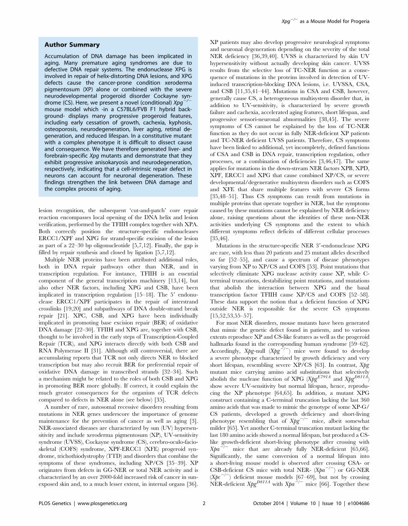
Author Summary
Accumulation of DNA damage has been implicated inaging. Many premature aging syndromes are due todefective DNA repair systems. The endonuclease XPG isinvolved in repair of helix-distorting DNA lesions, and XPGdefects cause the cancer-prone condition xerodermapigmentosum (XP) alone or combined with the severeneurodevelopmental progeroid disorder Cockayne syn-drome (CS). Here, we present a novel (conditional) Xpg2/2
mouse model which -in a C57BL6/FVB F1 hybrid back-ground- displays many progressive progeroid features,including early cessation of growth, cachexia, kyphosis,osteoporosis, neurodegeneration, liver aging, retinal de-generation, and reduced lifespan. In a constitutive mutantwith a complex phenotype it is difficult to dissect causeand consequence. We have therefore generated liver- andforebrain-specific Xpg mutants and demonstrate that theyexhibit progressive anisokaryosis and neurodegeneration,respectively, indicating that a cell-intrinsic repair defect inneurons can account for neuronal degeneration. Thesefindings strengthen the link between DNA damage andthe complex process of aging.
Xpg2/2 as a Mouse Model for Progeria
PLOS Genetics | www.plosgenetics.org 2 October 2014 | Volume 10 | Issue 10 | e1004686

data indicate a deleterious synergistic interaction between NER
deficiency and loss of non-NER activities that underlie CS.
Furthermore, they show that the C-terminus of XPG could play
a role in the CS symptoms, and that the XPG-deficient
Xpg2/2 mice may reproduce the phenotype of Xpa2/2Csb2/2,
Xpc2/2Csb2/2 or Xpa2/2Csa2/2 double mutant mice [62,66–69].
In previous analyses we clearly observed progeroid character-
istics in many NER mutant mouse models including Xpa/Csb, Xpb,
Xpd, and Ercc1 mutants [51,68,70–72], yet the occurrence of
progeroid features in Xpg2/2 mice has hitherto been poorly
established, mostly due to their very short lifespan. Since we are
particularly interested in the effect of Xpg deletion on organ-
specific aging, we generated a conditional Xpg mutant. As genetic
background can have a significant effect on phenotype de-
velopment, we first re-examined the pathological characteristics
of Xpg2/2 mice in a C57BL6/FVB F1 hybrid background, as was
previously described for Ercc1 mutant mice [51,72]. In this hybrid
background Xpg2/2 mice lived longer and presented progeroid
features including cachexia and osteoporosis with pronounced
degenerative phenotypes in both liver and brain. We next studied
the effect of liver- and forebrain-specific inactivation of Xpg,
showing that the observed phenotypes are indeed due to lack of
XPG protein. Together our data show that, consistent with
previous data in ERCC1- and CSB/XPA-deficient mice, Xpg2/2
mice develop a multisystem progeroid degenerative phenotype.
Results
Generation of NER-deficient Xpg2/2 miceTo generate a Cre-inducible Xpg knockout allele we flanked the
third exon of Xpg with LoxP sites (Figure 1A). Deletion of this
exon causes a frame shift and a premature translational stop
immediately after exon 2 at amino acid residue 89 (instead of the
full-length 1170). After transfection to 129 ES cells and selection of
properly targeted clones (Figure 1B and Materials and Methods),
two independent transfected clones were used to generate germ-
line transmitting chimeras (Figure 1C). Heterozygous males,
carrying the conditional Xpg allele, were crossed to females
ubiquitously expressing Flp for excision of the Neomycin cassette
and to yield mice that are heterozygous for the floxed Xpg (Xpgf)
allele. Xpgf/+ mice were backcrossed and maintained in FVB/N
background. To generate Xpg mice carrying a knockout allele
(Xpg2, Figure 1A), Xpgf/+ mice were crossed to Cag-Cre mice,
which ubiquitously express Cre recombinase from germline [73],
yielding heterozygous Xpg+/2 animals (Figure 1C). Xpg+/2 animals
were.10 times backcrossed into C57BL6 or FVB/N genetic
backgrounds. Unless otherwise stated, experiments were per-
formed with Xpg2/2 mice in the C57BL6/FVB F1 hybrid
background obtained from intercrossing C57BL6 Xpg+/26FVB/
N Xpg+/2 animals to minimalize background specific effects (see
below).
The presence of a premature stop codon in the Xpg2 allele was
confirmed by sequencing Xpg cDNA from liver of Xpg2/2 mice
(Figure S1A). Accordingly, Western immunoblot analysis with an
antibody raised against the central spacer region (R-domain) of
XPG shows the absence of XPG protein product in mouse dermal
fibroblasts (MDFs) isolated from Xpg2/2 mice (Figure 1D). Next,
we tested DNA repair deficiency of Xpg2/2 MDFs. In accordance
with complete NER deficiency, Xpg2/2 MDFs showed an almost
10-fold hypersensitivity to UV, similar to fully NER-defective
MDFs derived from Xpa2/2 mice (Figure 1E) [74]. In addition,
Xpg2/2 MDFs were hypersensitive to treatment with Illudin S
(Figure 1E), consistent with the loss of TC-NER function [75], and
were deficient in UV-induced unscheduled DNA synthesis in line
with loss of GG-NER activity (Figure 1F). Also, recovery of RNA
synthesis after UV exposure was almost completely abolished in
Xpg2/2 MDFs, further demonstrating loss of TC-NER activity
(Figure 1G). Xpg2/2 MDFs showed no increased sensitivity to
potassium bromate (KBrO3) which causes oxidative DNA lesions,
and a minimal increased sensitivity to the cross-linking agent
cisplatin (Figure S1B).
Genetic background affects perinatal viability andlifespan of Xpg2/2 mice
In view of a significant effect of genetic background on
embryonic lethality and lifespan in ERCC1-deficient mice
[51,72], we examined whether a similar genetic background effect
occurred in Xpg2/2 mice, by comparing birth frequencies and
lifespan of Xpg2/2 mice in a C57BL6, FVB/N or a C57BL6/FVB
F1 hybrid background. In C57BL6 background birth frequencies
were below Mendelian expectations (,8%, Table 1), whereas in
the FVB/N and C57BL6/FVB F1 hybrid background birth
frequencies were Mendelian and near-Mendelian, respectively
(Table 1). Also the lifespan of Xpg2/2 animals was strongly
dependent on genetic background, with C57BL6 Xpg2/2 mice
showing a lifespan of 3 weeks, and Xpg2/2 animals in FVB/N and
C57BL6/FVB F1 hybrid background living for 15–18 weeks
(Figure 2A).
Cachexia, short lifespan and osteoporosis in Xpg2/2 miceFurther analysis of C57BL6/FVB F1 Xpg2/2 mice showed that
they had the same size and weight as wild type and heterozygote
littermates at late embryonic stage (E17.5; Figure 2B and C).
However, after birth, Xpg2/2 mice showed reduced growth and
weight gain compared to controls, and stopped growing at 6–8
weeks when their body weight was about 65–70% of that of wild
type littermates (Figure 2C). From 10–11 weeks, body weights
declined and the Xpg2/2 mice became progressively cachectic
(Figure 2C and D). At 14 weeks all Xpg2/2 mice were severely
runted (Figure 2D), and the mice died a few weeks thereafter
between 15–18 weeks of age (Figure 2A and E). The growth
deficiency was paralleled by the development of kyphosis
(Figure 2D and F). In addition, Xpg2/2 mice progressively
developed neurological symptoms, including clasping of the
hind-limbs when lifted by their tails (Figure S2A), and at a later
time point fine tremors (Figure 2E). Accelerating rotarod and grip
strength tests in 14-week old Xpg2/2 mice revealed severe motor
deficits and muscle weakness at this age (Figure S2B and C).
Computed tomography (CT) confirmed severe kyphosis in
Xpg2/2 mice at 16 weeks of age (Figure 2F). To further examine
skeletal abnormalities and the occurrence of osteoporosis as
observed in other NER-deficient mouse models [68,76–79], we
measured several bone parameters using femoral bones. Analysis
of bone strength revealed decreased strength of the Xpg2/2
femoral bone at 14–16 weeks (Figure 2G). Micro-CT analysis
showed that the thickness of the trabeculae and cortex of the
femoral bones was significantly smaller in Xpg2/2 compared to
wild type mice at 14–17 weeks, but not yet at 7 weeks (Figure 2H).
Overall, these data indicate a progressive increase of age-related
features such as osteoporosis.
Mild progeroid features and a ‘survival-like’ stressresponse in livers of Xpg2/2 mice
Weight loss and reduced size of Xpg2/2 mice was associated
with reduced weight of internal organs (Figure S3A) and with
a strong reduction in the amount of subcutaneous fat (Figure S3B).
The Xpg2/2 mice previously reported by Harada et al. [63]
Xpg2/2 as a Mouse Model for Progeria
PLOS Genetics | www.plosgenetics.org 3 October 2014 | Volume 10 | Issue 10 | e1004686

Xpg2/2 as a Mouse Model for Progeria
PLOS Genetics | www.plosgenetics.org 4 October 2014 | Volume 10 | Issue 10 | e1004686
showed developmental abnormalities of the gastro-intestinal
tract. These gastro-intestinal abnormalities were proposed to be
a major contributor of the post-natal growth failure and short
lifespan (,3 weeks) of their animals [63]. However, in contrast to
their data, the gastro-intestinal tract of our Xpg2/2 mice had
a normal size and macroscopic appearance, and showed a normal
histological appearance in HE-stained sections (Figure 3A).
Furthermore, staining for the proliferative cell marker Ki-67
indicated that the number of proliferative cells in the intestinal
epithelium was similar between Xpg2/2 and wild type mice
(Figure 3A). In accord with normal function of the gastro-intestinal
tract we found that food intake per gram body weight was similar
between wild type and Xpg2/2 animals (Figure S3C).
The liver is a central organ in many aspects of metabolic
control, including regulation of circulating glucose levels and
detoxification, and it plays a key role in regulation of IGF1-
somatotrophic axis signaling. Previous studies have shown that
ERCC1/XPF-deficient mice develop multiple liver abnorma-
lities, in particular anisokaryosis resulting from polyploidy, and
intranuclear inclusions [72,80–83]. Analysis of HE-stained liver
sections of our Xpg2/2 mice revealed mild anisokaryosis, and
increased mean nuclear size at 14 weeks, but not at 4 weeks of age
(Figure 3B). In addition, sporadically, hepatocytes had intra-
nuclear inclusions. These liver nuclear changes are a well charac-
terized phenomenon in the aging liver, and indicate that
Xpg2/2 mice show features of accelerated aging in the liver
similar to Ercc1D/2 mice [72,82].
Liver cells of ERCC1-deficient and other progeroid NER-
deficient mouse mutants display changes in gene expression that
encompass a downregulation of catabolic and oxidative metabo-
lism and an upregulation of antioxidant and stress defense
pathways, suggestive of a compensatory survival response to cope
with increased DNA damage [51,68,84,85]. To determine
whether Xpg2/2 liver cells also display a ‘survival-like’ stress
response, we determined expression levels of selected antioxidant
and somatotrophic genes by real-time PCR. Indeed, mRNA levels
from a subset of antioxidant effector genes, including Nqo1, Srxn1,
Gstt2 and Gsta1, were significantly increased in liver homogenates
of young (7-week old) Xpg2/2 animals compared to controls. At 14
weeks of age, we observed a similar significant increased
expression of Nqo1 and Gsta1 while mRNA from the other
antioxidant effector genes tested showed unaltered expression
(Figure 3C). Expression levels of Nrf2, which is a potent inducer of
the antioxidant response element (ARE), were unaltered, in line
with the fact that NRF2-activation is largely achieved by post-
translational mechanisms [86]. As increased expression of
antioxidant genes could be an indication of increased genotoxic
stress, we also checked the expression of the p53-responsive kinase
inhibitor p21, a master regulator of cell survival and death [87],
which is generally increased after DNA damage and was
previously shown to be elevated in livers of Ercc1 mutant mice
[88]. Expression levels of p21 doubled at the age of 7 weeks and
were massively increased at the age of 14 weeks, indicative of
genotoxic stress caused by the absence of XPG. To determine
changes in somatotrophic gene expression we examined mRNA
levels of Ghr, Igf1r, Igf1 and Igfbp3. We found a two-fold
suppression of Ghr and Igf1r mRNA expression at 7 weeks, and
a significant downregulation of Ghr mRNA levels at 14 weeks of
age (Figure 3D). Together the data indicate that the Xpg2/2 liver
in part reproduces gene expression changes observed in other
short-living NER-deficient mice, which we refer to as a survival-
like stress response. Finally, consistent with other NER-deficient
progeroid mice [51,85], we found significantly reduced steady-
state blood glucose levels in Xpg2/2 mice (Figure 3E).
Age-related accumulation of neurodegenerative changesin Xpg2/2 central nervous system
The occurrence of neurological abnormalities and impaired
motor behavior in Xpg2/2 mice (Figure 2E and S2), as well as the
abundant neurodegenerative features in ERCC1-deficient and
combined XP/CS mouse models [89–91], prompted us to
investigate the central nervous systems of Xpg2/2 animals for
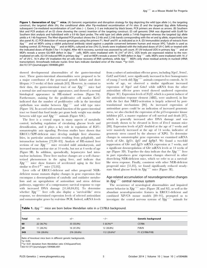
Figure 1. Generation of Xpg2/2 mice. (A) Genomic organization and disruption strategy for Xpg depicting the wild type allele (+), the targetingconstruct, the targeted allele (fn), the conditional allele after Flp-mediated recombination of Frt sites (f) and the targeted Xpg allele followingsubsequent Cre-mediated recombination of LoxP sites (2). Exons 2–5 are indicated by black boxes. PCR primers are shown as arrows. (B) Southernblot and PCR analysis of an ES clone showing the correct insertion of the targeting construct. ES cell genomic DNA was digested with EcoRI forSouthern blot analysis and hybridized with a 0.9 kb DpnI probe. The wild type (wt) allele yields a 7.4-kb fragment whereas the targeted (tg) alleleyields a 4.1-kb fragment. The NheI-digested PCR product shows the 2.3-kb and 2.2-kb bands corresponding with the wt and tg allele, respectively (seealso panel A). (C) PCR detection of mouse genotypes using the primers F1, NeoF and R1 as indicated as in A. (D) Immunoblot analysis of extracts fromXpg2/2 and wt MDFs using a rabbit polyclonal antibody raised against a peptide conserved between human and mouse XPG. Tubulin is used asloading control. (E) Primary Xpg2/2 and wt MDFs, cultured at low (3%) O2 levels were irradiated with the indicated doses of UV-C (left) or treated withthe indicated doses of Illudin S for 1 h (right). After 48 h recovery, survival was assessed by cell count. (F) UV-induced UDS in primary Xpg2/2 and wtMDFs reveals a severe GG-NER defect in Xpg2/2 cells. MDFs were irradiated with 16 J/m2 of UV-C. UDS levels are expressed relative to the non-irradiated wt cells. (G) UV-induced RRS in primary Xpg2/2 and wt MDFs reveals a severe TC-NER defect in Xpg2/2 cells. MDFs were irradiated with 16 J/m2 of UV-C. 16 h after UV irradiation the wt cells show recovery of RNA synthesis, while Xpg2/2 MDFs only show residual activity in nucleoli (rRNAtranscription). Arrowheads indicate nuclei. Error bars indicate standard error of the mean. **p,0.01.doi:10.1371/journal.pgen.1004686.g001
Table 1. Xpg2/2 mice are born below Mendelian ratio in a C57BL6 background.
Total +/+ +/2 2/2 Genetic background
60 22 (36.7%) 33 (55.0%) 5 (8.3%)** C57Bl6
39 11 (28.2%) 16 (41.0%) 12 (30.8%) FVB/N
545 156 (28.6%) 276 (50.6%) 112 (20.6%)* F1 (C57Bl6/FVB)
Ratio of knockout mice born in different genetic backgrounds.*p,0.05;**p,0.01 deviation from Mendelian ratio (ChiSquaredTest).doi:10.1371/journal.pgen.1004686.t001
Xpg2/2 as a Mouse Model for Progeria
PLOS Genetics | www.plosgenetics.org 5 October 2014 | Volume 10 | Issue 10 | e1004686
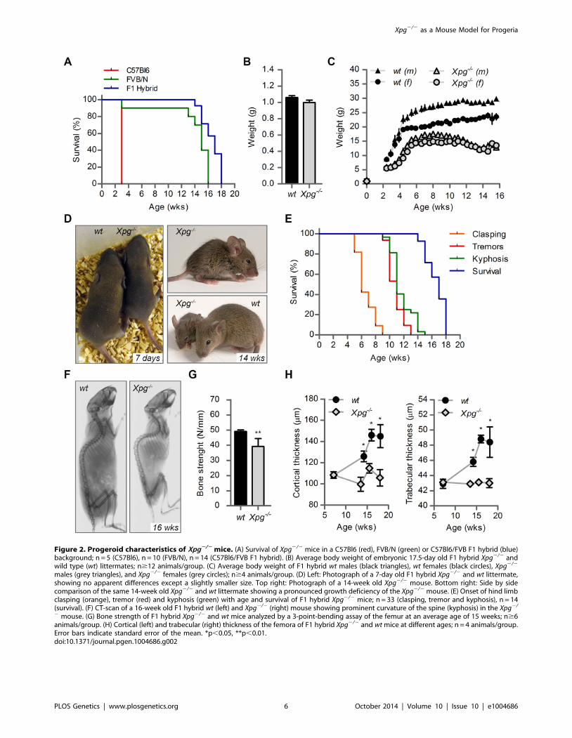
Figure 2. Progeroid characteristics of Xpg2/2 mice. (A) Survival of Xpg2/2 mice in a C57Bl6 (red), FVB/N (green) or C57Bl6/FVB F1 hybrid (blue)background; n = 5 (C57Bl6), n = 10 (FVB/N), n = 14 (C57Bl6/FVB F1 hybrid). (B) Average body weight of embryonic 17.5-day old F1 hybrid Xpg2/2 andwild type (wt) littermates; n$12 animals/group. (C) Average body weight of F1 hybrid wt males (black triangles), wt females (black circles), Xpg2/2
males (grey triangles), and Xpg2/2 females (grey circles); n$4 animals/group. (D) Left: Photograph of a 7-day old F1 hybrid Xpg2/2 and wt littermate,showing no apparent differences except a slightly smaller size. Top right: Photograph of a 14-week old Xpg2/2 mouse. Bottom right: Side by sidecomparison of the same 14-week old Xpg2/2 and wt littermate showing a pronounced growth deficiency of the Xpg2/2 mouse. (E) Onset of hind limbclasping (orange), tremor (red) and kyphosis (green) with age and survival of F1 hybrid Xpg2/2 mice; n = 33 (clasping, tremor and kyphosis), n = 14(survival). (F) CT-scan of a 16-week old F1 hybrid wt (left) and Xpg2/2 (right) mouse showing prominent curvature of the spine (kyphosis) in the Xpg2/
2 mouse. (G) Bone strength of F1 hybrid Xpg2/2 and wt mice analyzed by a 3-point-bending assay of the femur at an average age of 15 weeks; n$6animals/group. (H) Cortical (left) and trabecular (right) thickness of the femora of F1 hybrid Xpg2/2 and wt mice at different ages; n = 4 animals/group.Error bars indicate standard error of the mean. *p,0.05, **p,0.01.doi:10.1371/journal.pgen.1004686.g002
Xpg2/2 as a Mouse Model for Progeria
PLOS Genetics | www.plosgenetics.org 6 October 2014 | Volume 10 | Issue 10 | e1004686

neurodegenerative changes. Macroscopically, the brains and
spinal cords of Xpg2/2 mice showed a normal appearance, albeit
somewhat smaller. In addition, the gross histological organization
analyzed in thionin-stained sections appeared normal in all brain
regions. As a first step to examine the occurrence of neurodegen-
erative changes, we examined the brains of 4- and 14-week old
Xpg2/2 mice immunohistologically for glial acidic filament protein
(GFAP) expression, which outlines reactive astrocytosis in response
to neuronal injury. A mild increase in GFAP immunostaining
occurred in patches in multiple nervous system areas at 4 weeks
(Figure 4A and S4A). Instead, at 14 weeks, Xpg2/2 mice showed
a prominent ubiquitous increase in GFAP staining throughout the
entire central nervous system including spinal cord, indicative of
widespread astrocytosis (Figure 4A and S4A). Double-labelling of
GFAP and the microglia cell marker Iba-1 showed that the
increased GFAP staining was paralleled by microglia activation,
characterized by increased Iba-1 immunoreactivity and the
transformation of resting microglia cells into activated cells with
thicker processes and larger cell bodies (Figure S4B and C).
Next, to determine whether Xpg2/2 central nervous system cells
experience genotoxic stress, we studied the expression of the
transcription factor p53, which is activated by multiple types of
DNA damage and is expressed in neurons and macroglia of many
NER-deficient mouse models including mice defective in Ercc1,
Csa or Csb [89–91]. Immunohistochemistry revealed p53-positive
cells in all central nervous system regions. Analysis of the p53
density in neocortex and cerebellum indicated an increase in
number of p53-positive cells in brains of 14-week old compared to
4-week old Xpg2/2 mice (Figure 4B). Similar to our findings in
other NER mutant mice [89–91], double labelling of p53 with
neuronal (NeuN) and astrocytic (GFAP, S100b) markers, indicated
that these p53-positive cells include neurons, astrocytes (GFAP+or S100b+; Figure S4D), and oligodendrocytes. Although not
systematically investigated, we also noted that, as in other
Figure 3. Intestine and liver phenotype of Xpg2/2 mice. (A) Representative images of HE and Ki67 stained small intestine (SI) of 14-week oldXpg2/2 and wild type (wt) mice showing no gross morphological differences. (B) Average nucleus size of hepatocytes in the liver of 4- and 14-weekold Xpg2/2 and wt mice; n$3 animals/group. Bottom right: magnification of a nuclear inclusion found sporadically in liver sections of 14-week oldXpg2/2 mice. (C) Relative mRNA expression levels of several antioxidant genes and the DNA damage response gene p21 in liver tissue of 7- and 14-week old Xpg2/2 and wt mice. All values are corrected for TubG2, Hprt, and Rps9 (Table S1) expression as internal standard and normalized to the 7-week old wt expression levels; n = 4 animals/group. (D) Relative expression levels of the somatotrophic genes Ghr, Igf1r, Igf1, and Igfbp3 in liver tissueof 7- and 14-week old Xpg2/2 and wt mice. All values are corrected for TubG2, Hprt, and Rps9 expression and normalized to the 7-week old wtexpression levels; n = 4 animals/group. (E) Average basal blood glucose levels in groups of 4–7 and 12–18 week old Xpg2/2 and wt mice; n$15animals/group. Scale bars: 50 mm (A), 10 mm (B). Error bars indicate standard error of the mean. *p,0.05, **p,0.01.doi:10.1371/journal.pgen.1004686.g003
Xpg2/2 as a Mouse Model for Progeria
PLOS Genetics | www.plosgenetics.org 7 October 2014 | Volume 10 | Issue 10 | e1004686
NER-deficient mice, in neocortex and cerebellar cortex the
majority of p53-positive cells were neurons, while in spinal cord
a large proportion of p53-positive cells were astrocytes (Figure
S4E).
To obtain evidence for the occurrence of neuronal death, we
analyzed calbindin staining in cerebellar cortex where it outlines
Purkinje cells and enables easy detection of the degeneration of
these cells [90–92]. Calbindin staining revealed degeneration and
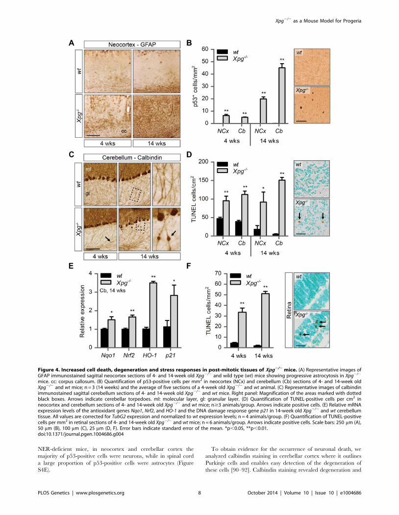
Figure 4. Increased cell death, degeneration and stress responses in post-mitotic tissues of Xpg2/2 mice. (A) Representative images ofGFAP immunostained sagittal neocortex sections of 4- and 14-week old Xpg2/2 and wild type (wt) mice showing progressive astrocytosis in Xpg2/2
mice. cc: corpus callosum. (B) Quantification of p53-positive cells per mm2 in neocortex (NCx) and cerebellum (Cb) sections of 4- and 14-week oldXpg2/2 and wt mice; n = 3 (14 weeks) and the average of five sections of a 4-week old Xpg2/2 and wt animal. (C) Representative images of calbindinimmunostained sagittal cerebellum sections of 4- and 14-week old Xpg2/2 and wt mice. Right panel: Magnification of the areas marked with dottedblack boxes. Arrows indicate cerebellar torpedoes. ml: molecular layer, gl: granular layer. (D) Quantification of TUNEL-positive cells per cm2 inneocortex and cerebellum sections of 4- and 14-week old Xpg2/2 and wt mice; n$3 animals/group. Arrows indicate positive cells. (E) Relative mRNAexpression levels of the antioxidant genes Nqo1, Nrf2, and HO-1 and the DNA damage response gene p21 in 14-week old Xpg2/2 and wt cerebellumtissue. All values are corrected for TubG2 expression and normalized to wt expression levels; n = 4 animals/group. (F) Quantification of TUNEL-positivecells per mm2 in retinal sections of 4- and 14-week old Xpg2/2 and wt mice; n = 6 animals/group. Arrows indicate positive cells. Scale bars: 250 mm (A),50 mm (B), 100 mm (C), 25 mm (D, F). Error bars indicate standard error of the mean. *p,0.05, **p,0.01.doi:10.1371/journal.pgen.1004686.g004
Xpg2/2 as a Mouse Model for Progeria
PLOS Genetics | www.plosgenetics.org 8 October 2014 | Volume 10 | Issue 10 | e1004686
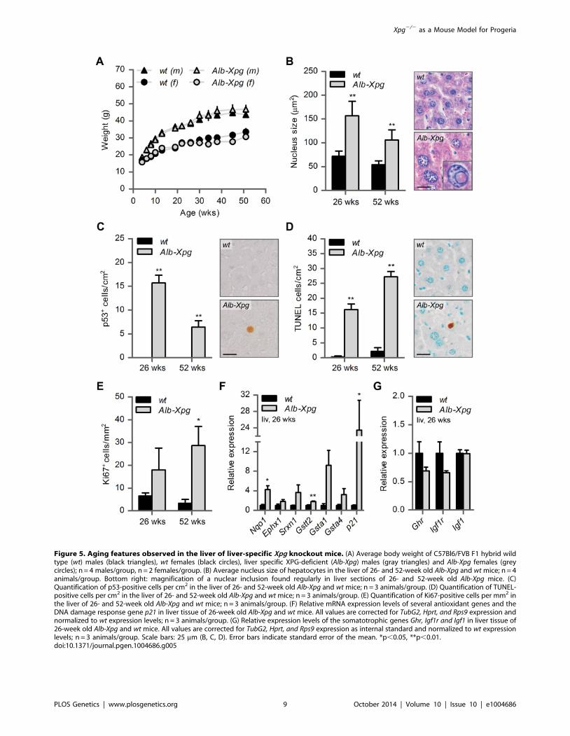
Figure 5. Aging features observed in the liver of liver-specific Xpg knockout mice. (A) Average body weight of C57Bl6/FVB F1 hybrid wildtype (wt) males (black triangles), wt females (black circles), liver specific XPG-deficient (Alb-Xpg) males (gray triangles) and Alb-Xpg females (greycircles); n = 4 males/group, n = 2 females/group. (B) Average nucleus size of hepatocytes in the liver of 26- and 52-week old Alb-Xpg and wt mice; n = 4animals/group. Bottom right: magnification of a nuclear inclusion found regularly in liver sections of 26- and 52-week old Alb-Xpg mice. (C)Quantification of p53-positive cells per cm2 in the liver of 26- and 52-week old Alb-Xpg and wt mice; n = 3 animals/group. (D) Quantification of TUNEL-positive cells per cm2 in the liver of 26- and 52-week old Alb-Xpg and wt mice; n = 3 animals/group. (E) Quantification of Ki67-positive cells per mm2 inthe liver of 26- and 52-week old Alb-Xpg and wt mice; n = 3 animals/group. (F) Relative mRNA expression levels of several antioxidant genes and theDNA damage response gene p21 in liver tissue of 26-week old Alb-Xpg and wt mice. All values are corrected for TubG2, Hprt, and Rps9 expression andnormalized to wt expression levels; n = 3 animals/group. (G) Relative expression levels of the somatotrophic genes Ghr, Igf1r and Igf1 in liver tissue of26-week old Alb-Xpg and wt mice. All values are corrected for TubG2, Hprt, and Rps9 expression as internal standard and normalized to wt expressionlevels; n = 3 animals/group. Scale bars: 25 mm (B, C, D). Error bars indicate standard error of the mean. *p,0.05, **p,0.01.doi:10.1371/journal.pgen.1004686.g005
Xpg2/2 as a Mouse Model for Progeria
PLOS Genetics | www.plosgenetics.org 9 October 2014 | Volume 10 | Issue 10 | e1004686
loss of Purkinje cells in 14-week old Xpg2/2 mice (Figure 4C).
Also, calbindin staining revealed sporadic Purkinje cells with
abnormal dendritic morphologies and, more frequently, Purkinje
cells with swellings in their proximal axon (Figure 4C). Axonal
swellings (also designated torpedoes or axonal spheroids) are
a common feature in neurodegenerative disorders and aging [93],
that is also well documented for Purkinje cell axons of CS and XP/
CS patients [94]. The presence of axonal pathology indicates that
many surviving Purkinje cells in 14-week old Xpg2/2 mice display
compromised health. Notably, few small axonal swellings occurred
in Purkinje cells axons in 4-week old Xpg2/2 mice.
To further examine the extent to which neurons in Xpg2/2 mice
show compromised health, we examined the morphology of the
Golgi apparatus in motor neurons. In a previous study in Ercc1mutant mice we noted that motor neurons displayed a variety of
morphological abnormalities of the Golgi apparatus, and we
proposed that these abnormalities reflect a heterogeneity of
cellular deficits resulting from stochastic DNA damage [90].
Immunostaining for the cis-Golgi marker GM130 showed that
motor neurons in Xpg2/2 mice developed the same heterogeneity
in morphological abnormalities of the Golgi apparatus as observed
in Ercc1D/2 mice. Double labelling of GM130 and p53 indicated
that only a small subset of neurons with abnormal Golgi apparatus
is p53 positive. This variability in p53 expression further illustrates
the heterogeneity of degenerative events that may occur in Xpg2/2
neurons (Figure S4F).
TUNEL staining to determine the amount of apoptotic cells
showed a significant increase in both the cerebrum and the
cerebellum at 4 as well as 14 weeks of age (Figure 4D). Finally,
real-time PCR in Xpg2/2 cerebellum revealed an upregulation of
the p53-responsive kinase inhibitor p21 consistent with the
activation of genotoxic stress pathways (Figure 4E), and increased
expression of several oxidative stress response genes (Figure 4E).
In addition to the brain and spinal cord, we also investigated the
retina, as retinal degeneration is a frequent symptom of CS and
XP/CS patients [38], that is also reproduced in CSA- and CSB-
deficient mice [23]. TUNEL staining revealed cell death in both
the inner and outer nuclear layers of the retina of 4- and 14-week
old Xpg2/2 mice (Figure 4F). Hence, Xpg2/2 mice display loss of
photoreceptor cells as well as degeneration of the retinal circuitry.
Together these data indicate the occurrence of widespread
progressive degenerative changes in Xpg2/2 nervous system,
strongly resembling the phenotype of ERCC1-deficient mice.
Liver-specific inactivation of XPG results in progeroidfeatures in the liver without causing early death orreduced body weight
Transgenic expression of ERCC1 in the liver has been shown to
alleviate growth deficiency and to extend lifespan of ERCC1-
deficient mice [83], suggesting that liver abnormalities are an
important determinant of the reduced lifespan of these mice. To
determine the importance of liver pathology in the runting and the
reduced lifespan of our Xpg2/2 mice, we generated mice with
liver-specific inactivation of the Xpg gene by crossing Xpgf/+ mice
carrying the floxed Xpg allele with heterozygous Xpg+/2 mice that
also express the albumin-Cre recombinase transgene that drives
Cre expression specifically in hepatocytes [95] to yield Xpgf/2/
Alb-Cre+ mice, hereafter designated Alb-Xpg mice. This Alb-Xpgmouse also has the advantage that it allows to study the effect of
liver XPG-deficiency in the absence of abnormalities in other
tissues.
A cohort of Alb-Xpg mice was allowed to reach the age of one
year. All Alb-Xpg mice displayed normal growth and weight gain,
and none of them died prematurely (Figure 5A). Livers of Alb-Xpg
mice had an increased size compared to wild type, while brain,
kidney and spleen displayed unaltered size and weight (Figure
S5A). Albumin and glucose blood levels were the same as in
control mice (Figure S5B and C). Histological analysis revealed
anisokaryosis with karyomegaly in the liver of Alb-Xpg mice
analyzed at 26 and 52 weeks (Figure 5B). The observed
karyomegaly was more prominent than that observed in 14-week
old Xpg2/2 mice, and cells with intranuclear inclusions were more
frequent. In addition, we identified p53-positive cells, increased
cell death and increased cell proliferation in Alb-Xpg liver
consistent with a progeroid degenerative phenotype (Figure 5C–
E). Furthermore, real-time PCR showed that livers of Alb-Xpgmice displayed a massive induction of the DNA damage response
gene p21 as well as increased expression of several antioxidant
effector genes (Figure 5F). We also observed a trend of reduced
expression of Ghr and Igf1r (Figure 5G), hence, reproducing gene
expression changes determined in livers of Xpg2/2 mice.
Activation of the Nrf2 antioxidant response genes, reduction of
the IGF1 axis, and increased proliferation shown by Ki67-staining
are all consistent with liver regeneration after tissue damage [96].
As an additional control we showed that the expression of these
genes is unaltered in livers from Emx1-Xpg mice that are XPG-
deficient in the dorsal forebrain (see below; Figure S5D and E).
Together the data from the Alb-Xpg mice show that progeroid
and gene expression changes in the liver triggered by the absence
of XPG are not sufficient to explain the runted short-living
phenotype of Xpg2/2 mice.
Dorsal forebrain specific inactivation of XPG results ina mixed macroglia/neuronopathy in cortex andhippocampus
Our neuropathological analyses of Xpg2/2 mice uncovered
severe neurodegenerative changes at 14 weeks of age, compatible
with neurological and motor deficits in these mice. Importantly,
the presence of p53 in glia and abundant astrocytosis and
microgliosis in the white matter indicate that abnormalities in
Xpg2/2 mice are not limited to neurons, but also involve glia cells.
This is consistent with our findings in CS mouse models [62,91],
and with the neuropathological changes found in CS and XP/CS
patients that is dominated by white matter pathology, in addition
to neuronal, glial and vascular pathology, and, in severe cases,
developmental abnormalities [39,94,97–99]. In previous studies,
using a Cre-lox approach with CamKIIa-Cre and L7-Cretransgenic mice that drive Cre expression in post-mitotic forebrain
neurons and Purkinje cells, respectively, we showed that neuron-
specific deficiency of ERCC1 or combined deficiency of XPA and
CSB was sufficient to trigger stochastic degeneration of these
neuronal populations [89,91,92]. These studies showed that
neurodegenerative changes in ubiquitous ERCC1- and XPA/
CSB-deficient mice are not a consequence of developmental
abnormalities, vascular problems, or degenerative changes in other
organs. Furthermore, these neuron-specific mice enabled us to
follow the degenerative process beyond the normal lifespan of the
short-living ubiquitous ERCC1- and XPA/CSB-deficient mice
[89,91,92]. In the present study we therefore used a similar
approach, but with an Emx1-Cre transgenic line that drives Cre
expression in progenitor cells of the dorsal telencephalon, to
achieve inactivation of the Xpg gene not only in excitatory neurons
of the neocortex and hippocampus, but also of astrocytes and
oligodendrocytes in these brain areas [100]. Analysis of a cohort of
Emx1-Xpg mice that were allowed to age for one year revealed no
early death and showed that body weights were indistinguishable
from that of wild type littermates until the age of 30 weeks. At
older age the mean weight gain of Emx1-Xpg mice was
Xpg2/2 as a Mouse Model for Progeria
PLOS Genetics | www.plosgenetics.org 10 October 2014 | Volume 10 | Issue 10 | e1004686

Figure 6. Age-related increase of neuronal stress in forebrain-specific Xpg knockout mice. (A) Average body weight of C57Bl6/FVB F1hybrid wild type (wt) females (black circles) and forebrain-specific XPG-deficient (Emx1-Xpg) females (gray circles); n$4 animals/group. (B) Onset ofclasping of the hind limbs in Emx1-Xpg mice; n = 7 animals/group. (C) Representative images of GFAP immunostained sagittal neocortex sections of26- and 52-week old Emx1-Xpg and wt mice showing progressive astrocytosis in Emx1-Xpg mice. (D) Representative images of Mac2 immunostainedsagittal brain sections of 26- and 52-week old Emx1-Xpg and wt mice showing Mac2-positive microgliosis and a progressive decrease in size of thecerebral cortex and hippocampus of Emx1-Xpg mice. Arrows indicate microgliosis in corpus callosum and fimbria fornix. A thionin counterstainingwas used. (E) Quantification of p53-positive cells in neocortex and cerebellum of 26- and 52-week old Emx1-Xpg and wt mice. Values are the averageof four sections per genotype. Arrows indicate p53 positive cells. (F) Representative confocal images showing double labeled p53-NeuN cells in theneocortex (left) and p53-S100ß in the fimbria fornix (right) of 26-week old Emx1-Xpg mice. Arrows indicate p53 positive cells. NCx: neocortex, cc:corpus callosum, Str: striatum, ff: fimbria fornix, Hip: hippocampus. Scale bars: 50 mm (C), 500 mm (D), 200 mm (E) and 20 mm (F). Error bars indicatestandard error of the mean. **p,0.01.doi:10.1371/journal.pgen.1004686.g006
Xpg2/2 as a Mouse Model for Progeria
PLOS Genetics | www.plosgenetics.org 11 October 2014 | Volume 10 | Issue 10 | e1004686
significantly smaller than in control littermates (Figure 6A). This
difference in weight was associated with a proportional reduced
weight of internal organs (Figure S6A). Basal blood glucose
concentrations were the same as in controls, indicating that
reduced weight of old Emx1-Xpg mice is not a consequence of
reduced energetic intake (Figure S6B).
To obtain a crude impression of the development of neurolog-
ical symptoms, we examined the time of onset of clasping of the
hind limbs when animals were lifted by their tails. This
abnormality developed in Emx1-Xpg between 14–24 weeks of
age and was not observed in control littermates (Figure 6B). The
Emx1-Xpg mice did not develop tremors and deficits in
accelerating rotarod performance (Figure S6C). Both the absence
of these motor deficits and the delayed onset of clasping in
comparison to the Xpg2/2 mice can be explained by the selective
inactivation of Xpg in neocortex and hippocampus, avoiding the
bulk of circuitries controlling motor behavior in mice [101].
Macroscopic inspection of brains of Emx1-Xpg mice at 26- and
52-week already revealed that the neocortex was considerably
smaller. Histological analysis confirmed that the neocortex was
thinner, and showed that also the hippocampus was dramatically
smaller, while other brain regions were unaltered (Figure S6D).
GFAP immunohistochemistry showed a very strong increase in
GFAP staining indicative of astrocytosis in both cortex and
hippocampus (Figure 6C), and no changes in other brain regions.
As in the Xpg2/2 nervous system, astrocytosis was paralleled by
microgliosis, identified by immunohistology with an Iba-1
antibody. Staining for Mac2 (also known as galectin-3), to outline
phagocytosing microglia cells [102], revealed very high levels of
Mac2-positive cells in the corpus callosum and the fimbria fornix
of Emx1-Xpg mice (Figure 6D and S6D,E), and a moderate
amount of phagocytosing microglia in the neocortex and
hippocampus (Figure 6D). Remarkably, the presence of Mac2
could not be explained by axonal degeneration of cortical and
hippocampal neurons solely, as we did not observe this phenom-
enon in our CamKIIa-Ercc1 mice [89]. Furthermore, the capsula
interna of Emx1-Xpg mice, which contains the descending
corticofugal axons and shows severe axonal degeneration, did
not show this dramatic increase of Mac2 staining (Figure 6D and
S6D). Hence, the presence of high levels of Mac2 labelling may
reflect the same phenomenon that we observed in our CS mice,
i.e. the presence of phagocytosing microglia in the absence of
axonal degeneration [91]. Accordingly, we also found an
upregulation of Hsp25 expression in astrocytes in the corpus
callosum and fimbria fornix (Figure S6F), a phenomenon that we
also observed in the white matter of CS mouse models [91].
Furthermore, analysis of p53 expression in Emx1-Xpg showed that
multiple p53-positive cells populated the fimbria-fornix and the
corpus callosum (Figure 6E). Double-labelling of p53 with NeuN
or the glia marker GFAP and S100b showed that some p53-
positve cells were neurons (NeuN), but a large proportion were
astrocytes (GFAP+, S100b+), or oligodendrocytes as identified on
the basis of nuclear morphology (Figure 6F). Together the data
indicate that Emx1-Xpg mice develop a combined neuro- and
gliopathy of cortex and hippocampus.
Discussion
XPG is a 39-endonuclease that is essential for the excision step
of the GG-NER and TC-NER subpathways of NER and that has
additional, yet incompletely defined, roles in other repair processes
and possibly in transcription [15,16,31,53,103–105]. Consistent
with multiple diverse functions, mutations in XPG cause
a spectrum of disease phenotypes varying from the UV-sensitivity
disorder XP to the multisystem developmental/degenerative
disorders XP/CS and COFS [52–55,57]. Several Xpg mouse
models have been generated that partially recapitulate the
spectrum of genetic defects and disorders found in patients
[63–66]. To better define the effect of the complete absence of
XPG, in the present study we have generated mice with a Cre-
inducible Xpg knockout allele and examined the phenotypes of
total (designated Xpg2/2), liver-specific (Alb-Xpg), and dorsal
forebrain-specific (Emx1-Xpg) XPG-deficient mice. Consistent
with previous data [63] we found that Xpg2/2 mice are born
with normal size and weight, but progressively fail to grow and to
gain weight after birth, combined with a short lifespan. In
addition, we found that lifespan was influenced by genetic
background, in that Xpg2/2 mice displayed a much shorter
lifespan in C57BL6 background (3 weeks), compared to FVB or
C57BL6/FVB F1 hybrid background (16–18 weeks). An effect of
genetic background is well known for many mouse models of
disease and was also noted in our Ercc1-mutant mice [51,72].
Differences in genetic background likely explain the differences in
lifespan of our C57BL6/FVB F1 hybrid Xpg2/2 mice and the
Xpg2/2 mice of Harada and coworkers [63], which all died at 3–4
weeks of age similar to our C57BL6 Xpg2/2 mice. However, what
mechanism defines the short lifespan of our C57BL6 Xpg2/2 mice
remains to be determined. Importantly, the effect of genetic
background in mice suggests that genetic context may also impact
the disease phenotype in CS and XP/CS patients. Interestingly,
preliminary analysis of neuropathological changes of our C57BL6
Xpg2/2 mice at 3 weeks of age revealed subtle degenerative
changes (Figure S7) that are comparable in severity to the
neurodegenerative changes of C57BL6/FVB Xpg2/2 mice of 4
weeks of age, and considerably milder than those in 14-week old
C57BL6/FVB F1 hybrid Xpg2/2 mice (compare Figure S7 with
Figure 4). These data suggest time-dependence of the neurode-
generative phenotype and indicate that the much shorter lifespan
of C57BL6 Xpg2/2 mice cannot be explained by enhanced
neurodegeneration. An alternative possibility suggested by the
data of Harada and coworkers [63] is that C57BL6 Xpg2/2 mice
display an exaggerated developmental phenotype with more
prominent developmental deficits, such as an improperly de-
veloped gastro-intestinal tract, that would reduce their ability to
cope with extra-uterine life. Recent evidence from XPA/CSA-
deficient mice bred in C57BL6 background suggest that providing
mutant pups with soft food before weaning is sufficient to increase
survival from 3–4 weeks to 12–18 weeks [106]. So far, we found no
beneficial effect of soft food in our C57BL6 Xpg2/2 mice, but this
remains to be further examined. Nevertheless, together the data
suggest that the ability of short-living NER mutant mice to survive
beyond weaning might also depend on the ability to cope with the
transition from mother milk to other food.
Xpg2/2 mice as a progeria modelThe main goal of the present study was to determine the extent
to which XPG deficiency in mice results in multisystem progeroid
degenerative changes as observed in CS and XP/CS patients, as
well as in other NER-deficient mouse models including Xpa/Csb,
Xpd, and Ercc1 mutants [48,51,68,69,72,77,107]. Our data show
that, although seemingly normal at late embryonic stage, and
showing only mild degenerative features at 4 weeks of age,
C57BL6/FVB Xpg2/2 at 14–16 weeks showed abundant de-
generative changes in multiple tissues, indicative of accelerated
aging. These degenerative/accelerated aging features included
kyphosis, cachexia, osteoporosis, liver aging, and abundant
nervous system degenerative changes. In addition, the data from
our liver- and dorsal forebrain-specific Xpg mice showed that
Xpg2/2 as a Mouse Model for Progeria
PLOS Genetics | www.plosgenetics.org 12 October 2014 | Volume 10 | Issue 10 | e1004686

a much more severe tissue-specific degenerative phenotype could
be obtained in these mice because of survival beyond the maximal
lifespan of Xpg2/2 mice. Hence, the data indicate that Xpg2/2
mice indeed develop a multisystem premature aging phenotype
reminiscent of the phenotypes of some of our other NER-deficient
mouse models, in particular the Ercc1D/2 mice, which show
a slightly longer lifespan and comparable pathological changes in
liver and nervous system (see below). Moreover, the Xpg2/2 mouse
phenotype shares many features with XP-G/CS patients
[17,94,108] including a cachectic ‘frail’ appearance with loss of
subcutaneous fat and signs of osteoporosis (Table 2).
Compensatory survival response in Xpg2/2 liverPreviously we have shown that transcription-blocking lesions
trigger a ‘‘survival response’’ involving somatotroph attenuation
and increased resistance to oxidative stress, which resembles the
response triggered by dietary restriction and is associated with
delaying many aspects of aging and increasing lifespan [51,68,84].
This mechanism has been observed in response to direct but
persistent DNA damage as well as during the course of aging, both
in natural aging as well as in specific progeroid mouse models, and
is activated in long-lived mutant mice and centenarians
[51,68,109]. A central hallmark of this survival response is the
downregulation of genes involved in the GH/IGF axis, combined
with upregulation of genes involved in the antioxidant response.
Using real-time PCR for selected genes of these pathways in the
present study, we show that Xpg2/2 mice display survival-
response-like changes in gene expression in liver cells, including
a decrease in Ghr mRNA levels and a robust increase in expression
of the antioxidant defense effector genes Nqo1, Srxn1, Gstt2, andGsta1. Similar changes were observed in the liver of liver-specific
XPG-deficient mice. Consistent with previous observations
concerning the survival response, we found significantly reduced
circulating glucose levels in the Xpg2/2 mouse. However, we
found no change in glucose in the liver-specific mouse, indicating
that reduced glucose levels are not necessarily a consequence of
gene expression changes in the liver. We did not observe
significantly lower levels of Igf1 and Igf1r, as previously found
for several other progeroid DNA repair mutants [51,84], although
we did find a trend of reduced expression.
Increased expression of antioxidant genes was also observed in the
central nervous system (i.e. cerebellum) of Xpg2/2 mice, but the
identity and the degree of changed expression is somewhat different.
For instance, Nrf2 shows a relatively increased expression in
cerebellum but not in liver, while Nqo1 shows a very large relative
increase in liver, and a modest relative increase in cerebellum. These
differences may be explained by altered stress responses in different cell
types [110]. In addition, in the nervous system, changes in gene
expression may result from the death and disappearance of neurons
and reactive proliferation of glial cells and as a consequence, reduced
and increased expression of neuronal and glial genes, respectively.
Progeroid features in the livers of Xpg2/2 animals are notsufficient to cause reduced growth and lifespan
The liver of Xpg2/2 mice showed several characteristics also
found in aging liver, i.e. increased nuclear size and the presence of
Table 2. Comparison of the Xpg2/2 phenotype to that of XP/CS patients.
Phenotypic characteristics XPG mouse XP(G)/CS patient
UV sensitivity
Cellular level y y
Organismal level nd y
Sensitivity to oxidative damage-cell n y
Growth and lifespan
Growth retardation y y
Reduced subcutaneous fat y y
Reduced lifespan y y
Sensory systems
Retinal degeneration y y
Sensorineuronal hearing loss nd y
Bones
Osteoporosis y y
Kyphosis y y
Nervous system
Gait ataxia y y
Action tremor y y
Cognitive decline y y
Brain calcifications nd y
Ventricle enlargement y y
White matter abnormalities nd y
Cerebral and cerebellar atrophy y y
Astrogliosis y y
y = present, n = not present, nd = not determined.doi:10.1371/journal.pgen.1004686.t002
Xpg2/2 as a Mouse Model for Progeria
PLOS Genetics | www.plosgenetics.org 13 October 2014 | Volume 10 | Issue 10 | e1004686

nuclear inclusions. These changes were much more prominent
and associated with additional degenerative changes in liver-
specific XPG-deficient mice. However, despite prominent liver
pathology, the liver-specific Xpg2/2 mice at 52 weeks did not show
altered weight, nor altered glucose and albumin levels suggestive of
metabolic problems. Although at this point we cannot exclude that
our liver-specific mutants will develop health problems in their
second year of life and might develop a shorter than normal
lifespan, it is safe to conclude that liver problems on their own
cannot be the main culprit of the reduced lifespan and the small
cachectic appearance of Xpg2/2 mice. In contrast to our data,
findings in Ercc1 mutant mice show that transgenic overexpression
of ERCC1 in the liver alleviates growth deficiency and extends
lifespan [83], indicating that liver abnormalities are an important
determinant of the reduced lifespan of these mice. A possible
explanation is that liver pathology is more prominent in Ercc1mutant mice, putatively as a consequence of additional defects in
interstrand crosslink and double-strand break repair [48]. An
alternative explanation for the differences in results could be that
the impact of liver pathology on survival depends on the severity of
pathology in other organs. This hypothesis is testable by studying
survival of liver-specific ERCC1-deficient mice, and vice versa by
generating liver-corrected Xpg2/2 mice using a transgenic ‘rescue’
strategy as reported by Selfridge et al. [83]. A possible conclusion
from these studies could be that Ercc1 and Xpg mutant mice die
prematurely as a consequence of a synergistic deleterious effect of
multi-organ failure. In view of the severe neurological and
neurodegenerative deficits in 14–16 week old Xpg2/2 mice, yet
an alternative scenario is that Xpg2/2 mice die as a consequence of
nervous system abnormalities (see below). This scenario is realistic
for a subset of human NER-deficient patients, in particular those
developing XP with neurological abnormalities [36,40]. Interest-
ingly, our dorsal forebrain-specific XPG-deficient mice also
showed reduced weight-gain after 30 weeks of age. At this age
these mice displayed severe neuronal degeneration in neocortex
and hippocampus, as a result of which the mice would be expected
to have severe cognitive deficits, whereas basal motor functions
remain unaltered. Weight loss was not associated with altered
glucose levels or gene expression changes in the liver (Figures S5
and S6). We noted the same reduced weight-gain in our forebrain
neuron-specific XPA/CSB-deficient mouse model [91]. The data
suggest that severe disruption of cortical or hippocampal circuitries
may result in the weight loss, but the precise mechanism remains
to be defined.
XPG deficiency causes age-dependent degeneration ofneurons and macroglia
Our data show that Xpg2/2 mice, within their relatively short
lifespan (16–18 weeks), develop neurological abnormalities of
increasing severity in association with degenerative changes
throughout the nervous system. These nervous system degenera-
tive changes seemingly are more severe than those previously
reported for another line of Xpg2/2 mice [111]. The differences in
nervous system neurodegenerative changes can be explained by
the very short lifespan (3 weeks) of previously reported Xpg2/2
mice, since our C57BL6 Xpg2/2 mice, which show the same short
lifespan, also displayed a low level of nervous system pathology at
the end of life (see above and Figure S7). Together with our
demonstration that dorsal forebrain-specific XPG-deficient mice,
allowed to age for one year, display very severe neurodegenerative
changes, our data indicate that age is a key determinant in the
development of neurodegenerative changes in Xpg2/2 mice. Our
data also indicate that XPG deficiency does not result in obvious
neurodevelopmental abnormalities, although subtle developmental
abnormalities cannot be excluded at this point.
The neurodegenerative changes in our Xpg2/2 mice strongly
resemble those of incomplete ERCC1-deficient Ercc1D/2 mice
that have a slightly longer lifespan (24–30 weeks). Our studies with
Ercc1D/2 mice and neuron-specific Ercc1 knock-out animals
indicate that ERCC1-deficient neurons in time stochastically
accumulate structural and functional deficits to eventually die and
disappear [89,90,92,112]. A stochastic accumulation of deficits
also appears to occur in Xpg2/2 neurons. Thus, the widespread
distribution of astrocytosis and microgliosis, as well as p53 and
TUNEL staining, indicates that degenerative changes in the
Xpg2/2 nervous system affect all neuronal populations. The
increased size and frequency of axonal spheroids in Purkinje cells,
and a diversity of Golgi apparatus morphological abnormalities in
motor neurons, on the other hand, illustrate that Xpg2/2 neurons
may asynchronously accumulate a variety of degenerative features
over time. This widespread and asynchronous accumulation of
cellular damage in Xpg2/2 neurons is consistent with a model in
which neurons are afflicted by stochastic DNA lesions that deregulate
gene expression, as we have proposed for Ercc1D/2 mice [89]. A
prime role for genotoxic stress in causing the degenerative phenotype
in the Xpg2/2 nervous system is further suggested by the presence of
stochastically distributed p53-positive cells and increased expression
of the DNA damage responsive p21 gene.
Together the degenerative changes in the Xpg2/2 nervous
system favor a pathogenic model involving deficient DNA repair
in the same way as proposed for ERCC1-deficient mice [48,89].
The central role of DNA damage in the Xpg2/2 nervous system
raises questions about the identity of the DNA lesions involved.
Importantly, the absence of a significant neurodegenerative
phenotype in entirely NER-deficient Xpa2/2 mice has led to the
conclusion that DNA lesions that accumulate in the nervous
system in the absence of NER are not sufficient to trigger neuronal
degeneration within the normal lifespan of mice [74,91,113]. In
the case of ERCC1-deficient mice, it therefore has been proposed
that the degenerative nervous system changes result from
combined deficiencies in NER and other DNA repair pathways,
i.e. interstrand crosslink repair and double-strand break repair
[48,89]. It is possible that a similar mechanism may contribute to
the severe neurodegenerative phenotype caused by XPG de-
ficiency, as XPG has been reported to play a role in repair of
crosslinks induced by mitomycin C [114]. An alternative
possibility is that XPG deficiency reproduces the severe de-
generative phenotype resulting from crossing NER-deficient Xpa2/
2 with the Csb2/2 or Csa2/2 CS mouse models [67–69,91,106]. In
this scenario XPG deficiency combines the deleterious synergistic
interaction between NER deficiency and loss of the yet in-
completely understood non-NER activities that underlie CS. CS
proteins operate together at the interface of DNA repair and
transcription regulation, and several mechanisms have been put
forward to explain CS symptoms [46].
XPG can interact directly with both CSB and RNA Polymerase
II [31], and may be implicated in the repair of transcription
blocking oxidative lesions, for instance by recruiting base excision
repair factors [32–34]. Accordingly, cells from CS patients
including XP-G/CS cells have been found to display increased
vulnerability to inducers of oxidative DNA lesions [55,115,116].
Importantly, Soltys et al. showed oxidative damage sensitivity of
XP-G/CS, but not of XP-G patient cells [55]. Yet, in accordance
with findings by Harada and coworkers [63], we found that cells
from our Xpg2/2 mice do not display increased vulnerability to
inducers of oxidative DNA lesions such as KBrO3 (Figure S1B).
This is despite clear indications of endogenous damage in, for
Xpg2/2 as a Mouse Model for Progeria
PLOS Genetics | www.plosgenetics.org 14 October 2014 | Volume 10 | Issue 10 | e1004686

example, the retina of these mice, as has been previously observed
for Csb/Xpa mice that are sensitive to oxidative damage [23]. It is
currently unclear whether this is a peculiarity of these particular
mouse cells in culture, as we have reported that cells from
CSB-deficient mice are sensitive to IR and paraquat [117,118]. It
has been argued that cultured cells may build up defense responses
that mask the increased vulnerability of these cells in vivo [119].
To what extent do Xpg2/2 mice reproduce the nervous system
abnormalities of patients carrying XPG mutations? Roughly, the
progressive widespread neurodegenerative changes of Xpg2/2
mice are reminiscent of neuropathological changes of patients with
‘XP-type neurological degeneration’ [39,40,97,120]. In well
documented cases these patients, carrying XPA mutations
resulting in complete NER deficiency, develop a wide array of
neurological symptoms that show early juvenile onset, over time
become more severe, and ultimately cause premature death in
mid-adult life [39,40,97]. However, the limited documented cases
indicate that XP-G patients either develop no neurological
symptoms, or reproduce mild to severe neurological and
neuropathological features of CS [39,40,57,94,120]. In CS and
XP/CS patients, neuronal degeneration generally is less promi-
nent. Instead, these patients, including documented XP-G/CS
cases, show prominent white matter degeneration, vascular
pathology, calcium depositions, and, in severe cases, developmen-
tal abnormalities [39,94,97–99]. We have recently noted that
CSA- and CSB-deficient CS mouse models, in addition to mild
neurodegenerative changes, develop subtle white matter abnor-
malities and glial pathology reminiscent of the glia and white
matter degenerative changes of CS patients, albeit milder [62,91].
The higher levels of neuronal degeneration in Xpg2/2 mice
hamper detection of primary glial and white matter pathology, due
to secondary glial pathology caused by neuronal degeneration.
However, the severe white matter pathology in the corpus
callosum and fimbria-fornix in our dorsal forebrain specific
XPG-deficient mice strongly indicates that XPG deficiency
triggers CS-like white matter pathology in mice.
Concluding remarksIn this study, we show that Xpg2/2 mice from young age
onwards develop a multisystem degenerative phenotype and die
before the age of 20 weeks. This phenotype strongly resembles the
progeroid features of CS and XP/CS patients. In addition, the
Xpg2/2 mouse model shows a number of similarities to other
NER-based mouse models of progeria such as Xpa/Csb and Ercc1mutants [3,62], pointing to the importance of NER in multiple
tissues. In particular, a detailed analysis of commonalities and
differences between Xpg2/2 and Ercc1 mutant mice may aid in
our understanding of the contribution of different types of DNA
damage and DNA repair defects in the accelerated aging process,
since both endonucleases have a joint role in the damage excision
step of NER but have divergent additional non-NER roles.
Together our findings further stress the relationship between
compromised DNA repair and acceleration of specific aging
features, as well as progressive neurodegeneration. Finally, the
neurodegenerative phenotype indicates that Xpg2/2 mice may
serve as a model to test intervention strategies aimed at reducing
the formation of detrimental DNA lesions in neurons.
Materials and Methods
Generation of a floxed Xpg alleleThe Xpg targeting construct was generated using multiple
elements. First, a cassette consisting of a Neomycin (NEO)
resistance marker, flanked by Frt sites, and followed by a single
LoxP site was cloned into a modified pBlueScript SK+ vector
containing a PGK-DTA negative selection marker, making use of
a klenow blunted ApaI (insert)/XbaI(vector) and a NotI restriction
site. Second, Xpg homologous arms were PCR amplified from
C57BL6 genomic DNA (originating from BAC clone RP24-
343K18) and cloned into the same plasmid. The following primers
(non-homologous regions indicated in italics; the LoxP sequence is
underlined) were used for amplification of the 59 and 39 arm,
respectively: LAF2 (59-CGCACCCGGGTGTGATCCTGTGGT-
CCTGTAGT-39) and LAR2 (59-CCATCGATATCCTCAGAA-
AGGTATCTCTTAAGCA-39), yielding a 3.2-kb XmaI-ClaI
fragment; RAF1 (59-CCCTGCTAGCGGGATGAGGAATCGT-
GACTAAGGAG-39) and RAR1 (59-CCGCAGCGGCCGCAAA-
CAAGGGACCCAAATGTAGGCT-39), yielding a 2.0-kb NheI-
NotI fragment, where the restriction sites were introduced in the
PCR primers. Last, the third exon of Xpg followed by a PCR-
introduced LoxP site was amplified using the primers Ex3Lox
F2 (59-GGGAACCGGTTTGAGTGTCCTTGGTGACAGG-39)
and Ex3LoxR2 (59-CCCTGCTAGCATAACTTCGTATAGCA-
TACATTATACGAAGTT ATCC-39), yielding a 350-bp AgeI-
NheI fragment, which was inserted between the neomycin cassette
and the 59 homology arm.
Next, a total of 10 mg of NotI-linearized targeting vector was
electroporated to Ola129 ES cells, and the targeted clones were
selected through the use of the Neomycin selection marker (G418
200 mg/ml). Clones resistant for G418 were initially screened
by PCR, using a forward primer in exon 3 (F3 59-GAGA-
CAGGCTCTGAAAACTGCTT-39) and a reverse primer outside
the 39 homologous region (R3 59-CACTGAACAAACAAGG-
GACCCAAA-39). ES clones showing a 2.2-kb fragment in
addition to the wild type 2.3-kb fragment after NheI digestion of
the PCR product were further screened by Southern blot. ES
genomic DNA was digested with EcoRI and hybridized with a 0.9-
kb DpnI restriction fragment from BAC RP24-343K18, spanning
the 2nd exon of Xpg. The probe hybridizes to a 7.4-kb fragment in
wild type DNA and to an additional 4.1-kb fragment in targeted
DNA.
ES cells from two independent targeted clones were micro-
injected into C57BL6 blastocysts. Heterozygous mutant mice were
generated by crossing the male chimeras with C57BL6 females
and verified by coat color and PCR genotyping. The Neomycin
(NEO) resistance gene was flanked by Frt sites to allow specific
elimination of this dominant selectable marker by an Flprecombinase to avoid potential undesired influence of the Neogene on Xpg transcription or mRNA processing. The NEO
cassette was removed by crossing mice carrying the targeted allele
with Cag-Flp recombinase FVB/N transgenic animals [121].
These mice carry the floxed allele, and are referred to as Xpgf
throughout this paper. Thereafter, the F3 offspring was crossed
with a Cag-Cre C57BL6 transgenic [73], resulting in Cre-
mediated recombination and excision of the third exon. Xpg+/2
animals were backcrossed to C57BL6 and FVB/N in parallel, at
least ten times, and interbred to obtain C57BL6, FVB/N and
C57BL6/FVB F1 hybrid Xpg2/2 mice. To achieve liver specific
Xpg gene inactivation, a transgenic line with Cre recombinase
under the control of the albumin promoter (hereafter referred to as
Alb-Cre) was used [95]. Female Alb-Cre+ mice were crossed with
male Xpg+/2 mice (both in a C57BL6 background). Female Xpg+/
2 Alb-Cre+ mice, obtained from these breedings, were crossed with
male Xpgf/f FVB/N mice to yield hybrid Xpgf/2 Alb-Cre+ mice.
Xpgf/2 Alb-Cre+ mice (in a C57BL6/FVB F1 hybrid background)
are heterozygous for Xpg in their entire body, except for the
hepatocytes in the liver, which are homozygous for Xpg after Cre
excision of the floxed allele. All littermates, with and without
Xpg2/2 as a Mouse Model for Progeria
PLOS Genetics | www.plosgenetics.org 15 October 2014 | Volume 10 | Issue 10 | e1004686

Cre-recombinase expression were used as controls (referred to as
wt).FVB/N Xpgf/f animals were similarly bred to the female
offspring from C57BL6 Xpg+/2 and C57BL6 Emx1-Cre mice
[100] to obtain forebrain specific XPG knock-out animals (referred
to as Emx1-Xpg). All animals used in the studies described in this
paper were of the same C57BL6/FVB F1 hybrid background
(unless otherwise stated) and had ad libitum access to water and
standard mouse food (CRM pellets, SDS BP Nutrition Ltd.; gross
energy content 4.39 kcal/g dry mass, digestible energy 3.2 kcal/g
or AIN93G synthetic pellets, Research Diet Services B.V.; gross
energy content 4.9 kcal/g dry mass, digestible energy 3.97 kcal/g).
Since the Xpg knockout animals were smaller, food was
administered within the cages and water bottles with long nozzles
were used from around two weeks of age. Experiments were
performed in accordance with the Principles of Laboratory Animal
Care (National Institutes of Health publication no. 86-23) and
with the guidelines approved by the Erasmus University Animal
Care Committee.
GenotypingFor PCR genotyping the following primers were used: F1
forward primer (59-TCTGTTTAGGTGGTGCCCATTT-39) an-
nealing 59 of the third exon of Xpg; NeoF forward primer (59-
GCTTCCTCGTGCTTTACGGTAT-39) located in the Neo-
mycin resistance marker; R1 reverse primer (59-CGACAG-
CACTTCTTTCTCCTTAGT-39) annealing 39 of the third exon
of Xpg. A 711-bp fragment was generated from the targeted allele
using primers NeoF and R1, whereas a 495-bp and 227-bp
fragment are amplified from the wild type and knockout allele,
respectively, using the F1/R1 primerset. Cycling conditions were
95uC for 45 sec, 58uC for 45 sec, 72uC for 1 min (35 cycles),
followed by an extension at 72uC for 5 min.
SequencingTotal RNA was extracted from wild type and Xpg2/2 liver
using TRIzol reagent and reverse transcribed with SuperScript II
Reverse Transcriptase (Life technologies), according to the
manufacturer’s instructions, to generate cDNA. A 0.4-kb PCR
fragment ranging from the 2nd to 6th of Xpg was produced using
the following primers: Ex2F (59-GCTCATCTTCTCACAT-
TATTCC-39) and Ex6R (59- GGTAAACTCTTTCATGT-
CAGTC-39) and analyzed by Sanger sequencing.
Phenotype scoringThe mice were weighed and visually inspected weekly, and were
scored for the onset of various phenotypical parameters. Clasping
was measured by suspending mice from their tails for 20 seconds.
A clasping event was scored when retraction of both hind limbs
towards the body was observed for at least 5 seconds. Whole body
tremor was scored if mice were trembling for a combined total of
at least 10 seconds, when put on a flat surface for 20 seconds.
Mice showing an abnormal curvature of the spine were scored as
having kyphosis.
Isolation and analysis of MDFsPrimary MDFs were isolated from the tail of 12–14 week old
Xpg2/2 animals and wild type littermates. Minced tail skin was
immersed in F10/DMEM culture medium supplemented with
20% fetal calf serum, 50 mg/ml penicillin/streptomycin, and
1.6 mg/ml type II collagenase (Gibco, Life Technologies). After
incubation at 37uC for 24 hours, MDFs were filtered through
a 40 mm cell strainer, centrifuged for removal of the collagenase,
and cultured at 37uC, 5% CO2, and 3% O2.
Immunoblot analysis of XPG protein in MDFsWhole cell extracts were prepared from cultured MDFs using
cells isolated from four wild type mice and four Xpg2/2
littermates. Proteins from 30 ml of each extract were separated
by electrophoresis on 7% SDS-PAGE gels and transferred
overnight onto a nitrocellulose membrane. As previously described
[31], XPG protein was detected with a rabbit polyclonal antibody
designated R2 (97727) that was raised against a conserved peptide
from the spacer region (R-domain) of XPG corresponding to
residues 267–281 of the human protein, which are identical with
the same residues in mouse XPG except for amino acid 267, which
is E in the human protein but Q in mouse. Whole cell extract from
human embryonic kidney 293 cells was used as a positive control
for XPG protein. As a loading control, tubulin was detected using
a commercial antibody.
Cell survival assaysMDFs were seeded in triplicates at equal densities and treated
24 h after seeding as indicated. After 48 h recovery cell survival
was determined by cell count on Beckman Coulter, Z2 Coulter
particle count and size analyzer.
Unscheduled DNA Synthesis (UDS)UDS was performed using the Click-iT EdU imaging kit (Life
technologies). MDFs were seeded on coverslips and 24 h later
washed with PBS and irradiated with 16 J/m2 UV-C (Philips) or
mock treated. Cells were directly labelled for 3 h in thymidine-
free Ham’s F10 medium supplemented with 10% dialyzed
serum, 50 mg/ml penicillin/streptomycin, 20 mM Ethynyl-deox-
yuridine and 1 mM Fluoro-desoxyuridine. After a PBS wash, the
cells were chased with 10 mM non-labelled thymidine in normal
medium for 15 minutes and fixed with 3.7% formaldehyde.
Slides were washed 36 with PBS/3%BSA and permeabilized
with PBS/0.5%Triton-X100 for 20 min. The Click-iT reaction,
linking azide-conjugated Alexa dye to ethynyl groups was
performed for 30 min in a dark, humid environment. After 36PBS/3%BSA and 26 PBS wash, the slides were mounted with
vectashield containing DAPI (Vector Laboratories) to stain
nuclei.
Recovery of RNA Synthesis (RRS)RRS was performed using the Click-iT EU imaging kit (Life
Technologies). MDFs were seeded on coverslips and 8 h later
washed with PBS and irradiated with 16 J/m2 UV-C (Philips) or
mock treated. After 14 h recovery, the cells were labelled for 2 h in
0.1 mM EU-containing medium (Ham’s F10 medium supplemen-
ted with 10% dialyzed serum, 50 mg/ml penicillin/streptomycin
and 20 mM HEPES). After a PBS wash, the cells were fixed and
processes as described for UDS.
Mechanical testing of bone strengthForce-deflection curves from the left femora of 14-week-old
and 16-week-old mice were acquired in a three-point bending
assay using a Chatillon TCD series mechanical test frame
(Technex BV, The Netherlands), equipped with 3 mm hemi-
cylindrical supports with a 8.5 mm total span. Width between
the supports was adjusted according to the anatomical
landmarks of the femur, i.e. lesser trochanter and condyles.
The femora were aligned such that the femoral head was in the
horizontal plane and the posterior aspect of the condyles was
Xpg2/2 as a Mouse Model for Progeria
PLOS Genetics | www.plosgenetics.org 16 October 2014 | Volume 10 | Issue 10 | e1004686
facing down. All samples were preconditioned for five cycles to 2
Newton (N) at a rate of 0.6 mm/min before testing to failure at a rate
of 0.1 mm/min. The obtained force-deflection curves were
analyzed for bone strength (N/mm), which was represented by
the D force/deflection of the linear part of the curve.
Micro-computed tomographic (micro-CT) quantificationof bone thickness
Xpg2/2 and wild type mice were sacrificed by cervical
dislocation at scheduled ages (7, 14, 16, 18 weeks), femora were
excised and non-osseous tissue was removed. Left femora were
placed in PBS and stored at 220uC until further use for
mechanical testing and the right femora were fixated (4%
formalin). Two days post-fixation, the right femora were scanned
using the Skyscan 1076 in vivo X-Ray computed tomography
(Skyscan, Kontich, Belgium) with a voxel size of 8.88 mm. Osseous
tissue was distinguished from non-osseous tissue by segmenting the
reconstructed grayscale images with an automated algorithm using
local thresholds [122]. The region of interest (ROI), i.e. distal
metaphysis of the femora, was selected by using 3D data analysis
software. To compensate for bone length differences between the
Xpg2/2 and wild type mice, the length of each ROI was
determined relative to the largest specimen femur of the cohort.
Cortex and trabeculae of the metaphysis were separated using
in-house developed automated software. Thickness of the trabec-
ulae and cortices were assessed using 3D analysis software as
described [123] using the CT analyzer software package (Skyscan).
A bone specimen with known bone morphometrics was included
within each scan as a quantitative control.
Real-time PCRTRIzol reagent (Life Technologies) was used to isolate total
RNA from mouse tissue specimens. 4 mg RNA was reverse
transcribed using SuperScript II (Life Technologies). Real-time
PCR was performed on a Bio-Rad CFX96 thermocycler using
SYBR Green (Sigma-Aldrich) and Platinum Taq polymerase (Life
Technologies). Generation of specific PCR products was con-
firmed by melting-curve analysis and gel electrophoresis. For data
analysis, the induction of target cDNA was calculated by the
method described by [124]. p-values were determined using two-
tailed t-tests. The used gene specific real-time PCR primers are
listed in Supplementary Table S1.
Blood glucose and albumin levelsMice were euthanized by CO2 asphyxiation and blood was
immediately collected from the heart. Glucose levels were
measured using a Freestyle mini blood glucose meter. Albumin
levels were measured in blood plasma using a mouse albumin
ELISA kit (Immunology Consultants Laboratory, Inc.).
AntibodiesPrimary antibodies (supplier; dilutions) used in this study were
as follows: rabbit anti-Calbindin (Swant; 1:10,000); goat anti-
ChAT (Millipore; 1:500); rabbit anti-GFAP (DAKO; 1:8,000);
mouse anti-GM130 (BD Transduction; 1:100); rabbit anti-Hsp25
(Enzo; 1:8,000); rabbit anti-Iba-1 (Wako; 1:5,000); rat anti-Ki67
(DAKO; 1:200); rat anti-Mac2 (Cedarlane;1:2,000); mouse anti-
NeuN (Millipore; 1:1,000); rabbit anti-p53 (Leica; 1:1,000). For
avidin– biotin–peroxidase immunocytochemistry biotinylated sec-
ondary antibodies from Vector Laboratories, diluted 1:200 were
used. FITC-, Cy3-, and Cy5-conjugated secondary antibodies
raised in donkey (Jackson ImmunoResearch) diluted at 1:200 were
used for confocal immunofluorescence.
Histological proceduresTUNEL staining on liver, brain and retina: To quantify
apoptotic cells, specimens were fixed overnight in 10% buffered
formalin, paraffin-embedded, sectioned at 5 mm, and mounted
on Superfrost Plus slides. Paraffin sections were employed for
TdT-mediated dUTP Nick-End Labeling (TUNEL) assay using
a commercial kit (Apoptag Plus Peroxidase in situ apoptosis
detection kit, Millipore). Sections were deparaffinized and in-
cubated as described by the manufacturer. HE staining on liver,
skin and small intestine: Liver, skin and intestine specimens were
processed using the same fixation and sectioning methods as
described for the TUNEL staining. Paraffin sections were
deparaffinized, rehydrated in decreasing concentrations of ethanol
and stained with haematoxilin and eosin.
For immunohistochemistry, paraffin sections of liver and
intestine specimens were deparaffinized, rehydrated in decreasing
concentrations of ethanol, treated for 10 minutes with 3% H2O2
to quench endogenous peroxidase activity and heated to 100uC for
1 h in 10 mM sodium citrate buffer, pH 6, for antigen retrieval.
The amount of damaged (p53) and proliferating (Ki67) cells were
subsequently detected using the avidin–biotin–immunoperoxidase
complex method (ABC, Vector Laboratories, USA) with diami-
nobenzidine (0.05%) as chromogen.
Gelatin sections of brain and spinal cord: Mice were
anaesthetized with pentobarbital and perfused transcardially with
4% paraformaldehyde. Brain and spinal cord were dissected out,
post-fixed for 1 h in 4% paraformaldehyde, cryoprotected,
embedded in 12% gelatin, rapidly frozen, and sectioned at
40 mm using a freezing microtome or stored at 280uC until use.
Frozen sections were processed free floating using the ABC
method (ABC, Vector Laboratories, USA) or single-, double-, and
triple-labelling immunofluorescence. Immunoperoxidase-stained
sections were analyzed and photographed using an Olympus
BX40 microscope. Immunofluorescence sections were analyzed
using a Zeiss LSM700 confocal.
Rotarod performanceAverage time spent on an accelerating rotarod (Ugo Basile).
Xpg2/2 mice and wild type controls were given four consecutive
trials of maximally 5 minutes with inter-trial intervals of 1 hour.
Emx1-Xpg mice were given two trials per day with a 1 hour inter-
trial interval for four consecutive days.
Grip strengthGrip strength was determined by placing mice with forelimbs or
all limbs on a grid attached to a force gauge, and steadily pulling
the mice by their tail. Grip strength is defined as the maximum
strength produced by the mouse before releasing the grid. For
each value the test is performed in triplicate.
Statement of ethical approvementThis study was performed in strict accordance with the
recommendations in the Guide for the Care and Use of
Laboratory Animals of the National Institutes of Health. The
protocol was approved by the Committee on the Ethics of Animal
Experiments of the Erasmus MC (Permit Numbers: 139-03-08,
139-09-03, 139-12-18).
Supporting Information
Figure S1 (A) Recombination of Frt and LoxP sites yield a Xpg2/
2 genetic status with a premature STOP codon in exon 3 as shown
by Sanger sequencing of mRNA isolated from Xpg2/2 liver tissue.
(B) Primary Xpg2/2 and wild type (wt) MDFs, cultured at low (3%)
Xpg2/2 as a Mouse Model for Progeria
PLOS Genetics | www.plosgenetics.org 17 October 2014 | Volume 10 | Issue 10 | e1004686
O2 levels were treated with the indicated doses of KBrO3 (left) or
cisplatin (right) for 1 h. After 48 h recovery, survival was assessed
by cell count. Error bars indicate standard error of the mean.
(TIF)
Figure S2 (A) Tail suspension test of 16-week old Xpg2/2 and
wild type (wt) mice showing normal spreading of the hind limbs in
wt mice, while Xpg2/2 mice display clasping. (B) Rotarod
performance of 14-week old Xpg2/2 and wt mice. Trials were
performed with 1 h intervals; n = 4 animals/group. (C) Average
grip strength of the forelimbs and all limbs of 14-week old Xpg2/2
and wt mice; n = 4 animals/group. Error bars indicate standard
error of the mean. **p,0.01.
(TIF)
Figure S3 (A) Absolute weight of liver, brain, kidney and spleen
from 4- and 14-week old Xpg2/2 and wild type (wt) males: n = 3
(4 weeks), n = 18 (14 weeks). (B) Representative images of HE
stained skin patches of 4-week old Xpg2/2 and wt mice. Dotted
arrows indicate dermis (d) thickness, which is similar between wtand Xpg2/2 mice, while subcutaneous fat (sf) is severely reduced in
Xpg2/2 mice as indicated with solid arrows. (C) Relative food
intake of Xpg2/2 males (grey triangles), Xpg2/2 females (grey
circles), wt males (black triangles) and wt females (black circles);
n = 6 animals/group. Scale bars: 50 mm (B). Error bars indicate
standard error of the mean. *p,0.05, **p,0.01.
(TIF)
Figure S4 (A) Representative images of GFAP immunostained
sagittal brain sections of 4- and 14-week old Xpg2/2 and wild type
(wt) mice showing progressive astrocytosis in Xpg2/2 mice.
Magnifications of the areas marked with black dotted squares
are shown in figure 4A. (B) Confocal images showing a double
labeling of Iba1-GFAP in the neocortex of 4- and 14-week old
Xpg2/2 and wt mice. Right panels are magnifications of the areas
marked with the white dotted boxes. Arrowheads indicate resting
microglia, while arrows indicate active microglia frequently found
in 14-week old Xpg2/2 mice. (C) Confocal images showing
a double labeling of Iba1-GFAP in the spinal cord of 14-week old
Xpg2/2 and wt mice. Right panels are magnifications of the areas
marked with the white dotted boxes. Arrowheads indicate resting
microglia, while arrows indicate active microglia frequently found
in Xpg2/2 mice. (D) Confocal images of double labeled p53-NeuN
and p53-S100ß cells in the neocortex of 14-week old Xpg2/2 mice
showing p53 staining in both neurons and astrocytes. (E) Confocal
images of double labeled p53-NeuN cells in the spinal cord of 14-
week old Xpg2/2 mice showing p53 staining mostly in non-
neuronal cells. Right panels are magnifications of the areas
marked with white dotted boxes. Arrowheads indicate non-
neuronal p53 positive cells, while the white arrow indicates a p53
positive neuronal cell. The double arrow points to a p53 positive
motor neuron. (F) Confocal images showing a triple immunostain-
ing of GM130, p53 and ChAT in the spinal cord of 14-week old
Xpg2/2 mice. Abnormal cis-Golgi in ChAT positive motor
neurons can be found in both p53 positive and negative cells.
The yellow arrow indicates a p53 positive motor neuron with
abnormal cis-Golgi, while the blue arrow points to a p53 negative
motor neuron. Scale bars: 1000 mm (A), 200 mm (B, C, E), 20 mm
(D, F).
(TIF)
Figure S5 (A) Absolute weight of liver, brain, kidney and spleen
from 26- and 52-week old Alb-Xpg and wild type (wt) males: n = 3
(26 weeks), n = 5 (52 weeks). (B) Albumin concentration in plasma
of 26- and 52-week old Alb-Xpg and wt mice; n$2 animals/group.
(C) Average basal blood glucose levels of 26- and 52-week old Alb-
Xpg and wt mice; n = 3 (26 weeks), n = 5 (52 weeks). (D) Relative
mRNA expression levels of several antioxidant genes and the DNA
damage response gene p21 in 26-week old Emx1-Xpg liver. All
values are corrected for TubG2, Hprt, and Rps9 (Table S1)
expression and normalized to wt expression levels; n = 3 animals/
group. (E) Relative expression levels of the somatotrophic genes
Ghr, Igf1r and Igf1 in liver tissue of 26-week old Emx1-Xpg mice.
All values are corrected for TubG2, Hprt, and Rps9 and
normalized to the 26-week wt expression levels; n = 3 animals/
group. Error bars indicate standard error of the mean. *p,0.05.
(TIF)
Figure S6 (A) Absolute weight of liver, brain, kidney and spleen
from 26- and 52-week old Emx1-Xpg and wild type (wt) females:
n = 3 (26 weeks), n = 6 (52 weeks). (B) Average basal blood glucose
levels of 26- and 52-week old Emx1-Xpg and wt mice; n = 3 (26
weeks), n = 6 (52 weeks). (C) Rotarod performance of 26-week old
Emx1-Xpg and wt mice. Average of two trials given for four
consecutive days; n = 5 animals/group. (D) Representative images
of Mac2 immunostained sagittal brain sections of 26- and 52-week
old Emx1-Xpg and wt mice showing Mac2-positive microgliosis
and a progressive decrease in size of the cerebral cortex and
hippocampus of Emx1-Xpg mice. A thionin counterstaining was
used. (E) Magnification of the marked areas indicated in S6D. (F)
Representative images of Hsp25 immunostained sagittal brain
sections of 26- and 52-week old Emx1-Xpg and wt mice showing
high levels of Hsp25 in the corpus callossum and fimbria fornix of
Emx1-Xpg mice (arrows), but not in the descending corticofugal
axons of the capsula interna which run through the striatum
(arrowhead). NCx: neocortex, Str: striatum, Hip: hippocampus,
Th: thalamus, Mes: mesencephalon, MeO: medulla oblongata,
Cb: cerebellum, cc: corpus callosum, ff: fimbria fornix. Scale bars:
1000 mm (D), 50 mm (E), 500 mm (F). Error bars indicate standard
error of the mean. *p,0.05, **p,0.01.
(TIF)
Figure S7 (A) Representative images of GFAP immunostained
neocortex sections of 3-week old C67Bl6 Xpg2/2 and wt mice
showing mild astrocytosis in the Xpg2/2 mice. cc:corpus
callossum. (B) Representative images of calbindin immunostained
cerebellum sections of 3-week old C67Bl6 Xpg2/2 and wt mice
showing subtle neuropathology in the Xpg2/2 mice. ml: molecular
layer, gl: granular layer. (C) Quantification of p53-positive cells per
mm2 in neocortex (NCx) and cerebellum (Cb) of 3-week old
C67Bl6 Xpg2/2 and wt mice. Values are the average of three
sections per genotype. Scale bars: 250 mm (A), 100 mm (B, C).
Error bars indicate standard error of the mean. **p,0.01.
(TIF)
Table S1 Primer sequences for real-time PCR.
(DOCX)
Acknowledgments
We would like to thank Nils Wijgers for providing the UDS and RRS
protocol & reagents and Yanto Ridwan and Sylvia Gabriels for general
assistance with mouse experiments. We would also like to thank Ruud
Koppenol for taking photographs of the Xpg2/2 mouse.
Author Contributions
Conceived and designed the experiments: SB LMU DJ WPV MCdW SMB
JHJH IvdP. Performed the experiments: SB LMU DJ WPV MT MW HM
RMCB SMB AHS. Analyzed the data: SB LMU DJ WPV SMB. Wrote
the paper: SB LMU DJ WPV NGJJ GTJvdH PKC JHJH IvdP.
Xpg2/2 as a Mouse Model for Progeria
PLOS Genetics | www.plosgenetics.org 18 October 2014 | Volume 10 | Issue 10 | e1004686
References
1. Hanahan D, Weinberg RA (2011) Hallmarks of cancer: the next generation.
Cell 144: 646–674.
2. Lopez-Otin C, Blasco MA, Partridge L, Serrano M, Kroemer G (2013) Thehallmarks of aging. Cell 153: 1194–1217.
3. Hoeijmakers JH (2009) DNA damage, aging, and cancer. N Engl J Med 361:
1475–1485.
4. Hoeijmakers JH (2001) Genome maintenance mechanisms for preventingcancer. Nature 411: 366–374.
5. Fagbemi AF, Orelli B, Scharer OD (2011) Regulation of endonuclease activity
in human nucleotide excision repair. DNA Repair (Amst) 10: 722–729.6. Friedberg EC, Aguilera A, Gellert M, Hanawalt PC, Hays JB, et al. (2006)
DNA repair: from molecular mechanism to human disease. DNA Repair
(Amst) 5: 986–996.
7. Scharer OD (2013) Nucleotide excision repair in eukaryotes. Cold Spring HarbPerspect Biol 5: a012609.
8. Hanawalt PC (2008) Emerging links between premature ageing and defective
DNA repair. Mech Ageing Dev 129: 503–505.9. Naegeli H, Sugasawa K (2011) The xeroderma pigmentosum pathway:
decision tree analysis of DNA quality. DNA Repair (Amst) 10: 673–683.
10. Fousteri M, Mullenders LH (2008) Transcription-coupled nucleotide excisionrepair in mammalian cells: molecular mechanisms and biological effects. Cell
Res 18: 73–84.
11. Vermeulen W, Fousteri M (2013) Mammalian transcription-coupled excisionrepair. Cold Spring Harb Perspect Biol 5: a012625.
12. Staresincic L, Fagbemi AF, Enzlin JH, Gourdin AM, Wijgers N, et al. (2009)
Coordination of dual incision and repair synthesis in human nucleotide
excision repair. EMBO J 28: 1111–1120.13. Egly JM, Coin F (2011) A history of TFIIH: two decades of molecular biology
on a pivotal transcription/repair factor. DNA Repair (Amst) 10: 714–721.
14. Giglia-Mari G, Coin F, Ranish JA, Hoogstraten D, Theil A, et al. (2004) Anew, tenth subunit of TFIIH is responsible for the DNA repair syndrome
trichothiodystrophy group A. Nat Genet 36: 714–719.
15. Ito S, Kuraoka I, Chymkowitch P, Compe E, Takedachi A, et al. (2007) XPGstabilizes TFIIH, allowing transactivation of nuclear receptors: implications for
Cockayne syndrome in XP-G/CS patients. Mol Cell 26: 231–243.
16. Le May N, Fradin D, Iltis I, Bougneres P, Egly JM (2012) XPG and XPFEndonucleases Trigger Chromatin Looping and DNA Demethylation for
Accurate Expression of Activated Genes. Mol Cell 47: 622–632.
17. Scharer OD (2008) XPG: its products and biological roles. Adv Exp Med Biol
637: 83–92.18. Lake RJ, Fan HY (2013) Structure, function and regulation of CSB: a multi-
talented gymnast. Mech Ageing Dev 134: 202–211.
19. Su Y, Orelli B, Madireddy A, Niedernhofer LJ, Scharer OD (2012) MultipleDNA binding domains mediate the function of the ERCC1-XPF protein in
nucleotide excision repair. J Biol Chem 287: 21846–21855.
20. Klein Douwel D, Boonen RA, Long DT, Szypowska AA, Raschle M, et al.(2014) XPF-ERCC1 Acts in Unhooking DNA Interstrand Crosslinks in
Cooperation with FANCD2 and FANCP/SLX4. Mol Cell 54: 460–471.
21. Ahmad A, Robinson AR, Duensing A, van Drunen E, Beverloo HB, et al.(2008) ERCC1-XPF endonuclease facilitates DNA double-strand break repair.
Mol Cell Biol 28: 5082–5092.
22. D’Errico M, Parlanti E, Teson M, de Jesus BM, Degan P, et al. (2006) New
functions of XPC in the protection of human skin cells from oxidative damage.EMBO J 25: 4305–4315.
23. Gorgels TG, van der Pluijm I, Brandt RM, Garinis GA, van Steeg H, et al.
(2007) Retinal degeneration and ionizing radiation hypersensitivity in a mousemodel for Cockayne syndrome. Mol Cell Biol 27: 1433–1441.
24. Melis JP, Luijten M, Mullenders LH, van Steeg H (2011) The role of XPC:
implications in cancer and oxidative DNA damage. Mutat Res 728: 107–117.25. Trapp C, Reite K, Klungland A, Epe B (2007) Deficiency of the Cockayne
syndrome B (CSB) gene aggravates the genomic instability caused by
endogenous oxidative DNA base damage in mice. Oncogene 26: 4044–4048.26. Menoni H, Hoeijmakers JH, Vermeulen W (2012) Nucleotide excision repair-
initiating proteins bind to oxidative DNA lesions in vivo. J Cell Biol 199: 1037–
1046.
27. Stevnsner T, Muftuoglu M, Aamann MD, Bohr VA (2008) The role ofCockayne Syndrome group B (CSB) protein in base excision repair and aging.
Mech Ageing Dev 129: 441–448.
28. Klungland A, Hoss M, Gunz D, Constantinou A, Clarkson SG, et al. (1999)Base excision repair of oxidative DNA damage activated by XPG protein. Mol
Cell 3: 33–42.
29. Bessho T (1999) Nucleotide excision repair 39 endonuclease XPG stimulatesthe activity of base excision repair enzyme thymine glycol DNA glycosylase.
Nucleic Acids Res 27: 979–983.
30. Oyama M, Wakasugi M, Hama T, Hashidume H, Iwakami Y, et al. (2004)Human NTH1 physically interacts with p53 and proliferating cell nuclear
antigen. Biochem Biophys Res Commun 321: 183–191.
31. Sarker AH, Tsutakawa SE, Kostek S, Ng C, Shin DS, et al. (2005) Recognitionof RNA polymerase II and transcription bubbles by XPG, CSB, and TFIIH:
insights for transcription-coupled repair and Cockayne Syndrome. Mol Cell 20:187–198.
32. Banerjee D, Mandal SM, Das A, Hegde ML, Das S, et al. (2011) Preferential
repair of oxidized base damage in the transcribed genes of mammalian cells.J Biol Chem 286: 6006–6016.
33. Reis AM, Mills WK, Ramachandran I, Friedberg EC, Thompson D, et al.
(2012) Targeted detection of in vivo endogenous DNA base damage revealspreferential base excision repair in the transcribed strand. Nucleic Acids Res
40: 206–219.
34. Guo J, Hanawalt PC, Spivak G (2013) Comet-FISH with strand-specific probes
reveals transcription-coupled repair of 8-oxoGuanine in human cells. Nucleic
Acids Res 41: 7700–7712.
35. Marteijn JA, Lans H, Vermeulen W, Hoeijmakers JH (2014) Understanding
nucleotide excision repair and its roles in cancer and ageing. Nat Rev Mol CellBiol 15: 465–481.
36. DiGiovanna JJ, Kraemer KH (2012) Shining a light on xeroderma
pigmentosum. J Invest Dermatol 132: 785–796.
37. Stefanini M, Botta E, Lanzafame M, Orioli D (2010) Trichothiodystrophy:
from basic mechanisms to clinical implications. DNA Repair (Amst) 9: 2–10.
38. Laugel V, Dalloz C, Durand M, Sauvanaud F, Kristensen U, et al. (2010)Mutation update for the CSB/ERCC6 and CSA/ERCC8 genes involved in
Cockayne syndrome. Hum Mutat 31: 113–126.
39. Kraemer KH, Patronas NJ, Schiffmann R, Brooks BP, Tamura D, et al. (2007)
Xeroderma pigmentosum, trichothiodystrophy and Cockayne syndrome:
a complex genotype-phenotype relationship. Neuroscience 145: 1388–1396.
40. Anttinen A KL, Nikoskelainen E, Portin R, Kurki T, Erkinjuntti M, et al.
(2008) Neurological symptoms and natural course of xeroderma pigmentosum.Brain 131: 1979–1989.
41. Nakazawa Y, Sasaki K, Mitsutake N, Matsuse M, Shimada M, et al. (2012)
Mutations in UVSSA cause UV-sensitive syndrome and impair RNApolymerase IIo processing in transcription-coupled nucleotide-excision repair.
Nat Genet 44: 586–592.
42. Schwertman P, Lagarou A, Dekkers DH, Raams A, van der Hoek AC, et al.(2012) UV-sensitive syndrome protein UVSSA recruits USP7 to regulate
transcription-coupled repair. Nat Genet 44: 598–602.
43. Zhang X, Horibata K, Saijo M, Ishigami C, Ukai A, et al. (2012) Mutations in
UVSSA cause UV-sensitive syndrome and destabilize ERCC6 in transcription-
coupled DNA repair. Nat Genet 44: 593–597.
44. Fei J, Chen J (2012) KIAA1530 is recruited by cockayne syndrome
complementation group protein A (CSA) to participate in transcription-coupled repair (TCR). J Biol Chem 287: 35118–35126.
45. Natale V (2011) A comprehensive description of the severity groups in
Cockayne syndrome. Am J Med Genet A 155A: 1081–1095.
46. Brooks PJ (2013) Blinded by the UV light: how the focus on transcription-
coupled NER has distracted from understanding the mechanisms of Cockayne
syndrome neurologic disease. DNA Repair (Amst) 12: 656–671.
47. Cho I, Tsai PF, Lake RJ, Basheer A, Fan HY (2013) ATP-dependent
chromatin remodeling by Cockayne syndrome protein B and NAP1-likehistone chaperones is required for efficient transcription-coupled DNA repair.
PLoS Genet 9: e1003407.
48. Gregg SQ, Robinson AR, Niedernhofer LJ (2011) Physiological consequencesof defects in ERCC1-XPF DNA repair endonuclease. DNA Repair (Amst) 10:
781–791.
49. Jaspers NG, Raams A, Silengo MC, Wijgers N, Niedernhofer LJ, et al. (2007)
First reported patient with human ERCC1 deficiency has cerebro-oculo-facio-
skeletal syndrome with a mild defect in nucleotide excision repair and severedevelopmental failure. Am J Hum Genet 80: 457–466.
50. Kashiyama K, Nakazawa Y, Pilz DT, Guo C, Shimada M, et al. (2013)
Malfunction of nuclease ERCC1-XPF results in diverse clinical manifestationsand causes Cockayne syndrome, xeroderma pigmentosum, and Fanconi
anemia. Am J Hum Genet 92: 807–819.
51. Niedernhofer LJ, Garinis GA, Raams A, Lalai AS, Robinson AR, et al. (2006)
A new progeroid syndrome reveals that genotoxic stress suppresses the
somatotroph axis. Nature 444: 1038–1043.
52. Lehmann J, Schubert S, Schafer A, Apel A, Laspe P, et al. (2014) An unusual
mutation in the XPG gene leads to an internal in-frame deletion and a XP/CScomplex phenotype. Br J Dermatol. E-pub ahead of print. doi:10.1111/
bjd.13035
53. Scharer OD (2008) Hot topics in DNA repair: the molecular basis for differentdisease states caused by mutations in TFIIH and XPG. DNA Repair (Amst) 7:
339–344.
54. Schafer A, Schubert S, Gratchev A, Seebode C, Apel A, et al. (2013)
Characterization of three XPG-defective patients identifies three missense
mutations that impair repair and transcription. J Invest Dermatol 133: 1841–1849.
55. Soltys DT, Rocha CR, Lerner LK, de Souza TA, Munford V, et al. (2013)Novel XPG (ERCC5) mutations affect DNA repair and cell survival after
ultraviolet but not oxidative stress. Hum Mutat 34: 481–489.
56. Nouspikel T, Lalle P, Leadon SA, Cooper PK, Clarkson SG (1997) A commonmutational pattern in Cockayne syndrome patients from xeroderma pigmen-
tosum group G: implications for a second XPG function. Proc Natl AcadSci U S A 94: 3116–3121.
Xpg2/2 as a Mouse Model for Progeria
PLOS Genetics | www.plosgenetics.org 19 October 2014 | Volume 10 | Issue 10 | e1004686

57. Emmert S, Slor H, Busch DB, Batko S, Albert RB, et al. (2002) Relationship of
neurologic degeneration to genotype in three xeroderma pigmentosum groupG patients. J Invest Dermatol 118: 972–982.
58. Schafer A, Gratchev A, Seebode C, Hofmann L, Schubert S, et al. (2013)
Functional and molecular genetic analyses of nine newly identified XPD-deficient patients reveal a novel mutation resulting in TTD as well as in XP/CS
complex phenotypes. Exp Dermatol 22: 486–489.
59. Friedberg EC, Meira LB (2006) Database of mouse strains carrying targeted
mutations in genes affecting biological responses to DNA damage Version 7.DNA Repair (Amst) 5: 189–209.
60. Wijnhoven SW, Hoogervorst EM, de Waard H, van der Horst GT, van Steeg
H (2007) Tissue specific mutagenic and carcinogenic responses in NERdefective mouse models. Mutat Res 614: 77–94.
61. Niedernhofer LJ (2008) Nucleotide excision repair deficient mouse models and
neurological disease. DNA Repair (Amst) 7: 1180–1189.
62. Jaarsma D, van der Pluijm I, van der Horst GT, Hoeijmakers JH (2013)
Cockayne syndrome pathogenesis: Lessons from mouse models. Mech AgeingDev 134: 180–195.
63. Harada YN, Shiomi N, Koike M, Ikawa M, Okabe M, et al. (1999) Postnatal
growth failure, short life span, and early onset of cellular senescence andsubsequent immortalization in mice lacking the xeroderma pigmentosum group
G gene. Mol Cell Biol 19: 2366–2372.
64. Tian M, Jones DA, Smith M, Shinkura R, Alt FW (2004) Deficiency in the
Nuclease Activity of Xeroderma Pigmentosum G in Mice Leads toHypersensitivity to UV Irradiation. Mol Cell Biol 24: 2237–2242.
65. Shiomi N, Kito S, Oyama M, Matsunaga T, Harada YN, et al. (2004)
Identification of the XPG Region That Causes the Onset of CockayneSyndrome by Using Xpg Mutant Mice Generated by the cDNA-Mediated
Knock-In Method. Mol Cell Biol 24: 3712–3719.
66. Shiomi N, Mori M, Kito S, Harada YN, Tanaka K, et al. (2005) Severe growthretardation and short life span of double-mutant mice lacking Xpa and exon 15
of Xpg. DNA Repair (Amst) 4: 351–357.
67. Laposa RR, Huang EJ, Cleaver JE (2007) Increased apoptosis, p53 up-
regulation, and cerebellar neuronal degeneration in repair-deficient Cockaynesyndrome mice. Proc Natl Acad Sci U S A 104: 1389–1394.
68. van der Pluijm I, Garinis GA, Brandt RM, Gorgels TG, Wijnhoven SW, et al.
(2007) Impaired genome maintenance suppresses the growth hormone–insulin-like growth factor 1 axis in mice with Cockayne syndrome. PLoS Biol 5: e2.
69. Murai M, Enokido Y, Inamura N, Yoshino M, Nakatsu Y, et al. (2001) Early
postnatal ataxia and abnormal cerebellar development in mice lacking
Xeroderma pigmentosum Group A and Cockayne syndrome Group B DNArepair genes. Proc Natl Acad Sci U S A 98: 13379–13384.
70. Andressoo JO, Weeda G, de Wit J, Mitchell JR, Beems RB, et al. (2009) An
Xpb mouse model for combined xeroderma pigmentosum and cockaynesyndrome reveals progeroid features upon further attenuation of DNA repair.
Mol Cell Biol 29: 1276–1290.
71. Andressoo JO, Mitchell JR, de Wit J, Hoogstraten D, Volker M, et al. (2006)
An Xpd mouse model for the combined xeroderma pigmentosum/Cockaynesyndrome exhibiting both cancer predisposition and segmental progeria.
Cancer Cell 10: 121–132.
72. Weeda G, Donker I, de Wit J, Morreau H, Janssens R, et al. (1997) Disruptionof mouse ERCC1 results in a novel repair syndrome with growth failure,
nuclear abnormalities and senescence. Curr Biol 7: 427–439.
73. Sakai K, Miyazaki J (1997) A transgenic mouse line that retains Cre
recombinase activity in mature oocytes irrespective of the cre transgenetransmission. Biochem Biophys Res Commun 237: 318–324.
74. Nakane H, Takeuchi S, Yuba S, Saijo M, Nakatsu Y, et al. (1995) High
incidence of ultraviolet-B-or chemical-carcinogen-induced skin tumours inmice lacking the xeroderma pigmentosum group A gene. Nature 377: 165–168.
75. Jaspers NG, Raams A, Kelner MJ, Ng JM, Yamashita YM, et al. (2002) Anti-
tumour compounds illudin S and Irofulven induce DNA lesions ignored by
global repair and exclusively processed by transcription- and replication-coupled repair pathways. DNA Repair (Amst) 1: 1027–1038.
76. Vo N, Seo HY, Robinson A, Sowa G, Bentley D, et al. (2010) Accelerated
aging of intervertebral discs in a mouse model of progeria. J Orthop Res 28:1600–1607.
77. de Boer J, Andressoo JO, de Wit J, Huijmans J, Beems RB, et al. (2002)
Premature aging in mice deficient in DNA repair and transcription. Science
296: 1276–1279.
78. Nicolaije C, Diderich KE, Botter SM, Priemel M, Waarsing JH, et al. (2012)Age-related skeletal dynamics and decrease in bone strength in DNA repair
deficient male trichothiodystrophy mice. PLoS ONE 7: e35246.
79. Diderich KE, Nicolaije C, Priemel M, Waarsing JH, Day JS, et al. (2012) Bonefragility and decline in stem cells in prematurely aging DNA repair deficient
trichothiodystrophy mice. Age (Dordr) 34: 845–861.
80. Tian M, Shinkura R, Shinkura N, Alt FW (2004) Growth Retardation, Early
Death, and DNA Repair Defects in Mice Deficient for the Nucleotide ExcisionRepair Enzyme XPF. Mol Cell Biol 24: 1200–1205.
81. McWhir J, Selfridge J, Harrison DJ, Squires S, Melton DW (1993) Mice with
DNA repair gene (ERCC-1) deficiency have elevated levels of p53, liver nuclearabnormalities and die before weaning. Nat Genet 5: 217–224.
82. Gregg SQ, Gutierrez V, Robinson AR, Woodell T, Nakao A, et al. (2012) A
mouse model of accelerated liver aging caused by a defect in DNA repair.
Hepatology 55: 609–621.
83. Selfridge J, Hsia KT, Redhead NJ, Melton DW (2001) Correction of liver
dysfunction in DNA repair-deficient mice with an ERCC1 transgene. Nucleic
Acids Res 29: 4541–4550.
84. Garinis GA, Uittenboogaard LM, Stachelscheid H, Fousteri M, van Ijcken W,
et al. (2009) Persistent transcription-blocking DNA lesions trigger somatic
growth attenuation associated with longevity. Nat Cell Biol 11: 604–615.
85. van de Ven M, Andressoo JO, Holcomb VB, von Lindern M, Jong WM, et al.
(2006) Adaptive stress response in segmental progeria resembles long-lived
dwarfism and calorie restriction in mice. PLoS Genet 2: e192.
86. Kensler TW, Wakabayashi N, Biswal S (2007) Cell survival responses to
environmental stresses via the Keap1-Nrf2-ARE pathway. Annu Rev
Pharmacol Toxicol 47: 89–116.
87. el-Deiry WS (1998) Regulation of p53 downstream genes. Semin Cancer Biol
8: 345–357.
88. Chipchase MD, O’Neill M, Melton DW (2003) Characterization of premature
liver polyploidy in DNA repair (Ercc1)-deficient mice. Hepatology 38: 958–
966.
89. Borgesius NZ, de Waard MC, van der Pluijm I, Omrani A, Zondag GC, et al.
(2011) Accelerated age-related cognitive decline and neurodegeneration,
caused by deficient DNA repair. J Neurosci 31: 12543–12553.
90. de Waard MC, van der Pluijm I, Zuiderveen Borgesius N, Comley LH,
Haasdijk ED, et al. (2010) Age-related motor neuron degeneration in DNA
repair-deficient Ercc1 mice. Acta Neuropathol 120: 461–475.
91. Jaarsma D, van der Pluijm I, de Waard MC, Haasdijk ED, Brandt R, et al.
(2011) Age-related neuronal degeneration: complementary roles of nucleotide
excision repair and transcription-coupled repair in preventing neuropathology.
PLoS Genet 7: e1002405.
92. de Graaf EL, Vermeij WP, de Waard MC, Rijksen Y, van der Pluijm I, et al.
(2013) Spatio-temporal analysis of molecular determinants of neuronal
degeneration in the aging mouse cerebellum. Mol Cell Proteomics 12: 1350–
1362.
93. Adalbert R, Coleman MP (2013) Review: Axon pathology in age-related
neurodegenerative disorders. Neuropathol Appl Neurobiol 39: 90–108.
94. Weidenheim KM, Dickson DW, Rapin I (2009) Neuropathology of Cockayne
syndrome: Evidence for impaired development, premature aging, and
neurodegeneration. Mech Ageing Dev 130: 619–636.
95. Postic C, Shiota M, Niswender KD, Jetton TL, Chen Y, et al. (1999) Dual roles
for glucokinase in glucose homeostasis as determined by liver and pancreatic
beta cell-specific gene knock-outs using Cre recombinase. J Biol Chem 274:
305–315.
96. Beyer TA XW, Teupser D, auf dem Keller U, Bugnon P, Hildt E, et al. (2008)
Impaired liver regeneration in Nrf2 knockout mice: role of ROS-mediated
insulin/IGF-1 resistance. EMBO J 9: 212–223.
97. Itoh M, Hayashi M, Shioda K, Minagawa M, Isa F, et al. (1999)
Neurodegeneration in hereditary nucleotide repair disorders. Brain Dev 21:
326–333.
98. Koob M, Laugel V, Durand M, Fothergill H, Dalloz C, et al. (2010)
Neuroimaging in Cockayne syndrome. AJNR Am J Neuroradiol 31: 1623–
1630.
99. Hayashi M, Miwa-Saito N, Tanuma N, Kubota M (2012) Brain vascular
changes in Cockayne syndrome. Neuropathology 32: 113–117.
100. Iwasato T, Nomura R, Ando R, Ikeda T, Tanaka M, et al. (2004) Dorsal
telencephalon-specific expression of Cre recombinase in PAC transgenic mice.
Genesis 38: 130–138.
101. Lemon RN, Johansson RS, Westling G (1995) Corticospinal control during
reach, grasp, and precision lift in man. J Neurosci 15: 6145–6156.
102. Rotshenker S (2009) The role of Galectin-3/MAC-2 in the activation of the
innate-immune function of phagocytosis in microglia in injury and disease.
J Mol Neurosci 39: 99–103.
103. Lee SK, Yu SL, Prakash L, Prakash S (2002) Requirement of yeast RAD2,
a homolog of human XPG gene, for efficient RNA polymerase II transcription.
implications for Cockayne syndrome. Cell 109: 823–834.
104. Thorel F, Constantinou A, Dunand-Sauthier I, Nouspikel T, Lalle P, et al.
(2004) Definition of a short region of XPG necessary for TFIIH interaction and
stable recruitment to sites of UV damage. Mol Cell Biol 24: 10670–10680.
105. Trego KS, Chernikova SB, Davalos AR, Perry JJ, Finger LD, et al. (2011) The
DNA repair endonuclease XPG interacts directly and functionally with the
WRN helicase defective in Werner syndrome. Cell Cycle 10: 1998–2007.
106. Brace LE, Vose SC, Vargas DF, Zhao S, Wang XP, et al. (2013) Lifespan
extension by dietary intervention in a mouse model of Cockayne syndrome
uncouples early postnatal development from segmental progeria. Aging Cell
12: 1144–1147.
107. Andressoo JO, Jans J, de Wit J, Coin F, Hoogstraten D, et al. (2006) Rescue of
progeria in trichothiodystrophy by homozygous lethal Xpd alleles. PLoS Biol 4:
e322.
108. Vermeulen W, Jaeken J, Jaspers NG, Bootsma D, Hoeijmakers JH (1993)
Xeroderma pigmentosum complementation group G associated with Cockayne
syndrome. Am J Hum Genet 53: 185–192.
109. Schumacher B, van der Pluijm I, Moorhouse MJ, Kosteas T, Robinson AR,
et al. (2008) Delayed and accelerated aging share common longevity assurance
mechanisms. PLoS Genet 4: e1000161.
110. Vermeij WP, Hoeijmakers JHJ, Pothof J (2014) Aging: Not All DNA Damage is
Equal. Curr Opin Genet Dev In press.
Xpg2/2 as a Mouse Model for Progeria
PLOS Genetics | www.plosgenetics.org 20 October 2014 | Volume 10 | Issue 10 | e1004686

111. Sun XZ, Harada YN, Takahashi S, Shiomi N, Shiomi T (2001) Purkinje cell
degeneration in mice lacking the xeroderma pigmentosum group G gene.J Neurosci Res 64: 348–354.
112. Vegh MJ, de Waard MC, van der Pluijm I, Ridwan Y, Sassen MJ, et al. (2012)
Synaptic proteome changes in a DNA repair deficient ercc1 mouse model ofaccelerated aging. J Proteome Res 11: 1855–1867.
113. Melis JP, Wijnhoven SW, Beems RB, Roodbergen M, van den Berg J, et al.(2008) Mouse models for xeroderma pigmentosum group A and group C show
divergent cancer phenotypes. Cancer Res 68: 1347–1353.
114. Lee YJ, Park SJ, Ciccone SL, Kim CR, Lee SH (2006) An in vivo analysis ofMMC-induced DNA damage and its repair. Carcinogenesis 27: 446–453.
115. D’Errico M, Parlanti E, Teson M, Degan P, Lemma T, et al. (2007) The role ofCSA in the response to oxidative DNA damage in human cells. Oncogene 26:
4336–4343.116. Spivak G, Hanawalt PC (2006) Host cell reactivation of plasmids containing
oxidative DNA lesions is defective in Cockayne syndrome but normal in UV-
sensitive syndrome fibroblasts. DNA Repair (Amst) 5: 13–22.117. de Waard H, de Wit J, Gorgels TG, van den Aardweg G, Andressoo JO, et al.
(2003) Cell type-specific hypersensitivity to oxidative damage in CSB and XPAmice. DNA Repair (Amst) 2: 13–25.
118. de Waard H, de Wit J, Andressoo JO, van Oostrom CT, Riis B, et al. (2004)
Different effects of CSA and CSB deficiency on sensitivity to oxidative DNA
damage. Mol Cell Biol 24: 7941–7948.
119. Halliwell B (2003) Oxidative stress in cell culture: an under-appreciated
problem? FEBS Letters 540: 3–6.
120. Totonchy MB, Tamura D, Pantell MS, Zalewski C, Bradford PT, et al. (2013)
Auditory analysis of xeroderma pigmentosum 1971–2012: hearing function,
sun sensitivity and DNA repair predict neurological degeneration. Brain 136:
194–208.
121. Schaft J, Ashery-Padan R, van der Hoeven F, Gruss P, Stewart AF (2001)
Efficient FLP recombination in mouse ES cells and oocytes. Genesis 31: 6–10.
122. Waarsing JH, Day JS, Weinans H (2004) An improved segmentation method
for in vivo microCT imaging. J Bone Miner Res 19: 1640–1650.
123. Botter SM, Glasson SS, Hopkins B, Clockaerts S, Weinans H, et al. (2009)
ADAMTS52/2 mice have less subchondral bone changes after induction of
osteoarthritis through surgical instability: implications for a link between
cartilage and subchondral bone changes. Osteoarthritis Cartilage 17: 636–645.
124. Pfaffl MW (2001) A new mathematical model for relative quantification in real-
time RT-PCR. Nucleic Acids Res 29: e45.
Xpg2/2 as a Mouse Model for Progeria
PLOS Genetics | www.plosgenetics.org 21 October 2014 | Volume 10 | Issue 10 | e1004686
Related Documents