Introduction Basic Principles Maternal & Fetal Doppler Doppler Studies Fetal Heart Links FETAL OXYGENATION Oxygenation is the process of transporting molecular oxygen from air to the tissues of the body. In the fetus, this involves, first, oxygen transfer across the placenta, second, reversible binding of oxygen to fetal hemoglobin and fetal blood flow, and, third, oxygen consumption for growth and metabolism. Energy is derived from the combination of oxygen and glucose to form carbon dioxide and water. Removal of carbon dioxide and protection against acidosis is by the reverse of the mechanisms for oxygen delivery and is helped by the rapid diffusion, high solubility and volatility of this gas. In the adult, carbon dioxide is excreted in the lungs while bicarbonate and hydrogen ions are removed by the kidney. In the fetus, both these functions are carried out by the placenta. When there is inadequate oxygen supply, the Krebs cycle cannot operate and pyruvate is converted to lactic acid. This enters the blood, leading to systemic acidosis unless it is either metabolized or excreted. The amount of oxygen bound to hemoglobin is not linearly related to oxygen tension (pO2). Each type of hemoglobin has a characteristic oxygen dissociation curve which can be modified by environmental factors, such as pH and the concentration of 2,3-diphosphoglycerate (2,3-DPG). For example, when 2,3-DPG rises, in response to anemia or hypoxia, it binds to and stabilizes the deoxygenated form of hemoglobin, resulting in a shift of the oxygen dissociation curve to the right and therefore release ofoxygen to the tissues. Although, in vitro , both adult (HbA) and fetal (HbF) hemoglobins have the same oxygen dissociation curves, human adult blood has a lower affinity for oxygen than fetal because of its greater binding of 2,3-DPG. The higher affinity of fetal blood helps placental transfer of oxygen. Furthermore, since the P50 of fetal blood is similar to the umbilical arterial pO2, the fetus operates over the steepest part of the hemoglobin oxygen dissociation curve and, therefore, a relatively large amount of oxygen is released from the hemoglobin for a given drop Side 1 af 26 Doppler Studies in fetal hypoxemic hypoxia 14-12-2004 file://C:\Documents and Settings\Olav\Skrivebord\Doppler Studies in fetal hypoxemic hypoxia.htm

Welcome message from author
This document is posted to help you gain knowledge. Please leave a comment to let me know what you think about it! Share it to your friends and learn new things together.
Transcript
Introduction Basic Principles Maternal & Fetal
Doppler Doppler Studies Fetal Heart Links
FETAL OXYGENATION
Oxygenation is the process of transporting molecular oxygen from air to the tissues of the body. In the fetus, this involves, first, oxygen transfer across the placenta, second, reversible binding of oxygen to fetal hemoglobin and fetal blood flow, and, third, oxygen consumption for growth and metabolism. Energy is derived from the combination of oxygen and glucose to form carbon dioxide and water. Removal of carbon dioxide and protection against acidosis is by the reverse of the mechanisms for oxygen delivery and is helped by the rapid diffusion, high solubility and volatility of this gas. In the adult, carbon dioxide is excreted in the lungs while bicarbonate and hydrogen ions are removed by the kidney. In the fetus, both these functions are carried out by the placenta. When there is inadequate oxygen supply, the Krebs cycle cannot operate and pyruvate is converted to lactic acid. This enters the blood, leading to systemic acidosis unless it is either metabolized or excreted. The amount of oxygen bound to hemoglobin is not linearly related to oxygen tension (pO2). Each type of hemoglobin has a characteristic oxygen dissociation curve which can be modified by environmental factors, such as pH and the concentration of 2,3-diphosphoglycerate (2,3-DPG). For example, when 2,3-DPG rises, in response to anemia or hypoxia, it binds to and stabilizes the deoxygenated form of hemoglobin, resulting in a shift of the oxygen dissociation curve to the right and therefore release ofoxygen to the tissues. Although, in vitro , both adult (HbA) and fetal (HbF) hemoglobins have the same oxygen dissociation curves, human adult blood has a lower affinity for oxygen than fetal because of its greater binding of 2,3-DPG. The higher affinity of fetal blood helps placental transfer of oxygen. Furthermore, since the P50 of fetal blood is similar to the umbilical arterial pO2, the fetus operates over the steepest part of the hemoglobin oxygen dissociation curve and, therefore, a relatively large amount of oxygen is released from the hemoglobin for a given drop
Side 1 af 26Doppler Studies in fetal hypoxemic hypoxia
14-12-2004file://C:\Documents and Settings\Olav\Skrivebord\Doppler Studies in fetal hypoxemic hypoxia.htm
in pO2.
Normal fetal oxygenation
In normal fetuses, the blood oxygen tension is much lower than the maternal, and it has been suggested that this is due either to incomplete venous equilibration of uterine and umbilical circulations and/or to high placental oxygen consumption 1,2. Studies in a variety of animals have also demonstrated that the umbilical venous blood pO2 is less than half the maternal arterial pO2 and this observation led to the concept of ‘Mount Everest in utero'. However, the high affinity of fetal hemoglobin for oxygen, together with the high fetal cardiac output in relation to oxygen demand, compensates for the low fetal pO2 (3). The umbilical venous and arterial pO2 and pH decrease, while pCO2 increases, with gestation1,2. The blood oxygen content increases with gestational age because of the rise in fetal hemoglobin concentration 2. Fetal blood lactate concentration does not change with gestation and the values are similar to those in samples obtained at elective Cesarean section at term 2. The umbilical venous concentration is higher than the umbilical arterial, suggesting that the normoxemic human fetus is, like the sheep fetus, a net consumer of lactate 4. Furthermore, the concentration of lactate in umbilical cord blood is higher than in the maternal blood and the two are correlated significantly. This suggests a common source of lactate, which is likely to be the placenta.
Fetal hypoxia
Fetal hypoxia, oxygen deficiency in the tissues, of any cause leads to a conversion from aerobic to anaerobic metabolism, which produces less energy and more acid. If the oxygen supply is not restored, the fetus dies. Hypoxia may result from:
(1) Reduced placental perfusion with maternal blood and consequent decrease in fetal arterial blood oxygen content due to low pO2 (hypoxemic hypoxia);
(2) Reduced arterial blood oxygen content due to low fetal hemoglobin concentration (anemic hypoxia);
(3) Reduced blood flow to the fetal tissues (ischemic hypoxia).
Side 2 af 26Doppler Studies in fetal hypoxemic hypoxia
14-12-2004file://C:\Documents and Settings\Olav\Skrivebord\Doppler Studies in fetal hypoxemic hypoxia.htm
Hypoxemic hypoxia (uteroplacental insufficiency)
Small-for-gestational age fetuses may be constitutionally small, with no increased perinatal death or morbidity, or they may be growth-restricted due to either low growth potential, the result of genetic disease or environmental damage, or due to reduced placental perfusion and ‘uteroplacental insufficiency’. Analysis of samples obtained by cordocentesis has demonstrated that some small-for-gestation fetuses are hypoxemic, hypercapnic, hyperlacticemic and acidemic 2,5. Furthermore, both respiratory and metabolic acidemia increase with hypoxemia. In umbilical venous blood, mild hypoxemia may be present in the absence of hypercapnia or acidemia. In severe uteroplacental insufficiency, the fetus cannot compensate hemodynamically and hypercapnia and acidemia increase exponentially 2. The carbon dioxide accumulation is presumably the result of reduced exchange between the uteroplacental and fetal circulations due to reduced blood flow. The association between hypoxemia and hyperlacticemia supports the concept of reduced oxidative metabolism of lactate being the cause of hyperlacticemia, and, under these circumstances, the fetus appears to be a net producer of lactate. Hypoxemic growth-restricted fetuses also demonstrate a whole range of hematological and metabolic abnormalities, including erythroblastemia, thrombocytopenia, hypoglycemia, deficiency in essential amino acids, hypertriglyceridemia, hypoinsulinemia and hypothyroidism 5-10 . Cross-sectional studies in pregnancies with growth-restricted fetuses have shown that increased impedance to flow in the uterine and umbilical arteries is associated with fetal hypoxemia and acidemia 11,12 . These data support the findings from histopathological studies that, in some pregnancies with small-for-gestation fetuses, there are: (1) Failure of the normal development of maternal placental arteries into lowresistance vessels and therefore reduced oxygen and nutrient supply to the intervillous space 13; (2) Reduction in the number of placental terminal capillaries and small muscular arteries in the tertiary stem villi and therefore impaired maternal–fetal transfer 14. Animal studies have demonstrated that, in fetal hypoxemia, there is a redistribution in blood flow, with increased blood supply to the brain, heart and adrenals and a simultaneous reduction in the perfusion of the carcase, gut and kidneys 15. Doppler ultrasound has enabled the non-invasive confirmation of the so-called ‘brain-sparing’ effect in human fetuses.
PATHOLOGICAL FINDINGS IN PRE-ECLAMPSIA AND INTRAUTERINE GROWTH RESTRICTION
Side 3 af 26Doppler Studies in fetal hypoxemic hypoxia
14-12-2004file://C:\Documents and Settings\Olav\Skrivebord\Doppler Studies in fetal hypoxemic hypoxia.htm
Pre-eclampsia and intrauterine growth restriction are associated with an inadequate quality and quantity of the maternal vascular response to placentation. In both conditions, there are characteristic pathological findings in the placental bed. Brosens et al. examined placental bed biopsies from pregnancies complicated by pre-eclampsia and reported absence of physiological changes in the spiral arteries beyond the decidual– myometrial junction in more than 80% of the cases 13. Robertson et al. examined placental bed biopsies from hypertensive women and found a difference between the lesions seen in women with pre-eclampsia and those with essential hypertension 16. In pre-eclampsia, there was a necrotizing lesion with foam cells in the wall of the basal and spiral arteries, which was referred to as ‘acute atherosis’. In essential hypertension, there were hyperplastic lesions in the basal and spiral arteries. Sheppard and Bonnar reported that, in pregnancies with intrauterine growth restriction (irrespective of whether there is coexistent pre-eclampsia or not), there are atheromatous-like lesions that completely or partially occlude the spiral arteries; these changes are not present in pregnancies with pre-eclampsia in the absence of intrauterine growth restriction 17. In contrast, Brosens et a. reported lack of physiological changes in all cases of pre-eclampsia, irrespective of the birth weight, and in most cases of intrauterine growth restriction; however, acute atherosis was found only in preeclampsia 18. Khong et al. reviewed some of the archived biopsies of Brosens et al. 18,19. They assessed the proportion of spiral arteries converted to uteroplacental arteries. In all cases of pre-eclampsia and in two-thirds of those with intrauterine growth restriction (defined as birth weight < 10th centile), there was no evidence of physiological change in the myometrial segments. Furthermore, complete absence of physiological change throughout the entire length of some spiral arteries was seen in approximately half the cases of pre-eclampsia and intrauterine growth restriction.
DOPPLER STUDIES
Uterine arteries
In pregnancies complicated by pre-eclampsia and/or intrauterine growth restriction, impedance to flow in the uterine arteries is increased (Figure 1). Studies in women with hypertensive disease of pregnancy have reported that, in those with increased impedance (increased resistance index or the presence of an early diastolic notch), compared to hypertensive women with normal flow velocity waveforms, there is a higher incidence of pre-eclampsia, intrauterine growth restriction, emergency Cesarean delivery, placental abruption, shorter duration of pregnancy and poorer neonatal outcome 20-23.
Side 4 af 26Doppler Studies in fetal hypoxemic hypoxia
14-12-2004file://C:\Documents and Settings\Olav\Skrivebord\Doppler Studies in fetal hypoxemic hypoxia.htm
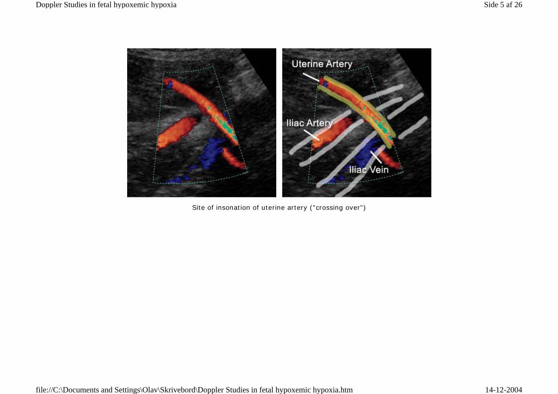
Site of insonation of uterine artery ("crossing over")
Side 5 af 26Doppler Studies in fetal hypoxemic hypoxia
14-12-2004file://C:\Documents and Settings\Olav\Skrivebord\Doppler Studies in fetal hypoxemic hypoxia.htm
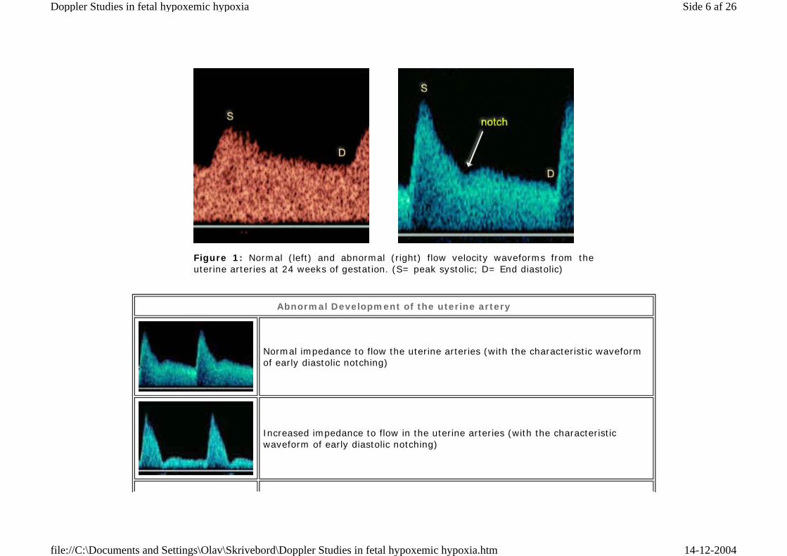
Figure 1: Normal (left) and abnormal (right) flow velocity waveforms from the uterine arteries at 24 weeks of gestation. (S= peak systolic; D= End diastolic)
Abnormal Development of the uterine artery
Normal impedance to flow the uterine arteries (with the characteristic waveform of early diastolic notching)
Increased impedance to flow in the uterine arteries (with the characteristic waveform of early diastolic notching)
Side 6 af 26Doppler Studies in fetal hypoxemic hypoxia
14-12-2004file://C:\Documents and Settings\Olav\Skrivebord\Doppler Studies in fetal hypoxemic hypoxia.htm
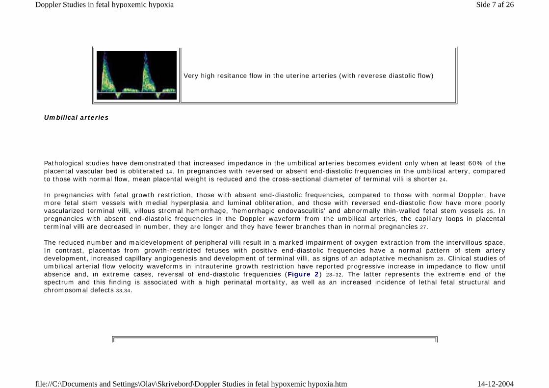
Very high resitance flow in the uterine arteries (with reverese diastolic flow)
Umbilical arteries
Pathological studies have demonstrated that increased impedance in the umbilical arteries becomes evident only when at least 60% of the placental vascular bed is obliterated 14. In pregnancies with reversed or absent end-diastolic frequencies in the umbilical artery, compared to those with normal flow, mean placental weight is reduced and the cross-sectional diameter of terminal villi is shorter 24. In pregnancies with fetal growth restriction, those with absent end-diastolic frequencies, compared to those with normal Doppler, have more fetal stem vessels with medial hyperplasia and luminal obliteration, and those with reversed end-diastolic flow have more poorly vascularized terminal villi, villous stromal hemorrhage, ‘hemorrhagic endovasculitis’ and abnormally thin-walled fetal stem vessels 25. In pregnancies with absent end-diastolic frequencies in the Doppler waveform from the umbilical arteries, the capillary loops in placental terminal villi are decreased in number, they are longer and they have fewer branches than in normal pregnancies 27. The reduced number and maldevelopment of peripheral villi result in a marked impairment of oxygen extraction from the intervillous space. In contrast, placentas from growth-restricted fetuses with positive end-diastolic frequencies have a normal pattern of stem artery development, increased capillary angiogenesis and development of terminal villi, as signs of an adaptative mechanism 28. Clinical studies of umbilical arterial flow velocity waveforms in intrauterine growth restriction have reported progressive increase in impedance to flow until absence and, in extreme cases, reversal of end-diastolic frequencies (Figure 2) 28–32. The latter represents the extreme end of the spectrum and this finding is associated with a high perinatal mortality, as well as an increased incidence of lethal fetal structural and chromosomal defects 33,34.
Side 7 af 26Doppler Studies in fetal hypoxemic hypoxia
14-12-2004file://C:\Documents and Settings\Olav\Skrivebord\Doppler Studies in fetal hypoxemic hypoxia.htm
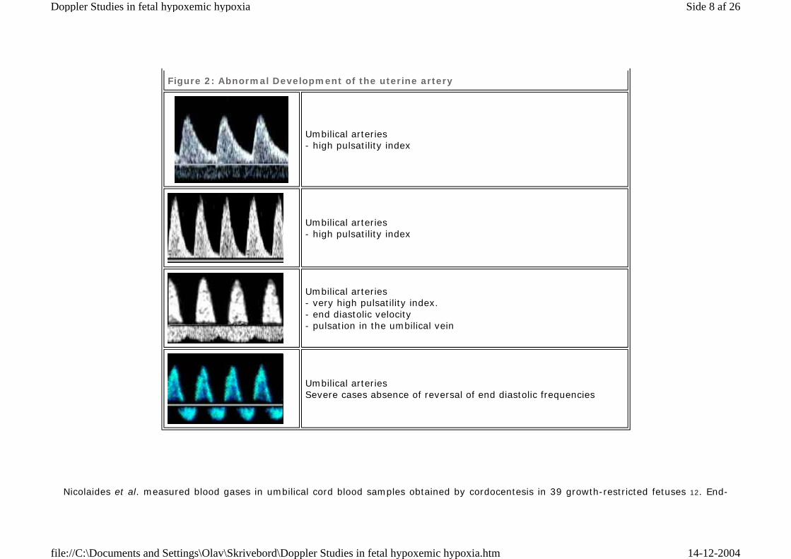
Figure 2: Abnormal Development of the uterine artery
Umbilical arteries - high pulsatility index
Umbilical arteries - high pulsatility index
Umbilical arteries - very high pulsatility index. - end diastolic velocity - pulsation in the umbilical vein
Umbilical arteries Severe cases absence of reversal of end diastolic frequencies
Nicolaides et al. measured blood gases in umbilical cord blood samples obtained by cordocentesis in 39 growth-restricted fetuses 12. End-
Side 8 af 26Doppler Studies in fetal hypoxemic hypoxia
14-12-2004file://C:\Documents and Settings\Olav\Skrivebord\Doppler Studies in fetal hypoxemic hypoxia.htm
diastolic frequencies were absent in 22 cases; 80% of these fetuses were found to be hypoxemic and 46% also acidemic. In contrast, only 12% of the fetuses with positive end-diastolic frequencies were hypoxemic and none was acidemic. In a multicenter study involving high-risk pregnancies, the patients were subdivided into three groups depending on the flow velocity waveforms in the umbilical artery (positive end-diastolic frequencies, n = 214; absent end-diastolic frequencies, n = 178); and reversed end-diastolic frequencies, n = 67) 35. The overall perinatal mortality rate was 28% and the relative risk was 1.0 for patients with present frequencies, 4.0 for those with absent frequencies and 10.6 for those with reversed frequencies. Significantly more neonates in the groups with absent or reversed frequencies needed admittance to the neonatal intensive care unit and they had a higher risk of cerebral hemorrhage, anemia or hypoglycemia 35. In addition to increased fetal and neonatal mortality, gowth restriction with absent or reversed end-diastolic frequencies in the umbilical artery is associated with increased incidence of long-term permanent neurological damage 36. A review of 12 randomized, controlled trials of Doppler ultrasonography of the umbilical artery in high-risk pregnancies reported that, in the Doppler group, there was a significant reduction in the number of antenatal admissions (44%, 95% confidence interval (CI) 28–57%), induction of labor (20%, 95% CI 10–28%), and Cesarean section for fetal distress (52%, 95% CI 24–69%) 37. Furthermore, the clinical action guided by Doppler ultrasonography reduced the odds of perinatal death by 38% (95% CI 15–55%). Post hoc analyses revealed a statistically significant reduction in elective delivery, intrapartum fetal distress, and hypoxic encephalopathy in the Doppler group. It was concluded that there is now compelling evidence that women with high-risk pregnancies, including pre-eclampsia and suspected intrauterine growth restriction, should be offered Doppler ultrasonographic study of umbilical artery waveforms 37. In terms of monitoring growth-restricted pregnancies, abnormal waveforms in the umbilical artery are an early sign of fetal impairment. For example, Bekedam et al. followed up growth-restricted fetuses longitudinally and reported that abnormalities in the umbilical artery preceded the occurrence of cardiotocographic signs of fetal hypoxemia in more than 90% of cases 38. The median time interval between absence of end-diastolic frequencies and the onset of late decelerations was 12 days (range 0–49 days).
Fetal arterial blood flow Redistribution
In fetal hypoxemia, there is an increase in the blood supply to the brain, myocardium and the adrenal glands and reduction in the perfusion of the kidneys, gastrointestinal tract and the lower extremities (Figure 3 and 4, Table 1 ) 39–66. Although knowledge of the factors governing circulatory readjustments and their mechanism of action is incomplete, it appears that partial pressures of oxygen and carbon dioxide play a role, presumably through their action on chemoreceptors. This mechanism allows preferential delivery of nutrients and oxygen to vital organs, thereby compensating for diminished placental resources. However, compensation through cerebral vasodilatation is limited and a plateau corresponding to a nadir of pulsatility index (PI) in cerebral vessels is reached at least 2 weeks before the
Side 9 af 26Doppler Studies in fetal hypoxemic hypoxia
14-12-2004file://C:\Documents and Settings\Olav\Skrivebord\Doppler Studies in fetal hypoxemic hypoxia.htm
development of the fetus is jeopardized. Consequently, arterial vessels are unsuitable for longitudinal monitoring of growth-restricted fetuses. Cardiac and venous velocity waveforms give more information regarding fetal well-being or compromise.
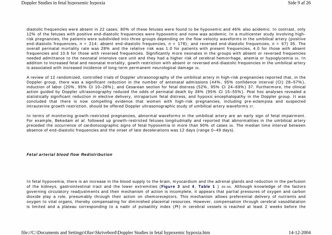
Arduini et al. examined growth-restricted fetuses longitudinally and described a curvilinear relationship between impedance in cerebral vessels and the state of fetal oxygenation; the progressive fall in impedance reached a nadir 2 weeks before the onset of late fetal heart rate decelerations 55. This suggests that the maximum degree of vascular adaptation to hypoxemia precedes the critical degree of impairment of fetal oxygenation. Similarly, Potts et al. described a curvilinear relationship between cerebral vascular response and hypercapnia 56. Vyas et al. reported that, concomitant to severe oxygen deficit, there was a sudden rise in middle cerebral artery PI; they suggested that vascular dilatation may be suppressed by the development of cerebral edema 53. An alternative explanation may be that, in severe hypoxemia, an increase in PI may be the consequence of alterations in flow due to reduced cardiac contractility and to a fall in absolute cardiac output. Moreover, in fetal sheep, chronic hypoxemia was found to alter cerebral vascular contractility through changes in vascular smooth muscle and endothelial cells, with the net result of a relative decrease in cerebral blood flow 57.
Figure 3: Color Doppler examination of the circle of Willis (left). Flow velocity waveforms from the middle cerebral artery in a normal fetus with low diastolic velocities (right, top) and in a growth-restricted fetus with high diastolic velocities (right, bottom).
Side 10 af 26Doppler Studies in fetal hypoxemic hypoxia
14-12-2004file://C:\Documents and Settings\Olav\Skrivebord\Doppler Studies in fetal hypoxemic hypoxia.htm
Fetal arterial Doppler studies are useful in the differential diagnosis of small-forgestation fetuses. In the hypoxemic group, due to impaired placental perfusion, the PI in the umbilical artery is increased and, in the fetal middle cerebral artery, the PI is decreased; consequently, the ratio in PI between the umbilical artery and middle cerebral artery (UA/MCA) is increased 58–61. Bahado-Singh et al . reported that an abnormally low cerebroplacental ratio is associated with increased perinatal morbidity and mortality and that the ratio improves the prediction of perinatal outcome compared with umbilical artery PI alone 65. However, the cerebroplacental ratio did not appear to correlate
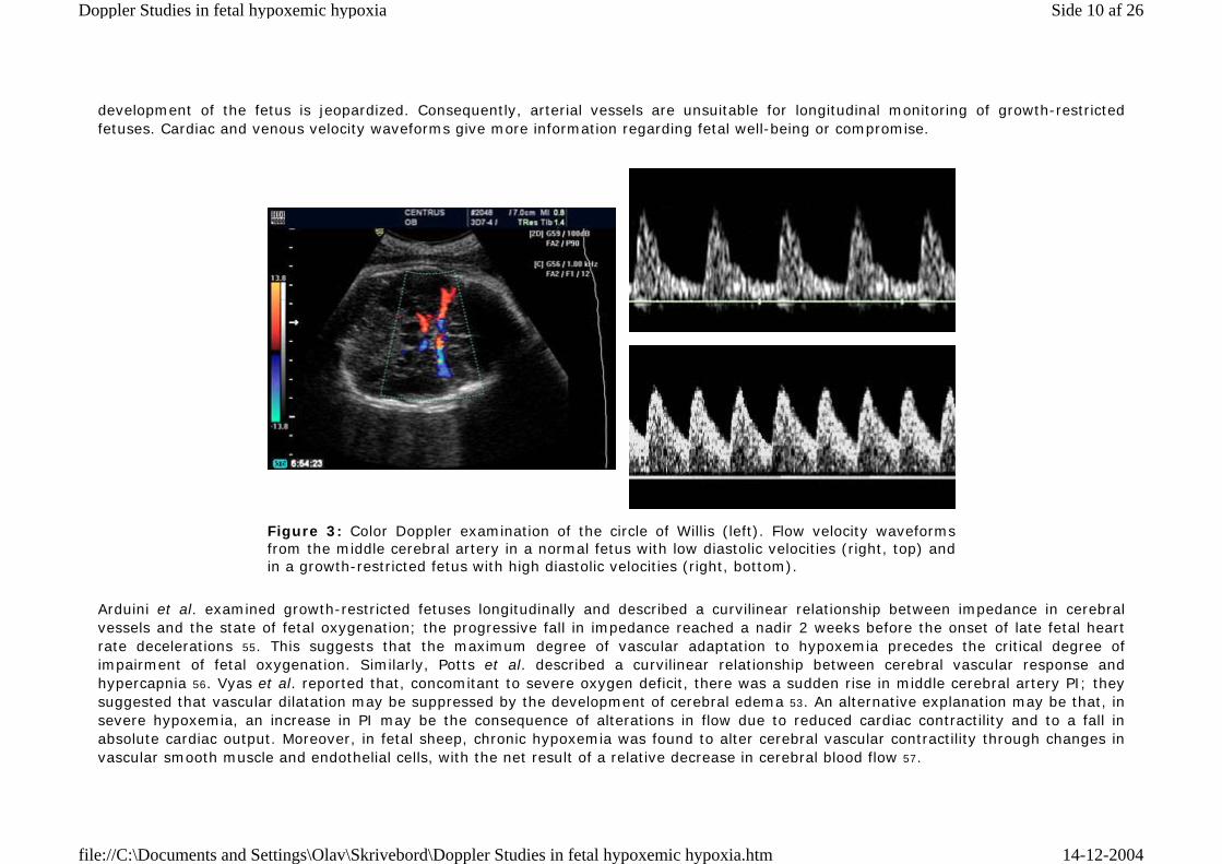
Table 1 - Hemodynamic changes occurring in fetal arterial vessels during hypoxemia and acidemia induced by uteroplacental insufficiency.
Vessel Impedance
to flow
Descending aorta increased
Renal artery increased
Femoral artery increased
Peripheral pulmonary artery increased
Mesenteric arteries increased
Cerebral arteries decreased
Adrenal arteries decreased
Splenic arteries decreased
Coronary arteries decreased
Side 11 af 26Doppler Studies in fetal hypoxemic hypoxia
14-12-2004file://C:\Documents and Settings\Olav\Skrivebord\Doppler Studies in fetal hypoxemic hypoxia.htm
significantly with outcome after 34 weeks. In third-trimester fetuses, the ratio in PI between the fetal descending thoracic aorta and the middle cerebral artery may be more useful 66.
There is no evidence that the use of other peripheral arterial fetal vessels, such as renal artery 51, splenic artery 64 or peripheral pulmonary arteriesprovides any advantage in the identification of intrauterine growth-restricted fetuses.
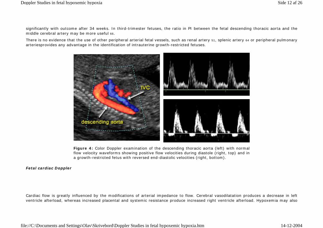
Figure 4: Color Doppler examination of the descending thoracic aorta (left) with normal flow velocity waveforms showing positive flow velocities during diastole (right, top) and in a growth-restricted fetus with reversed end-diastolic velocities (right, bottom).
Fetal cardiac Doppler
Cardiac flow is greatly influenced by the modifications of arterial impedance to flow. Cerebral vasodilatation produces a decrease in left ventricle afterload, whereas increased placental and systemic resistance produce increased right ventricle afterload. Hypoxemia may also
Side 12 af 26Doppler Studies in fetal hypoxemic hypoxia
14-12-2004file://C:\Documents and Settings\Olav\Skrivebord\Doppler Studies in fetal hypoxemic hypoxia.htm
impair cardiac contractility directly, while changes in blood viscosity due to polycythemia may alter preload 5. Consequently, growth-restricted fetuses show, at the level of the atrioventricular valves, impaired ventricular filling (lower ratio of early passive to late active ventricular filling phase – E/A ratio) 67, lower peak velocities in the aorta and pulmonary arteries. These hemodynamic intracardiac changes are compatible with a preferential shift of cardiac output in favor of the left ventricle, leading to improved cerebral perfusion. Thus, in the first stages of the disease, the supply of substrates and oxygen can be maintained at near normal levels despite any absolute reduction of placental transfer 71. Longitudinal studies of deteriorating growth-restricted fetuses have shown that peak velocity and cardiac output gradually decline, suggesting a progressive worsening in cardiac function 71.
Similarly, there is a symmetrical decrease in ventricular ejection force at the level of both ventricles, despite the dramatically different hemodynamic conditions present in the vascular district of ejection of the two ventricles (i.e. reduced cerebral resistances for the left ventricle and increased splachnic and placental resistance for the right ventricle) 72. This supports a pivotal role of the intrinsic myocardial function in the compensatory mechanism of the growth-restricted fetus following the establishment of the brain-sparing effect. Ventricular ejection force dramatically decreases in a short time interval (about 1 week), showing an impairment of ventricular force close to fetal distress. As a consequence, cardiac filling also is impaired.
Fetal venous Doppler
Animal studies have shown that, in severe hypoxemia, there is redistribution in the umbilical venous blood towards the ductus venosus at the expense of hepatic blood flow. Consequently, the proportion of umbilical venous blood contributing to the fetal cardiac output is increased. There is a doubling of umbilical venous-derived oxygen delivery to the myocardium and an increase in oxygen delivery to the fetal brain 73,74. In vitro perfusion studies have shown that, at reduced umbilical venous pressures, a proportionally greater fraction of umbilical venous flow is directed through the ductus venosus in comparison to blood flow through the liver 75. The same is true during perfusion with blood of high hematocrit. Mechanical forces seem to play a key role in the regulation of umbilical venous flow distribution between the liver and the ductus venosus. Under unfavorable conditions, the ductus venosus seems to ensure blood flow directly to the fetal heart and, in extreme conditions, umbilical blood may pass exclusively through the ductus venosus. This may lead to an impaired perfusion of the liver with potential impact on its metabolic properties. Blood flow measurements with chronically implanted electromagnetic flow transducers in fetal sheep have shown an increase of the amplitude of vena caval pulsations during hypoxemia and increased afterload 76. Flow waveforms show an increase in peak systolic forward flow, and during atrial contraction retrograde flow occurs. In contrast, reductions in afterload are associated with an increase in peak diastolic forward flow, indicating that fetal systemic vascular resistance has a major influence on venous return and filling patterns of the right heart. Increased placental resistance and peripheral vasoconstriction, as seen in
Side 13 af 26Doppler Studies in fetal hypoxemic hypoxia
14-12-2004file://C:\Documents and Settings\Olav\Skrivebord\Doppler Studies in fetal hypoxemic hypoxia.htm
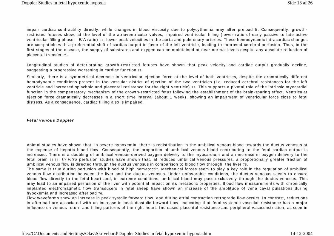
fetal arterial redistribution, cause an increase in right ventricular afterload, and thus ventricular end-diastolic pressure increases. This may result in highly pulsatile venous blood flow waveforms and umbilical venous pulsations due to transmission of atrial pressure waves through the ductus venosus 77. Studies in growth-restricted human fetuses have demonstrated that, in the inferior vena cava, an increase of reverse flow during atrial contraction occurs with progressive fetal deterioration, suggesting a higher pressure gradient in the right atrium (Figure 5) 78,79.
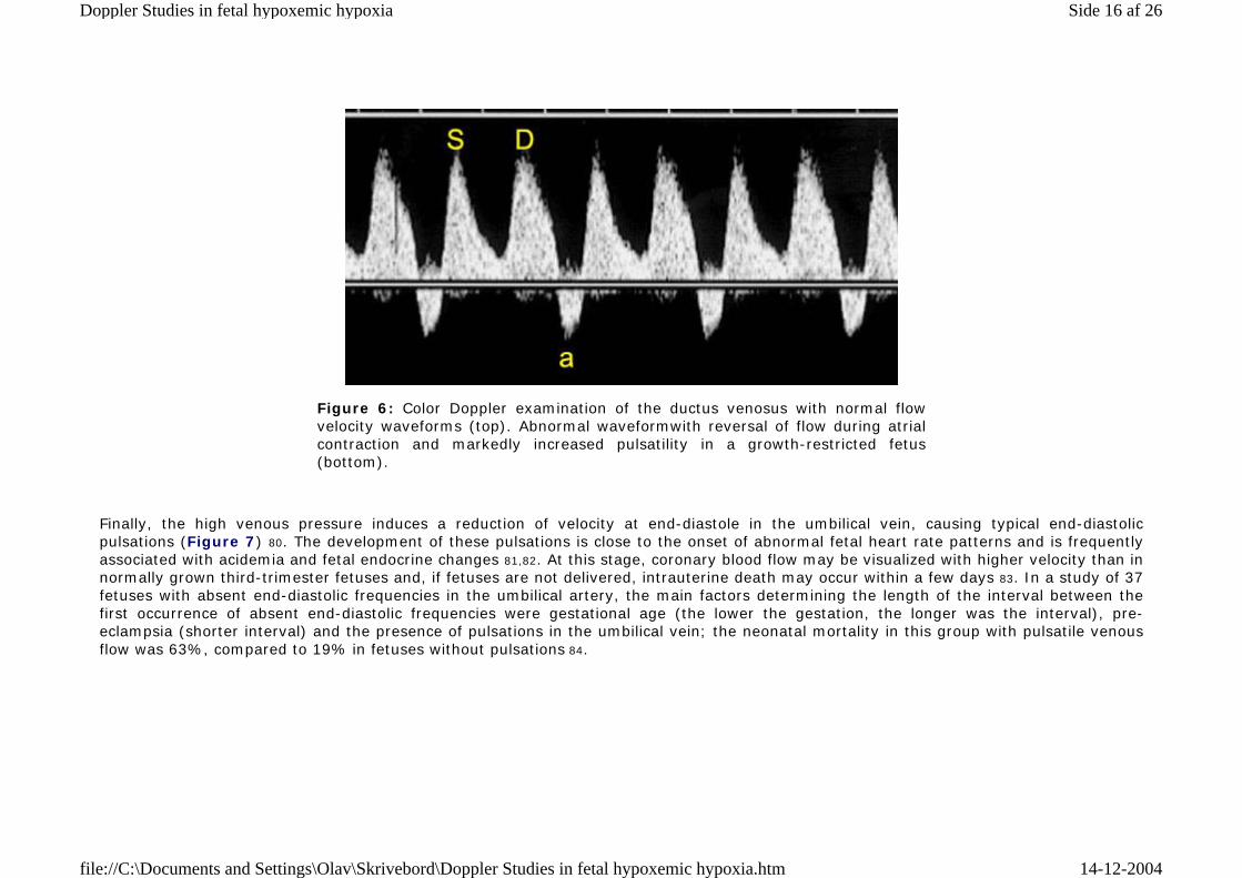
The next step of the disease is the extension of the abnormal reversal of blood velocities in the inferior vena cava to the ductus venosus, inducing an increase of the S/A ratio, mainly due to a reduction of the A component of the velocity waveforms (Figure 6).
Figure 5: Color Doppler examination of the inferior vena cava with normal flow velocity waveforms (top). Abnormal waveform with increase in reversed flow during atrial contraction in a growth-restricted fetus (bottom).
Side 14 af 26Doppler Studies in fetal hypoxemic hypoxia
14-12-2004file://C:\Documents and Settings\Olav\Skrivebord\Doppler Studies in fetal hypoxemic hypoxia.htm
Side 15 af 26Doppler Studies in fetal hypoxemic hypoxia
14-12-2004file://C:\Documents and Settings\Olav\Skrivebord\Doppler Studies in fetal hypoxemic hypoxia.htm
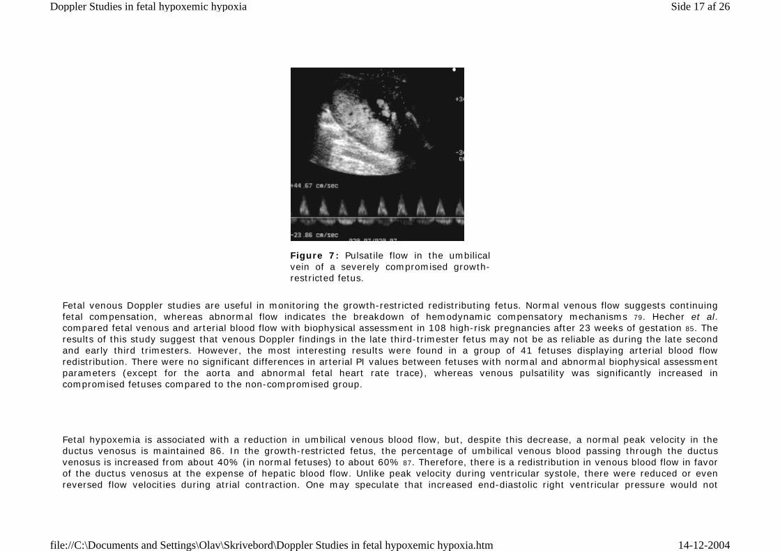
Finally, the high venous pressure induces a reduction of velocity at end-diastole in the umbilical vein, causing typical end-diastolic pulsations (Figure 7) 80. The development of these pulsations is close to the onset of abnormal fetal heart rate patterns and is frequently associated with acidemia and fetal endocrine changes 81,82. At this stage, coronary blood flow may be visualized with higher velocity than in normally grown third-trimester fetuses and, if fetuses are not delivered, intrauterine death may occur within a few days 83. In a study of 37 fetuses with absent end-diastolic frequencies in the umbilical artery, the main factors determining the length of the interval between the first occurrence of absent end-diastolic frequencies were gestational age (the lower the gestation, the longer was the interval), pre-eclampsia (shorter interval) and the presence of pulsations in the umbilical vein; the neonatal mortality in this group with pulsatile venous flow was 63%, compared to 19% in fetuses without pulsations 84.
Figure 6: Color Doppler examination of the ductus venosus with normal flow velocity waveforms (top). Abnormal waveformwith reversal of flow during atrial contraction and markedly increased pulsatility in a growth-restricted fetus (bottom).
Side 16 af 26Doppler Studies in fetal hypoxemic hypoxia
14-12-2004file://C:\Documents and Settings\Olav\Skrivebord\Doppler Studies in fetal hypoxemic hypoxia.htm
Fetal venous Doppler studies are useful in monitoring the growth-restricted redistributing fetus. Normal venous flow suggests continuing fetal compensation, whereas abnormal flow indicates the breakdown of hemodynamic compensatory mechanisms 79. Hecher et al. compared fetal venous and arterial blood flow with biophysical assessment in 108 high-risk pregnancies after 23 weeks of gestation 85. The results of this study suggest that venous Doppler findings in the late third-trimester fetus may not be as reliable as during the late second and early third trimesters. However, the most interesting results were found in a group of 41 fetuses displaying arterial blood flow redistribution. There were no significant differences in arterial PI values between fetuses with normal and abnormal biophysical assessment parameters (except for the aorta and abnormal fetal heart rate trace), whereas venous pulsatility was significantly increased in compromised fetuses compared to the non-compromised group.
Figure 7: Pulsatile flow in the umbilical vein of a severely compromised growth-restricted fetus.
Fetal hypoxemia is associated with a reduction in umbilical venous blood flow, but, despite this decrease, a normal peak velocity in the ductus venosus is maintained 86. In the growth-restricted fetus, the percentage of umbilical venous blood passing through the ductus venosus is increased from about 40% (in normal fetuses) to about 60% 87. Therefore, there is a redistribution in venous blood flow in favor of the ductus venosus at the expense of hepatic blood flow. Unlike peak velocity during ventricular systole, there were reduced or even reversed flow velocities during atrial contraction. One may speculate that increased end-diastolic right ventricular pressure would not
Side 17 af 26Doppler Studies in fetal hypoxemic hypoxia
14-12-2004file://C:\Documents and Settings\Olav\Skrivebord\Doppler Studies in fetal hypoxemic hypoxia.htm
influence ductus venosus blood flow velocities during atrial contraction, as flow is preferentially directed through the foramen ovale to the left atrium. However, the foramen ovale is closed during atrial contraction and blood flow velocity through the foramen ovale decreases to zero. Alterations of venous flow velocity waveforms are in a closer temporal relationship to intrauterine fetal jeopardy, compared to changes in arterial flow, which may occur quite early during the course of impaired placental function. The degree of fetal acidemia can be estimated from Doppler measurements of pulsatility in both the arterial system and the ductus venosus. This was shown in a cross-sectional study of 23 severely growth-retstricted fetuses, examining the relationship between Doppler measurements and umbilical venous blood gases obtained at cordocentesis 88. With moderate acidemia (pH between -2 and -4 standard deviations from the normal mean for gestational age), almost all fetuses had a middle cerebral artery PI below two standard deviations, whereas there was a wide scatter of individual results for the ductus venosus, with the majority of measurements being still within the reference ranges. With increasing severity of hypoxemia and acidemia, ductus venosus PIs increased and, in the most severe cases, velocities with atrial contraction were reduced to zero or even became negative. In a study investigating the association of arterial and venous Doppler findings with adverse perinatal outcome in severe fetal growth restriction, abnormal Doppler velocimetry of the ductus venosus was the only significant parameter associated with perinatal death and low 5-min Apgar scores 89. There are two possible mechanisms for abnormal venous blood flow waveforms: increasing right ventricular afterload and myocardial failure. As long as the fetus is able to compensate for a reduced placental supply by arterial redistribution, there is preferential myocardial oxygenation, which delays development of right heart failure, despite an increasing afterload. Therefore, fetal Doppler measurements show high placental resistance and arterial redistribution in the presence of normal venous waveforms. At this stage, the majority of fetuses have normal, reactive heart rate traces and biophysical profiles. Progressive changes in the venous circulation may indicate failure of the compensatory mechanism and herald the development of right heart failure due to myocardial hypoxia. Another interesting aspect has been raised by a study examining fetal central venous pressure. The pressure waveform from the inferior vena cava was recorded by following the movement of the vessel wall and thereby recording changes in the vessel lumen diameter 90. There were two groups of abnormal waveforms: one with a high pulsatile pattern and the other with a shallow and low pulsatile pattern. Both groups had significantly worse clinical outcomes compared to the normal waveform group. However, fetuses in the low pulsatile group were the most severely compromised, all of them showing an abnormal heart rate pattern. It was postulated that impaired contractility and reduced ventricular output, with a concomitantly reduced ventricular filling, were responsible for this waveform pattern.
Timing of delivery
Side 18 af 26Doppler Studies in fetal hypoxemic hypoxia
14-12-2004file://C:\Documents and Settings\Olav\Skrivebord\Doppler Studies in fetal hypoxemic hypoxia.htm
In the management of the very preterm (before 33 weeks of gestation) growthrestricted fetus, there is uncertainty as to whether iatrogenic delivery should be undertaken before the development of signs of severe hypoxemia, with a consequent risk of prematurity-related neonatal complications, or whether delivery should be delayed, but with the risks of prolonged exposure to hypoxia and malnutrition imposed by a hostile intrauterine environment. A growth-restricted fetus leading an ascetic existence from chronic starvation during the late second or early third trimester is capable of tolerating chronic hypoxemia without damage for much longer than a well-nourished late third-trimester fetus with a high energy consumption.
Postnatal follow-up studies, at the age of 7 years, have reported that growthrestricted fetuses with abnormal aortic velocity waveforms had minor neurological dysfunction and impaired intellectual outcome 91,92. If these findings are confirmed in prospective studies with adequate controls for confounding variables, such as degrees of prematurity, smallness, and management, it may be advisable to deliver growthrestricted fetuses before these blood flow alterations occur. On the other hand, fetuses showing the brain-sparing effect did not have an increased risk for moderate or severe neurological handicap at the age of 2 years 93. It will always be a challenge to weigh the risks and benefits of early interventions against each other and it is a dynamic process, in which advancements in both fetal and neonatal medicine are of crucial importance for the counselling of parents and the management of these pregnancies.
In the growth-restricted hypoxemic fetus, redistribution of well-oxygenated blood to vital organs, such as the brain, heart and adrenals, represents a compensatory mechanism to prevent fetal damage. When the reserve capacities of the circulatory redistribution reach their limits, fetal deterioration may occur rapidly. In clinical practice, it is necessary to carry out serial Doppler investigations to estimate the duration of fetal blood flow redistribution. The onset of abnormal venous Doppler results indicates deterioration in the fetal condition and iatrogenic delivery should be considered.
In the sequence of deterioration of the condition of the growth-restricted fetus, the first pathological finding is increased impedance to flow in the umbilical artery. This is followed by evidence of arterial redistribution in the fetal circulation and, subsequently, the development of pathological fetal heart rate patterns. On average, the time interval between the onset of abnormal umbilical arterial Doppler results and the onset of late fetal heart rate decelerations is about 2 weeks, but this interval differs considerably among fetuses and is shorter in late than early pregnancy and in the presence of hypertensive disease 31,38,84,94,95. Increased impedance to flow in the umbilical artery is usually associated with evidence of arterial redistribution in the fetal circulation; this is best monitored by examining the PI in the middle cerebral artery, which is decreased. Late fetal heart rate decelerations are preceded by approximately 2 weeks with Doppler evidence of a nadir in the brain-sparing effect and by a few days with an abrupt increase in impedance in the umbilical arteries 55 . In the first stages of the disease, there is a preferential shift of cardiac output in favor of the left ventricle, leading to improved cerebral perfusion 71, but, with deterioration in the fetal condition, there are a decline in cardiac output and progressive worsening in cardiac function 71. Normal venous flow suggests continuing fetal compensation, whereas abnormal flow indicates the breakdown of hemodynamic compensatory mechanisms 79. An abrupt increase in pulsatility of ductus venosus waveforms with loss of forward flow velocity during atrial contraction precede the onset of pathological fetal heart rate patterns and decreased short-term variation. However, the interval may be as short as a few hours in late
Side 19 af 26Doppler Studies in fetal hypoxemic hypoxia
14-12-2004file://C:\Documents and Settings\Olav\Skrivebord\Doppler Studies in fetal hypoxemic hypoxia.htm
gestation and in patients with pre-eclampsia; in contrast, during the second trimester, severely abnormal venous waveforms can be present for several days before intrauterine death.
REFERENCES
1. Soothill PW, Nicolaides KH, Rodeck CH, Campbell S. Effect of gestational age on fetal and intervillous blood gas and acid–base values in human pregnancy. Fetal Ther 1986;1:168–75
2. Nicolaides KH, Economides DL, Soothill PW. Blood gases and pH and lactate in appropriate and small for gestational age fetuses. Am J Obstet Gynecol 1989;161:996–1001
3. Battaglia FC, Meschia G. An Introduction to Fetal Physiology. London: Academic Press, 1986:154–67
4. Burd LI, Jones MD, Simmons MA. Placental production and fetal utilisation of lactate and pyruvate. Nature (London) 1975;254:210–1
5. Soothill PW, Nicolaides KH, Campbell S. Prenatal asphyxia, hyperlacticaemia, hypoglycaemia and erythroblastosis in growth retarded fetuses. Br Med J 1987;294:1051–3
6. Economides DL, Nicolaides KH. Blood glucose and oxygen tension levels in small for gestational age fetuses. Am J Obstet Gynecol 1989;160:385–9
7. Economides DL, Proudler A, Nicolaides KH. Plasma insulin in appropriate and small for gestational age fetuses. Am J Obstet Gynecol 1989;160:1091–4
8. Economides DL, Nicolaides KH, GahlW, Bernardini I, Evans M. Plasma amino acids in appropriate and small for gestational age fetuses. Am J Obstet Gynecol 1989;161:1219–27
9. Economides DL, Crook D, Nicolaides KH. Hypertriglyceridemia and hypoxemia in small for gestational age fetuses. Am J Obstet Gynecol 1990;162:382–6
10. Thorpe-Beeston JG, Nicolaides KH, Snijders RJM, Felton CV, McGregor AM. Thyroid function in small for gestational age fetuses. Obstet Gynecol 1991;77:701–6
11. Soothill PW, Nicolaides KH, Bilardo CM, Hackett G, Campbell S. Utero-placental blood flow velocity resistance index and venous pO2, pCO2, pH, lactate and erythroblast count in growth retarded fetuses. Fetal Ther 1986;l:l76–9
12. Nicolaides KH, Bilardo CM, Soothill PW, Campbell S. Absence of end diastolic frequencies in the umbilical artery: a sign of fetal hypoxia and acidosis. Br Med J 1988;297:1026–7
13. Brosens I, Robertson WB, Dixon HG. The role of the spiral arteries in the pathogenesis of pre-eclampsia. Obstet Gynecol Annu 1972;1:177–91
14. Giles WB, Trudinger BJ, Baird PJ. Fetal umbilical artery flow velocity waveforms and placental resistance: pathological correlation. Br J Obstet Gynaecol 1985;92:31–8
15. Peeters LL, Sheldon RE, Jones MD, Makowsky EL, Meschia G. Blood flow to fetal organs as a function of arterial oxygen content. Am J Obstet Gynecol 1979;135:637–46
Side 20 af 26Doppler Studies in fetal hypoxemic hypoxia
14-12-2004file://C:\Documents and Settings\Olav\Skrivebord\Doppler Studies in fetal hypoxemic hypoxia.htm
16. Robertson WB, Brosens I, Dixon HG. The pathological response of the vessels of the placental bed to hypertensive pregnancy. J Pathol Bacteriol 1967;93:581–92
17. Sheppard BL, Bonnar J. An ultrastructural study of utero-placental spiral arteries in hypertensive and normotensive pregnancy and fetal growth retardation. Br J Obstet Gynaecol 1981;88:695–705
18. Brosens IA. Morphological changes in the utero-placental bed in pregnancy hypertension. Clin Obstet Gynecol 1977;4:573–93
19. Khong TY, De Wolf F, Robertson WB, Brosens I. Inadequate maternal vascular response to placentation in pregnancies complicated by pre-eclampsia and by small-for-gestational age infants. Br J Obstet Gynaecol 1986;93:1049–59
20. Campbell S, Griffin DR, Pearce JM, Diaz-Recasens J, Cohen-Overbeek T,Wilson K, Teague MJ. New Doppler technique for assessing uteroplacental blood flow. Lancet 1983;26:675–7
21. Trudinger BJ, Giles WB, Cook CM. Uteroplacental blood flow velocity-time waveforms in normal and complicated pregnancy. Br J Obstet Gynaecol 1985;92:39–45
22. Campbell S, Pearce JM, Hackett G, Cohen-Overbeek T, Hernandez C. Qualitative assessment of uteroplacental blood flow: an early screening test for high risk pregnancies. Obstet Gynecol 1986;68: 649–53
23. Fleisher A, Schulman H, Farmakides G, Bracero L, Rochelson B, Koenigsberg M. Uterine artery Doppler velocimetry in pregnant women with hypertension. Am J Obstet Gynecol 1986;154:806–13
24. Karsdorp VH, Dirks BK, van der Linden JC, van Vugt JM, Baak JP, van Geijn HP. Placenta morphology and absent or reversed end diastolic flow velocities in the umbilical artery: a clinical and morphometrical study. Placenta 1996;17:393–9
25. Salafia CM, Pezzullo JC, Minior VK, Divon MY. Placental pathology of absent and reversed end-diastolic flow in growth-restricted fetuses. Obstet Gynecol 1997;90:830–6
26. Schulman H, Fleisher A, Stern W, Farmakides G, Jagani N, Blattner P. Umbilical velocity wave ratios in human pregnancy. Am J Obstet Gynecol 1984;148:985–90
27. Krebs C, Macara LM, Leiser R, Bowman AW, Greer IA, Kingdom JCP. Intrauterine growth restriction with absent end-diastolic flow velocity in the umbilical artery is associated with maldevelopment of the placental terminal villous tree. Am J Obstet Gynecol 1996;175:1534–42
28. Todros T, Sciarrone A, Piccoli E, Guiot C, Kaufmann P, Kingdom J. Umbilical Doppler waveforms and placental villous angiogenesis in pregnancies complicated by fetal growth restriction. Obstet Gynecol 1999;93:499–503
29. Erskine RL, Ritchie JW. Umbilical artery blood flow characteristics in normal and growth retarded fetuses. Br J Obstet Gynaecol 1985;92:605–10
30. Trudinger BJ, Giles WB, Cook CM, Bombardieri J, Collins L. Fetal umbilical artery flow velocity waveforms and placental resistance: clinical significance. Br J Obstet Gynaecol 1985;92:23–30
31. Reuwer PJ, Sijmons EA, Rietman GW, van Tiel MW, Bruinse HW. Intrauterine growth retardation: prediction of perinatal distress by Doppler ultrasound. Lancet 1987;22:415–18
Side 21 af 26Doppler Studies in fetal hypoxemic hypoxia
14-12-2004file://C:\Documents and Settings\Olav\Skrivebord\Doppler Studies in fetal hypoxemic hypoxia.htm
32. Rochelson B, Shulman H, Farmakides G, Bracero L, Ducey J, Fleisher A, Penny B,Winter D. The significance of absent end-diastolic velocity in umbilical artery velocity waveforms. Am J Obstet Gynecol 1987;156:1213–38
33. Mandruzzato GP, Bogatti P, Fischer L, Gigli C. The clinical significance of absent or reverse end-diastolic flow in the fetal aorta and umbilical artery. Ultrasound Obstet Gynecol 1991;1:192–6
34. Brar HS, Platt LD. Reverse end-diastolic flow velocity on umbilical artery velocimetry in high risk pregnancies: an ominous finding with adverse pregnancy outcome. Am J Obstet Gynecol 1988;159: 559–61
35. Karsdorp VH, van Vugt JM, van Geijn HP, Kostense PJ, Arduini D, Montenegro N, Todros T. Clinical significance of absent or reversed end diastolic velocity waveforms in umbilical artery. Lancet 1994;344:1664–8
36. Valcamonico A, Danti L, Frusca T, Soregaroli M, Zucca S, Abrami F, Tiberti A. Absent enddiastolic velocity in umbilical artery: risk of neonatal morbidity and brain damage. Am J Obstet Gynecol 1994;170:796–801
37. Alfirevic Z, Neilson JP. Doppler ultrasonography in high-risk pregnancies: systematic review with meta-analysis. Am J Obstet Gynecol 1995;172:1379–87
38. Bekedam DJ, Visser GHA, van der Zee AGJ, Snijders RJM, Poelmann-Weesjes G. Abnormal velocity waveforms of the umbilical artery in growth-retarded fetuses: relationship to antepartum late heart rate decelerations and outcome. Early Hum Dev 1990;24:79–89
39. Soothill PW, Nicolaides KH, Bilardo KM, Campbell S. The relationship of fetal hypoxia in growth retardation to the mean blood velocity in the fetal aorta. Lancet 1986;2:1118–20
40. Wladimiroff JW, Tonge HM, Stewart PA. Doppler ultrasound assessment of cerebral blood flow in the human fetus. Br J Obstet Gynaecol 1986;93:471–5
41. Tonge HM, Wladimiroff JW, Noordam MJ, van Kooten C. Blood flow velocity waveforms in the descending fetal aorta: comparison between normal and growth retarded pregnancies. Obstet Gynecol 1986;67:851–5
42. van Eyck J, Wiadimiroff JW, Noordam MJ, Tonge HM, Prechtl HFR. The blood flow velocity waveformin the fetal descending aorta: its relationship to behavioural states in growth retarded fetus at 37–38 weeks of gestation. Early Hum Dev 1986;14:99–107
43. Wladimiroff JW, vanWijngaard JAGW, Degani S, Noordam MJ, van Eijck J, Tonge HM. Cerebral and umbilical arterial blood flow velocity waveforms in normal and growth retarded pregnancies. Obstet Gynecol 1987;69:705–9
44. Laurin J, Lingman G, Marsal K, Persson PH. Fetal blood flow in pregnancies complicated by intrauterine growth retardation. Obstet Gynecol 1987;69:895–902
45. Laurin J, Marsal K, Persson P, Lingman H. Ultrasound measurements of fetal blood flow in predicting fetal outcome. Br J Obstet Gynaecol 1987;94:940–8
46. Arduini D, Rizzo D, Romanini C, Mancuso S. Fetal blood flow velocity waveforms as predictors of growth retardation. Obstet Gynecol 1987;70:7–10
47. van Eyck J, Wladimiroff JW, van den Wijngaard JAGW, Noordam MJ, Prechtl HFR. The blood flow velocity waveformin the internal carotid artery: its relationship to behavioural states in growth retarded fetus at 37–38 weeks of gestation. Br J Obstet Gynaecol 1987;94:736–41
Side 22 af 26Doppler Studies in fetal hypoxemic hypoxia
14-12-2004file://C:\Documents and Settings\Olav\Skrivebord\Doppler Studies in fetal hypoxemic hypoxia.htm
48. Arabin B, Bergmann PL, Saling E. Simultaneous assessment of blood flow velocity waveforms in uteroplacental vessels, umbilical artery, fetal aorta and common carotid artery. Fetal Ther 1987;2: 17–26
49. Hackett G, Campbell S, Gamsu H, Cohen-Overbeek T, Pearce JMF. Doppler studies in the growth retarded fetus and prediction of neonatal necrotising enterocolitis, haemorrhage, and neonatal morbidity. Br Med J 1987;294:13–16
50. Bilardo CM, Campbell S, Nicolaides KH. Mean blood velocities and impedance in the fetal descending thoracic aorta and common carotid artery in normal pregnancy. Early Hum Dev 1988; 18:213–21
51. Vyas S, Nicolaides KH, Campbell S. Renal artery flow velocity waveforms in normal and hypoxemic fetuses. Am J Obstet Gynecol 1989;161:168–72
52. Bilardo CM, Nicolaides KH, Campbell S. Doppler measurements of fetal and uteroplacental circulations: relationship with umbilical venous blood gases measured at cordocentesis. Am J Obstet Gynecol 1990;162:115–20
53. Vyas S, Nicolaides KH, Bower S, Campbell S. Middle cerebral artery flow velocity waveforms in fetal hypoxemia. Br J Obstet Gynaecol 1990;97:797–803
54. Vyas S, Campbell S, Bower S, Nicolaides KH. Maternal abdominal pressure alters fetal cerebral blood flow. Br J Obstet Gynaecol 1990;97:740–2
55. Arduini D, Rizzo G, Romanini C. Changes of pulsatility index from fetal vessels preceding the onset of late decelerations in growth-retarded fetuses. Obstet Gynecol 1992;79:605–10
56. Potts P, Connors G, Gillis S, Hunse C, Richardson B. The effect of carbon dioxide on Doppler flow velocity waveforms in the human fetus. J Dev Physiol 1992;17:119–23
57. Longo LD, Pearce WJ Fetal and newborn cerebral vascular responses and adaptations to hypoxia. Semin Perinatol 1991;15:49–57
58. Arduini D, Rizzo G. Prediction of fetal outcome in small for gestational age fetuses: comparison of Doppler measurements obtained from different fetal vessels. J Perinat Med 1992;20:29–38 59. Hecher K, Spernol R, Stettner H, Szalay S. Potential for diagnosing imminent risk to appropriateand small-for-gestational-age fetuses by Doppler sonographic examination of umbilical and cerebral arterial blood flow. Ultrasound Obstet Gynecol 1992;2:266–71
60. Gramellini D, Folli MC, Raboni S, Vadora E, Merialdi A. Cerebral–umbilical Doppler ratio as a predictor of adverse perinatal outcome. Obstet Gynecol 1992;74:416–20
61. Arias F. Accuracy of the middle-cerebral-to-umbilical-artery resistance index ratios in the prediction of neonatal outcome in patients at high risk for fetal and neonatal complications. Am J Obstet Gynecol 1994;171:1541–5
62. Akalin-Sel T, Nicolaides KH, Peacock J, Campbell S. Doppler dynamics and their complex interrelation with fetal oxygen pressure, carbon dioxide pressure, and pH in growth-retarded fetuses. Obstet Gynecol 1994;84:439–44
63. Rizzo G, Capponi A, Chaoui R, Taddei F, Arduini D, Romanini C. Blood flow velocity waveforms from peripheral pulmonary arteries in normally grown and growth-retarded fetuses. Ultrasound Obstet Gynecol 1996;8:87–92
64. Capponi A, Rizzo G, Arduini D, Romanini C. Splenic artery velocity waveforms in small for gestational age fetuses: relationship with pH and blood gases measured in umbilical blood at cordocentesis. Am J Obstet Gynecol 1997;176:300–7
Side 23 af 26Doppler Studies in fetal hypoxemic hypoxia
14-12-2004file://C:\Documents and Settings\Olav\Skrivebord\Doppler Studies in fetal hypoxemic hypoxia.htm

65. Bahado-Singh RO, Kovanci E, Jeffres A, Oz U, Deren O, Copel J, Mari G. The Doppler cerebroplacental ratio and perinatal outcome in intrauterine growth restriction. Am J Obstet Gynecol 1999;180:750–6
66. Harrington K, Thompson MO, Carpenter RG, Nguyen M, Campbell S. In third trimester fetuses the ratio in pulsatility index between the fetal descending thoracic aorta and the middle cerebral artery may be more useful. Doppler fetal circulation in pregnancies complicated by pre-eclampsia or delivery of a small for gestational age baby. 2. Longitudinal analysis. Br J Obstet Gynaecol 1999; 106:453–66
67. Rizzo G, Arduini D, Romanini C, Mancuso S. Doppler echocardiographic assessment of atrioventricular velocity waveforms in normal and small for gestational age fetuses. Br J Obstet Gynaecol 1988;95:65–9
68. Groenenberg IA, Baerts W, Hop WC, Wladimiroff JW. Relationship between fetal cardiac and extra-cardiac Doppler flow velocity waveforms and neonatal outcome in intrauterine growth retardation. Early Hum Dev 1991;26:185–92
69. Rizzo G, Arduini D, Romanini C, Mancuso S. Doppler echocardiographic evaluation of time to peak velocity in the aorta and pulmonary artery of small for gestational age fetuses. Br J Obstet Gynaecol 1990;97:603–7
70. Al-GhazaiiW, Chita SK, Chapman MG, Allan LD. Evidence of redistribution of cardiac output in asymmetrical growth retardation. Br J Obstet Gynaecol 1989;96:697–70 71. Rizzo G, Arduini D. Fetal cardiac function in intrauterine growth retardation. Am J Obstet Gynecol 1991;165:876–82
72. Rizzo G, Capponi A, Rinaldo D, Arduini D, Romanini C. Ventricular ejection force in growth retarded fetuses. Ultrasound Obstet Gynecol 1995;5:247–55
73. Reuss ML, Rudolph AM. Distribution and recirculation of umbilical and systemic venous blood flow in fetal lambs during hypoxia. J Dev Physiol 1980;2:71–84
74. Tchirikov M, Eisermann K, Rybakowski C, Schröder HJ. Doppler ultrasound evaluation of ductus venosus blood flow during acute hypoxemia in fetal lambs. Ultrasound Obstet Gynecol 1998;11: 426–31
75. Kiserud T, Stratford L, Hanson MA. Umbilical flow distribution to the liver and the ductus venosus: an in vitro investigation of the fluid dynamic mechanism in the fetal sheep. Am J Obstet Gynecol 1997;177:86–90
76. Reuss ML, Rudolph AM, Dae MW. Phasic blood flow patterns in the superior and inferior venae cavae and umbilical vein of fetal sheep. Am J Obstet Gynecol 1983;145:70–8
77. Kiserud T, Crowe C, Hanson M. Ductus venosus agenesis prevents transmission of central venous pulsations to the umbilical vein in fetal sheep. Ultrasound Obstet Gynecol 1998;11:190–4
78. Rizzo G, Arduini D, Romanini C. Inferior vena cava flow velocity waveforms in appropriate and small for gestational age fetuses. Am J Obstet Gynecol 1992;166:1271–80
79. Hecher K, Hackeloer BJ. Cardiotocogram compared to Doppler investigation of the fetal circulation in the premature growth-retarded fetus: longitudinal observations. Ultrasound Obstet Gynecol 1997;9:152–61
80. Gudmundusson S, Tulzer G, Huhta JC, Marsal K. Venous Doppler in the fetus with absent end diastolic flow in umbilical artery. Ultrasound Obstet Gynecol 1996;7:262–7
Side 24 af 26Doppler Studies in fetal hypoxemic hypoxia
14-12-2004file://C:\Documents and Settings\Olav\Skrivebord\Doppler Studies in fetal hypoxemic hypoxia.htm
81. Rizzo G, Capponi A, Soregaroli M, Arduini D, Romanini C. Umbilical vein pulsations and acid base status at cordocentesis in growth retarded fetuses with absent end diastolic velocity in umbilical artery. Biol Neonate 1995;68:163–8
82. Capponi A, Rizzo G, De Angelis C, Arduini D, Romanini C. Atrial natriuretic peptide levels in fetal blood in relation to inferior vena cava velocity waveforms. Obstet Gynecol 1997;89:242–7
83. Baschat AA, Gembruch U, Reiss I, Gortner L, Diedrich K. Demonstration of fetal coronary blood flow by Doppler ultrasound in relation to arterial and venous flow velocity waveforms and perinatal outcome. The heart sparing effect. Ultrasound Obstet Gynecol 1997;9:162–72
84. Arduini D, Rizzo G, Romanini C. The development of abnormal heart rate patterns after absent end-diastolic velocity in umbilical artery: analysis of risk factors. Am J Obstet Gynecol 1993;168: 43–50
85. Hecher K, Campbell S, Doyle P, Harrington K, Nicolaides KH. Assessment of fetal compromise by Doppler ultrasound investigation of the fetal circulation. Arterial, intracardiac, and venous blood flow velocity studies. Circulation 1995;91:129–38
86. Kiserud T, Eik-Nes SH, Blaas HG, Hellevik LR, Simensen B. Ductus venosus blood velocity and the umbilical circulation in the seriously growth-retarded fetus. Ultrasound Obstet Gynecol 1994;4: 109–14
87. Tchirikov M, Rybakowski C, Hüneke B, Schröder HJ. Blood flow through the ductus venosus in singleton and multifetal pregnancies and in fetuses with intrauterine growth retardation. Am J Obstet Gynecol 1998;178:943–9
88. Hecher K, Snijders R, Campbell S, Nicolaides KH. Fetal venous, intracardiac, and arterial blood flow measurements in intrauterine growth retardation: relationship with fetal blood gases. Am J Obstet Gynecol 1995;173:10–15
89. Ozcan T, Sbracia M, d’Ancona RL, Copel JA, Mari G. Arterial and venous Doppler velocimetry in the severely growth-restricted fetus and association with adverse perinatal outcome. Ultrasound Obstet Gynecol 1998;12:39–44
90. Mori A, Trudinger B, Mori R, Reed V, Takeda Y. The fetal central venous pressure waveform in normal pregnancy and in umbilical placental insufficiency. Am J Obstet Gynecol 1995;172:51–7
91. Ley D, Laurin J, Bjerre I, Marsal K. Abnormal fetal aortic velocity waveform and minor neurological dysfunction at 7 years of age. Ultrasound Obstet Gynecol 1996;8:152–9
92. Ley D, Tideman E, Laurin J, Bjerre I, Marsal K. Abnormal fetal aortic velocity waveform and intellectual function at 7 years of age. Ultrasound Obstet Gynecol 1996;8:160–5
93. Chan FY, Pun TC, Lam P, Lam C, Lee CP, Lam YH. Fetal cerebral Doppler studies as a predictor of perinatal outcome and subsequent neurologic handicap. Obstet Gynecol 1996;87:981–8
94. Divon MY, Girz BA, Lieblich R, Langer O. Clinical management of the fetus with markedly diminished umbilical artery end-diastolic flow. Am J Obstet Gynecol 1989;161:1523–7
95. Arabin B, SiebertM, Jimenez E, Saling E. Obstetrical characteristics of a loss of end-diastolic velocities in the fetal aorta and/or umbilical artery using Doppler ultrasound. Gynecol Obstet Invest 1988;25:173–80
Side 25 af 26Doppler Studies in fetal hypoxemic hypoxia
14-12-2004file://C:\Documents and Settings\Olav\Skrivebord\Doppler Studies in fetal hypoxemic hypoxia.htm

Doppler in Obstetrics Copyright © 2002 by Kypros Nicolaides, Giuseppe Rizzo, Kurt Hecker and Renato Ximenes
produced at Centrus ®
Side 26 af 26Doppler Studies in fetal hypoxemic hypoxia
14-12-2004file://C:\Documents and Settings\Olav\Skrivebord\Doppler Studies in fetal hypoxemic hypoxia.htm
Related Documents