CD8+ Lymphocytes Control Viral Replication in SIVmac239-Infected Rhesus Macaques without Decreasing the Lifespan of Productively Infected Cells Nichole R. Klatt 1,2 , Emi Shudo 3 , Alex M. Ortiz 1 , Jessica C. Engram 1 , Mirko Paiardini 1 , Benton Lawson 2 , Michael D. Miller 4 , James Else 2 , Ivona Pandrea 5 , Jacob D. Estes 6 , Cristian Apetrei 5 , Joern E. Schmitz 7 , Ruy M. Ribeiro 3 , Alan S. Perelson 3 , Guido Silvestri 1,2 * 1 Department of Pathology and Laboratory Medicine, University of Pennsylvania, Philadelphia, Pennsylvania, United States of America, 2 Yerkes National Primate Research Center, Emory University, Atlanta, Georgia, United States of America, 3 Theoretical Biology and Biophysics, Los Alamos National Laboratory, Los Alamos, New Mexico, United States of America, 4 Gilead Sciences, Inc., Foster City, California, United States of America, 5 Tulane National Primate Research Center and Tulane Health Sciences Center, Tulane University, New Orleans, Louisiana, United States of America, 6 AIDS and Cancer Virus Program, Science Applications International Corporation-Frederick, Inc., National Cancer Institute, Frederick, Maryland, United States of America, 7 Beth Israel Deaconess Medical Center, Harvard Medical School, Boston, Massachusetts, United States of America Abstract While CD8+ T cells are clearly important in controlling virus replication during HIV and SIV infections, the mechanisms underlying this antiviral effect remain poorly understood. In this study, we assessed the in vivo effect of CD8+ lymphocyte depletion on the lifespan of productively infected cells during chronic SIVmac239 infection of rhesus macaques. We treated two groups of animals that were either CD8+ lymphocyte-depleted or controls with antiretroviral therapy, and used mathematical modeling to assess the lifespan of infected cells either in the presence or absence of CD8+ lymphocytes. We found that, in both early (day 57 post-SIV) and late (day 177 post-SIV) chronic SIV infection, depletion of CD8+ lymphocytes did not result in a measurable increase in the lifespan of either short- or long-lived productively infected cells in vivo. This result indicates that the presence of CD8+ lymphocytes does not result in a noticeably shorter lifespan of productively SIV- infected cells, and thus that direct cell killing is unlikely to be the main mechanism underlying the antiviral effect of CD8+ T cells in SIV-infected macaques with high virus replication. Citation: Klatt NR, Shudo E, Ortiz AM, Engram JC, Paiardini M, et al. (2010) CD8+ Lymphocytes Control Viral Replication in SIVmac239-Infected Rhesus Macaques without Decreasing the Lifespan of Productively Infected Cells. PLoS Pathog 6(1): e1000747. doi:10.1371/journal.ppat.1000747 Editor: Danny C. Douek, NIH/NIAID, United States of America Received March 25, 2009; Accepted January 5, 2010; Published January 29, 2010 This is an open-access article distributed under the terms of the Creative Commons Public Domain declaration which stipulates that, once placed in the public domain, this work may be freely reproduced, distributed, transmitted, modified, built upon, or otherwise used by anyone for any lawful purpose. Funding: This work was supported by NIH grants AI66998 (to GS), AI28433, RR06555, and P20-RR18754 (to ASP), AI065335 (to JES), and RR-00165 (Yerkes National Primate Research Center). Portions of this work were done under the auspices of the U. S. Department of Energy under contract DE-AC52-06NA25396. The funders had no role in study design, data collection and analysis, decision to publish, or preparation of the manuscript. Competing Interests: The authors have declared that no competing interests exist. * E-mail: [email protected] Introduction The global spread of the HIV pandemic, currently affecting over 30 million individuals worldwide, emphasizes the urgency to develop a safe and effective vaccine. While many challenges face the AIDS vaccine development effort, the most fundamental obstacles are still at the level of the basic biology of the interaction between HIV and the human immune system [1–3]. These obstacles are: (i) the extreme heterogeneity of the virus; (ii) the lack of known correlates of immune protection against transmission or disease progression; (iii) the ability of the virus to become immunologically silent when the infection is latent; and (iv) the fact that any adaptive immune response to HIV or its non-human primate counterpart simian immunodeficiency virus (SIV) results in the generation of virus- specific, activated CD4+ T cells that are preferential targets for HIV and SIV. This latter effect may favor virus transmission and/or disease progression [1–3]. In this context, the disappointing results of the Merck STEP phase IIb clinical trial of a human adenovirus type 5 (AdHu5)-based candidate vaccine are just another indication of the tremendous challenge presented by these biological obstacles [4]. Due to the current absence of immunogens that can elicit HIV- specific neutralizing antibodies [5–7], numerous vaccine strategies have been proposed that are based on antiviral cellular immunity [8]. Virus-specific T cell responses, and, in particular, those mediated by CD8+ cytotoxic T lymphocytes (CTL) confer protection against many viral infections by favoring both viral clearance and resistance to re-infection [9,10]. Several lines of evidence indicate that CD8+ T cells play an important role in anti- lentiviral immunity. First, CD8+ T cells can inhibit HIV and SIV replication in vitro [11,12]. Second, there is a strong association between specific major histocompatibility alleles and rates of disease progression during HIV and SIV infection (reviewed in [13]). Third, CD8+ T cell escape mutants consistently arise during both acute and chronic HIV/SIV infections, indicating selective immune pressure on the virus population (reviewed in [14]). Fourth, there is a temporal association between post-peak decline of acute viremia and emergence of CD8+ T cell responses [15,16]. While very informative, all these studies are correlative in nature and fail to establish a direct cause-effect relationship. The most convincing evidence for a direct antiviral effect of CD8+ T cells PLoS Pathogens | www.plospathogens.org 1 January 2010 | Volume 6 | Issue 1 | e1000747

Welcome message from author
This document is posted to help you gain knowledge. Please leave a comment to let me know what you think about it! Share it to your friends and learn new things together.
Transcript
CD8+ Lymphocytes Control Viral Replication inSIVmac239-Infected Rhesus Macaques withoutDecreasing the Lifespan of Productively Infected CellsNichole R. Klatt1,2, Emi Shudo3, Alex M. Ortiz1, Jessica C. Engram1, Mirko Paiardini1, Benton Lawson2,
Michael D. Miller4, James Else2, Ivona Pandrea5, Jacob D. Estes6, Cristian Apetrei5, Joern E. Schmitz7,
Ruy M. Ribeiro3, Alan S. Perelson3, Guido Silvestri1,2*
1 Department of Pathology and Laboratory Medicine, University of Pennsylvania, Philadelphia, Pennsylvania, United States of America, 2 Yerkes National Primate Research
Center, Emory University, Atlanta, Georgia, United States of America, 3 Theoretical Biology and Biophysics, Los Alamos National Laboratory, Los Alamos, New Mexico,
United States of America, 4 Gilead Sciences, Inc., Foster City, California, United States of America, 5 Tulane National Primate Research Center and Tulane Health Sciences
Center, Tulane University, New Orleans, Louisiana, United States of America, 6 AIDS and Cancer Virus Program, Science Applications International Corporation-Frederick,
Inc., National Cancer Institute, Frederick, Maryland, United States of America, 7 Beth Israel Deaconess Medical Center, Harvard Medical School, Boston, Massachusetts,
United States of America
Abstract
While CD8+ T cells are clearly important in controlling virus replication during HIV and SIV infections, the mechanismsunderlying this antiviral effect remain poorly understood. In this study, we assessed the in vivo effect of CD8+ lymphocytedepletion on the lifespan of productively infected cells during chronic SIVmac239 infection of rhesus macaques. We treatedtwo groups of animals that were either CD8+ lymphocyte-depleted or controls with antiretroviral therapy, and usedmathematical modeling to assess the lifespan of infected cells either in the presence or absence of CD8+ lymphocytes. Wefound that, in both early (day 57 post-SIV) and late (day 177 post-SIV) chronic SIV infection, depletion of CD8+ lymphocytesdid not result in a measurable increase in the lifespan of either short- or long-lived productively infected cells in vivo. Thisresult indicates that the presence of CD8+ lymphocytes does not result in a noticeably shorter lifespan of productively SIV-infected cells, and thus that direct cell killing is unlikely to be the main mechanism underlying the antiviral effect of CD8+ Tcells in SIV-infected macaques with high virus replication.
Citation: Klatt NR, Shudo E, Ortiz AM, Engram JC, Paiardini M, et al. (2010) CD8+ Lymphocytes Control Viral Replication in SIVmac239-Infected Rhesus Macaqueswithout Decreasing the Lifespan of Productively Infected Cells. PLoS Pathog 6(1): e1000747. doi:10.1371/journal.ppat.1000747
Editor: Danny C. Douek, NIH/NIAID, United States of America
Received March 25, 2009; Accepted January 5, 2010; Published January 29, 2010
This is an open-access article distributed under the terms of the Creative Commons Public Domain declaration which stipulates that, once placed in the publicdomain, this work may be freely reproduced, distributed, transmitted, modified, built upon, or otherwise used by anyone for any lawful purpose.
Funding: This work was supported by NIH grants AI66998 (to GS), AI28433, RR06555, and P20-RR18754 (to ASP), AI065335 (to JES), and RR-00165 (YerkesNational Primate Research Center). Portions of this work were done under the auspices of the U. S. Department of Energy under contract DE-AC52-06NA25396.The funders had no role in study design, data collection and analysis, decision to publish, or preparation of the manuscript.
Competing Interests: The authors have declared that no competing interests exist.
* E-mail: [email protected]
Introduction
The global spread of the HIV pandemic, currently affecting over
30 million individuals worldwide, emphasizes the urgency to develop
a safe and effective vaccine. While many challenges face the AIDS
vaccine development effort, the most fundamental obstacles are still
at the level of the basic biology of the interaction between HIV and
the human immune system [1–3]. These obstacles are: (i) the
extreme heterogeneity of the virus; (ii) the lack of known correlates of
immune protection against transmission or disease progression; (iii)
the ability of the virus to become immunologically silent when the
infection is latent; and (iv) the fact that any adaptive immune
response to HIV or its non-human primate counterpart simian
immunodeficiency virus (SIV) results in the generation of virus-
specific, activated CD4+ T cells that are preferential targets for HIV
and SIV. This latter effect may favor virus transmission and/or
disease progression [1–3]. In this context, the disappointing results of
the Merck STEP phase IIb clinical trial of a human adenovirus type
5 (AdHu5)-based candidate vaccine are just another indication of the
tremendous challenge presented by these biological obstacles [4].
Due to the current absence of immunogens that can elicit HIV-
specific neutralizing antibodies [5–7], numerous vaccine strategies
have been proposed that are based on antiviral cellular immunity
[8]. Virus-specific T cell responses, and, in particular, those
mediated by CD8+ cytotoxic T lymphocytes (CTL) confer
protection against many viral infections by favoring both viral
clearance and resistance to re-infection [9,10]. Several lines of
evidence indicate that CD8+ T cells play an important role in anti-
lentiviral immunity. First, CD8+ T cells can inhibit HIV and SIV
replication in vitro [11,12]. Second, there is a strong association
between specific major histocompatibility alleles and rates of
disease progression during HIV and SIV infection (reviewed in
[13]). Third, CD8+ T cell escape mutants consistently arise during
both acute and chronic HIV/SIV infections, indicating selective
immune pressure on the virus population (reviewed in [14]).
Fourth, there is a temporal association between post-peak decline
of acute viremia and emergence of CD8+ T cell responses [15,16].
While very informative, all these studies are correlative in nature
and fail to establish a direct cause-effect relationship. The most
convincing evidence for a direct antiviral effect of CD8+ T cells
PLoS Pathogens | www.plospathogens.org 1 January 2010 | Volume 6 | Issue 1 | e1000747
comes from a series of elegant studies demonstrating that
antibody-mediated in vivo depletion of CD8+ lymphocytes is
consistently associated with increased virus replication in SIV-
infected rhesus macaques (RMs) [17–20]. Although this observa-
tion is very clear, the mechanisms by which CD8+ T cells exert
anti-viral effects in vivo are still poorly understood. Conceivably,
these mechanisms can be summarized into three major, non-
mutually exclusive categories: CD8+ T cells may reduce pro-
duction of virions by (i) direct killing of productively infected cells
(thus decreasing their average lifespan); (ii) direct killing of infected
cells before they begin producing virus, (iii) inhibition of the rate of
virus production by non-cytolytic mechanisms; and (iv) reduction
of the number of available target cells (i.e., activated CD4+ T cells)
and hence the number of cells that become productively infected.
Elucidating the basis for the in vivo antiviral effect of CD8+ T
cells will be important in designing of an effective, CD8+ T cell-
based AIDS vaccine. In this study, our goal was to assess the
relative contribution of cytotoxic T lymphocyte (CTL) activity to
the antiviral effect of CD8+ lymphocytes. As previously proposed
in [21], we reasoned that CD8+ T cell-mediated CTL activity will
result in reduced production of virions per infected cell due to a
significant shortening of the average in vivo lifespan of productively
SIV-infected cells. In order to directly measure the impact of
CD8+ lymphocytes on the lifespan of productively infected cells,
we treated two groups of chronically SIVmac239-infected RMs
with antiretroviral therapy (PMPA and FTC) in the absence or
presence of CD8+ lymphocytes. We next calculated the lifespan of
productively infected cells based on the slope of the decline of SIV
plasma viremia after initiation of ART using a mathematical
model [22]. We found that, during chronic SIVmac239 infection
of RMs, depletion of CD8+ lymphocytes did not result in a
significantly prolonged lifespan of infected cells in vivo. This result
suggests that the CD8+ lymphocyte-mediated, direct killing of cells
producing virus that results in shorter lifespan of these cells is
unlikely to be the main mechanism underlying the antiviral effect
of CD8+ T cells in SIV-infected macaques.
Results
Experimental designIn this study, we sought to better understand the mechanisms
underlying the in vivo antiviral role of CD8+ lymphocytes during
SIVmac239 infection of rhesus macaques (RM) by measuring the
lifespan of productively infected cells in the presence or absence of
CD8+ cells. To this end, we first infected ten RMs with 3,000
TCID50 of SIVmac239 and observed them throughout the acute
phase of infection (peak and post-peak decline of viral load). We
subsequently divided these ten SIVmac239-infected animals in
two groups of five and treated them with potent antiretroviral
therapy (ART) either alone (control animals) or after depletion of
CD8+ lymphocytes with the OKT8F mAb (Figure 1). Several
previous studies have demonstrated that analysis of changes in
viral load after initiation of ART provides substantial insight into
the dynamics of HIV and SIV infection [22–30]. Since reverse
transcriptase inhibitors efficiently block de novo infections while
not affecting productively infected cells, essentially all measurable
virus originates from cells that were infected prior to treatment.
As these cells die, measurable plasma viral loads decrease, and
mathematical modeling can be used to determine the lifespan
(or death-rate) of productively infected cells in vivo based on the
rate of viral decay [22]. We applied this experimental/modeling
approach and used two potent reverse transcriptase inhibitors,
9-R-(2-phosphono-methoxypropyl)adenine (PMPA) and beta-29,
39-dideoxy-39-thia-5-fluorocytidine (FTC) immediately after
Author Summary
Despite overwhelming evidence that CD8+ T cells areimportant in controlling virus replication during HIV andsimian immunodeficiency virus (SIV) infections, the mech-anisms responsible for this antiviral effect in vivo remainpoorly understood. This lack of knowledge represents akey obstacle to our efforts to develop a CD8+ T cell-basedAIDS vaccine. In this study, we implemented a newexperimental system in which we determined the lifespanof productively SIV-infected cells in vivo, either in thepresence or absence of CD8+ lymphocytes. The lifespan ofproductively infected cells was calculated based on theslope of the decline of SIV plasma viremia after initiation ofART using a widely accepted mathematical model. Usingthis novel approach, we determined that CD8+ lympho-cytes control virus replication without noticeably decreas-ing the lifespan of productively infected cells, thussuggesting that the major mechanism of antiviral activityby CD8+ lymphocytes during pathogenic SIV infection maynot be direct cell killing of productively SIV-infected cells.
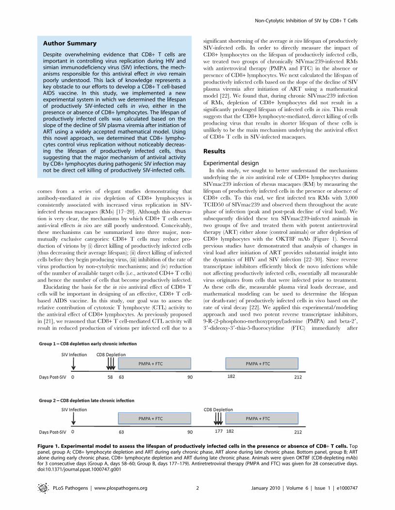
Figure 1. Experimental model to assess the lifespan of productively infected cells in the presence or absence of CD8+ T cells. Toppanel, group A; CD8+ lymphocyte depletion and ART during early chronic phase, ART alone during late chronic phase. Bottom panel, group B; ARTalone during early chronic phase, CD8+ lymphocyte depletion and ART during late chronic phase. Animals were given OKT8F (CD8-depleting mAb)for 3 consecutive days (Group A, days 58–60; Group B, days 177–179). Antiretretroviral therapy (PMPA and FTC) was given for 28 consecutive days.doi:10.1371/journal.ppat.1000747.g001
Non-Cytolytic Inhibition of SIV by CD8+ T Cells
PLoS Pathogens | www.plospathogens.org 2 January 2010 | Volume 6 | Issue 1 | e1000747

CD8+ lymphocyte depletion (or alone in the control animals). All
RMs were given a cycle of ART for 28 days in two occasions:
during early and late chronic infection. In the early chronic phase
of infection (day 57) group A RMs (n = 5) were depleted of CD8+lymphocytes immediately prior to treatment with ART, while
group B animals (control, n = 5) were treated with ART alone.
Conversely, during the late chronic phase of infection (day 177),
group B animals (n = 4) were CD8+ lymphocyte depleted prior to
ART, while group A animals (control, n = 3) was treated with
ART alone. Note that the reduction in the number of animals per
group during the late phase of our experiment was due to the fact
that two RMs in group A and one in group B were sacrificed after
the first cycle of ART because of severe weight loss and possible
signs of simian AIDS. In all cases, the lifespan of productively
infected cells in vivo (in the presence and absence of CD8+ T cells)
was estimated using a mathematical model that allows the
calculation of the lifespan of short and long-lived productively
infected cells [22]. In addition, we analyzed the viral load decline
data with a linear mixed effects model, where only the slope of the
first phase of viral decline was estimated directly by a linear
regression procedure.
Treatment with OKT8F results in increased plasmaviremia
As expected based on our previous experience with the use
of the OKT8F monoclonal antibody [31] (Engram, J. C. and
Silvestri G., unpublished observations), all RMs treated with this
antibody showed a very rapid and near complete depletion of
CD8+ lymphocytes from both peripheral and mucosal tissues.
During the first phase of this experiment (early chronic infection,
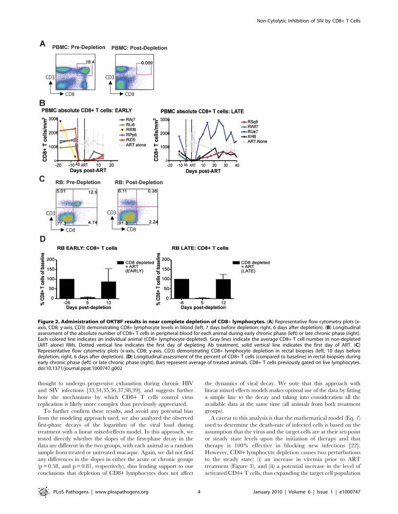
i.e., day 57 post-inoculation), in group A animals, CD8+ T cells
were depleted by an average of 99.97% (60.01 s.d.) in peripheral
blood (Figure 2A, 2B), 99.29% (60.50 s.d.) in rectal biopsies
(Figure 2C, 2D), and 99.23% (61.07 s.d.) in bronchoalveolar
lavage (data not shown) as measured by flow cytometry. During
the second phase of this experiment (late chronic infection, i.e.,
day 177 post inoculation), in group B animals, CD8+ T cells were
depleted by an average of 99.95% (60.03 s.d.) in peripheral blood
(Figure 2B) and 98.07% (61.54 s.d.) in rectal biopsies (Figure 2D)
as measured by flow cytometry. Extent of depletion in mucosal
tissues was corrected for non-CD8+ T cell fluctuations (as
described in [32]). In all cases, and consistent with previous
studies [18], CD8+ T cells were depleted very rapidly (.98%
depletion after 24 hours) and CD8+ T cell depletion was sustained
for 8–13 days with nadir depletion occurring between 5-6 days
after the first infusion. Of note, the OKT8F Ab induced a loss
of both CD3+CD8+ cells as well as CD32CD8+ cells, thus
indicating that not only CD8+ T cells, but also NK cells, NKT
cells, and TCRcd T cells that express CD8 are also efficiently
depleted in this experimental setting. Similar to previous studies
in which CD8+ T cells were depleted during pathogenic SIV
infection [17,18,19,20], we observed an increase in viremia
between 0.7–2.2 logs (Figure 3).
Suppression of virus replication after treatment withPMPA and FTC
In this study, antiretroviral therapy with PMPA and FTC
(30mg/kg/day i.m. for each drug, for a total of 28 days of
treatment) was conducted during both early and late phases of the
study (i.e., starting at days 63 and 182 post-SIV infection,
respectively). In the CD8+ lymphocyte-depleted animals, this
timing corresponded to initiation of ART three days after the last
OKT8F infusion. As expected, during both phases of the study,
ART effectively suppressed virus replication in all RMs by at least
0.5 log10 (and in 16 out of 17 instances of treatment by at least 1.5
log10) within a week after initiation of therapy (Figure 4). The
observation that ART induced a rapid and dramatic suppression
of SIV replication allowed us to proceed to the next phase of the
study in which the kinetics of decline of plasma viremia were used
to calculate the lifespan of cells producing virions in vivo.
CD8+ lymphocyte depletion does not prolong thelifespan of SIV-infected cells in vivo
Previous studies [22–24,26] demonstrated that there are two
phases of viral decay; an initial rapid, exponential decline of 1–2
logs, in which productively infected short-lived cells are lost,
followed by the second phase, which is characterized by a slower
decline, where long-lived infected cells are lost. In order to
quantify the contribution of CD8+ T cells to the lifespan of
productively infected cells in vivo, we quantified this parameter in
the presence or absence of CD8+ T cells by analyzing the viral
decline after initiation of ART using the equation:
V tð Þ~V0 A exp {dtð ÞzC exp {mtð Þz 1{A{Cð Þexp {ctð Þð Þ ð1Þ
where A = (NkTo)/(c2d), C = (c2NkTo)/(c2m) and V0 is the initial
viral load, k is the infection rate, N is the viral burst size, d is the
death rate of short-lived productively infected cells, m is the death
rate of long-lived productively infected cells, and c is the rate of
virion clearance [22] (Figure 4). By fitting the natural logarithm of
V(t) given by equation (1) to the natural logarithm of the measured
SIV RNA between initiation and termination of therapy, we were
able to estimate d and m, the death-rate of short-lived and long-
lived productively infected cells, for each animal either in the
presence or absence of CD8+ T cells (Table S1). However, for
macaques RAj7, RMm6 and RPp6 in early infection, we could
not fit a second phase decline due to too few data points or a flat
second phase. Thus for these animals we used a monophasic decline
model [22,24], appropriate for reverse transcription inhibitor
therapy, by making C = 0 in (Eq. 1), i.e., setting NKT0 = c, and
estimated d but not m.
During both the early and late phases of this study, we found
that the lifespan of short-lived productively infected cells (1/d) is
similar regardless of the presence or absence of CD8+ T cells.
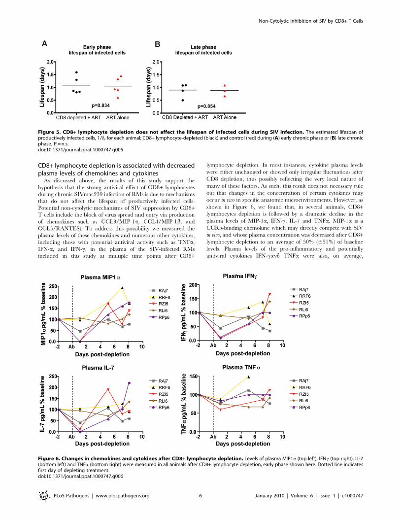
Specifically, during the early phase, the mean lifespan for group A
(CD8+ lymphocyte depleted RMs) was 1.11 (60.39 s.d.) days
(median = 0.87), while the mean lifespan for group B (control non-
CD8+ lymphocyte depleted animals) was 1.05 (60.35 s.d.) days
(median = 0.93) (p = 0.83) (Figure 5A, Table S1). During the late
phase, the mean lifespan for group A (control non-CD8+lymphocyte depleted) was 0.87 (60.21 s.d.) days (median = 0.86),
while the mean lifespan for group B (CD8+ lymphocyte depleted)
was 0.89 (60.28 s.d.) days (median = 0.98) (p = 0.85) (Figure 5B,
Table S1). These data indicate that the depletion of CD8+lymphocytes does not prolong the lifespan of short-lived
productively infected cells in vivo during pathogenic SIV infection
of RMs. Similarly, the estimated lifespans of long-lived infected
cells (1/m) were also not different between CD8+ lymphocyte
depleted and not depleted animals (9.7610.7 vs. 8.565.6 days,
respectively, p = 0.71). A notable observation, however, is that of
the seven RMs that participated in both the early and late phase
studies, five had a shorter lifespan of short-lived infected cells (by
an average of 36%) later in infection. This finding, together with
the slightly higher increase in viremia that we observed after
CD8+ lymphocyte depletion in the late phase as compared to the
early phase (Figure 3) is somewhat unexpected as CD8+ T cells are
Non-Cytolytic Inhibition of SIV by CD8+ T Cells
PLoS Pathogens | www.plospathogens.org 3 January 2010 | Volume 6 | Issue 1 | e1000747
thought to undergo progressive exhaustion during chronic HIV
and SIV infections [33,34,35,36,37,38,39], and suggests further
how the mechanisms by which CD8+ T cells control virus
replication is likely more complex than previously appreciated.
To further confirm these results, and avoid any potential bias
from the modeling approach used, we also analyzed the observed
first-phase decays of the logarithm of the viral load during
treatment with a linear mixed-effects model. In this approach, we
tested directly whether the slopes of the first-phase decay in the
data are different in the two groups, with each animal as a random
sample from treated or untreated macaque. Again, we did not find
any differences in the slopes in either the acute or chronic groups
(p = 0.58, and p = 0.81, respectively), thus lending support to our
conclusions that depletion of CD8+ lymphocytes does not affect
the dynamics of viral decay. We note that this approach with
linear mixed effects models makes optimal use of the data by fitting
a simple line to the decay and taking into consideration all the
available data at the same time (all animals from both treatment
groups).
A caveat to this analysis is that the mathematical model (Eq. 1)
used to determine the death-rate of infected cells is based on the
assumption that the virus and the target cells are at their set-point
or steady state levels upon the initiation of therapy and that
therapy is 100% effective in blocking new infections [22].
However, CD8+ lymphocyte depletion causes two perturbations
to the steady state: (i) an increase in viremia prior to ART
treatment (Figure 3), and (ii) a potential increase in the level of
activated CD4+ T cells, thus expanding the target cell population
Figure 2. Administration of OKT8F results in near complete depletion of CD8+ lymphocytes. (A) Representative flow cytometry plots (x-axis, CD8; y-axis, CD3) demonstrating CD8+ lymphocyte levels in blood (left, 7 days before depletion; right, 6 days after depletion). (B) Longitudinalassessment of the absolute number of CD8+ T cells in peripheral blood for each animal during early chronic phase (left) or late chronic phase (right).Each colored line indicates an individual animal (CD8+ lymphocyte-depleted). Gray lines indicate the average CD8+ T cell number in non-depleted(ART alone) RMs. Dotted vertical line indicates the first day of depleting Ab treatment, solid vertical line indicates the first day of ART. (C)Representative flow cytometry plots (x-axis, CD8; y-axis, CD3) demonstrating CD8+ lymphocyte depletion in rectal biopsies (left, 10 days beforedepletion; right, 6 days after depletion). (D) Longitudinal assessment of the percent of CD8+ T cells (compared to baseline) in rectal biopsies duringearly chronic phase (left) or late chronic phase (right). Bars represent average of treated animals. CD8+ T cells previously gated on live lymphocytes.doi:10.1371/journal.ppat.1000747.g002
Non-Cytolytic Inhibition of SIV by CD8+ T Cells
PLoS Pathogens | www.plospathogens.org 4 January 2010 | Volume 6 | Issue 1 | e1000747

for virus replication. First, to determine the effect of changes in
viremia after CD8+ lymphocyte depletion, surrogate data for
SIV kinetics with virus not in steady state were created by
equation (2) (in Text S1) with a known value of d and then fit
using equation (1) to assess if, and to what extent, viral load
increases before the start of therapy altered estimated d values
(Text S1, Figure S1). Second, to take into account the possibility
that significant changes in the activation state of CD4+ T cells
occurs after CD8+ lymphocyte depletion, we created surrogate
data that include changes in target cells (Text S2, Figure S2).
Third, the above analyses were repeated with various drug
effectiveness less than 100% to study the influence of this factor
on our estimate of d (Text S2). All three analyses demonstrated
that errors due to lack of steady-state viremia, to changes in target
cell pools after CD8+ lymphocyte depletion as well as to drug
effectiveness ,100% lead to a potential underestimation of both
d and m (Text S1 and S2). Further, when the drug effectiveness
was high, i.e. 99%, the maximum error in estimating d and m was
,3.5%. This analysis shows that the actual values of d and m in
systems with CD8+ lymphocyte depletion may be even higher
than we estimate, thus supporting our conclusion that lack of
CD8+ T cells does not increase the lifespan of productively
infected cells.
A conceivable conceptual limitation of our experimental system
is that antiretroviral treatment might have an immediate impact
on the number and/or function of SIV-specific CD8+ T cells, thus
introducing a potential bias in our effort to assess the impact of
CTL activity on the lifespan of infected cells based on the decline
of viremia after ART. To directly address this issue, we measured
the magnitude and functionality of SIV-specific CD8+ T cells
before and after ART in non-CD8+ lymphocyte depleted animals
and found that ART did not cause any significant changes in SIV-
specific CD8+ T cell responses during either the early or late phase
of the study (data not shown), therefore not supporting the
possibility that the use of ART generated an intrinsic bias in our
assessment of the impact of CD8+ lymphocytes on the lifespan of
SIV-infected cells.
Figure 3. CD8+ lymphocyte depletion results in a 0.7–2.2 log10
rise in viral load. Change of viral load from baseline for eachindividual animal after CD8 depletion, during early chronic phase (whitebars, left) or late chronic phase (black bars, right).doi:10.1371/journal.ppat.1000747.g003
Figure 4. Treatment with PMPA and FTC effectively suppresses virus replication in SIVmac239-infected RMs. (A, B) Plasma viral load(log10) measured longitudinally for each individual animal (black lines, CD8+ lymphocyte-depleted, red lines, control) during (A) early chronic phaseor (B) late chronic phase. (C,D) Average plasma viral load (log10) for each group (black, CD8+ lymphocyte-depleted; red, control) during (C) earlychronic phase or (D) late chronic phase. Error bars represent standard deviation. Dotted vertical line indicates the first day of depleting Ab treatment,solid vertical line indicates the first day of ART.doi:10.1371/journal.ppat.1000747.g004
Non-Cytolytic Inhibition of SIV by CD8+ T Cells
PLoS Pathogens | www.plospathogens.org 5 January 2010 | Volume 6 | Issue 1 | e1000747
CD8+ lymphocyte depletion is associated with decreasedplasma levels of chemokines and cytokines
As discussed above, the results of this study support the
hypothesis that the strong antiviral effect of CD8+ lymphocytes
during chronic SIVmac239 infection of RMs is due to mechanisms
that do not affect the lifespan of productively infected cells.
Potential non-cytolytic mechanisms of SIV suppression by CD8+T cells include the block of virus spread and entry via production
of chemokines such as CCL3/MIP-1a, CCL4/MIP-1b, and
CCL5/RANTES). To address this possibility we measured the
plasma levels of these chemokines and numerous other cytokines,
including those with potential antiviral activity such as TNFa,
IFN-a, and IFN-c, in the plasma of the SIV-infected RMs
included in this study at multiple time points after CD8+
lymphocyte depletion. In most instances, cytokine plasma levels
were either unchanged or showed only irregular fluctuations after
CD8 depletion, thus possibly reflecting the very local nature of
many of these factors. As such, this result does not necessary rule
out that changes in the concentration of certain cytokines may
occur in vivo in specific anatomic microenvironments. However, as
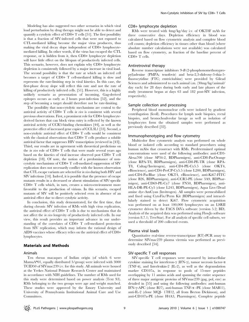
shown in Figure 6, we found that, in several animals, CD8+lymphocytes depletion is followed by a dramatic decline in the
plasma levels of MIP-1a, IFN-c, IL-7 and TNFa. MIP-1a is a
CCR5-binding chemokine which may directly compete with SIV
in vivo, and whose plasma concentration was decreased after CD8+lymphocyte depletion to an average of 50% (651%) of baseline
levels. Plasma levels of the pro-inflammatory and potentially
antiviral cytokines IFN-cand TNFa were also, on average,
Figure 5. CD8+ lymphocyte depletion does not affect the lifespan of infected cells during SIV infection. The estimated lifespan ofproductively infected cells, 1/d, for each animal; CD8+ lymphocyte-depleted (black) and control (red) during (A) early chronic phase or (B) late chronicphase. P = n.s.doi:10.1371/journal.ppat.1000747.g005
Figure 6. Changes in chemokines and cytokines after CD8+ lymphocyte depletion. Levels of plasma MIP1a (top left), IFNc (top right), IL-7(bottom left) and TNFa (bottom right) were measured in all animals after CD8+ lymphocyte depletion, early phase shown here. Dotted line indicatesfirst day of depleting treatment.doi:10.1371/journal.ppat.1000747.g006
Non-Cytolytic Inhibition of SIV by CD8+ T Cells
PLoS Pathogens | www.plospathogens.org 6 January 2010 | Volume 6 | Issue 1 | e1000747

reduced to 49% (640%) and to 76% (610%) of baseline levels,
respectively, after CD8+ lymphocyte depletion. Plasma concen-
trations of the lympho-tropic cytokine IL-7 were also decreased to
51% (648%) of baseline levels after CD8+ lymphocyte depletion.
As all of these cytokines may have an important antiviral effect
during SIV infection, lower levels of these molecules after CD8+lymphocyte depletion may contribute to the observed rise in
viremia. While these data are not conclusive, they suggest that
soluble factors produced by CD8+ lymphocytes may play a key
role in the suppression of virus replication mediated by these cells
in SIV-infected RMs.
Effects of CD8+ lymphocyte depletion on CD4+ T cellactivation
The finding that CD8+ lymphocyte depletion does not result in
a prolonged lifespan of productively infected cells is also consistent
with the possibility that the observed increase in virus replication is
caused, at least in part, by increased CD4+ T cell activation, which
would result in an increased availability of target cells for SIV
infection. Several factors may be involved in this CD4+ T cell
activation, including homeostatic responses to lymphopenia,
increased availability of CD4+ T cell tropic and/or pro-inflam-
matory cytokines, reactivation of latent virus infections, and other
potential changes in the lymphoid microenvironment(s). To
address this possibility, we measured the expression of activation
and proliferation markers in CD4+ T cells before and after CD8+lymphocyte depletion. As shown in Figure 7, we found that
CD8+ lymphocyte depletion was followed by a marked increase in
CD4+ T cell activation that occurred in all examined tissues. In
peripheral blood, the peak of CD4+ T cell activation occurred at
day 15 post-depletion, and most activation did not increase at
all until day 8. On average at peak activation, the fraction of
CD4+Ki67+ T cells was 6.7 fold higher than baseline levels, the
fraction of CD4+CCR5+ T cells was 6.2 fold higher than baseline,
the fraction of CD4+HLA-DR+ T cells was 19.2 fold higher than
baseline, and the fraction of CD4+CD69+ T cells was 10.6 fold
higher than baseline levels (Figure 7A). The kinetics of CD4+ T
cell activation was also delayed in mucosal tissues, although it
should be noted that the relative infrequent sampling schedule
raises the possibility that we missed the peak of CD4+ T cell
Figure 7. CD8+ lymphocyte depletion results in a rise in activated CD4+ T cells. (A) Longitudinal assessment (individual animals from CD8-depleted group and mean and s.d. from control group) of the percent of CD4+CCR5+ (top left), CD4+Ki67+ (top right), CD4+HLA-DR+ (bottom left),and CD4+CD69+ (bottom right) T cells during early chronic infection. (B) Longitudinal assessment of the mean (and s.d.) percent of CD4+Ki67+ T cellsin rectal biopsies (left) and bronchoalveolar lavage (right).doi:10.1371/journal.ppat.1000747.g007
Non-Cytolytic Inhibition of SIV by CD8+ T Cells
PLoS Pathogens | www.plospathogens.org 7 January 2010 | Volume 6 | Issue 1 | e1000747

activation in these tissues. In rectal biopsies, during early chronic
infection, the fraction of CD4+Ki67+ T cells was 1.4 fold higher
than baseline levels at day 6 post-depletion and 1.8 fold higher
than baseline levels at day 13 post-depletion (Figure 7B, left).
Similarly, during late chronic infection, CD4+Ki67+ T cells were
0.9 fold higher than baseline at day 5 post-depletion, and 1.2 fold
higher than baseline levels at day 12 post-depletion (data not
shown). The same trend was observed in bronchoalveolar lavage,
where CD4+Ki67+ T cells were 0.7 fold higher than baseline at
day 6 post-depletion, and 1.5 fold higher at day 13 post-depletion
(Figure 7B, right). Importantly, the observed changes in CD4+ T
cell activation followed, rather than preceded, the increase in
plasma viral load, thus suggesting that the activation of CD4+ T
cells that occurs after CD8+ lymphocyte depletion is unlikely to be
the predominant source of the increased viremia. As such, these
data support a model in which CD8+ T cells play a key, direct role
in maintaining the steady state of viral load during chronic SIV
infection.
Discussion
Numerous studies indicate that CD8+ lymphocytes play an
important role in suppressing virus replication in vivo during
pathogenic HIV and SIV infections [11,13–17]. However, the
mechanisms underlying this activity are still poorly understood and
may involve several non-mutually exclusive factors, whose relative
contribution to the net in vivo antiviral effect of CD8+ lymphocytes
is unknown. Direct killing of productively HIV- or SIV-infected
cells by CD8+ T cells (i.e., CTL activity) has been shown in many
in vitro settings and is very likely to occur in vivo as well [40]. In
addition, suppression of HIV replication by CD8+ T cells via
non-cytolytic mechanisms that inhibit virus transcription was first
observed by Levy and colleagues in 1986 [11,41,42], although the
nature of this antiviral activity has not been fully elucidated [43].
Furthermore, CD8+ T cells may block HIV/SIV spread from cell-
to-cell by releasing factors such as CCR5-binding chemokines (i.e.,
MIP-1a/CCL3, MIP-1b/CCL4, and RANTES/CCL5) that act
as competitive inhibitors of CCR5-mediated virus entry [44,45].
Finally, it is conceivable that the increased HIV/SIV replication
observed after CD8+ lymphocyte depletion is caused, at least in
part, by changes in the activation state of CD4+ T cells that render
these cells more intrinsically ‘‘permissive’’ to virus replication [31].
In this study, we set to address the relative contribution of
cytolytic vs. non-cytolytic mechanisms of CD8+ lymphocyte-
mediated control of virus replication by measuring the in vivo
lifespan of productively infected cells during chronic SIVmac239
infection of RMs in the presence or absence of CD8+lymphocytes. The assessment of the turnover of infected cells
was conducted using a well-characterized mathematical model
that is based on the analysis of the decline of viral load after
initiation of antiretroviral therapy [22]. Of note, the experimental
design of this study (Figure 1) is based on the premise that SIV-
specific CTL activity will ostensibly reduce virus replication by
shortening the average in vivo lifespan of productively infected cells
[21]. To the best of our knowledge, this is the first direct
assessment of the impact of CD8+ T cells on the longevity of
productively SIV-infected cells in vivo.
Perhaps surprisingly, the results of this experiment indicate that
CD8+ T cells do not affect the lifespan of productively infected
cells during SIVmac239 infection of rhesus macaques. Our
experiments further confirmed the important role of CD8+ T
cells in antiviral immunity since, in all circumstances, the in vivo
depletion of CD8+ lymphocytes is associated with a marked
and consistent increase in viral load (Figure 3). However, our
experiments do challenge the common assumption that the main
antiviral effect of CD8+ T cells is related to the direct killing of
productively infected CD4+ T cells (i.e., CTL activity) that
suppresses virus replication by reducing the amount of time in
which infected cells are able to produce virions (Figure 5). Instead,
these results indicate that non-cytolytic mechanisms of SIV
inhibition are potentially involved, or that CD8+ T cells have
cytolytic affects prior to productive virus replication.
While CD8+ T cell-mediated CTL activity may play a key role
in killing infected cells before they start producing virus and/or
in ‘‘elite controller’’ SIV-infected RMs with very low viremia, this
study is consistent with a model wherein, during chronic
SIVmac239 infection of RMs with high viremia, the main
antiviral effect of CD8+ lymphocytes may be due to non-cytolytic
mechanisms that do not impact the average lifespan of infected
cells. These non-cytolytic mechanisms may include the inhibition
of SIV production by factors acting at the level of virus
transcriptions and/or the block of virus spread and entry via
production of chemokines and cytokines. This latter possibility is
supported by our observation that, in several animals, CD8+lymphocyte depletion is followed by a dramatic decline of the
plasma levels of molecules such as the CCR5-binding chemokine
MIP-1a, the antiviral cytokine IFN-c, the pro-inflammatory
cytokine TNFa and the homeostatic cytokine IL-7 (Figure 6).
The finding that the absence of CD8+ T cells does not noticeably
increase the lifespan of productively infected cells is also consistent
with the possibility that the increase in viral load after CD8+lymphocyte depletion is caused by increased availability of
activated CD4+ T cells as targets for virus replication. In this
experiment, CD8+ lymphocyte depletion was indeed followed by a
marked increase in CD4+ T cell activation that occurred in all
examined tissues (Figure 7). However, the observed changes in
CD4+ T cell activation followed, rather than preceded, the
increase in viremia, thus suggesting that activation of CD4+ T cells
after CD8+ lymphocyte depletion is unlikely to be the main
mechanism for the increase in viremia. Further studies in which
the level of CD4+ T cell activation following CD8+ lymphocyte
depletion is examined at earlier time points and in more tissues
may be needed to better assess how changes in CD4+ T cell
activation may contribute to the increase in virus replication in
SIV-infected, CD8+ lymphocyte depleted RMs.
One caveat of this study is that the OKT8F depleting Ab also
depletes NK cells, NKT cells, and TCRcd T cells that express the
CD8 molecule. NK cells and NKT cells are known to have an
antiviral role during HIV infection, including the production of
proinflammatory cytokines and chemokines which drive a Th1
antiviral immune response [46–49]. TCRcd T cells also may play a
role in antiviral immunity during SIV/HIV infection, as these cells
have a specific role in the recognition of microbial pathogens and
produce both Th1 and Th2 cytokines that can influence the adaptive
immune response after infection [50]. Therefore, loss of any of these
cell types may influence viremia after CD8+ lymphocyte depletion.
However, these considerations do not change the conclusion that
removal of CD8+ lymphocytes does not affect the lifespan of cells
productively infected with SIV. As mentioned above, an additional
caveat to this study is that, while it is clear that CD8+ lymphocyte
depletion does not prolong the lifespan of productively SIV-infected
cells in vivo, it remains possible that CD8+ lymphocytes exert their
antiviral effect by killing infected cells before they start producing
new virions. In this case, this cytolytic antiviral effect would not
translate into a net change of the average lifespan of productively
infected cells. Further investigation will be required to quantify the in
vivo impact of this putative antiviral effect, and to compare it with the
impact of non-cytolytic mechanisms of SIV suppression.
Non-Cytolytic Inhibition of SIV by CD8+ T Cells
PLoS Pathogens | www.plospathogens.org 8 January 2010 | Volume 6 | Issue 1 | e1000747
Modeling has also suggested two other scenarios in which viral
load perturbation by drug therapy might not be able to detect and
quantify a cytolytic effect of CD8+ T cells [51]. The first possibility
is that a fraction of SIV-infected cells that were not exposed to
CTL-mediated killing become the major virus producers, thus
making the viral decay slope independent of CD8+ lymphocyte-
mediated killing. In other words, if the virus has escaped the CTL
response, or is hidden from it, then CD8+ lymphocyte depletion
will have little effect on the lifespan of productively infected cells.
This scenario, however, does not explain why CD8+ lymphocyte
depletion is consistently followed by a major increase in viral load.
The second possibility is that the rate at which an infected cell
becomes a target of CD8+ T cell-mediated killing is slow and
represents the rate-limiting step in viral kinetics. In this case, the
first-phase decay slope will reflect this rate and not the rate of
killing of productively infected cells. [51]. However, this is a highly
unlikely scenario as presentation of incoming, Gag-derived
epitopes occurs as early as 2 hours post-infection [52], and the
step of becoming a target should therefore not be rate-limiting.
The possibility that non-cytolytic mechanisms are central to the
antiviral activity of CD8+ T cells in vivo is consistent with several
previous observations. First, a prominent role for CD8+ lymphocyte-
derived factors that can block virus entry is reflected by the known
antiviral activity of CCR5-binding chemokines [44], as well as the
protective effect of increased gene copies of CCL3L1 [53]. Second, a
non-cytolytic antiviral effect of CD8+ T cells would be consistent
with the classical observations that CD8+ T cells produce a soluble
antiviral factor that suppresses HIV transcription (reviewed in [43]).
Third, our results are in agreement with theoretical predictions on
the in vivo role of CD8+ T cells that were made several years ago
based on the kinetics of viral increase observed post CD8+ T cell
depletion [18]. Of note, the notion of a predominance of non-
cytolytic mechanisms of CD8+ T cell-mediated suppression of SIV
replication does not necessarily conflict with the known observation
that CTL escape variants are selected for in vivo during both HIV and
SIV infections [14]. Indeed, it is possible that the presence of escape
mutants results in a decreased or absent stimulation of SIV-specific
CD8+ T cells which, in turn, creates a micro-environment more
favorable to the production of virions. In this scenario, escaped
mutants of SIV will be positively selected even in absence of an
antiviral effect due to direct cytolytic activity.
In conclusion, this study demonstrated, for the first time, that
during chronic SIV infection of RMs with high virus replication,
the antiviral effect of CD8+ T cells is due to mechanisms that do
not affect the in vivo longevity of productively infected cells. In our
view, this result provides an important advance in our under-
standing of the correlates of CD8+ T cell-mediated protection
from SIV replication, which may inform the rational design of
AIDS vaccines whose efficacy relies on the antiviral effect of CD8+lymphocytes.
Materials and Methods
AnimalsTen rhesus macaques of Indian origin (of which 6 were
MamuA*01, equally distributed 3/group) were infected with 3000
TCID50 of SIVmac239 i.v. for this study. All animals were housed
at the Yerkes National Primate Research Center and maintained
in accordance with NIH guidelines. The number of RMs used for
this study were determined based on power analysis (Text S3).
RMs belonging to the two groups were age and weight matched.
These studies were approved by the Emory University and
University of Pennsylvania Institutional Animal Care and Use
Committees.
CD8+ lymphocyte depletionRMs were treated with 4mg/kg/day i.v. of OKT8F mAb for
three consecutive days. Depletion efficiency in blood was
calculated based on flow cytometric analysis and complete blood
cell counts; depletion efficiency in tissues other than blood (where
absolute number calculations were not available) was calculated
based on flow cytometry, as fraction of the baseline percent of
CD8+ T cells.
Antiretroviral therapyReverse transcriptase inhibitors 9-R-(2-phosphonomethoxypro-
pyl)adenine (PMPA; tenofovir) and beta-2,3-dideoxy-3-thia-5-
fluorocytidine (FTC; emtricitabine) were provided by Gilead
Sciences and administered to each animal i.m. (30mg/kg/animal/
day each) for 28 days during both early and late phases of the
study (treatment began at days 63 and 182 post-SIV infection,
respectively).
Sample collection and processingPeripheral blood mononuclear cells were isolated by gradient
centrifugation (ficoll). Procedures for lymph node biopsies, rectal
biopsies, and bronochoalveolar lavage as well as isolation of
lymphocytes form the obtained samples were performed as
previously described [32].
Immunophenotyping and flow cytometryMulticolor flow cytometric analysis was performed on whole
blood or isolated cells according to standard procedures using
human mAbs that crossreact with RMs. Predetermined optimal
concentrations were used of the following antibodies: anti-CD3-
Alexa700 (clone SP34-2, BDPharmigen), anti-CD8-PacOrange
(clone RPA-T8, BDPharmigen), anti-CD8-PE-TR (clone RPA-
T8, Caltag/Invitrogen), anti-CD4-PE-Cy5.5 (clone OKT4,
eBioscience), anti-CD4-PerCP-Cy5.5 (clone L200, BDPharmigen),
anti-CD4-PacBlue (clone OKT4, eBioscience), anti-Ki67-FITC
(clone B26, BDPharmigen), anti-CCR5-PE (clone 3A9, BDPhar-
migen), anti-CD69-PE-Cy7 (clone FN50, BDPharmigen), anti-
HLA-DR-PE-Cy5 (clone L243, BDPharmigen), Aqua Live/Dead
amine dye-AmCyan (Invitrogen). All samples were permeabilized
and fixed using CytoFix/Perm Kit (BDPharmigen) and intracel-
lularly stained to detect Ki67. Flow cytometric acquisition
was performed on at least 100,000 lymphocytes on an LSRII
cytometer driven by the FACS DiVa software (version 5.2; BD).
Analysis of the acquired data was performed using FlowJo software
(version 8.7.1; TreeStar). For all analysis of specific cell subsets, we
used a threshold of 200 collected events.
Plasma viral loadsQuantitative real-time reverse-transcriptase (RT)-PCR assay to
determine SIVmac239 plasma viremia was performed as previ-
ously described [54].
SIV-specific T cell responsesSIV-specific T cell responses were measured by intracellular
cytokine staining for interferon-c (IFN-c), tumor necrosis factor-a(TNF-a), and Interleukin-2 (IL-2), as well as the degranulation
marker CD107a, in response to pools of 15-mer peptides
overlapping by 11 amino acids and spanning the entire sequence
of three major antigenic proteins of SIVmac239 (gag, pol, env) as
detailed in [55] and using the following antibodies: anti-human
IFN-c-APC (clone B27), anti-human TNF-a -PE (clone MAB11),
anti-IL-2 (clone MQI- 17H12, all from Becton Dickinson), and
anti-CD107a-PE (clone H4A3, Pharmingen). Complete peptide
Non-Cytolytic Inhibition of SIV by CD8+ T Cells
PLoS Pathogens | www.plospathogens.org 9 January 2010 | Volume 6 | Issue 1 | e1000747
sets for SIVmac239 were obtained from the NIH AIDS Research
& Reference Reagent Program. In all experiments at least 200,000
T cells were acquired and analyzed.
Plasma levels of chemokines and cytokinesPlasma levels of the beta-chemokines CCL3/MIP-1a, CCL4/
MIP-1b, and CCL5/RANTES in conjunction with other
cytokines and chemokines were measured using a sandwich
immunoassay-based protein array system, the human cytokine 25-
Plex (BioSource International), as instructed by the manufacturer
and then read by the Bio-Plex array reader (Bio-Rad Laborato-
ries), which uses fluorescent bead-based technology from Luminex.
Supporting Information
Text S1 Supplementary figure legends, text, and references.
Found at: doi:10.1371/journal.ppat.1000747.s001 (0.05 MB
DOC)
Table S1 Values for d and m for each animal, including lower
and upper 95% confidence intervals. Values for d (half-life of
short-lived cells) and m (half-life of long-lived cells) were estimated
based on Eq. 1. 95% confidence intervals were calculated from
500 bootstrap replicates.
Found at: doi:10.1371/journal.ppat.1000747.s002 (0.02 MB XLS)
Figure S1 Effects of fitting the viral load data with a model that
assumes the viral load is in steady state, when in reality viral load is
increasing. Surrogate data for SIV kinetics with virus not in steady
state (black dots) was created using Eq. 2 (Text S1) with the rate
of virion production p allowed to increase as CD8 levels decline
in order to account for changes in viremia caused by CD8+lymphocyte depletion. This data was generated to agree with the
change in viremia observed for animal Rsq8. At t = 0, the model
assumes combination drug therapy begins with an effectiveness of
99%. The surrogate data was then fit with Eq. 1 and parameters
estimated. The best fitting solution is shown by the orange line.
The parameters estimated in this way were ,3.5% different than
the ‘‘true’’ parameters used to generate the data.
Found at: doi:10.1371/journal.ppat.1000747.s003 (1.69 MB TIF)
Figure S2 CD4+ T cell data used to estimate the change in
target cells after CD8+ lymphocyte depletion. Measured CD4+ T
cell values for Rsq8 in late chronic infection, (black line) and data
smoothed by using a 3 point moving average (purple line). The 3-
point moving average was then fit using linear regression to obtain
the parameters a and T0 used in the supplemental text to define
the T cell increase during CD8+ lymphocyte depletion. Analysis of
the surrogate SIV RNA data indicates that the effect of changes in
CD4+ T-cells and SIV RNA due to CD8+ lymphocyte depletion
has a negligible (,3.5%) effect on the estimates of d and m when
the drug effectiveness is high (,99%).
Found at: doi:10.1371/journal.ppat.1000747.s004 (1.97 MB TIF)
Acknowledgments
We would like to acknowledge Stephanie Ehnert, Elizabeth Strobert, and
all the animal care and veterinary staff at the Yerkes National Primate
Research Center, the Virology Core of the Emory Center for AIDS
Research (CFAR), the University of Pennsylvania Center for AIDS
Research (CFAR), the University of Pennsylvania Flow Cytometry and
Cell Sorting Core, and the NIH nonhuman primate reagent resource. The
OKT8F CD8 depleting mAb used in this study was kindly provided by Dr.
Robert Mittler, Emory University. The SIVmac239 used to infect the RMs
was provided by Dr. Louis Picker and Dr. Michael Axthelm, Oregon
Health and Science University.
Author Contributions
Conceived and designed the experiments: NRK JES GS. Performed the
experiments: NRK AMO JCE BL IP JDE CA. Analyzed the data: NRK
ES MP RMR ASP. Contributed reagents/materials/analysis tools: ES MP
BL MDM JE JES RMR ASP GS. Wrote the paper: NRK RMR ASP GS.
References
1. Garber DA, Silvestri G, Feinberg MB (2004) Prospects for an AIDS vaccine:three big questions, no easy answers. Lancet Infect Dis 4: 397–413.
2. Coordinating Committee of the Global HIV/AIDS Vaccine Enterprise (2005)
The Global HIV/AIDS Vaccine Enterprise: scientific strategic plan. PLoS Med2: e25. doi:10.1371/journal.pmed.0020025.
3. Walker BD, Burton DR (2008) Toward an AIDS vaccine. Science 320: 760–764.
4. Watkins DI, Burton DR, Kallas EG, Moore JP, Koff WC (2008) Nonhuman
primate models and the failure of the Merck HIV-1 vaccine in humans. Nat
Med 14: 617–621.
5. Pantophlet R, Burton DR (2006) GP120: target for neutralizing HIV-1
antibodies. Annu Rev Immunol 24: 739–769.
6. Hu SL, Stamatatos L (2007) Prospects of HIV Env modification as an approach
to HIV vaccine design. Curr HIV Res 5: 507–513.
7. Karlsson Hedestam GB, Fouchier RA, Phogat S, Burton DR, Sodroski J, et al.(2008) The challenges of eliciting neutralizing antibodies to HIV-1 and to
influenza virus. Nat Rev Microbiol 6: 143–155.
8. Letvin NL (2007) Correlates of immune protection and the development of ahuman immunodeficiency virus vaccine. Immunity 27: 366–369.
9. Miller JD, Masopust D, Wherry EJ, Kaech S, Silvestri G, et al. (2005)Differentiation of CD8 T cells in response to acute and chronic viral infections:
implications for HIV vaccine development. Curr Drug Targets Infect Disord 5:
121–129.
10. Harari A, Dutoit V, Cellerai C, Bart PA, Du Pasquier RA, et al. (2006)
Functional signatures of protective antiviral T-cell immunity in human virusinfections. Immunol Rev 211: 236–254.
11. Walker CM, Moody DJ, Stites DP, Levy JA (1986) CD8+ lymphocytes can
control HIV infection in vitro by suppressing virus replication. Science 234:1563–1566.
12. Kannagi M, Chalifoux LV, Lord CI, Letvin NL (1988) Suppression of simianimmunodeficiency virus replication in vitro by CD8+ lymphocytes. J Immunol
140: 2237–2242.
13. Brumme ZL, Harrigan PR (2006) The impact of human genetic variation onHIV disease in the era of HAART. AIDS Rev 8: 78–87.
14. Goulder PJ, Watkins DI (2004) HIV and SIV CTL escape: implications forvaccine design. Nat Rev Immunol 4: 630–640.
15. Koup RA, Safrit JT, Cao Y, Andrews CA, McLeod G, et al. (1994) Temporal
association of cellular immune responses with the initial control of viremia in
primary human immunodeficiency virus type 1 syndrome. J Virol 68:4650–4655.
16. Borrow P, Lewicki H, Hahn BH, Shaw GM, Oldstone MB (1994) Virus-specific
CD8+ cytotoxic T-lymphocyte activity associated with control of viremia inprimary human immunodeficiency virus type 1 infection. J Virol 68: 6103–6110.
17. Schmitz JE, Kuroda MJ, Santra S, Sasseville VG, Simon MA, et al. (1999)Control of viremia in simian immunodeficiency virus infection by CD8+lymphocytes. Science 283: 857–860.
18. Jin X, Bauer DE, Tuttleton SE, Lewin S, Gettie A, et al. (1999) Dramatic rise inplasma viremia after CD8(+) T cell depletion in simian immunodeficiency virus-
infected macaques. J Exp Med 189: 991–998.
19. Matano T, Shibata R, Siemon C, Connors M, Lane HC, et al. (1998)
Administration of an anti-CD8 monoclonal antibody interferes with theclearance of chimeric simian/human immunodeficiency virus during primary
infections of rhesus macaques. J Virol 72: 164–169.
20. Lifson JD, Rossio JL, Piatak M Jr, Parks T, Li L, et al. (2001) Role of CD8(+)
lymphocytes in control of simian immunodeficiency virus infection andresistance to rechallenge after transient early antiretroviral treatment. J Virol
75: 10187–10199.
21. Van Rompay KK, Singh RP, Pahar B, Sodora DL, Wingfield C, et al. (2004)CD8+-cell-mediated suppression of virulent simian immunodeficiency virus
during tenofovir treatment. J Virol 78: 5324–5337.
22. Perelson AS, Essunger P, Cao Y, Vesanen M, Hurley A, et al. (1997) Decay
characteristics of HIV-1-infected compartments during combination therapy.Nature 387: 188–191.
23. Ho DD, Neumann AU, Perelson AS, Chen W, Leonard JM, et al. (1995) Rapid
turnover of plasma virions and CD4 lymphocytes in HIV-1 infection. Nature
373: 123–126.
24. Wei X, Ghosh SK, Taylor ME, Johnson VA, Emini EA, et al. (1995) Viral dynamicsin human immunodeficiency virus type 1 infection. Nature 373: 117–122.
25. Nowak MA, Lloyd AL, Vasquez GM, Wiltrout TA, Wahl LM, et al. (1997) Viral
dynamics of primary viremia and antiretroviral therapy in simian immunode-
ficiency virus infection. J Virol 71: 7518–7525.
Non-Cytolytic Inhibition of SIV by CD8+ T Cells
PLoS Pathogens | www.plospathogens.org 10 January 2010 | Volume 6 | Issue 1 | e1000747
26. Gordon SN, Dunham RM, Engram JC, Estes J, Wang Z, et al. (2008) Short-
lived infected cells support virus replication in sooty mangabeys naturallyinfected with simian immunodeficiency virus: implications for AIDS pathogen-
esis. J Virol 82: 3725–3735.
27. Pandrea I, Ribeiro RM, Gautam R, Gaufin T, Pattison M, et al. (2008) Simianimmunodeficiency virus SIVagm dynamics in African green monkeys. J Virol
82: 3713–3724.28. Mittler JE, Markowitz M, Ho DD, Perelson AS (1999) Improved estimates for
HIV-1 clearance rate and intracellular delay. AIDS 13: 1415–1417.
29. Notermans DW, Goudsmit J, Danner SA, de Wolf F, Perelson AS, et al. (1998)Rate of HIV-1 decline following antiretroviral therapy is related to viral load at
baseline and drug regimen. AIDS 12: 1483–1490.30. Perelson AS, Neumann AU, Markowitz M, Leonard JM, Ho DD (1996) HIV-1
dynamics in vivo: virion clearance rate, infected cell life-span, and viralgeneration time. Science 271: 1582–1586.
31. Barry AP, Silvestri G, Safrit JT, Sumpter B, Kozyr N, et al. (2007) Depletion of
CD8+ Cells in Sooty Mangabey Monkeys Naturally Infected with SimianImmunodeficiency Virus Reveals Limited Role for Immune Control of Virus
Replication in a Natural Host Species. J Immunol 178: 8002–8012.32. Klatt NR, Villinger F, Bostik P, Gordon SN, Pereira L, et al. (2008) Availability
of activated CD4+ T cells dictates the level of viremia in naturally SIV-infected
sooty mangabeys. J Clin Invest 118: 2039–2049.33. Day CL, Kaufmann DE, Kiepiela P, Brown JA, Moodley ES, et al. (2006) PD-1
expression on HIV-specific T cells is associated with T-cell exhaustion anddisease progression. Nature 443: 350–354.
34. D’Souza M, Fontenot AP, Mack DG, Lozupone C, Dillon S, et al. (2007)Programmed death 1 expression on HIV-specific CD4+ T cells is driven by viral
replication and associated with T cell dysfunction. J Immunol 179: 1979–1987.
35. Petrovas C, Casazza JP, Brenchley JM, Price DA, Gostick E, et al. (2006) PD-1 isa regulator of virus-specific CD8+ T cell survival in HIV infection. J Exp Med
203: 2281–2292.36. Trautmann L, Janbazian L, Chomont N, Said EA, Gimmig S, et al. (2006)
Upregulation of PD-1 expression on HIV-specific CD8+ T cells leads to
reversible immune dysfunction. Nat Med 12: 1198–1202.37. Jones RB, Ndhlovu LC, Barbour JD, Sheth PM, Jha AR, et al. (2008) Tim-3
expression defines a novel population of dysfunctional T cells with highlyelevated frequencies in progressive HIV-1 infection. J Exp Med 205: 2763–2779.
38. Velu V, Kannanganat S, Ibegbu C, Chennareddi L, Villinger F, et al. (2007)Elevated expression levels of inhibitory receptor programmed death 1 on simian
immunodeficiency virus-specific CD8 T cells during chronic infection but not
after vaccination. J Virol 81: 5819–5828.39. Petrovas C, Price DA, Mattapallil J, Ambrozak DR, Geldmacher C, et al. (2007)
SIV-specific CD8+ T cells express high levels of PD1 and cytokines but haveimpaired proliferative capacity in acute and chronic SIVmac251 infection.
Blood 110: 928–936.
40. Tsubota H, Lord CI, Watkins DI, Morimoto C, Letvin NL (1989) A cytotoxic T
lymphocyte inhibits acquired immunodeficiency syndrome virus replication in
peripheral blood lymphocytes. J Exp Med 169: 1421–1434.
41. Mackewicz CE, Blackbourn DJ, Levy JA (1995) CD8+ T cells suppress human
immunodeficiency virus replication by inhibiting viral transcription. Proc Natl
Acad Sci U S A 92: 2308–2312.
42. Walker CM, Levy JA (1989) A diffusible lymphokine produced by CD8+ T
lymphocytes suppresses HIV replication. Immunology 66: 628–630.
43. Levy JA (2003) The search for the CD8+ cell anti-HIV factor (CAF). Trends
Immunol 24: 628–632.
44. Cocchi F, DeVico AL, Garzino-Demo A, Arya SK, Gallo RC, et al. (1995)
Identification of RANTES, MIP-1 alpha, and MIP-1 beta as the major HIV-
suppressive factors produced by CD8+ T cells. Science 270: 1811–1815.
45. Moore JP, Trkola A, Dragic T (1997) Co-receptors for HIV-1 entry. Curr Opin
Immunol 9: 551–562.
46. Alter G, Altfeld M (2009) NK cells in HIV-1 infection: evidence for their role in
the control of HIV-1 infection. J Intern Med 265: 29–42.
47. Alter G, Altfeld M (2006) NK cell function in HIV-1 infection. Curr Mol Med 6:
621–629.
48. Alter G, Malenfant JM, Delabre RM, Burgett NC, Yu XG, et al. (2004)
Increased natural killer cell activity in viremic HIV-1 infection. J Immunol 173:
5305–5311.
49. Fauci AS, Mavilio D, Kottilil S (2005) NK cells in HIV infection: paradigm for
protection or targets for ambush. Nat Rev Immunol 5: 835–843.
50. Kosub DA, Lehrman G, Milush JM, Zhou D, Chacko E, et al. (2008) Gamma/
Delta T-cell functional responses differ after pathogenic human immunodefi-
ciency virus and nonpathogenic simian immunodeficiency virus infections.
J Virol 82: 1155–1165.
51. Klenerman P, Phillips RE, Rinaldo CR, Wahl LM, Ogg G, et al. (1996)
Cytotoxic T lymphocytes and viral turnover in HIV type 1 infection. Proc Natl
Acad Sci U S A 93: 15323–15328.
52. Sacha JB, Chung C, Rakasz EG, Spencer SP, Jonas AK, et al. (2007) Gag-
specific CD8+ T lymphocytes recognize infected cells before AIDS-virus
integration and viral protein expression. J Immunol 178: 2746–2754.
53. Dolan MJ, Kulkarni H, Camargo JF, He W, Smith A, et al. (2007) CCL3L1 and
CCR5 influence cell-mediated immunity and affect HIV-AIDS pathogenesis via
viral entry-independent mechanisms. Nat Immunol 8: 1324–1336.
54. Garber DA, Silvestri G, Barry AP, Fedanov A, Kozyr N, et al. (2004) Blockade
of T cell costimulation reveals interrelated actions of CD4+ and CD8+ T cells in
control of SIV replication. J Clin Invest 113: 836–845.
55. Dunham R, Pagliardini P, Gordon S, Sumpter B, Engram J, et al. (2006) The
AIDS resistance of naturally SIV-infected sooty mangabeys is independent of
cellular immunity to the virus. Blood 108: 209–217.
Non-Cytolytic Inhibition of SIV by CD8+ T Cells
PLoS Pathogens | www.plospathogens.org 11 January 2010 | Volume 6 | Issue 1 | e1000747
Related Documents