UC Irvine UC Irvine Previously Published Works Title Casting the lungs In-situ. Permalink https://escholarship.org/uc/item/4wp7t79n Journal The Anatomical record, 177(2) ISSN 0003-276X Authors Phalen, RF Yeh, HC Raabe, OG et al. Publication Date 1973-10-01 DOI 10.1002/ar.1091770207 Copyright Information This work is made available under the terms of a Creative Commons Attribution License, availalbe at https://creativecommons.org/licenses/by/4.0/ Peer reviewed eScholarship.org Powered by the California Digital Library University of California

Welcome message from author
This document is posted to help you gain knowledge. Please leave a comment to let me know what you think about it! Share it to your friends and learn new things together.
Transcript

UC IrvineUC Irvine Previously Published Works
TitleCasting the lungs In-situ.
Permalinkhttps://escholarship.org/uc/item/4wp7t79n
JournalThe Anatomical record, 177(2)
ISSN0003-276X
AuthorsPhalen, RFYeh, HCRaabe, OGet al.
Publication Date1973-10-01
DOI10.1002/ar.1091770207
Copyright InformationThis work is made available under the terms of a Creative Commons Attribution License, availalbe at https://creativecommons.org/licenses/by/4.0/ Peer reviewed
eScholarship.org Powered by the California Digital LibraryUniversity of California

Casting the Lungs In-situ ROBERT F. PHALEN, HSU-CHI YEH, OTTO G . RAABE AND DAVID J. VELASQUEZ Inhalation Toxicology Research Institute, Lovelace Foundation for Medical Education and Research, 5200 Gibson Boulevard, SE Albuquerque, New Mexico 87108
ABSTRACT A method for producing flexible silicone rubber casts of the airways of the lungs in-situ is described. Casts are made to correspond to lung volumes occurring during normal breathing. The lung is prepared for casting by replacing the air within with COZ followed by filling with degassed physio- logical saline. The saline dissolves the COZ gas within the airways allowing for a bubble-free finished cast. Casting compound is then slowly injected through the trachea. The saline diffuses out of the lung and passes out of the thorax through several small slits in the thoracic wall. After the injection is completed, the cast lung is allowed to cure in-situ before it is removed and the tissue digested away. Finished casts have an overall shape corresponding closely to the shape of the thorax. Casts produced by this in-situ method appear to have more realistic geometrical relationships than those produced from excised lungs.
The accurate description of the ana- tomical details of respiratory airways is essential for producing satisfactory lung models for predicting particle deposition and localized dose patterns for inhaled ma- terials within the respiratory tract in man and other animals. Morphometric data currently available are incomplete and therefore inadequate for these purposes. Equations for predicting regional particle deposition within the lungs are commonly based on the inertial, gravitational and diffusional forces acting on inhaled par- ticles (Beeckmans, '65). The anatomical features considered to be of importance in particle deposition are airway segment shapes and sizes (usually diameters and lengths), the inclination of airways to the force of gravity, and the branching angles. Radii of curvature of airway segments and branch regions, airflow patterns, air mix- ing within the lungs and cyclic changes in structural relationships during breathing must also be considered in meaningful deposition calculations.
Flexible casts of the airways are useful for replicating anatomical detail. Two major criteria for evaluating such casts are fidelity to original airway anatomy and suitability of the cast for precise mor- phometry. This report presents a method
for producing acceptable flexible casts of the respiratory system by the injection of a casting material in-situ. This method a p pears to overcome some short-comings in- herent in making casts from excised lungs.
After an examination of several casting materials, including paraffin, latex, low melting point metals, and several varieties of silicone molding compounds, two sili- cone rubber industrial molding compounds were chosen. These two materials, de- scribed in table 1, produce strong, flexible casts with good representation of anatomi- cal detail of structures as small as 100 p. They also cure at room temperature and have very small shrinkages during curing.
The procedure for making lung casts in the thorax is outlined in figure 1. Before the cast is made, several measurements of the functional residual capacity and the tidal volume are taken if possible. These measurements are used to determine the
METHODS
Received Mar. 27, '73. Accepted May 24, '73. 1 Research conducted under an interagency agree-
ment between the National Institute of Environmental Health Sciences and the U. S. Atomic Energy Commis- sion under AEC Contract AT(29-2)-1013 and in ani- mal care facilities fully accredited by the American Association for Accreditation of Laboratory Animal Care.
ANAT. REC., 177: 255-264. 255

256 PHALEN ET AL.
TABLE 1 Physical pronerties of silicone rubber industrial molding compounds
Silastic E 1 RTV-700 2
Uncured properties color density viscosity
white white/green/red 1.12 gm/cmz 1.07 gm/cm3 1,200 poises 600 poises
Cured properties bensile strength 700 psi elongation at rupture 400% temp. stability 500" F
(upper limit)
(after 7 days) shrinkage 0.1%
600 r s i 400% -
about 1.2%
1 From Dow Corning Bulletin 61-008a, 1970, Midland, Michigan. 2 General Electric Product data sheet, Silicone Products Department, Waterford, New York.
PRE-CASTING PROCEUURE
CASTING PROCEDURE
F L U S H L U N G S
I
INTO LUNGS INJEC r CASTING MATERIAL
EXCISE LUNGS 7 POST-CASTING PROCEDURE
VOLUME
Fig. 1 Flow chlart for preparing a cast of the lung in-situ. *At this time, the animal dies.
injection volume and to evaluate the final cast fidelity.
The experimental animal is anesthetized with sodium pentobarbital and the trachea is intubated. The animal is then placed on its back and the tracheal tube connected to a CO, reservior. Breathing this gas kills the anesthetized animal and replaces 0, and N, in the lungs with COz. The COz is con- siderably more water soluble than 0% and N, and helps to produce a bubble-free cast. When casting the lungs of cadavers, as in the case of the human, the lungs are flushed with COz using a respirator pump.
The inside of the thorax is then vented to the atmosphere by making six or more small ventral-lateral incisions between the ribs and through the thoracic wall allow- ing the lungs to collapse. The space be- tween the lungs and parietal pleura is fUed with physiological saline that has been pre- viously degassed under vacuum. This pro- vides support for the lungs during the re- mainder of the casting procedure. In the case of a rodent, the entire body may be suspended in degassed saline.
Degassed saline is next slowly intro- duced into the lungs through the tracheal catheter using the pressure of a 5- to 10- cm column of saline. This pressure inflates the lungs and enhances the diffusion of saline out of the lungs which are perme- able to saline. Several (3 to 5) lung vol- umes of saline are infused, dissolving the CO, gas within. This saline infusion re- quires about 15 to 30 minutes. Fluid, dif- fusing from the lungs, flows out of the tho- racic incisions during the infusion. These incisions, on each side of the thorax, at the apex, mid-region and near the diaphragm, are situated so that fluid will not be trapped within the thorax and interfere with full inflation of the lungs. The animal has now been prepared to receive the casting ma- terial.
The casting material is thoroughly mixed with its catalyst using an electric stirrer and is then degassed under vacuum. A syringe-type injection system (fig. 2) is used to slowly inject catalyzed silicone rubber into the lungs through the tracheal catheter. Syringes with volumes of 50 cM", 4 1 and 10 1 are used for injecting mdents,

CASTING THE LUNGS IN-SITU
Fig. 2 Air-driven syringe (diameter, 4 inches) used for the metered dispensing of sili- cone rubber mold-making materials.
257

258 PHALEN ET AL.
CASTING THE LUNGS rN-srTu
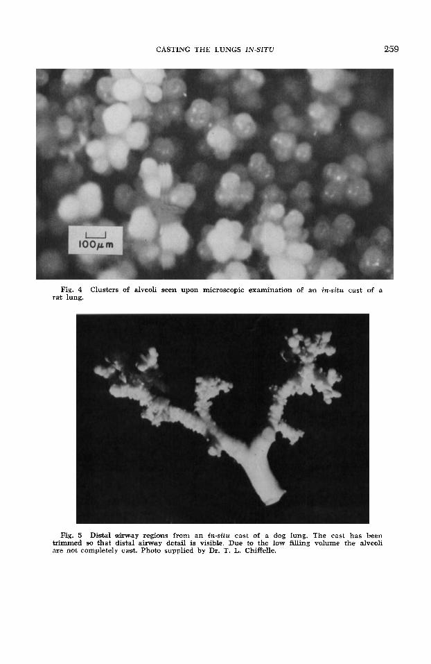
Fig. 4 rat lung.
Clusters of alveoli seen upon microscopic examination of an in-situ cast of a
Fig. 5 Distal &way regions from an in-situ cast of a &og Iung. The cast has been trimmed so that distal airway detail is visible. Due to the low filling volume the alveoli are not completely cast. Photo supplied by Dr. T. L. Chiffelle.
259

260 PHALEN ET AL.
Fig. 6 In-situ cast of the dog lung after digestion of lung tissee.

CASTING THE LUNGS IN-SITU 261
dogs and humans, respectively. A filling time for the lungs of about one hour is con- sidered optimal. This avoids excessive pres- sures within the lungs and is within the working time of the casting material. Saline in the lungs diffuses freely out of the lungs and through the pleural mem- brane as it is displaced by the casting ma- terial. The injected casting material is metered and the injection stopped when filling corresponds to a predetermined lung volume.
Casts are made to volumes that corre. spond to lung volumes occurring during normal breathing. Three methods are used for determining the filling volume of the lungs. Whenever possible, measurements of functional residual capacity and tidal volume made on unanesthetized animals in our pulmonary physiology laboratory (Mauderly, '72) are used. These measure- ments are always performed on our dogs. When such data are not available, as in the cases of the rodent or human, published values of lung volumes for the appropriate species, size, sex and age are used (Comroe et al., '62; Tenney and Remmers, '63; Weibel, '63; Crosfill and Widdicombe, '61). In those cases where the lung volume values from the literature are inappropri- ate, molding compound is injected until the lungs appear to be well-filled and just fill the available space in the thorax with- out distention of the chest wall. Ideally, 12 hours at room temperature are allowed for curing of the cast before it is removed from the thorax. The cast lungs are then carefully excised from the thorax and placed in a fresh one molar NaOH solu- tion for about 24 hours at room tempera- ture for tissue digestion. A final washing with a weak acid solution may be used to provide a more durable cast.
DISCUSSION
Although lung casts of excised lungs have been prepared by many investigators, (Eisman, '70; Frank and Yoder, '66; Lie- bow et al., '47; Narat et al., '36; Nowell et al., '72; Parker et al., '71; Rahn and Ross, '57; Tucker, Jr. and Krementz, '57) the in-situ method was developed to pro- vide casts of the lungs in their normal anatomical configuration. Gross lung shape is determined by the interplay of the lungs'
intrinsic structural properties and con- straints imposed by the thoracic walls, diaphragm, heart, major vessels, and other tissues within the thoracic cavity. When detailed airway geometry, including branch angles, must be replicated, these shape constraints become important.
Casts of the lungs of female rats (280- 330 gm body weight) made by three tech- niques, ( 1 ) excision - air drying - vac- uum filling (Rahn and Ross, '57); (2) ex- cision - saline replacement (Tompsett, '70); and (3) in-situ - saline replace- ment, as herein described, were compared to a cast of the rat thoracic cavity (fig. 3) . The overall shape of the cast made in-situ best approximated that of the thoracic cavity. The lobes of excised lungs had a tendency to overinflate and move relative to one another, thus changing the gross lung shape and the branching angles of the major bronchi. Also, hanging the ex- cised lungs from the tracheas during cast- ing caused the tracheas to be oriented to the Iungs at abnormal angles in the ex- cised lung casts. The in-situ method mini- mizes these and other obvious distortions caused by the lack of physiological ana- tomical support during casting of excised lungs.
The in-situ technique has three basic drawbacks : (1) the technique requires a relatively fresh and complete animal or human body which, in many instances, may not be available; (2) it requires more time and more careful attention than ex- cised lung techniques; and (3) artifacts may be produced by filling the lungs with 100% COz or saline. For example, flush- ing the lungs with COz gas causes a con- dition of lowered pH that may lead to constriction of the smooth muscle of the airways. Upon close examination, how- ever, the casts show no evidence of sig- nificant muscular constriction. Alveoli with slightly angular shapes are clearly seen upon microscopic examination of the casts (fig. 4) and the transition of conducting airways to resniratory bronchioles is clearly evident (fig. 5) .
The in-situ technique, which has been applied successfully to hamster, rat, mon- key, Beagle dog and human, has pro- vided our laboratory with lung casts suit- able for detailed airway morphometry. A
262 PHALEN ET AL.

CASTING THE LUNGS IN-SITU 263
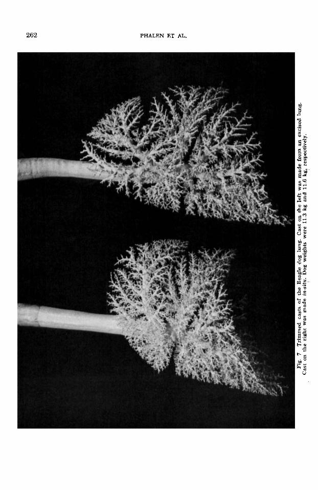
complete in-situ cast of a Beagle dog lung is shown in figure 6. When the casts of dog lungs are trimmed to show the major bronchioles, the more anatomically correct orientation of the airway of casts prepared by the in-situ method becomes more ap- parent. In figure 7, a cast prepared by the excision-saline replacement method shows spreading of lobes not found in the in-situ cast. Also, the gross artifacts previously noted in excised lung casts are seen.
ACKNOWLEDGMENTS
We wish to acknowledge our indebted- ness to Dr. R. W. Kilpper of the University of Rochester for beneficial advice on lung casting techniques and to Dr. Thomas L. Chiffelle of Lovelace Clinic for his con- tribution. We are also grateful for the valu- able suggestions of Dr. R. 0. McClellan, Dr. J. L. Mauderly, Dr. J. A. Mewhinney, and Mrs. Randi L. Thomas; the illustra- tions of Mr. E. E. Goff; and the editorial assistance of Mr. F. C. Rupprecht.
LITERATURE CITED Beeckmans, J. M. 1965 The deposition of aero-
sols in the respiratory tract. I. Mathematical analysis and comparison with experimental data. Can. J. Physiol. Pharmacol., 43: 157-172.
Comroe, J. H., R. E. Forslter, A. B. Bubois, W. A. Briscoe and E. Carlsen 1962 The Lung. Second ed. Year Book Medical Publishers Inc., Chicago.
Cro&ll, M. L., and J. G. Widdicombe 1961 Physical characteristics of the chest and lungs and the work of breathing in different mam- malian species. J. Physiol., 158: 1-14.
Eisman, M. 1970 Lung models: Hollow, flexible reproductions. J. Appl. Physiol., 29: 531-533.
Frank, N. R , and R. E. Yoder 1966 A method of malting a flexible oast of the lung. J. Appl. Physiol., 21: 1925-1926.
Liebow, A. A., M. R. Hales, G. E. Lindskag and W. E. Bloomer 1947 Plasltic demonstrations of pulmonary pathology. The Bulletin of the International Association of Medical Museums, 27: 116-129.
Mauderly, J. L., and J. A. Pichell 1972 Pul- m o n a ~ y function testing of unanesthetized beagle dogs. To be published in the Proceed- ings of the National Conference on Research Animals in Medicine, Washineon, D.C., Jan- uary 28-30.
1936 On the preparation of multicolored corrosion specimens. Anat. Rec., 64: 155-160.
Nowell, J. A., J. Panghorn and W. S. Tyler 1972 Stabilization and replication of soft tubular and alveolar systems; a scanning electron mi- crosoope study of the lung. In Scanning Elec- tron Microsoopy, 1972 (Part 11) Proceedings of the workshop on Biological Specimen Prep- aration Techniques for SEM, IIT Rsh. Inst., Chicago, Ill. 60616.
1971 Miorphology of distal airways in the human lung. J. Appl. Physiol., 31. 386-391.
Rahn, H., and B. B. Ross 1957 Bronchial tree casts, lobe weights and anatomical dead space measurements in the dog’s lung. J. Appl. Physiol., 10: 154-157.
Temey, S. M., and J. E. Remuners 1963 Com- parative morphology of the mammalian lung diffusing area. Nature, 197: 54-56.
Tompsett, D. H. 1970 Anatomical Techniques. Second ed. London, E. S. Livingstone, pp. xi and 123-128.
Tucker, J. L., Jr., and E. T. Ksementz 1957 Anatomical oomosion specimenis. 11. Broncho- pulmonary anatomy in the dog. Anat. Rec.,
Weibel, E. R. 1963 Morphometry of the human
Narat, J. K., J. A. Loef and M. Narat
Parker, P., K. Horsefield and G. Cumming
127: 667-676.
lung. Academic Press, New York.
Related Documents