Case Report The Use of Invisalign„ System in the Management of the Orthodontic Treatment before and after Class III Surgical Approach Renato Pagani, 1,2 Fabrizio Signorino, 3 Pier Paolo Poli, 3 Pietro Manzini, 1 and Irene Panisi 1,4 1 Maxillofacial Surgery Unit, Carlo Poma Hospital, Strada Lago Paiolo 10, 46100 Mantua, Italy 2 Specialization School in Maxillofacial Surgery, University of Milan, Via Commenda 10, 20122 Milan, Italy 3 Department of Dental Implants, Maxillofacial Surgery and Odontostomatology Unit, Fondazione IRCCS C` a Granda, University of Milan, Via Commenda 10, 20122 Milan, Italy 4 University of Milan, Via Commenda 10, 20122 Milan, Italy Correspondence should be addressed to Fabrizio Signorino; [email protected] Received 9 March 2016; Accepted 6 June 2016 Academic Editor: Maria Beatriz Duarte Gavi˜ ao Copyright © 2016 Renato Pagani et al. is is an open access article distributed under the Creative Commons Attribution License, which permits unrestricted use, distribution, and reproduction in any medium, provided the original work is properly cited. e approach to skeletal dysmorphisms in the maxillofacial area usually requires an orthodontic treatment by means of fixed appliances, both before and aſter the surgical phase. Since its introduction, Invisalign system has become a popular treatment choice for the clinicians because of the aesthetics and comfort of the removable clear aligners compared with the traditional appliances. erefore, the aim of the present report was to illustrate the management of a malocclusion by means of Invisalign system associated with the traditional surgical technique. e present paper shows a case of a 23-year-old male patient characterized by a Class III malocclusion with lateral deviation of the mandible to the leſt side and cross-bite on teeth 2.2, 2.3, and 2.4. Invisalign system was used during the pre- and postsurgical phases rather than fixed appliances. e posttreatment cephalometric analysis emphasized the stability of the dental and skeletal symmetry corrections, occlusion and functional balance, over a 6-year follow-up. e results achieved at the end of the treatment showed how Invisalign can be effective in the management of the orthodontic phases in orthognathic surgery. e follow-up aſter 6 years emphasizes the stability of the treatment over time. 1. Introduction For many patients, the surgical treatment of Class III maloc- clusion represents the only available therapy. It requires time, due to not only the diagnostic and planning phases, but also the duration of the treatment itself. For this reason, in these patients, the motivation is an important issue that must not be underestimated [1]. One of the most relevant problems, particularly with regard to adult patients, is represented by the need to undergo orthodontic therapy for several months. e aesthetic problem, associated to the worsening of oral hygiene conditions, may discourage many patients even before the beginning [2]. Invisalign system (Align Technology, Inc., San Jos` e, California, USA) could represent a suitable solution to solve such problematic [3–5]. It consists in a series of transparent aligners that are able to perform orthodontic movements without compromising the aesthetic of the smile. Hence, the purpose of this paper was to show the effectiveness and the advantages of this device in a surgical treatment of a Class III malocclusion. 2. Case Presentation 2.1. Diagnosis. A 23-year-old male patient presenting a Class III malocclusion with a lateral deviation of the mandible to the leſt side associated with a cross-bite of teeth 2.2, 2.3, and 2.4 came to our attention (Figure 1). Articular dysfunction in both Temporomandibular Joints (TMJs) was present, partic- ularly focused on the leſt side. On the working side it was pos- sible to observe a shorter and thicker condyle and mandibular ramus; conversely, in the opposite side, both areas appeared Hindawi Publishing Corporation Case Reports in Dentistry Volume 2016, Article ID 9231219, 10 pages http://dx.doi.org/10.1155/2016/9231219

Welcome message from author
This document is posted to help you gain knowledge. Please leave a comment to let me know what you think about it! Share it to your friends and learn new things together.
Transcript
Case ReportThe Use of Invisalign„ System in the Managementof the Orthodontic Treatment before and after Class IIISurgical Approach
Renato Pagani,1,2 Fabrizio Signorino,3 Pier Paolo Poli,3 Pietro Manzini,1 and Irene Panisi1,4
1Maxillofacial Surgery Unit, Carlo Poma Hospital, Strada Lago Paiolo 10, 46100 Mantua, Italy2Specialization School in Maxillofacial Surgery, University of Milan, Via Commenda 10, 20122 Milan, Italy3Department of Dental Implants, Maxillofacial Surgery and Odontostomatology Unit, Fondazione IRCCS Ca Granda,University of Milan, Via Commenda 10, 20122 Milan, Italy4University of Milan, Via Commenda 10, 20122 Milan, Italy
Correspondence should be addressed to Fabrizio Signorino; [email protected]
Received 9 March 2016; Accepted 6 June 2016
Academic Editor: Maria Beatriz Duarte Gaviao
Copyright © 2016 Renato Pagani et al. This is an open access article distributed under the Creative Commons Attribution License,which permits unrestricted use, distribution, and reproduction in any medium, provided the original work is properly cited.
The approach to skeletal dysmorphisms in the maxillofacial area usually requires an orthodontic treatment by means of fixedappliances, both before and after the surgical phase. Since its introduction, Invisalign system has become a popular treatment choicefor the clinicians because of the aesthetics and comfort of the removable clear aligners compared with the traditional appliances.Therefore, the aim of the present report was to illustrate themanagement of amalocclusion bymeans of Invisalign system associatedwith the traditional surgical technique. The present paper shows a case of a 23-year-old male patient characterized by a Class IIImalocclusion with lateral deviation of the mandible to the left side and cross-bite on teeth 2.2, 2.3, and 2.4. Invisalign system wasused during the pre- and postsurgical phases rather than fixed appliances. The posttreatment cephalometric analysis emphasizedthe stability of the dental and skeletal symmetry corrections, occlusion and functional balance, over a 6-year follow-up.The resultsachieved at the end of the treatment showed how Invisalign can be effective in the management of the orthodontic phases inorthognathic surgery. The follow-up after 6 years emphasizes the stability of the treatment over time.
1. Introduction
For many patients, the surgical treatment of Class III maloc-clusion represents the only available therapy. It requires time,due to not only the diagnostic and planning phases, but alsothe duration of the treatment itself. For this reason, in thesepatients, the motivation is an important issue that must notbe underestimated [1]. One of the most relevant problems,particularlywith regard to adult patients, is represented by theneed to undergo orthodontic therapy for several months.Theaesthetic problem, associated to theworsening of oral hygieneconditions, may discourage many patients even before thebeginning [2]. Invisalign system (Align Technology, Inc., SanJose, California, USA) could represent a suitable solutionto solve such problematic [3–5]. It consists in a series oftransparent aligners that are able to perform orthodontic
movements without compromising the aesthetic of the smile.Hence, the purpose of this paperwas to show the effectivenessand the advantages of this device in a surgical treatment of aClass III malocclusion.
2. Case Presentation
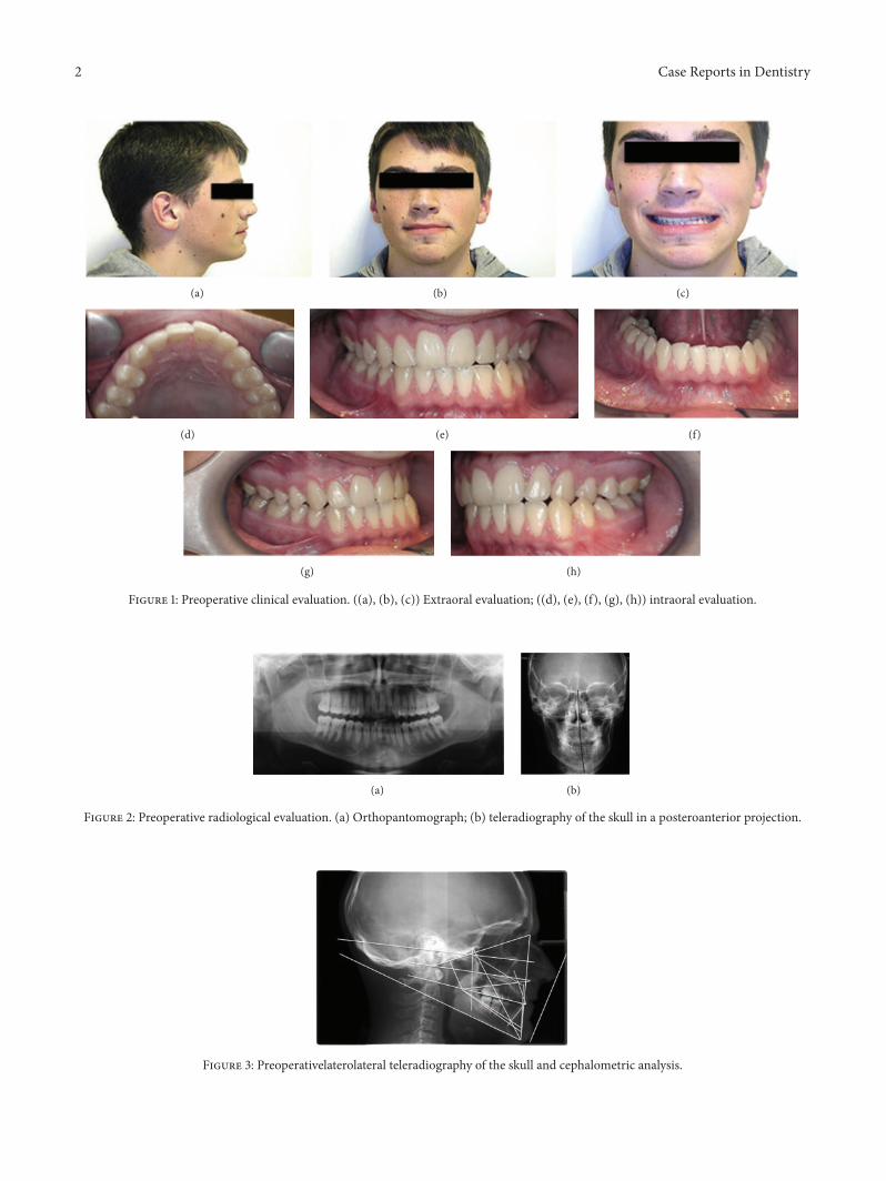
2.1. Diagnosis. A 23-year-old male patient presenting a ClassIII malocclusion with a lateral deviation of the mandible tothe left side associated with a cross-bite of teeth 2.2, 2.3, and2.4 came to our attention (Figure 1). Articular dysfunction inboth Temporomandibular Joints (TMJs) was present, partic-ularly focused on the left side. On theworking side it was pos-sible to observe a shorter and thicker condyle andmandibularramus; conversely, in the opposite side, both areas appeared
Hindawi Publishing CorporationCase Reports in DentistryVolume 2016, Article ID 9231219, 10 pageshttp://dx.doi.org/10.1155/2016/9231219
2 Case Reports in Dentistry
(a) (b) (c)
(d) (e) (f)
(g) (h)
Figure 1: Preoperative clinical evaluation. ((a), (b), (c)) Extraoral evaluation; ((d), (e), (f), (g), (h)) intraoral evaluation.
(a) (b)
Figure 2: Preoperative radiological evaluation. (a) Orthopantomograph; (b) teleradiography of the skull in a posteroanterior projection.
Figure 3: Preoperativelaterolateral teleradiography of the skull and cephalometric analysis.
Case Reports in Dentistry 3
ClinCheck software
(a)
ClinCheck software
(b)
ClinCheck software
(c)
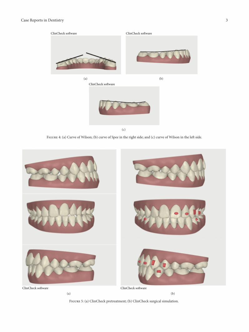
Figure 4: (a) Curve of Wilson; (b) curve of Spee in the right side; and (c) curve of Wilson in the left side.
ClinCheck software(a)
ClinCheck software(b)
Figure 5: (a) ClinCheck pretreatment; (b) ClinCheck surgical simulation.
4 Case Reports in Dentistry
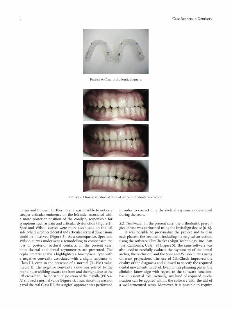
Figure 6: Clear orthodontic aligners.
Figure 7: Clinical situation at the end of the orthodontic correction.
longer and thinner. Furthermore, it was possible to notice asteeper articular eminence on the left side, associated witha more posterior position of the condyle, responsible forsymptoms such as pain and articular dysfunction (Figure 2).Spee and Wilson curves were more accentuate on the leftside, where a reduced dental and articular vertical dimensionscould be observed (Figure 3). As a consequence, Spee andWilson curves underwent a remodelling to compensate theloss of posterior occlusal contacts. In the present case,both skeletal and dental asymmetries are presented. Thecephalometric analysis highlighted a brachyfacial type witha negative convexity associated with a slight tendency toClass III, even in the presence of a normal (Xi-PM) value(Table 1). The negative convexity value was related to themandibular shifting toward the front and the right, due to theleft cross-bite. The horizontal position of the maxilla (Pf-Na-A) showed a normal value (Figure 4).Thus, since this was nota real skeletal Class III, the surgical approach was performed
in order to correct only the skeletal asymmetry developedduring the years.
2.2. Treatment. In the present case, the orthodontic presur-gical phase was performed using the Invisalign device [6–8].
It was possible to previsualize the project and to planeach phase of the treatment, including the surgical correction,using the software ClinCheck� (Align Technology, Inc., SanJose, California, USA) [9] (Figure 5). The same software wasalso used to carefully evaluate the asymmetry of the dentalarches, the occlusion, and the Spee and Wilson curves usingdifferent projections. The use of ClinCheck improved thequality of the diagnosis and allowed to specify the requireddental movements in detail. Even in this planning phase, theclinician knowledge with regard to the software functionshas an essential role. Actually, any kind of required modi-fication can be applied within the software with the aid ofa well-structured setup. Moreover, it is possible to require
Case Reports in Dentistry 5
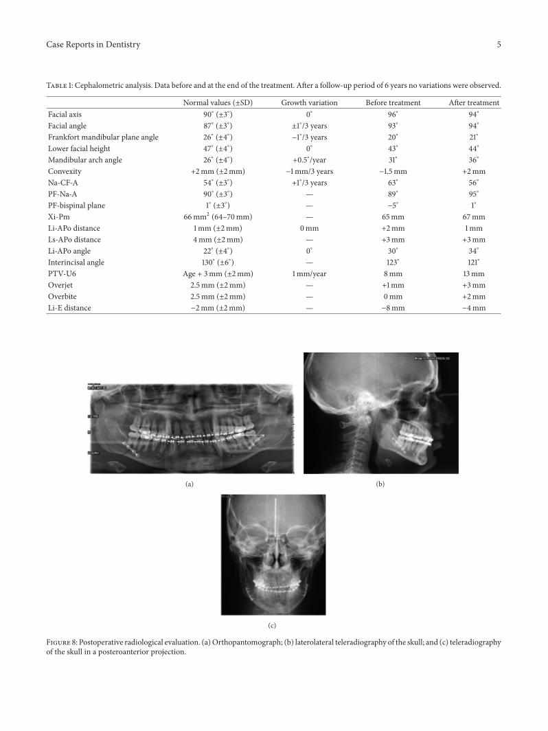
Table 1: Cephalometric analysis. Data before and at the end of the treatment. After a follow-up period of 6 years no variations were observed.
Normal values (±SD) Growth variation Before treatment After treatmentFacial axis 90∘ (±3∘) 0∘ 96∘ 94∘
Facial angle 87∘ (±3∘) ±1∘/3 years 93∘ 94∘
Frankfort mandibular plane angle 26∘ (±4∘) −1∘/3 years 20∘ 21∘
Lower facial height 47∘ (±4∘) 0∘ 43∘ 44∘
Mandibular arch angle 26∘ (±4∘) +0.5∘/year 31∘ 36∘
Convexity +2mm (±2mm) −1mm/3 years −1.5mm +2mmNa-CF-A 54∘ (±3∘) +1∘/3 years 63∘ 56∘
PF-Na-A 90∘ (±3∘) — 89∘ 95∘
PF-bispinal plane 1∘ (±3∘) — −5∘ 1∘
Xi-Pm 66mm2 (64–70mm) — 65mm 67mmLi-APo distance 1mm (±2mm) 0mm +2mm 1mmLs-APo distance 4mm (±2mm) — +3mm +3mmLi-APo angle 22∘ (±4∘) 0∘ 30∘ 34∘
Interincisal angle 130∘ (±6∘) — 123∘ 121∘
PTV-U6 Age + 3mm (±2mm) 1mm/year 8mm 13mmOverjet 2.5mm (±2mm) — +1mm +3mmOverbite 2.5mm (±2mm) — 0mm +2mmLi-E distance −2mm (±2mm) — −8mm −4mm
(a) (b)
(c)
Figure 8: Postoperative radiological evaluation. (a)Orthopantomograph; (b) laterolateral teleradiography of the skull; and (c) teleradiographyof the skull in a posteroanterior projection.
6 Case Reports in Dentistry
ClinCheck software(a)
ClinCheck software(b)
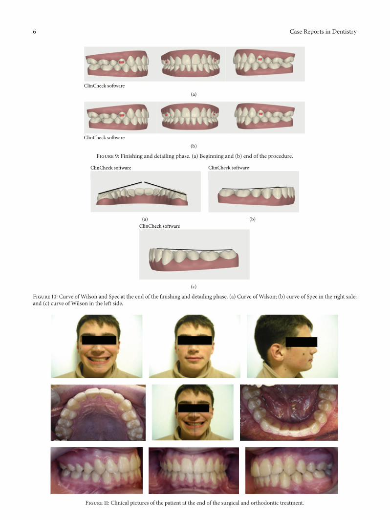
Figure 9: Finishing and detailing phase. (a) Beginning and (b) end of the procedure.
ClinCheck software
(a)
ClinCheck software
(b)ClinCheck software
(c)
Figure 10: Curve of Wilson and Spee at the end of the finishing and detailing phase. (a) Curve of Wilson; (b) curve of Spee in the right side;and (c) curve of Wilson in the left side.
Figure 11: Clinical pictures of the patient at the end of the surgical and orthodontic treatment.
Case Reports in Dentistry 7
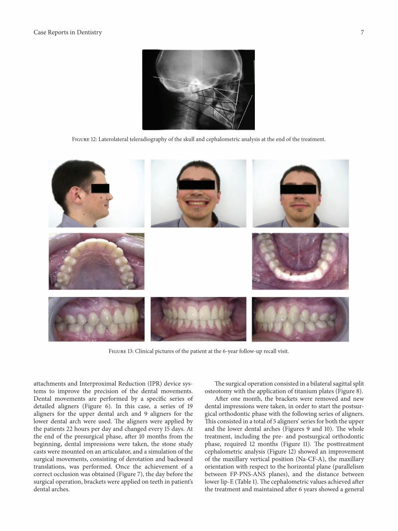
Figure 12: Laterolateral teleradiography of the skull and cephalometric analysis at the end of the treatment.
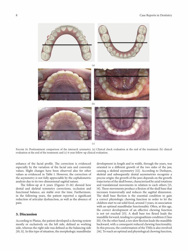
Figure 13: Clinical pictures of the patient at the 6-year follow-up recall visit.
attachments and Interproximal Reduction (IPR) device sys-tems to improve the precision of the dental movements.Dental movements are performed by a specific series ofdetailed aligners (Figure 6). In this case, a series of 19aligners for the upper dental arch and 9 aligners for thelower dental arch were used. The aligners were applied bythe patients 22 hours per day and changed every 15 days. Atthe end of the presurgical phase, after 10 months from thebeginning, dental impressions were taken, the stone studycasts were mounted on an articulator, and a simulation of thesurgical movements, consisting of derotation and backwardtranslations, was performed. Once the achievement of acorrect occlusion was obtained (Figure 7), the day before thesurgical operation, brackets were applied on teeth in patient’sdental arches.
The surgical operation consisted in a bilateral sagittal splitosteotomy with the application of titanium plates (Figure 8).
After one month, the brackets were removed and newdental impressions were taken, in order to start the postsur-gical orthodontic phase with the following series of aligners.This consisted in a total of 5 aligners’ series for both the upperand the lower dental arches (Figures 9 and 10). The wholetreatment, including the pre- and postsurgical orthodonticphase, required 12 months (Figure 11). The posttreatmentcephalometric analysis (Figure 12) showed an improvementof the maxillary vertical position (Na-CF-A), the maxillaryorientation with respect to the horizontal plane (parallelismbetween FP-PNS-ANS planes), and the distance betweenlower lip-E (Table 1).The cephalometric values achieved afterthe treatment and maintained after 6 years showed a general
8 Case Reports in Dentistry
(a)
(b)
(c)
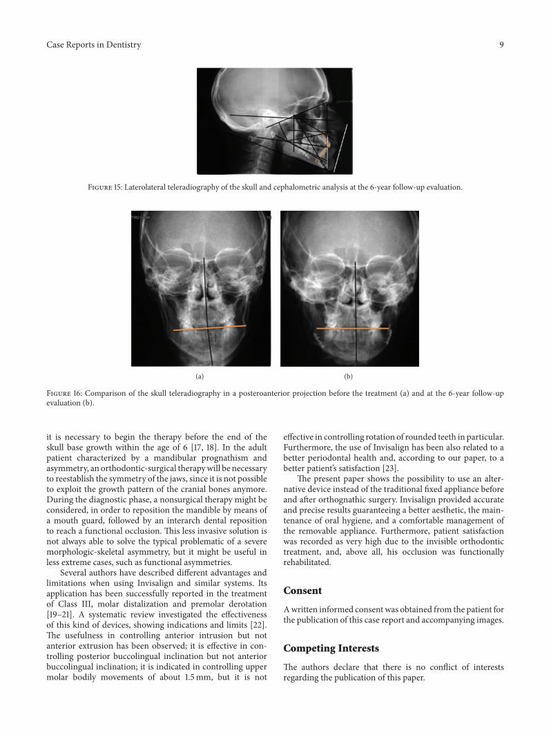
Figure 14: Posttreatment comparison of the interarch symmetry. (a) Clinical check evaluation at the end of the treatment; (b) clinicalevaluation at the end of the treatment; and (c) 6-year follow-up clinical evaluation.
enhance of the facial profile. The correction is evidencedespecially by the variation of the facial axis and convexityvalues. Slight changes have been observed also for othervalues as evidenced in Table 1. However, the correction ofthe asymmetry is not fully appreciable by the cephalometricanalysis due to its two-dimensional sagittal nature.
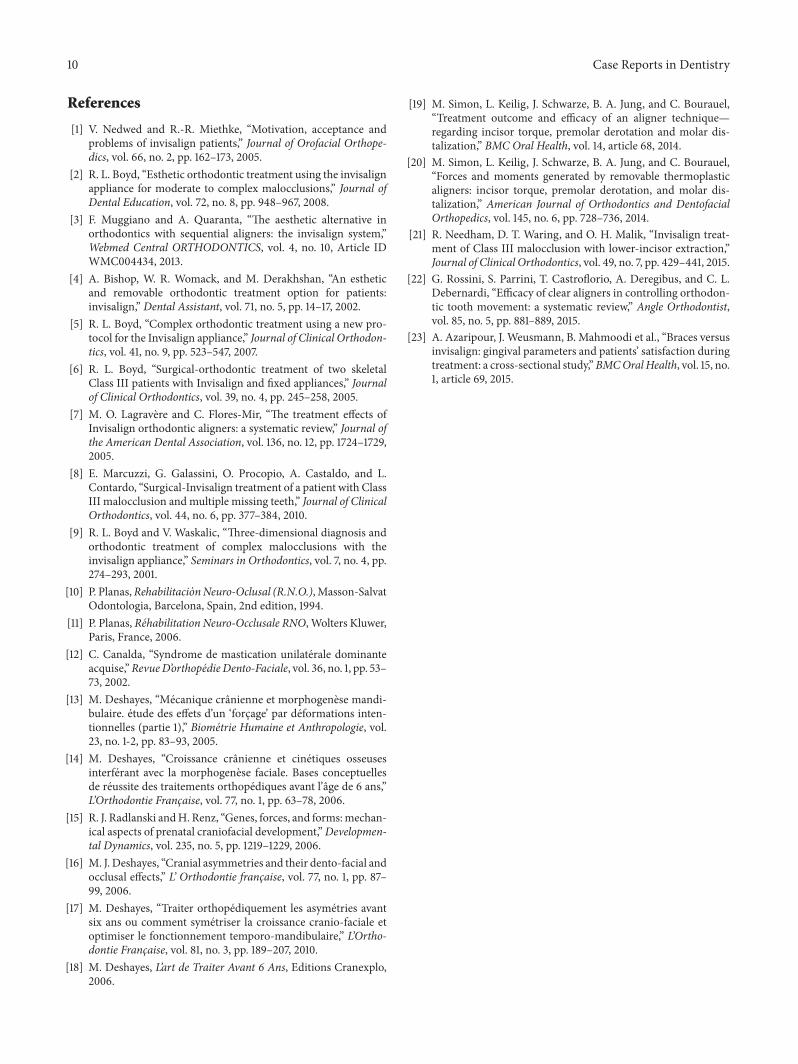
The follow-up at 6 years (Figures 13–16) showed howdental and skeletal symmetry corrections, occlusion andfunctional balance, are stable over the time. Furthermore,in the following years, the patient reported a significantreduction of articular dysfunction, as well as the absence ofpain.
3. Discussion
According to Planas, the patient developed a chewing systemmostly or exclusively on the left side, defined as workingside, whereas the right side was defined as the balancing side[10, 11]. In this type of situation, the morphologic mandibular
development in length and in width, through the years, wasoriented to a different growth of the two sides of the jaw,causing a skeletal asymmetry [12]. According to Deshayes,skeletal and subsequently dental asymmetries recognize aprecise origin: the growth of the jaws depends on the growthtrajectories of the skull bones, characterized by axial rotationsand translational movements in relation to each others [13,14].These movements produce a flexion of the skull base thatincreases transversally and reduces the sagittal dimension.The skull base flection is the essential condition to gaina correct physiologic chewing function in order to let thechildren start to eat solid food, around 3 years, in associationwith an optimal mandibular functionality. Often, at this age,the correct development of an effective chewing functionis not yet reached [15]. A skull base too flexed leads themandible forward, tending to a prognathism condition (ClassIII). On the other hand, a too slow flection leads themandiblebackward, with a consequent reduced chewing functionality.In this process, the conformation of the TMJs is also involved[16]. To reach an optimal and physiological chewing function,
Case Reports in Dentistry 9
Figure 15: Laterolateral teleradiography of the skull and cephalometric analysis at the 6-year follow-up evaluation.
(a) (b)
Figure 16: Comparison of the skull teleradiography in a posteroanterior projection before the treatment (a) and at the 6-year follow-upevaluation (b).
it is necessary to begin the therapy before the end of theskull base growth within the age of 6 [17, 18]. In the adultpatient characterized by a mandibular prognathism andasymmetry, an orthodontic-surgical therapywill be necessaryto reestablish the symmetry of the jaws, since it is not possibleto exploit the growth pattern of the cranial bones anymore.During the diagnostic phase, a nonsurgical therapy might beconsidered, in order to reposition the mandible by means ofa mouth guard, followed by an interarch dental repositionto reach a functional occlusion. This less invasive solution isnot always able to solve the typical problematic of a severemorphologic-skeletal asymmetry, but it might be useful inless extreme cases, such as functional asymmetries.
Several authors have described different advantages andlimitations when using Invisalign and similar systems. Itsapplication has been successfully reported in the treatmentof Class III, molar distalization and premolar derotation[19–21]. A systematic review investigated the effectivenessof this kind of devices, showing indications and limits [22].The usefulness in controlling anterior intrusion but notanterior extrusion has been observed; it is effective in con-trolling posterior buccolingual inclination but not anteriorbuccolingual inclination; it is indicated in controlling uppermolar bodily movements of about 1.5mm, but it is not
effective in controlling rotation of rounded teeth in particular.Furthermore, the use of Invisalign has been also related to abetter periodontal health and, according to our paper, to abetter patient’s satisfaction [23].
The present paper shows the possibility to use an alter-native device instead of the traditional fixed appliance beforeand after orthognathic surgery. Invisalign provided accurateand precise results guaranteeing a better aesthetic, the main-tenance of oral hygiene, and a comfortable management ofthe removable appliance. Furthermore, patient satisfactionwas recorded as very high due to the invisible orthodontictreatment, and, above all, his occlusion was functionallyrehabilitated.
Consent
Awritten informed consent was obtained from the patient forthe publication of this case report and accompanying images.
Competing Interests
The authors declare that there is no conflict of interestsregarding the publication of this paper.
10 Case Reports in Dentistry
References
[1] V. Nedwed and R.-R. Miethke, “Motivation, acceptance andproblems of invisalign patients,” Journal of Orofacial Orthope-dics, vol. 66, no. 2, pp. 162–173, 2005.
[2] R. L. Boyd, “Esthetic orthodontic treatment using the invisalignappliance for moderate to complex malocclusions,” Journal ofDental Education, vol. 72, no. 8, pp. 948–967, 2008.
[3] F. Muggiano and A. Quaranta, “The aesthetic alternative inorthodontics with sequential aligners: the invisalign system,”Webmed Central ORTHODONTICS, vol. 4, no. 10, Article IDWMC004434, 2013.
[4] A. Bishop, W. R. Womack, and M. Derakhshan, “An estheticand removable orthodontic treatment option for patients:invisalign,” Dental Assistant, vol. 71, no. 5, pp. 14–17, 2002.
[5] R. L. Boyd, “Complex orthodontic treatment using a new pro-tocol for the Invisalign appliance,” Journal of Clinical Orthodon-tics, vol. 41, no. 9, pp. 523–547, 2007.
[6] R. L. Boyd, “Surgical-orthodontic treatment of two skeletalClass III patients with Invisalign and fixed appliances,” Journalof Clinical Orthodontics, vol. 39, no. 4, pp. 245–258, 2005.
[7] M. O. Lagravere and C. Flores-Mir, “The treatment effects ofInvisalign orthodontic aligners: a systematic review,” Journal ofthe American Dental Association, vol. 136, no. 12, pp. 1724–1729,2005.
[8] E. Marcuzzi, G. Galassini, O. Procopio, A. Castaldo, and L.Contardo, “Surgical-Invisalign treatment of a patient with ClassIII malocclusion andmultiple missing teeth,” Journal of ClinicalOrthodontics, vol. 44, no. 6, pp. 377–384, 2010.
[9] R. L. Boyd and V. Waskalic, “Three-dimensional diagnosis andorthodontic treatment of complex malocclusions with theinvisalign appliance,” Seminars in Orthodontics, vol. 7, no. 4, pp.274–293, 2001.
[10] P. Planas,RehabilitacionNeuro-Oclusal (R.N.O.),Masson-SalvatOdontologia, Barcelona, Spain, 2nd edition, 1994.
[11] P. Planas, Rehabilitation Neuro-Occlusale RNO, Wolters Kluwer,Paris, France, 2006.
[12] C. Canalda, “Syndrome de mastication unilaterale dominanteacquise,”RevueD’orthopedie Dento-Faciale, vol. 36, no. 1, pp. 53–73, 2002.
[13] M. Deshayes, “Mecanique cranienne et morphogenese mandi-bulaire. etude des effets d’un ‘forcage’ par deformations inten-tionnelles (partie 1),” Biometrie Humaine et Anthropologie, vol.23, no. 1-2, pp. 83–93, 2005.
[14] M. Deshayes, “Croissance cranienne et cinetiques osseusesinterferant avec la morphogenese faciale. Bases conceptuellesde reussite des traitements orthopediques avant l’age de 6 ans,”L’Orthodontie Francaise, vol. 77, no. 1, pp. 63–78, 2006.
[15] R. J. Radlanski andH. Renz, “Genes, forces, and forms:mechan-ical aspects of prenatal craniofacial development,”Developmen-tal Dynamics, vol. 235, no. 5, pp. 1219–1229, 2006.
[16] M. J. Deshayes, “Cranial asymmetries and their dento-facial andocclusal effects,” L’ Orthodontie francaise, vol. 77, no. 1, pp. 87–99, 2006.
[17] M. Deshayes, “Traiter orthopediquement les asymetries avantsix ans ou comment symetriser la croissance cranio-faciale etoptimiser le fonctionnement temporo-mandibulaire,” L’Ortho-dontie Francaise, vol. 81, no. 3, pp. 189–207, 2010.
[18] M. Deshayes, L’art de Traiter Avant 6 Ans, Editions Cranexplo,2006.
[19] M. Simon, L. Keilig, J. Schwarze, B. A. Jung, and C. Bourauel,“Treatment outcome and efficacy of an aligner technique—regarding incisor torque, premolar derotation and molar dis-talization,” BMC Oral Health, vol. 14, article 68, 2014.
[20] M. Simon, L. Keilig, J. Schwarze, B. A. Jung, and C. Bourauel,“Forces and moments generated by removable thermoplasticaligners: incisor torque, premolar derotation, and molar dis-talization,” American Journal of Orthodontics and DentofacialOrthopedics, vol. 145, no. 6, pp. 728–736, 2014.
[21] R. Needham, D. T. Waring, and O. H. Malik, “Invisalign treat-ment of Class III malocclusion with lower-incisor extraction,”Journal of Clinical Orthodontics, vol. 49, no. 7, pp. 429–441, 2015.
[22] G. Rossini, S. Parrini, T. Castroflorio, A. Deregibus, and C. L.Debernardi, “Efficacy of clear aligners in controlling orthodon-tic tooth movement: a systematic review,” Angle Orthodontist,vol. 85, no. 5, pp. 881–889, 2015.
[23] A. Azaripour, J. Weusmann, B. Mahmoodi et al., “Braces versusinvisalign: gingival parameters and patients’ satisfaction duringtreatment: a cross-sectional study,”BMCOralHealth, vol. 15, no.1, article 69, 2015.

Submit your manuscripts athttp://www.hindawi.com
Hindawi Publishing Corporationhttp://www.hindawi.com Volume 2014
Oral OncologyJournal of
DentistryInternational Journal of
Hindawi Publishing Corporationhttp://www.hindawi.com Volume 2014
Hindawi Publishing Corporationhttp://www.hindawi.com Volume 2014
International Journal of
Biomaterials
Hindawi Publishing Corporationhttp://www.hindawi.com Volume 2014
BioMed Research International
Hindawi Publishing Corporationhttp://www.hindawi.com Volume 2014
Case Reports in Dentistry
Hindawi Publishing Corporationhttp://www.hindawi.com Volume 2014
Oral ImplantsJournal of
Hindawi Publishing Corporationhttp://www.hindawi.com Volume 2014
Anesthesiology Research and Practice
Hindawi Publishing Corporationhttp://www.hindawi.com Volume 2014
Radiology Research and Practice
Environmental and Public Health
Journal of
Hindawi Publishing Corporationhttp://www.hindawi.com Volume 2014
The Scientific World JournalHindawi Publishing Corporation http://www.hindawi.com Volume 2014
Hindawi Publishing Corporationhttp://www.hindawi.com Volume 2014
Dental SurgeryJournal of
Drug DeliveryJournal of
Hindawi Publishing Corporationhttp://www.hindawi.com Volume 2014
Hindawi Publishing Corporationhttp://www.hindawi.com Volume 2014
Oral DiseasesJournal of
Hindawi Publishing Corporationhttp://www.hindawi.com Volume 2014
Computational and Mathematical Methods in Medicine
ScientificaHindawi Publishing Corporationhttp://www.hindawi.com Volume 2014
PainResearch and TreatmentHindawi Publishing Corporationhttp://www.hindawi.com Volume 2014
Preventive MedicineAdvances in
Hindawi Publishing Corporationhttp://www.hindawi.com Volume 2014
EndocrinologyInternational Journal of
Hindawi Publishing Corporationhttp://www.hindawi.com Volume 2014
Hindawi Publishing Corporationhttp://www.hindawi.com Volume 2014
OrthopedicsAdvances in
Related Documents