Top Banner
Case Report #0431 Submitted by: Jin T. Kim, M.D. Faculty reviewer: Clark W. Sitton, M.D. Date accepted: 25 November 2007 Radiological Category: Principal Modality (1): Principal Modality (2): Neuroradiology Nuclear Scintigraphy CT
49

Case Report #0431 Submitted by:Jin T. Kim, M.D. Faculty reviewer:Clark W. Sitton, M.D. Date accepted:25 November 2007 Radiological Category:Principal Modality.

Dec 14, 2015

Download

Documents

Braedon Martell
Welcome message from author
This document is posted to help you gain knowledge. Please leave a comment to let me know what you think about it! Share it to your friends and learn new things together.
Transcript
Page 1: Case Report #0431 Submitted by:Jin T. Kim, M.D. Faculty reviewer:Clark W. Sitton, M.D. Date accepted:25 November 2007 Radiological Category:Principal Modality.

Case Report #0431

Submitted by: Jin T. Kim, M.D.

Faculty reviewer: Clark W. Sitton, M.D.

Date accepted: 25 November 2007

Radiological Category: Principal Modality (1):

Principal Modality (2):

Neuroradiology

Nuclear Scintigraphy

CT

Page 2: Case Report #0431 Submitted by:Jin T. Kim, M.D. Faculty reviewer:Clark W. Sitton, M.D. Date accepted:25 November 2007 Radiological Category:Principal Modality.

Case History

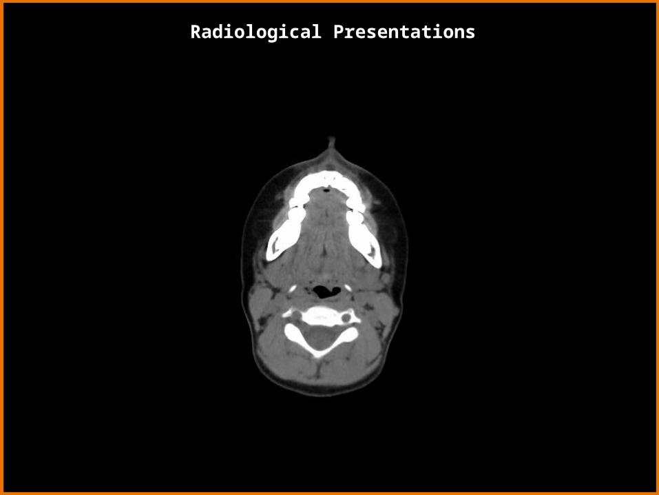
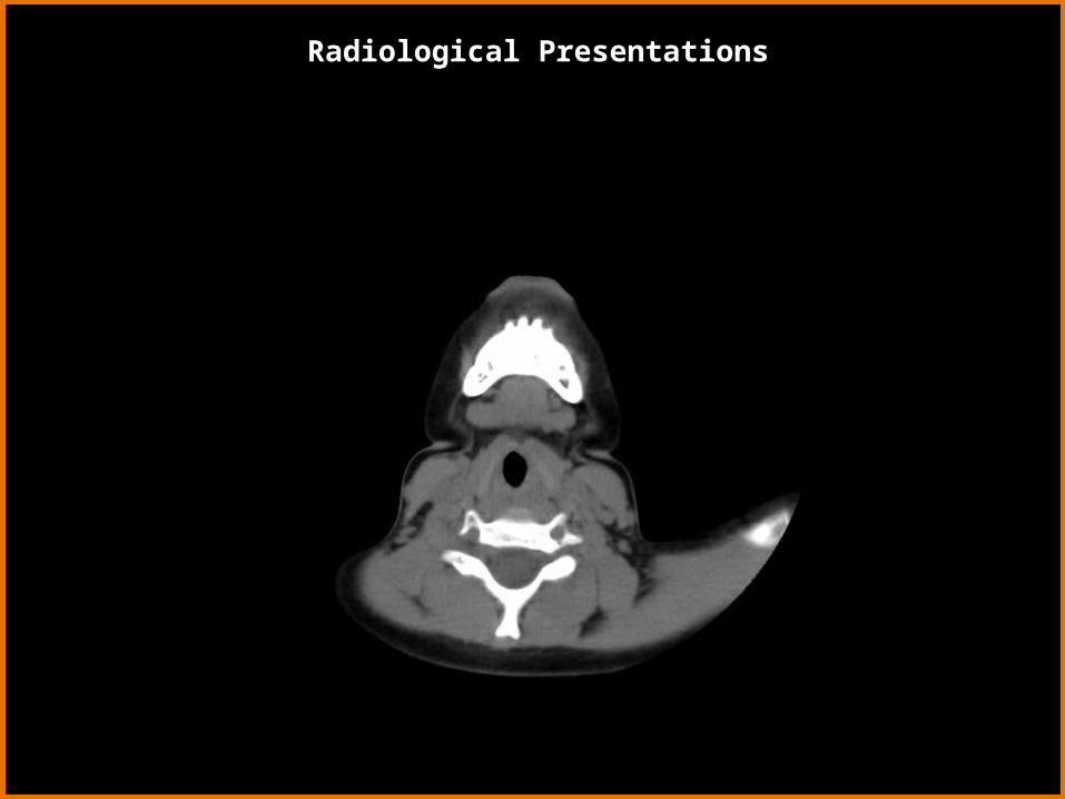
6 year-old girl with dysphagia.

Page 3: Case Report #0431 Submitted by:Jin T. Kim, M.D. Faculty reviewer:Clark W. Sitton, M.D. Date accepted:25 November 2007 Radiological Category:Principal Modality.

Radiological Presentations

Non-contrast CT – Axial View

Page 4: Case Report #0431 Submitted by:Jin T. Kim, M.D. Faculty reviewer:Clark W. Sitton, M.D. Date accepted:25 November 2007 Radiological Category:Principal Modality.

Radiological Presentations

Page 5: Case Report #0431 Submitted by:Jin T. Kim, M.D. Faculty reviewer:Clark W. Sitton, M.D. Date accepted:25 November 2007 Radiological Category:Principal Modality.

Radiological Presentations

Page 6: Case Report #0431 Submitted by:Jin T. Kim, M.D. Faculty reviewer:Clark W. Sitton, M.D. Date accepted:25 November 2007 Radiological Category:Principal Modality.

Radiological Presentations

Page 7: Case Report #0431 Submitted by:Jin T. Kim, M.D. Faculty reviewer:Clark W. Sitton, M.D. Date accepted:25 November 2007 Radiological Category:Principal Modality.

Radiological Presentations

Page 8: Case Report #0431 Submitted by:Jin T. Kim, M.D. Faculty reviewer:Clark W. Sitton, M.D. Date accepted:25 November 2007 Radiological Category:Principal Modality.

Radiological Presentations

Page 9: Case Report #0431 Submitted by:Jin T. Kim, M.D. Faculty reviewer:Clark W. Sitton, M.D. Date accepted:25 November 2007 Radiological Category:Principal Modality.

Radiological Presentations

Page 10: Case Report #0431 Submitted by:Jin T. Kim, M.D. Faculty reviewer:Clark W. Sitton, M.D. Date accepted:25 November 2007 Radiological Category:Principal Modality.

Radiological Presentations

Page 11: Case Report #0431 Submitted by:Jin T. Kim, M.D. Faculty reviewer:Clark W. Sitton, M.D. Date accepted:25 November 2007 Radiological Category:Principal Modality.

Radiological Presentations

Page 12: Case Report #0431 Submitted by:Jin T. Kim, M.D. Faculty reviewer:Clark W. Sitton, M.D. Date accepted:25 November 2007 Radiological Category:Principal Modality.

Radiological Presentations

Page 13: Case Report #0431 Submitted by:Jin T. Kim, M.D. Faculty reviewer:Clark W. Sitton, M.D. Date accepted:25 November 2007 Radiological Category:Principal Modality.

Radiological Presentations

Page 14: Case Report #0431 Submitted by:Jin T. Kim, M.D. Faculty reviewer:Clark W. Sitton, M.D. Date accepted:25 November 2007 Radiological Category:Principal Modality.

Radiological Presentations

Page 15: Case Report #0431 Submitted by:Jin T. Kim, M.D. Faculty reviewer:Clark W. Sitton, M.D. Date accepted:25 November 2007 Radiological Category:Principal Modality.

Radiological Presentations

Page 16: Case Report #0431 Submitted by:Jin T. Kim, M.D. Faculty reviewer:Clark W. Sitton, M.D. Date accepted:25 November 2007 Radiological Category:Principal Modality.

Radiological Presentations

Page 17: Case Report #0431 Submitted by:Jin T. Kim, M.D. Faculty reviewer:Clark W. Sitton, M.D. Date accepted:25 November 2007 Radiological Category:Principal Modality.

Radiological Presentations

Page 18: Case Report #0431 Submitted by:Jin T. Kim, M.D. Faculty reviewer:Clark W. Sitton, M.D. Date accepted:25 November 2007 Radiological Category:Principal Modality.

Radiological Presentations

Non-contrast CT – Coronal View

Page 19: Case Report #0431 Submitted by:Jin T. Kim, M.D. Faculty reviewer:Clark W. Sitton, M.D. Date accepted:25 November 2007 Radiological Category:Principal Modality.

Radiological Presentations

Page 20: Case Report #0431 Submitted by:Jin T. Kim, M.D. Faculty reviewer:Clark W. Sitton, M.D. Date accepted:25 November 2007 Radiological Category:Principal Modality.

Radiological Presentations

Page 21: Case Report #0431 Submitted by:Jin T. Kim, M.D. Faculty reviewer:Clark W. Sitton, M.D. Date accepted:25 November 2007 Radiological Category:Principal Modality.

Radiological Presentations

Page 22: Case Report #0431 Submitted by:Jin T. Kim, M.D. Faculty reviewer:Clark W. Sitton, M.D. Date accepted:25 November 2007 Radiological Category:Principal Modality.

Radiological Presentations

Page 23: Case Report #0431 Submitted by:Jin T. Kim, M.D. Faculty reviewer:Clark W. Sitton, M.D. Date accepted:25 November 2007 Radiological Category:Principal Modality.

Radiological Presentations

Page 24: Case Report #0431 Submitted by:Jin T. Kim, M.D. Faculty reviewer:Clark W. Sitton, M.D. Date accepted:25 November 2007 Radiological Category:Principal Modality.

Radiological Presentations

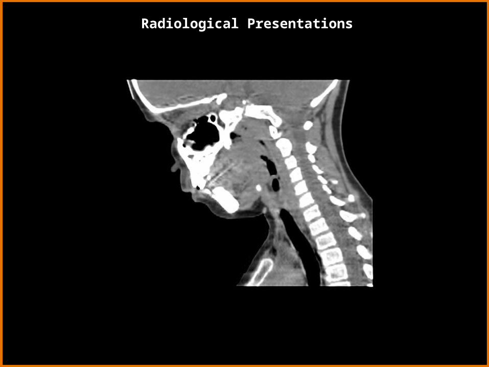
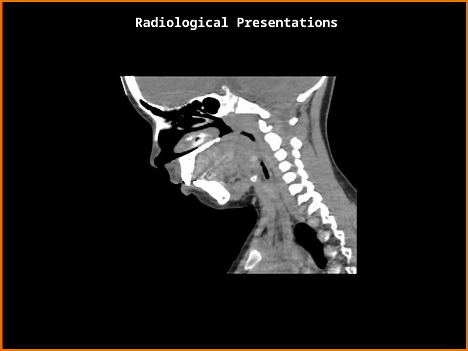
Non-contrast CT – Sagittal View

Page 25: Case Report #0431 Submitted by:Jin T. Kim, M.D. Faculty reviewer:Clark W. Sitton, M.D. Date accepted:25 November 2007 Radiological Category:Principal Modality.

Radiological Presentations

Page 26: Case Report #0431 Submitted by:Jin T. Kim, M.D. Faculty reviewer:Clark W. Sitton, M.D. Date accepted:25 November 2007 Radiological Category:Principal Modality.

Radiological Presentations

Page 27: Case Report #0431 Submitted by:Jin T. Kim, M.D. Faculty reviewer:Clark W. Sitton, M.D. Date accepted:25 November 2007 Radiological Category:Principal Modality.

Radiological Presentations

Page 28: Case Report #0431 Submitted by:Jin T. Kim, M.D. Faculty reviewer:Clark W. Sitton, M.D. Date accepted:25 November 2007 Radiological Category:Principal Modality.

Radiological Presentations

Page 29: Case Report #0431 Submitted by:Jin T. Kim, M.D. Faculty reviewer:Clark W. Sitton, M.D. Date accepted:25 November 2007 Radiological Category:Principal Modality.

Radiological Presentations

Page 30: Case Report #0431 Submitted by:Jin T. Kim, M.D. Faculty reviewer:Clark W. Sitton, M.D. Date accepted:25 November 2007 Radiological Category:Principal Modality.

Radiological Presentations

Page 31: Case Report #0431 Submitted by:Jin T. Kim, M.D. Faculty reviewer:Clark W. Sitton, M.D. Date accepted:25 November 2007 Radiological Category:Principal Modality.

Radiological Presentations

Page 32: Case Report #0431 Submitted by:Jin T. Kim, M.D. Faculty reviewer:Clark W. Sitton, M.D. Date accepted:25 November 2007 Radiological Category:Principal Modality.

Radiological Presentations

Page 33: Case Report #0431 Submitted by:Jin T. Kim, M.D. Faculty reviewer:Clark W. Sitton, M.D. Date accepted:25 November 2007 Radiological Category:Principal Modality.

Radiological Presentations

Page 34: Case Report #0431 Submitted by:Jin T. Kim, M.D. Faculty reviewer:Clark W. Sitton, M.D. Date accepted:25 November 2007 Radiological Category:Principal Modality.

Radiological Presentations

Page 35: Case Report #0431 Submitted by:Jin T. Kim, M.D. Faculty reviewer:Clark W. Sitton, M.D. Date accepted:25 November 2007 Radiological Category:Principal Modality.

Radiological Presentations

Page 36: Case Report #0431 Submitted by:Jin T. Kim, M.D. Faculty reviewer:Clark W. Sitton, M.D. Date accepted:25 November 2007 Radiological Category:Principal Modality.

Radiological Presentations

Page 37: Case Report #0431 Submitted by:Jin T. Kim, M.D. Faculty reviewer:Clark W. Sitton, M.D. Date accepted:25 November 2007 Radiological Category:Principal Modality.

Radiological Presentations

Page 38: Case Report #0431 Submitted by:Jin T. Kim, M.D. Faculty reviewer:Clark W. Sitton, M.D. Date accepted:25 November 2007 Radiological Category:Principal Modality.

Radiological Presentations

Page 39: Case Report #0431 Submitted by:Jin T. Kim, M.D. Faculty reviewer:Clark W. Sitton, M.D. Date accepted:25 November 2007 Radiological Category:Principal Modality.

Radiological Presentations

Anterior View Lateral View

Page 40: Case Report #0431 Submitted by:Jin T. Kim, M.D. Faculty reviewer:Clark W. Sitton, M.D. Date accepted:25 November 2007 Radiological Category:Principal Modality.

Radiological Presentations

Page 41: Case Report #0431 Submitted by:Jin T. Kim, M.D. Faculty reviewer:Clark W. Sitton, M.D. Date accepted:25 November 2007 Radiological Category:Principal Modality.

• Hemangioma

• Enlarged Lingual Tonsils

• Lymphoma

• Thyroglossal Duct Cyst

• (Ectopic) Lingual Thyroid

• Squamous Cell Carcinoma

• Dermoid Cyst

Which one of the following is your choice for the appropriate diagnosis? After your selection, go to next page.

Test Your Diagnosis

Page 42: Case Report #0431 Submitted by:Jin T. Kim, M.D. Faculty reviewer:Clark W. Sitton, M.D. Date accepted:25 November 2007 Radiological Category:Principal Modality.

Noncontrast enhanced CT demonstrates a hyperdense mass at the base of the tongue centered in the midline 1.5 cm TV x 1.4 cm AP x 1.5 cm CC. The mass impinges on the oral airway at the back of the tongue.

Tc-99m pertechnate scan demonstrates focal area of intense radiotracer activity in the midline of the oropharyngeal region. A small curvilinear activity consistent with saliva activity was noted anterosuperiorly on the lateral and obliques images. Otherwise, no focal radiotracer activity in the neck or upper thorax.

• Hemangioma

• Enlarged lingual tonsils

• Thyroglossal duct cyst

• Lingual thyroid

Findings:

Differentials:

Findings and Differentials

Page 43: Case Report #0431 Submitted by:Jin T. Kim, M.D. Faculty reviewer:Clark W. Sitton, M.D. Date accepted:25 November 2007 Radiological Category:Principal Modality.

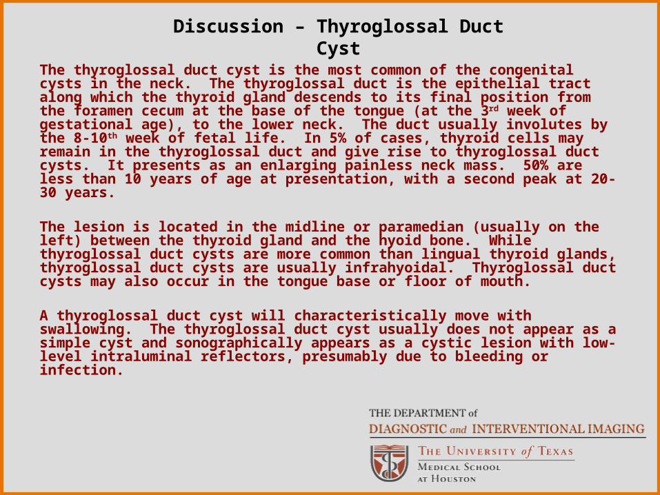
The thyroglossal duct cyst is the most common of the congenital cysts in the neck. The thyroglossal duct is the epithelial tract along which the thyroid gland descends to its final position from the foramen cecum at the base of the tongue (at the 3rd week of gestational age), to the lower neck. The duct usually involutes by the 8-10th week of fetal life. In 5% of cases, thyroid cells may remain in the thyroglossal duct and give rise to thyroglossal duct cysts. It presents as an enlarging painless neck mass. 50% are less than 10 years of age at presentation, with a second peak at 20-30 years.

The lesion is located in the midline or paramedian (usually on the left) between the thyroid gland and the hyoid bone. While thyroglossal duct cysts are more common than lingual thyroid glands, thyroglossal duct cysts are usually infrahyoidal. Thyroglossal duct cysts may also occur in the tongue base or floor of mouth.

A thyroglossal duct cyst will characteristically move with swallowing. The thyroglossal duct cyst usually does not appear as a simple cyst and sonographically appears as a cystic lesion with low-level intraluminal reflectors, presumably due to bleeding or infection.

Discussion – Thyroglossal Duct Cyst

Page 44: Case Report #0431 Submitted by:Jin T. Kim, M.D. Faculty reviewer:Clark W. Sitton, M.D. Date accepted:25 November 2007 Radiological Category:Principal Modality.

The thyroid gland originates at the tip of the foramen cecum of the developing tongue and migrates inferiorly, beginning in the first month of gestation. The thyroid reaches the final location in the 7th week.

Only the lingual thyroid tissue shows marked hyperdensity on the noncontrast enhanced CT scan due to its natural iodine content. The nuclear medicine study will demonstrate whether or not there is the presence of additional thyroid tissue. The nuclear medicine scan should clinch the diagnosis with the absence of thyroid tissue in the usual expected location.

The lesion usually manifests during young adulthood and adolescence because of thyroid enlargement, leading to dysphagia or airway obstruction. In 80% of cases the lingual thyroid tissue is the only functioning thyroid tissue in the body. Because thyroid hormone is vital for healthy growth as a child, the surgeon will often leave the lingual thyroid tissue in through adolescence, unless there is secondary airway obstruction. The presence of the ectopic thyroid does place the patient at risk (5% chance) of papillary cell carcinoma of the thyroid gland at the base of the tongue.

Discussion – Lingual Thyroid

Page 45: Case Report #0431 Submitted by:Jin T. Kim, M.D. Faculty reviewer:Clark W. Sitton, M.D. Date accepted:25 November 2007 Radiological Category:Principal Modality.

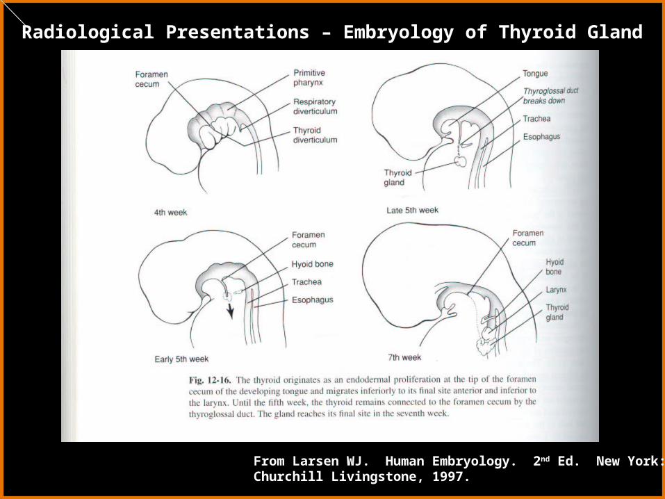
Radiological Presentations – Embryology of Thyroid Gland

From Larsen WJ. Human Embryology. 2nd Ed. New York: Churchill Livingstone, 1997.

Page 46: Case Report #0431 Submitted by:Jin T. Kim, M.D. Faculty reviewer:Clark W. Sitton, M.D. Date accepted:25 November 2007 Radiological Category:Principal Modality.

The hemangioma will enhance intensely and appear similar in appearance to that of an ectopic thyroid on a noncontrast CT. An ectopic thyroid gland may be mistaken for a hemangioma if an enhanced CT is performed prior to a noncontrast CT. Phleboliths may be present.

Discussion – Hemangioma

Page 47: Case Report #0431 Submitted by:Jin T. Kim, M.D. Faculty reviewer:Clark W. Sitton, M.D. Date accepted:25 November 2007 Radiological Category:Principal Modality.

The lingual tonsillar tissue may be also seen at the base of the tongue at the midline position. The lingual tonsillar tissue, however, will not be hyperattenuating on the noncontrast CT and the thyroid gland will be in the expected location on the nuclear medicine study. Hypertrophied tonsillar tissue may simulate an exophytic lymphoma and will appear slightly hyperintense on T1-weighted MRI and very hyperintense on T2-weighted MRI.

Discussion – Enlarged Lingual Tonsils

Page 48: Case Report #0431 Submitted by:Jin T. Kim, M.D. Faculty reviewer:Clark W. Sitton, M.D. Date accepted:25 November 2007 Radiological Category:Principal Modality.

Lingual thyroid

Diagnosis

Page 49: Case Report #0431 Submitted by:Jin T. Kim, M.D. Faculty reviewer:Clark W. Sitton, M.D. Date accepted:25 November 2007 Radiological Category:Principal Modality.

Chung EM. Diseases Affecting the Pediatric Airway. In Levy AD, Chung EM, eds. Radiologic Pathology. Washington, D.C.: American Registry of Pathology, 2006:1369-1370.

Grossman RI and Yousem DM. Neuroradiology: the Requisites. 2nd Ed. Philadelphia: Mosby, 2003:651-653, 661, 738-739.

Larsen WJ. Human Embryology. 2nd Ed. New York: Churchill Livingstone, 1997: 372.

Yousem DM and Da Motta ACBS, Head and Neck Imaging: Case Review Series.2nd Ed. Philadelphia: Mosby, 2006: 215-216.

References