UNIVERSIDADE FEDERAL DO AMAZONAS - UFAM INSTITUTO DE CIÊNCIAS BIOLÓGICAS - ICB PROGRAMA DE PÓS-GRADUAÇÃO EM DIVERSIDADE BIOLÓGICA – PPGDB NÉLLY MARA VINHOTE DA SILVA MANAUS 2008 CARACTERIZAÇÃO TAXONÔMICA E ATIVIDADE ANTIMICROBIANA DE ACTINOMICETOS ASSOCIADOS A LIQUENS FOLHOSOS DE ECOSSISTEMAS AMAZÔNICOS

Welcome message from author
This document is posted to help you gain knowledge. Please leave a comment to let me know what you think about it! Share it to your friends and learn new things together.
Transcript
UNIVERSIDADE FEDERAL DO AMAZONAS - UFAM INSTITUTO DE CIÊNCIAS BIOLÓGICAS - ICB PROGRAMA DE PÓS-GRADUAÇÃO EM DIVERSIDADE BIOLÓGICA – PPGDB
NÉLLY MARA VINHOTE DA SILVA
MANAUS
2008
CARACTERIZAÇÃO TAXONÔMICA E ATIVIDADE
ANTIMICROBIANA DE ACTINOMICETOS ASSOCIADOS A
LIQUENS FOLHOSOS DE ECOSSISTEMAS AMAZÔNICOS

2
UNIVERSIDADE FEDERAL DO AMAZONAS - UFAM INSTITUTO DE CIÊNCIAS BIOLÓGICAS - ICB PROGRAMA DE PÓS-GRADUAÇÃO EM DIVERSIDADE BIOLÓGICA – PPGDB
NÉLLY MARA VINHOTE DA SILVA
Orientador: Prof. Doutor Spartaco Astolfi Filho
Co-Orientador: Prof. Doutor Takeshi Matsuura
MANAUS
2008
CARACTERIZAÇÃO TAXONÔMICA E ATIVIDADE
ANTIMICROBIANA DE ACTINOMICETOS ASSOCIADOS A
LIQUENS FOLHOSOS DE ECOSSISTEMAS AMAZÔNICOS
Dissertação de Mestrado submetida ao Programa de Pós-Graduação em Diversidade Biológica da Universidade Federal do Amazonas, em cumprimento as exigências para obtenção do grau de Mestre em Diversidade Biológica, área de concentração Caracterização da Biota Amazônica.
3
NÉLLY MARA VINHOTE DA SILVA
Aprovado em 11 de março de 2008
BANCA EXAMINADORA
PRESIDENTE: Doutor Takeshi Matsuura (UFAM)
TITULAR: Doutora Maria Ivone Lopes (UFAM)
TITULAR: Doutora Luciana Leomil (UFAM)
TITULAR: Doutor José Odair Pereira (UFAM)
Dissertação de Mestrado submetida ao Programa de Pós-Graduação em Diversidade Biológica da Universidade Federal do Amazonas, em cumprimento as exigências para obtenção do grau de Mestre em Diversidade Biológica, área de concentração Biodiversidade Amazônica.
CARACTERIZAÇÃO TAXONÔMICA E ATIVIDADE
ANTIMICROBIANA DE ACTINOMICETOS ASSOCIADOS A
LIQUENS FOLHOSOS DE ECOSSISTEMAS AMAZÔNICOS

4
Dedico in memorium a meu pai,
Moysés Cirilo da Silva
5
AGRADECIMENTOS
A DEUS, por seu amor incondicional, por sua graça maravilhosa, por sua misericódia que se
renova dia após dia e por seu cuidado dispensado a mim!
À minha família, mãe Nelma Vinhote por seu amor e cuidado. E aos meus manos Marcelo, Nellyane e Murilo, por me darem o suporte de que necessito vocês são muito preciosos!!! A Thiago Marinho e sua família, por seu amor, carinho e atenção e por me ajudar a concluir esta etapa da minha vida! Aos meus orientadores, Professor Doutor Spartaco Astolfi Filho e ao Professor Doutor Takeshi Matsuura, por terem aceitado mais este desafio; A Professora Doutora. Maria Francisca Simas Teixeira, simplesmente por sua vida ser um exemplo de bravura e amor à ciência; Aos Professores, técnicos e estagiários do Laboratório de Microbiologia, especialmente ao Professor MSc. Januário Gama, MSc. Felipe Cruz e ao técnico Beckman; A Coordenação de Aperfeiçoamento a Pesquisa do Ensino Superior – CAPES, a Universidade Federal do Amazonas – UFAM e a Fundação de Amparo a Pesquisa do Estado do Amazonas – FAPEAM, pelo apoio e auxílio financeiro para o desenvolvimento da pesquisa.
Aos amigos do Laboratório de Micologia, Doutora Ormezinda, Profª Doutora Ivone, Teresa, Hérlon, Larissa, Josy, Rosana, Renata, Lorisa e Michel, foram tantos momentos de aprendizado!!! A Professora Doutora Ana Porto, as amigas Germana Michelle, Márcia Karine e Suanni Lemos, pela recepção e auxílio na cidade do Recife. A Professora Doutora Janete Magali do Departamento de Antibióticos-UFPE, pelos ensinamentos e auxílio na pesquisa, e a todos do Laboratório do Deptº. de Antibióticos (Professores, Técnicos e alunos) em especial a Fátima Regina, Orlando, Vânia, Ricardo e Ivana. Aos alunos da primeira turma do Programa de Pós-graduação em Diversidade Biológica (minha turma) Adriano Oliveira, Dhane Eyre Albuquerque, Ilton Oliveira, Josy Caldas, Laís Dias, Michel Martins, Sihame Araújo e Wanessa Cruz. A todos aqueles que colaboraram direta ou indiretamente para realização deste teabalho, em especial as amigas que estão sempre por perto, mesmo quando à distância nos separa: Janaína Vasconcelos, Nayelen Oliveira e Vera Lúcia Cavalcante.
6
Por amor, subimos montanhas, atravessamos mares,
cruzamos desertos e enfrentamos todo tipo de adversidade.
Sem amor, montanhas tornam-se insuperáveis, mares
intransponíveis, desertos insuportáveis e dificuldades
avolumam-se pela vida afora.
Gary Chapman

7
RESUMO O estudo de habitats pouco explorados com a finalidade de obter biocompostos de interesse biotecnológicos produzidos pela diversidade biológica da Amazônia é uma estratégia que permitirá a descoberta de importantes princípios bioativos. Dentre a biodiversidade microbiana, destacam-se os actinomicetos, que são um grupo de bactérias de organização filamentosa que ocorrem em uma grande variedade de substratos e apresentam múltiplas aplicações na indústria farmacêutica, principalmente no que tange a produção de antimicrobianos. Este trabalho teve como escopo analisar taxonomicamente os actinomicetos associados a liquens e determinar a capacidade destas bactérias em produzir antibióticos. Foram coletadas dez amostras de liquens folhosos das árvores presentes na área do Campus Universitário, Setor Sul, da Universidade Federal do Amazonas (UFAM). Para o isolamento dos actinomicetos foram utilizados os meios de cultivo: Ágar Extrato de Levedura-Extrato de Malte-Amido (ISP-2A), Ágar Caseína-Amido (CAA), Ágar Rafinose-Histidina (RHA) e Ágar-Água (AA), suplementados com antifúngicos. Como resultados foram isolados 71 actinomicetos associados aos liquens. Os isolados foram testados quanto à caracterização da atividade antimicrobiana contra oito microrganismos-teste, através de técnicas em meio sólido e em meio líquido. Das linhagens de actinomicetos testados, 80% apresentaram atividade antimicrobiana em meio sólido, principalmente contra Aspergillus niger, Candida albicans e Staphylococcus aureus. No ensaio em meio líquido, 79% das linhagens foram capazes de inibir o crescimento dos microrganismos-teste, sendo que as maiores atividades foram detectadas contra Mycobacterium smegmatis e Staphylococcus aureus. A atividade antimicrobiana dos isolados variou de moderada (halo=13 a 18 mm) a alta (halo=19 a 35 mm) atividade. Observou-se que 68% dos isolados em meio sólido apresentaram alta atividade antimicrobiana frente aos microrganismos-teste. A identificação dos actinomicetos se deu em nível de gênero, através da determinação da micromorfologia, testes fisiológicos e da determinação de aminoácidos da parede celular, sendo a grande maioria pertencente ao gênero Streptomyces. Os microrganismos foram preservados por congelamento a -20 oC e através da técnica de preservação em água (método de Castellani). Palavras-chave: Biodiversidade Amazônica, Metabólitos Secundários, Atividade Antimicrobiana, Actinomicetos, Liquens.

8
ABSTRACT The study of habitats that have been few explored with purpose of obtaining composts with biotechnological interest that has been produced by the Amazonian’s biological diversity it’s a strategy that will allow the discovery of important bioactive principles. In microbial biodiversity, the actinomycetes represents the most important bacterial group. They have a filamentous organization that occur in great varieties of substrata and present a great application in pharmaceutical industry. The scope of this work was to analyze taxonomically the actinomycetes associated at lichens and to determine the capability of these bacteria for antibiotic producing. Ten samples of foliose lichens were collected in area Federal University of Amazonas (UFAM), South Section. For the isolation of actinomycetes were utilized the culture media Yeast Extract-Malt Extract Agar–Starch (ISP-2A), Casein-Starch Agar (CAA), Raffinose-Histidin Agar (RHA) and Water Agar (AA), added with antifungics. Were isolated 71 actinomycetes associated to foliose lichens. The isolated were tested for antimicrobial activity against eight microorganisms-test by the techniques with cultivation in solid medium and broth culture. Among the actinomycetes tested by solid medium 80% shown antimicrobial activity, mainly against Aspergillus niger, Candida albicans and Staphylococcus aureus. In assay by broth cultivation 79% of the actinomycetes inhibited the growth of microorganisms-test, although the higher activities were against Mycobacterium smegmatis and Staphylococcus aureus. The isolated antimicrobial activity varied from moderate (halo=13 at 18 mm) to high (halo=19 at 35 mm) activity. It was observed that 68% of isolated presented high antimicrobial activity. The identification of the actinomycetes was done by the macro and micromorphological determination, physiologic tests and by the determination of aminoacids from the cell wall, and most of them belonging to genus Streptomyces. The microorganisms were preserved by freezing at -20 oC and by preservation of actinomycetes colony directly in water (Castellani’s method). Key words: Amazon Biodiversity, Secundary Methabolites, Antimicrobial Activity, Actinomycetes, Lichens.

9
LISTA DE ILUSTRAÇÕES
Quadro 01 Microrganismos-teste utilizados na caracterização da atividade
antimicrobiana............................................................................................ 20
Figura 01 Microrganismo-teste utilizado na caracterização da Atividade
Antimicrobiana...........................................................................................
28
Figura 02 Identificação dos actinomicetos isolados de liquens folhosos de
ecossistemas amazônicos........................................................................... 35
Figura 03 Atividade antimicrobiana em bloco de gelose dos actinomicetos
isolados de liques....................................................................................... 36
Figura 04 Atividade antimicrobiana pelo método de difusão em disco dos
actinomicetos isolados...............................................................................
37

10
LISTA DE TABELAS
Tabela 1 Quantitativo de amostras de actinomicetos isolados de liquens nos meios
de cultivo..................................................................................................... 31
Tabela 2 Caracterização morfológica de amostras de actinomicetos isolados de
liquens......................................................................................................... 32
11
SUMÁRIO
INTRODUÇÃO
1.1 Diversidade microbiana 12
1.2 Os actinomicetos 13
1.3 Habitats inivadores 13
1.4 Os liquens 14
1.5 Potencial Biotecnológico 15
1.6 Potencial Farmacológico dos actinomicetos 16
2. OBJETIVOS
2.1 Geral 17
2.2 Específicos 17
3. MATERIAL E MÉTODOS
3.1 Amostra 18
3.2 Coleta, acondicionamento e transporte das amostras 18
3.3 Isolamento dos actinomicetos dos liquens 18
3.4 Identificação dos actinomicetos 18
3.4.1 Macromorfologia e micromorfologia 18
3.4.2 Determinação de aminoácidos presentes na parede celular 19
3.5 Caracterização da atividade antimicrobiana 19
3.5.1 Ensaio da atividade antimicrobiana em meio sólido 20
3.5.2 Ensaio para atividade antimicrobiana por difusão em disco 20
3.6 Preservação dos actinomicetos 21
3.7 Análise estatística 21
4. ARTIGO - Taxonomic Characterization and Antimicrobial Activi ty of
Actinomycetes Associated with Foliose Lichens from the
Amazonian Ecosystems
22
REFERÊNCIAS 43
ANEXOS 49

12
INTRODUÇÃO
1.1 Diversidade Microbiana
Os microrganismos são entidades bióticas que são capazes de colonizar com sucesso
cada nicho ecológico possível do planeta. Além disso, representam uma importante fonte de
recursos genéticos para o avanço biotecnológico e para o desenvolvimento econômico
sustentável (OLIVEIRA et al., 2006).
O número de espécies microbianas identificadas cresce a cada ano, sendo formalmente
descritos mais de 70.000 fungos, 36.000 protozoários, 30.000 algas, 5.000 bactérias e 3.600
vírus. Mas esses números ainda estão longe do total de espécies microbianas, estimado em
mais de dois milhões (ROSSELÓ-MORA, 2001; AMANN, 2001; FAORO, 2006).
O Brasil detém cerca de 20% da diversidade biológica mundial (SUDAM, 1995;
DIAS, 1996; SOUZA et al., 2004) e uma parcela considerável desta biodiversidade está
localizada nos ecossistemas Amazônicos, que abriga uma das últimas extensões contínuas de
florestas tropicais úmidas da Terra, detendo cerca de 1/3 do estoque genético planetário. O
aproveitamento dos recursos genéticos amazônicos, a partir de seus usos pelas novas
biotecnologias, embora seja uma questão emergente, é ainda pouco compreendida, mensurada
e principalmente incorporada às políticas governamentais e às estratégias empresariais
direcionadas para a região (ALBAGLI, 2001).
Apesar da imensa diversidade biológica da Amazônia, as espécies que a compõem e
suas relações filogenéticas são pouco conhecidas, especialmente as interações dos
microrganismos com os outros seres (SEIDL, 1993; SOUZA et al., 2004). A razão para o
baixo número de espécies formalmente descritas são as necessidades do cultivo celular no
processo de identificação dos microrganismos, o que na grande maioria das vezes não é
possível (PACE et al., 1986). Bull et al. (2000) e Conti (2007) relataram que menos de 1%
das bactérias e apenas 7% dos fungos, foram isolados e cultivados em laboratório, precisando
de novas tecnologias para o cultivo destes microrganismos.
O conhecimento da biodiversidade e bioprospecção de novos microrganismos
tornaram-se uns dos focos principais da era biotecnológica, visto que a utilização destes

13
organismos na busca de soluções nas áreas de alimento, saúde, meio ambiente e indústria vêm
crescendo de forma acelerada no atual cenário mundial (OLIVEIRA et al., 2006).
1.2 Os actinomicetos
Dentre a biodiversidade bacteriana, destacam-se os actinomicetos, que são um grupo
de bactérias de organização filamentosa, muitas vezes ramificada, cuja característica comum é
a formação de micélio aéreo e/ou vegetativo em algum estágio de seu ciclo de vida
(McCARTHY; WILLIAMS, 1990). As características essenciais da natureza procariótica dos
actinomicetos são a ausência de membrana nuclear, a ausência de organelas citoplasmáticas e
a presença de ribossomos 70S (STANIER et al., 1980; WOESE, 1992). Essas bactérias são na
grande maioria aeróbias estritas, Gram-positivas com alto conteúdo de Guanina e Citosina (G
+ C), e formam filamentos ramificados ou hifas que podem persistitir como micélio estável ou
podem se quebrar em elementos na forma de bacilos ou cocos (HOLT et al., 1994).
Os actinomicetos ocorrem em uma grande diversidade de habitats naturais e artificiais,
crescendo em uma variedade de substratos. O solo é o ambiente mais comum para os
actinomicetos, onde ocorrem em uma proporção de aproximadamente um milhão de células
por grama de solo. Embora a maioria seja sapróbio estrito, alguns formam associações
parasíticas ou simbióticas com plantas ou animais. (GOODFELLOW; WILLIAMS, 1983).
Apesar de o solo ser o principal substrato para os actinomicetos, estes também são
encontrados na água, em tecidos vegetais e em tecidos animais (WILLIAMS; CROSS, 1974).
Essa ubiqüidade dos actinomicetos sapróbios em ambientes naturais possuem dois fatores
principais: diversidade metabólica e evolução de mecanismos específicos de dispersão
(McCARTHY; WILLIAMS, 1990).
1.3 Habitats inovadores
O estudo de habitats pouco explorados buscando isolar actinomicetos da Amazônia
produtores de novos compostos bioativos é uma estratégia que permitirá a descoberta de
princípios ativos ainda desconhecidos e com importantes aplicações biotecnológicas.
Pesquisas realizadas por González et al. (2005); Terkina et al. (2006); Chin et al. (2006);
Cardinale et al.(2006) e Gunatilaka (2006) mostram a importância de se explorar a
biodiversidade na busca por produtos naturais e sua importância no desenvolvimento de
diversos bioprodutos.
14
1.4 Os liquens
O líquen é uma associação simbiótica entre um fungo e um microrganismo
fotossintetizante. O componente fúngico de um líquen (o micobionte) é, em sua grande
maioria, um fungo do Filo Ascomycota (acima de 95%) e, mais raramente, Basidiomycota. O
componente fotossintetizante (fotobionte, também chamado de ficobionte em alusão à alga) é,
em geral, uma Chlorophyta ou uma Cyanobactéria, no qual o micobionte protege o fotobionte
dos extremos de temperatura e umidade, que em contrapartida oferece os produtos da
fotossíntese (SEAWARD, 1977; NASH, 1996). A organização estrutural e morfológica dos
talos liquênicos se apresenta sob muitos hábitos (formas de crescimento; aparência). Esses
hábitos separam morfologicamente os liquens em tipos, como: fruticuloso (fruticoso,
arbustiva) o talo pode se apresentar na forma de um arbusto em miniatura, com ou sem
ramificações, mas sem diferenciação dorsi-ventral; quando o lado ventral está firmemente
ligado ao substrato ou ao ponto de fixação, sendo impossível separá-los, denomina-se
crustoso (crustáceo) e em outros casos, há uma nítida diferenciação dorsiventral, sendo o talo
formado por uma fita ramificada ou não, facilmente separável do ponto de fixação,
apresentando o formato folhoso (foliáceo) (MARCELLI, 1996).
Dessa associação resultam muitas substâncias liquênicas que participam ativamente da
dinâmica microbiana no solo, como também possuem grande espectro de aplicação
biotecnológica (NASH III, 1996). Os metabólitos secundários dos liquens, especialmente os
ácidos liquênicos, possuem atividade antibiótica e citotóxica, sendo fontes potenciais de
novos antibióticos e drogas anti-neoplásicas. Por serem extremamente sensíveis a poluentes
atmosféricos, inclusive os acumulando, os líquens são excelentes bioindicadores e
biomonitores da qualidade do ar. Testes com liquens comprovam a ocorrência frequente de
metabólitos com propriedades antibacteriana, antimicobacteriana, antiviral, antitumoral,
analgésica e antipirética (MULLER, 2001).
Os liquens são cosmopolitas, sendo encontrados em todos os possíveis tipos de
ambientes terrestres, inclusive aqueles mais extremos, como desertos, geleiras e afloramentos
rochosos. Mesmo os componentes simbiônticos dos liquens já terem sido descritos
extensivamente, porém a comunidade microbiana que habita estes nichos ainda não foi bem
caracterizada. Os liquens constituem um reservatório rico, para o isolamento de uma grande
diversidade de actinomicetos, muitos deles representando uma fonte inexplorada, rica de
metabólitos secundários (GONZÁLEZ et al., 2005).

15
1.5 Potencial Biotecnológico
Existe em todo o mundo uma crescente busca de microrganismos com potencialidade
biotecnológica. A descoberta de novos microrganismos, bem como sua preservação e
classificação apropriada, passa a ser imprescindível para que a biodiversidade seja
convenientemente utilizada, mantida e transformada em riquezas (AZEVEDO, 1998).
A comunidade de microrganismos existentes possui uma ampla diversidade biológica
e bioquímica e representa um recurso ainda pouco explorado e de enorme valor para o futuro
(LABEDA, 1990; BULL; HARDMAN, 1991). Desde a segunda metade do século XX foram
originados cerca de 50.000 produtos naturais provenientes de microrganismos, dos quais mais
de 10.000 são biologicamente ativos e mais de 8.000 são agentes antibióticos e antitumorais
(FENICAL, 1993).
A exploração dos microrganismos pela indústria gera bilhões de dólares a cada ano. O
valor dos microrganismos é geralmente avaliado pela potencial aplicação direta nos processos
biotecnológicos ou valor de mercado dos produtos derivados (KURTBÖKE et al., 2004).
Os microrganismos podem produzir uma variedade muito grande de metabólitos tanto
primários quanto secundários (SOUZA et al., 2004) incluindo enzimas (STAMFORD et al.,
1998), aminoácidos (KLEINKAUF; VON DOHREN, 1990), vitaminas, antibióticos,
pigmentos (DEMAIN, 1992), agentes moduladores de respostas imunológicas (TRILLI et al.,
1978), toxinas (BACH; KIMATI, 1999), agentes anti-tumorais (WANG et al., 2000), fatores
de crescimento de plantas (ALEXOPOULOS, 1996), anti-helmínticos (RODRIGUES et al.,
2000) e antifúngicos (LI; STROBEL, 2001).
Em decorrência do potencial biotecnológico dos microrganismos, esses metabólitos
constituem os principais produtos de interesse econômico para a agricultura, medicina e
indústria farmacêutica (DEMAIN, 2000; ADRIO; DEMAIN, 2003).
Pesquisas demonstram que a segunda maior causa de morte no mundo é ocasionada
por doenças infecciosas em decorrência a alta prevalência de microrganismos patogênicos
resistentes a diversos antibióticos sendo necessário, portanto, a busca por novos
antimicrobianos mais eficazes e de baixa toxicidade (LEVY; MARSHALL, 2004;
APPELBAUM; JACOB, 2005; YONEYAMA; KATSUMATA, 2006; BUTLER; BUSS,
2006; RATTI, 2007; CONTI, 2007).
Atualmente, mais de 70% das espécies das bactérias que causam infecções, são
resistentes a pelo menos um dos antibióticos comumente utilizados na terapêutica como
enfatizado por Overbye; Barret (2005). Contudo, Ujikawa (2003) afirma que está se tornando

16
cada vez mais raro a descoberta de novos tipos de antibióticos, encontrando-se apenas
variações de classes conhecidas (KNOWLES, 1977; OMURA, 1992).
Para contornar esta situação, a biospropecção de metabólitos bioativos precisa ser
melhorada, através da utilização de novas tecnologias e alternativas como: engenharia
genética; biossíntese mutacional; triagem de alta produtividade; e uma maior eficiência na
identificaçao de moléculas, de forma a viabilizar economicamente o processo (BULL et al.,
2000; PAREKH et al., 2000; HIGGS et al., 2001; FLOSS, 2001; BOGGS; MILLER, 2004;
BUTLER, 2004; GULLO; HUGHES, 2005; SAMIULLA et al., 2005; WEIST; SUSSMUTH,
2005; CONTI, 2007).
1.6 Potencial farmacológico dos actinomicetos
Entre os antibióticos isolados, aproximadamente 68% são elaborados por
actinomicetos, 14% por vegetais superiores, 10 a 15% por fungos imperfeitos, próximo do
mesmo valor por bactérias do gênero Bacillus, cerca de 6% são sintetizados por
Basidiomycetes e Ascomycetes e aproximadamente 2% são de origem animal (PHAFF,
1991).
Os actinomicetos têm sido especialmente úteis na indústria farmacêutica por sua
capacidade ilimitada de produzir metabólitos secundários com diversas estruturas químicas e
atividades biológicas. A produção de 2/3 dos antibióticos de ocorrência natural é de
actinomicetos.
Dentre os actinomicetos, o que mais desperta interesse científico é o gênero
Streptomyces, responsável pela produção de mais de 80% dos antibióticos atualmente
utilizados (CHALIS; HOPWOOD, 2003; RATTI, 2007), seguido pelos gêneros
Micromonospora, Nocardia, Streptosporangium e Actinoplanes (CROSS, 1981).
A devida caracterização e preservação dos recursos microbianos são fatores de
fundamental importância para o desenvolvimento da bioeconomia no século 21 (OLIVEIRA
et al., 2006).
17
2. OBJETIVOS
2.1. Geral
Caracterizar taxonomicamente os actinomicetos isolados de liquens folhosos e
determinar a capacidade destas bactérias em produzir antibióticos.
2.2 Específicos
• Isolar actinomicetos de liquens folhosos, empregando diferentes meios de cultivo;
• Classificar taxonomicamente os actinomicetos isolados;
• Testar as amostras isoladas quanto à capacidade de produção de compostos com atividade
antimicrobiana contra microrganismos patogênicos ao homem.

18
3. MATERIAL E MÉTODOS
3.1 Amostra
Foram coletados 10 espécimes de um líquen folhoso das árvores presente na área da
floresta do Campus Universitário, Setor Sul, da Universidade Federal do Amazonas - UFAM
(Sul 3º 5’ 56’’ / Oeste 59º 58’56’’) Manaus/AM.
3.2 Coleta, acondicionamento e transporte das amostras
As amostras foram coletadas com auxílio de uma espátula metálica limpa com solução
de etanol a 70%, acondicionadas individualmente em placas de Petri vazias estéreis, mantidas
em caixas isotérmicas e processadas em laboratório.
3.3 Isolamento dos actinomicetos dos liquens
Foram pesados 300 mg de cada líquen, estes foram lavados duas vezes com água
destilada estéril, e em seguida triturados e homogeneizados com 30 mL de água destilada
estéril. Após esse processo foram realizadas diluições sucessivas para isolamento dos
actinomicetos. Foram inoculados 0,1 mL da diluição nos meios de cultura Ágar Extrato de
Levedura-Extrato de Malte - Amido (ISP-2A), Ágar Caseína-Amido (CAA), Ágar Rafinose-
Histidina (RHA) e Ágar-Água (AA), contendo o antifúngico cicloheximida (80 µg/mL)
(González et al., 2005) ou nistatina, e as placas foram incubadas a 30 oC por até 21 dias.
3.4 Identificação dos actinomicetos
3.4.1 Macromorfologia e micromorfologia
A inoculação das actinobactérias nos meios ISP-2, ISP-6 e ISP-7 a 30 oC por até 21
dias, permitiram o estudo da macromorfologia das colônias, avaliando-se visualmente a
coloração do verso e anverso das colônias e a produção de pigmento melanínico solúvel.
19
O estudo da micromorfologia das linhagens cultivadas nos meios ISP-2, ISP-3, ISP-4 e
ISP-5, e incubadas a 30 oC por até 21 dias, foi realizada através de microscopia óptica,
avaliando-se a morfologia e o formato das cadeias de esporos.
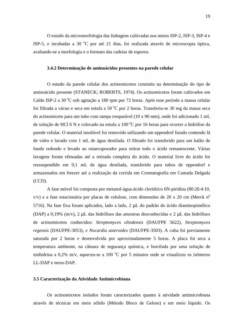
3.4.2 Determinação de aminoácidos presentes na parede celular
O estudo da parede celular dos actinomicetos consistiu na determinação do tipo de
aminoácido presente (STANECK; ROBERTS, 1974). Os actinomicetos foram cultivados em
Caldo ISP-2 a 30 oC sob agitação a 180 rpm por 72 horas. Após esse período a massa celular
foi filtrada a vácuo e seca em estufa a 50 oC por 2 horas. Transferiu-se 30 mg da massa seca
do actinomiceto para um tubo com tampa rosqueável (10 x 90 mm), onde foi adicionado 1 mL
de solução de HCl 6 N e colocado na estufa a 100 oC por 16 horas para ocorrer a hidrólise da
parede celular. O material insolúvel foi removido utilizando um eppendorf furado contendo lã
de vidro e lavado com 1 mL de água destilada. O filtrado foi transferido para um balão de
fundo redondo e levado ao rotaevaporador para retirar todo o ácido remanescente. Várias
lavagens foram efetuadas até a retirada completa do ácido. O material livre do ácido foi
ressuspendido em 0,1 mL de água destilada, transferido para tubos de eppendorf e
armazenados em freezer até a realização da corrida em Cromatografia em Camada Delgada
(CCD).
A fase móvel foi composta por metanol-água-ácido clorídrico 6N-piridina (80:26:4:10,
v/v) e a fase estacionária por placas de celulose, com dimensões de 20 x 20 cm (Merck no
5716). Na fase fixa foram aplicados, lado a lado, 2 µL do padrão do ácido diaminopimélico
(DAP) a 0,19% (m/v), 2 µL das hidrólises das amostras desconhecidas e 2 µL das hidrólises
de actinomicetos conhecidos: Streptomyces olindensis (DAUFPE 5622), Streptomyces
regensis (DAUFPE-3053), e Nocardia asteroides (DAUFPE-3503). A cuba foi previamente
saturada por 2 horas e desenvolvida por aproximadamente 5 horas. A placa foi seca a
temperatura ambiente, na câmara de segurança química, e borrifada por uma solução de
ninhidrina a 0,2% m/v, aqueceu-se a 100 oC por 5 minutos onde se visualizou os isômeros
LL-DAP e meso-DAP.
3.5 Caracterização da Atividade Antimicrobiana
Os actinomicetos isolados foram caracterizados quanto à atividade antimicrobiana
através de técnicas em meio sólido (Método Bloco de Gelose) e em meio líquido. Os
20
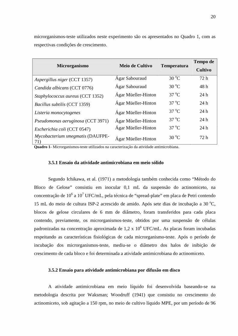
microrganismos-teste utilizados neste experimento são os apresentados no Quadro 1, com as
respectivas condições de crescimento.
Microrganismo Meio de Cultivo Temperatura Tempo de
Cultivo
Aspergillus niger (CCT 1357) Ágar Sabouraud 30 oC 72 h
Candida albicans (CCT 0776) Ágar Sabouraud 30 oC 48 h
Staphylococcus aureus (CCT 1352) Ágar Müeller-Hinton 37 oC 24 h
Bacillus subtilis (CCT 1359) Ágar Müeller-Hinton 37 oC 24 h
Listeria monocytogenes Ágar Müeller-Hinton 37 oC 24 h
Pseudomonas aeruginosa (CCT 3971) Ágar Müeller-Hinton 37 oC 24 h
Escherichia coli (CCT 0547) Ágar Müeller-Hinton 37 oC 24 h
Mycobacterium smegmatis (DAUFPE-71)
Ágar Müeller-Hinton 30 oC 72 h
Quadro 1- Microrganismos-teste utilizados na caracterização da atividade antimicrobiana.
3.5.1 Ensaio da atividade antimicrobiana em meio sólido
Segundo Ichikawa, et al. (1971) a metodologia também conhecida como “Método do
Bloco de Gelose” consistiu em inocular 0,1 mL da suspensão do actinomiceto, na
concentração de 106 a 107 UFC/mL, pela técnica de “spread-plate” em placa de Petri contendo
15 mL do meio de cultura ISP-2 acrescido de amido. Após sete dias de incubação a 30 oC,
blocos de gelose circulares de 6 mm de diâmetro, foram transferidos para cada placa
contendo, previamente, os microrganismos-teste, obtidos por uma suspensão de células
padronizadas na concentração aproximada de 1,2 x 10⁶ UFC/mL. As placas foram incubadas
respeitando as características fisiológicas de cada microrganismo-teste. Após o período de
incubação dos microrganismos-teste, mediu-se o diâmetro dos halos de inibição de
crescimento de cada bloco e foi determinada a atividade antimicrobiana do actinomiceto.
3.5.2 Ensaio para atividade antimicrobiana por difusão em disco
A atividade antimicrobiana em meio líquido foi desenvolvida baseando-se na
metodologia descrita por Waksman; Woodruff (1941) que consistiu no crescimento do
actinomiceto, sob agitação a 150 rpm, no meio de cultivo líquido MPE, por um período de 96

21
horas a 30o C. A concentração de células inoculadas foi de 106 a 107 UFC/mL. Ao final do
período de crescimento, 10 µL do líquido metabólico foi retirado e transferido para um disco
de papel com 6 mm de diâmetro, e introduzido em placas de Petri contendo, previamente, os
microrganismos-teste, obtidos por uma suspensão de células padronizadas na concentração
aproximada de 1,2 x 10⁶ UFC/mL, semeados pela técnica de “spread-plate”. Após o período
de incubação, os halos de inibição de crescimento dos microrganismos-teste, de cada disco,
foram mensurados e determinou-se a atividade inibitória do actinomiceto.
3.6 Preservação dos actinomicetos
Os microrganismos isolados foram preservados por congelamento a -20 oC, e através
da técnica de preservação em água (Método de Castellani), descritos em Muro; Luchi (1989).
3.7 Análise estatística
Os dados foram submetidos à análise estatística descritiva cujo objetivo é o de
sintetizar uma série de valores de mesma natureza, permitindo dessa forma que se tenha uma
visão global da variação desses valores, organizar e descrever os dados de três maneiras: por
meio de tabelas, de gráficos e de medidas descritivas (REIS, 1998).

22
4. ARTIGO
Australian Journal of Basic and Applied Sciences
Manuscript Review Form
(To be returned to the author, no reviewer identifiers please)
Manuscript Title:
Taxonomic Characterization and Antimicrobial Activity of Actinomycetes Associated with
Foliose Lichens from the Amazonian Ecosystems
Manuscript No.: 2575-AJBAS
Manuscript Type: Research ✔ Review Case Study Short Communication
Note: ✔ shows selected option.
Other comments (If any): no comments
Conclusion: 1 - Publish as it is
2 - Publish with minor corrections ✔
3 - Send for corrections
4 - Reject
Date: 05-04-2010

23
Taxonomic Characterization and Antimicrobial Activi ty of Actinomycetes Associated
with Foliose Lichens from the Amazonian Ecosystems
Nélly Mara Silva-Vinhote¹; Thiago Marinho-Pereira2; Spartaco Astolfi-Filho3; Takeshi
Matsuura¹
¹ Departamento de Parasitologia, Universidade Federal do Amazonas – UFAM, Laboratório
de Microbiologia; Av. Gal. Rodrigo Octavio Jordão Ramos, 3000, Aleixo; CEP 69.000-070;
Manaus-Amazonas-Brasil.
2 Coordenação de Pesquisas em Aqüicultura, Instituto Nacional de Pesquisas da Amazônia –
CPAQ/INPA; Av. André Araújo, 2936, Aleixo; CEP 69.083-000; Manaus-Amazonas-Brasil.
3 Departamento de Biologia, Universidade Federal do Amazonas – UFAM/CAM – Centro de
Apoio Multidisciplinar; Av. Gal. Rodrigo Octávio Jordão Ramos, 3000, Aleixo; CEP 69.000-
070; Manaus-Amazonas-Brasil.
* e-mails: [email protected]
24
Caracterização Taxonômica e Atividade Antimicrobiana de Actinomicetos Associados a
Liquens Folhosos de Ecossistemas Amazônicos
Resumo
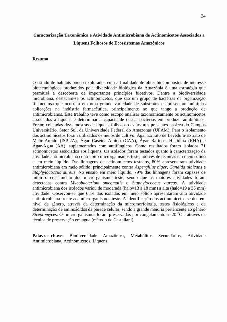
O estudo de habitats pouco explorados com a finalidade de obter biocompostos de interesse biotecnológicos produzidos pela diversidade biológica da Amazônia é uma estratégia que permitirá a descoberta de importantes princípios bioativos. Dentre a biodiversidade microbiana, destacam-se os actinomicetos, que são um grupo de bactérias de organização filamentosa que ocorrem em uma grande variedade de substratos e apresentam múltiplas aplicações na indústria farmacêutica, principalmente no que tange a produção de antimicrobianos. Este trabalho teve como escopo analisar taxonomicamente os actinomicetos associados a liquens e determinar a capacidade destas bactérias em produzir antibióticos. Foram coletadas dez amostras de liquens folhosos das árvores presentes na área do Campus Universitário, Setor Sul, da Universidade Federal do Amazonas (UFAM). Para o isolamento dos actinomicetos foram utilizados os meios de cultivo: Ágar Extrato de Levedura-Extrato de Malte-Amido (ISP-2A), Ágar Caseína-Amido (CAA), Ágar Rafinose-Histidina (RHA) e Ágar-Água (AA), suplementados com antifúngicos. Como resultados foram isolados 71 actinomicetos associados aos liquens. Os isolados foram testados quanto à caracterização da atividade antimicrobiana contra oito microrganismos-teste, através de técnicas em meio sólido e em meio líquido. Das linhagens de actinomicetos testados, 80% apresentaram atividade antimicrobiana em meio sólido, principalmente contra Aspergillus niger, Candida albicans e Staphylococcus aureus. No ensaio em meio líquido, 79% das linhagens foram capazes de inibir o crescimento dos microrganismos-teste, sendo que as maiores atividades foram detectadas contra Mycobacterium smegmatis e Staphylococcus aureus. A atividade antimicrobiana dos isolados variou de moderada (halo=13 a 18 mm) a alta (halo=19 a 35 mm) atividade. Observou-se que 68% dos isolados em meio sólido apresentaram alta atividade antimicrobiana frente aos microrganismos-teste. A identificação dos actinomicetos se deu em nível de gênero, através da determinação da micromorfologia, testes fisiológicos e da determinação de aminoácidos da parede celular, sendo a grande maioria pertencente ao gênero Streptomyces. Os microrganismos foram preservados por congelamento a -20 oC e através da técnica de preservação em água (método de Castellani). Palavras-chave: Biodiversidade Amazônica, Metabólitos Secundários, Atividade Antimicrobiana, Actinomicetos, Liquens.

25
Taxonomic Characterization and Antimicrobial Activity of Actinomycetes Associated
with Foliose Lichens from the Amazonian Ecosystems
Abstract The study of habitats that have been few explored with purpose of obtaining composts with biotechnological interest that has been produced by the Amazonian’s biological diversity it’s a strategy that will allow the discovery of important bioactive principles. In microbial biodiversity, the actinomycetes represents the most important bacterial group. They have a filamentous organization that occur in great varieties of substrata and present a great application in pharmaceutical industry. The scope of this work was to analyze taxonomically the actinomycetes associated at lichens and to determine the capability of these bacteria for antibiotic producing. Ten samples of foliose lichens were collected in area of Universidade Federal do Amazonas (UFAM), South Section. For the isolation of actinomycetes were utilized the culture media Yeast Extract-Malt Extract Agar–Starch (ISP-2A), Casein-Starch Agar (CAA), Raffinose-Histidin Agar (RHA) and Water Agar (AA), added with antifungics. Were isolated 71 actinomycetes associated to foliose lichens. The isolated were tested for antimicrobial activity against eight microorganisms-test by the techniques with cultivation in solid medium and broth culture. Among the actinomycetes tested by solid medium 80% shown antimicrobial activity, mainly against Aspergillus niger, Candida albicans and Staphylococcus aureus. In assay by broth cultivation 79% of the actinomycetes inhibited the growth of microorganisms-test, although the higher activities were against Mycobacterium smegmatis and Staphylococcus aureus. The isolated antimicrobial activity varied from moderate (halo=13 at 18 mm) to high (halo=19 at 35 mm) activity. It was observed that 68% of isolated presented high antimicrobial activity. The identification of the actinomycetes was done by the macro and micromorphological determination, physiologic tests and by the determination of aminoacids from the cell wall, and most of them belonging to genus Streptomyces. The microorganisms were preserved by freezing at -20 oC and by preservation of actinomycetes colony directly in water (Castellani’s method). Key words: Amazon Biodiversity, Secundary Methabolites, Antimicrobial Activity, Actinomycetes, Lichens.

26
Introduction
In global level is considered that the diversity of microorganisms exceeds in order of
some thousands the diversity of plants and animals. Brazil has about 20% of the world
biological diversity (SUDAM, 1995; Dias, 1996; Souza et al., 2004) and a considerable
portion of this biodiversity is located in Amazonian ecosystems, incommensurable source of
raw materials for the most several areas of biotechnological application.
Among the microbial biodiversity, the actinomycetes represent a bacteria group of
filamentous organization, many times ramified, whose common characteristic is the formation
of aerial and/or vegetative mycelium in some stage of its life cycle (McCarthy; Williams,
1990). The actinomycetes occur in a great diversity of natural and artificial habitats, growing
in a large variety of substrata (Williams; Cross, 1974).
The lichen is a symbiotic association between a fungal and a microorganism
photosynthetic. The lichen fungal component (mycobiont) is in great majority a fungal of the
phylo Ascomycota (above 95%) and, rarely, Basidiomycota. The photosynthetic component
(photobiont, also knew ficobionte in allusion to the algae) it is, in general, a Chlorophyta or a
cyanobacteria (Seaward, 1977; Nash, 1996).
Even the symbiotic components of the lichens have already been described
extensively, however the microbial community that habits these niches still remain not
characterized. The lichens constitute a rich reservoir for the isolation of a great variety of
actinomycetes diversity, many of them representing an unexplored source, rich in secondary
metabolites (González et al., 2005).
In nowadays, more than 70% of the species of the bacteria that cause infections are
resistant at least one of the antibiotics commonly used on therapeutics as it is emphasizes for
Overbye and Barret (2005). The actinomycetes have especially been useful in the
pharmaceutical industry for limitless capacity to produce secondary metabolites with many
chemistries structures and biological activities.
The actinomycetes bioprospection in innovative habitats is a strategy that makes
possible the discovery of relevant biotechnological bioactive principles, more scientific
knowledge about the microbial diversity, better understanding about functions of microbial
communities in the environment and knowledge of these interactions with others components
of the biodiversity.
This work proposes to characterize taxonomically the actinomycetes isolated from
folioses lichens and to determine the capacity of these bacteria in producing antibiotics.

27
Material and Methods
Samples
Ten samples of foliose lichen were collected from the trees of Amazon Federal
University (UFAM) campus, south sector, (Sul 3º 5’ 56’’ / Oeste 59º 58’56’’), Manaus/AM.
Collect, packing and samples transportation
The samples were collected with a cleaned metal spatula, individually packing in
sterile Petri’s dishes and kept in isotherm boxes, being processed at laboratory.
Isolation of actinomycetes from liquens
Around 300 mg of each lichen were weighted washed twice with sterile distilled water
and homogeneized with 30 mL of sterile distilled water. After this, it was made successive
dilutions for the actinomycetes isolation. Were inoculated 0,1 mL of dilutions in culture
medium Yeast Extract-Malt Extract Agar (ISP-2), Starch-Casein Agar (SCA), Rafinosis-
Histidine Agar (RHA) and Water-Agar (WA), added with antifungal cycloheximide (80
µg/mL) (González et al., 2005) or nistatine, and the plates were incubated at 30 oC during 21
days.
Actinomycetes identification
Macromorphology and micromorphology determination
The actinobacteria inoculated on Petri’s dishes with media ISP-2, ISP-6 and ISP-7 at
30 oC per until 21 days permitted the colony macromorphology study. The color and
production of soluble melaninic pigment were visually evaluated.
The micromorphology study of isolated actinomycetes on media ISP-2, ISP-3, ISP-4
and ISP-5, incubated at 30 oC until 21 days, and morphology and spore chains format were
evaluated through optic microscopy.

28
Determination of amino acid in cell wall
The study of actinomycetes cell wall consisted in determination of present amino acid
type (Staneck; Roberts, 1974). The actinomycetes were cultivated in ISP-2 Broth at 30 oC
under shaking at 180 rpm per 72 hours. After this period, the cell mass was filtrated at
vacuum and dry at 50 oC per two hours. Were transferred 30 mg of actinomycete dry mass to
tube (10 x 90 mm), acidified with 1 mL of HCl 6 N solution and the cell wall was hydrolysed
at 100 oC per 16 hours. The insoluble material was removed using one holed eppendorf
containing glass wool and washed with 1 mL of distilled water. The filtrated was transferred
to balloon of round bottom and evaporated to remove the acid remaining. Several washes
were made until the complete retreat of acid. The material free of the acid was resuspensed in
0.1 mL of distilled water, transferred for eppendorf tubes and stored in freezer until the
accomplishment of thin layer chromatography (TLC).
The mobile phase was composed by methanol-water-acid chloridric 6N-piridine
(80:26:4:10, v/v) and the stationary phase by cellulose plates with 20 x 20 cm of dimentions
(Merck no 5716). In stationary phase were applied, side by side, 2 µL of diaminopimelic acid
standard (DAP) at 0.19% (m/v), 2 µL of the hydrolysis of unknown samples and 2 µL of the
hydrolysis of known actinomycetes: Streptomyces olindensis (DAUFPE 5622), Streptomyces
regensis (DAUFPE-3053), and Nocardia asteroides (DAUFPE-3503). The cube was
previously saturated for two hours and run per approximately five hours. The cellulose plate
was dry at room temperature, sprinkled with ninhidrine solution at 0.2% m/v, and warmed at
100 oC during five minutes and then visualizated the LL-DAP e meso-DAP isomers.
Antimicrobial activity characterization
The isolated actinomycetes were characterizated for antimicrobial activity through
solid (Gelose Block Method) and broth media. The microrganisms-test used in this
experiment are presented in Figure 1, with the respective growth conditions.
Microorganisms-test Culture media Temperature Period of
cultivation
Aspergillus niger (CCT 1357) Sabouraud Agar 30 oC 72 h
Candida albicans (CCT 0776) Sabouraud Agar 30 oC 48 h
Staphylococcus aureus (CCT Müeller-Hinton Agar 37 oC 24 h

29
1352)
Bacillus subtilis (CCT 1359) Müeller-Hinton Agar 37 oC 24 h
Listeria monocytogenes Müeller-Hinton Agar 37 oC 24 h
Pseudomonas aeruginosa (CCT 3971)
Müeller-Hinton Agar 37 oC 24 h
Escherichia coli (CCT 0547) Müeller-Hinton Agar 37 oC 24 h
Mycobacterium smegmatis (DAUFPE-71)
Müeller-Hinton Agar 30 oC 72 h
Fig. 1: Microrganisms-test used for characterization of antimicrobial activity.
Antimicrobial activity assay in solid media
According to Ichikawa et al. (1971), the methodology also known as "Gelose Block
Method" consisted in inoculate 0.1 mL of actinomycete suspension in the concentration of 106
to 107 CFU/mL, for the "spread-plate" technique in Petri plates containing 15 mL of the ISP-2
culture medium added with starch. After seven days of incubation at 30 oC, circular gelose
blocks of 6 mm diameter were transferred for each plate containing, previously, the test
microorganism, obtained by a suspension of standardized cells in approximated concentration
of 1,2 x 10⁶ CFU/mL. The plates were incubated respecting the physiologic characteristics of
each test microorganism. After the incubation period of the test microorganism, the diameter
of the growth inhibition of each block was measured and determinate the antimicrobial
activity of the actinomycete.
Antimicrobial activity assay in broth culture
The antimicrobial activity by cultivation of actinomycetes in broth was developed with
based on methodology describes by Waksman and Woodruff (1941) that consisted in growth
the actinomycete under shaking at 150 rpm in MPE broth until a period of 96 hours at 30 oC.
The concentration of inoculated cells was 106 a 107 CFU/mL. At ending of the growth period,
10 µL of metabolic liquid was transferred into a paper disk with 6 mm diameter and
introduced in Petri plates containing, previously, the test microorganism, obtained by a
suspension of standardized cells in approximate concentration of 1.2 x 10⁶ CFU/mL, showed
by the "spread-plate" technique. After the incubation period, the inhibition growth halo of
microorganisms-test, of each disk, were measured and determinated the inhibitory activity of
actinomycetes.

30
Preservation of actinomycetes
The isolated microorganisms were preserved by freezing at -20 oC and by water
preservation technique according Castellani (Muro; Luchi, 1989).

31
Results and Discussion
Microorganisms Isolation
From ten samples of foliose lichens, collected on tropical trees of Amazon area, it was
isolated a total of 71 actinomycetes (Table 1).
Table 1: Isolation of actinomycetes from liquens in differents culture media.
CULTURE MEDIA LICHENS
ISP-2A SCA TOTAL
L1 03 01 04
L2 12 02 14
L3 04 01 05
L4 02 00 02
L5 17 04 21
L6 09 02 11
L7 05 01 06
L8 03 nd 03
L9 03 01 04
L10 01 nd 01
TOTAL 59 (83%) 12 (17%) 71 (100%)
According to Table 1, these results show that among the four culture media used for
the isolation of this filamentous bacteria, the higher efficiency was ISP-2A (83%), followed
by SCA (17%), do not being detected none actinomycete growing on RHA or WA, at this
experimental conditions. Kitouni et al., (2005), mention that the addition of some sources of
carbon and nitrogen as starch, chitin, glycerol, casein, arginine, asparagines, in culture media
make favors the growth of actinomycetes/microorganisms isolated of natural substrata in
detriment of the nonfilamentous bacteria. Similar observations were verified by Matsuura
(1998) for isolation of endophytic actinomycetes.
González (2005) isolated 337 actinomycetes from 25 samples from lichens of three
different environments and the isolation rate varied 1 to 45 isolated per lichen, while our work

32
varied 1 to 21 isolated. On the other hand, Cardinalle (2006) studying the microorganisms in
nine lichens through molecular techniques obtained only four actinomycetes among 34
bacteria. Those results based the hypothesis that the bacterial communities composition in
lichens can be influenced for several biotic and abiotic factors, which it can detach the lichens
phylogenetic position, the geographical origin, the substrata, the microhabitat conditions and
the pattern of the fungal secondary metabolites (mycobiont).
Identification of Actinomycetes
The isolated actinomycetes were identificated by genus level with based on the macro
and micromorphologic characteristics and in cell wall study. The morphologic
characterization was determinate looking the spore chain format, the mycelia colors (aerial
and vegetative mycelium) and pigment production in culture media (Table 2).
Table 2: Morphological characterization of actinomycetes isolated from lichens.
Spore Chain Samples
Format Length Colony color
Color of soluble
pigments
Melanin Pigment
L1-A1 spirales short dark grey - - L1-A2 spirales short grey - - L1-A3 spirales larger light grey - - L1-A4 spirales larger brown - - L2-A5 spirales larger dark brown - - L2-A6 spirales larger brown - - L2-A7 spirales short dark brown - - L2-A8 retinaculiaperti larger cream-colored yellow - L2-A9 retinaculiaperti larger dark cream-colored yellow - L2-A10 * * dark grey - - L2-A11 spirales larger dark grey - - L2-A12 spirales middle light brown - - L2-A13 retinaculiaperti larger brown - -
L2-A14 rectiflexibles larger cream-colored with
borders - -
L2-A15 rectiflexibles larger
cream-colored with borders
- -
L2-A16 retinaculiaperti larger brown dark - - L2-A17 spirales larger grey - - L2-A18** * larger white with exsudate - - L3-A19
spirales middle brown with exsud.
amarelo - -
L3-A20 spirales middle light grey - - L3-A21 spirales larger brown - - L3-A22 spirales larger brown with grey - - L3-A23 retinaculiaperti larger dark grey - -

33
Table 2: Morphological characterization of actinomycetes isolated from lichens. (continuation) Spore Chain
Samples Format Length
Colony color Color of soluble
pigments
Melanin Pigment
L4-A24 rectiflexibles larger grey red - L4-A25 spirales larger cream-colored yellow - L5-A26 spirales larger brown - + L5-A27 retinaculiaperti larger dark grey yellow - L5-A28 spirales middle light brown - + L5-A29 spirales middle brown - + L5-A30 * * white with grey - -
L5-A31 retinaculiaperti larger cream-colored with
brown - +
L5-A32** retinaculiaperti larger light grey yellow + L5-A33 spirales larger light grey - +
L5-A34** rectiflexibles larger cream-colored with
orange - -
L5-A35 retinaculiaperti larger dark grey - - L5-A36 * * light grey yellow - L5-A37 spirales short dark brown - + L6-A38** spirales short dark brown - + L6-A39 spirales larger brown - + L6-A40 spirales short brown - + L6-A41 spirales short light brown - + L6-A42** spirales short brown - + L7-A43 spirales short cream-colored - - L7-A44 spirales short cream-colored - - L7-A45 rectiflexibles larger brown - + L5-A46 spirales short dark brown - + L5-A47 spirales short brown - + L5-A48 spirales short white with grey - -
L5-A49 spirales short cream-colored with
brown - -
L5-A50** retinaculiaperti larger white with yellow yellow - L5-A51 spirales short white with grey - - L6-A52 * * dark grey - - L5-A53 N I - L5-A54 spirales larger dark grey - - L5-A55 spirales larger dark grey - - L6-A56 spirales larger brown - + L6-A57 spirales larger dark brown - + L6-A58 spirales larger light brown - + L6-A59 spirales larger brown - + L6-A60 NI - L7-A61 spirales larger brown - + L8-A62 spirales short light brown - + L7-A63 spirales larger brown - - L7-A64 spirales larger brown - + L8-A65 spirales short brown - +
34
Table 2: Morphological characterization of actinomycetes isolated from lichens. (continuation) Spore Chain
Samples Format Length
Colony color Color of soluble
pigments
Melanin Pigment
L8-A66 spirales short brown - - L9-A67 spirales short brown - + L9-A68 spirales short brown - + L9-A69 spirales short brown - + L9-A70 spirales short brown - + L10-A71 spirales short brown - + L: Lichen A: Actinomicete NI: no identification * Samples with differentiated morphology ** Exsudate presence + Strains grew in media ISP6 e ISP7 that present melanin pigmentation. - no detect
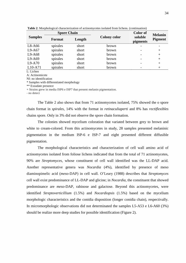
The Table 2 also shows that from 71 actinomycetes isolated, 75% showed the o spore
chain format in spirales, 14% with the format in retinaculiaperti and 8% has rectiflexibles
chains spore. Only in 3% did not observe the spore chain formation.
The colonies showed mycelium coloration that variated between grey to brown and
white to cream-colored. From this actinomycetes in study, 28 samples presented melaninic
pigmentation in the medium ISP-6 e ISP-7 and eight presented different diffusible
pigmentation.
The morphological characteristics and characterization of cell wall amino acid of
actinomycetes isolated from foliose lichens indicated that from the total of 71 actinomycetes,
90% are Streptomyces, whose constituent of cell wall identified was the LL-DAP acid.
Another representative genera was Nocardia (4%), identified by presence of meso
diaminopimelic acid (meso-DAP) in cell wall. O’Leary (1988) describes that Streptomyces
cell wall exist predominance of LL-DAP and glicine; in Nocardia, the constituent that showed
predominance are meso-DAP, rabinose and galactose. Beyond this actinomycetes, were
identified Streptoverticillium (1.5%) and Nocardiopsis (1.5%) based on the mycelium
morphologic characteristics and the conidia disposition (longer conidia chain), respectivally.
In micromorphologic observations did not determinated the samples L5-A53 e L6-A60 (3%)
should be realize more deep studies for possible identification (Figure 2).

35
Fig. 2: Identification of actinomycetes genus isolated from foliose lichens.
Determination of Antimicrobial Activity
The antimicrobial activity analisys of isolated actinomycetes supplied informations
about the antimicrobial spectrum. Of the 71 actinomycetes tested 80% presented antimicrobial
activity in Gelose Block Method. The average of isolated inhbition halo varied since low
(halo = 8 at 12 mm), moderate (halo = 13 at 18 mm) to high (halo = 19 at 35 mm) activity.
From this, 68% showed higher antimicrobial activity against the microorganisms-test.
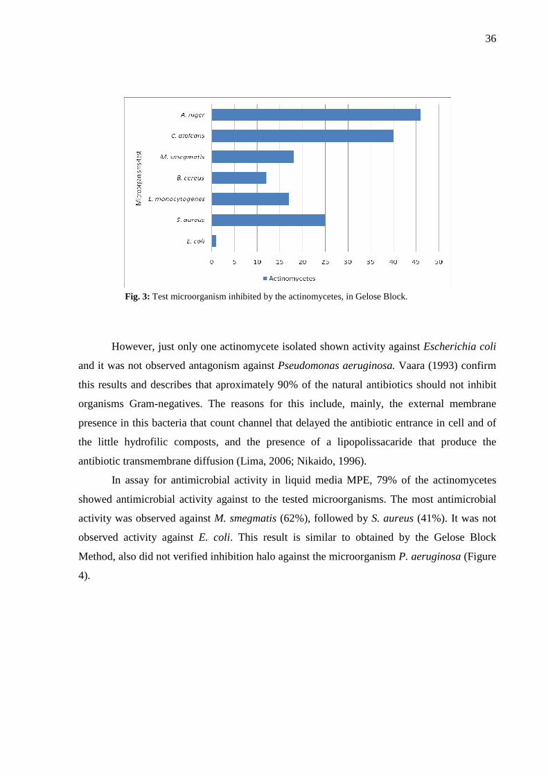
The data referring to the antimicrobial activity of the 71 isolated in Gelose Block
against the tested microorganism (E. coli CCT0547, P. Aeruginosa CCT3971, S. aureus
CCT1352, L. monocytogene, B. subitilis CCT1359, M. smegmatis DPUFPE-71, C. albicans
CCT0776, A. niger CCT1357) demonstrated the great majority of the actinomycetes
expressed activity against Aspergillus niger (65%) and Candida albicans (56%), (Figure 3).
90,0%
4,0%1,5%
1,5%3,0%
Streptomyces spp. Nocardia spp. Nocardiopsis spp.
Streptoverticil ium spp. no identification
36
Fig. 3: Test microorganism inhibited by the actinomycetes, in Gelose Block.
However, just only one actinomycete isolated shown activity against Escherichia coli
and it was not observed antagonism against Pseudomonas aeruginosa. Vaara (1993) confirm
this results and describes that aproximately 90% of the natural antibiotics should not inhibit
organisms Gram-negatives. The reasons for this include, mainly, the external membrane
presence in this bacteria that count channel that delayed the antibiotic entrance in cell and of
the little hydrofilic composts, and the presence of a lipopolissacaride that produce the
antibiotic transmembrane diffusion (Lima, 2006; Nikaido, 1996).
In assay for antimicrobial activity in liquid media MPE, 79% of the actinomycetes
showed antimicrobial activity against to the tested microorganisms. The most antimicrobial
activity was observed against M. smegmatis (62%), followed by S. aureus (41%). It was not
observed activity against E. coli. This result is similar to obtained by the Gelose Block
Method, also did not verified inhibition halo against the microorganism P. aeruginosa (Figure
4).

37
Fig. 4: Test microorganism inhibited by actinomycetes by disk diffusion.
The inhibiton halo average varied from low to high activity. From this, 51% of the
isolated showed moderada activity and e 7% present higher antimicrobial activity.
The media MPE demonstrated to be efficient for the bioactive metabolites production.
As Huck et al. (1991) the use of yeasts extract, soy bean and other raw complex material
could be utilizated as nitrogen source for better production of antibiotics by actinomycetes.
Lima (2006) and Ismet et al. (2004) affirmed that the combination of different carbon sources
increased the antimicrobial activity in comparision with a only source and that the production
increase of bioactive substance was depend of nitrogen source added to the media.
The antimicrobial activity simultaneous detection against filamentous fungi, yeasts
and bacteria could suggest the action of more of one antibiotic with different target (González
et al., 1999) or the evenctual presence of a new antimicrobial substance capable of cross the
cell wall, as much bacterial species as to fungal (Sacramento et al., 2004; Tsvetanova; Prince,
2001; Chino et al., 1996).
The screening for new microorganisms capable to produce substances that can action
against bacteria and pathogenic fungi is extremelly relevant, but nowadays the application of
actinomycetes has changed to more diverse antimicrobials, like against Plasmodium sp.
(Castillo et al., 2002; 2003; Pullen et al., 2002).
These results suggest that actinomycetes isolated from lichens are promissors
organisms for the discovery of new antibiotics, conquering great importance in pharmaceutic
industry.

38
The biotechnological processes are directly related to the diversity of molecules
produced by microorganisms, as result of primary and secondary metabolism, and the
conservation of their genetic resources. Besides the manufacturing of new pharmaceuticals
and bio-industry products, microbial diversity can be widely used in the Amazon region.
Acknowledgements
To the CAPES (Coordenação de Aperfeiçoamento de Pessoal de Nível Superior),
UFAM (Universidade Federal do Amazonas) and FAPEAM (Fundação de Amparo à Pesquisa
do Estado do amazonas), for help and finance support for the research development.

39
References
Cardinale, M., Pugcglial, A. M., Brube, M.: Molecular analysis of lichen-associated
bacterial communities. Dipartimento di Biologia Cellulare e dello Sviluppo, Universita degli
Studi di Palermo, Palermo, Italy and Institute of Plant Sciences, Karl-Franzens-University
Graz, Graz, Austria. Federation of European Microbiological Societies FEMS. Microbiol Ecol
57. 484–495 (2006).
Castillo, U. F.; Strobel, G. A.; Ford, E. J.; Hess, W. M.; Porter, H.; Jensen, J. B.;
Albert, H.; Robison, R.; Condron, M. A. M.; Teplow, D. B.; Stevens, D.; Yaver, D.
Munumbicins, wide-spectrum antibiotics produced by Streptomyces NRRL 30562,
endophytic on Kennedia nigriscans. Microbiology, v. 148, p. 2675-2685 (2002).
Castillo, U., Harper, J.K., Strobel, G.A., Sears, J., Alesi, K., Ford, E., Lin, J., Hunter,
M.: Kakadumycins, novel antibiotics from Streptomyces sp. NRRL 30566, an endophyte of
Grevillea pteridifolia. FEMS Microbiology Letters 224, 183–190 (2003).
Chino, M., Nishimura, K., Umekita, M., Hayashi, C., Yumazaki, T., Tsuchida, T.,
Sawa, T., Hamada, M., Takeuchi, T.: Heliquinomycin, a new inhibitor of DNA-helicase,
produced by Streptomyces sp. MJ929-SF2. I – Taxonomy, production, isolation, physico-
chemical properties and biological activities. Journal of Antibiotics 49, 752–757 (1996).
Dias, B. F. S.: A implantação da convenção sobre diversidade biológica no Brasil:
desafios e oportunidades. Anais do Workshop sobre Biodiversidade perspectiva e
oportunidades tecnológicas. Campinas, Brasil (1996).
González, I., Niebla, A., Lemus, M., González, L., Otero, I., Iznaga, Y., Pérez, M. E.,
Vallin, C.: Ecological approach of macrolide-lincosamides-streptogramin producing
Actinomyces from Cuban soils. Letters in Applied Microbiology 29, 147– 150 (1999).
González, I., Ayuso-Sacido, A., Anderson, A., Genilloud, O.: Actinomycetes isolated
from lichens: Evaluation of their diversity and detection of biosynthetic gene sequences.
FEMS, Microbiol Ecol 54:401-415 Madrid, Spain (2005).

40
Huck, T. A., Porter, N., Bushell, M. E.: Positive selection of antibiotic-producing soil
isolates. Journal of General Microbiology, v. 137, p. 2321-2329 (1991).
Ichikawa, T., Ishikura, T., Ozaki, A.: Improvement of Kasugamycin – producing strain
by the agar piece method and the prototroph method. Folia Microbiologica, v. 16, p. 218-224
(1971).
Ismet, A., Vikineswary, S., Paramaswari, S., Wong, W. H., Ward, A., Seki, T.,
Fiedler, H. P., Goodfellow, M.: Production and chemical characterizations of antifungal
metabolites from Micromonospora sp. M39 isolated from mangrove rhizosphere soil. World
Journal of Microbiology and Biotecnology, v. 20, p. 523-528 (2004).
Kitouni M., Boudemagh A., Oulmi L., Reghioua S., Boughachiche F., ZerizerH.,
HamdikenH., Couble A., Mouniee D., Boulahrouf A. and BoironP. Isolation of
actinomycetes producing bioactive substances from water, soil and tree bark samples of the
north-east of Algeria. J. Med. Mycol. 15, 45–51 (2005).
Lima, V. T.: Isolamento e Atividade Antimicrobiana de Actinomicetos Endofíticos e
da Rizosfera de Melão-de-São-Caetano (Momordica charantia L.). Dissertação (mestrado).
Universidade Federal de Pernambuco. PE, Brasil (2006).
Matsuura, T.: Ocorrência de actinomicetos endofíticos produtores de antibióticos
isoaldos de folhas e raízes de feijão Caupi (Vigna unguiculata). Dissertação (mestrado),
Universidade Federal de Pernambuco, PE, Brasil (1998).
McCarthy, A. J., Williams, S. T.: Methods for studying the ecology of actinomycetes.
In: Grigorova, R. and Norris, J. R. Methods in Microbiology: techniques in microbial
ecology. v. 22. London: Academic (1990).
Muro, M. A., Luchi, M. R.: Preservação de microrganismos. Campinas: Fundação
Tropical de Pesquisa e Tecnologia “André Toselo” (1989).
41
Nash, T. H. Lichen Biology. Cambridge, USA, Cambridge University Press led. p.303
(1996).
Nikaido, H.: Multidrug efflux pumps of gram-negative bacteria. J. Bacteriol. 178:
5853-5859 (1996).
O'leary. W. M.: Practical Handbook of Microbiology, New York, CRC Press. p.688
(1988).
Overbye, K. M., Barret, J. F.: Antibiotics: where did we go wrong? Drug Discovery
Today. 10(1): 45-52 (2005).
Pullen, C., Schmitz, P., Meurer, K., Bamberg, D. D., Lohmann, S., Franca, S. D. C.,
Groth, I., Schlegel, B.: New and bioactive compounds from Streptomyces strains residing in
the wood of Celastraceae. Planta 216, 162–167 (2002).
Sacramento, D. R, Coelho, R. R. R., Wigg, M. D., Linhares, L. F. T. L., Santos, M. G.
M., Semêdo, L. T. A. S., Silva, A. J. R.: Antimicrobial and antiviral activities of an
actinomycete (Streptomyces sp.) isolated from a Brazilian tropical forest soil. World Journal
of Microbiology & Biotechnology 20: 225–229 (2004).
Seaward, M. R. D.: Lichen Ecology. Academic Press, Inc. London (1977).
Souza, A. Q. L., Souza, A. D. L., Astolfi-Filho, S., Belém-Pinheiro, M. L., Sarquis, M.
I. M., Pereira, J. O.: Atividade antimicrobiana de fungos endofíticos isolados de plantas
tóxicas da Amazônia: Palicourea longiflora (aubl.) rich e Strychnos cogens bentham. Acta
Amazônica, vol. 34(2), p.185 – 195 (2004).
Staneck, J. L., Roberts, G. D.: Simplified approach to identification of aerobic
actinomycetes by thin-layer chromatography. Applied Microbiology, v. 28, p. 226-231
(1974).
42
SUDAM (Superintendência de Desenvolvimento da Amazônia). Rede para
conservação e uso de recursos genéticos amazônicos. Grupo de Ciências e Tecnologia. Belém,
Brasil (1995).
Tsvetanova, B. C., Prince, N. P. J.: Liquid chromatographyelectrospray mass
spectrometry of tunicamycin-type antibiotics. Analytical Biochemistry 289, 147–156 (2001).
Vaara, M.: Antibiotic-supersusceptible of Escherichia coli and Salmonella
typhimurium. Antimicrobial Agents and Chemotherapy, v. 37, p. 2255-2260 (1993).
Waksman, S. A., Woodruff, H. B.: Actinomyces antibioticus a new soil organism
antagonistic to pathogenic and non-pathogenic bacteria. Journal of Bacteriology, v. 42, p.
231-249 (1941).
Williams, S. T., Cross. T.: Actinomycetes. Methods in Microbiology, v. 6, p. 295-334
(1974).
43
5. REFERÊNCIAS ADRIO, J.L.; DEMAIN, A.L. Fungal biotechnology. International Microbiology. 6 (3): 191-199, 2003. ALBAGLI, S., Amazônia: fronteira geopolítica da biodiversidade. Parcerias Estratégicas, no. 12, 2001. ALEXOPOULOS, C. J.; MIMS, C.W. Introductory Mycology. 4th edition. John Wiley and Sons INC, USA. p.869, 1996. APPELBAUM, P.C.; JACOBS, M.R. Recently approved and investigational antibiotics for treatment of severe infections caused by Gram-positive bacteria. Current Opinion in Microbiology. 8 (5): 510-517, 2005. AZEVEDO, J.L. Microrganismos endofíticos. In: I.S. MELO e J.L. Ecologia Microbiana. Editora da EMBRAPA, Jaguariúna, SP. p. 117-137, 1998 BACH, E. E.; KIMATI, H. Purification and Characterization of Toxins from Wheat Isolates of Drechslera tririci-repentis, Bipolaris bicolor, and Bipolaris sorokiniana. Journal of Venomous Animals and Toxins. 5(2): 184-199, 1999. BOGGS, A.F.; MILLER, G.H. Antibacterial drug discovery: is small pharma the solution? Clinical Microbiology and Infection.10 (suppl. 4): 32-36, 2004. BULL, A. T.; HARDMAN, D. J. Microbial diversity . Curr. Opin Biotechnology, 2: 421-428, 1991. BULL, A.T.; WARD, A.C.; GOODFELLOW, M. Search and discovery strategies for biotechnology: the Paradigm Shift. Micrbiology and Molecular Biology Reviews. 64 (3): 573-606, 2000. BUTLER, P.J. Butler, Metabolic regulation in diving birds and mammals, Respir. Physiol. Neurobiol. Summary Plus. Cited By in Scopus (18), p.297–315, 2004. BUTLER, M.S.; BUSS, A.D. Natural products – The future scaffolds for novel antibiotics? Biochemical Pharmacology. 71 (7): 919-929, 2006. CARDINALE, M., PUGCGLIAL, A. M., BRUBE, M.: Molecular analysis of lichen-associated bacterial communities. Dipartimento di Biologia Cellulare e dello Sviluppo, Universita degli Studi di Palermo, Palermo, Italy and Institute of Plant Sciences, Karl-Franzens-University Graz, Graz, Austria. Federation of European Microbiological Societies FEMS. Microbiol Ecol 57. 484–495 (2006). CASTILLO, U. F.; STROBEL, G. A.; FORD, E. J.; HESS, W. M.; PORTER, H.; JENSEN, J. B.; ALBERT, H.; ROBISON, R.; CONDRON, M. A. M.; TEPLOW, D. B.; STEVENS, D.; YAVER, D. Munumbicins, wide-spectrum antibiotics produced by Streptomyces NRRL 30562, endophytic on Kennedia nigriscans. Microbiology , v. 148, p. 2675-2685. 2002.

44
CASTILLO, U., HARPER, J.K., STROBEL, G.A., SEARS, J., ALESI, K., FORD, E., LIN, J., HUNTER, M.: Kakadumycins, novel antibiotics from Streptomyces sp. NRRL 30566, an endophyte of Grevillea pteridifolia. FEMS Microbiology Letters 224, 183–190 (2003) CHALIS, G.L.; HOPWOOD, D.A. Synergy and contingency as driving forces for the evolution of multiple secondary metabolite production by Streptomyces species. PNAS, 100, p.14555-14561, 2003. CHIN, Y-W.; BALUNAS, M.J.; CHAI, H.B.; KINGHRN, A.D.; Drug discovery from natural sources. The AAPS Journal. 8(2): 239-253, 2006. CHINO, M., NISHIMURA, K., UMEKITA, M., HAYASHI, C., YUMAZAKI, T.,
TSUCHIDA, T., SAWA, T., HAMADA, M., TAKEUCHI, T.: Heliquinomycin, a new
inhibitor of DNA-helicase, produced by Streptomyces sp. MJ929-SF2. I – Taxonomy,
production, isolation, physico-chemical properties and biological activities. Journal of
Antibiotics 49, 752–757. 1996.
CONTI, R. Diversidade e atividade antimicrobiana de microrganismos endofíticos da planta medicinal Borreria verticillata (L.) G.F.W. Meyer. Dissertação (mestrado). Universidade Federal de Pernambuco. PE, Brasil. 2007. CROSS T. Aquatic Actinomycetes: A critical survey of the ocurrence, growth and role of Actinomycetes in aquatic habitats. J Appl Bacteriol 1981; 50: 397-423. 1981. DEMAIN, A. Microbial Secondary Metabolism: a New Opportunity for Industry. Ciba Foundation Symposium. p.3-23, 1992. DEMAIN, A.L. Microbial biotechnology. Trends in Biotechnology. 18 (1): 26-31, 2000. DIAS, B. F. S. A implantação da convenção sobre diversidade biológica no Brasil: desafios e oportunidades. Anais do Workshop sobre Biodiversidade perspectiva e oportunidades tecnológicas. Campinas, 1996. FAORO, H. Determinação da Biodiversidade de Archaea e Bacteria da mata atlântica paranaense Dissertação (mestrado), Universidade Federal do Paraná, PR, Brasil. 2006. FENICAL W. Chemical studies of marine bacteria: developing a new resource. Chem Rev; 93: 1673-83, 1993. FLOSS, H.G. Antibiotic biosynthesis: from natural to unnatural compounds. Journal of Industrial Microbiology & Biotechnology. 27(3): 183-194, 2001. GOODFELLOW, M.; WILLIAMS, S. T. Ecology of actinomycetes. Annual Review of Microbiology , v. 37, p. 189-216, 1983. GONZÁLEZ, I., AYUSO-SACIDO, A., ANDERSON, A., GENILLOUD, O.: Actinomycetes isolated from lichens: Evaluation of their diversity and detection of biosynthetic gene sequences. FEMS, Microbiol Ecol 54:401-415 Madrid, Spain (2005)

45
GULLO, V.P.; HUGHES, D.E. Exploiting new approaches for natural product drug discovery in the biotechnology industry. Drug Discovery Today: Technologies. 2(3): 281-286, 2005. GUNATILAKA, A.A.L.; Natural products from plant-associated microorganisms: distribuition, structural diversity, bioactivity, and implications of their occurrence. Journal of Natural Products. 69(3): 509-526, 2006. HIGGS, R.E.; ZAHN, J.A.; GIGI, J.D.; HILTON, M.D. Rapid method to estimate the presence os secondary metabolites in microbial extracts. Applied and Environmental Microbiology. 67(1): 371-376, 2001. HOLT, J. G.; KRIEG, N. R.; SNEATH, P. H. A.; STALEY, J. T.; WILLIAMS, S. T. Bergey’s Manual of Determinative Bacteriology. 9. ed. Baltimore: Williams & Wilkins, 1994. HUCK, T. A., PORTER, N., BUSHELL, M. E.: Positive selection of antibiotic-producing soil isolates. Journal of General Microbiology, v. 137, p. 2321-2329. 1991. ICHIKAWA, T., ISHIKURA, T., OZAKI, A.: Improvement of Kasugamycin – producing strain by the agar piece method and the prototroph method. Folia Microbiologica, v. 16, p. 218-224 (1971). ISMET, A., VIKINESWARY, S., PARAMASWARI, S., WONG, W. H., WARD, A., SEKI, T., FIEDLER, H. P., GOODFELLOW, M.: Production and chemical characterizations of antifungal metabolites from Micromonospora sp. M39 isolated from mangrove rhizosphere soil. World Journal of Microbiology and Biotecnology, v. 20, p. 523-528. 2004. KITOUNI M., BOUDEMAGH A., OULMI L., REGHIOUA S., BOUGHACHICHE F., ZERIZERH., HAMDIKENH., COUBLE A., MOUNIEE D., BOULAHROUF A. and BoironP. Isolation of actinomycetes producing bioactive substances from water, soil and tree bark samples of the north-east of Algeria. J. Med. Mycol. 15, 45–51 (2005) KLEINKAUF, H.; VON DOHREN, H. Biosynthesis of Peptide Antibiotics. European Journal of Biochemistry, 192: 1-15, 1990. KNOWLES, D.J.C. New strategies for antibacterial drug design. Trends Microbiol . v. 5, p. 379-382, 1977. KURTBÖKE, D.I., SWINGS, J. & STORMS, V. 2004. Microbial genetic resources and Biodiscovery. In Ipek Kurtböke & Jean Swings (eds.), Microbial Genetic Resources and Biodiscovery WFCC Publications, UK. LABEDA, D.P. Isolations of Biotechnologiacal Organisms from Nature. New York: McGRAW-HILL, 1990. LEVY, S.B.; MARSHALL, B.; Antibacterial resistance worldwide: causes, challenges and responses. Nature Medicine. 10 (suppl. 12): 122-129, 2004.
46
LI, J. Y; STROBEL, G. A. Jesterone and Hydroxy-Jesterone Antioomycete Cyclohexenone Epoxides From the Endophytic Fungus Pestalotiopsis jesteri. Phytochemistry, 57(2): 262-265, 2001. LIMA, V. T.: Isolamento e Atividade Antimicrobiana de Actinomicetos Endofíticos e da Rizosfera de Melão-de-São-Caetano (Momordica charantia L.). Dissertação (mestrado). Universidade Federal de Pernambuco. PE, Brasil. 2006. McCARTHY, A. J. & WILLIAMS, S. T. Methods for studying the ecology of actinomycetes. In: GRIGOROVA, R. & NORRIS, J. R. Methods in Microbiology: techniques in microbial ecology. v. 22. London: Academic, 1990. MARCELLI, M. P. Biodiversity assessment in lichenized fungi: the necessary naive roll makers. In Bicudo, C.E. de M. & Menezes, N.A. (eds.). Biodiversity in Brazil: a first approach. São Paulo, CNPq, p.93-107, 1996. MATSUURA, T.: Ocorrência de actinomicetos endofíticos produtores de antibióticos isoaldos de folhas e raízes de feijão Caupi (Vigna unguiculata). Dissertação (mestrado), Universidade Federal de Pernambuco, PE, Brasil. 1998. MULLER, K., Pharmaceutically relevant metabolites from lichens. Applied Microbiology and Biotechnology. 56, 9-16, 2001. MURO, M. A., LUCHI, M. R.: Preservação de microrganismos. Campinas: Fundação Tropical de Pesquisa e Tecnologia “André Toselo” (1989). NASH, T. H. Lichen Biology. Cambridge, USA, Cambridge University Press led. p.303 (1996) NASH III, T.H. Lichen Biology – Introduction; In Lichen Biology; Nash III, T. H..; Ed; Cambridge University Press, Cambrige; p. 1-315, 1996. NIKAIDO, H.: Multidrug efflux pumps of gram-negative bacteria. J. Bacteriol. 178: 5853-5859. 1996. O'LEARY. W. M.: Practical Handbook of Microbiology, New York, CRC Press. p.688. 1988.
OLIVEIRA, V. M.; SETTE, L. D.; FANTINATTI-GARBOGGINI, F. Construindo a História dos Produtos Naturais, v. 7, MultiCiência - Preservação e Prospecção de Recursos Microbianos. São Paulo, 2006. OMURA, S. Trends in the search for bioactive microbianl metabolites. J. Industrial Microbiol. v. 10, p. 135-156, 1992. OVERBYE, K. M., BARRET, J. F.: Antibiotics: where did we go wrong? Drug Discovery Today. 10(1): 45-52,2005. PACE, N.R.; STAHL, D.A.; LANE, D.J.; OLSEN, G.J.. The analysis of natural microbial populations by ribosomal RNA sequences. Adv. Microb. Ecol., v. 9, p. 1-55, 1986.
47
PAREKH, D. B.; ZIEGLER, W.; PARKER, P. J.; Multiple pathways control protein kinase C phosphorylation. EMBO J. 19, 496-503, 2000. PHAFF, H. J. Industrial microorganisms. Scientific American, v. 295, p. 52-65, 1991. PULLEN, C., SCHMITZ, P., MEURER, K., BAMBERG, D. D., LOHMANN, S., FRANCA, S. D. C., GROTH, I., SCHLEGEL, B.: New and bioactive compounds from Streptomyces strains residing in the wood of Celastraceae. Planta 216, 162–167 (2002) RATTI, R. P.; SERRANO, N. F. G.; HOKKA, C. O.; SOUSA, C. P. Isolamento e atividade antimicrobiana de microrganismos endofíticos de “pêssego do cerrado”. Congresso de Pós-Graduação, 4. São Carlos. Anais de Eventos da UFSCar, v. 3, p. 1249, 2007. REIS, E. Estatística Descritiva. Lisboa: Silabo, ed. 4, 1998.
RODRIGUES, K. F.; HESSE, M.; WERNER, C. Antimicrobial activities of secondary metabolites produced by endophytic fungi from Spondias mombin. Journal Basic Microbial, 40(4): 261-267, 2000. ROSSELÓ-MORA, R.; AMANN, R. The species concept for prokaryotes. FEMS Microbiology Review, v. 25, n. 1, p. 39-67, 2001 SACRAMENTO, D. R, COELHO, R. R. R., WIGG, M. D., LINHARES, L. F. T. L., SANTOS, M. G. M., SEMÊDO, L. T. A. S., SILVA, A. J. R.: Antimicrobial and antiviral activities of an actinomycete (Streptomyces sp.) isolated from a Brazilian tropical forest soil. World Journal of Microbiology & Biotechnology 20: 225–229. 2004. SAMIULLA, DS.; VAIDYANATHAN, V.V.; ARUN, P.C.; BALA N, G.; BLAZE, M.; BONDRE, S.; CHANDRASEKHAR, G.; GADAKH, A.; KUMAR, R.; KHARVI, G.; KIM, H.-O.; KUMAR, S.; MALIKAYIL, J.A.; MOGER, M.; MONE, M.K.; NAGARJUNA, P.; OGBU, C.; PENDHALKAR, D.; RAJA RAO, A.V.S.; VENKATESHWAR RAO, G.; SARMA, V.K.; V.K.; SHAIK, S.; SHARMA, G.V.R.; SINGH, S.; SREEDHAR, C.; SONAWANE, R.; TIMMANNA, U.; HARDY, L.W. Rational selection of structurally diverse natural product scaffolds with favorable ADME properties for drug discovery. Molecular Diversity. 9(1-3): 131-139, 2005. SEAWARD, M.R.D..Lichen Ecology. Academic Press, Inc. London. 1977. SEIDL, P. R. The use of biodiversity for sustaintable development: investigation of bioactive products and their commercial applications. Proceedings of a Workshop. Manaus, 1993. SOUZA, A. Q. L.; SOUZA, A. D. L.; ASTOLFI-FILHO, S.; BELÉM-PINHEIRO, M. L. SARQUIS, M. I. M.; PEREIRA, J. O. Atividade antimicrobiana de fungos endofíticos isolados de plantas tóxicas da amazônia: Palicourea longiflora (aubl.) rich e Strychnos cogens bentham. . Acta Amazônica VOL. 34(2) 2004: 185 – 195 STANIER, R.Y.; INGRAHAM, J.L.; WHEELIS, M.L.; PAINTER, P.R. The Microbial World . 5.ed. New Jersey: Prentice-Hall, 1980.

48
STAMFORD, T. L. M.; ARAÚJO, J. M.; STAMFORD, N. P. Atividade enzimática de microrganismos isolados do jacatupé (Pachyrhizus erosus L. Urban). Ciência e Tecnologia dos Alimentos, vol. 18, no. 4, p. 382-385, 1998.
STANECK, J. L., ROBERTS, G. D.: Simplified approach to identification of aerobic actinomycetes by thin-layer chromatography. Applied Microbiology, v. 28, p. 226-231 (1974). SUDAM. Rede para conservação e uso de recursos genéticos amazônicos. Grupo de Ciências e Tecnologia - Superintendência de Desenvolvimento da Amazônia. Belém, 1995. TERKINA, I. A.; PARFENOVA, V. V., AHN, T. S. Antagonistic Activity of Actinomycetes of Lake Baikal. Applied Biochemistry and Microbiology, 2006, Vol 42, no.2, pp 173-176. TRILLI, V.; MICHILINI, V.; MONTOVANI, V.; PIRT, S.J. Development of the agar disk method for the rapid selection of cephalosporim producers with improved yields. Antimicrobial Agents and Chemotherapy, 13(1): 7-13, 1978. TSVETANOVA, B. C., PRINCE, N. P. J.: Liquid chromatographyelectrospray mass spectrometry of tunicamycin-type antibiotics. Analytical Biochemistry 289, 147–156. 2001. UJIKAWA, K.; Antibióticos antifúngicos produzidos por actinomicetos do Brasil e sua determinação preliminar nos meios experimentais. Revista Brasileira de Ciências Farmacêuticas. Brazilian Journal of Pharmaceutical Sciences. vol. 39, n. 2, abr.jun., 2003. VAARA, M.: Antibiotic-supersusceptible of Escherichia coli and Salmonella typhimurium. Antimicrobial Agents and Chemotherapy, v. 37, p. 2255-2260.1993. WANG, J.; LI, G.; LU, H.; ZHENG, Z.; HUANG, Y.; SU, W. Taxol from Tubercularia sp. strain TF5, an endophytic fungus of Taxus mairei. FEMS-Microbiology Letters, 193: 249-253. 2000. WAKSMAN, S. A., WOODRUFF, H. B.: Actinomyces antibioticus a new soil organism antagonistic to pathogenic and non-pathogenic bacteria . Journal of Bacteriology, v. 42, p. 231-249 (1941) WEIST, S.; SUSSMUTH, R.D. Mutation biosynthesis – a tool the generation of structural diversity in the biosynthesis of antibiotics. Applied Microbiology and Biotechnology. 68(2): 140-150, 2005. WILLIAMS, S. T. & CROSS. T. Actinomycetes. Methods in Microbiology, v. 6, p. 295-334, 1974. WOESE, C. R. Prokariotic systematics: The evolution of a science. In: BALOWS, A., TRÜPER, H. G.; DWORKIN, M.; HARDER, W.; SCHEIFER, K. M. The Prokaryotes. 2. ed. New York: Sporinger-Verlag, 1992. YONEYAMA, H.; KATSUMATA, R, R.; Antibiotic resistance in bacteria and its Future for novel antibiotic development. Bioscience, Biotechnology and Biochemistry. 70 (5): 1060-1075, 2006.
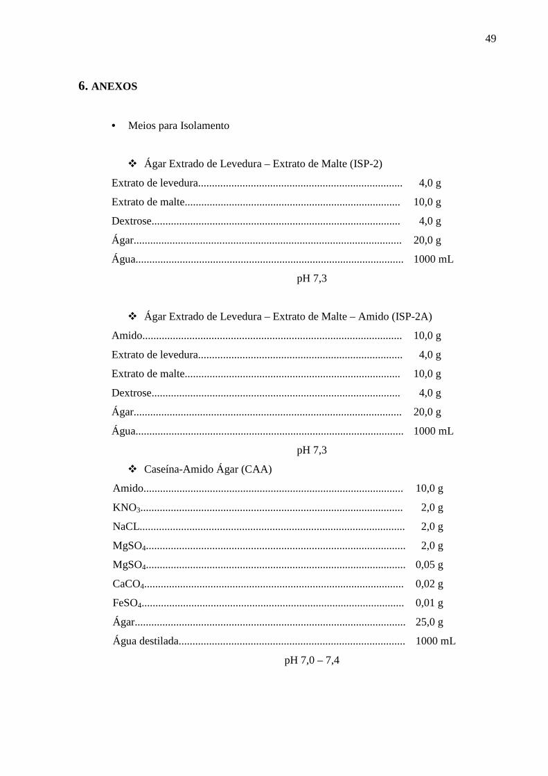
49
6. ANEXOS
• Meios para Isolamento
� Ágar Extrado de Levedura – Extrato de Malte (ISP-2)
Extrato de levedura.......................................................................... 4,0 g
Extrato de malte.............................................................................. 10,0 g
Dextrose.......................................................................................... 4,0 g
Ágar................................................................................................. 20,0 g
Água................................................................................................. 1000 mL
pH 7,3
� Ágar Extrado de Levedura – Extrato de Malte – Amido (ISP-2A)
Amido.............................................................................................. 10,0 g
Extrato de levedura.......................................................................... 4,0 g
Extrato de malte.............................................................................. 10,0 g
Dextrose.......................................................................................... 4,0 g
Ágar................................................................................................. 20,0 g
Água................................................................................................. 1000 mL
pH 7,3
� Caseína-Amido Ágar (CAA)
Amido.............................................................................................. 10,0 g
KNO3............................................................................................... 2,0 g
NaCL................................................................................................ 2,0 g
MgSO4.............................................................................................. 2,0 g
MgSO4.............................................................................................. 0,05 g
CaCO4.............................................................................................. 0,02 g
FeSO4............................................................................................... 0,01 g
Ágar.................................................................................................. 25,0 g
Água destilada.................................................................................. 1000 mL
pH 7,0 – 7,4

50
� Rafinose – Histidina Agar (RHA)
Rafinose .......................................................................................... 10,0 g
L-histidina........................................................................................ 1,0 g
MgSO4.................................................................................................... 0,5 g
FeSO4............................................................................................... 0,01 g
Ágar.................................................................................................. 25,0 g
Água destilada.................................................................................. 1000 mL
pH 7,0 – 7,4
• Meio para Atividade Antimicrobiana em Meio Sólido (ISP-2A)
Amido.............................................................................................. 10,0 g
Extrato de levedura......................................................................... 4,0 g
Extrato de malte.............................................................................. 10,0 g
Dextrose.......................................................................................... 4,0 g
Ágar................................................................................................ 20,0 g
Água destilada................................................................................. 1000 mL
pH 7,3
• Meio para Atividade Antimicrobiana em Meio Líquido (MPE)
Glicose........................................................................................... 20,0 g
Farinha de Soja.............................................................................. 20,0 g
NaCL............................................................................................. 5,0 g
CaCO3........................................................................................... 2,0 g
Água destilada............................................................................... 1000 mL
pH 7,0 – 7,4
• Meios para Identificação dos actinomicetos
� Extrato de levedura - Extrato de malte - Ágar (ISP-2)
Extrato de levedura ........................................................................ 4,0 g
Extrato de malte ............................................................................. 10,0 g
Dextrose ......................................................................................... 4,0 g
Ágar ............................................................................................... 20,0 mL
Água destilada ................................................................................ 1000 mL pH 7,3

51
� Ágar Farinha de Aveia (ISP-3)
Solução de traços de sais:
Sulfato ferroso (FeSO4.7H2O) ....................................................... 0,1 g
Cloreto manganoso (MnCl2.4H2O) ............................................... 0,1 g
Sulfato de zinco (ZnSO4.7H2O) ..................................................... 0,1 g
Água destilada ................................................................................ 100,0 mL
Farinha de aveia ............................................................................. 20,0 g
Solução de traços de sais ............................................................... 1,0 g
Ágar ............................................................................................... 18,0 g
Água destilada ................................................................................ 1000 mL
pH 7,2
� Ágar Sais inorgânicos - Amido (ISP-4)
Solução de traços de sais:
Sulfato ferroso (FeSO4.7H2O) ....................................................... 0,1 g
Cloreto manganoso (MnCl2.4H2O) ............................................... 0,1 g
Sulfato de zinco (ZnSO4.7H2O) ..................................................... 0,1 g
Água destilada ................................................................................ 100,0 mL
Solução I: Amido .......................................................................... 10,0 g
Água destilada ................................................................................ 500 mL
Solução II: Fosfato hidrogeno dipotássico (K2HPO4) ................... 1,0 g
Sulfato de magnésio (MgSO4.7H2O) ............................................. 1,0 g Cloreto de sódio (NaCl) .................................................................
1,0 g
Sulfato de amônio ( (NH4)2SO4 ) ................................................. 2,0 g Carbonato de cálcio (CaCO3) ........................................................ 2,0 g
Solução de traços de sais ................................................................ 1,0 mL
Ágar ............................................................................................... 20,0 g
Água destilada ................................................................................ 500 mL
pH 7,0 - 7,

52
� Glicerol – Asparagina - Ágar (ISP-5)
L-asparagina (base anidra).............................................................. 1,0 g
Glicerol............................................................................................ 10,0 g
K2HPO4 (base anidra)...................................................................... 1,0 g
Água destilada ................................................................................ 1000 mL
Solução de traços de sais (ISP-3) 1,0 mL Ágar 15,0g
pH 7,4
� Ágar (ISP-6)
Peptona............................................................................................ 15,0 g
Protease Peptona............................................................................. 5,0 g
Citrato de Ferro e Amônio.............................................................. 0,5 g
R2HPO4........................................................................................... 1,0 mL
Tiossulfato de Sódio (S2O3Na2)...................................................... 0,08 g Extrato de Levedura........................................................................ 1,0 g Ágar 15,0 g Água destilada ................................................................................ 1000 mL
pH 7,3
� Tirosina - Ágar (ISP-7)
Glicerol........................................................................................... 15,0 g
L-tirosina......................................................................................... 0,5 g
L-asparagina.................................................................................... 1,0g
R2HPO4 . 7H2O ............................................................................... 0,5 g
MgSO4 0,5 g NaCl................................................................................................ 0,5 g FeSO4 . 7H2O.................................................................................. 0,01 g Água destilada ................................................................................ 1000 mL Solução de traços de sais (ISP-3).................................................... 1,0 mL Ágar................................................................................................. 20,0 g
pH 7,2 – 7,4
Related Documents


