Calcium Channel b Subunit Promotes Voltage-Dependent Modulation of a1B by Gbg Alon Meir, Damian C. Bell, Gary J. Stephens, Karen M. Page, and Annette C. Dolphin Department of Pharmacology, University College London, London WC1E 6BT, United Kingdom ABSTRACT Voltage-dependent calcium channels (VDCCs) are heteromultimers composed of a pore-forming a1 subunit and auxiliary subunits, including the intracellular b subunit, which has a strong influence on the channel properties. Voltage-dependent inhibitory modulation of neuronal VDCCs occurs primarily by activation of G-proteins and elevation of the free Gbg dimer concentration. Here we have examined the interaction between the regulation of N-type (a1B) channels by their b subunits and by Gbg dimers, heterologously expressed in COS-7 cells. In contrast to previous studies suggesting antagonism of G protein inhibition by the VDCC b subunit, we found a significantly larger Gbg-dependent inhibition of a1B channel activation when the VDCC a1B and b subunits were coexpressed. In the absence of coexpressed VDCC b subunit, the Gbg dimers, either expressed tonically or elevated via receptor activation, did not produce the expected features of voltage-dependent G protein modulation of N-type channels, including slowed activation and prepulse facilitation, while VDCC b subunit coexpression restored all of the hallmarks of Gbg modulation. These results suggest that the VDCC b subunit must be present for Gbg to induce voltage-dependent modulation of N-type calcium channels. INTRODUCTION VDCCs play an essential role in the control of many cellular processes, including synaptic transmission, by transducing a voltage signal into elevation of intracellular Ca 21 (Tsien et al., 1991; Dunlap et al., 1995). In presynaptic nerve termi- nals, the VDCCs that are most involved in transmitter release are N-type (a1B) and P/Q-type (a1A) channels (Meir et al., 1999). Many neurotransmitters, including do- pamine, GABA, and opiates, are involved in a widespread form of inhibitory synaptic modulation, by binding to pre- synaptic seven transmembrane domain (7TM) receptors, liberating activated G protein subunits, and inhibiting cal- cium currents (Bean, 1989; Dolphin, 1995). This inhibition is produced by the Gbg dimers, which associate with a1B VDCCs in particular (Ikeda, 1996; Herlitze et al., 1996) (but also with a1A and a1E), in a membrane-delimited manner, causing the channels to lose the property of fast opening in response to depolarization, so that less Ca 21 enters the cell. This modulation involves a retardation of the activation pro- cess of VDCCs (Carabelli et al., 1996; Patil et al., 1996), which can be overcome (voltage-dependent facilitation) by a strong depolarizing prepulse (Ikeda, 1991) or a train of action poten- tials (Patil et al., 1998). G protein modulation of VDCCs embeds three major elements: reduction of the current ampli- tude, slowing of the activation kinetics, and relief of inhibition by depolarizing voltages. Recent single-channel and gating current analysis has suggested that the modulation consists mainly of Gbg-bound channels failing to gate to the open state, implying a requirement that Gbg unbinds before gating (add- ing this relatively slow component to the activation kinetics) (Patil et al., 1996; Jones et al., 1997). Therefore, it was pro- posed that the kinetic slowing alone caused the amplitude decrease. In this model, facilitation is explained by voltage- dependent unbinding of the modulating G protein during a strong depolarizing prepulse (Elmslie and Jones, 1994). How- ever, other data suggest that “reluctant” or Gbg-bound calcium channels may also open (Elmslie et al., 1990; Boland and Bean, 1993; Colecraft et al., 2000). VDCCs are heteromultimers composed of a pore-forming a1 subunit and several auxiliary subunits (Jones, 1998). The a1 subunits are the primary determinants of the resultant properties; however, the auxiliary subunits, particularly the intracellular b subunit, strongly influence the properties of the assembled channels (Walker and De Waard, 1998). VDCC b subunits (of which four isoforms have been cloned) are involved in membrane targeting of the putative pore-forming a1 subunits (Chien et al., 1995; Brice et al., 1997) and modulate the biophysical properties of a1C (Costantin et al., 1998; Gerster et al., 1999), a1E (Stephens et al., 1997), and a1B (Wakamori et al., 1999) channels. A common effect of the different b subunits is to enhance the coupling between depolarization and activation and to in- crease the channel open probability (for a review see Walker and De Waard, 1998). There is biochemical evidence for overlapping binding sites for the VDCC b subunit and for Gbg dimers on the intracellular I-II loop (De Waard et al., 1997) and on the C and N termini (Walker et al., 1998, 1999; Canti et al., 1999) of various VDCC a1 subunits. This led to the idea that there is competition between VDCC b and Gbg for the same binding site. The competition hypothesis is supported by data showing reduced G protein-mediated inhibition with overexpression of certain VDCC b subunits for several types of heterologously expressed VDCCs in Xenopus oo- cytes (Roche et al., 1995; Bourinet et al., 1996). Further- more, previous work from our laboratory shows that partial Received for publication 5 November 1999 and in final form 9 May 2000. Address reprint requests to Dr. Alon Meir, Department of Pharmacology, University College London, Gower St., London WC1E 6BT, UK. Tel.: 144-20-7769-4485; Fax: 144-20-7813-2808; E-mail [email protected]. © 2000 by the Biophysical Society 0006-3495/00/08/731/16 $2.00 731 Biophysical Journal Volume 79 August 2000 731–746

Welcome message from author
This document is posted to help you gain knowledge. Please leave a comment to let me know what you think about it! Share it to your friends and learn new things together.
Transcript

Calcium Channel b Subunit Promotes Voltage-Dependent Modulation ofa1B by Gbg
Alon Meir, Damian C. Bell, Gary J. Stephens, Karen M. Page, and Annette C. DolphinDepartment of Pharmacology, University College London, London WC1E 6BT, United Kingdom
ABSTRACT Voltage-dependent calcium channels (VDCCs) are heteromultimers composed of a pore-forming a1 subunitand auxiliary subunits, including the intracellular b subunit, which has a strong influence on the channel properties.Voltage-dependent inhibitory modulation of neuronal VDCCs occurs primarily by activation of G-proteins and elevation of thefree Gbg dimer concentration. Here we have examined the interaction between the regulation of N-type (a1B) channels bytheir b subunits and by Gbg dimers, heterologously expressed in COS-7 cells. In contrast to previous studies suggestingantagonism of G protein inhibition by the VDCC b subunit, we found a significantly larger Gbg-dependent inhibition of a1Bchannel activation when the VDCC a1B and b subunits were coexpressed. In the absence of coexpressed VDCC b subunit,the Gbg dimers, either expressed tonically or elevated via receptor activation, did not produce the expected features ofvoltage-dependent G protein modulation of N-type channels, including slowed activation and prepulse facilitation, whileVDCC b subunit coexpression restored all of the hallmarks of Gbg modulation. These results suggest that the VDCC b subunitmust be present for Gbg to induce voltage-dependent modulation of N-type calcium channels.
INTRODUCTION
VDCCs play an essential role in the control of many cellularprocesses, including synaptic transmission, by transducing avoltage signal into elevation of intracellular Ca21 (Tsien etal., 1991; Dunlap et al., 1995). In presynaptic nerve termi-nals, the VDCCs that are most involved in transmitterrelease are N-type (a1B) and P/Q-type (a1A) channels(Meir et al., 1999). Many neurotransmitters, including do-pamine, GABA, and opiates, are involved in a widespreadform of inhibitory synaptic modulation, by binding to pre-synaptic seven transmembrane domain (7TM) receptors,liberating activated G protein subunits, and inhibiting cal-cium currents (Bean, 1989; Dolphin, 1995). This inhibitionis produced by the Gbg dimers, which associate witha1BVDCCs in particular (Ikeda, 1996; Herlitze et al., 1996) (butalso witha1A anda1E), in a membrane-delimited manner,causing the channels to lose the property of fast opening inresponse to depolarization, so that less Ca21 enters the cell.This modulation involves a retardation of the activation pro-cess of VDCCs (Carabelli et al., 1996; Patil et al., 1996), whichcan be overcome (voltage-dependent facilitation) by a strongdepolarizing prepulse (Ikeda, 1991) or a train of action poten-tials (Patil et al., 1998). G protein modulation of VDCCsembeds three major elements: reduction of the current ampli-tude, slowing of the activation kinetics, and relief of inhibitionby depolarizing voltages. Recent single-channel and gatingcurrent analysis has suggested that the modulation consistsmainly of Gbg-bound channels failing to gate to the open state,implying a requirement that Gbg unbinds before gating (add-ing this relatively slow component to the activation kinetics)
(Patil et al., 1996; Jones et al., 1997). Therefore, it was pro-posed that the kinetic slowing alone caused the amplitudedecrease. In this model, facilitation is explained by voltage-dependent unbinding of the modulating G protein during astrong depolarizing prepulse (Elmslie and Jones, 1994). How-ever, other data suggest that “reluctant” or Gbg-bound calciumchannels may also open (Elmslie et al., 1990; Boland andBean, 1993; Colecraft et al., 2000).
VDCCs are heteromultimers composed of a pore-forminga1 subunit and several auxiliary subunits (Jones, 1998). Thea1 subunits are the primary determinants of the resultantproperties; however, the auxiliary subunits, particularly theintracellularb subunit, strongly influence the properties ofthe assembled channels (Walker and De Waard, 1998).VDCC b subunits (of which four isoforms have beencloned) are involved in membrane targeting of the putativepore-forminga1 subunits (Chien et al., 1995; Brice et al.,1997) and modulate the biophysical properties ofa1C(Costantin et al., 1998; Gerster et al., 1999),a1E (Stephenset al., 1997), anda1B (Wakamori et al., 1999) channels. Acommon effect of the differentb subunits is to enhance thecoupling between depolarization and activation and to in-crease the channel open probability (for a review seeWalker and De Waard, 1998).
There is biochemical evidence for overlapping bindingsites for the VDCCb subunit and for Gbg dimers on theintracellular I-II loop (De Waard et al., 1997) and on the Cand N termini (Walker et al., 1998, 1999; Canti et al., 1999)of various VDCCa1 subunits. This led to the idea that thereis competition between VDCCb and Gbg for the samebinding site. The competition hypothesis is supported bydata showing reduced G protein-mediated inhibition withoverexpression of certain VDCCb subunits for severaltypes of heterologously expressed VDCCs inXenopusoo-cytes (Roche et al., 1995; Bourinet et al., 1996). Further-more, previous work from our laboratory shows that partial
Received for publication 5 November 1999 and in final form 9 May 2000.
Address reprint requests to Dr. Alon Meir, Department of Pharmacology,University College London, Gower St., London WC1E 6BT, UK. Tel.:144-20-7769-4485; Fax:144-20-7813-2808; E-mail [email protected].
© 2000 by the Biophysical Society
0006-3495/00/08/731/16 $2.00
731Biophysical Journal Volume 79 August 2000 731–746

depletion of endogenous VDCCb subunits, by an antisenseoligonucleotide, enhanced inhibition of native VDCCs in sen-sory neurons by GABAB receptor activation, although not thatby GTP-g-s (Campbell et al., 1995b). However, facilitation ordepolarization-induced relief of inhibition of N-type channelsexpressed inXenopusoocytes was enhanced by coexpressionof the VDCC b3 subunit (Roche and Treistman, 1998b),despite the reduced inhibition induced by this subunit. Inaddition, most descriptions of an antagonism between VDCCb and Gbg dimers are based on isopotential measurements,ignoring voltage shifts related to the separate expression ofVDCC b subunit or Gbg with thea1 subunit. Our recent datafor Xenopusoocytes suggest that almost all of the apparentbsubunit-related antagonism ofa1B modulation is due tobsubunit-mediated hyperpolarization of the voltage dependenceof current activation (C. Canti, Y. Bogdanov, and A. C. Dol-phin, manuscript submitted for publication).
To examine the correlation between inhibition, kineticslowing, and facilitation, we have applied several differentapproaches to examine the effect of VDCCb subunits onthe modulation ofa1B channels by G proteins in the mam-malian cell line COS-7. First, we recorded single-channela1B or a1B/b currents in two groups of cells. The firstgroup was transfected with VDCC subunits and a minigenederived from the C terminus of ab-adrenergic receptorkinase (bARK1) (Koch et al., 1993; Stephens et al., 1998)that binds Gbg and prevents basal modulation of the chan-nels by endogenous free Gbg. In the second group, wetransfected VDCC subunits and Gb1g2 dimers to producetonic modulation. In a second approach, we recorded whole-cell currents from cells transfected with eithera1B alone ora1B/b2a and the dopamine D-2 receptor to allow reversibleagonist activation of endogenous G proteins. These exper-iments are not possible inXenopusoocytes because of theirendogenous VDCCb subunit (Tareilus et al., 1997).
We show that both the pronounced Gbg-dependent kineticslowing of the channel response and the accompanying volt-age-dependent facilitation depend on VDCCb subunit coex-pression. In the absence of an expressed VDCCb subunit,Gb1g2 expressed tonically does not produce significant kineticslowing of the activation of thea1B channels, while coexpres-sion ofa1B with b2a (orb1b) restores full Gbg modulation.Furthermore, in the COS-7 cell expression system, transientelevation of Gbg by dopamine D-2 receptor activation inducesless inhibition ofa1B than ofa1B/b2a channels, and this is notaccompanied by kinetic slowing or facilitation.
Our present findings support interdependence betweenthe two modulatory processes induced by Gbg dimers andby VDCC b subunits and may be interpreted in severalways. Either the VDCCb subunit is functionally requiredfor the Gbg-induced voltage-dependent inhibition ofa1Bchannels, or Gbg is bound more strongly to thea1B subunitin the absence of a bound auxiliaryb subunit. Therefore, itwould unbind less easily with a depolarizing prepulse, lead-
ing to a failure to detect facilitation. The implications ofboth models will be discussed.
MATERIALS AND METHODS
Materials
Thea1B, b1, b2a,b3, a2-d1, bARK1 Gbg binding domain minigene, D-2receptor, and Gb1 and Gg2 cDNAs used in this study have been describedpreviously (Page et al., 1997, 1998; Stephens et al., 1998; Bogdanov et al.,2000).
Transfection of COS-7 cells
COS-7 cells were cultured and transfected, using the electroporation tech-nique, essentially as described previously (Campbell et al., 1995a; Ste-phens et al., 1998).
Reverse transcriptase-polymerase chain reaction
Total RNA was isolated from a pellet of COS-7 cells with an RNeasyminiprep kit (Qiagen, Crawley, UK). Residual genomic and plasmid DNAwere removed by digestion with RQ DNase (Promega, Madison, WI) for30 min at 37°C, in the presence of 40 mM Tris-HCl (pH 8.0), 10 mMMgSO4, 1 mM CaCl2, 40 units RNasin (Promega), and 5 units RQ DNase.Reverse transcription was carried out in a final volume of 50ml, in thepresence of 50 mM Tris-HCl (pH 8.3), 75 mM KCl, 3 mM MgCl2, 10 mMdithiothreitol, 1.25 mM each deoxyribonucleoside triphosphate, 1.25mgrandom hexamer primers (Promega), 40 units RNasin, and 500 unitsMoloney murine leukemia virus reverse transcriptase (Promega) (37°C,60–120 min). The samples were diluted to 100ml, and 5ml was used perpolymerase chain reaction (PCR) reaction. PCR was carried out using 1.25units Taq DNA polymerase (Promega) in 25ml, containing 10 mMTris-HCl (pH 9), 50 mM KCl, 0.1% Triton X-100, 1.5 mM MgCl2, 200mM each of deoxyribonucleoside triphosphates, and 0.4mM primers. Thefollowing primers were used:b1bF: CCT ATG ACG TGG TGC CTT CC;b1bR: TGC GGT TCA GCA GCG GAT TG;b2F: AAG AAG ACA GAGCAC ACT CC,b2R ACT CTG AAC TTC CGC TAA GC;b3F: CTC TAGCCA AGC AGA AGC AA; b3R: CTG GTA CAG GTC CTG GTA GG;b4F: TGA GGT AAC AGA CAT GAT GCA G; b4R: CCA CCA GTTGAA CAT TCA AGT G.
Immunocytochemistry
COS-7 cells were fixed 48 h after transfection. Cells were washed twice inTris-buffered saline (TBS) (154 mM NaCl, 20 mM Tris, pH 7.4) then fixedin 4% paraformaldehyde in TBS as described (Brice et al., 1997). The cellswere permeabilized in 0.02% Triton X-100 in TBS and incubated withblocking solution (20% (v/v) goat serum, 4% (w/v) bovine serum albumin(BSA), 0.1% (w/v)D,L-lysine in TBS). Cells were incubated for 14 h at 4°Cwith the appropriate primary antibody diluted in 10% goat serum, 2% BSA,0.05% D,L-lysine. The VDCC antibodies used in this study were eitherraised in rabbits against specific peptides derived from a sequence commonto all b subunits (bcommon) (Campbell et al., 1995a), used at 1:500 dilution,or were produced as monoclonal antibodies to fusion proteins derived fromthe C-terminal sequences ofb1b orb3, used at 5mgzml21. Theb1b andb3fusion proteins consisted of amino acids 453–646 of humanb1b and403–525 of humanb3. Theb1b andb3 antibodies recognize the rat andhuman orthologs and are therefore likely to recognize the African greenmonkeyb subunits. These specific antibodies do not cross-react with otherb subunits (Day et al., 1998). The primary polyclonal antibody wasdetected using biotin-conjugated goat anti-rabbit IgG (0.5mgzml21) (Sig-ma), then streptavidin fluorescein isothiocyanate (15mgzml21; Molecular
732 Meir et al.
Biophysical Journal 79(2) 731–746

Probes, Eugene, OR). For the primary monoclonal antibodies, detectionwas carried out with goat anti-mouse fluorescein isothiocyanate (10mgzml21; Molecular Probes). COS-7 cells were then incubated for 20 minwith the double-stranded DNA dye YO-YO (2.4 nM; Molecular Probes) tovisualize the nucleus. Cells were then washed in TBS for 53 5 min.Coverslips were mounted directly on a microscope slide with Vectashield(Vector Laboratories, Burlingame, CA). Cells were examined on a laser-scanning confocal microscope (Leica TCS SP, Milton Keynes, UK), usingconditions of constant aperture and gain, ensuring that the image was notsaturated. The optical sections shown are 1mm horizontally through thecenter of the cell.
Whole-cell recording
Recordings were made as described previously (Stephens et al., 1997). Theinternal (pipette) and external solutions and recording techniques weresimilar to those previously described (Campbell et al., 1995b). The patchpipette solution contained (in mM) 140 Cs-aspartate, 5 EGTA, 2 MgCl2,0.1 CaCl2, 2 K2ATP, 10 HEPES (pH 7.2), and 310 mOsm sucrose. Theexternal solution contained (in mM) 160 tetraethylammonium (TEA) bro-mide, 3 KCl, 1.0 NaHCO3, 1.0 MgCl2, 10 HEPES, 4 glucose, 1 or 10 BaCl2
(pH 7.4), and 320 mOsm sucrose. Pipettes of resistance 2–4 MV wereused. An Axopatch 1D or Axon 200A amplifier (Axon Instruments, FosterCity, CA) was used, and data were filtered at 1–2 kHz and digitized at 5–10kHz. Analysis was performed using Pclamp6 and Origin 5. Current recordsafter leak and residual capacitance current subtraction (P/4 or P/8 protocol)are shown.I-V relations were fitted with a combined Boltzmann and linearfit (because of the rectification of the channel amplitude near its reversalpotential the current should be fitted with the Goldman–Hodgkin–Katzequation, but because we examined mainly the activation phase of theI-Vrelations, we used the linear approximation instead):
Gmax z ~V 2 Vrev!/~1 1 e2(V2V1/2)/k! (1)
Voltages were not corrected for liquid junction potential (Neher, 1995),which was measured to be 6 mV in the 1 and 10 mM Ba21 solutions.
Single-channel recording
All recordings were performed on green fluorescent protein (GFP)-positivecells at room temperature (20–22°C). Recording pipettes were pulled fromborosilicate tubes (World Precision Instruments, Sarasota, FL) coated withSylgard (Sylgard 184; Dow Corning, Wiesbaden, Germany) and fire pol-ished to create high resistance pipettes (;10 MV with 100 mM BaCl2).The bath solution, designed to zero the resting membrane potential (Meirand Dolphin, 1998) was composed of (in mM) 135 K-aspartate, 1 MgCl2,5 EGTA, and 10 HEPES (titrated with KOH, pH 7.3), and the patchpipettes were filled with a solution of the following composition (in mM):100 BaCl2, 10 TEA-Cl, 10 HEPES, 200 nM tetrodotoxin, titrated withTEA-OH to pH 7.4. Both solutions were adjusted to an osmolarity of 320mOsmol with sucrose. Data were sampled (Axopatch 200B and Digidata1200 interface; Axon Instruments) at 10 kHz and filtered on-line at 1 kHz.Voltages were not corrected for liquid junction potential (Neher, 1995),which was measured to be215 mV in these solutions, so that the resultscould be compared with other published material. Although the junctionpotentials are comparable in all of the solutions used here, the activationcurves are shifted to more depolarized potentials with increased Ba21
concentration. This is likely to be due to surface charge effects caused bythe elevated Ba21. TheV1/2 values for theI-V curves (fora1B/b2a/Gbg ora1B/b2a/D-21quinpirole) were13.5mV with 1 mM (Fig. 1b), 124.8 mVwith 10 mM (Fig. 6e), and143.5mV with 100 mM Ba21 (a fit with Eq.1 to the data shown in Fig. 3f, definingVrev as the extrapolatedVrev fromthe linear regression fitted to the single-channel amplitudes).
Single-channel analysis
Leak subtraction was performed by averaging segments of traces with noactivity from the same voltage protocol in the same experiment andsubtracting this average from each episode, using pClamp6. Event detec-tion was carried out using the half-amplitude threshold method. Single-channel amplitude was determined, either by a Gaussian fit to the binnedamplitude distributions, or by the mean amplitude in cases when there wasa small number of events or multiple conductance modes (see Fig. 2c).However, the values obtained here (13pS) are smaller than those reportedelsewhere (see, for example, Carabelli et al., 1996) and by us (Meir andDolphin, 1998). This may be due to the inclusion of the low-amplitudeevents in the present study (see below). The open time was determined asthe arithmetic mean of the binned open times. In this way the events shorterthan 0.1 ms are ignored, and the resulting value is higher than the one givenby an exponential fit to the open time distributions.
As reported previously (Meir and Dolphin, 1998), HVA VDCCs alsoshow a lower amplitude mode, which may represent a subconductancestate. Here the low-amplitude events were included in the ensemble currentand cumulative first latency (CFL) analysis. The low-amplitude eventsrepresent, on average, between all subunit compositions (n 5 9, singlechannel patches), 25.16 3.7% of the openings, and in this study we did notexamine their particular contribution to the processes tested. No patcheswere included in this study that only showed low-amplitude events, asreported previously (Meir and Dolphin, 1998). For mean open time mea-surements, we did not include the low-amplitude events. Latency to firstopening was measured in 2-ms bins and, if necessary, was corrected for thenumber of channels in the patch (see below).
Estimation of the number ofchannels in the patch
We assumed the number of detectable multiple openings to represent thenumber of channels active in the patch. This assumption is supported byconfirmation that in patches where no multiple openings were detected,there is a high probability that it results from only a single channel present.For all of the single detected channel patches, we calculated the number ofexpected double openings if there were two channels in the patch. Thenumber of double openings expected was calculated from
noo 5 N z ~mean open time!/~2 z ~mean closed time!! (2)
noo is the expected number of double openings, andN is the number ofsingle openings (Colquhoun and Hawkes, 1995).noo/N gives the proportionof double opening events expected fora1B/bARK1 (12.36 5.5%,n 5 3),a1B/Gbg (5.76 2.7%,n 5 3), a1B/b2a/bARK1 (46.56 13.5%,n 5 2),anda1B/b2a/Gbg (29.3 6 5.6%,n 5 4), all at 130 mV. In all of thesepatches, no double openings were detected at any voltage examined. Themean number of estimated channels was similar for all subunit composi-tions. Here it is given with the distribution of the number of channels, foreach subunit composition:a1B/bARK1 (2.8 6 0.3, n 5 13; two patcheswith one channel, three with two, three with three, and five with four),a1B/Gbg (2.7 6 0.4, n 5 13; three patches with one channel, three withtwo, five with three, one with four, and one with seven),a1B/b2a/bARK1(2.56 0.4,n 5 11; two patches with one channel, four with two, four withthree, and one with five), anda1B/b2a/Gbg (2.2 6 0.3, n 5 14; fivepatches with one channel, four with two, three with three, one with four,and one with five). This method provides an underestimate of the numberof channels.
Latency analysis and correction formultichannel patches
First latency histograms from each experiment were divided by the numberof episodes collected, to express the data as a probability (Imredy and Yue,
N-Type Calcium Channel Modulation 733
Biophysical Journal 79(2) 731–746
1994). The plots were then accumulated, using an Origin built-in function,according to
CFLN 5 S~FLN!/E (3)
FLN and CFLN are the first latency and the cumulative first latencydistributions, respectively, andE is the number of stimulations. Multichan-nel patches were also corrected for the apparent number of channels in thepatch, according to
CFL1 5 1 2 ~1 2 CFLN!(1/N) (4)
CFL1 and CFLN are the single-channel and multichannel cumulative firstlatency functions, respectively, andN is the apparent number of channels.
Data are expressed as mean6 SEM. Statistical analysis was performedusing a paired or unpaired Student’st-test or two-way analysis of variance(ANOVA), as appropriate.
RESULTS
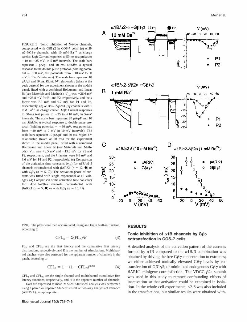
Tonic inhibition of a1B channels by Gbgcotransfection in COS-7 cells
A detailed analysis of the activation pattern of the currentsformed bya1B compared to thea1B/b combination wasobtained by driving the free Gbg concentration to extremes;we either achieved tonically elevated Gbg levels by co-transfection of Gb1g2, or minimized endogenous Gbg withbARK1 minigene cotransfection. The VDCCb2a subunitwas used in this study to remove confounding effects ofinactivation so that activation could be examined in isola-tion. In the whole-cell experiments,a2-d was also includedin the transfections, but similar results were obtained with-
FIGURE 1 Tonic inhibition of N-type channels,coexpressed with Gb1g2 in COS-7 cells. (a) a1B/a2-d/Gbg channels, with 10 mM Ba21 as chargecarrier.Left: Current responses to 50-ms test pulses to210 to 135 mV, in 5-mV intervals. The scale barsrepresent 5 pA/pF and 10 ms.Middle: A typicalresponse to the double pulse protocol (holding poten-tial 5 280 mV, test potentials from210 mV to 30mV in 10-mV intervals). The scale bars represent 10pA/pF and 50 ms.Right: I-V relationship (taken at thepeak current) for the experiment shown in the middlepanel, fitted with a combined Boltzmann and linearfit (see Materials and Methods).V1/2 was126.6 mVand126.8 mV for P1 and P2, respectively, and thekfactor was 7.9 mV and 9.7 mV for P1 and P2,respectively. (b) a1B/a2-d/b2a/Gbg channels with 1mM Ba21 as charge carrier.Left: Current responsesto 50-ms test pulses to235 to 110 mV, in 5-mVintervals. The scale bars represent 20 pA/pF and 10ms.Middle: A typical response to double pulse pro-tocol (holding potential5 280 mV, test potentialsfrom 240 mV to 0 mV in 10-mV intervals). Thescale bars represent 10 pA/pF and 50 ms.Right: I-Vrelationship (taken at 50 ms) for the experimentshown in the middle panel, fitted with a combinedBoltzmann and linear fit (see Materials and Meth-ods).V1/2 was13.5 mV and213.0 mV for P1 andP2, respectively, and thek factors were 6.8 mV and3.6 mV for P1 and P2, respectively. (c) Comparisonof the activation time constants (tact) for a1B/a2-dchannels cotransfected withbARK1 (n 5 12, F) orwith Gbg (n 5 5, E). The activation phase of cur-rents was fitted with single exponential at all volt-ages. (d) Comparison of the activation time constantsfor a1B/a2-d/b2a channels cotransfected withbARK1 (n 5 5, F) or with Gbg (n 5 10,E).
734 Meir et al.
Biophysical Journal 79(2) 731–746

out the additional inclusion of this subunit (data not shown,but see the single-channel results).
a1B/a2-d/Gb1g2 whole-cell currents were rapidly acti-vating (Fig. 1a, left) and showed no facilitation in responseto a prepulse to1120 mV, but rather showed some inacti-vation (Fig. 1a, middle). TheI-V relation parameters did notchange with a depolarizing prepulse, indicating the lack offacilitation (Fig. 1 a, right). In sharp contrast, withb2acoexpression (Fig. 1b), thea1B/a2-d/b2a/Gb1g2 currentsactivated slowly (Fig. 1b, left) and were strongly facilitatedby a large depolarizing prepulse (Fig. 1b, middleandright).The prepulse produced a216-mV hyperpolarizing shiftin the I-V relation and increased the current amplitude(Fig. 1 b, right).
The speed of the activation process and its voltage de-pendence were estimated by fitting a single exponential to
the activation phase of the whole-cell current. Comparisonof activation time constants (tact) obtained fora1B/a2-dchannels, cotransfected withbARK1 or Gb1g2, showed nosignificant differences (Fig. 1c). Nevertheless, a slightdepolarizing shift in thetact-voltage relation was evidentwith Gb1g2 (compareemptyto filled circles in Fig. 1c). Incontrast, with the additional coexpression ofb2a (Fig. 1d),tact was about fivefold slower with Gb1g2 compared tobARK1 cotransfection.
Single-channel analysis of tonic inhibition of a1Bchannels by Gbg
We analyzed single-channel records over a wide range ofvoltages (Fig. 2a) and compared a number of parameters,
FIGURE 2 Examples of single-channel activity and its analysis, fora1B in combination withb2a in the presence of Gb1g2. (a) Top voltage trace:Holding potential2100 mV, test potential in both P1 and P2, to the indicated value, 100 ms, separated by a strong depolarizing prepulse (1120 mV, 50ms). Steps were delivered every 2 s. The voltage protocol is followed by five representative traces and an ensemble of 200 episodes (bottom trace) for eachvoltage. The zero current line that runs through the traces represents the closed state; openings are downward deflections. The bars to the right of the firsttrace represent 1 pA and 100 ms, and the bar to the right of the ensemble current trace represents 0.2 pA (this applies to all of the voltages). (b) Open timedistribution histograms corresponding to the experiment shown ina for P1 (top histogram) and P2 (bottom histogram). For each voltage 200 episodes werecollected. The bin width was 0.5 ms. The mean open time at each voltage is indicated. (c) Open level amplitude histograms for the different voltages inP1 (bars) and P2 (line). The bin width was 0.02 pA. The mean amplitude is indicated for P1 and P2.
N-Type Calcium Channel Modulation 735
Biophysical Journal 79(2) 731–746

including single-channel ensemble current (Fig. 2a), meanopen time (Fig. 2b), mean amplitude (Fig. 2c), and latencyto first opening. The behavior of single channels recordedfrom cell-attached patches of COS-7 cells transfected witha1B with or without a VDCC auxiliaryb subunit and eitherGbg or bARK1 (Fig. 3 and 4) was compared using theseparameters.
These parameters enable one to distinguish between theeffects of either modulatory protein (Gbg and/or VDCCb)on activation (all of the processes leading to the first open-ing of a channel) or postactivation processes, includingmean open time. The hypothesis suggested by the whole-cell data is that the related voltage-dependent processes ofkinetic slowing and prepulse facilitation, which are boththought to involve Gbg unbinding (Stephens et al., 1998;Zamponi and Snutch, 1998), are dependent on the presenceof the VDCCb subunit. We also examined whether param-eters independent of the activation process, such as ampli-tude and mean open time, are affected by either modulatoryprotein.
Both the mean open time and mean amplitude representcompound processes (see Fig. 2c for the compound ampli-tude distributions). The open time distribution of expressed(Wakamori et al., 1999) N-type channels was described bythe sum of several exponentials. However, because of theexclusion of the shortest open times in our analysis, thecontribution of the shorter exponent could be misinter-preted. We therefore chose to examine the arithmetic meanopen time (Fig. 2b). Changes in this parameter couldtherefore be a result of either a change in the distributionbetween short and long opening populations or in the timeconstants for channel closure.
Cells expressinga1B/bARK1 (Fig. 3 a, left) showedchannels with a single-channel conductance of 13 pS and areversal potential of171 mV (Fig. 3b, filled triangles). Thechannels had a mean open time at120 mV of 1.76 0.1 ms(n 5 7; Fig. 3b, top) and showed a tendency to inactivateduring the test pulse (seetracesin Fig. 3 a, left). All of thepatches were subjected to double pulse protocols, with asecond test step after a 50-ms prepulse to1120 mV (Fig. 3a, top trace). The ensemble average current at130 mVshowed no significant difference in the mean responses tothe same voltage step in P1 and P2 (Fig. 3a, left, bottomtrace). This was true for all of the voltages examined (Fig. 3b, middle, comparefilled circles andsquares).
Coexpression of the VDCCa1B subunit with Gb1g2(Fig. 3 a, right) yielded channels with very similar single-channel conductance and reversal potential (Fig. 3b, emptytriangles). The mean open time was shorter over the voltagerange examined (p , 0.001 between thea1B/bARK anda1B/Gbg populations; two-way ANOVA). For example, at120 mV the mean open time was 1.16 0.1 ms (n 5 9),which is a 35% reduction compared to that obtained fora1B/bARK (p , 0.05, Studentt-test; Fig. 3b, top). Inagreement with the whole-cell data (Fig. 1a), the responses
to P1 and P2 were similar within the voltage range exam-ined (Fig. 3,a andb), indicating a lack of facilitation. Therewere no significant differences between these two groups ofdata. Nevertheless, a small;5-mV depolarizing shift of thesingle-channelI-V relationship (Fig. 3b, middle) was de-tected in the presence of Gb1g2, which was similar to theshifts observed in whole-cell recordings.
The coexpression of VDCCb2a with a1B markedlyaffected the activity of the resulting channels (Fig. 3c).However, the single-channel conductance was unchanged(Fig. 3 d). The mean open time was increased significantlyby VDCC b subunit expression at all of the voltages exam-ined (Student’st-test, p , 0.05 for all six pairs of data)(compare Fig. 3b, top, and Fig. 3d, top). However, themean open time was similar regardless of the presence orabsence of Gbg (Fig. 3 d, top). The a1B/b2a/bARK1channels showed fast activation and no inactivation (Fig. 3c, left). The double pulse protocol did not reveal significantdifferences in the responses to P1 and P2, but slight facili-tation was evident at most voltages (Fig. 3c, left, and Fig. 3d, filled symbols). This is in agreement with whole-cellrecordings showing that coexpression of VDCCs withbARK1 removed most but not all of the facilitation (Ste-phens et al., 1998). The ensemble average currents of thesechannels were about twofold larger in amplitude than in theabsence of coexpressedb2a (comparefilled squaresin Fig.3, b andd).
Transfection of Gb1g2 instead ofbARK1 with a1B/b2ayielded channels with a similar mean open time and single-channel conductance (Fig. 3,c, right, and d, empty sym-bols). However,a1B/b2a/Gb1g2 channels frequently failedto gate to the open state during the 100-ms test pulse, givingrise to a large proportion of null episodes. If the channelopened, it was usually with a long delay (Fig. 3c, right).Under this condition, the double pulse protocol was veryeffective at eliciting facilitation of both the ensemble cur-rent amplitude (Fig. 3c, right, bottom trace, and Fig. 3d,compareempty symbols) and the kinetics of the response.Superimposing the responses to a140-mV test pulse (P1)for a1B/Gb1g2 anda1B/b2a/Gb1g2 shows clearly that inthe additional presence ofb2a the mean single-channelresponse ofa1B/Gb1g2 developed much more slowly(Fig. 3 e).
Influence of VDCC b2a on Gbg modulation oflatency to the first opening
Single-channel studies that have analyzed the effect ofneurotransmitter-activated G protein modulation of N-typechannels (Carabelli et al., 1996; Patil et al., 1996) suggestthat the main parameter change is a slowing in the activationprocess, manifested by a longer latency to first opening.Therefore we compared the first latency, presented in theform of CFL (see Materials and Methods), for the differentcombinations of expressed subunits. CFL represents the
736 Meir et al.
Biophysical Journal 79(2) 731–746

FIGURE 3 Single-channel activity ofa1B in combination withbARK1 or Gb1g2 in the presence or absence ofb2a. (a) Top voltage trace: Test potentialin both P1 and P2,130 mV, separated by a depolarizing prepulse to1120 mV.Left column: The single-channel records represent a single-channel patchwith a1B/bARK1. The bar to the left of the first trace represents 1 pA (this applies to all of the single-channel traces).Bottom trace: Average6 SEM (errorsshown only every 5 ms for clarity), single-channel ensemble current (see Materials and Methods) at130 mV (n 5 13, 1895 episodes included). The barsapply to all of the ensemble traces ina andc and represent 0.1 pA and 100 ms. In all panels, the interpulse response was clipped for clarity.Right column:A representative, apparently single-channel patch witha1B/Gb1g2. The format is the same as ina. Bottom trace: n 5 13, 2640 episodes included. (b) Top:Mean channel open time for data of the type shown ina, a1B/bARK1 (filled columns, n 5 7) anda1B/Gbg (empty columns, n 5 9). Statistical significanceis p , 0.05 (Student’st-test, noted by * for a single voltage level) andp . 0.001 (two-way ANOVA, noted with bracket and **) for the difference betweenthe two populations.Bottom: Current-voltage relationship for ensemble and unitary currents (note the break in scale).a1B/bARK1: P1 f and P2F;a1B/Gbg: P1 M and P2E. Single-channel ensemble current amplitudes were taken as the current at 20 ms (the average of 19–21 ms) after the onset ofthe test pulse (mean6 SEM). Unitary amplitudes were fit with a linear regression. The single-channel conductance was 13 pS and 12 pS, and the reversalpotential was170.9 mV and 73.8 mV fora1B/bARK1 (Œ) anda1B/Gbg (‚), respectively. (c) Representative single-channel patches witha1B/b2a.Leftcolumn: a1B/b2a/bARK1, the same format as ina. Bottom trace: n 5 13, 1460 episodes included.Right column: a1B/b2a/Gbg. Bottom trace: n 5 12,3120 episodes included. (d) Top: Mean channel open time for data of the type shown inc, a1B/b2a/bARK1 (filled column, n 5 5) anda1B/b2a/Gbg (emptycolumn, n 5 5). Bottom: I-V curves fora1B/b2a channels. The same format as inb, a1B/b2a/bARK1: P1f] and P2F; a1B/b2a/Gbg: P1M and P2E.The single-channel conductance was 14 pS and 12 pS, and the reversal potential was168.7 mV and171.8 mV fora1B/b2a/bARK1 (Œ) anda1B/b2a/Gbg(‚), respectively. (e) The activation phases at140 mV of the ensemble average responses to P1 with Gbg are compared fora1B (n 5 13) anda1B/b2a(n 5 11). The scale bars represent 0.05 pA and 20 ms.
N-Type Calcium Channel Modulation 737
Biophysical Journal 79(2) 731–746

kinetics of the opening probability of the channel, i.e., theprobability that the channel will open before any given timein the test pulse. Thus higher values at a given point in timerepresent faster activation kinetics.
Both a1B/Gb1g2 and a1B/bARK1 channels showedvoltage-dependent kinetics of activation that were insensi-tive to the large depolarizing prepulse (Fig. 4a; compare thealmost superimposedsquareandcircular symbols). A Boltz-mann fit to these data revealed that the activation processremained partial, reaching saturation at 50–60% (Fig. 4b).This may have been due to closed-state inactivation (Patil etal., 1998). There were no significant differences betweenthe individual CFL values 20 ms after the onset of the testpulse, but there was a15-mV shift in theV1/2 with Gb1g2(Fig. 4 b).
The expression ofa1B/b2a/bARK1 resulted in rapidlyactivating channels (Fig. 4c, filled symbols), with a morenegative midpoint of activation than in the absence of absubunit, and maximum activation of;85% at 20 ms (Fig. 4d). On the other hand, expression ofa1B/b2a/Gb1g2 re-sulted in channels that were slowly activating and thereforeshowed very low opening probabilities (Fig. 4c, emptysquares). Application of a strong depolarizing prepulse re-sulted in a biphasic activation waveform (Fig. 4c, emptycircles). The added fast component facilitated the openingprobability (Fig. 4 d, compare empty circlesand emptysquares).
To examine whether the effects ofb2a on facilitation(compare the P2/P1 ratio in Fig. 5,a andb) were a generalfeature of VDCCb subunit coexpression, we also used the
FIGURE 4 First latency ofa1B/bARK1 compared toa1B/Gbg and of a1B/b2a/bARK1 compared toa1B/b2a/Gbg. Latency to first opening wasmeasured in single and multichannel patches (see Materials and Methods). The cumulative first latency (CFL) plots for all of the experiments with thesamesubunit composition were averaged, and the means6 SEM are shown. The CFL represents the probability of the channel opening before a given time (seeMaterials and Methods). (a) Opening probability (CFL) without theb subunit at three different voltages; the voltage step is indicated above each column.a1B/bARK1: P1 (f) and P2 (F); a1B/Gbg: P1 (M) and P2 (E). Thex axis was broken between 94 and 95 ms to expand the last few symbols for clarityof the symbol definition. (b) The opening probability (CFL) 20 ms after the onset of the test pulse (the same symbol code as ina). Boltzmann fits to thedata are plotted as continuous lines. The extracted parameters are as follows: fora1B/bARK1: P1, P2 (n 5 13); for a1B/Gbg: P1 and P2 (n 5 13),respectively. Maximum (%), 58, 55, 56, and 56.V1/2 (mV), 29, 29, 34, 34. (c) Opening probability with the combinations including theb subunit at threedifferent voltages. Symbols are as ina. (d) The opening probability 20 ms after the onset of the test pulse fora1B/b2a/bARK1 anda1B/b2a/Gbg (thesame symbol code as ina). Boltzmann fits to the data are plotted as continuous lines. The extracted parameters are as follows: fora1B/b2a/bARK1: P1,P2 (n 5 13); for a1B/b2a/Gbg: P1 and P2 (n 5 12), respectively. Maximum (%), 88, 83, 53, and 59.V1/2 (mV), 25, 22, 40, 32.
738 Meir et al.
Biophysical Journal 79(2) 731–746
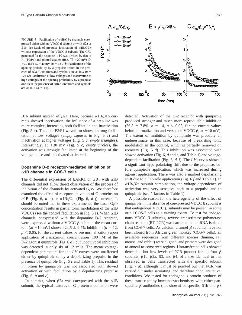
b1b subunit instead ofb2a. Here, becausea1B/b1b cur-rents showed inactivation, the influence of a prepulse wasmore complex, increasing both facilitation and inactivation(Fig. 5 c). Thus the P2/P1 waveform showed strong facili-tation at low voltages (empty squaresin Fig. 5 c) andinactivation at higher voltages (Fig. 5c, empty triangles).Interestingly, at130 mV (Fig. 5 c, empty circles), theactivation was strongly facilitated at the beginning of thevoltage pulse and inactivated at its end.
Dopamine D-2 receptor-mediated inhibition ofa1B channels in COS-7 cells
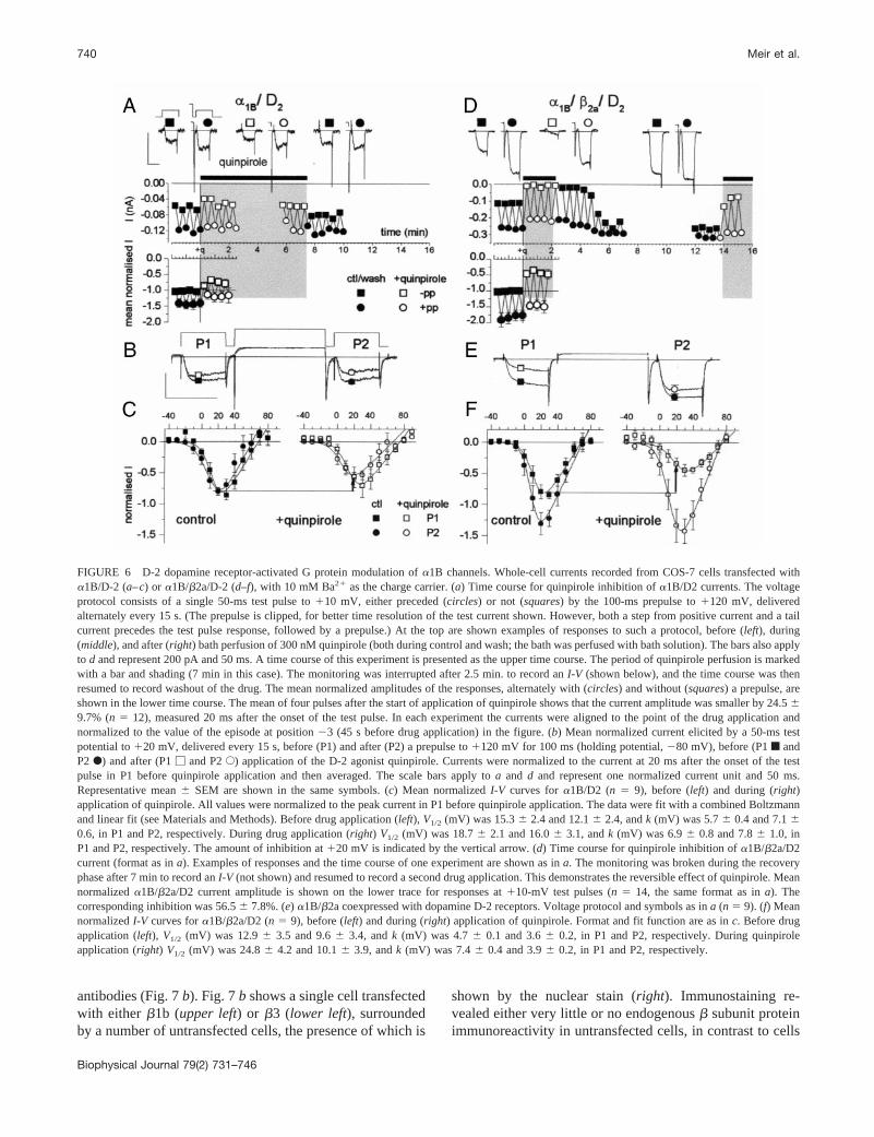
The differential expression ofbARK1 or Gbg with a1Bchannels did not allow direct observation of the process ofinhibition of the channels by activated Gbg. We thereforeexamined the effect of receptor activation of G proteins ona1B (Fig. 6, a–c) or a1B/b2a (Fig. 6, d–f) currents. Itshould be noted that in these experiments, the basal Gbgconcentration results in partial tonic modulation of thea1BVDCCs (see the control facilitation in Fig. 6e). Whena1Bchannels, coexpressed with the dopamine D-2 receptor,were expressed without a VDCCb subunit, the mean cur-rent (at110 mV) showed 24.56 9.7% inhibition (n 5 12,p , 0.05, for the current values before normalization) uponapplication of a maximum concentration (100 nM) of theD-2 agonist quinpirole (Fig. 6a), but unequivocal inhibitionwas detected in only six of 12 cells. The mean voltage-dependent parameters for theI-V curves were unaffectedeither by quinpirole or by a depolarizing prepulse in thepresence of quinpirole (Fig. 6c and Table 1). This residualinhibition by quinpirole was not associated with slowedactivation or with facilitation by a depolarizing prepulse(Fig. 6, a andc).
In contrast, whenb2a was coexpressed with thea1Bsubunit, the typical features of G protein modulation were
detected. Activation of the D-2 receptor with quinpiroleproduced stronger and much more reproducible inhibition(56.5 6 7.8%, n 5 14, p , 0.05, for the current valuesbefore normalization and versus no VDCCb, at 110 mV).The extent of inhibition by quinpirole was probably anunderestimate in this case, because of preexisting tonicmodulation in the control, which is partially removed onrecovery (Fig. 6d). This inhibition was associated withslowed activation (Fig. 6,d ande, and Table 1) and voltage-dependent facilitation (Fig. 6,d–f). The I-V curves showeda significant hyperpolarizing shift due to the prepulse, be-fore quinpirole application, which was increased duringagonist application. There was also a marked depolarizingshift due to quinpirole application (Fig. 6f and Table 1). Ina1B/b2a subunit combination, the voltage dependence ofactivation was very sensitive both to a prepulse and toquinpirole (seek factors in Table 1).
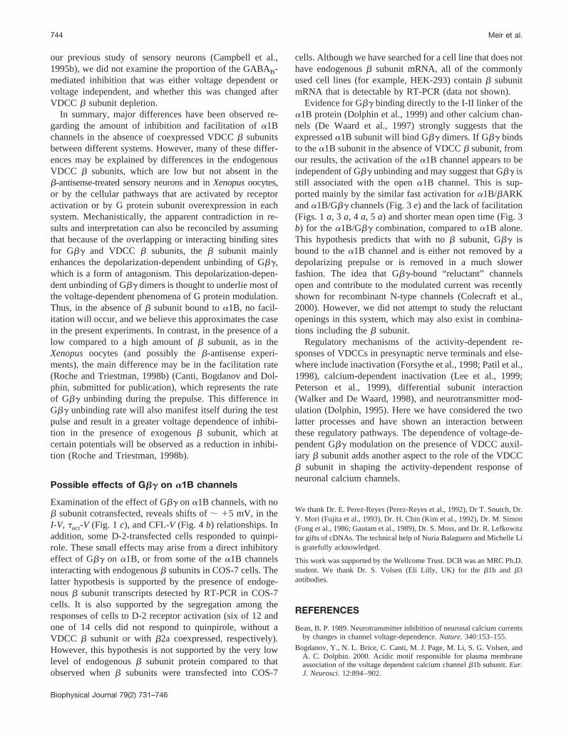
A possible reason for the heterogeneity of the effect ofquinpirole in the absence of coexpressed VDCCb subunit isthat endogenous VDCCb subunits may be present in someor all COS-7 cells to a varying extent. To test for endoge-nous VDCCb subunits, reverse transcriptase-polymerasechain reaction (RT-PCR) was carried out on mRNA isolatedfrom COS-7 cells. As calcium channelb subunits have notbeen cloned from African green monkey (COS-7 cells), allavailable sequences from different species (human, rat,mouse, and rabbit) were aligned, and primers were designedto anneal to conserved regions. Untransfected cells showeddetectable but low levels of PCR product for all fourbsubunits,b1b, b2a, b3, andb4, of a size identical to thatobserved in cells transfected with the specific subunit(Fig. 7 a), although it must be pointed out that PCR wascarried out under saturating, and therefore nonquantitative,conditions. We tested for endogenous protein products ofthese transcripts by immunocytochemistry with either pan-specific b antibodies (not shown) or specificb1b andb3
FIGURE 5 Facilitation ofa1B/Gbg channels coex-pressed either with no VDCCb subunit or withb2a orb1b. (a) Lack of prepulse facilitation ofa1B/Gbgwithout expression of the VDCCb subunit. The CFLgenerated for the response to P2 was divided by that ofP1 (P2/P1) and plotted against time.M, 120 mV; E,130 mV;‚, 140 mV (n 5 13). (b) Facilitation of theopening probability by a prepulse occurs in the pres-ence ofb2a. Conditions and symbols are as ina (n 512). (c) Facilitation at low voltages and inactivation athigh voltages of the opening probability by a prepulseoccurs in the presence ofb1b. Conditions and symbolsare as ina (n 5 10).
N-Type Calcium Channel Modulation 739
Biophysical Journal 79(2) 731–746
antibodies (Fig. 7b). Fig. 7b shows a single cell transfectedwith eitherb1b (upper left) or b3 (lower left), surroundedby a number of untransfected cells, the presence of which is
shown by the nuclear stain (right). Immunostaining re-vealed either very little or no endogenousb subunit proteinimmunoreactivity in untransfected cells, in contrast to cells
FIGURE 6 D-2 dopamine receptor-activated G protein modulation ofa1B channels. Whole-cell currents recorded from COS-7 cells transfected witha1B/D-2 (a–c) or a1B/b2a/D-2 (d–f), with 10 mM Ba21 as the charge carrier. (a) Time course for quinpirole inhibition ofa1B/D2 currents. The voltageprotocol consists of a single 50-ms test pulse to110 mV, either preceded (circles) or not (squares) by the 100-ms prepulse to1120 mV, deliveredalternately every 15 s. (The prepulse is clipped, for better time resolution of the test current shown. However, both a step from positive current and atailcurrent precedes the test pulse response, followed by a prepulse.) At the top are shown examples of responses to such a protocol, before (left), during(middle), and after (right) bath perfusion of 300 nM quinpirole (both during control and wash; the bath was perfused with bath solution). The bars also applyto d and represent 200 pA and 50 ms. A time course of this experiment is presented as the upper time course. The period of quinpirole perfusion is markedwith a bar and shading (7 min in this case). The monitoring was interrupted after 2.5 min. to record anI-V (shown below), and the time course was thenresumed to record washout of the drug. The mean normalized amplitudes of the responses, alternately with (circles) and without (squares) a prepulse, areshown in the lower time course. The mean of four pulses after the start of application of quinpirole shows that the current amplitude was smaller by 24.569.7% (n 5 12), measured 20 ms after the onset of the test pulse. In each experiment the currents were aligned to the point of the drug application andnormalized to the value of the episode at position23 (45 s before drug application) in the figure. (b) Mean normalized current elicited by a 50-ms testpotential to120 mV, delivered every 15 s, before (P1) and after (P2) a prepulse to1120 mV for 100 ms (holding potential,280 mV), before (P1f andP2F) and after (P1M and P2E) application of the D-2 agonist quinpirole. Currents were normalized to the current at 20 ms after the onset of the testpulse in P1 before quinpirole application and then averaged. The scale bars apply toa and d and represent one normalized current unit and 50 ms.Representative mean6 SEM are shown in the same symbols. (c) Mean normalizedI-V curves fora1B/D2 (n 5 9), before (left) and during (right)application of quinpirole. All values were normalized to the peak current in P1 before quinpirole application. The data were fit with a combined Boltzmannand linear fit (see Materials and Methods). Before drug application (left), V1/2 (mV) was 15.36 2.4 and 12.16 2.4, andk (mV) was 5.76 0.4 and 7.160.6, in P1 and P2, respectively. During drug application (right) V1/2 (mV) was 18.76 2.1 and 16.06 3.1, andk (mV) was 6.96 0.8 and 7.86 1.0, inP1 and P2, respectively. The amount of inhibition at120 mV is indicated by the vertical arrow. (d) Time course for quinpirole inhibition ofa1B/b2a/D2current (format as ina). Examples of responses and the time course of one experiment are shown as ina. The monitoring was broken during the recoveryphase after 7 min to record anI-V (not shown) and resumed to record a second drug application. This demonstrates the reversible effect of quinpirole. Meannormalizeda1B/b2a/D2 current amplitude is shown on the lower trace for responses at110-mV test pulses (n 5 14, the same format as ina). Thecorresponding inhibition was 56.56 7.8%. (e) a1B/b2a coexpressed with dopamine D-2 receptors. Voltage protocol and symbols as ina (n 5 9). (f) MeannormalizedI-V curves fora1B/b2a/D2 (n 5 9), before (left) and during (right) application of quinpirole. Format and fit function are as inc. Before drugapplication (left), V1/2 (mV) was 12.96 3.5 and 9.66 3.4, andk (mV) was 4.76 0.1 and 3.66 0.2, in P1 and P2, respectively. During quinpiroleapplication (right) V1/2 (mV) was 24.86 4.2 and 10.16 3.9, andk (mV) was 7.46 0.4 and 3.96 0.2, in P1 and P2, respectively.
740 Meir et al.
Biophysical Journal 79(2) 731–746

that were transfected with eitherb1b orb3. Furthermore, inadditional experiments we have found that expression ofa1B alone in these cells does not induce the expression ofb subunit protein, as evidenced from immunocytochemistry(results not shown). This result is also supported by ourprevious study, which did not detect endogenousb subunitsin COS-7 cells by Western blotting (Campbell et al., 1995a).
DISCUSSION
We have examined the influence of Gbg dimers and VDCCb subunits, both separately and combined, on the kineticproperties of N-type VDCCs by coexpression with the pore-forming a1B subunits in COS-7 cells. With a VDCCb
subunit coexpressed, G protein modulation produced theexpected kinetic alterations of the N-type channels (Figs. 1,b and d; 2; 3, c and d; 4, c and d; 5, b and c; 6, d–f), aspredicted by a previous model (Patil et al., 1996). In con-trast, with nob subunit coexpressed, these kinetic alter-ations were largely absent (Figs. 1,a andc; 3, a andb; 4, candd; 5 a; 6, a–c), although there was still some residualnon-voltage-dependent inhibition in this system (Fig. 6a,bottom).
The differential coexpression ofa1B with eitherbARKGbg binding domain or Gbg dimers is advantageous be-cause it allows a comparison between Gbg-depleted andsaturated populations, respectively. The ensemble averagecurrents obtained here (Fig. 2a) represent the changes in
FIGURE 7 Examination of the presence of endogenous VDCCb subunits in COS-7 cells. (a) RT-PCR detection ofb subunit mRNAs in COS-7 cells.For eachb subunit, three lanes are shown: o represents untransfected cells,1 represents cells transfected with the specificb subunit, and2 represents anegative (H2O) control. Markers at either side represent a 100-bp ladder; the arrow on the left indicates 800 bp. The details of the primers used are givenin the Materials and Methods section. (b) Immunostaining for calcium channelb subunits in cells transfected with eitherb1b (top) or b3 (bottom). Left:A field in which one cell in each case was positively transfected. This shows immunostaining for the specificb subunit; using monoclonalb1b (upper panel)or b3 (lower panel) antibodies. Untransfected cells present in the same field show little or no immunoreactivity.Right: Nuclear staining with the DNA dyeYO-YO to show the presence of several cells in each field. The white bar (bottom right) represents 10mm for all panels.
TABLE 1 Influence of quinpirole and a depolarizing prepulse (pp) on I-V parameters
a1B/D-2 (n 5 9) a1B/b2a/D-2 (n 5 9)
Shift in V1/2
(mV)Change ink
(%)Shift in V1/2
(mV)Change ink
(%)
Influence of quinpirole 13.36 1.8 (NS) 121.06 10.6 (NS) 111.96 2.3* 160.76 10.3*Influence of pp,2quinpirole 23.36 0.5* 125.46 9.4* 23.36 0.3* 77.46 4.5*Influence of pp,1quinpirole 22.66 2.1 (NS) 112.66 8.6 (NS) 214.76 1.4* 52.76 3.4*
I-V relations were fitted with a combined Boltzmann and linear fit (see Materials and Methods and Fig. 1) in each experiment. The shifts inV1/2 wereobtained by subtraction, and the changes in thek factors are presented as a percentage. Increases in thek factor ratio indicate shallower voltage dependenceand vice versa.*Statistically significant difference (p , 0.05, Student’st-test).
N-Type Calcium Channel Modulation 741
Biophysical Journal 79(2) 731–746

open probability of a single channel. Therefore, it providesa means of “normalization” that could not be achieved withwhole-cell current. The whole-cell current can be expressedas current per membrane area (current density), a parameterthat still includes the number of channels, which may beaffected differentially by the subunit content. The observa-tion of the number of overlapping openings in single/mul-tichannel records allows estimation of the number of chan-nels in the patch and therefore direct comparison betweenpopulations transfected with different subunit combinations.
In the experiments in which the dopamine D-2 receptorwas coexpressed, thea1B channels are partially tonicallymodulated because of a certain level of endogenous Gbg,(see Fig. 6,d ande). The Gbg level is then further elevatedbecause of receptor activation. In our work, this (receptor-mediated modulation) system had variability and showedsome residual inhibition by quinpirole in the absence ofexpressed VDCCb subunits (Fig. 6). This may reflect thegreater complexity of receptor-mediated modulation in ac-tivating both voltage-dependent and non-voltage-dependentpathways by multiple signal transduction pathways. How-ever, in general it supports the findings from the former(tonic Gbg modulation) system, with significantly lowerinhibition and lack of significant kinetic slowing or facili-tation, when a VDCCb subunit was not coexpressed.
The tonic modulation system provides strongly groundeddata showing the role of the VDCCb subunit, both in thesuppression by Gbg of the activation process (Figs. 1, 3e,and 4) and in Gbg unbinding, as manifested by prepulsefacilitation (Figs. 1b and 5). The single-channel data pro-vide strong evidence that the main effect of theb subunit inthe bARK1-expressing cells is a twofold increase in themean open time (compare Fig. 3b with Fig. 3 d) and anincrease in the activation probability (CFL). The residualeffect of Gbg on a1B expressed without a VDCCb subunitcould be partially explained by a reduction in the mean opentime (Fig. 3b) or possibly by endogenous VDCCb subunits(Fig. 7a). In contrast, in the presence of a VDCCb subunitit is clear that the activation ofa1B was slowed by Gbg,and the number of failures increased (Fig. 4c). To the bestof our knowledge, this is the first kinetic examination ofboth the separate and combined effects of VDCCb subunitsand Gbg dimers.
The role of the auxiliary b subunits in theregulation of N-type channel properties
The involvement of VDCCb subunits in the trafficking ofa1 subunits to the plasma membrane is well established(Chien et al., 1995; Brice et al., 1997). Nevertheless, itremains undecided whether they are essential for functionalexpression. Several groups have provided evidence forbsubunit involvement in the modulation of the channel bio-physical properties, suggesting interaction with a number ofintracellular sites (Stephens et al., 1997; Costantin et al.,
1998; Gerster et al., 1999). The differentb subunits alsohave differential effects on inactivation, withb1b, b3, andb4 supporting, andb2a reducing, inactivation (Walker andDe Waard, 1998).
Because we wished to avoid the confounding factor ofinactivation in our analysis, we used theb2a subunit in mostexperiments, but broadly similar results were obtained forb1b (Fig. 5). The role ofb2a in reducing closed-stateinactivation (Patil et al., 1998) is seen here (Fig. 4) as anincrease in the maximum opening probability (Imredy andYue, 1994; Meir et al., 1998), but we made no attempt tostudy inactivation properties in this study.
Coexpression of the auxiliaryb subunit is reported tocaused a hyperpolarizing shift in current activation by;5–10 mV (Wakamori et al., 1999), with no effect on thesingle-channel conductance (Fig. 3,b andd). In our hands,only a small nonsignificant shift due tob coexpression wasdetected in the activation of the whole-cell populations(Fig. 6,c andf). This may be due to the absence ofa2-d inthese experiments, which augments the VDCCb subuniteffects (Wakamori et al., 1999). In another study wherea1Bwas cotransfected witha2-d, using identical Ba21 concen-trations to enable direct comparison, a 15-mV hyperpolar-izing shift in the activation of the whole-cell current wasdetected withb subunit coexpression in COS-7 cells (Ste-phens et al., 2000).
The VDCC b subunit also increased the mean channelopen time (Fig. 3,b and d; p , 0.05, Student’st-test, forcomparison ofa1B/bARK versusa1B/b2a/bARK and fora1B/Gbg versusa1B/b2a/Gbg at all voltages examined)(Meir et al., 1998; Wakamori et al., 1999). VDCCb2acoexpression enhanced the first opening probability (Fig. 4,a and c), which is in agreement with improvement of thecoupling between gating charge movement and channelopening (Jones et al., 1997).
Because the effects of VDCCb subunit coexpressionwere significant, and taking into account the immunocyto-chemistry data on the level of endogenousb subunits, weassume thata1B can be expressed in the membrane withoutthe permanent attachment of this auxiliary subunit. In ad-dition, expression ofa1B with an antisenseb3 subunitcDNA construct did not prevent the expression ofa1B inCOS-7 cells (Stephens and Dolphin, unpublished results).However, we cannot entirely discount the possibility thata1B can only be functionally expressed in the plasmamembrane with the support of a VDCCb subunit duringtrafficking.
The effect of auxiliary b2a subunit on themodulation of N-type VDCCs by Gbg
The VDCC a1B/b2a combination, coexpressed with theD-2 receptor, generated currents that were markedly andreversibly inhibited during quinpirole application (Fig. 6d).On the other hand,a1B channels expressed without an
742 Meir et al.
Biophysical Journal 79(2) 731–746

auxiliary b subunit showed much less inhibition, and noslowed activation or enhanced facilitation, when exposed toquinpirole (Fig. 6a). Furthermore, comparison of the sin-gle-channel populations formed bya1B when coexpressedwith eitherbARK1 or Gbg also indicated a much strongerinhibition by Gbg when VDCC b2a was coexpressed(Fig. 3,a andc). These findings are supported in our systemby kinetic measurements of the response of thea1B chan-nels to moderate voltage steps. Thea1B/b2a/Gbg channelswere very slowly activating (Figs. 1d and 3e). In sharpcontrast,a1B/Gb1g2 currents activated rapidly (Fig. 1c),which would not be expected for modulated channels thatare thought to unbind the Gbg before opening (Patil et al.,1996), with this process being responsible for the slowgating. A marked slowing of the activation process has beenproposed to underlie G protein inhibition (Patil et al., 1996)and has been detected in many systems and for severalnative and heterologously expressed VDCCs (Dolphin,1995).
The facilitation that is a hallmark of voltage-dependent Gprotein modulation (Dolphin, 1995) was also strongly de-pendent on VDCCb subunit coexpression. Withb2a, clearfacilitation due to a depolarizing prepulse was evident forwhole-cell (Stephens et al., 1998) (Figs. 1b and 6,d ande)and single-channel (Figs. 2–5) currents. With the VDCCb1b subunit coexpressed, a more complex behavior wasobserved, but clear facilitation (although partially overlaidby inactivation at higher potentials) was also evident (Fig. 5c). The facilitation was time dependent at all voltages(Fig. 5 b), involving the addition of a fast Gbg-unboundpopulation to the gating probability. The apparent amountof facilitation for theb2a-containing combinations variedbetween the different sets of experiments (compare Figs. 1b and 6,d ande, to Fig. 3c, right). One possible reason forthese discrepancies may be the different voltage protocolused for cell-attached (100-ms test pulses and 50-ms pre-pulse) and whole-cell (50-ms test pulses and 100-ms pre-pulse) experiments.
Two main questions regarding the role of the VDCCbsubunit and Gbg binding are not answered by the presentstudy. First, as discussed above, cana1 subunits be ex-pressed in the membrane without a chaperoningb subunit(either endogenous or coexpressed)? Second, area1B chan-nels inhibited to any extent by Gbg in the absence of theVDCC b subunit? Alternatively, are the effects detected fora1B with no VDCCb subunit coexpressed due solely to afraction of the channels interacting with endogenousbsubunits, allowing the observation of residual inhibition?
While recognizing these limitations, our data stronglysupport a mechanism in which the VDCCb subunit isnecessary for the voltage-dependent modulation of VDCCs.However, this result appears to contradict previous resultsproposing antagonistic effects of VDCCb subunits on Gprotein modulation.
Comparison with previous literature: does theVDCC b subunit facilitate or antagonizeGbg modulation?
Biochemical evidence for overlapping binding sites for aux-iliary VDCC b subunit and Gbg dimers on variousa1subunits (De Waard et al., 1997; Walker et al., 1998, 1999;Canti et al., 1999) suggests a physical basis for an interac-tion between their effects.
In light of previous results obtained in our laboratory(Campbell et al., 1995b) for native VDCCs in sensoryneurons and in other laboratories forXenopusoocytes, wewere initially surprised by the lack of Gbg-mediated kineticslowing in the absence of overexpressed VDCCb subunit.Although our present results do not support the hypothesisof antagonism betweenb subunits and Gbg dimers, severalexplanations may reconcile these apparently contradictorystudies.
A number of groups previously reported that the expres-sion of certainb subunits reduced receptor-mediated Gprotein inhibition of expressed VDCCs (Roche et al., 1995;Bourinet et al., 1996; Qin et al., 1997; Roche and Treistman,1998a; Canti et al., 1999). Antagonism is supported mainlyby recordings fromXenopusoocytes, in which an endoge-nousb3 subunit is active, so that the extent to which theexpressed channels represent freea1 subunits is unknown(Tareilus et al., 1997). In the studies cited above, a com-parison of the percentage inhibition upon receptor activationshowed more inhibition at certain potentials in the absencethan in the presence of a coexpressedb subunit. At least partof this effect can be explained by the VDCCb subunit-induced hyperpolarizing shift in current activation, makingcomparisons of receptor-mediated inhibition at a singlepotential difficult to interpret. However, facilitation or volt-age-dependent relief of inhibition of N-type channels wasalso enhanced by coexpression ofb3, which is similar to theresults presented here (Roche and Treistman, 1998b).
In a converse experiment with sensory neurons (Camp-bell et al., 1995b), VDCCb subunits were partially depletedby an antisense oligonucleotide, and the GABAB-inducedinhibition of the native VDCCs was found to be greater inthe b subunit-depleted than in control neurons. The reasonfor the apparent contradiction compared to our presentresults is unknown, but multiple signaling pathways areactivated by G proteins in native neurons, and although Gbgis widely accepted to be responsible for the voltage-depen-dent (Gbg-mediated) modulation of N- and P/Q-type chan-nels, voltage-independent modulation has also been de-scribed and may be mediated by a number of differentpathways (Diverse-Pierluissi et al., 1991; Fitzgerald andDolphin, 1997). Furthermore, these pathways may alsocross-talk with Gbg-mediated voltage-dependent inhibition(Zamponi et al., 1997). Voltage-dependent receptor-medi-ated inhibition of N-type channels is also influenced by theGa subunit involved (Kammermeier and Ikeda, 1999). In
N-Type Calcium Channel Modulation 743
Biophysical Journal 79(2) 731–746
our previous study of sensory neurons (Campbell et al.,1995b), we did not examine the proportion of the GABAB-mediated inhibition that was either voltage dependent orvoltage independent, and whether this was changed afterVDCC b subunit depletion.
In summary, major differences have been observed re-garding the amount of inhibition and facilitation ofa1Bchannels in the absence of coexpressed VDCCb subunitsbetween different systems. However, many of these differ-ences may be explained by differences in the endogenousVDCC b subunits, which are low but not absent in theb-antisense-treated sensory neurons and inXenopusoocytes,or by the cellular pathways that are activated by receptoractivation or by G protein subunit overexpression in eachsystem. Mechanistically, the apparent contradiction in re-sults and interpretation can also be reconciled by assumingthat because of the overlapping or interacting binding sitesfor Gbg and VDCC b subunits, theb subunit mainlyenhances the depolarization-dependent unbinding of Gbg,which is a form of antagonism. This depolarization-depen-dent unbinding of Gbg dimers is thought to underlie most ofthe voltage-dependent phenomena of G protein modulation.Thus, in the absence ofb subunit bound toa1B, no facil-itation will occur, and we believe this approximates the casein the present experiments. In contrast, in the presence of alow compared to a high amount ofb subunit, as in theXenopusoocytes (and possibly theb-antisense experi-ments), the main difference may be in the facilitation rate(Roche and Triestman, 1998b) (Canti, Bogdanov and Dol-phin, submitted for publication), which represents the rateof Gbg unbinding during the prepulse. This difference inGbg unbinding rate will also manifest itself during the testpulse and result in a greater voltage dependence of inhibi-tion in the presence of exogenousb subunit, which atcertain potentials will be observed as a reduction in inhibi-tion (Roche and Triestman, 1998b).
Possible effects of Gbg on a1B channels
Examination of the effect of Gbg on a1B channels, with nob subunit cotransfected, reveals shifts of; 15 mV, in theI-V, tact-V (Fig. 1 c), and CFL-V (Fig. 4 b) relationships. Inaddition, some D-2-transfected cells responded to quinpi-role. These small effects may arise from a direct inhibitoryeffect of Gbg on a1B, or from some of thea1B channelsinteracting with endogenousb subunits in COS-7 cells. Thelatter hypothesis is supported by the presence of endoge-nousb subunit transcripts detected by RT-PCR in COS-7cells. It is also supported by the segregation among theresponses of cells to D-2 receptor activation (six of 12 andone of 14 cells did not respond to quinpirole, without aVDCC b subunit or withb2a coexpressed, respectively).However, this hypothesis is not supported by the very lowlevel of endogenousb subunit protein compared to thatobserved whenb subunits were transfected into COS-7
cells. Although we have searched for a cell line that does nothave endogenousb subunit mRNA, all of the commonlyused cell lines (for example, HEK-293) containb subunitmRNA that is detectable by RT-PCR (data not shown).
Evidence for Gbg binding directly to the I-II linker of thea1B protein (Dolphin et al., 1999) and other calcium chan-nels (De Waard et al., 1997) strongly suggests that theexpresseda1B subunit will bind Gbg dimers. If Gbg bindsto thea1B subunit in the absence of VDCCb subunit, fromour results, the activation of thea1B channel appears to beindependent of Gbg unbinding and may suggest that Gbg isstill associated with the opena1B channel. This is sup-ported mainly by the similar fast activation fora1B/bARKanda1B/Gbg channels (Fig. 3e) and the lack of facilitation(Figs. 1a, 3 a, 4 a, 5 a) and shorter mean open time (Fig. 3b) for the a1B/Gbg combination, compared toa1B alone.This hypothesis predicts that with nob subunit, Gbg isbound to thea1B channel and is either not removed by adepolarizing prepulse or is removed in a much slowerfashion. The idea that Gbg-bound “reluctant” channelsopen and contribute to the modulated current was recentlyshown for recombinant N-type channels (Colecraft et al.,2000). However, we did not attempt to study the reluctantopenings in this system, which may also exist in combina-tions including theb subunit.
Regulatory mechanisms of the activity-dependent re-sponses of VDCCs in presynaptic nerve terminals and else-where include inactivation (Forsythe et al., 1998; Patil et al.,1998), calcium-dependent inactivation (Lee et al., 1999;Peterson et al., 1999), differential subunit interaction(Walker and De Waard, 1998), and neurotransmitter mod-ulation (Dolphin, 1995). Here we have considered the twolatter processes and have shown an interaction betweenthese regulatory pathways. The dependence of voltage-de-pendent Gbg modulation on the presence of VDCC auxil-iary b subunit adds another aspect to the role of the VDCCb subunit in shaping the activity-dependent response ofneuronal calcium channels.
We thank Dr. E. Perez-Reyes (Perez-Reyes et al., 1992), Dr T. Snutch, Dr.Y. Mori (Fujita et al., 1993), Dr. H. Chin (Kim et al., 1992), Dr. M. Simon(Fong et al., 1986; Gautam et al., 1989), Dr. S. Moss, and Dr. R. Lefkowitzfor gifts of cDNAs. The technical help of Nuria Balaguero and Michelle Liis gratefully acknowledged.
This work was supported by the Wellcome Trust. DCB was an MRC Ph.D.student. We thank Dr. S. Volsen (Eli Lilly, UK) for theb1b and b3antibodies.
REFERENCES
Bean, B. P. 1989. Neurotransmitter inhibition of neuronal calcium currentsby changes in channel voltage-dependence.Nature.340:153–155.
Bogdanov, Y., N. L. Brice, C. Canti, M. J. Page, M. Li, S. G. Volsen, andA. C. Dolphin. 2000. Acidic motif responsible for plasma membraneassociation of the voltage dependent calcium channelb1b subunit.Eur.J. Neurosci.12:894–902.
744 Meir et al.
Biophysical Journal 79(2) 731–746

Boland, L. M., and B. P. Bean. 1993. Modulation of N-type calciumchannels in bullfrog sympathetic neurons by luteinizing hormone-releasing hormone: kinetics and voltage dependence.J. Neurosci.13:516–533.
Bourinet, E., T. W. Soong, A. Stea, and T. P. Snutch. 1996. Determinantsof the G protein-dependent opioid modulation of neuronal calciumchannels.Proc. Natl. Acad. Sci. USA.93:1486–1491.
Brice, N. L., N. S. Berrow, V. Campbell, K. M. Page, K. Brickley, I.Tedder, and A. C. Dolphin. 1997. Importance of the differentb subunitsin the membrane expression of thea1A and a2 calcium channelsubunits: studies using a depolarisation-sensitivea1A antibody. Eur.J. Neurosci.9:749–759.
Campbell, V., N. Berrow, K. Brickley, K. Page, R. Wade, and A. C.Dolphin. 1995a. Voltage-dependent calcium channelb-subunits in com-bination witha1 subunits have a GTPase activating effect to promote thehydrolysis of GTP by Gao in rat frontal cortex.FEBS Lett.370:135–140.
Campbell, V., N. S. Berrow, E. M. Fitzgerald, K. Brickley, and A. C.Dolphin. 1995b. Inhibition of the interaction of G protein Go withcalcium channels by the calcium channelb-subunit in rat neurones.J. Physiol. (Lond.).485:365–372.
Canti, C., K. M. Page, G. J. Stephens, and A. C. Dolphin. 1999. Identifi-cation of residues in the N-terminus ofa1B critical for inhibition of thevoltage-dependent-calcium channel by Gbg. J. Neurosci. 19:6855–6864.
Carabelli, V., M. Lovallo, V. Magnelli, H. Zucker, and E. Carbone. 1996.Voltage-dependent modulation of single N-type Ca21 channel kineticsby receptor agonists in IMR32 cells.Biophys. J.70:2144–2154.
Chien, A. J., X. L. Zhao, R. E. Shirokov, T. S. Puri, C. F. Chang, D. Sun,E. Rios, and M. M. Hosey. 1995. Roles of a membrane-localizedbsubunit in the formation and targeting of functional L-type Ca21 chan-nels.J. Biol. Chem.270:30036–30044.
Colecraft, H. M., P. G. Patil, and D. T. Yue. 2000. Differential occurrenceof reluctant openings in G-protein-inhibited N- and P/Q-type calciumchannels.J. Gen. Physiol.115:175–192.
Colquhoun, D., and G. H. Hawkes. 1995. The principles of the stochasticinterpretation of ion channel mechanisms.In Single Channel Recording.B. Sakmann and E. Neher, editors. Plenum, New York. 397–482.
Costantin, J., F. Noceti, N. Qin, X. Y. Wei, L. Birnbaumer, and E. Stefani.1998. Facilitation by theb2a subunit of pore openings in cardiac Ca21
channels.J. Physiol. (Lond.).507:93–103.
Day, N. C., S. G. Volsen, A. L. McCormack, P. J. Craig, W. Smith, R. E.Beattie, P. J. Shaw, S. B. Ellis, M. M. Harpold, and P. G. Ince. 1998. Theexpression of voltage-dependent calcium channel beta subunits in hu-man hippocampus.Mol. Brain Res.60:259–269.
De Waard, M., H. Y. Liu, D. Walker, V. E. S. Scott, C. A. Gurnett, andK. P. Campbell. 1997. Direct binding of G-proteinbgamma complex tovoltage-dependent calcium channels.Nature.385:446–450.
Diverse-Pierluissi, M., K. Dunlap, and E. W. Westhead. 1991. Multipleactions of extracellular ATP on calcium currents in cultured bovinechromaffin cells.Proc. Natl. Acad. Sci. USA.88:1261–1265.
Dolphin, A. C. 1995. Voltage-dependent calcium channels and their mod-ulation by neurotransmitters and G proteins: G. L. Brown prize lecture.Exp. Physiol.80:1–36.
Dolphin, A. C., K. M. Page, N. S. Berrow, G. J. Stephens, and C. Canti.1999. Dissection of the calcium channel domains responsible for mod-ulation of neuronal voltage-dependent calcium channels by G proteins.Ann. N.Y. Acad. Sci.868:160–174.
Dunlap, K., J. I. Luebke, and T. J. Turner. 1995. Exocytotic Ca21 channelsin mammalian central neurons.Trends Neurosci.18:89–98.
Elmslie, K. S., and S. W. Jones. 1994. Concentration dependence ofneurotransmitter effects on calcium current kinetics in frog sympatheticneurones.J. Physiol. (Lond.).481:35–46.
Elmslie, K. S., W. Zhou, and S. W. Jones. 1990. LHRH and GTPgS modifycalcium current activation in bullfrog sympathetic neurons.Neuron.5:75–80.
Fitzgerald, E. M., and A. C. Dolphin. 1997. Regulation of rat neuronalvoltage-dependent calcium channels by endogenous p21-ras.Eur.J. Neurosci.9:1252–1261.
Fong, H. K., J. B. Hurley, R. S. Hopkins, R. Miake-Ley, M. S. Johnson,R. F. Doolittle, and M. I. Simon. 1986. Repetitive segmental structure ofthe transduction beta subunit: homology with the CDC4 gene andidentification of related mRNAs.Proc. Natl. Acad. Sci. USA.83:2162–2166.
Forsythe, I. D., T. Tsujimoto, M. Barnes-Davies, M. F. Cuttle, and T.Takahashi. 1998. Inactivation of presynaptic calcium current contributesto synaptic depression at a fast central synapse.Neuron.20:797–807.
Fujita, Y., M. Mynlieff, R. T. Dirksen, M.-S. Kim, T. Niidome, J. Nakai,T. Friedrich, N. Iwabe, T. Miyata, T. Furuichi, D. Furutama, K. Miko-shiba, Y. Mori, and K. G. Beam. 1993. Primary structure and functionalexpression of thev-conotoxin-sensitive N-type calcium channel fromrabbit brain.Neuron.10:585–598.
Gautam, N., M. Baetscher, R. Aebersold, and M. I. Simon. 1989. A Gprotein g subunit shares homology with ras proteins.Science.244:971–974.
Gerster, U., B. Neuhuber, K. Groschner, J. Striessnig, and B. E. Flucher.1999. Current modulation and membrane targeting of the calcium chan-nel a1C subunit are independent functions of theb subunit.J. Physiol.(Lond.).517:353–368.
Herlitze, S., D. E. Garcia, K. Mackie, B. Hille, T. Scheuer, and W. A.Catterall. 1996. Modulation of Ca21 channels by G-proteinbgammasubunits.Nature.380:258–262.
Ikeda, S. R. 1991. Double-pulse calcium channel current facilitation inadult rat sympathetic neurones.J. Physiol. (Lond.).439:181–214.
Ikeda, S. R. 1996. Voltage-dependent modulation of N-type calcium chan-nels by G proteinbgamma subunits.Nature.380:255–258.
Imredy, J. P., and D. T. Yue. 1994. Mechanism of Ca21-sensitive inacti-vation of L-type Ca21 channels.Neuron.12:1301–1318.
Jones, L. P., P. G. Patil, T. P. Snutch, and D. T. Yue. 1997. G-proteinmodulation of N-type calcium channel gating current in human embry-onic kidney cells (HEK 293).J. Physiol. (Lond.).498:601–610.
Jones, S. W. 1998. Overview of voltage-dependent calcium channels.J. Bioenerg. Biomembr.30:299–312.
Kammermeier, P. J., and S. R. Ikeda. 1999. Expression of RGS2 alters thecoupling of metabotropic glutamate receptor 1a to M-type K1 andN-type Ca21 channels.Neuron.22:819–829.
Kim, H.-L., H. Kim, P. Lee, R. G. King, and H. Chin. 1992. Rat brainexpresses an alternatively spliced form of the dihydropyridine-sensitiveL-type calcium channela2 subunit.Proc. Natl. Acad. Sci. USA.89:3251–3255.
Koch, W. J., J. Inglese, W. C. Stone, and R. J. Lefkowitz. 1993. Thebinding site for thebgamma subunits of heterotrimeric G proteins on theb-adrenergic receptor kinase.J. Biol. Chem.268:8256–8260.
Lee, A., S. T. Wong, D. Gallagher, B. Li, D. R. Storm, T. Scheuer, andW. A. Catterall. 1999. Ca21/calmodulin binds to and modulates P/Q-type calcium channels.Nature.399:155–159.
Meir, A., and A. C. Dolphin. 1998. Known calcium channela1 subunitscan form low threshold, small conductance channels, with similarities tonative T type channels.Neuron.20:341–351.
Meir, A., E. M. Fitzgerald, and A. C. Dolphin. 1998. Influence of auxiliarysubunits on single channel properties of calcium channela1 subunitsco-expressed in COS7 cells.Soc. Neurosci. Abstr.24:1575 (Abstr.).
Meir, A., S. Ginsburg, A. Butkevich, S. G. Kachalsky, I. Kaiserman, R.Ahdut, S. Demirgoren, and R. Rahamimoff. 1999. Ion channels inpresynaptic nerve terminals and control of transmitter release.Physiol.Rev.79:1019–1088.
Neher, E. 1995. Voltage Offsets in patch-clamp experiments.In Single-Channel Recording. B. Sakmann and E. Neher, editors. Plenum Press,New York. 147–153.
Page, K. M., C. Canti, G. J. Stephens, N. S. Berrow, and A. C. Dolphin.1998. Identification of the amino terminus of neuronal Ca21 channela1subunitsa1B anda1E as an essential determinant of G protein modu-lation. J. Neurosci.18:4815–4824.
Page, K. M., G. J. Stephens, N. S. Berrow, and A. C. Dolphin. 1997. Theintracellular loop between domains I and II of the B type calciumchannel confers aspects of G protein sensitivity to the E type calciumchannel.J. Neurosci.17:1330–1338.
N-Type Calcium Channel Modulation 745
Biophysical Journal 79(2) 731–746

Patil, P. G., D. L. Brody, and D. T. Yue. 1998. Preferential closed-stateinactivation of neuronal calcium channels.Neuron.20:1027–1038.
Patil, P. G., M. De Leon, R. R. Reed, S. Dubel, T. P. Snutch, and D. T. Yue.1996. Elementary events underlying voltage-dependent G-protein inhi-bition of N-type calcium channels.Biophys. J.71:2509–2521.
Perez-Reyes, E., A. Castellano, H. S. Kim, P. Bertrand, E. Baggstrom,A. E. Lacerda, X. Wei, and L. Birnbaumer. 1992. Cloning and expres-sion of a cardiac/brainb subunit of the L-type calcium channel.J. Biol.Chem.267:1792–1797.
Peterson, B. Z., C. D. De Maria, and D. T. Yue. 1999. Calmodulin is theCa21 sensor for Ca21-dependent inactivation of L-type calcium chan-nels.Neuron.22:542–558.
Qin, N., D. Platano, R. Olcese, E. Stefani, and L. Birnbaumer. 1997. Directinteraction of Gbgamma with a C-terminal Gbgamma binding domainof the calcium channela1 subunit is responsible for channel inhibitionby G protein coupled receptors.Proc. Natl. Acad. Sci. USA.94:8866–8871.
Roche, J. P., V. Anantharam, and S. N. Treistman. 1995. Abolition of Gprotein inhibition ofa1A anda1B calcium channels by co-expression ofthe b3 subunit.FEBS Lett.371:43–46.
Roche, J. P., and S. N. Treistman. 1998a. The Ca21 channelb3 subunitdifferentially modulates G-protein sensitivity ofa1A and a1B Ca21
channels.J. Neurosci.18:878–886.Roche, J. P., and S. N. Treistman. 1998b. Ca21 channel b3 subunit
enhances voltage-dependent relief of G-protein inhibition induced bymuscarinic receptor activation and Gbg. J. Neurosci.18:4883–4890.
Stephens, G. J., N. L. Brice, N. S. Berrow, and A. C. Dolphin. 1998.Facilitation of rabbita1B calcium channels: involvement of endogenousGbgamma subunits.J. Physiol. (Lond.).509:15–27.
Stephens, G. J., K. M. Page, Y. Bogdanov, and A. C. Dolphin. 2000. Thea1B calcium channel amino terminus contributes determinants forbsubunit mediated voltage-dependent inactivation properties.J. Physiol.(Lond.).525:377–390.
Stephens, G. J., K. M. Page, J. R. Burley, N. S. Berrow, and A. C. Dolphin.1997. Functional expression of rat brain cloneda1E calcium channels inCOS-7 cells.Pflugers Arch.433:523–532.
Tareilus, E., M. Roux, N. Qin, R. Olcese, J. M. Zhou, E. Stefani, and L.Birnbaumer. 1997. AXenopusoocyteb subunit: evidence for a role inthe assembly/expression of voltage-gated calcium channels that is sep-arate from its role as a regulatory subunit.Proc. Natl. Acad. Sci. USA.94:1703–1708.
Tsien, R. W., P. T. Ellinor, and W. A. Horne. 1991. Molecular diversity ofvoltage-dependent Ca21 channels.Trends Pharmacol. Sci.12:349–354.
Wakamori, M., G. Mikala, and Y. Mori. 1999. Auxiliary subunits operateas a molecular switch in determining gating behaviour of the unitaryN-type Ca21 channel current inXenopusoocytes.J. Physiol. (Lond.).517:659–672.
Walker, D., D. Bichet, K. P. Campbell, and M. De Waard. 1998. Ab4
isoform-specific interaction site in the carboxyl-terminal region of thevoltage-dependent Ca21 channel a1A subunit. J. Biol. Chem.273:2361–2367.
Walker, D., D. Bichet, S. Geib, E. Mori, V. Cornet, T. P. Snutch, Y. Mori,and M. De Waard. 1999. A new beta subtype-specific interaction inalpha1A subunit controls P/Q-type Ca21 channel activation.J. Biol.Chem.274:12383–12390.
Walker, D., and M. De Waard. 1998. Subunit interaction sites in voltage-dependent Ca21 channels.Trends Neurosci.21:148–154.
Zamponi, G. W., E. Bourinet, D. Nelson, J. Nargeot, and T. P. Snutch.1997. Crosstalk between G proteins and protein kinase C mediated bythe calcium channela1 subunit.Nature.385:442–446.
Zamponi, G. W., and T. P. Snutch. 1998. Decay of prepulse facilitation ofN type calcium channels during G protein inhibition is consistent withbinding of a single Gbgammasubunit.Proc. Natl. Acad. Sci. USA.95:4035–4039.
746 Meir et al.
Biophysical Journal 79(2) 731–746
Related Documents